Embed Size (px)
Citation preview

Reproducibility of the World Health Organization 2008 criteria formyelodysplastic syndromes
by Leonor Senent, Leonor Arenillas, Elisa Luño, Juan Carlos Ruiz, Guillermo Sanz,and Lourdes Florensa
Haematologica 2012 [Epub ahead of print]
Citation: Senent L, Arenillas L, Luño E, Ruiz JC, Sanz G, and Florensa L. Reproducibility of the World Health Organization 2008 criteria for myelodysplastic syndromes. Haematologica. 2012; 97:xxx doi:10.3324/haematol.2012.071449
Publisher's Disclaimer. E-publishing ahead of print is increasingly important for the rapid dissemination of science.Haematologica is, therefore, E-publishing PDF files of an early version of manuscripts thathave completed a regular peer review and have been accepted for publication. E-publishingof this PDF file has been approved by the authors. After having E-published Ahead of Print,manuscripts will then undergo technical and English editing, typesetting, proof correction andbe presented for the authors' final approval; the final version of the manuscript will thenappear in print on a regular issue of the journal. All legal disclaimers that apply to the journal also pertain to this production process.
Haematologica (pISSN: 0390-6078, eISSN: 1592-8721, NLM ID: 0417435, www.haemato-logica.org) publishes peer-reviewed papers across all areas of experimental and clinicalhematology. The journal is owned by the Ferrata Storti Foundation, a non-profit organiza-tion, and serves the scientific community with strict adherence to the principles of openaccess publishing (www.doaj.org). In addition, the journal makes every paper publishedimmediately available in PubMed Central (PMC), the US National Institutes of Health (NIH)free digital archive of biomedical and life sciences journal literature.
Official Organ of the European Hematology AssociationPublished by the Ferrata Storti Foundation, Pavia, Italy
www.haematologica.org
Early Release Paper
Support Haematologica and Open Access Publishing by becoming a member of the European Hematology Association (EHA)and enjoying the benefits of this membership, which include free participation in the online CME program
Copyright 2012 Ferrata Storti Foundation.Published Ahead of Print on October 12, 2012, as doi:10.3324/haematol.2012.071449.

1
Reproducibility of the World Health Organization 2008 criteria for myelodysplastic syndromes Short title: Reproducibility of WHO 2008 criteria for MDS
Leonor Senent, 1,2¤ Leonor Arenillas, 1,3¤ Elisa Luño, 1,4 Juan C. Ruiz,5
Guillermo Sanz, 1,2 and Lourdes Florensa1,3
1On behalf of the Spanish MDS cooperative group (GESMD); 2Servicio de
Hematología, Hospital Universitario La Fe, Valencia, Spain; 3Laboratori de Citologia
Hematològica, Servei de Patologia, Hospital del Mar, GRETNHE, IMIM (Hospital del
Mar Research Institute), Barcelona, Spain; 4Servicio de Hematología, Hospital Central
de Asturias, Oviedo, Spain, and 5Departamento de Metodología de las Ciencias del
Comportamiento, Facultad de Psicología, Universidad de Valencia, Valencia, Spain
¤These authors contributed equally to this work
Correspondence
Leonor Senent, Servicio de Hematología, Hospital Universitario La Fe, Torre F, Planta
7, Bulevar Sur s/n, 46026, Valencia, Spain. Phone: international +34.96.1245876.
Fax: international +34.96.1246201. E-mail: [email protected].
DOI: 10.3324/haematol.2012.071449

2
Abstract Background
The reproducibility of the World Health Organization 2008 classification for
myelodysplastic syndromes is uncertain and its assessment was the major aim of this
study.
Design and methods
The different peripheral blood and bone marrow variables required for an adequate
morphological classification were blindly evaluated by four cytomorphologists in
samples from 50 patients with myelodysplastic syndromes. The degree of agreement
among observers was calculated using intraclass correlation coefficient and
generalized kappa statistic for multiple raters.
Results
The degree of agreement for the percentages of blasts in bone marrow and peripheral
blood, ring sideroblasts in bone marrow, and erythroid, granulocytic and
megakaryocytic dysplastic cells was strong (P < .001 in all instances). After stratifying
the percentages according to the categories required for the assignment of World
Health Organization subtypes, the degree of agreement was not statistically
significant for cases with 5 – 9% blasts in bone marrow (P = .07) and 0.1 – 1%, in
peripheral blood (P = .47) as well as for the percentage of erythroid dysplastic cells (P
= .49). Finally, the interobserver concordance for World Health Organization -defined
subtypes showed a moderate overall agreement (P < 0.001), being the reproducibility
lower for cases with refractory anemia with excess of blasts type 1 (P = .05) and
refractory anemia with ring sideroblasts (P = .09).
Conclusions
The reproducibility of the World Health Organization 2008 classification for
myelodysplastic syndromes is acceptable but definition of blast cells and features of
erythroid dysplasia needs to be properly refined.
DOI: 10.3324/haematol.2012.071449

3
Introduction
For more than 20 years the French-American-British morphologic classification was
the base for the diagnosis of myelodysplastic syndromes (MDS), a group of acquired
clonal hematopoietic stem cell disorders with very heterogeneous outcomes and
characterized by ineffective hematopoiesis, cytopenias, dysplastic morphologic
features, and an increased risk of development of acute myeloid leukemia (AML) 1,2. In
an attempt to improve its prognostic value, to incorporate other relevant morphological
and biological prognostic characteristics, such as grade of myelodysplasia and
cytogenetics, and to redefine the border between MDS and AML, an expert panel of
the World Health Organization (WHO) proposed in 2001 a new classification system
for MDS 3 that was refined in 2008 4.Taking into account the type and number of
cytopenias, percentage of cells with dysplastic changes in the different myeloid cell
lineages, percentage of blasts in peripheral blood (PB) and bone marrow (BM),
percentage of ring sideroblasts in BM, absolute monocyte count and conventional
cytogenetics, the revised 2008 WHO classification recognizes 7 subcategories of MDS
that are shown in Table 1 (modified from Vardiman JW et al.5). Although the
usefulness of the WHO classification was initially criticized 6,7, it has gained
widespread acceptance1,8. The 2001 WHO classification prognostic value is clearly
superior than the FAB classification9,10 and has been incorporated into the recently
defined WHO classification-based Prognostic Scoring System (WPSS)11,12. However,
the reproducibility of the WHO classification is uncertain. The only published study on
interobserver agreement according to the 2001 WHO criteria showed an acceptable
reproducibility 13. A recent study has evidenced a 20% discrepancy in the WHO 2008
classification-based diagnosis between referring and tertiary care centers14. The
concordance between observers of the 2008 WHO criteria has never been addressed.
This issue is not only academically but clinically relevant.
The aim of this study was to assess the reproducibility of the WHO 2008
morphological classification. For this purpose PB and BM samples from 50 patients
with MDS were blindly and independently reviewed by four cytomorphologists from
three different referral centers with expertise in the diagnosis of MDS. The
interobserver concordance in the quantification of the three morphologic
characteristics required for the assignment of patients to the appropriate WHO
morphological subtype (the number of blast cells in PB and BM, percentage of ring
sideroblasts in BM and myelodysplastic features) was evaluated.
DOI: 10.3324/haematol.2012.071449
4
Design and Methods
Patients and samples
Samples of BM aspirates and PB from 50 patients with a clearly established diagnosis
of MDS according to WHO 2008 criteria and diagnosed at two of the participating
centers (Hospital del Mar, Barcelona and Hospital Universitari i Politecnic La Fe,
Valencia) were included in this retrospective analysis. In all instances the analyzed
samples had been obtained at initial evaluation. The number of cases of the different
WHO 2008 morphologic subtypes selected for review was prefixed according to their
expected incidence in previous studies4. Preference for inclusion in the study was
given to cases diagnosed in more recent years and with good quality samples
available. Table 2 summarizes the main characteristics of the patients.
Morphologic studies
Four smears from each included patient in the study were available for blind and
independent microscopical review by four experienced cytologists from three centers.
Two BM and one PB May-Grünwald-Giemsa smears were used for assessing
percentages of blasts in PB and BM and percentages of dysplastic cells of the three
myeloid cell lines. An additional Prussian blue stained BM smear was used for
assessing the percentage of ring sideroblasts. The cytologists had a meeting to
discuss the evaluation of dysplasia and diagnosis using training slides. Blasts and ring
sideroblasts were defined according to the recent consensus proposals of the
International Working Group on Morphology of Myelodysplastic syndrome (IWGM-
MDS)15. The WHO 2008 recommendations for evaluating the morphologic diagnosis of
MDS were strictly followed. Thus, BM blast counts were calculated as percentages of
all BM nucleated cells. PB and BM differential counts were performed on at least 200
and 500 cells, respectively. Evaluation of dysplasia was based on morphological
criteria described in the 2008 WHO publication4. Briefly, the following morphologic
features of dysplasia were evaluated: 1) dyserythropoiesis: nuclear budding,
internuclear bridging, karyorrhexis, multinuclearity, nuclear hyperlobation,
megaloblastic changes, stippling basophil, ring sideroblasts and vacuolization, 2)
dysgranulopoiesis: nuclear hypolobation (pseudo Pelger-Huët), irregular
hypersegmentation, agranularity, pseudo Chediak-Higashi granules, Auer rods and
Döhle bodies, and 3) dysmegakaryocytopoiesis: micromegakaryocytes, nuclear
hypolobation, and multinucleation. As defined in the WHO 2008 classification, the
threshold used for considering a myeloid cell line as dysplastic was the presence of ≥
10% abnormal cells in the corresponding myeloid lineage. To assess dysplasia at least
DOI: 10.3324/haematol.2012.071449

5
200 neutrophils, 200 erythroid precursors and 30 megakaryocytes were evaluated in
BM. Information on hemoglobin level, absolute neutrophil and platelet counts was
available for observers when performing the morphologic review. In contrast, the
observers were blinded to the clinical and cytogenetic data. Consequently, for the
purpose of this study, cases of MDS associated with isolated 5q deletion were
classified into other MDS morphologic subtypes. All the morphologic characteristics
analyzed were recorded in specific forms designed for this purpose by the Spanish
Group on Myelodysplastic Syndromes (GESMD) and transferred into a specific
database. Once finished the review, no attempt was made to reach a consensus
agreement on cases with discrepant results in any of the variables analyzed. The local
ethics committees approved the studies which followed the 2000 revision of the
Helsinki Declaration.
Statistical analysis
Agreement between the four observers for continuous quantitative variables
(percentages of blasts in PB and BM, percentages of ring sideroblasts in BM and
percentages of dysplastic cells of erythroid, granulocytic and megakaryocytic lineages)
was evaluated using the intraclass correlation coefficient (ICC)16. The ICC has
advantages over Spearman correlation coefficient, because it is adjusted for the
effects of the scale of measurements, and allows the assessment of agreement when
there are more than two observers. Quantitative variables (percentages of blast cells
in PB and BM, percentages of ring sideroblasts in BM and percentage of dysplastic
cells in each myeloid lineage) were categorized and also evaluated as categorical
variables (Table 3). For this purpose, we used the cutoff levels defined by the WHO
classification and an additional cutoff level of 40% of dysplastic cells according to
previously data17. To evaluate the concordance between observers in qualitative and
categorized quantitative variables, the generalized kappa statistic for multiple raters (к)
was calculated. An ICC or generalized к statistic value of 1 denotes complete
agreement between the different observers, while an ICC value of 0 denotes
agreement equivalent to chance. Both ICC and generalized к statistic can be
interpreted as follows: 0-0.2 indicates poor agreement: 0.3-0.4 indicates fair
agreement; 0.5-0.6 indicates moderate agreement; 0.7-0.8 indicates strong
agreement; and >0.8 indicates almost perfect agreement 18. The statistical package
SPSS, version 19.0 (SPSS Inc., Chicago, IL, USA) was used for ICC calculation and a
Microsoft Excel Template to calculate the Kappa statistic.
DOI: 10.3324/haematol.2012.071449

6
Results
The degree of concordance between observers for the different morphological
characteristics is summarized in Table 3.
Interobserver concordance in blast cells count
There was a strong agreement in the percentage of blast cells in BM considered as a
continuous variable. The ICC for this parameter was 0.95 (95% confidence interval
[CI], 0.92-0.97; P < 0.001). When its degree of concordance was assessed stratifying
the variable into 3 categories (<5%, 5 to 9%, and ≥ 10%), according to the thresholds
used in WHO classification subtypes, the interobserver concordance was moderate
(overall к, 0.57; P < 0.001). The degree of agreement was higher and significant when
the BM blast percentage was less than 5% (к, 0.72; P < 0.001) or equal or greater to
10% (к, 0.65 P < 0.001), but it was lower for cases with an intermediate percentage of
BM blast cells (5% to 9%) where only a fair agreement was reached (к, 0.29; P =
0.07). When this variable was further stratified into four categories, adding a cut-off
point of 2% blast cells in BM, the interobserver concordance was fair (overall к 0.42;
P < 0.001). The к values were 0.50 (P = 0.002) for cases with less or equal to 2%,
0.28 (P = 0.065) for cases with more than 2 and less than 5%, 0.29 (P = 0.048) for
cases with 5 to less than 10% and 0.60 (P < 0.001) for cases with more or equal than
10%.
Interobserver agreement for the percentage of blast cells in PB showed a very good
agreement. The ICC for this parameter was 0.82 (95% CI, 0.72-0.89). When the
variable was evaluated according to the subcategories used in the WHO classification
(absence, less or equal to 1% and more than 1%), the overall kappa score was 0.30
(P < 0.002). The agreement between observers was significant in the condition with
more than 1% blasts (к, 0.37 P = 0.009), but there was no significant agreement
between observers in the condition without blasts (к, 0.37; P = 0.20) and in the
intermediate category of less or equal to 1% blasts (к, 0.09; P = 0.47).
Interobserver concordance in ring sideroblasts count
The percentage of ring sideroblasts in BM showed a nearly perfect agreement
between observers both analyzed as a continuous variable (ICC, 0.96; 95% CI, 0.93-
0.98; P < 0.001) and with the 15% cut-off point used in WHO criteria (к, 0.82; P <
0.001).
DOI: 10.3324/haematol.2012.071449

7
Interobserver concordance in assessment of dysplasia When the degree of dysplasia of the three different hematopoietic cell lines was
studied as a continuous variable, the degree of concordance between observers was
strong and almost perfect for megakaryocytic lineage with an ICC of 0.91 (95% CI,
0.85-0.95; P < 0.001) and for granulocytic lineage with an ICC of 0.89 (95% CI, 0.83-
0.94; P < 0.001). A substantial agreement was observed for erythroid lineage with an
ICC of 0.75 (95% CI, 0.60-0.85; P < 0.001). When those variables where stratified
according to the 10% cut-off point required by WHO criteria to define a hematopoietic
cell lineage as dysplastic, the interobserver agreement was statistically significant for
the granulocytic (к, 0.40; P=0.04) and megakaryocytic (к, 0.49; P < 0.001) lineages.
In the erythroid lineage there was poor agreement (к, 0.19; P=0.49). When a cut-off
point of 40% dysplastic cells was used, the concordance between raters improved for
the megakaryocytic and granulocytic lineages but not improved for the erythroid
lineage.
Reproducibility of WHO-defined MDS subtypes The overall interobserver concordance for WHO-defined MDS subtypes showed a
moderate overall agreement (к, 0.43; P < 0.001) (Table 3 and Figure 1). A greater
reproducibility was found for patients with RAEB-2 (к, 0.60, P < 0.001), RCUD (к, 0.5;
P < 0.001) and RCMD (к, 0.46; P < 0.01). Concordance was lower for RARS (к, 0.26,
P = 0.09) and RAEB-1 (к, 0.29; P = 0.05).
Discussion
Despite major advances in the diagnosis of hematologic diseases, cytomorphologic
diagnosis remains the cornerstone in the diagnosis of myelodysplastic syndromes
(MDS). The current study was designed to evaluate interobserver variability to assign
a diagnosis of MDS according to the WHO 2008 classification criteria and to define
potential morphological difficulties. In our study, we observed a moderate
reproducibility of the WHO 2008 classification.
Mufti et al. (2008)15 pointed out the difficulty of morphological diagnosis of blast cells,
however the percentage of blasts in BM is one of the main known prognostic factor7,19-
20 and which has been included in the most commonly prognostic scoring systems for
MDS, such as the International prognostic scoring system (IPSS) and WPSS. In our
work we have an almost perfect agreement in BM blast cells count in cases with < 5%
or with ≥ 10%. In those cases with ≥ 5% and < 10% blast cell count, the rate of
concordance showed a moderate agreement. This implies that one patient could be
classified as RCMD, RAEB-1 or RAEB-2 depending on the observer. We decided to
DOI: 10.3324/haematol.2012.071449

8
evaluate the interobserver concordance for an additional cut-off point of 2% blasts in
bone marrow for cases without excess of blasts because this threshold seems to
portray prognostic relevance in the revised version of the IPSS21. The degree of
agreement was adequate for cases with ≤ 2% blast cells but, again, it was not as good
for cases between 2% and 5%. Discrepancies in the blast count in BM of patients with
myeloid malignancies, including MDS, is partly due to the difficulty in distinguishing
between granular blast cells and promyelocytes and the irregular distribution of blast
cells in BM. In spite of a good correlation between the percentage of blasts determined
by morphologic examination and percentage of CD34+ cells determined by flow
cytometry is usually observed, blast enumeration by morphology is the gold-standard
method4,22.
The correct assignment of the percentage of blasts in PB is also crucial for a correct
diagnosis and classification of patients23. In our study, we found a fair agreement in PB
blast cell count. This result may be due to the low level of blast cells present in PB.
Interobserver discrepancies may best be resolved by increasing the number of cells in
the differential counts.
The recognition of dysplastic signs has a crucial value not only for diagnosis and
classification of MDS patients, but also a prognostic role in low risk MDS patients. In
this regard, Pseudo-Pelger-Huët anomaly in neutrophils and micromegakaryocytes
had been correlated with overall survival17,24. Besides, several investigators support
that cases with multilineage dysplasia have a less favorable prognosis than those with
only dyserythropoietic dysplasia 10, 25-26. Thus, the WHO classification separated cases
with refractory anemia in the previous FAB classification into two categories depending
on the presence or absence of multilineage dysplastic features. This distinction has
been criticized by some groups6,7, because in clinical practice the assessment of the
features of dysplasia is not always easy due to the lack of definition of objective
parameters. Poor technical quality of the specimen could also be an obstacle to an
accurate diagnosis of dysplasia. In our work we found a moderate but significant
interobserver agreement for megakaryocytic and granulocytic dysplasia and a poor
agreement for erythroid dysplasia. This is probably because features of
dysgranulopoiesis (Pseudo-Pelger-Huët, hypogranularity) and dysmegakaryopoiesis
(micromegakaryocytes, non-lobulated nuclei and multiple widely separated nuclei) are
less subjective and more reproducible than features of erythroid dysplasia.
WHO classification includes a uniform threshold of 10% for dysplasia in each myeloid
lineage; however, as discussed in the paper of Parmentier et al27, this level of
dysplasia is highly questionable, it is particularly low in the megakaryocytic lineage
where the number of cells analyzed is smaller than in the other series. We analyzed
DOI: 10.3324/haematol.2012.071449

9
the interobserver concordance with a cut-off point of 40% dysplastic cells and we
found that the agreement improved in the megakaryocytic and granulocytic lineage but
not in the erythroid lineage. These results agree with those by Matsuda et al.17 and
Germing et al.28 who proposed to raise the threshold of dysmegakaryopoiesis from 10
to 40%.
The prognostic value of the WHO classification is already known 7,9,10,12. Howe et al.13
analyzed the reproducibility of the 2001 WHO classification showing a 92% of
agreement among three reviewers. Their discrepancies were related to the
identification and enumeration of dyspoiesis in neutrophils and megakaryocytes and in
those cases with borderline blast percentages.
Recently, Naqvi et al.14 analyses the discrepancies in morphologic diagnosis of MDS
between referral and tertiary centers setting up morphological differences in 12% of
patients. However, they did not analyze the causes for that discrepancy.
The current work, although reviewing a rather limited number of samples, is the first to
analyze the correlation between observers of the 2008 WHO morphological criteria.
We found a nearly moderate and significant concordance to define the 2008 WHO
MDS subtypes (к, 0.43; P < 0.001). Most differences concerned the distinction of
unilineage and multilineage dysplasia; consequently some patients were classified as
RARS or RCMD depending on the recognition of dysplasia in one or more myeloid
lineages. As previously described by Howe et al.13, we also had difficulties in assigning
MDS subtype in those cases with borderline blast cell percentages. In fact, a
substantial agreement was obtained only in cases with less than 2% or 5% or more
than 10% of blast cells in BM.
To sum up, the WHO 2008 classification can be applied with a moderate interobserver
concordance. Discrepancies are frequent and may have a potential negative impact in
the assignment of prognosis and therapy planning in the individual patient. The degree
of agreement could improve if dyserythropoiesis features are refined. Future studies
should evaluate the potential increment in the threshold for considering a cell lineage
as dysplastic in order to enhance the recognition of multilineage dysplasia. Finally,
MDS diagnosis is complex, requires an accurate application of the WHO criteria, and
should be performed by experienced morphologists. Despite all those measures, it
must be highlighted that the value of cytomorphology alone for the classification of
MDS is limited. In this regard, the development, standardization, and incorporation into
our daily practice of other techniques, such as flow cytometry and molecular studies 29-
31, will likely allow us in the near future to better diagnose, characterize, and classify
this heterogeneous group of myeloid neoplasms.
DOI: 10.3324/haematol.2012.071449
10
References
1. Tefferi A, Vardiman JW. Myelodysplastic syndromes. N Engl J Med
2009;361(19):1872-85.
2. Bennett JM, Catovsky D, Daniel MT, Flandrin G, Galton DA, Gralnick HR, et al.
Proposals for the classification of the myelodysplastic syndromes. Br J Haematol
1982;51:189–99.
3. Jaffe ES, Harris NL, Stein H, Vardiman JW. World Health Organization
Classification of Tumours of Hematopoietic and
Lymphoid Tissues. IARC Press: Lyon, France, 2001, pp 1–351
4. Brunning RD, Orazi A, Germing U, LeBeau MM, Porwit A, Baumann I, et al.
Myelodysplastic syndromes/neoplasms overview. In: Jaffe Es, Harris NL, Swerdlow
SH, Vardiman JWl, editors. WHO classification of tumours of haematopoietic and
lympoid tissues. Lyon: IARC Press: 2008. p. 88–93.
5. Vardiman J, Thiele J, Arber D, Brunning R, Borowitz M, Porwit A, et al. The 2008
revision of the World Health Organization (WHO) classification of myeloid neoplasms
and acute leukemia: rationale and important changes. Blood 2009;114:937-51.
6. Greenberg P, Anderson J, de Witte T, Estey E; Fenoux P, Gupta P, et al.
Problematic WHO reclassification of myelodysplastic syndromes. Members of the
International MDS Study Group. J Clin Oncol 2000;19:3447-52.
7. Nosslinger T, Reisner R, Koller E, Gruner H, Tuchler H, Nowotny H et al.
Myelodysplastic syndromes, from French-American-British to World Health
Organization: comparison of classifications on 431 unselected patients from a single
institution. Blood 2001;98:2935-41.
8. Gree
nberg PL, Attar E, Bennett J, Bloomfield C, De Castro C, Deeg J, et al. NCCN clinical
practice guidelines in Oncology: myelodysplastic syndromes. J Natl Compr Canc
Netw 2011;9(1):30-56.
9. Germing U, Gattermann N, Strupp C, Aivado M, Aul C. Validation of the WHO
proposal for a new classification of primary myelodysplastic syndromes: a
retrospective analysis of 1600 patients. Leuk Res 2000;24:983-92.
10. Germing U, Strupp C, Kuendgen A, Isa S, Knipp S, Hildebrandt B, et al.
Prospective validation of the WHO proposals for the classification of myelodysplastic
syndromes. Haematologica 2006;91:1596-04.
DOI: 10.3324/haematol.2012.071449
11
11. Malcovati L, Germing U, Kuendgen A, et al. Time-dependent prognostic scoring
system for predicting survival and leukemic evolution in myelodysplastic syndromes. J
Clin Oncol 2007;25:3503-10.
12 Malcovati L, Germing U, Kuendgen A, DellaPorta MG, Pascutto C, Invernizzi R, et
al. Prognostic factors and life expectancy in myelodysplastic syndromes classified
according to WHO criteria: a basis for clinical decision making. J Clin Oncol
2005;23:7594-03.
13. Howe RB, Porwit-MacDonald A, Wanat R, Tehranchi R, Hellström-Lindberg E.
The WHO classification of MDS does make a difference. Blood 2004;103:3265-70.
14. Naqvi K, Jabbour E, Bueso-Ramos C, Pierce S, Borthakur G, Estrov Z, et al.
Implications of discrepancy in morphologic diagnosis of myelodysplastic syndrome
between referral and tertiary care centres. Blood 2011;118:4690-93.
15. Mufti GJ, Bennett JM, Goasguen J, Bain BJ, Baumann I, Brunning R, et al.
Diagnosis and classification of myelodysplastic syndrome: International Working
Group on Morphology of myelodysplastic syndrome (IWGM-MDS) consensus
proposals for the definition and enumeration of myeloblasts and ring sideroblasts.
Haematologica 2008;93(11):1712-7.
16. McGraw KO, Wong SP. Forming inferences about some intraclass correlation
coeficients. Psychol Methods 1996;1:30-46.
17. Matsuda A, Germing U, Jinnai I, Iwanaga M, Misumi M, A Kuendgen A, et al.
Improvement of criteria for refractory cytopenia with multilineage dysplasia according
to the WHO classification based on prognostic significance of morphological features
in patients with refractory anemia according to the FAB classification. Leukemia
2007;21:678-86.
18. Anthony J. Viera, MD; Joanne M. Garrett, PhD (2005). Understanding
interobserver agreement: the kappa statistic. Fam Med 2005;37(5):360-63.
19. Wang H, Wang XQ, Xu XP, Lin GW. Bone marrow blasts level predicts prognosis
in patients with refractory cytopenia with multilineage dysplasia. Eur J Haematol
2009;83:550-8.
20. Bennett JM. A Comparative review of classification systems in myelodysplastic
syndromes (MDS). Semin Oncol 2005;32(5):S3-S10.
21. Greenberg PL, Tuechler H, Schanz J, Sanz G, Garcia-Manero G, Solé F et al.
Revised International Prognostic Scoring System (IPSS-R) for myelodysplastic
syndromes. Blood 2012: Jun 27 [Epub ahead of print]
22. Kern W, Haferlach C, Schnittger S, Haferlach T. Clinical Utility of multiparameter
flow cytometry in the diagnosis of 1013 patients with suspected myelodysplastic
syndrome. Cancer 2010;116:4549-63.
DOI: 10.3324/haematol.2012.071449

12
23. Knipp S, Strupp C, Gattermann N, Hildebrandt B, Schapira M, Giagounidis A et al.
Presence of peripheral blasts in refractory anemia and refractory cytopenia with
multilineage dysplasia predicts an unfavourable outcome. Leuk Res 2008;32:33-37.
24. Matsuda A, Jinnai I, Yagasaki F, Kusumoto S, Minamihisamatsu M, Honda S, et al.
Refractory anemia with severe dysplasia: clinical significance of morphological
features in refractory anemia. Leukemia 1998;12:482–5.
25. Dunkley SM, Manoharan A, Kwan YL. Myelodysplastic syndromes: prognostic
significance of multilineage dysplasia in patients with refractory anemia or refractory
anemia with ringed sideroblasts. Blood 2002;99:3870-71.
26. Bennett JM, Brunning R, Vardiman JW. Myelodysplastic syndromes: from French-
American-British to World Health Organization: a commentary. Blood 2002;
99(8):3074-75.
27. Parmentier S, Scheteligm J, Lorenz K, Kramer M, Ireland R, Schuler U, et al..
Assessment of dysplastic hematopoiesis: lessons from healthy bone marrow donors.
Haematologica 2012;97(5):723-30
28.Germing U, Strupp C, Giagounidis A, Haas R, Gattermanna N, Starkea C, et al.
Evaluation of dysplasia through detailed cytomorphology in 3156 patients from the
Düsseldorf Registry on myelodysplastic syndromes. Leuk Res 2012;36(6):727–3
29 Van Dongen JJM, Lhermitte L, Böttcher S, Almeida J, Van der Velden VHJ, Flores-
Montero J, et al. EuroFlow antibody panels for standardized n-dimensional flow
cytometric immunophenotyping of normal, reactive and malignant leukocytes.
Leukemia accepted article preview 3 May 2012; doi: 10.1038/leu.2012.120
30. Cazzola M. Risk assessment in myelodysplastic syndromes and
myelodysplastic/myeloproliferative neoplasms. Haematologica 2011;96(3):349-51
31. Felicitas Thol F, Kade S, Schlarmann C, Löffeld P, Morgan M, Krauter J et al.
Frequency and prognostic impact of mutations in SRSF2, U2AF1, and ZRSR2 in
patients with myelodysplastic syndromes. Blood 2012;119(15):3578-84
DOI: 10.3324/haematol.2012.071449
13
Acknowledgments
The authors wish to thank Dr. Ignacio Lorenzo for his helpful comments on statistical
issues and Luis Benlloch for data management.
Funding
This work was supported in part by research funding from “RTICC” (Red Temática de
Investigación Cooperativa en Cáncer, RD06/0020/0031, RD07/0020/2004), Instituto
de Salud Carlos III, Ministerio de Sanidad y Consumo, Spain (FI07/00107; PI07/1009
and PI11/02010); SGR 541 ("Agència de Gestió d'Ajuts Universitaris i de Recerca",
“Departament d'Innovació, Universitats i Empresa”) y COST-European Genetic and
Epigentic Study on AML ans MDS (BM080).
Authorship and Disclosures
Conception and design: LS, LA, EL, GS, and LF. Collection of samples: LS, LA, and
LF; Morphological review: LS, LA, EL, and LF; Statistical analysis: JCR; Data
interpretation: All authors; Manuscript writing: LS, LA, EL, GS, and LF; Final approval
of manuscript: All authors. The authors have no financial conflict of interest to declare.
Figure legends
Figure 1.
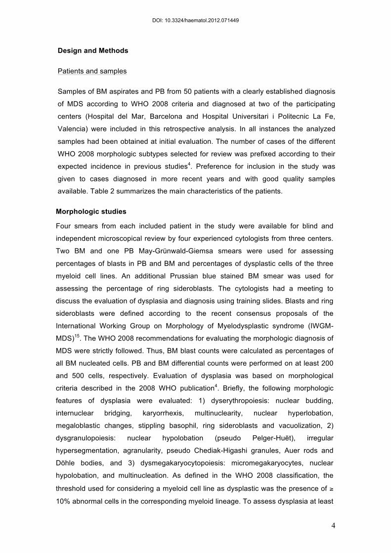
The presence of granulated blast cells (arrow) makes difficult the distinction between
blast cells and promyelocytes (discontinous arrow) so that the number of blast cells
may differ and the same patient be classified as MDS with or without excess of blasts
Figure 2.
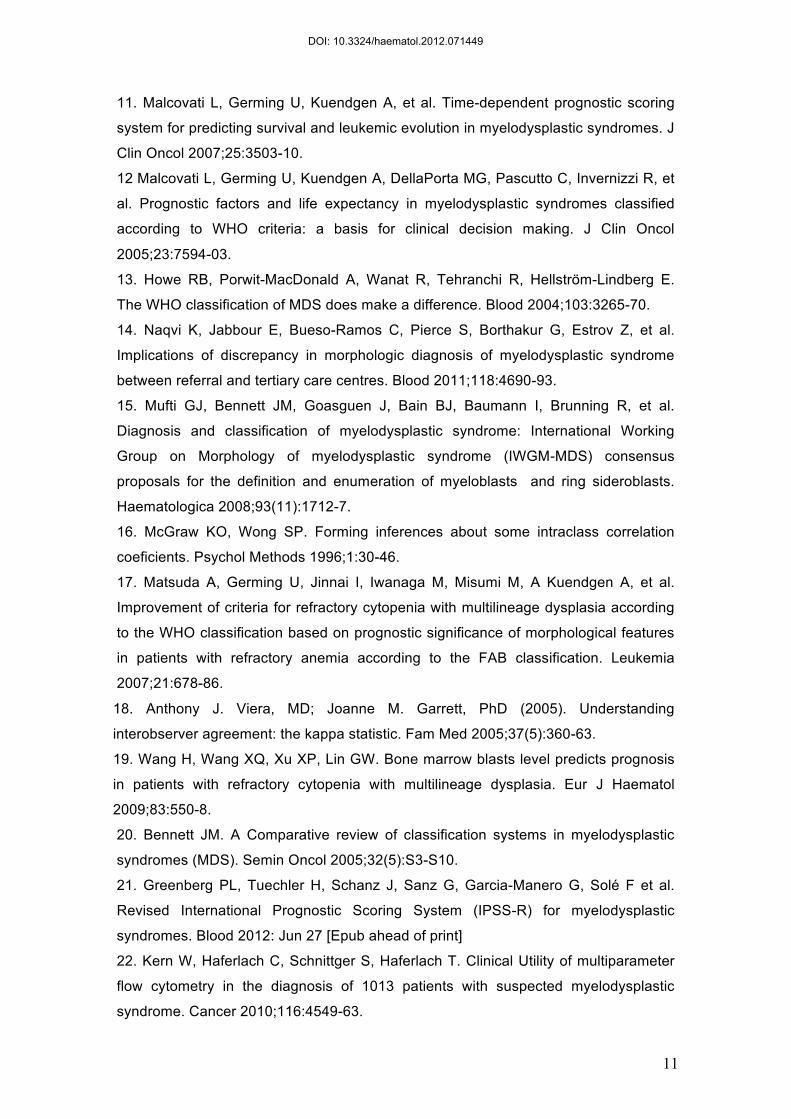
Evaluation of dysplastic features in erythropoiesis such as megaloblastoid
changes (arrow) and cytoplasmic changes (discontinous arrow) are poorly
reproducible and this justifies that the agreement between observers in
evaluation of dyserythropoiesis is not good.
DOI: 10.3324/haematol.2012.071449

Figure 1
DOI: 10.3324/haematol.2012.071449

Figure 2.
DOI: 10.3324/haematol.2012.071449