Embed Size (px)
Citation preview
Hindawi Publishing CorporationEvidence-Based Complementary and Alternative MedicineVolume 2013 Article ID 703024 10 pageshttpdxdoiorg1011552013703024
Research ArticleAntimicrobial Brazilian Propolis (EPP-AF) ContainingBiocellulose Membranes as Promising Biomaterial for SkinWound Healing
Hernane da Silva Barud1 Adalberto Miguel de Arauacutejo Juacutenior1 Sybele Saska12
Letiacutecia Boldrin Mestieri2 Juliana Alvares Duarte Bonini Campos2
Rubens Moreno de Freitas2 Nathaacutelia Ursoli Ferreira3 Andresa Piacezzi Nascimento3
Felipe Galeti Miguel3 Mirela Mara de Oliveira Lima Leite Vaz34 Edna Aparecida Barizon3
Franciane Marquele-Oliveira3 Ana Maria Minarelli Gaspar2 Sidney Joseacute Lima Ribeiro1
and Andresa Aparecida Berretta34
1 Instituto de Quımica Universidade Estadual Paulista (UNESP) CP 355 14800-900 Araraquara SP Brazil2 Faculdade de Odontologia Universidade Estadual Paulista (UNESP) Rua Humaita 1680 14801-903 Araraquara SP Brazil3 Laboratorio de Pesquisa Desenvolvimento e Inovacao (P D and I)-Apis Flora Industrial e Comercial Ltda Rua Triunfo 94514020-670 Ribeirao Preto SP Brazil
4Departamento de Ciencias Farmaceuticas Faculdade de Ciencias Farmaceuticas de Ribeirao Preto Universidade de Sao PauloAvenida do Cafe sn 14040-903 Ribeirao Preto SP Brazil
Correspondence should be addressed to Sidney Jose Lima Ribeiro sjlribeiroyahoocombrand Andresa Aparecida Berretta andresaberrettahotmailcom
Received 11 January 2013 Revised 30 April 2013 Accepted 13 May 2013
Academic Editor Zenon Czuba
Copyright copy 2013 Hernane da Silva Barud et alThis is an open access article distributed under the Creative Commons AttributionLicense which permits unrestricted use distribution and reproduction in any medium provided the original work is properlycited
Among remarkable discoveries concerning propolis such as antifungal antiviral and antioxidant activities its anti-inflammatoryandmainly its antibacterial properties deserve special attention when skin wound healing is concerned Based on this and knowingthe distinctive performance of bacterial (BC) membranes on wound healing in this work it is proposed to demonstrate the potentantimicrobial activity and wound healing properties of a novel propolis containing biocellulose membrane The obtained pro-polisBCmembranewas able to adsorb propolis not only on the surface but also in its interstices demonstrated by scanning electronmicroscopy X-ray diffraction Fourier transform infrared (FT-IR) spectroscopy and thermogravidimetric assays Additionally thepolyphenolic compounds determination and the prominent antibacterial activity in the membrane are demonstrated to be dosedependent supporting the possibility of obtaining propolisBCmembranes at the desired concentrations taking into considerationits application and its skin residence time Finally it could be suggested that propolisBC membrane may favor tissue repair in lesstime and more effectively in contaminated wounds
1 Introduction
The comprehensive therapeutic applicability of propolis hasbeen demonstrated for centuries if not for millennia andnowadays there are innumerous scientific reports elucidatingthe actionmechanism claimed for the compounds of this bal-samic resin Among remarkable discoveries such as antifun-gal [1 2] antiviral [3] and antioxidant activities [4ndash6] its
anti-inflammatory [7] and mainly its antibacterial [8ndash10]properties deserve special attentionwhen skinwoundhealingis concerned
The worldwide occurrence of skin wounds such as burninjuries remains high despite efforts to reduce injury inci-dence through public awareness campaigns and improve-ments in living conditions Severe burn injuries as well asother forms of stress and trauma trigger a hypermetabolic
2 Evidence-Based Complementary and Alternative Medicine
response requiring aggressive resuscitation nutrition exci-sion grafting and pharmacotherapeutic regimen [11] On theother hand infection is still one major problem that may leadto death It has been estimated that 75 of deaths followingburn injuries are related to infection [12 13]
Additionally it has been reported that a challenge facedin the treatment of skin wounds and in burn injuries is thedressing Some properties for dressing have been addressed(i) low cost (ii) safe (iii) relatively painless (iv) preventionof infection (v) promotion of fast wound healing (vi) lownumber of dressing changes during healing and so forth [13]
In this regard the last decades have witnessed anincreased interest in the use of biomaterials for example bio-polymers in healthcare products especially in dressing forwounds a fact that is predominantly associated to the renew-able nature biocompatibility and biodegradability of thesematerials [14] Among several biopolymers of interest biocel-lulose or bacterial cellulose (BC) produced by theGluconace-tobacter genus is extremely pure and allows obtaining highlyswollen membranes with around 99 water on the culturemedium surface [15] In addition distinctive tridimensionaland branched nano- and microfibrillar structure is formedleading to considerable interest not only as dressing forwound healing but also as substitutes of natural skin [16] andas drug delivery systems [14 17] Moreover it has beendemonstrated that BC present mechanical properties such astensile strength and extensibility similar to human skin andit allows the growth spreading and migration of humankeratinocytes [18]
Meanwhile BC membrane itself has no antimicrobialactivity to prevent wound infection [19] In the last yearsantimicrobial silver nanoparticles containing BCmembraneshave been developed using different routes and reducingagents [19ndash21]
Based on the exposed advantages provided using biocel-lulose and following our interest in the elucidation of propolisactivity especially regarding its properties on skin [5 6 8]in this work we reported preparation comprehensive charac-terization and efficacy of novel Brazilian propolis containingbiocellulose membrane Therefore the main purpose was todemonstrate the antimicrobial activity and thewoundhealingproperties of this new device material
2 Material and Methods
21Materials Green propolis standardized extract (EPP-AF)was kindly provided by Apis Flora Co (Ribeirao Preto SaoPaulo Brazil) (patent PI 0405483-0 Revista de PropriedadeIndustrial No 1778 of 01022005) For quantitative anal-ysis caffeic p-coumaric and trans-cinnamic acids (Sigma-Aldrich Sao Paulo Brazil) artepillin C (Wako Pure ChemicalIndustries Co Osaka Japan) and aromaden-drin-41015840-O-methyl ether (previously isolated and identified as describedby Souza et al [4] and kindly donated by the authors) wereused Methanol HPLC-grade was obtained from J T Bakerand water was treated in a Milli-Q water purification systemAll other chemicals were of reagent grade and were used
without further purification Mueller Hinton agar (DifcoDetroit MI USA) was used for the antibacterial assay
22 Methods
221 Bacterial CellulosePropolis Membranes PreparationBC membranes were obtained from cultivation of the Glu-conacetobacter hansenii ATCC 23769 Culture media wereestablished for 120 h at 28∘C in trays of 30times 50 cm containingthe sterile media composed of glucose 50 gsdotLminus1 yeast extract4 gsdotLminus1 anhydrous disodium phosphate 2 gsdotLminus1 heptahy-drated magnesium sulphate 08 gsdotLminus1 and ethanol 20 gsdotLminus1After 120 h hydrated BC pellicles (5mm thick) were obtain-edThese membranes were several times washed in water 2aqueousNaOHat 70∘C in order to remove bacteria andwateruntil neutral pH Next the BC membranes were immersedin ethanol for 24 h with continuous exchanges performed inorder to ensure substitution of water for ethanol Ethanolswollen BC membrane was used to prepare the BCpropolissamples
In the preparation of BCpropolis membranes firstlyalcoholic propolis solutions were prepared at 12 24 and36 (wv considering the dried matter in the propolisextract) employing 11 of Green Propolis StandardizedExtract EPP-AF (wv) Ethanol swollen BC membranes wereimmersed for 24 h in these solutions BCpropolis mem-branes were dried at 40∘C for 24 h and were set in nylonmolds Samples were named BCpropolis A BCpropolis Band BCpropolis C respectively
222 Physical-Chemical Characterization
Scanning Electron Microscopy (SEM) Scanning electronmicroscopy FEG-SEM (JEOL JMF-6700FmdashField EmissionScanning Electron Microscopy) was used to observe thesurface topography of all samples All specimens were placedin copper supports covered with a tick carbon layer
X-Ray Diffraction The X-ray diffractograms were obtainedusing a Siemens Kristalloflex diffractometer (Siemens Knox-ville TN USA) with a nickel filter and radiation CuK
120572
between 2120579 angle from 4 to 70∘ counting time of 2 s and glasssample holder
Fourier Transform Infrared (FT-IR) Spectroscopy FT-IR spec-tra were obtained with a Perkin-Elmer spectrometer model2000 Samples were milled and mixed with dried KBr inknown proportions and pressed into pellets
Thermal AnalysisThe thermogravimetric assays (TGAs) werecarried out using a SDT 2960 equipment from TA Instru-ments Samples were heated at a constant rate of 10∘C minminus1from 25∘C to 450∘C under a nitrogen flow of 70mmLminminus1
Chemical Characterization Analyses were conducted usinga Shimadzu (Kyoto Japan) liquid chromatograph equippedwith a CBM-20A controller a LC-20AT quaternary pump
Evidence-Based Complementary and Alternative Medicine 3
a SPD-M 20A diode-array detector and Shimadzu LC solu-tion software version 121 SP1 A Shimadzu Shim-Pack CLC-ODS column (46mm times 250mm particle diameter of 5 120583mand pore diameter of 100 A) was used The mobile phaseconsisted of methanol (B) and of a solution of water-formicacid (01 vv) pH 27 (A) The method consisted of a lineargradient of 20ndash95 of B over a period of 77min at a flow rateof 08 gmLminus1 The injection volume was 10 120583L The columnoven was set at 40∘C Detection was set at 275 nm [8 22]
Working solutions were prepared daily in methanolin the following concentration ranges caffeic acid 106ndash1696 120583gmLminus1 p-coumaric acid 504ndash8064 120583gmLminus1 trans-cinnamic acid 040ndash640 120583gmLminus1 aromadendrin-41015840-O-methyl ether 20ndash320120583gmLminus1 and artepillin C 1006ndash16096 120583gmLminus1
The samples of BCpropolis were weighed (25mg) onan analytical balance and transferred into 10mL volumetricflasks Next the volume was completed with methanol and itwas sonicated for 30min for maximum release of propolisThe samples were filtered through a 045 120583m filter beforeanalysis
223 Antibacterial Assay The disk diffusion method (Clin-ical and Laboratory Standards Institute (CLSI)) [23] wasemployed in this study with some modifications to evaluatethe antibacterial activity of the BCpropolis against Staphy-lococcus aureus ATCC 25923 Staphylococcus aureus ATCC43300 and Staphylococcus epidermidis ATCC 14990
The bacterial suspension was prepared in a sterile 085physiological solution with turbidity equivalent to a 05McFarland standard (approximately 108 CFUmLminus1) A sterilecotton swab was used to seed the suspension on the surfaceof Mueller Hinton agar contained in a plate (90 times 15mm)
Membranes were cut in disks (diameter 55mm) andapplied to the agar surface Next 5 120583L of sterile 085 phys-iological solution were put on the surface of each biomem-brane Biomembranes without propolis submitted to thesame procedure of the BCpropolis were used to control theexperimentThe plates were incubated at 35∘C aerobically for18 h After the incubation period the diameters of the zonesof inhibition were measured using a ruler The experimentswere replicated three times for each microorganism
224 In Vivo Experiment This experiment was approvedby the Animal Experimentation Ethics Committee of Arara-quara University (UNIARA) Araraquara SP Brazil Twenty-four male rats (Rattus Norvegicus Holtzman) weighingapproximately 250 g were used in this study General anes-thesia was induced using intramuscular injections of ket-amine hydrochloride (25mg kgminus1) and xylazine hydrochlo-ride (5mg kgminus1) All surgical procedures were performedunder strict aseptic protocol After shaving andpreparation ofthe dorsal region on each animal were made three incisionsof 6mm in diameter with a circular scalpel and a distance ofapproximately 2 cm among them (Figure 1) [8]
On the left side group I (G1) propolis-free membranewas put on the wound (positive control) and on the right sidegroup II (G2) BCpropolis membrane (B) was put on the
Group I Group II
Group III
Figure 1 Identification of the wounds created surgically for therespective treatment groups
wound In group III (G3) median region the wound wasuntreated (negative control) Subsequently the animals wereaccommodated in cages properly isolated and were moni-tored daily with appropriate conditions of food water andhygiene In the immediate postoperative period all animalsreceived an oral administration of salicylic acid (120ndash300mg kgminus1 single dose) as recommended by the CanadianCouncil on Animal Care (CCAC)
The animals (119899 = 6 per group) were euthanized after 37 15 and 30 days postoperatively Firstly the animals wereanesthetized again following the protocol described previ-ously and after the specimens were removed the animalsreceived deepening anesthetic The specimens were fixed in10 buffered formaldehyde solution and processed accordingto histological routine for light microscopy The specimenswere then cut in 6 120583m sections in the longitudinal directionof the skin and stained with hematoxylin-eosin (HampE) Nextthey were analyzed and photographed under a microscope(Jenaval-Zeiss) coupled to a digital camera (Leica DFC425)The parameters analyzed were descriptive and examined by asingle researcher who was blinded to analysis of the groupsThe parameters analyzed were (1) inflammatory reaction (2)angiogenesis process and (3) quality of tissue repair
Macroscopic Assessment The wounds were analyzed macro-scopically after periods of 3 7 and 15 days postoperativelyThe 30-day period was excluded from the analysis becauseall the wounds of the three study groups had already beenfully repaired Evaluation of the wounds was performed bymeasuring lesion in the longitudinal and transverse direction(mm) using a caliper rule during the preeuthanasia proce-dure For statistical analysis the measurements longitudinaland transverse of eachwoundwere added and divided by twofor obtaining a repair average value
225 Statistical Analysis The data of antibacterial assaywere submitted to the nonparametric Kruskal-Wallis test
4 Evidence-Based Complementary and Alternative Medicine
Statistical significance was established at 119875 lt 001 Statisticalanalysis of data was performed using the software GraphPad Prism 4 For the in vivo study statistical analysis wasperformed using SPSS 200 software Sphericity and nor-mality tests were performed previously A two-way ANOVAwas applied to macroscopic assessment Statistical analysis inrelation to intensity of inflammatory reaction was measuredby score (score 0 to 4 no reaction = 0 very slight reaction = 1mild reaction = 2 and moderate reaction = 3 markedreaction = 4) following the standards of ASTM F981-04 [24]In this analysis twenty histological cuts were analyzed foreach specimen in the respective periods The ANOVA testwas carried out to evaluate measured scores to intensity ofinflammatory reaction and Tukeyrsquos test was used as posttestfor statistical significance Statistical significance was estab-lished at 119875 lt 005
3 Results and Discussion
31 Physical-Chemical Characterization
311 Scanning Electron Microscopy (SEM) BC is a semi-transparent paper-like material and the final BCpropoliswas obtained as a flexible and macroscopic homogeneousmembrane BCpropolis membranes presented amber col-orations in a dose-dependent way SEM measurements wereperformed for some representative samples including BCmembrane and BCpropolis B Figure 2
BC membrane (Figure 2(a)) clearly presents a compactstructure composed of long fibers of hundreds of micronswith nanometer thickness [16] Figure 2(b) reveals that BCmicrofibril is swollen and homogeneously covered by propo-lis extract solution Cross-section images Figures 2(c) and2(d) confirm that propolis is present not only in the surfaceof BC membrane but also inside the cellulose chains
312 X-Ray Diffraction Figure 3 shows the results of X-raydiffraction for all samples The BC membrane presented twodiffraction angles 15 and 225∘ degrees (Figure 3(a)) Eachpeak has a contribution of diffractions corresponding to I120572and I120573 phases characteristics of native cellulose type I [25]
Dried propolis extract also showed an amorphous struc-ture and three broad peaks in 15∘ 17∘ and 23∘ could bemarked corroboratingwith previous results for propolis [22]
All BCpropolis membranes showed considerable changeon X-ray profile when compared to BCmembrane A gradualdecrease in crystallinity is observed with increasing of propo-lis contents inferring that the propolis extract solutions isnot only on BC surface but it is found in the interstices andbetween the crystal planes of the polymer making it moreamorphous
313 Fourier Transforms Infrared (FT-IR) SpectroscopyFigure 4 shows the results of FT-IR spectroscopy for BCpropolis and all BCpropolis membranes
BC (Figure 4(a)) shows bands in the 400ndash700 cmminus1 rangecharacteristic of the OH bending 120573-glucosidic linkagesbetween the glucose units at sim896 cmminus1 and CndashO symmetric
stretching of primary alcohol and CndashOndashC antisymmetricbridge stretching at 1040 and 1168 cmminus1 respectively The CndashH deformation (CH
3or OndashH in plane bending) is seen at
1340 cmminus1 and the band centered at 1400 cmminus1 is related toCH2bending and OH in plane bending Other bands are
related to HndashOndashH bending of adsorbed water (at 1650 cmminus1)CH stretching of CH
2and CH
3groups (at 2900 cmminus1) and
OH stretching (broad band at 3500 cmminus1) [26]Some bands that characterize propolis in the IR spectrum
are absorptions related to the presence of C=C doublebond (]max 1641 cmminus1) carboxyl (]max 1703 cmminus1) andhydroxyl (]max 3487 cmminus1) groups with concern to organiccompounds
Spectra concerning BCpropolis-based membranes canbe considered as the sum of the bands present in BC mem-brane and in propolis in other words there are observed con-tributions relating to characteristic vibrations both of the BCmembrane [27] and propolis [22] showing therefore theformation a composite material Some peaks were slightlyshifted due to the formation of intermolecular interactionslike hydrogen bonding between propolis component andcellulose
Broadening was observed for the band around 3500 cmminus1with the increase in the propolis relative content suggestingthe formation of hydrogen bonds between cellulose andpropolis components such as flavonoids
314 Thermogravidimetric Assay Thermogravimetry (TG)curves for BC membrane and BCpropolis membranes areshown in Figure 5 BC membrane displayed a typical weight-loss degradation profile (Figure 5(a)) with two main separatedegradation steps the first step (45ndash150∘C) can be attributedto the cellulose dehydration while the second step with largemass loss (250ndash350∘C) is related to processes of cellulosedegradation as depolymerization and subsequent decompo-sition of the glycosidic units followed by the formation ofcarbonaceous residues [27 28]
Propolis presented a single weight loss profile with largemass loss (65) in the temperature range of 150ndash450∘Cwhich can be attributed to simultaneous events includingcondensation of groups (OHndash) carbonic bond breaks andsubsequent degradation of organic compounds [22]
BC-propolis samples showed a thermal profile similar tothe one obtained for dried propolis BCpropolis membranespresented a dehydration process starting around 50∘C sug-gesting that after being topically applied they are able to keepthewound surface hydrated avoiding loss of electrolytes [29]
A continuous weight loss in the temperature range of 90ndash450∘C is observed for all BC-propolis samples Figures 5(c)and 5(d) These events include condensation of groups(OHndash) carbonic bond breaks and subsequent degradation ofpropolis organic compounds and they also include BCmem-brane degradation [22 27 29]
An increase in the final residue content with the increasein the relative propolis content was observed confirming thatthe propolis mass is dose-dependent on the initial propolissolutions (12 24 and 36 wv) used in BCpropolis mem-brane preparation
Evidence-Based Complementary and Alternative Medicine 5
1120583m
(a)
1120583m
(b)
1120583m
(c)
1120583m
(d)
Figure 2 Representative SEM images of (a) BC membrane and (b) BCpropolis B surface images and (c) pristine BC membrane and (d)BCpropolis B cross-sections images
Determination of dry matter of propolis in the samplesof BCpropolis A B or C was performed by an indirect wayusing HPLC results and comparing with the standard valuesfound in propolis standardized extract used The resultsshowed that BCpropolis A B and C possess respectively4367 plusmn 1062 5678 plusmn 1522 and 6659 plusmn 1345 ww ofpropolis fact observed because of the large superficial areaof never-dried BC membrane in contact for a long time withpropolis solutions (large ability to adsorb large mass) and theopen porous present during this kind of manufacture process[30]
315 Chemical Characterization The quantification of somestandards present in BCpropolis membranes was performedby HPLC The employed method was fully validated asreported by Rocha et al [31] The results for BCpropolis bio-membranes A B and C and the propolis standardizedextract (EPP-AF) used may be observed in Table 1 Asexpected the quantified compounds were dose dependenttherefore assuming that there is a greater amount of activecompounds inserted in biomembrane C followed by bio-membrane B and biomembrane A
Analysis of BCpropolis composition comparing withpropolis extract used after dry matter normalization showsthat the flavonoid aromadendrin was the component betteradsorbed in BC probably because of intermediary polarity ofthis component On the other hand artepillin C the major
constituent of EPP-AF propolis extract [8] showed rela-tively low adsorption in BC (7534 7259 and 8469BCpropolis A B andC resp) probably due to lower polarityof this structure when compared with caffeic acid derivativesand because of intrinsic properties of BC Caffeic acid p-coumaric and cinnamic acids were well adsorbed
32 Antibacterial Assay Propolis-free membranes (negativecontrol) showed no antibacterial activity On the other handthe different membranes presented antibacterial activity(Table 2) with the BCpropolis membrane C being the mosteffective (119875 lt 001)
The results presented in Table 2 are lower than the resultspresented by de Rezende et al [32] for propolis ethanolicextract especially for S aureus ATCC 25923 (15mm) Saureus ATCC 6538 (17mm) and S aureus ATCC 29213(11mm) using the same methodology However it is impor-tant to consider that the diffusion of the substances in thistype of methodology completely affects the results found It ispossible that the strong interaction previously demonstratedfor propolis and BC decreases the liberation and diffusionof the propolis compounds in the medium and therefore alower inhibition zone is observed
The samples were effective against all staphylococcitested including S aureus ATCC 43300 a methicillin-resistant S aureus (MRSA) These strains are responsiblefor difficult-to-treat infections in humans as they exhibit
6 Evidence-Based Complementary and Alternative Medicine
Table 1 Chemical composition of propolis standardized extract (EPP-AF) and bacterial cellulosepropolis A B and C (mgg) Values aremean plusmn SD obtained from analyses in triplicate
Standards Researched Average plusmn SDEPP-AF A B C
Caffeic acid 0345 plusmn 0011 130 plusmn 0074 165 plusmn 0113 195 plusmn 0130119901-coumaric acid 1712 plusmn 0069 610 plusmn 0389 786 plusmn 0195 925 plusmn 0266Trans-cinnamic acid 0167 plusmn 0006 066 plusmn 0075 088 plusmn 0031 099 plusmn 0060Aromadendrin 0808 plusmn 0024 450 plusmn 0274 601 plusmn 0089 661 plusmn 0306Artepillin C 6621 plusmn 0306 1978 plusmn 370 2481 plusmn 504 3395 plusmn 224SD standard deviation
10 20 30 40
(e)
(d)
(c)
Inte
nsity
(au
)
(a)
(b)
2120579 (deg)
Figure 3 XRD diffractogram of (a) BC membrane (b) dried pro-polis (c) BCpropolis A (d) BCpropolis B and (e) BCpropolis C
4000 3500 3000 2500 2000 1500 1000 500
(e)
(c)
(d)
(b)
Tran
smitt
ance
()
(a)
Wavenumber (cmminus1)
Figure 4 FT-IR spectra of (a) BC membrane (b) Dried propolis(c) BCpropolis A (d) BCpropolis B and (e) BCpropolis C
50 100 150 200 250 300 350 4000
20
40
60
80
100
(c)
(b) (d)
(e)W
eigh
t (
)
(a)
Temperature (∘C)
Figure 5 TG curves of (a) BC membrane (b) dried propolis (c)BCpropolis A (d) BCpropolis B and (e) BCpropolis C
Table 2Mean diameter (inmm) of the zones of inhibition providedby biomembranes A B and C Values are mean plusmn SD (119899 = 3)
Bacterium Bacterial cellulosepropolisA B C
BC without propolis 119877 119877 119877
Staphylococcus aureusATCC 25923 8 plusmn 058 9 plusmn 000 10 plusmn 000
Staphylococcus aureusATCC 43300 7 plusmn 000 8 plusmn 000 9 plusmn 000
Staphylococcus epidermidisATCC 14990 7 plusmn 058 8 plusmn 000 9 plusmn 000
SD standard deviation R resistant
multidrug resistance such as penicillins cephalosporins andcarbapenems
S aureus and coagulase-negative staphylococci (like Sepidermidis) are between the most frequently isolated bacte-ria from burn wounds Furthermore in burn wound micro-bial colonization there is an initial predominance of thesebacteria and other gram-positive cocci over gram-negativebacilli [12]These results suggest that the BCpropolis may bean effective material to be used for infected wounds whichwould work not only as an excellent dressing but also as
Evidence-Based Complementary and Alternative Medicine 7
G1
BCv
lowast
200120583m
(a)
G2
BC + P
v
lowast
200120583m
(b)
G3
v lowast
200120583m
(c)
Figure 6 3 days of treatmentmdashG1 necrotic tissue (lowast) BC membrane (BC) and presence of moderate inflammatory infiltrate and numerousblood vessels (v) G2 necrotic tissue (lowast) BCpropolis membrane (BC) presence of moderate inflammatory infiltrate and numerous bloodvessels (v) G3 necrotic tissue (lowast) presence of moderate inflammatory infiltrate and numerous blood vessels (v)
200120583mG1
(a)
200120583mG2
(b)
200120583mG3
(c)
Figure 7 30 days of treatmentmdashrepair tissue with normal characteristics in all three groups
Table 3 The levels of the inflammation reaction to the respectivestudied groups in comparison with the analyzed periods
Period GroupG1 G2 G3
3 +++lowast +++lowast +++lowast
7 minus ++lowast minus
15 minus minus minus
30 minus minus minus
G1 propolis-free membrane G2 BCpropolis Bmembrane and G3 negativecontrol Score minus no reaction + very slight ++ mild +++ moderate ++++marked inflammatory reaction lowastno statistically significant difference (119875 =019)
an antibacterial one Additionally the results obtained inthe physical-chemical characterization and the antibacterialactivity of the BCpropolismembrane show that these param-eters are dose dependent supporting an interesting possibil-ity that is obtaining propolis membranes at the desired con-centrations taking into consideration its application and itsskin residence time
33 Histological Analysis BCmembrane in contrast to othersynthetic membranes is highly resistant to chemical cor-rosion biocompatible porous and also has good tensilestrength [33] unique properties that make it a polymer withgreat potential to be explored by biotechnology and other
0123456789
3 days 7 days 15 days
Wou
nd si
ze (m
m)
Period
ControlBCBC-propolis
Figure 8 Macroscopic results obtained with tissue repair with BCBCpropolis and without treatment (control) (119875 gt 005)
areas of biomedical science mainly employing the BC aspotential carrier for drug delivery systems
In the last decades BC membranes have been used asa temporary dressing of skin in burns and other conditionssuch as chronic ulcers and grafts [34ndash38] In addition in vivostudies have demonstrated that BC membranes are biocom-patible and can be used as scaffold for tissue engineering [39ndash41]
8 Evidence-Based Complementary and Alternative Medicine
The studies were performed only for the BCpropolis Bonce this membrane presented good results in the previousantibacterial studies The inflammatory reactions presentedin the studied groups were shown in Table 3
Thehistological analyses of thewounds healing are shownin Figures 6 and 7 At day 3 the wound bed appeared in reor-ganization necrotic tissue (fibrin-leukocytes crust) mainlyin the control group whose tissue can also be observed underrespective membranes BC and BCpropolis also appearedBeneath this tissue it is observed that formation of granula-tion tissue abundant constituted of few disorganized collagenfibers with large fibroblastic activity (loose connective tissue)and presence of numerous blood vessels in stasis (Figure 6)
After 7 days a thin layer of epidermal reepithelializationcan be observed and loose connective tissue is still shownin repair process in all tested groups but collagen fiberswere denser and organized in relation to earlier period andpresence of numerous blood vessels mainly in the treatedgroups with membranes as dressing G1 and G2 Moreoverthe repair of the epithelium also appeared in amore advancedstage in these respective groups compared to the controlgroup (G3) presenting continuity in the epithelium surfaceIt was observed the presence of necrotic tissue in all groupsAt day 15 repair of the epithelium (keratinized stratified squa-mous epithelium) absence of a discontinuity and presence ofkeratohyalin granules in the three groups (stratum corneum)were observed The repaired fibrous conjunctive tissue pre-sented normal characteristics where were observed a greatnumber of thick-regular collagen fibers decreasing theamount of blood vessels At day 30 (Figure 7) the histologicalcharacteristics were similar to the period of 15 days withgreat deposition of collagen in this tissue and accentuatedcompression of the newly formed capillaries
Propolis is a known anti-inflammatory agent [42 43] andthen the reduction of inflammatory process was expected inG2 group Some authors attribute this activity to the impor-tant biomarker of green propolis artepillin C [44] The anti-inflammatory effect of artepillin C was observed and theauthors have claimed that this property ismediated at least inpart by prostaglandin E2 and nitric oxide inhibition throughNF-120581B modulation [44]
The results obtained are important and satisfactory andrevealed that BC and BCpropolis membranes were biocom-patible and promoted in earlier periods a better tissue repairAlso BCpropolis presented a delivery system since pre-liminary results of propolis delivery from BCpropolis wasobtained through p-coumaric acid isossakuranetin andartepillin C release evaluation using Franz Cell assay Resultswith 24 hours of evaluation demonstrated that p-coumaricacid was released in a maximum of 6021 isosakuranetin5389 and artepillin C 3726 However it is important toconsider that epithelial tissue was completely reconstitutedwith the application of a thermoreversible gel with propolisstandardized extract (EPP-AF) within 3 days after injury [8]In this case the gel promotes hydration of wound for alonger time favoring the healing process thus with woundhydration and covering there is no crust formation factorsthat favor a shorter wound resolution A hypothesis to solvethe problem of dryness of the BC membranes would be
including in the treatment protocol the rehydration of mem-brane this way we could assess if there would be improve-ment and decrease of the time of wound healing
So the results of the macroscopic analysis revealed repairvalues around 76mm 58mm and 4mm after 3 7 and 15days postoperatively (Figure 8) Macroscopically the repairof wounds showed no statistically significant differences (119875 gt005) among the treatments employed with the BC and BCpropolis membranes and the control group (no dressing)during the period studied the results were confirmed byhistological analysis in relation to layer of epidermal reepithe-lialization and these results also suggest that the describedhypothesis above may have relevance Therefore the type oftreatment does not affect the repair time for uncontaminatedwounds
4 Conclusion
The present study suggests the potential applicability ofpropolis containing biocellulose membranes for treatingwoundsThe obtained BCpropolis was able to adsorb propo-lis not only on the surface but also in its interstices demon-strated by SEM X-ray diffraction FT-IR spectroscopy andthermogravidimetric assays Additionally the caffeic acidderivatives and flavonoid determination and the prominentantibacterial activity in the BCpropolis demonstrated to bedose dependent supporting an interesting possibility theobtention of propolis based membranes at the desired con-centrations taking into consideration its application and itsskin residence time Finally it could be suggested that BCpropolis membrane may favor tissue repair in less time andmore effectively in contaminated wounds
Acknowledgments
The authors would like to thank the special colleague JuliaSgarioni Lima Ribeiro for the important participation in thepresent work which could justify her name as an authortoo However this is not possible due to sad circumstancesinvolved The authors also are grateful to Coordenacao deAperfeicoamento de Pessoal de Nıvel Superior (CAPES)Conselho Nacional de Desenvolvimento Cientıfico e Tec-nologico (CNPq) Fundacao de Amparo a Pesquisa do Estadode Sao Paulo (FAPESP) and Apis Flora Indl Coml Ltdafor financial support and for granting research fellowshipsand the present coauthors are grateful to Bruno Alves RochaPaula Carolina Pires Bueno and Alexandre HenriqueOliveira for suggestions of some protocols used during theexperiments used
References
[1] P A de Castro M Savoldi D Bonatto et al ldquoMolecular char-acterization of propolis-induced cell death in Saccharomycescerevisiaerdquo Eukaryotic Cell vol 10 no 3 pp 398ndash411 2011
[2] A A da Silva Filho J P B de Sousa S Soares et al ldquoAnti-microbial activity of the extract and isolated compounds fromBaccharis dracunculifolia D C (Asteraceae)rdquo Zeitschrift furNaturforschung C vol 63 no 1-2 pp 40ndash46 2008
Evidence-Based Complementary and Alternative Medicine 9
[3] T Urushisaki T Takemura S Tazawa et al ldquoCaffeoylquinicacids aremajor constituentswith potent anti-influenza effects inbrazilian green propolis water extractrdquo Evidence-Based Comple-mentary and Alternative Medicine vol 2011 Article ID 2549147 pages 2011
[4] R M Souza M C De Souza M L Patitucci and J F M SilvaldquoEvaluation of antioxidant and antimicrobial activities andcharacterization of bioactive components of two Brazilian pro-polis samples using a pK a-guided fractionationrdquo Zeitschrift furNaturforschung C vol 62 no 11-12 pp 801ndash807 2007
[5] F Marquele-Oliveira Y M Fonseca O de Freitas and M J VFonseca ldquoDevelopment of topical functionalized formulationsadded with propolis extract stability cutaneous absorption andin vivo studiesrdquo International Journal of Pharmaceutics vol 342no 1-2 pp 40ndash48 2007
[6] Y M Fonseca F Marquele-Oliveira F T M C Vicentini et alldquoEvaluation of the potential of Brazilian propolis against UV-induced oxidative stressrdquo Evidence-Based Complementary andAlternative Medicine vol 2011 Article ID 863917 8 pages 2011
[7] S R Gregory N Piccolo M T Piccolo M S Piccolo and JP Heggers ldquoComparison of propolis skin cream to silver sul-fadiazine a naturopathic alternative to antibiotics in treatmentof minor burnsrdquo Journal of Alternative and ComplementaryMedicine vol 8 no 1 pp 77ndash83 2002
[8] A A Berretta A P Nascimento P C P Bueno M M D O LLeite Vaz and J M Marchetti ldquoPropolis standardized extract(EPP-AF) an innovative chemically and biologically repro-ducible pharmaceutical compound for treating woundsrdquo Inter-national Journal of Biological Sciences vol 8 no 4 pp 512ndash5212012
[9] H Koo B P F A Gomes P L Rosalen G M B Ambrosano YK Park and J A Cury ldquoIn vitro antimicrobial activity of pro-polis and Arnica montana against oral pathogensrdquo Archives ofOral Biology vol 45 no 2 pp 141ndash148 2000
[10] R Jorge N A J C Furtado J P B Sousa et al ldquoBrazilian pro-polis seasonal variation of the prenylated p-coumaric acids andantimicrobial activityrdquo Pharmaceutical Biology vol 46 no 12pp 889ndash893 2008
[11] Y Rojas C C Finnerty R S Radhakrishnan S Ravi and DN Herndon ldquoBurns an update on current pharmacotherapyrdquoExpert Opinion on Pharmacotherapy vol 13 no 17 pp 2485ndash2494 2012
[12] S Nasser A Mabrouk and A Maher ldquoColonization of burnwounds in Ain Shams University Burn Unitrdquo Burns vol 29 no3 pp 229ndash233 2003
[13] R L Gerding C L Emerman D Effron T Lukens A LImbembo and R B Fratianne ldquoOutpatient management ofpartial-thickness burns biobrane versus 1 silver sulfadiazinerdquoAnnals of Emergency Medicine vol 19 no 2 pp 121ndash124 1990
[14] E Trovatti C S R Freire P C Pinto et al ldquoBacterial cellulosemembranes applied in topical and transdermal delivery of lido-caine hydrochloride and ibuprofen in vitro diffusion studiesrdquoInternational Journal of Pharmaceutics vol 435 no 1 pp 83ndash87 2012
[15] D KlemmD Schumann U Udhardt and SMarsch ldquoBacterialsynthesized cellulosemdashartificial blood vessels formicrosurgeryrdquoProgress in Polymer Science vol 26 no 9 pp 1561ndash1603 2001
[16] W Czaja A Krystynowicz S Bielecki and R M Brown JrldquoMicrobial cellulosemdashthe natural power to heal woundsrdquo Bio-materials vol 27 no 2 pp 145ndash151 2006
[17] S Saska R M Scarel-Caminaga L N Teixeira et al ldquoChar-acterization and in vitro evaluation of bacterial cellulose mem-branes functionalized with osteogenic growth peptide for bonetissue engineeringrdquo Journal of Materials Science vol 23 no 9pp 2253ndash2266 2012
[18] N Sanchavanakit W Sangrungraungroj R Kaomongkolgit TBanaprasert P Pavasant and M Phisalaphong ldquoGrowth ofhuman keratinocytes and fibroblasts on bacterial cellulose filmrdquoBiotechnology Progress vol 22 no 4 pp 1194ndash1199 2006
[19] H S Barud T Regiani R F C Marques W R Lustri YMessaddeq and S J L Ribeiro ldquoAntimicrobial bacterial cell-ulose-silver nanoparticles composite membranesrdquo Journal ofNanomaterials vol 2011 Article ID 721631 8 pages 2011
[20] L C S Maria A L C Santos P C Oliveira et al ldquoPreparationand antibacterial activity of silver nanoparticles impregnated inbacterial celluloserdquo Polımeros vol 20 no 1 pp 72ndash77 2010
[21] T Maneerung S Tokura and R Rujiravanit ldquoImpregnationof silver nanoparticles into bacterial cellulose for antimicrobialwound dressingrdquo Carbohydrate Polymers vol 72 no 1 pp 43ndash51 2008
[22] B A Rocha M R Rodrigues P C P Bueno et al ldquoPreparationand thermal characterization of inclusion complex of Braziliangreen propolis and hydroxypropyl-120573-cyclodextrin increasedwater solubility of the chemical constituents and antioxidantactivityrdquo Journal of Thermal Analysis and Calorimetry vol 108no 1 pp 87ndash94 2012
[23] Clinical and Laboratory Standards Institute (CLSI) ldquoPerfor-mance standards for antimicrobial disk susceptibility testsrdquoApproved Standard 10th Edition CLSI document M02-A102009
[24] ASTMF981-04 Standard Practice For Assessment of Compatibil-ity of Biomaterials For Surgical Implants With Respect To Effectof Materials on Muscle and Bone 2010
[25] A C OrsquoSullivan ldquoCellulose the structure slowly unravelsrdquoCellulose vol 4 no 3 pp 173ndash207 1997
[26] M Kakurakova A C Smith M J Gidley and R H WilsonldquoMolecular interactions in bacterial cellulose composites stud-ied by 1D FR-IR and dynamic 2D FT-IR spectroscopyrdquo Carbo-hydrate Research vol 337 pp 1145ndash1153 2002
[27] H S Barud R M N Assuncao M A U Martines et alldquoBacterial cellulose-silica organic-inorganic hybridsrdquo Journal ofSol-Gel Science and Technology vol 46 no 3 pp 363ndash367 2008
[28] H S Barud C A Ribeiro M S Crespi et al ldquoThermal chara-cterization of bacterial cellulose-phosphate composite mem-branesrdquo Journal of Thermal Analysis and Calorimetry vol 87no 3 pp 815ndash818 2007
[29] D T B De Salvi H S Barud J M A Caiut Y Messaddeqand S J L Ribeiro ldquoSelf-supported bacterial celluloseboehmiteorganic-inorganic hybrid filmsrdquo Journal of Sol-Gel Science andTechnology vol 63 no 2 pp 211ndash218 2012
[30] D Klemm F Kramer S Moritz et al ldquoNanocelluloses a newfamily of nature-based materialsrdquo Angewandte Chemie vol 50no 24 pp 5438ndash5466 2011
[31] B A Rocha P C P Bueno M M O L L Vaz et al ldquoEvalua-tion of a propolis water extract using a reliable RP-HPLCmethodology and in vitro and in vivo efficacy and safety char-acterisationrdquo Evidence-Based Complementary and AlternativeMedicine vol 2013 Article ID 670451 11 pages 2013
[32] G P S R de Rezende F C Pimenta and L R R S da CostaldquoAntimicrobial activity of two Brazilian commercial propolisextractsrdquo Brazilian Journal of Oral Sciences vol 5 no 16 pp967ndash970 2006
10 Evidence-Based Complementary and Alternative Medicine
[33] U Geyer T Heinze A Stein et al ldquoFormation derivatizationand applications of bacterial celluloserdquo International Journal ofBiological Macromolecules vol 16 no 6 pp 343ndash347 1994
[34] C Rebello D A Almeida E M Lima Jr and M P DornelasldquoBio-fill umnovo substituto de pele nossa experienciardquoRevistaBrasileira De Cirurgia vol 77 pp 407ndash414 1987
[35] J D Fontana A M de Souza C K Fontana et al ldquoAcetobactercellulose pellicle as a temporary skin substituterdquo Applied Bio-chemistry and Biotechnology vol 24-25 pp 253ndash264 1990
[36] R C Mayall A C Mayall L C Mayall H C Rocha and L CMarques ldquoTratamento das ulceras troficas dos membros comum novo substituto de pelerdquo Revista Brasileira De Cirurgia vol80 pp 257ndash283 1990
[37] A F P FWouk JMDiniz SM Cirio H Santos E L Baltazarand A Acco ldquoMembrana biologica (Biofill)mdashestudo compara-tivo com outros agentes promotores da cicatrizacao de pele emsuınos aspectos clınicos histopatologicos e morfometricosrdquoArchives of Veterinary Science vol 3 pp 31ndash37 1998
[38] F K Andrade R Costa L Domingues R Soares and MGama ldquoImproving bacterial cellulose for blood vessel replace-ment functionalization with a chimeric protein containinga cellulose-binding module and an adhesion peptiderdquo ActaBiomaterialia vol 6 no 10 pp 4034ndash4041 2010
[39] G Helenius H Backdahl A Bodin U Nannmark P Gaten-holm and B Risberg ldquoIn vivo biocompatibility of bacterialcelluloserdquo Journal of Biomedical Materials Research A vol 76no 2 pp 431ndash438 2006
[40] P N Mendes S C Rahal O C M Pereira-Junior et alldquoIn vivo and in vitro evaluation of an Acetobacter xylinumsynthesized microbial cellulose membrane intended for guidedtissue repairrdquoActaVeterinaria Scandinavica vol 51 no 1 article12 2009
[41] W L Amorim H O Costa F C de Souza M G de Castroand L da Silva ldquoEstudo experimental da resposta tecidual apresenca de celulose produzida por Acetobacter xylinum nodorso nasal de coelhosrdquo Brazilian Journal of Otorhinolaryngol-ogy vol 75 pp 200ndash207 2009
[42] N Paulino C Teixeira R Martins et al ldquoEvaluation of theanalgesic and anti-inflammatory effects of a Brazilian greenpropolisrdquo Planta Medica vol 72 no 10 pp 899ndash906 2006
[43] J L Machado A K M Assuncao M C P da Silva et alldquoBrazilian green propolis anti-inflammatory property by animmunomodulatory activityrdquo Evidence-Based Complementaryand Alternative Medicine vol 2012 Article ID 157652 10 pages2012
[44] N Paulino S R L Abreu Y Uto et al ldquoAnti-inflammatoryeffects of a bioavailable compound artepillin C in Brazilianpropolisrdquo European Journal of Pharmacology vol 587 no 1ndash3pp 296ndash301 2008
Submit your manuscripts athttpwwwhindawicom
Stem CellsInternational
Hindawi Publishing Corporationhttpwwwhindawicom Volume 2014
Hindawi Publishing Corporationhttpwwwhindawicom Volume 2014
MEDIATORSINFLAMMATION
of
Hindawi Publishing Corporationhttpwwwhindawicom Volume 2014
Behavioural Neurology
EndocrinologyInternational Journal of
Hindawi Publishing Corporationhttpwwwhindawicom Volume 2014
Hindawi Publishing Corporationhttpwwwhindawicom Volume 2014
Disease Markers
Hindawi Publishing Corporationhttpwwwhindawicom Volume 2014
BioMed Research International
OncologyJournal of
Hindawi Publishing Corporationhttpwwwhindawicom Volume 2014
Hindawi Publishing Corporationhttpwwwhindawicom Volume 2014
Oxidative Medicine and Cellular Longevity
Hindawi Publishing Corporationhttpwwwhindawicom Volume 2014
PPAR Research
The Scientific World JournalHindawi Publishing Corporation httpwwwhindawicom Volume 2014
Immunology ResearchHindawi Publishing Corporationhttpwwwhindawicom Volume 2014
Journal of
ObesityJournal of
Hindawi Publishing Corporationhttpwwwhindawicom Volume 2014
Hindawi Publishing Corporationhttpwwwhindawicom Volume 2014
Computational and Mathematical Methods in Medicine
OphthalmologyJournal of
Hindawi Publishing Corporationhttpwwwhindawicom Volume 2014
Diabetes ResearchJournal of
Hindawi Publishing Corporationhttpwwwhindawicom Volume 2014
Hindawi Publishing Corporationhttpwwwhindawicom Volume 2014
Research and TreatmentAIDS
Hindawi Publishing Corporationhttpwwwhindawicom Volume 2014
Gastroenterology Research and Practice
Hindawi Publishing Corporationhttpwwwhindawicom Volume 2014
Parkinsonrsquos Disease
Evidence-Based Complementary and Alternative Medicine
Volume 2014Hindawi Publishing Corporationhttpwwwhindawicom
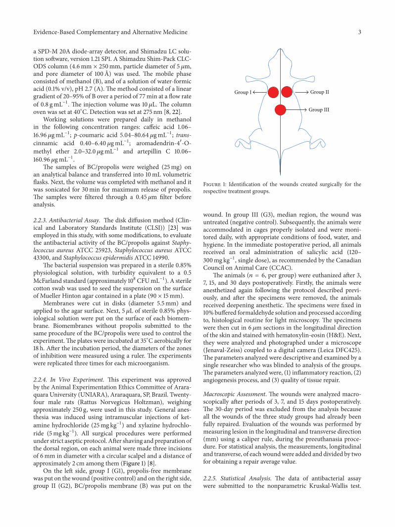
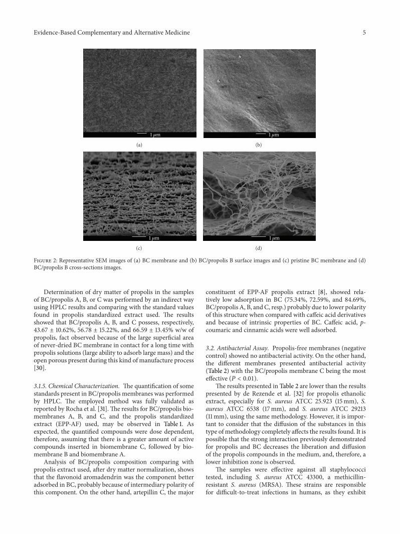
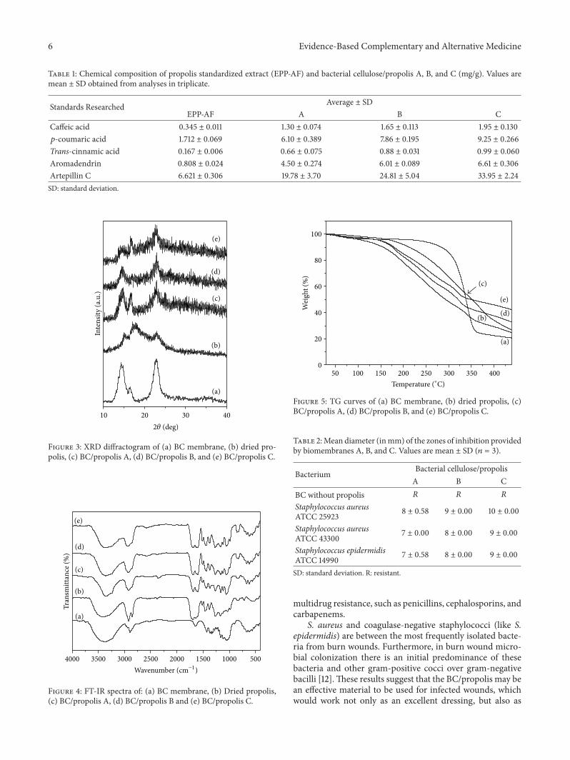
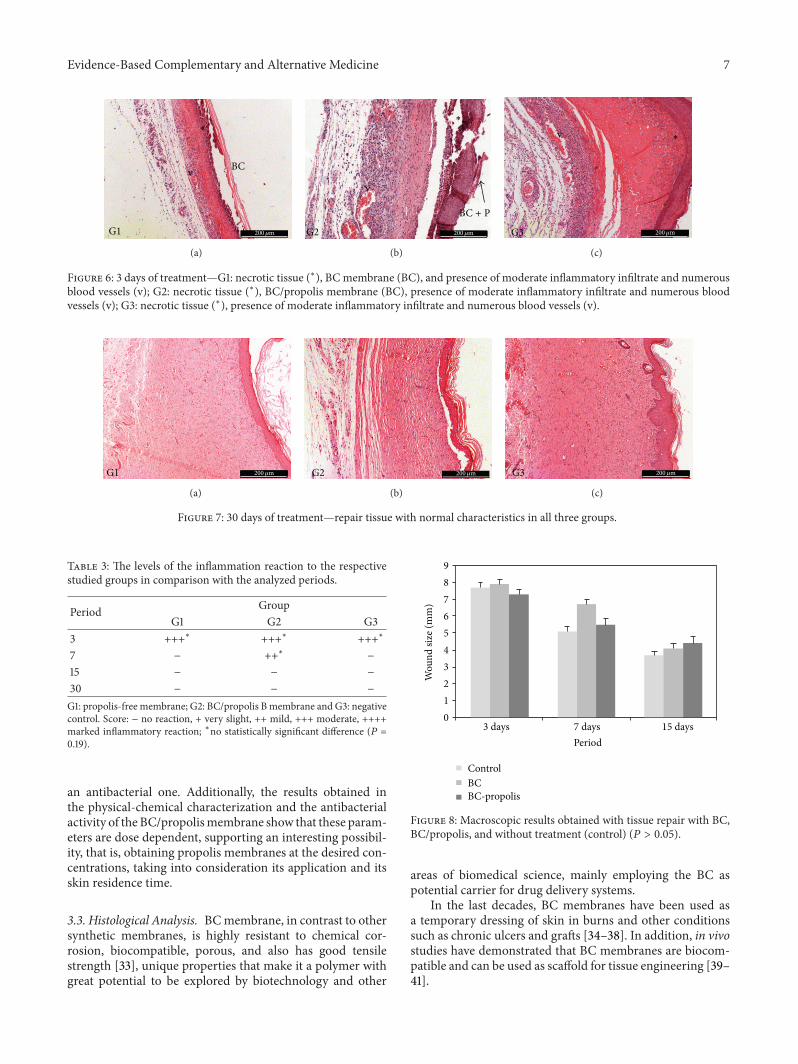
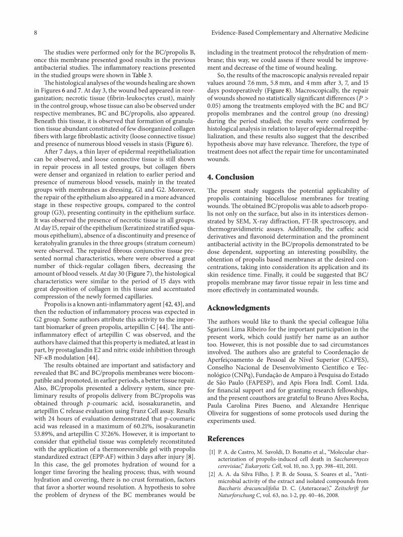