Embed Size (px)
Citation preview
Wimuttisuk et al. BMC Cell Biology 2014, 15:28http://www.biomedcentral.com/1471-2121/15/28
RESEARCH ARTICLE Open Access
Novel Cul3 binding proteins function to remodelE3 ligase complexesWananit Wimuttisuk2,4, Mark West1, Brittney Davidge1, Kebing Yu3, Arthur Salomon2 and Jeffrey D Singer1*
Abstract
Background: Cullins belong to a family of scaffold proteins that assemble multi-subunit ubiquitin ligase complexesto recruit protein substrates for ubiquitination via unique sets of substrate adaptor, such as Skp1 or Elongin B, and asubstrate-binding protein with a conserved protein-protein interacting domain, such as leucine-rich repeats (LRR), aWD40 domain, or a zinc-finger domain. In the case of the Cullin3 (Cul3), it forms a BTB-Cul3-Rbx1 (BCR) ubiquitinligase complex where it is believed that a BTB domain-containing protein performs dual functions where it servesas both the substrate adaptor and the substrate recognition protein.
Results: Tandem affinity purification and LC/MS-MS analysis of the BCR complex led to the identification of 10,225peptides. After the SEQUEST algorithm and CDART program were used for protein identification and domain prediction,we discovered a group of Cul3-bound proteins that contain either the LRR or WD40 domain (CLWs). Further biochemicalanalysis revealed that the LRR domain-containing CLWs could bind both Cul3 and BTB domain-containing proteins. Thedual binding role for the LRR domain-containing CLWs results in causing the BTB-domain protein to become a substrateinstead of an adaptor.To further distinguish potential substrates from other components that are part of the BCR ubiquitin ligase complex, wealtered the parameters in the SEQUEST algorithm to select for peptide fragments with a modified lysine residue. Thismethod not only identifies the potential substrates of the BCR ubiquitin ligase complex, but it also pinpoints the lysineresidue in which the post-translational modification occurs. Interestingly, none of the CLWs were identified by thismethod, supporting our hypothesis that CLWs were not potential substrates but rather additional components of theBCR ubiquitin ligase complex.
Conclusion: Our study identified a new set of Cul3-binding proteins known as CLWs via tandem affinity purificationand LC/MS-MS analysis. Subsequently, our biochemical analysis revealed that some CLWs modify binding of BTBdomain-containing proteins to the complex, causing degradation of the BTB domain-containing protein. As theseCLWs were excluded from our list of substrates, we propose that CLWs serve as unique Cul3 binding proteins thatprovide an alternative regulatory mechanism for the complex.
Keywords: Cullin3, Tandem-affinity purification, BTB domain-containing protein, BCR ubiquitin ligase complex,Mass spectrometry, E3 ubiquitin ligase, Protein purification, Ubiquitin, Ubiquitin ligase
BackgroundSeven cullins have been identified in mammalian cells(Cul1, 2, 3, 4A, 4B, 5, and 7); each cullin assembles aunique set of ubiquitin ligase complexes that catalyzethe formation of polyubiquitin chains to signal the deg-radation of target proteins by the ubiquitin-dependentproteolytic pathway [1,2]. The three best-characterizedcullins, Cul1, Cul2, and Cul3, form diverse groups of
* Correspondence: [email protected] of Biology, Portland State University, Portland, Oregon, USAFull list of author information is available at the end of the article
© 2014 Wimuttisuk et al.; licensee BioMed CenCreative Commons Attribution License (http:/distribution, and reproduction in any mediumDomain Dedication waiver (http://creativecomarticle, unless otherwise stated.
ubiquitin ligase complexes with different sets of substraterecognition modules. For instance, Cul1 forms an Skp1-Cullin-F-box (SCF) complex that acquires substratespecificity via two proteins: a Skp1 linker protein and asubstrate adaptor protein containing an F-box domain[3,4]. Likewise, Cul2 forms an Elongin-Cul2-SOCS-box(ECS) ubiquitin ligase complex with a ubiquitin-likeprotein Elongin B, a Skp1-like linker protein Elongin C anda substrate adaptor SOCS-box containing protein [5,6].Conversely, Cul3 forms a BTB-Cul3-Rbx1 (BCR) ubiquitinligase complex, in which a BTB domain-containing protein
tral Ltd. This is an Open Access article distributed under the terms of the/creativecommons.org/licenses/by/2.0), which permits unrestricted use,, provided the original work is properly credited. The Creative Commons Publicmons.org/publicdomain/zero/1.0/) applies to the data made available in this
Wimuttisuk et al. BMC Cell Biology 2014, 15:28 Page 2 of 17http://www.biomedcentral.com/1471-2121/15/28
serves as both a linker and a substrate adaptor module[7-9]. It is estimated that more than 200 BTB domain-containing proteins are expressed in human cells, whichcould accommodate a very large number of substrates forthe BCR ubiquitin ligase complex [9].Previous studies have uncovered new substrates and
unique components of cullin ubiquitin ligase complexesthat suggest new mechanisms for substrate recruitment[8,10-13]. For example, DDB1 was originally identifiedas a substrate adaptor that bound to the N-terminus ofthe Cul4A complex using its one β-propeller domain(BPB), while utilizing the double-β-propeller domain(BPA-BPC) to provide substrate specificity for the Cul4Aubiquitin ligase complex [14]. However, a study usingmass spectrometry analysis of the Cul4A complex hasidentified a group of WD40 domain-containing proteinscalled DWD that can bind to both Cul4A and DDB1.These DWD proteins were later shown to serve asadditional substrate adaptors for the Cul4A complex[12,13]. Because of the functional similarity betweenDDB1 and BTB domain-containing proteins, this findingsuggests a possibility that the substrate adaptor compo-nent of the BCR complex may not be limited to BTBdomain-containing proteins.Here we performed tandem affinity purification (TAP)
followed by Multidimensional Protein Identification Tech-nology (MudPIT) mass spectrometry to identify newcomponents of the BCR complex. Several known Cul3-binding proteins and substrates of the BCR ubiquitinligase complex were identified by this method. In anattempt to rapidly analyze the potential of the BCRubiquitin ligase complex from this list of proteins, wemodified the parameters in the SEQUEST algorithm sothat it would recognize the peptide sequence with alysine residue that is attached to two glycine residueson its side chain, which is indicative of a protein thathas been conjugated to ubiquitin or a ubiquitin-likemolecule prior to the tryptic digest.In addition to many known Cul3 binding proteins and
new substrates, identified by remnants of conjugatedubiquitin, we have identified two classes of Cul3-boundproteins with either Leucine-rich repeats (LRR) or a WD40domain, which will be referred to as CLWs in this study.
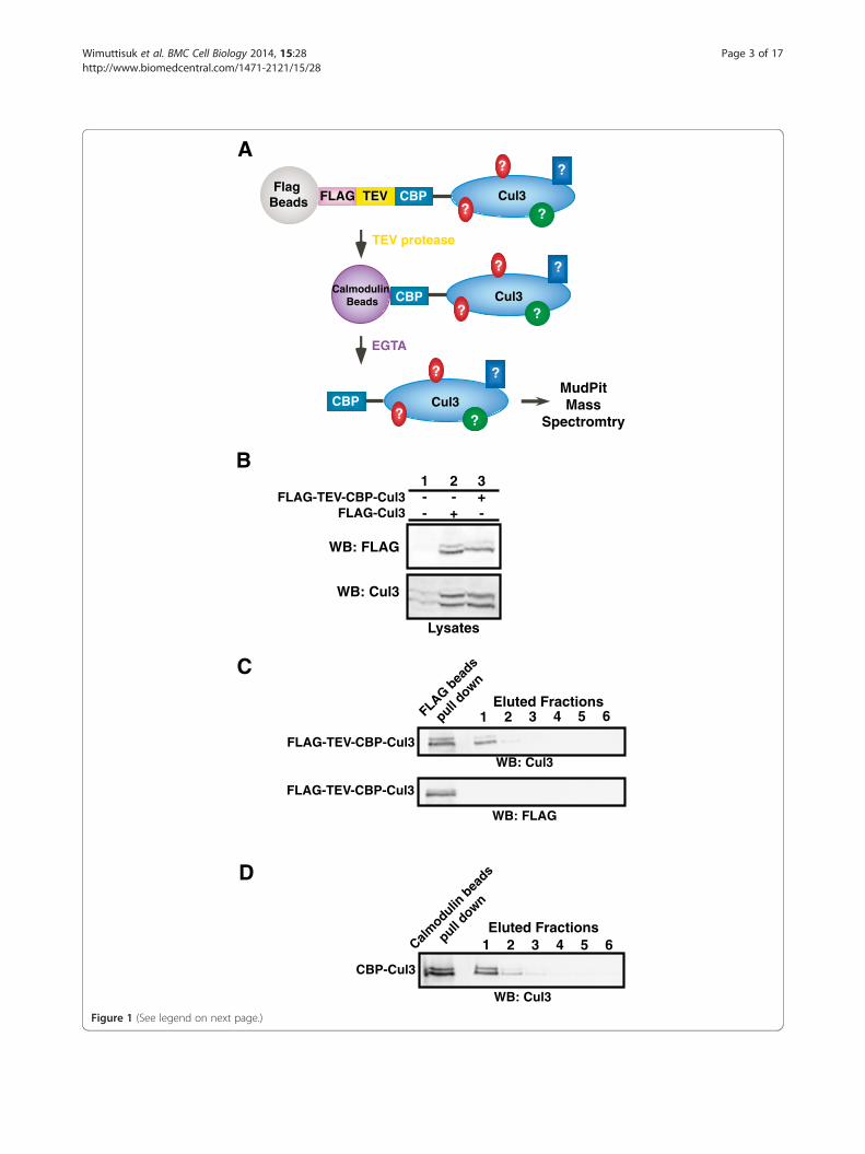
ResultsTandem affinity purification of in vivo Cul3 complexesIn this study, a combination of tandem affinity purification(TAP) and MudPIT mass spectrometry were used to iden-tify new members of the BCR ubiquitin ligase complex.To obtain highly purified Cul3 complexes, an expressioncassette encoding a FLAG-TEV-CBP-Cul3 fusion proteinwas constructed so that the Cul3 complex could bepurified using anti-FLAG Sepharose beads and calmodulin-conjugated beads (Figure 1A). The size and expression
levels of the FLAG-TEV-CBP-Cul3 fusion protein wereverified by immunoblotting and probing against eitheranti-Cul3 antibody or anti-FLAG antibody (Figure 1B). Asthe proportion of neddylated to unneddylated Cul3 wasroughly the same in endogenous Cul3 and overexpressedCul3 (Figure 1B, bottom panel, lane 1 and 3), we reasonedthat these purified Cul3 complexes would likely to be partof a functional BCR ubiquitin ligase complex rather than afree Cul3 monomer, and thus we proceeded with ourexperiment. The Cul3 complexes were purified withanti-FLAG Sepharose beads and TEV protease cleavage,where the lack of protein bands in the FLAG immuno-blot confirmed that the FLAG-tag had been cleaved offthe eluted Cul3 complexes (Figure 1C). Subsequently, theCBP-Cul3 complexes were purified with calmodulin-conjugated beads (Figure 1D).
Data analysis on the MS/MS spectra and potentialbinding proteins of the BCR ubiquitin ligase complexFive independent MudPIT experiments were performedfor the Cul3 fusion protein as well as one actin control.The SEQUEST algorithm was set to match the experi-mentally obtained mass spectra with theoretical peptidesfrom the human NCBI nr database, revealing a total of5,964 peptides from the purified actin control (Table 1, top)and 10,225 peptides from the purified Cul3 complexes(Table 1, bottom) [15,16]. Low quality spectra with a masserror greater than 20 ppm, low Xcorr values (charge 1 < 2,charge 2 < 2.5, and charge 3 < 3), and other known con-taminants were discarded. The resulting peptides werecategorized into two groups based on the number ofpeptide sequences that match the predicted protein. Thefirst group of Cul3-binding protein includes proteins thatwere either identified by at least two different peptidesequences or known Cul3-binding proteins that wereidentified with at least one peptide sequence in thisstudy, such as CAND1, Nedd8, KLHL5, Rpn1, and Rpn5(Additional file 1: Table S1). The second group of potentialCul3-binding proteins was identified via mass spectra ofone peptide sequence, which required additional manualvalidation of the MS/MS spectra as well as further bio-chemical analysis to confirm their interactions with Cul3complexes (Additional file 2: Table S2). This list includesproteins that may be involved in the ubiquitination path-way, such as pVHL-interacting deubiquitinating enzyme 1,STIP1 homology and U-box containing protein 1, andubiquitin specific proteases.
Identification of leucine-rich repeats and WD40domain-containing proteins as Cul3 binding proteinsProteins that corresponded to the identified peptide spectrawere subjected to the domain prediction program CDARTand categorized based on conserved domains [17]. Whencompared with the list of proteins identified from purified
A
B
C
D
MudPitMass
Spectromtry
Eluted Fractions
Eluted Fractions
1 2 3 4 5 6
1 2 3 4 5 6
1 2 3
WB: Cul3
WB: FLAG
WB: Cul3
WB: Cul3
WB: FLAG
FLAG-TEV-CBP-Cul3
FLAG-TEV-CBP-Cul3
CBP-Cul3
Lysates
FLAG-TEV-CBP-Cul3FLAG-Cul3
+- -- -+
Calmodulin
bea
ds
pull down
FLAG bea
ds
pull down
FlagBeads
Cul3
Cul3
Cul3
CBP
CBP
EGTA
CBP
TEV protease
TEVFLAG
CalmodulinBeads
??
? ?
?
?
?
?
?
?
??
Figure 1 (See legend on next page.)
Wimuttisuk et al. BMC Cell Biology 2014, 15:28 Page 3 of 17http://www.biomedcentral.com/1471-2121/15/28
(See figure on previous page.)Figure 1 Tandem affinity purification of 3XFLAG-TEV-CBP-Cul3 complexes. A. Diagram for the tandem affinity purification of the Cul3 bindingproteins. 3XFLAG-TEV-CBP-Cul3 was first affinity purified using anti-FLAG M2 affinity gel resin. Subsequently, the Cul3 complexes were cleavedoff the gel resin by the TEV protease enzyme. The eluted CBP-Cul3 complexes were affinity purified using calmodulin-conjugated beads andthe Cul3 complexes were subsequently subjected to analysis by MudPIT mass spectrometry. B. Untransfected HEK293 cells (lane 1) and HEK293 cellstransfected with either FLAG-tagged Cul3 (lane 2) or FLAG-TEV-CBP-Cul3 (lane 3) were harvested and sonicated in RIPA buffer. Cell lysates wereimmunoblotted and probed with anti-FLAG (top panel) and anti-Cul3 antibodies (bottom panel). C. Complexes containing Cul3 were isolatedin the first step of the tandem affinity purification. Cell lysates containing FLAG-TEV-CBP-Cul3 protein were subjected to immunoprecipitationusing anti-FLAG beads (left lane). CBP-Cul3 was released from the beads using the TEV protease (eluted fractions, lane 1). The FLAG beadswere subsequently washed with TEV protease buffer (eluted fractions, lanes 2–4) and calmodulin-binding buffer (eluted fractions, lanes 5–6).The collected supernatants were immunoblotted and probed with anti-Cul3 antibody (top panel) and anti-FLAG antibody (bottom panel). D.The CBP-Cul3 complex was isolated in the second step of the tandem affinity purification. Ten milliliters of combined CBP-Cul3 fractions from the firststep of the tandem affinity purification were incubated with calmodulin-conjugated beads, followed by several washes with the calmodulin-bindingbuffer and the calmodulin-rinsing buffer. A sample of protein-bound calmodulin beads was collected (left lane). Cul3 complexes were eluted fromthe calmodulin beads using the calmodulin-eluting buffer containing EGTA (lane 1–6). The eluted fractions were immunoblotted and probed withanti-Cul3 antibody. The fractions that contained the most Cul3 were selected for MudPIT analysis. See Additional file 3: Table S3.
Wimuttisuk et al. BMC Cell Biology 2014, 15:28 Page 4 of 17http://www.biomedcentral.com/1471-2121/15/28
actin complexes, we observed enrichment for proteins thatcontain WD40 domains and leucine-rich repeats (LRR) inthe purified Cul3 data set with the percent enrichmentscores of 73.3% of WD40 domain and 90.9% for LRRdomain-containing proteins in the Cul3-bound complexin contrast to 26.7% of WD40 domain and 9.1% of theLRR domain-containing protein in the actin-boundcomplex (Additional file 3: Tables S3 and Additionalfile 4: Table S4). Because domain enrichment maybeindicative of preferential binding between the BCRcomplex and proteins with LRR/WD40 domains, wechose to further explore the role of these LRR/WD40domains in the mammalian BCR complex.
The leucine-rich repeats (LRR) and WD40 domain-containing proteins (CLWs) bind Cul3Three leucine-rich repeat proteins (LRRs, LRR1, LRR3 andLRR5 (fibromodulin)) and two WD40 domain-containingproteins (W14 and W16) were cloned from the list of32 MudPIT-identified Cul3-bound proteins with LRRor WD40 domains (CLWs) to verify their binding toCul3. The cDNAs were fully sequenced and cloned intomammalian expression vectors in frame with the HA-tag.The proteins were co-expressed with FLAG-tagged Cul3
Table 1 Summary of TAP tag experiments to find Cul3 and ac
Exp. # # of cells Total protein (mg) TAP yield (mg) Separ
Actin-binding proteins bound to 3XFLAG-TEV-CBP-Actin:
1 9.5 × 10⁷ 264 64 Mud
Cul3-binding proteins bound to 3XFLAG-TEV-CBP-Cul3:
1 6.9 × 10⁷ 192 280 Mud
2 6.9 × 10⁷ 192 52 Mud
3 7.5 × 10⁷ 208 36 Mud
4 6.5 × 10⁷ 180 39 Mud
5 6.5 × 10⁷ 180 39 Mud
Total Cul3 bo
See also Additional file 1: Table S1.
(FLAG-Cul3). Immunoprecipitation assays revealed thatFLAG-Cul3 was able to specifically bind the HA-taggedCLWs with either the LRR (Figure 2A) or the WD40domains (Figure 2B), which further confirms the resultsfrom the proteomic analysis.
Cul3 mutants that cannot bind BTB domain-containingproteins have enhanced binding to LRRsTo clarify how other Cul3 binding proteins affect orregulate the binding of LRRs to Cul3, we tested a varietyof Cul3 mutants that were unable to bind various Cul3binding partners. Mutants that could not be neddylated(K712R) or could not bind RBX1 (F665D) were able tobind the LRR proteins in an identical fashion to wildtype Cul3 (not shown). Surprisingly, mutants thatcould not bind BTB domain-containing proteins (Δ51-67, Figure 2C) showed enhanced binding to bothLRR1 and LRR5 (Figure 2D). This observation led usto investigate a possible interaction between LRRs andBTB domain-containing proteins.
LRRs bind to BTB domain-containing proteinsBased on our observation that removal of the BTB bindingregion of Cul3 enhanced LRR binding we hypothesized
tin-binding proteins
ation MS input (mg) Peptides Peptides </=20 ppm error
PIT 15 5964 883
PIT 15 4158 562
PIT 15 1395 234
PIT 15 1862 393
PIT 15 1405 276
PIT 15 1405 117
und peptides: 10225 1582
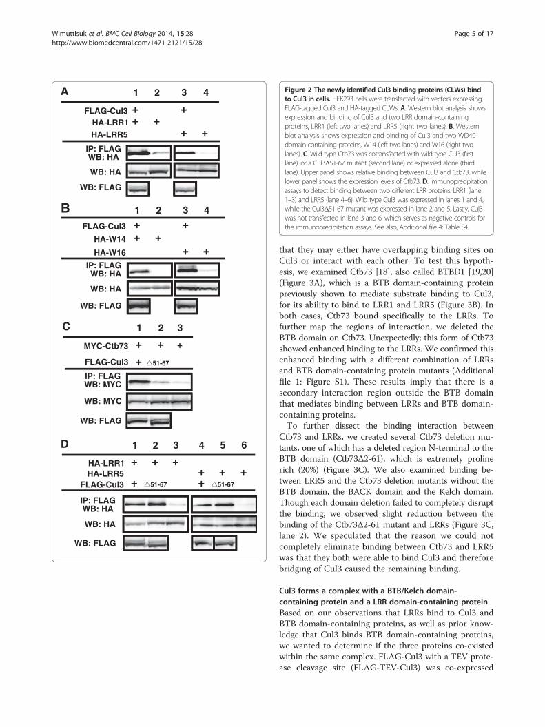
Figure 2 The newly identified Cul3 binding proteins (CLWs) bindto Cul3 in cells. HEK293 cells were transfected with vectors expressingFLAG-tagged Cul3 and HA-tagged CLWs. A. Western blot analysis showsexpression and binding of Cul3 and two LRR domain-containingproteins, LRR1 (left two lanes) and LRR5 (right two lanes). B. Westernblot analysis shows expression and binding of Cul3 and two WD40domain-containing proteins, W14 (left two lanes) and W16 (right twolanes). C. Wild type Ctb73 was cotransfected with wild type Cul3 (firstlane), or a Cul3Δ51-67 mutant (second lane) or expressed alone (thirdlane). Upper panel shows relative binding between Cul3 and Ctb73, whilelower panel shows the expression levels of Ctb73. D. Immunoprecipitationassays to detect binding between two different LRR proteins: LRR1 (lane1–3) and LRR5 (lane 4–6). Wild type Cul3 was expressed in lanes 1 and 4,while the Cul3Δ51-67 mutant was expressed in lane 2 and 5. Lastly, Cul3was not transfected in lane 3 and 6, which serves as negative controls forthe immunoprecipitation assays. See also, Additional file 4: Table S4.
Wimuttisuk et al. BMC Cell Biology 2014, 15:28 Page 5 of 17http://www.biomedcentral.com/1471-2121/15/28
that they may either have overlapping binding sites onCul3 or interact with each other. To test this hypoth-esis, we examined Ctb73 [18], also called BTBD1 [19,20](Figure 3A), which is a BTB domain-containing proteinpreviously shown to mediate substrate binding to Cul3,for its ability to bind to LRR1 and LRR5 (Figure 3B). Inboth cases, Ctb73 bound specifically to the LRRs. Tofurther map the regions of interaction, we deleted theBTB domain on Ctb73. Unexpectedly; this form of Ctb73showed enhanced binding to the LRRs. We confirmed thisenhanced binding with a different combination of LRRsand BTB domain-containing protein mutants (Additionalfile 1: Figure S1). These results imply that there is asecondary interaction region outside the BTB domainthat mediates binding between LRRs and BTB domain-containing proteins.To further dissect the binding interaction between
Ctb73 and LRRs, we created several Ctb73 deletion mu-tants, one of which has a deleted region N-terminal to theBTB domain (Ctb73Δ2-61), which is extremely prolinerich (20%) (Figure 3C). We also examined binding be-tween LRR5 and the Ctb73 deletion mutants without theBTB domain, the BACK domain and the Kelch domain.Though each domain deletion failed to completely disruptthe binding, we observed slight reduction between thebinding of the Ctb73Δ2-61 mutant and LRRs (Figure 3C,lane 2). We speculated that the reason we could notcompletely eliminate binding between Ctb73 and LRR5was that they both were able to bind Cul3 and thereforebridging of Cul3 caused the remaining binding.
Cul3 forms a complex with a BTB/Kelch domain-containing protein and a LRR domain-containing proteinBased on our observations that LRRs bind to Cul3 andBTB domain-containing proteins, as well as prior know-ledge that Cul3 binds BTB domain-containing proteins,we wanted to determine if the three proteins co-existedwithin the same complex. FLAG-Cul3 with a TEV prote-ase cleavage site (FLAG-TEV-Cul3) was co-expressed

Figure 3 The BTB domain-containing proteins bind LRRs, while the ΔBTB mutant enhances the binding interaction. A. Ctb73 containsthree protein-protein interaction domains; BTB domain, BACK domain and PHR (kelch) domain. B. MYC-Ctb73 was immunoprecipitated and probedwith anti-HA antibodies to detect binding to LRR1 and LRR5 (upper panel). Lower panels show the expression levels of the HA-LRRs in transfected cells.C. MYC-Ctb73 wild-type or deletion mutants were pulled down by anti-MYC antibody and probed against HA (upper panel) to locate LRR5 binding siteon Ctb73. Relative expression levels of HA-LRR5 are shown in lower panel. D. Two sequential immunoprecipitations were performed on cell lysatesexpressing FLAG-TEV-Cul3, MYC-Ctb73 and HA-LRR5. FLAG beads brought down Cul3 complexes that were cleaved off the resin using TEV protease.The eluted proteins were verified by Western blot for Cul3. Subsequent immunopreciptiation with MYC antibody brought down a protein complexcontaining Ctb73. Western blot analysis showed LRR5 binding to the Cul3-Ctb73 complex. Relative protein levels are shown in bottom three panels.E. Cell lysates co-expressing Cul3, Ctb73 and LRRs were used in the immunoprecipitation of FLAG-Cul3 that pulled down HA-LRR1 and HA-LRR5. Inthe same transfection set, immunoprecipitation of FLAG-Cul3 also brought down MYC-Ctb73. Relative expression levels of LRR1, LRR5 and MYC-Ctb73were verified by Western blotting. See also Additional file 1: Figure S1.
Wimuttisuk et al. BMC Cell Biology 2014, 15:28 Page 6 of 17http://www.biomedcentral.com/1471-2121/15/28
with MYC-Ctb73 and HA-LRR5. FLAG-beads were usedto purify the Cul3 complex, which were subsequentlytreated with TEV protease to release the protein complexfrom the beads. The resulting soluble proteins were sub-jected to a second round of immunoprecipitation withMYC antibody to pull down MYC-Ctb73. The resulting
protein complexes were analyzed by Western blot analysisto detect the presence of the third protein, HA-LRR5.Two sequential affinity purifications of FLAG-Cul3 andMYC-Ctb73 pulled down significant amounts HA-LRR5only in the presence of all three proteins (Figure 3D, toppanel). These data verify our hypothesis that Cul3, a BTB-
Wimuttisuk et al. BMC Cell Biology 2014, 15:28 Page 7 of 17http://www.biomedcentral.com/1471-2121/15/28
domain containing protein and an LRR could co-existwithin the same complex.Because our results in Figures 2 and 3 suggested a poten-
tial for overlapping or co-regulation of binding betweenBTB domain-containing proteins and LRRs on Cul3, wealso compared complex formation on wild type Cul3versus the mutant that cannot bind BTB domain-containing proteins (FLAG-Cul3Δ51-67). We observeda slight increase in the binding between the LRRs andthe Cul3 mutant when compared to wild type (Figure 3Elanes 2 and 4), which further supports a overlappingbinding model.
A B/C-like box sequence mediates LRR binding to Cul3complexesAs several LRRs are very small and the LRR domaindominates the coding sequence, we began determiningthe regions on the LRRs that mediate interactions withCul3 and/or the BTB domain-containing proteins. Inthis case, the deletion of LRR domain would not beinformative because the remaining portion would beshort peptides that were unlikely to fold properly. Wecould not find any conserved regions outside of the LRRdomains that were shared by the identified LRR domaincontaining proteins. Further examination however revealeda sequence adjacent to the LRR domain that stronglyresembles a B/C box (Figure 4A). To determine if thissequence may be playing a role in the interactionbetween the LRR domain containing proteins and Cul3complexes, we created I330G C334A mutations in theputative B/C-like box of the LRR5 (HA-LRR5-B/C)and tested its binding to Cul3 (Figure 4B) and Ctb73(Figure 4C). When compared with wild-type LRR5, weobserved that the LRR5-B/C mutant showed enhancedbinding to wild type Cul3 (Figure 4B, lanes 1 and 4).However, the LRR-B/C interaction with Cul3Δ51-67mutant that could not bind BTB domain was com-pletely eliminated, though this form of Cul3 mutantshowed enhanced binding to wild type LRRs (Figure 4B,lanes 2 and 5). Additionally, the binding between theLRR5-B/C mutant and various Ctb73 deletion mutantswere examined. We observed that LRR5-B/C mutantmaintained the same binding interaction with wild-typeCtb73 and most of the deletion mutants (Figure 4C).However, the binding interaction between the LRR5-B/Cmutant and the Ctb73Δ2-61 was eliminated (Figure 4C,lane2), indicating that this B/C-like box is the majormediator of binding the LRR domain-containing proteinto the BTB domain-containing protein. This B/C-like boxbinds the N-terminal sequence (residues 2–61) on theBTB domain containing protein. In addition, the residualbinding between Ctb73Δ2-61 and wild type LRR5 was dueto bridging via Cul3. We speculated that the enhancedbinding of the LRR-B/C-like box mutant to wild type Cul3
was due to the reduced interaction between the LRR-B/Cmutant and a BTB domain-containing protein in thecomplex, thus eliminating the interaction we havepreviously shown to regulate LRRs binding to Cul3.Consistent with this model, LRR5-B/C does not showenhanced binding to the version of Ctb73 that is missingits BTB domain, unlike wild-type LRR5 (Figure 4D, lane 2vs. lane 5). A graphical summary of these interactions isshown in Figure 4E.
FMOD (LRR5) causes FAZF to become degradedIn order to determine the potential cellular role for thesecomplexes that contain a BTB domain-containing protein,an LRR domain-containing protein and Cul3, we per-formed two hybrid screens using LRR5 as bait. Afterscreening greater than a million clones we identified onlyone strong binding partner, FAZF. FAZF is a BTBdomain-containing protein that has been shown to beimportant in transcriptional regulation of B-cell differ-entiation [21]. This was a surprise since we have seenLRR5 bind several BTB domain-containing proteins, andthe library we used contains many BTB domain-containingproteins [18]. In order to determine specificity and verifythese two proteins bind in mammalian cells LRR5 wasco-expressed with either FAZF or the highly similarprotein PLZF [22] (Figure 5A). Interestingly, LRR5 boundFAZF and not PLZF and also appeared to cause a reductionin expression levels of FAZF (Figure 5A). We therefore feltthat the binding interaction was potentially biologicallysignificant. FAZF overexpression has been shown to causeG1 arrest and apoptosis in B cell lines [21], in additionLRR5 (FMOD) is highly overexpressed in some types ofleukemias [23]. Enforced down regulation of LRR5 inthese leukemias also results in apoptosis [24]. Based onthis observation and on our previous data regardingthe relationship between LRR5 binding Cul3 and BTBdomain-containing proteins, we postulated that LRR5might be involved in regulating the levels of the BTBdomain-containing protein FAZF. We reasoned that acomplex of all three might convert the BTB domain-containing protein to a substrate instead of a substrateadaptor by modulating how it binds to Cul3. Consistentwith the proposed hypothesis, the reduced levels of FAZFcaused by coexpression of both LRR5 and FAZF couldbe restored by inhibiting the proteasome with MG-132(Figure 5B). We then focused on a potential for LRR5to modulate expression levels of endogenous FAZF usinga well-characterized antibody against FAZF [21]. Wefound that high expression of LRR5 dramatically reducedendogenous levels of FAZF (Figure 5C). As LRR5 levelswere decreased using coexpression of shRNA againstLRR5, the levels of FAZF increase demonstrating aninverse relationship between expression of LRR5 andFAZF. An analogous experiment in which LRR1 was
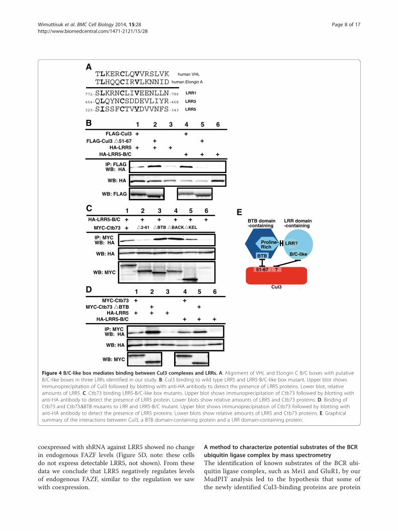
Figure 4 B/C-like box mediates binding between Cul3 complexes and LRRs. A. Alignment of VHL and Elongin C B/C boxes with putativeB/C-like boxes in three LRRs identified in our study. B. Cul3 binding to wild type LRR5 and LRR5-B/C-like box mutant. Upper blot showsimmunoprecipitation of Cul3 followed by blotting with anti-HA antibody to detect the presence of LRR5 proteins. Lower blot, relativeamounts of LRR5. C. Ctb73 binding LRR5-B/C-like box mutants. Upper blot shows immunoprecipitation of Ctb73 followed by blotting withanti-HA antibody to detect the presence of LRR5 protein. Lower blots show relative amounts of LRR5 and Ctb73 proteins. D. Binding ofCtb73 and Ctb73ΔBTB mutants to LRR and LRR5-B/C mutant. Upper blot shows immunoprecipitation of Ctb73 followed by blotting withanti-HA antibody to detect the presence of LRR5 proteins. Lower blots show relative amounts of LRR5 and Ctb73 proteins. E. Graphicalsummary of the interactions between Cul3, a BTB domain-containing protein and a LRR domain-containing protein.
Wimuttisuk et al. BMC Cell Biology 2014, 15:28 Page 8 of 17http://www.biomedcentral.com/1471-2121/15/28
coexpressed with shRNA against LRR5 showed no changein endogenous FAZF levels (Figure 5D, note: these cellsdo not express detectable LRR5, not shown). From thesedata we conclude that LRR5 negatively regulates levelsof endogenous FAZF, similar to the regulation we sawwith coexpression.
A method to characterize potential substrates of the BCRubiquitin ligase complex by mass spectrometryThe identification of known substrates of the BCR ubi-quitin ligase complex, such as Mei1 and GluR1, by ourMudPIT analysis led to the hypothesis that some ofthe newly identified Cul3-binding proteins are protein
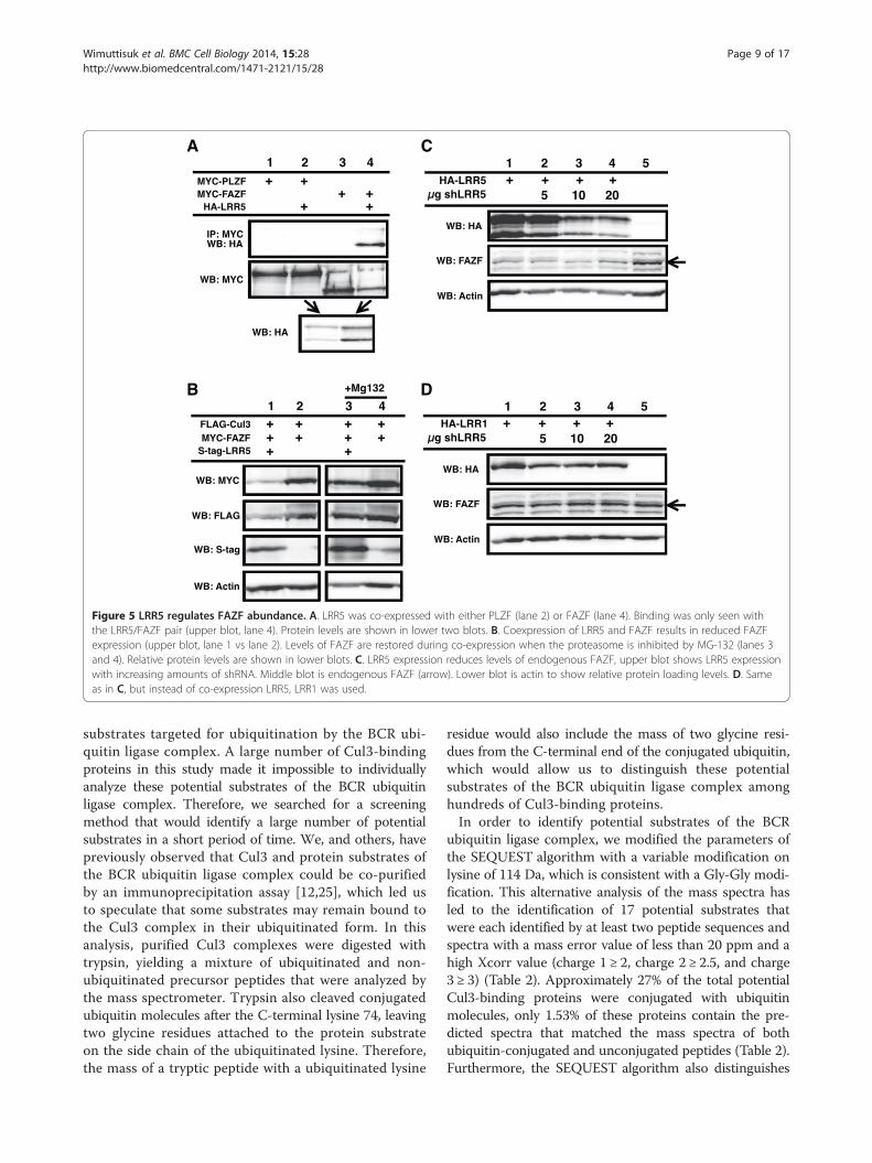
Figure 5 LRR5 regulates FAZF abundance. A. LRR5 was co-expressed with either PLZF (lane 2) or FAZF (lane 4). Binding was only seen withthe LRR5/FAZF pair (upper blot, lane 4). Protein levels are shown in lower two blots. B. Coexpression of LRR5 and FAZF results in reduced FAZFexpression (upper blot, lane 1 vs lane 2). Levels of FAZF are restored during co-expression when the proteasome is inhibited by MG-132 (lanes 3and 4). Relative protein levels are shown in lower blots. C. LRR5 expression reduces levels of endogenous FAZF, upper blot shows LRR5 expressionwith increasing amounts of shRNA. Middle blot is endogenous FAZF (arrow). Lower blot is actin to show relative protein loading levels. D. Sameas in C, but instead of co-expression LRR5, LRR1 was used.
Wimuttisuk et al. BMC Cell Biology 2014, 15:28 Page 9 of 17http://www.biomedcentral.com/1471-2121/15/28
substrates targeted for ubiquitination by the BCR ubi-quitin ligase complex. A large number of Cul3-bindingproteins in this study made it impossible to individuallyanalyze these potential substrates of the BCR ubiquitinligase complex. Therefore, we searched for a screeningmethod that would identify a large number of potentialsubstrates in a short period of time. We, and others, havepreviously observed that Cul3 and protein substrates ofthe BCR ubiquitin ligase complex could be co-purifiedby an immunoprecipitation assay [12,25], which led usto speculate that some substrates may remain bound tothe Cul3 complex in their ubiquitinated form. In thisanalysis, purified Cul3 complexes were digested withtrypsin, yielding a mixture of ubiquitinated and non-ubiquitinated precursor peptides that were analyzed bythe mass spectrometer. Trypsin also cleaved conjugatedubiquitin molecules after the C-terminal lysine 74, leavingtwo glycine residues attached to the protein substrateon the side chain of the ubiquitinated lysine. Therefore,the mass of a tryptic peptide with a ubiquitinated lysine
residue would also include the mass of two glycine resi-dues from the C-terminal end of the conjugated ubiquitin,which would allow us to distinguish these potentialsubstrates of the BCR ubiquitin ligase complex amonghundreds of Cul3-binding proteins.In order to identify potential substrates of the BCR
ubiquitin ligase complex, we modified the parameters ofthe SEQUEST algorithm with a variable modification onlysine of 114 Da, which is consistent with a Gly-Gly modi-fication. This alternative analysis of the mass spectra hasled to the identification of 17 potential substrates thatwere each identified by at least two peptide sequences andspectra with a mass error value of less than 20 ppm and ahigh Xcorr value (charge 1 ≥ 2, charge 2 ≥ 2.5, and charge3 ≥ 3) (Table 2). Approximately 27% of the total potentialCul3-binding proteins were conjugated with ubiquitinmolecules, only 1.53% of these proteins contain the pre-dicted spectra that matched the mass spectra of bothubiquitin-conjugated and unconjugated peptides (Table 2).Furthermore, the SEQUEST algorithm also distinguishes
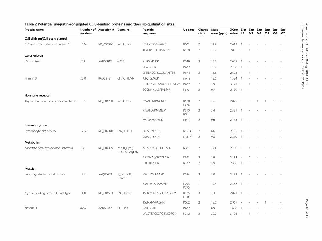
Table 2 Potential ubiquitin-conjugated Cul3-binding proteins and their ubiquitination sites
Protein name Number ofresidues
Accession # Domains Peptidesequence
Ub-sites Chargestate
Masserror (ppm)
XCorrvalue
ExpL2
ExpM3
ExpM4
ExpM5
ExpM6
ExpM7
Cell division/Cell cycle control
Rb1-inducible coiled coil protein 1 1594 NP_055596 No domain LTHLGTAVSVMAK* K201 2 12.4 2.012 1 - - - - -
TFVQK*EQCDFSNSLK K828 2 19.7 2.885 - 1 - - - -
Cytoskeleton
DST protein 258 AAH04912 GAS2 K*SPASKLDK K249 2 15.5 2.055 1 - - - - -
SPASKLDK none 1 18.7 2.136 1 - - - - -
EKFILADGASQGMAAFRPR none 2 16.6 2.693 - 1 - - - -
Filamin B 2591 BAD52434 CH, IG_FLMN ATQTGDASK none 1 18.6 1.584 1 - - - - -
ETTDFKVDTKAAGSGELGVTMK none 2 3.9 3.121 - 1 - - - -
SGCIVNNLAEFTVDPK* K673 2 9.7 2.139 1 - - - - -
Hormone receptor
Thyroid hormone receptor interactor 11 1979 NP_004230 No domain K*VAFDVK*MENEK K670,K676
2 17.8 2.879 - - 1 1 2 -
K*VAFDVKMENEK* K670,K681
2 5.4 2.581 1 - - - - -
MQLLQSLQEQK none 2 0.6 2.463 1 - - - - -
Immune system
Lymphocyte antigen 75 1722 NP_002340 FN2, CLECT DGAICYK*PTK K1514 2 6.6 2.182 1 - - - - -
DGAICYKPTK* K1517 2 9.8 2.260 1 - - - - -
Metabolism
Aspartate beta-hydroxylase isoform a 758 NP_004309 Asp-B_Hydr,TPR, Asp-Arg-Hy
ARYGK*AQCEDDLAEK K381 2 12.1 2.730 - 1 - - - -
ARYGKAQCEDDLAEK* K391 2 3.9 2.338 - 2 - - - -
PKLLNK*FDK K332 2 3.9 2.338 1 - - - - -
Muscle
Long myosin light chain kinase 1914 AAQ02673 S_TKc, FN3,IGcam
ESK*LDSLEAAAK K284 2 5.0 2.382 1 - - - - -
ESKLDSLEAAAK*SK* K293,K295
1 19.7 2.338 1 - - - - -
Myosin binding protein C, fast type 1141 NP_004524 FN3, IGcam TSEKK*SDTAGELDFSGLLK* K175,K185
3 1.4 2.821 1 - - - - -
TSENAIVVVAGNK* K562 2 12.6 2.967 - - - 1 - -
Nesprin-1 8797 AAN60442 CH, SPEC SAREKGER none 1 8.9 1.688 1 - - - - -
WVQYTAGKQTGIEVKDFGK* K212 3 20.0 3.426 - 1 - - - -
Wim
uttisuket
al.BMCCellBiology
2014,15:28Page
10of
17http://w
ww.biom
edcentral.com/1471-2121/15/28
Table 2 Potential ubiquitin-conjugated Cul3-binding proteins and their ubiquitination sites (Continued)
Neuron
Synaptojanin 2A 1288 AAN73051 Syja_N, IPPc SSGKIFKDFHEGAINFGPTYK none 2 0.9 2.005 1 - - - - -
TGMGGK*AGNKGAVGIR K678 2 3.6 2.048 1 - - - - -
Oncoprotein
BRCA2 and CDKN1A interacting protein 322 CAI12093 No domain AGLIQSR none 1 9.2 1.813 1 - - - - -
NCEK*SMVEQLDK K159 1 19.7 1.922 1 - - - - -
Signal transduction
Endothelial differentiation, sphingolipidG-protein-coupled receptor, 1
382 NP_001391 7tm_1 CPSGDSAGKFK*R K340 2 6.1 2.170 1 - - - - -
HYNYTGK* K34 1 14.8 1.973 1 - - - - -
Triple functional domain(PTPRF interacting) variant
2202 BAD92991 SPEC, SH3,RhoGEF, PH_TRIO
AFAAALDER none 2 15.9 3.027 - - 1 - - -
LVNASVAFYK* K1003 2 1.4 2.046 1 - - - - -
Ubiquitination
Cullin3 768 NP_003581 Cullin GVKGLTEQEVETILDK* K414 2 11.9 2.443 1 - - - - -
Unknown function
Chromosome 10 open reading frame 92 876 AAH34223 No domain GRKGSIPR none 1 17.3 1.551 1 - - - - -
ARVQTPAVVADSGKSK none 2 6.7 2.024 1 - - - - -
K*GSIPR K233 1 1.2 1.535 1 - - - - -
KIAA1602 protein 906 AAH33253 No domain EGAGGGSPLR none 2 11.5 2.039 1 - - - - -
SKGLPK* K62 1 3.8 1.931 1 - - - - -
RING finger and CHY zinc fingerdomain containing protein 1 variant
263 BAD92309 zf_CHY DKK*QYHCENCGICR K65 2 10.2 2.581 - 1 - - - -
GYRCPLCMHSALDMTR none 2 1.1 2.126 1 - - - - -
PYK*CMHCEK*VFR K306,K312
2 16.4 2.036 1 - - - - -
Legend:*Charge state represents the charge state of the precursor peptide ion.*Mass error (ppm; parts per million) represents the calculated mass difference between the mass of theoretical precursor peptide and the mass of the experimental precursor peptide.*Xcorr value represents the calculated value of the accuracy between the peaks of the theoretical spectra and the experimental spectra.*Ub-sites represents the position of the ubiquitin-conjugated lysine residues in a given protein.*Number of the peptides based on the corresponding mass spectra from each mass spectrometry experiment shown in Table 1 (L = long gradient; M =MudPIT).The ubiquitinated lysine residues are marked by an asterisk (*). See also Additional file 1: Figure S2.
Wim
uttisuket
al.BMCCellBiology
2014,15:28Page
11of
17http://w
ww.biom
edcentral.com/1471-2121/15/28
Wimuttisuk et al. BMC Cell Biology 2014, 15:28 Page 12 of 17http://www.biomedcentral.com/1471-2121/15/28
the position of the modified lysine residues within eachprecursor peptide. Therefore, this MudPIT approach notonly identifies potential substrates of the BCR ubiquitinligase complex in less time than traditional biochemicalassays, it also predicts the ubiquitin conjugation sites oneach potential substrate that would otherwise requiremany mutational analyses to obtain. Based on our ana-lysis, we observed that none of the CLW proteins wereidentified by this method, thus further supporting ourhypothesis that they are not substrates for the ubiquitina-tion process, but rather part of the BCR ubiquitin ligasecomplex.To verify this algorithm for predicting potentially real
ubiquitination sites, we mutated the predicted site onCul3 (K414R) based on our MudPIT data indicating thatthe K414 residue on Cul3 contains a ubiquitination sitebased on the extra mass that is equivalent to two glycineresidues (Table 2). Biochemical analysis demonstratedthat K414 was a genuine ubiquitination site for Cul3 incells (Additional file 1: Figure S2).
DiscussionSince the discovery of Cul3, the characterization of theBCR ubiquitin ligase has progressed rapidly and hasresulted in the identification of protein substrates andnovel subunits of the BCR ubiquitin ligase complex[18,26-30]. In this study, a MudPIT approach was chosenfor the analysis of Cul3 complexes to identify novel proteinsthat could be part of the Cul3-dependent ubiquitinationpathway. As a result, we have identified new members ofthe Cul3 complex using two different approaches: (1) theidentification of potential substrates of the BCR ubiquitinligase complex and their ubiquitination sites by alteringthe criteria in the SEQUEST algorithm to identify Cul3-bound ubiquitin-conjugated proteins, and (2) the analysisof proteins with conserved domains among the MudPIT-identified Cul3-binding proteins that resulted in thecharacterization of previously unidentified Cul3-boundproteins containing either LRR domains or WD40 domains(CLWs).Based on our chemical analysis, we propose that the
LRR domain-containing proteins (LRRs) function to modifythe binding of the BTB domain containing protein to Cul3such that the BTB domain containing protein becomes asubstrate. LRRs bind to Cul3 not only via the BTB domain-containing protein, but also through other regions of theCul3 complex, as the Cul3 mutant that cannot bind BTBdomain-containing proteins (Cul3Δ51-67) can still bindLRRs. In addition, these Cul3-bound LRRs were not ubi-quitinated, suggesting they are unlikely to be substratesof the BCR ubiquitin ligase complex. This conclusion isfurther supported by the fact that substrates are unlikelyto share a domain (such as an LRR domain) because the
mechanism of substrate recognition is usually based onshort sequences [31,32].
LRR domain-containing proteins regulate bindingbetween Cul3 and BTB-domain containing proteinsWe discovered that the LRR domain-containing proteinsmodulated the binding of the BTB domain-containingproteins binding to Cul3 such that they became substratesinstead of substrate adaptors. We found that a region onthe BTB domain-containing protein just N-terminal to theBTB domain appears to be mediating the interaction withLRR domain-containing proteins. This region on Ctb73 isextremely proline-rich; it is interesting to note that otherBTB domain proteins that have been shown to bind Cul3also have similar proline rich regions in their N-terminus,like actinfilin, BTBD2, and FAZF (PLZF does not have thisregion and does not bind LRR5). Thus, this region mayplay a role in a number of other BTB-domain protein/LRR-domain protein interactions. In addition the LRRdomain-containing proteins contain a sequence that re-sembles a B/C box that mediates binding to the BTBdomain-containing proteins.Based on the identification of the WD40 domain in
substrate adaptor subunits of the Cul4 complex, we firstproposed that our MudPIT-identified WD40 domain-containing proteins also behave similarly in the Cul3complex. However, mutational analysis revealed that theinteraction site for the WD40 domain-containing proteinslies within the conserved cullin domain in C-terminalregion of Cul3, suggesting that their binding interactionsdid not resemble those found on the LRRs (not shown).Therefore, the WD40 domain-containing proteins mayfunction in a completely different way than the LRRs.
Potential substrates of the BCR ubiquitin ligase complexidentified by a method that involves an altered SEQUESTalgorithmTo distinguish novel Cul3-binding proteins from poten-tial substrates of the BCR ubiquitin ligase complex, weadjusted the parameters in the SEQUEST algorithm torecognize the ubiquitin-modified lysine residue in thepeptide spectra. Manual validation of the mass spectrarevealed that the altered SEQUEST parameters could beused to identify ubiquitinated precursor peptides thatare derived from the ubiquitin-conjugated proteins. Thistechnique for the identification of ubiquitin-conjugatedproteins using MudPIT mass spectrometry is an approachthat can be applied to the analysis of ubiquitinated pro-teins bound to other ubiquitin ligase complexes. Usingthis technique, our list of MudPIT-identified Cul3-bindingproteins also includes GluR-1, Mei1 and p60/katanin(Additional file 1: Table S1), which are known substratesof the BCR ubiquitin ligase complex [18,33,34], further
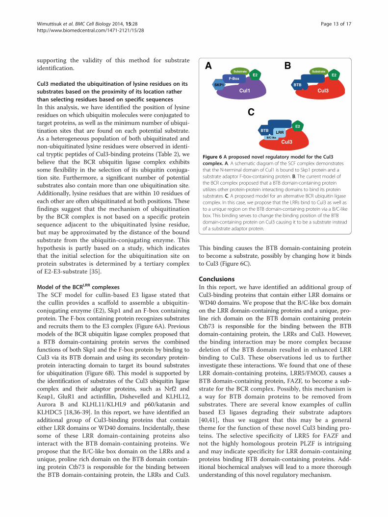
Figure 6 A proposed novel regulatory model for the Cul3complex. A. A schematic diagram of the SCF complex demonstratesthat the N-terminal domain of Cul1 is bound to Skp1 protein and asubstrate adaptor F-box-containing protein. B. The current model ofthe BCR complex proposed that a BTB domain-containing proteinutilizes other protein-protein interacting domains to bind its proteinsubstrates. C. A proposed model for an alternative BCR ubiquitin ligasecomplex. In this case, we propose that the LRRs bind to Cul3 as well asto a unique region on the BTB domain-containing protein via a B/C-likebox. This binding serves to change the binding position of the BTBdomain-containing protein on Cul3 causing it to be a substrate insteadof a substrate adaptor protein.
Wimuttisuk et al. BMC Cell Biology 2014, 15:28 Page 13 of 17http://www.biomedcentral.com/1471-2121/15/28
supporting the validity of this method for substrateidentification.
Cul3 mediated the ubiquitination of lysine residues on itssubstrates based on the proximity of its location ratherthan selecting residues based on specific sequencesIn this analysis, we have identified the position of lysineresidues on which ubiquitin molecules were conjugated totarget proteins, as well as the minimum number of ubiqui-tination sites that are found on each potential substrate.As a heterogeneous population of both ubiquitinated andnon-ubiquitinated lysine residues were observed in identi-cal tryptic peptides of Cul3-binding proteins (Table 2), webelieve that the BCR ubiquitin ligase complex exhibitssome flexibility in the selection of its ubiquitin conjuga-tion site. Furthermore, a significant number of potentialsubstrates also contain more than one ubiquitination site.Additionally, lysine residues that are within 10 residues ofeach other are often ubiquitinated at both positions. Thesefindings suggest that the mechanism of ubiquitinationby the BCR complex is not based on a specific proteinsequence adjacent to the ubiquitinated lysine residue,but may be approximated by the distance of the boundsubstrate from the ubiquitin-conjugating enzyme. Thishypothesis is partly based on a study, which indicatesthat the initial selection for the ubiquitination site onprotein substrates is determined by a tertiary complexof E2-E3-substrate [35].
Model of the BCRLRR complexesThe SCF model for cullin-based E3 ligase stated thatthe cullin provides a scaffold to assemble a ubiquitin-conjugating enzyme (E2), Skp1 and an F-box containingprotein. The F-box containing protein recognizes substratesand recruits them to the E3 complex (Figure 6A). Previousmodels of the BCR ubiquitin ligase complex proposed thata BTB domain-containing protein serves the combinedfunctions of both Skp1 and the F-box protein by binding toCul3 via its BTB domain and using its secondary protein-protein interacting domain to target its bound substratesfor ubiquitination (Figure 6B). This model is supported bythe identification of substrates of the Cul3 ubiquitin ligasecomplex and their adaptor proteins, such as Nrf2 andKeap1, GluR1 and actinfillin, Dishevelled and KLHL12,Aurora B and KLHL11/KLHL9 and p60/katanin andKLHDC5 [18,36-39]. In this report, we have identified anadditional group of Cul3-binding proteins that containeither LRR domains or WD40 domains. Incidentally, thesesome of these LRR domain-containing proteins alsointeract with the BTB domain-containing proteins. Wepropose that the B/C-like box domain on the LRRs and aunique, proline rich domain on the BTB domain contain-ing protein Ctb73 is responsible for the binding betweenthe BTB domain-containing protein, the LRRs and Cul3.
This binding causes the BTB domain-containing proteinto become a substrate, possibly by changing how it bindsto Cul3 (Figure 6C).
ConclusionsIn this report, we have identified an additional group ofCul3-binding proteins that contain either LRR domains orWD40 domains. We propose that the B/C-like box domainon the LRR domain-containing proteins and a unique, pro-line rich domain on the BTB domain containing proteinCtb73 is responsible for the binding between the BTBdomain-containing protein, the LRRs and Cul3. However,the binding interaction may be more complex becausedeletion of the BTB domain resulted in enhanced LRRbinding to Cul3. These observations led us to furtherinvestigate these interactions. We found that one of theseLRR domain-containing proteins, LRR5/FMOD, causes aBTB domain-containing protein, FAZF, to become a sub-strate for the BCR complex. Possibly, this mechanism isa way for BTB domain proteins to be removed fromsubstrates. There are several know examples of cullinbased E3 ligases degrading their substrate adaptors[40,41], thus we suggest that this may be a generaltheme for the function of these novel Cul3 binding pro-teins. The selective specificity of LRR5 for FAZF andnot the highly homologous protein PLZF is intriguingand may indicate specificity for LRR domain-containingproteins binding BTB domain-containing proteins. Add-itional biochemical analyses will lead to a more thoroughunderstanding of this novel regulatory mechanism.
Wimuttisuk et al. BMC Cell Biology 2014, 15:28 Page 14 of 17http://www.biomedcentral.com/1471-2121/15/28
MethodsPlasmidsFull-length cDNA sequences of Cul3, BTBs, and CLWswere cloned from a human testis cDNA library andinserted in-frame into the 3XFLAG-24 (Sigma-Aldrich),CS2 +MT, and CS2 +HA vectors, respectively. All cloneswere fully sequenced. Mutations in the Cul3 protein weremade using the QuikChange site-directed mutagenesis kit(Stratagene). All plasmids were sequenced after muta-genesis. Expression plasmids for MudPIT analysis wereconstructed by cloning the full-length Cul3 sequence intothe 3XFLAGNEO-26 plasmid (Sigma-Aldrich) in-framewith the FLAG tag. A TEV recognition sequence and acalmodulin-binding protein sequence were insertedin-frame between the 3XFLAG epitope and the Cul3sequence, creating the 3XFLAG-TEV-CBP-Cul3 plasmidthat was used for tandem affinity purification. As a control,a 3XFLAG-TEV-CBP-actin plasmid was created by cloningthe full-length actin sequence from a human testis cDNAlibrary and inserting it in-frame into the 3XFLAG-TEV-CBP plasmid.
Cell culture and protein expressionHEK293 cell lines were maintained in DMEM mediasupplemented with 10% fetal bovine serum (FBS). Fortransient protein expression, HEK293 cells were transfectedwith either 3XFLAG-TEV-CBP-Cul3 or 3XFLAG-TEV-CBP-actin plasmids as described previously [42].
Two-hybrid screeningpGildaLRR5 (FMOD) was screened using the MatchmakerLexA two hybrid system (Clontech). A human testis librarywas used and 2.7 million clones were screened. The onlyinteracting clone identified was FAZF.
Immunoblotting, immunoprecipitation andimmunofluorescenceTransfected HEK293 cells were harvested in 2.7 mMEDTA in phosphate-buffered saline and cell lysates wereprepared by sonication for 10 seconds in radioimmuno-precipitation assay (RIPA) buffer (1% NP-40, 1% sodiumdeoxycholate, 0.1% SDS, 150 mM NaCl, 0.01 M sodiumphosphate buffer pH 7.2 and 2 mM EDTA). For immuno-precipitation assays, the clarified lysates were incubatedwith primary antibody, followed by protein A-Sepharosebeads for 1 hr each at 4°C. Beads were spun down andwashed twice with RIPA buffer. Protein samples wereseparated by electrophoresis on 10% polyacrylamide gels,transferred to PVDF membranes, and incubated overnightin primary antibodies. Proteins were visualized using en-hanced chemiluminescence followed by digital acquisitionusing an AlphaInnotech Fluorochem SP system. Thefollowing antibodies were used in these experiments:anti-FLAG M2 monoclonal antibody (Sigma-Aldrich),
anti-actin polyclonal antibody (Sigma-Aldrich), anti-HAmonoclonal antibody (Covance), anti-MYC sc-789 poly-clonal antibody (Santa Cruz Biotechnology), and anti-Cul3antibody which has been previously described [43]. Theanti-FAZF antibody was a kind gift of Dr. MaureenHoatlin (OHSU). Immunofluorescence was performedas previously described [18].
FLAG immunoprecipitation and TEV protease cleavageClarified cell lysates were incubated with anti-FLAG M2agarose slurry (Sigma-Aldrich) at 4°C for 1 hour. Theprotein-bound beads were washed twice in RIPA bufferand three times in tobacco etch virus (TEV) buffer(10 mM HEPES-KOH pH 8.0, 150 mM NaCl, 0.1% NP-40, 0.5 mM EDTA and 1 mM DTT). The FLAG beadswere then incubated with the AcTEV protease enzyme(Life Technologies) in TEV buffer overnight at 4°C withconstant agitation. After TEV cleavage was complete, thesupernatant containing Cul3 complexes was collectedand the FLAG beads were washed three times with TEVprotease buffer and twice with calmodulin-binding buffer(10 mM 2-mercaptoethanol, 10 mM HEPES-KOH pH 8.0,150 mM NaCl, 1 mM MgOAc, 1 mM Imidazole, 0.1%NP-40, and 2 mM CaCl2) to recover the Cul3 complexesthat were cleaved but remained bound to the beads. Asample of FLAG beads prior to TEV cleavage and proteinsamples from each eluted fraction were immunoblottedand probed with the appropriate antibodies to determinethe efficiency of TEV cleavage and to estimate the proteinyield for the next purification step.
Purification with calmodulin-conjugated beadsCollected protein samples from the FLAG immunoprecipi-tation and the TEV protease cleavage were supplementedwith 1 M CaCl2 solution at a 25:1 ratio, followed by incuba-tion with Calmodulin Sepharose™ 4B beads (AmershamBiosciences) for 90 minutes at 4°C. The beads were spundown at 750 × g and washed three times with the calmodu-lin-binding buffer and twice with the calmodulin-rinsingbuffer (50 mM NH4HCO3 pH 8.0, 75 mM NaCl, 1 mMMgOAc, 1 mM imidazole, and 2 mM CaCl2). The boundprotein complexes were then eluted from the calmodulinbeads using calmodulin-eluting buffer (50 mM ammoniumbicarbonate, pH 8.0 and 25 mM EGTA) and collected inseparate fractions. A sample of protein-bound calmodulinbeads and protein samples of each eluted fraction wereimmunoblotted and probed with the appropriate antibodiesto estimate the relative protein yield of each eluted fraction.The fractions with the most amount of protein were se-lected for further analysis using MudPIT techonology.
Trypsin digestion and peptide desaltingAn equal volume of 8 M urea, pH 8.3, was added to theeluants from the calmodulin bead purification and the
Wimuttisuk et al. BMC Cell Biology 2014, 15:28 Page 15 of 17http://www.biomedcentral.com/1471-2121/15/28
mixture was denatured at 95°C for 5 minutes. After themixture had cooled to room temperature, trypsin (Pro-mega) was added to purified proteins in a 1:40 w/w ratioand the mixture was incubated overnight at 37°C. Oncethe digestion was complete, the peptide mixture wasdiluted with 0.1% acetic acid, adjusted to pH 3, and thenloaded onto a C18 reverse phase peptide macrotrap cart-ridge (Michrom Bioresources) that had been equilibratedwith 0.1% acetic acid. The cartridge with the bound proteinwas washed twice with 0.1% acetic acid and the peptideswere eluted with 70% acetonitrile in 0.1% acetic acid. Thepeptide mixture was dried using a SAVANT SC110ASpeedVac centrifuge with an RVT400 refrigerated vaportrap (Thermo Electron Corp.) and stored at −80°C.
Strong cation exchange chromatographyThe dried peptides were reconstituted in 0.1% acetic acidto a concentration of 0.5 mg/ml. They were subsequentlyloaded onto a strong cation capillary column (home-packed on a pressure bomb) with an inner diameter of247.0 μm (Polymicro technology), 20 cm in length, andfritted by M-520 (Upchurch) containing polysulfoethylA™ bulk material of 5 μm diameter and 300 Å pore size(The Nest Group, Inc.). The samples were eluted with a12-step salt gradient between buffer C (30% aceto-nitrile, 5 mM phosphate buffer pH 3) and buffer D(30% acetonitrile, 5 mM phosphate buffer pH 3, and500 mM ammonium acetate) with 0.01 ml/min flowrate for 10 minutes in each gradient step. Each solutionstep consists of a different ratio of buffer C to buffer D(C/D): step 1 = 100/0, step 2 = 98/2, step 3 = 96/4, step4 = 94/6, step 5 = 92/8, step 6 = 90/10, step 7 = 88/12, step8 = 86/14, step 9 = 84/16, step 10 = 80/20, step 11 = 50/50,and step 12 = 0/100. The eluted peptides from each saltgradient step were collected separately, dried and storedat −80°C until the next step.
On-line reverse phase column and tandem massspectrometryIn preparation for analysis by mass spectrometry, thedried peptide samples from each salt step fraction werereconstituted in 0.1% acetic acid. The peptides were loadedonto a pre-column (360 μm outer diameter × 75 μm innerdiameter) containing 2 cm of 5 μm Monitor C18 resin(Column Engineering). They were then eluted into themass spectrometer (LTQ-FTICR, Thermo Electron Corp.)through an analytical column (360 μm outer diameter ×75 μm inner diameter fused silica packed with 12 cm of5 μm Monitor C18 particles with an integrated ESI emittertip having a 4 μm opening fritted with 3 μm silica particles;Bangs Labs, Fishers, IN) by using a linear HPLC gradientranging from 0-70% acetonitrile over 30 minutes usingbuffer A (0.1 M acetic acid) and buffer B (0.1 M aceticacid in acetonitrile) with peak parting. The eluted
peptides were then processed by an LTQ-FTICR massspectrometer (data-dependent scanning with 1 MS scanfollowed by 5 MS/MS scans). MS/MS spectra were gener-ated and automatically searched against the human NCBInon-redundant protein database using the SEQUEST al-gorithm provided with Bioworks 3.2 SR (Thermo ElectronCorp.). The list of predicted proteins was imported intoa custom-made FileMaker Pro relational database fordata analysis.
Data analysis and domain predictionMS/MS spectra with mass error of less than 20 ppm andhigh Xcorr value (charge 1 ≥ 2, charge 2 ≥ 2.5, and charge3 ≥ 3) were chosen for further analysis. The exact pre-dicted peptide sequences were searched against sequencesin the NCBI protein-protein database to uncover potentialCul3-binding proteins [44]. All corresponding proteinsequences were then scanned for predicted domainswith the NCBI conserved domain architecture retrievaltool (CDART) [45]. MudPIT mass spectrometry was alsoperformed on tandem affinity purified actin complexes toeliminate proteins that may be mistaken for binding part-ners. These may be contaminants from the purificationthat bind non-specifically to the CBP tag or to the surfaceof large globular proteins, such as actin. Predicted proteincandidates that were identified in both the actin andthe Cul3 complexes were regarded as false positivesand removed from the list of potential Cul3-bindingproteins. The remaining proteins were then categorizedbased on their conserved domains and their roles incellular pathways.
Availability of supporting dataThe data sets supporting the results of this article areincluded within the article (and its additional supple-mental files).
Additional files
Additional file 1: Table S1. Potential Cul3-binding proteins that wereidentified by MudPIT.
Additional file 2: Table S2. Potential Cul3-binding proteins that wereidentified by one peptide sequence from MudPIT analysis.
Additional file 3: Table S3. Summary of conserved domains of potentialCul3 and actin-binding proteins. Figure S1. LRR3 binds Ctb62. HA-taggedLRR3 was transfected into HEK293 cells, either alone (third lane) or withMYC-tagged Ctb62 (first lane) or with MYC-tagged Ctb62 deleted for its BTBdomain. Lower gel shows an immunoblot of levels of expression of LRR3 andthe upper blot shows binding of LRR3 to Ctb62 by immunoprecipitatingCtb62 followed by an immunoblot for LRR3. Figure S2. Cul3 is ubiquitinatedat the lysine 414 residue. HEK293 cells were transfected with vectorsexpressing wild-type Cul3, Cul3K414R mutant, and HA-tagged ubiquitin.Lysates were prepared, checked for protein expression (bottom), andimmunoprecipitated with anti-HA antibody. The precipitates were separatedby SDS-PAGE and analyzed by immunoblot for Cul3.
Additional file 4: Table S4. Summary of proteins that contain conserveddomains of interest from MudPIT-identified Cul3-binding proteins.
Wimuttisuk et al. BMC Cell Biology 2014, 15:28 Page 16 of 17http://www.biomedcentral.com/1471-2121/15/28
AbbreviationsBACK: BTB and C-terminal Kelch; BCR: BTB-Cul3-Rbx1; BPB: β-propeller domain;BTB: Broad-Complex, Tramtrack and Bric a brac; CAND1: Cullin-associated andneddylation-dissociated 1; CBP: Calmodulin-binding protein; CLW: Cul3-boundproteins that contain either the LRR or WD40 domain; Ctb: Cul3-bindingprotein; DDB1: DNA damage-binding protein1; DWD: DDB1 binding WD40;ECS: Elongin-Cul2-SOCS-box; Gly: Glycine; KLHL5: Kelch-like family member 5;LC/MS-MS: Liquid chromatography-tandem mass spectrometry; LRR: Leucine-rich repeat; Mei1: Meiosis inhibitor 1; MudPIT: Multidimensional ProteinIdentification Technology; Nedd8: Neural precursor cell expressed; Rbx1:RING-box protein 1; Rpn: Ribophorin; SCF: Skp1-Cullin-F-box; Skp1: S-phasekinase-associated protein 1; Skp2: S-phase kinase-associated protein 2, SOCS,suppressors of cytokine signaling; STIP1: Stress-induced-phosphoprotein 1;TAP: Tandem affinity purification, TEV, Tobacco etch virus.
Competing interestsThe authors declare that they have no competing interests.
Authors’ contributionsWW did the cloning, expressed the Cul3 complexes, performed tandemaffinity purification and immunoprecipitation (results shown in Figures 1, 6,Additional file 3: Figure S2, Table S3 and Additional file 4: Table S4), andwrote the manuscript. MW performed experiments shown in Figures 2, 3and 4. BD performed experiments shown in Figure 5. KY performed the LC/MS-MS and analyzed the data using the SEQUEST algorithm, as shown inAdditional file 1: Table S1 and Additional file 2: Table S2. AS conceived theidea about altering the SEQUEST algorithm to select for substrates of theBCR ubiquitin ligase complex. JDS conceived the study. All authors read andapproved the final manuscript.
AcknowledgementsThis work was supported by NIH grant R01GM082940 to J.D.S.
Author details1Department of Biology, Portland State University, Portland, Oregon, USA.2Department of Molecular Biology, Cell Biology and Biochemistry, the Centerfor Genomics and Proteomics Brown University, Providence, RI, USA.3Department of Chemistry, Brown University, Providence, RI, USA. 4Presentaddress: National Center for Genetic Engineering and Biotechnology(BIOTEC), Bangkok, Thailand.
Received: 5 November 2013 Accepted: 2 July 2014Published: 10 July 2014
References1. Petroski MD, Deshaies RJ: Function and regulation of cullin-RING ubiquitin
ligases. Nat Rev Mol Cell Biol 2005, 6(1):9–20.2. Petroski MD, Deshaies RJ: In vitro reconstitution of SCF substrate
ubiquitination with purified proteins. Methods Enzymol 2005, 398:143–158.3. Zheng J, Yang X, Harrell JM, Ryzhikov S, Shim EH, Lykke Andersen K, Wei N,
Sun H, Kobayashi R, Zhang H: CAND1 Binds to Unneddylated CUL1 andRegulates the Formation of SCF Ubiquitin E3 Ligase Complex. Mol Cell2002, 10(6):1519–1526.
4. Schulman BA, Carrano AC, Jeffrey PD, Bowen Z, Kinnucan ER, Finnin MS,Elledge SJ, Harper JW, Pagano M, Pavletich NP: Insights into SCF ubiquitinligases from the structure of the Skp1-Skp2 complex. Nature 2000,408(6810):381–386.
5. Pause A, Peterson B, Schaffar G, Stearman R, Klausner RD: Studyinginteractions of four proteins in the yeast two-hybrid system: structuralresemblance of the pVHL/elongin BC/hCUL-2 complex with the ubiquitinligase complex SKP1/cullin/F-box protein. Proc Natl Acad Sci U S A 1999,96(17):9533–9538.
6. Kamura T, Sato S, Haque D, Liu L, Kaelin WG Jr, Conaway RC, Conaway JW:The Elongin BC complex interacts with the conserved SOCS-box motifpresent in members of the SOCS, ras, WD-40 repeat, and ankyrin repeatfamilies. Genes Dev 1998, 12(24):3872–3881.
7. Furukawa M, He YJ, Borchers C, Xiong Y: Targeting of protein ubiquitinationby BTB-Cullin 3-Roc1 ubiquitin ligases. Nat Cell Biol 2003, 5(11):1001–1007.
8. Geyer R, Wee S, Anderson S, Yates J, Wolf DA: BTB/POZ domain proteinsAre putative substrate adaptors for cullin 3 ubiquitin ligases. Mol Cell2003, 12:783–790.
9. Pintard L, Willems A, Peter M: Cullin-based ubiquitin ligases: Cul3-BTBcomplexes join the family. Embo J 2004, 23(8):1681–1687.
10. Deshaies RJ, Seol JH, McDonald WH, Cope G, Lyapina S, Shevchenko A,Shevchenko A, Verma R, Yates JR 3rd: Charting the protein complexome inyeast by mass spectrometry. Mol Cell Proteomics 2002, 1(1):3–10.
11. Xu L, Wei Y, Reboul J, Vaglio P, Shin TH, Vidal M, Elledge SJ, Harper JW: BTBproteins are substrate-specific adaptors in an SCF-like modular ubiquitinligase containing CUL-3. Nature 2003, 425(6955):316–321.
12. Angers S, Li T, Yi X, MacCoss MJ, Moon RT, Zheng N: Molecular architectureand assembly of the DDB1-CUL4A ubiquitin ligase machinery. Nature 2006,443(7111):590–593.
13. He YJ, McCall CM, Hu J, Zeng Y, Xiong Y: DDB1 functions as a linker torecruit receptor WD40 proteins to CUL4-ROC1 ubiquitin ligases. GenesDev 2006, 20(21):2949–2954.
14. Li T, Chen X, Garbutt KC, Zhou P, Zheng N: Structure of DDB1 in complexwith a paramyxovirus V protein: viral hijack of a propeller cluster inubiquitin ligase. Cell 2006, 124(1):105–117.
15. Eng JK, McCormack AL, Yates JR: An approach to correlate tandem massspectral data of peptides with amino acid sequences in a proteindatabase. J Amer Soc Mass Spectrom 1994, 5:976–989.
16. Tabb DL, Eng JK, Yates JR: Protein Identification by SEQUEST. In ProteomeResearch: Mass Spectrometry. Springer Berlin Heidelberg; 2001:126–142.
17. Geer LY, Domrachev M, Lipman DJ, Bryant SH: CDART: protein homologyby domain architecture. Genome Res 2002, 12(10):1619–1623.
18. Cummings CM, Bentley CA, Perdue SA, Baas PW, Singer JD: The Cul3/Klhdc5 E3 ligase regulates p60/katanin and is required for normalmitosis in mammalian cells. J Biol Chem 2009, 284(17):11663–11675.
19. Pisani DF, Cabane C, Derijard B, Dechesne CA: The topoisomerase 1-interacting protein BTBD1 is essential for muscle cell differentiation.Cell Death Differ 2004, 11(11):1157–1165.
20. Xu L, Yang L, Hashimoto K, Anderson M, Kohlhagen G, Pommier Y,D’Arpa P: Characterization of BTBD1 and BTBD2, two similar BTB-domain-containing Kelch-like proteins that interact with TopoisomeraseI. BMC Genomics 2002, 3:1.
21. Dai MS, Chevallier N, Stone S, Heinrich MC, McConnell M, Reuter T,Broxmeyer HE, Licht JD, Lu L, Hoatlin ME: The effects of the Fanconianemia zinc finger (FAZF) on cell cycle, apoptosis, and proliferation aredifferentiation stage-specific. J Biol Chem 2002, 277(29):26327–26334.
22. Beaulieu AM, Sant’Angelo DB: The BTB-ZF family of transcription factors: keyregulators of lineage commitment and effector function development inthe immune system. J Immunol 2011, 187(6):2841–2847.
23. Jelinek DF, Tschumper RC, Stolovitzky GA, Iturria SJ, Tu Y, Lepre J, Shah N,Kay NE: Identification of a global gene expression signature of B-chroniclymphocytic leukemia. Mol Cancer Res 2003, 1(5):346–361.
24. Choudhury A, Derkow K, Daneshmanesh AH, Mikaelsson E, Kiaii S, Kokhaei P,Osterborg A, Mellstedt H: Silencing of ROR1 and FMOD with siRNA resultsin apoptosis of CLL cells. Br J Haematol 2010, 151(4):327–335.
25. Wimuttisuk W, Singer JD: The Cullin3 ubiquitin ligase functions as aNedd8-bound heterodimer. Mol Biol Cell 2007, 18(3):899–909.
26. Sumara I, Quadroni M, Frei C, Olma MH, Sumara G, Ricci R, Peter M: A Cul3-based E3 ligase removes Aurora B from mitotic chromosomes, regulatingmitotic progression and completion of cytokinesis in human cells. DevCell 2007, 12(6):887–900.
27. Rondou P, Haegeman G, Vanhoenacker P, Van Craenenbroeck K: BTBProtein KLHL12 targets the dopamine D4 receptor for ubiquitination bya Cul3-based E3 ligase. J Biol Chem 2008, 283(17):11083–11096.
28. Marshall J, Blair LA, Singer JD: BTB-Kelch proteins and ubiquitination ofkainate receptors. Adv Exp Med Biol 2011, 717:115–125.
29. Huotari J, Meyer-Schaller N, Hubner M, Stauffer S, Katheder N, Horvath P,Mancini R, Helenius A, Peter M: Cullin-3 regulates late endosomematuration. Proc Natl Acad Sci U S A 2012, 109(3):823–828.
30. Sambuughin N, Swietnicki W, Techtmann S, Matrosova V, Wallace T, Goldfarb L,Maynard E: KBTBD13 interacts with Cullin 3 to form a functional ubiquitinligase. Biochem Biophys Res Commun 2012, 421(4):743–749.
31. Rechsteiner M, Rogers SW: PEST sequences and regulation by proteolysis.Trends Biochem Sci 1996, 21(7):267–271.
32. Rogers S, Wells R, Rechsteiner M: Amino acid sequences common to rapidlydegraded proteins: the pest hypothesis. Science 1986, 234:364–368.
33. Schaefer H, Rongo C: KEL-8 is a substrate receptor for CUL3-dependentubiquitin ligase that regulates synaptic glutamate receptor turnover.Mol Biol Cell 2006, 17(3):1250–1260.
Wimuttisuk et al. BMC Cell Biology 2014, 15:28 Page 17 of 17http://www.biomedcentral.com/1471-2121/15/28
34. Pintard L, Willis JH, Willems A, Johnson JL, Srayko M, Kurz T, Glaser S, Mains PE,Tyers M, Bowerman B, Peter M: The BTB protein MEL-26 is a substrate-specificadaptor of the CUL-3 ubiquitin-ligase. Nature 2003, 425(6955):311–316.
35. Petroski MD, Kleiger G, Deshaies RJ: Evaluation of a diffusion-drivenmechanism for substrate ubiquitination by the SCF-Cdc34 ubiquitinligase complex. Mol Cell 2006, 24(4):523–534.
36. Angers S, Thorpe CJ, Biechele TL, Goldenberg SJ, Zheng N, MacCoss MJ,Moon RT: The KLHL12-Cullin-3 ubiquitin ligase negatively regulates theWnt-beta-catenin pathway by targeting Dishevelled for degradation.Nat Cell Biol 2006, 8(4):348–357.
37. Cullinan SB, Gordan JD, Jin J, Harper JW, Diehl JA: The Keap1-BTB proteinis an adaptor that bridges Nrf2 to a Cul3-based E3 ligase: oxidative stresssensing by a Cul3-Keap1 ligase. Mol Cell Biol 2004, 24(19):8477–8486.
38. Kobayashi A, Kang MI, Okawa H, Ohtsuji M, Zenke Y, Chiba T, Igarashi K,Yamamoto M: Oxidative stress sensor keap1 functions as an adaptorfor cul3-based e3 ligase to regulate proteasomal degradation of nrf2.Mol Cell Biol 2004, 24(16):7130–7139.
39. Salinas GD, Blair LA, Needleman LA, Gonzales JD, Chen Y, Li M, Singer JD,Marshall J: Actinfilin is a CUL3 substrate adaptor, linking GluR6 kainatereceptor subunits to the ubiquitin-proteasome pathway. J Biol Chem2006, 281(52):40164–40173.
40. Zhou P, Howley PM: Ubiquitination and degradation of the substraterecognition subunits of SCF ubiquitin-protein ligases. Mol Cell 1998,2(5):571–580.
41. Geng Y, Whoriskey W, Park MY, Bronson RT, Medema RH, Li T, Weinberg RA,Sicinski P: Rescue of cyclin D1 deficiency by knockin cyclin E. Cell 1999,97:767–777.
42. Clurman BE, Sheaff RJ, Thress K, Groudine M, Roberts JM: Turnover of cyclinE by the ubiquitin-proteasome pathway is regulated by cdk2 bindingand cyclin phosphorylation. Genes Dev 1996, 10:1979–1990.
43. Singer JD, Gurian-West M, Clurman B, Roberts JM: Cullin-3 targets cyclin Efor ubiquitination and controls S phase in mammalian cells. Genes Dev1999, 13(18):2375–2387.
44. Altschul SF, Lipman DJ: Protein database searches for multiplealignments. Proc Natl Acad Sci U S A 1990, 87(14):5509–5513.
45. Gish W, States DJ: Identification of protein coding regions by databasesimilarity search. Nat Genet 1993, 3(3):266–272.
doi:10.1186/1471-2121-15-28Cite this article as: Wimuttisuk et al.: Novel Cul3 binding proteins functionto remodel E3 ligase complexes. BMC Cell Biology 2014 15:28.
Submit your next manuscript to BioMed Centraland take full advantage of:
• Convenient online submission
• Thorough peer review
• No space constraints or color figure charges
• Immediate publication on acceptance
• Inclusion in PubMed, CAS, Scopus and Google Scholar
• Research which is freely available for redistribution
Submit your manuscript at www.biomedcentral.com/submit