Embed Size (px)
Citation preview
Research ArticleOxidative Stress: Dual Pathway Induction in CardiorenalSyndrome Type 1 Pathogenesis
Grazia Maria Virzì,1,2 Anna Clementi,2,3 Massimo de Cal,1,2 Alessandra Brocca,1,2,4,5
Sonya Day,1,2 Silvia Pastori,1,2,6 Chiara Bolin,7 Giorgio Vescovo,7,8 and Claudio Ronco1,2
1Department of Nephrology, Dialysis and Transplant, San Bortolo Hospital, 36100 Vicenza, Italy2International Renal Research Institute of Vicenza (IRRIV), Italy3Department of Nephrology and Dialysis, San Giovanni Di Dio Hospital, 92100 Agrigento, Italy4Department of Medicine DIMED, University of Padua Medical School, 35128 Padua, Italy5Laboratory of Experimental Hepatology, Department of Medicine, University of Padua, 35128 Padua, Italy6Department of Information Engineering, University of Padua, 35128 Padua, Italy7Internal Medicine, San Bortolo Hospital, 36100 Vicenza, Italy8Internal Medicine Unit, Sant’Antonio Hospital, 35127 Padua, Italy
Correspondence should be addressed to Massimo de Cal; [email protected]
Received 3 December 2014; Revised 11 February 2015; Accepted 15 February 2015
Academic Editor: Noriko Noguchi
Copyright © 2015 Grazia Maria Virzı et al. This is an open access article distributed under the Creative Commons AttributionLicense, which permits unrestricted use, distribution, and reproduction in any medium, provided the original work is properlycited.
Cardiorenal Syndrome Type 1 (Type 1) is a specific condition which is characterized by a rapid worsening of cardiac functionleading to acute kidney injury (AKI). Even though its pathophysiology is complex and not still completely understood, oxidativestress seems to play a pivotal role. In this study, we examined the putative role of oxidative stress in the pathogenesis of CRS Type 1.Twenty-three patientswith acute heart failure (AHF)were included in the study. Subsequently, 11 patientswhodevelopedAKI due toAHF were classified as CRS Type 1. Quantitative determinations for IL-6, myeloperoxidase (MPO), nitric oxide (NO), copper/zincsuperoxide dismutase (Cu/ZnSOD), and endogenous peroxidase activity (EPA) were performed. CRS Type 1 patients displayedsignificant augmentation in circulating ROS and RNS, as well as expression of IL-6. Quantitative analysis of all oxidative stressmarkers showed significantly lower oxidative stress levels in controls and AHF compared to CRS Type 1 patients (𝑃 < 0.05). Thispilot study demonstrates the significantly heightened presence of dual oxidative stress pathway induction inCRSType 1 compared toAHF patients. Our findings indicate that oxidative stress is a potential therapeutic target, as it promotes inflammation by ROS/RNS-linked pathogenesis.
1. Introduction
Heart performance and kidney function are strictly inter-connected and communication between these two organsoccurs through a variety of pathways, including hemody-namic and nonhemodynamic mechanisms [1–6]. Heart andkidney disease often coexist in the same patient in acute andchronic states. Clinical trial data have indeed demonstratedthat cardiac disease can directly contribute to worseningkidney function and vice versa. These critical, dynamic, andbidirectional connections between both acute and chroniccardiac dysfunction and acute and chronic kidney disease are
well recognized and have been defined as cardiorenal syn-dromes (CRS) by the consensus conference of Acute DialysisQuality Initiative (ADQI) [3, 4]. The current definition hasbeen expanded into 5 subtypes whose etymology reflects theprimary and secondary pathology, the time frame, and thesimultaneous cardiac and renal codysfunction secondary tosystemic disease [2, 4].
A large body of data indicates that the bidirectionalityand the temporal pattern of cellular and humoral signalingbetween these two organs occurs through a variety of mech-anisms [6], including oxidative damage, sustained cell acti-vation, metabolic dysregulation and inflammation leading
Hindawi Publishing CorporationOxidative Medicine and Cellular LongevityVolume 2015, Article ID 391790, 9 pageshttp://dx.doi.org/10.1155/2015/391790
2 Oxidative Medicine and Cellular Longevity
to monocyte phenotype transition, myocyte apoptosis, andactivation of matrix metalloproteinases [3, 5, 7–17].
Cardiorenal Syndrome Type 1 (CRS Type 1) is a specificcondition which is characterized by a rapid worsening ofcardiac function leading to acute kidney injury (AKI). CRSType 1 occurs in approximately 25% to 33% of patients withacute decompensate heart failure (ADHF) and represents animportant consequence of hospitalization with a myriad ofimplications in terms of diagnosis, management, prognosis,and cost of care [5, 18–21].
The pathophysiology of CRS Type 1 is complex and itis not still completely understood. Oxidative stress seemsto play a pivotal role in the pathogenesis of this syndrome.Oxidative loss of redox homeostasis in reactive oxygenspecies (ROS) and reactive nitrogen species (RNS) results,indeed, in an immune system activation and in a proin-flammatory and profibrotic milieu via distinct mechanismswhich stimulate renal and cardiovascular structural andfunctional abnormalities [5, 22–25]. Although physiologicallevels of ROS are necessary for a normal cellular function,the overproduction of these molecules is responsible for bothcardiac dysfunction and renal dysfunction.Thus, therapeuticattempts to substantially attenuate oxidative stress, in theory,hold promise for large benefits in patients with CRS Type 1[3, 21].
In this study, we examined the putative role of ROS andRNS in the pathogenesis of CRS Type 1. We evaluated IL-6and the oxidative stress levels, bymeasuringmyeloperoxidaseand endogenous peroxidase activity and quantifying nitricoxide and copper/zinc superoxide dismutase levels in ourstudy population.
2. Materials and Methods
2.1. Study Population. Patients admitted to the InternalMedicine Department of San Bortolo Hospital in Vicenza,Italy, between September 2011 and December 2011 werescreened. A total of 40 patients with acute heart failure (AHF)were further examined for inclusion into the study. Patientswith acute kidney injury (AKI) prior to the episode of AHF,patients with other potential causes of AKI or patients withestimated glomerular filtration rate (eGFR) < 45mL/min/1.73m2 (CKD stage 3a), and patients with previous kidneytransplantation were excluded. Septic patients and hypoten-sive patients who required inotropic support prior to thediagnosis of AKI were not included into the study. Weconsidered, as the baseline value, the creatinine level of the 3months before the admission of all the patients enrolled intothe study.
Twenty-three patients were finally enrolled. Subsequen-tly, 11 patients who developed AKI due to acute heart failureduring the course of hospitalization were classified as CRSType 1. Acute kidney injury was presumed to be relatedto cardiac dysfunction after having excluded other possiblecauses of renal damage based on the review of the clinicalcourse of the patients. The 12 patients with AHF who didnot develop AKI during hospitalization were analysed to
better understand the contribution of cardiac dysfunction onoxidative stress.
Clinical data, blood pressure, serum creatinine (SCr),blood urea, haemoglobin, serum albumin, brain natriureticpeptide (BNP), aspartate aminotransferase (AST), alanineaminotransferase (ALT), lactic acid dehydrogenase (LDH),and creatine phosphokinase (CPK) were evaluated and col-lected at admission. Echocardiogramswere performedwithin6 hours from the admission into the Internal Medicine ward.AHF was defined by the European Society of Cardiology(ESC) guidelines [26]. AKI was defined by the Acute KidneyInjury Network (AKIN) criteria [27]. SCr was measuredby Jaffe-Method and the eGFR was calculated with the 4-variable standardized-MDRD study equations. CRS Type 1was defined according to the current classification system[2–4]. Hypertension was defined according to the EuropeanSociety of Cardiology (ESC) guidelines for the managementof arterial hypertension (normal range: systolic blood pres-sure (SBP) < 130–139mmHg and diastolic blood pressure(DBP) < 85–89mmHg) [28]. Obesity was defined by theOMS classification (normal range of Body Mass Index 18.5 <(BMI) < 24.9) [29–31]. Diabetes was defined according to theAmerican Diabetes Association (ADA) guidelines [32].
All the procedures were in accordance with the HelsinkiDeclaration. The protocol and consent form were approvedby the Ethics Committee of San Bortolo Hospital. All thepatients were informed about the experimental protocol andthe objectives of the study before providing informed consentand blood samples.
In addition, 15 healthy volunteers without AHF or AKIwere recruited as control group for this study (CTR).
2.2. Sample Collection. Peripheral venous blood sampleswere collected from all 23 patients within 8 hours fromthe admission into the Internal Medicine ward. We alsocollected blood sample within 24 h of AKI for patients whodeveloped CRS Type 1. The blood samples were collectedin EDTA tubes and subsequently centrifuged for 10 minutesat 3500 rpm. After centrifugation, plasma was immediatelyseparated from blood cells and stored at −80∘C. All thesamples were processed within 4 hours after collection.
2.3. IL-6 Enzyme-Linked Immunosorbent Assay (ELISA).Quantitative determination of IL-6 in plasma samples wasperformed by Human Instant ELISA kit (eBioscience, SanDiego, CA, USA).
Cytokine determination was performed according tomanufacturer’s protocol and instructions. Optical densitywas read by using a VICTORX4 Multilabel Plate Reader(PerkinElmer Life Sciences, Waltham, MA, USA) at 450 nm.The levels of this molecule were calculated from standardcurves, according to the manufacturer’s protocol. All thetests were performed in triplicate. Standard samples for IL-6 ranged from 3.1 to 200 ng/mL and the sensitivity of this testwas 0.92 ng/mL.
Oxidative Medicine and Cellular Longevity 3
2.4. Oxidative Stress Detection. A quantitative determinationof oxidative stress was performed in the plasma samples ofthe patients with acute heart failure, CRS Type 1.
2.4.1. Myeloperoxidase (MPO) ELISADetection. Quantitativedetermination of plasmaMPO concentration was performedby Human Instant ELISA kit (eBioscience, San Diego, CA,USA).
Preliminary plasma dilution 1 : 100 was performed foreach sample with Sample Diluent (eBioscience, San Diego,CA, USA). MPO determination was performed accordingto the manufacturer’s protocol and instructions. Opticaldensity was read by using a VICTORX4 Multilabel PlateReader (PerkinElmer Life Sciences, Waltham, MA, USA) at450 nm. The levels of this molecule were calculated fromthe standard curve according to the manufacturer’s protocol.Standard samples ranged from 0.16 to 10.0 ng/mL. HumanMPO Instant ELISA Kit sensitivity is 0.03 pg/mL. All testswere performed in triplicate.
2.4.2. Colorimetric Assay forNitricOxide (NO)Quantification.Immunoaffinity purified nitrate reductase (NaR) enables themeasurement of the total nitric oxide (NO) produced in invitro experimental systems. Nitric oxide can be spectropho-tometrically assayed by measuring the accumulation of itsstable degradation products, nitrate and nitrite. Quantitativedetermination of NO concentration in plasma samples wasperformed by Nitric Oxide Colorimetric Assay Kit (OxfordBiomedical Research, Aachen, Germany). NO determinationwas performed according to the manufacturer’s protocoland instructions. Optical density was read by using a VIC-TORX4 Multilabel Plate Reader (PerkinElmer Life Sciences,Waltham,MA, USA) at 540 nm.The levels of these moleculeswere calculated from the standard curve according to themanufacturer’s protocol. Standard samples ranged from 0.5to 100.0 𝜇M. All the tests were performed in triplicate.
2.4.3. Copper/Zinc Superoxide Dismutase (Cu/ZnSOD) ELISADetection. Quantitative determination of Cu/ZnSOD con-centration in plasma samples was performed by HumanELISA kit (eBioscience, San Diego, CA, USA).
Preliminary plasma dilution 1 : 20 was performed for eachsample with Sample Diluent (eBioscience, San Diego, CA,USA). Cu/ZnSOD determination was performed accordingto the manufacturer’s protocol and instructions. Opticaldensity was read by using a VICTORX4 Multilabel PlateReader (PerkinElmer Life Sciences, Waltham, MA, USA) at450 nm. The levels of these molecules were calculated fromthe standard curve according to the manufacturer’s protocol.Standard samples ranged from 0.08 to 5.0 ng/mL. HumanMPO Instant ELISAKit sensitivity is 0.04 ng/mL. All the testswere performed in triplicate.
2.4.4. Endogenous Peroxidase Activity (EPA) Quantification.EPA is a colorimetric test for the quantitative determinationof endogenous peroxidase activity in EDTA-plasma. Thedetermination of the endogenous peroxidase activity is basedon the reaction of peroxides with peroxidase followed by
a color reaction of the chromogenic substrate tetramethyl-benzidine.
Its blue color turns to yellow after the addition of thestop solution, and it can be measured photometrically at450 nmbyVICTORX4Multilabel Plate Reader (PerkinElmerLife Sciences,Waltham,MA, USA). EPA quantifications wereperformed according to the manufacturer’s protocol andinstructions. Quantification was achieved by serial dilutionsof a standard peroxidase solution. The calibration curve wasobtained by plotting the extinction values measured for the 5standards against the corresponding concentrations (0, 5, 10,15, and 20U/L). The results can be calculated using a linearfit. Each sample was performed in triplicate.
2.5. Statistical Analysis. Statistical analysis was performedusing the STATA Software package. Categorical variableswere expressed as percentages; continuous variables wereexpressed as means ± standard deviation (parametric vari-ables) ormedian and interquartile range (IQR) (nonparamet-ric variables).
Comparisons of the two cohorts were made using a chi-square test or Fisher’s exact test for categorical variables anda Mann-Whitney 𝑈 test or 𝑡-test for continuous variables, asappropriate.TheKruskal-Wallis test formultiple comparisonswas applied to compare the groups. A 𝑃 value of <0.05 wasconsidered statistically significant.
3. Results
3.1. Subjects Characteristics. Acute heart failure, defined bythe European Society of Cardiology (ESC) guidelines [26],was caused by non-ST segment elevation myocardial infarc-tion in 8.7%of patients, excessive salt and fluid intake in 39.1%of patients, hypertensive crisis in 21.7% of patients, and othercauses in 17.4% of patients. In 13.1% of patients, no cause ofacute heart failure was recognized.
The mean age of 11 patients with CRS Type 1 was 76 ±10 years and 54% of the patients were males. The medianbaseline SCr of CRS Type 1 patients was 0.96mg/dL (IQR0.88–1.02), and the median eGFR was 62mL/min/1.73m2(IQR 55–75). Seven (63%) CRS Type 1 subjects had diabetesmellitus and 10 (90%) had hypertension.Three patients (27%)developed AKI during the first day of hospitalization, 6(55%) patients developed it during the second day, and 2patients (18%) developed it during the third day. All CRSType 1 patients were classified in AKIN stage 1. The meanage of 12 patients with AHF was 80 ± 8 years and 58% ofthese patients were males. The median baseline SCr of AHFsubjects was 0.98mg/dL (IQR 0.87–1.15), and the medianeGFR was 67mL/min/1.73m2 (IQR 53–82). 5 (42%) AHFsubjects had diabetes mellitus and 11 (92%) AHF subjectshad hypertension. Characteristics of CRS Type 1 and AHFpatients are described in Table 1. Medications of CRS Type1 and AHF patients are described in Table 2.
No patients were exposed to radiocontrast media in the72 hours preceding AKI. No patients developed the need ofmechanical ventilation and renal replacement therapy (RRT).Urea, haemoglobin, serum albumin, BNP, and Troponin I
4 Oxidative Medicine and Cellular Longevity
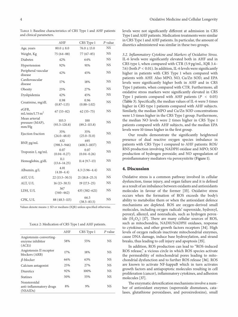
Table 1: Baseline characteristics of CRS Type 1 and AHF patientsand clinical parameters.
AHF CRS Type 1 𝑃 valueAge, years 80.0 ± 8.0 76.0 ± 13.0 NS
Weight, Kg 75 (64–88) 77 (67–85) NS
Diabetes 42% 64% NS
Hypertension 92% 91% NSPeripheral vasculardisease 42% 45% NS
Cardiovasculardisease 17% 18% NS
Obesity 25% 27% NS
Dyslipidemia 42% 45% NS
Creatinine, mg/dL 0.98(0.87–1.15)
0.96(0.88–1.02)
NS
eGFR,mL/min/1.73m2 67 (53–82) 62 (55–75) NS
Mean arterialpressure (MAP),mm/Hg
103.3(93.3–120.8)
100(89.2–115.8)
NS
Ejection fraction 35%(24.0–48.0)
35%(25.0–51.0)
NS
BNP, pg/mL 632(398.3–946)
695(408.5–1837)
NS
Troponin I, ng/mL 0.07(0.04–0.27)
0.07(0.04–0.26)
NS
Hemoglobin, g/dL 11.1(13.6–14.25) 11.4 (9.7–13) NS
Albumin, g/L 4.01(4.18–4.4) 4.3 (3.96–4.4) NS
AST, U/L 22 (13.5–30.5) 21 (18.8–25.3) NS
ALT, U/L 16 (13–30.5) 19 (17.3–25) NS
LDH, U/L 367(340–462) 435 (382–621) NS
CPK, U/L 88 (48.5–115) 66.5(38.5–83.5)
NS
Values denote means ± SD or medians (IQR) unless specified otherwise.
Table 2: Medication of CRS Type 1 and AHF patients.
AHF CRS Type 1 𝑃 valueAngiotensin-converting-enzyme inhibitor(ACEi)
58% 55% NS
Angiotensin II receptorblockers (ARB) 17% 18% NS
𝛽-blocker 66% 63% NS
Calcium antagonist 25% 27% NS
Diuretics 92% 100% NS
Statines 50% 55% NSNonsteroidalanti-inflammatory drugs(NSAIDs)
8% 9% NS
levels were not significantly different at admission in CRSType 1 andAHF patients.Medication treatments were similarin CRS Type 1 and AHF patients. In particular, the amount ofdiuretics administered was similar in these two groups.
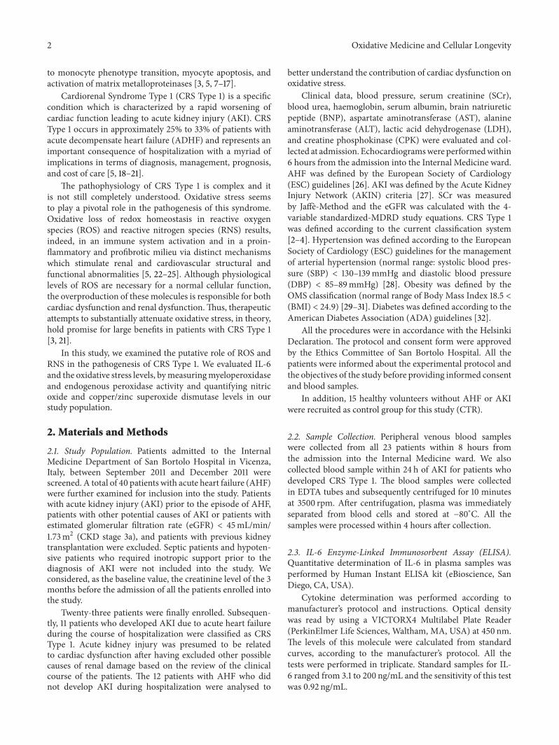
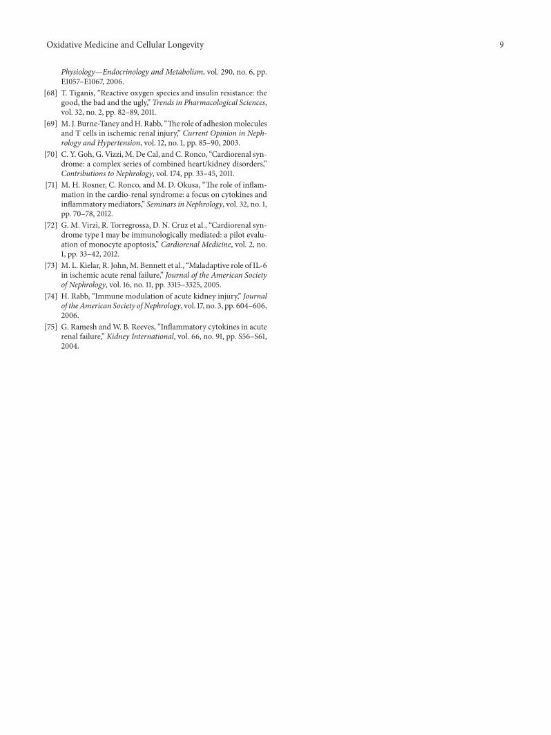
3.2. Inflammatory Cytokine and Markers of Oxidative Stress.IL-6 levels were significantly elevated both in AHF and inCRS type 1, when compared with CTR (5.9 pg/mL, IQR 3.4–7.6) (both𝑃 < 0.01). In addition, IL-6 levels were significantlyhigher in patients with CRS Type 1 when compared withpatients with AHF. Also MPO, NO, Cu/Zn SOD, and EPAlevels were significantly higher both in AHF and in CRSType 1 patients, when compared with CTR. Furthermore, alloxidative stress markers were significantly elevated in CRSType 1 patients compared with AHF patients (𝑃 < 0.05)(Table 3). Specifically, the median values of IL-6 were 5 timeshigher in CRS type 1 patients compared with AHF subjects.Similarly, the median MPO and Cu/Zn SOD concentrationswere 1.5 times higher in the CRS Type 1 group. Furthermore,the median NO levels were 2 times higher in CRS Type 1patients compared with AHF subjects, and the median EPAlevels were 10 times higher in the first group.
Our results demonstrate the significantly heightenedpresence of dual reactive oxygen species imbalance inpatients with CRS Type 1 compared to AHF patients: ROS/RNS production involving NADPH oxidase and MPO; SODproduction of hydrogen peroxide; and NO upregulation ofproinflammatory mediators via peroxynitrite (Figure 1).
4. Discussion
Oxidative stress is a common pathway involved in cellulardysfunction, tissue injury, and organ failure and it is definedas a result of an imbalance between oxidants and antioxidantsmolecules in favour of the former [33]. Oxidative stressoccurs when the formation of ROS exceeds the body’sability to metabolize them or when the antioxidant defencemechanisms are depleted. ROS are oxygen-derived smallmolecules, including oxygen radicals’ superoxide, hydroxyl,peroxyl, alkoxyl, and nonradicals, such as hydrogen perox-ide (H
2O2) [17]. There are many cellular sources of ROS,
such as mitochondria, NADH/NADPH oxidases, responseto cytokines, and other growth factors receptors [34]. Highlevels of oxygen radicals inactivate mitochondrial enzymes,cause DNA damage, induce base hydroxylation, and strandbreaks, thus leading to cell injury and apoptosis [35].
In addition, ROS production can lead to “ROS-inducedROS release,” a vicious circle in which ROS species activatethe permeability of mitochondrial pores leading to mito-chondrial dysfunction and to further ROS release [36]. ROSare known to activate NF-kappaB which in turn activatesgrowth factors and antiapoptotic molecules resulting in cellproliferation (cancer), inflammatory cytokines, and adhesionmolecules [37].
The enzymatic detoxificationmechanisms involve a num-ber of antioxidant enzymes (superoxide dismutases, cata-lases, glutathione peroxidases, and peroxiredoxins), small

Oxidative Medicine and Cellular Longevity 5
Table 3: Oxidative stress and IL-6 levels in AHF, CRS Type 1 patients, and CTR.
AHF CRS Type 1 CTR 𝑃 valueMPO, pg/mL 505.6 (421.7–547.8) 746.9 (665.2–940.0) 10.1 (6.0–19.3) <0.01NO, 𝜇M 205.6 (95.0–277.5) 507.3 (404.7–557.3) 9.5 (6.1–12.2) <0.01Cu/ZnSOD, pg/mL 184.5 (160.5–192.0) 274.5 (191.8–326.8) 58.9 (51.7–70.9) <0.01EPA, U/L 274.5 (191.8–326.8) 2978.4 (2071.8–4069.9) 2.0 (0.9–3.9) <0.01IL-6, pg/mL 22.19 (16.6–24.6) 90.68 (59.9–105.3) 5.9 (3.4–7.6) <0.01Values denote medians (IQR).
2000
1500
1000
500
0
AHF AHF AHF AHFP = 0.00017
MPO
(ng/
mL)
800
600
400
200
0
P = 0.00005
400
300
200
100
0 0
Cu/Z
nSO
D (n
g/m
L)
P = 0.0489
5000
4000
3000
2000
1000
P = 0.00209
EPA
(U/L
)
NO
(𝜇M
)Oxidative stress in AHF and CRS Type 1 patients
CRS Type 1 CRS Type 1 CRS Type 1 CRS Type 1
Figure 1
molecular-weight antioxidants, and adaptive mechanismleading to antioxidant gene expression [38].
Antioxidant systems can stabilize free radicals, conse-quently reducing the oxidative stress. Enzymatic antioxidantsare the most important defense against radical-induceddamage [39].
Our results demonstrate a significant increase in bothROS and RNS redox disequilibrium in patients with CRSType 1 compared to patients with acute heart failure andcontrol subjects. In particular, CRS Type 1 patients presenteda significant increase in circulating ROS and RNS, as wellas an increased expression of inflammatory cytokines, inparticular that of IL-6. Increased levels of NADPH oxidaseand MPO as well as SOD upregulation with concomitantupregulation of proinflammatorymediators via peroxynitritehave not been previously reported. MPO is a haeme enzymethat is abundant in granules of human inflammatory cellssuch as activated neutrophils, macrophages, and monocytes.MPO acts as a master enzyme in the generation of a rangeof ROS by catalyzing the conversion of hydrogen peroxide(H2O2) into species including OH, ONOO, hypochlorous
acid (HOCl), and NO2. MPO-catalyzed species are involved
further in oxidative damage of various biological molecules
(e.g., lipids, lipoproteins, and proteins low) and tissue degra-dation and are implicated in atherosclerosis, cancer, diabeticvascular complications, kidney diseases, and other disorders[40–43]. In prospective studies, high MPO levels were ableto predict increased risk of developing CAD in healthyindividuals [44] and cardiovascular events in patients pre-senting to emergency with chest pain [45] and increasedrisk of myocardial infarction and death in patients withacute coronary syndrome [46]. Furthermore, MPO has beenspeculated to be a major oxidative stress pathway in ESRD[47].
Furthermore, it is known that superoxide production ofhydrogen peroxide and nitric oxide upregulation is responsi-ble for an increase in IL-6 production and secretion. In ourstudy, IL-6 resulted to be higher in patients with CRS Type 1who presented higher levels of oxidative stress markers.
Increased levels of SOD have not been previouslyobserved in CRS Type 1 patients. SODs are a unique familyof metalloproteins that catalytically enhances the normaldismutation of superoxide. SOD is normally present at lowmicromolar concentrations in cells. Four types of SOD havebeen defined on the basis of distinctions in their metalcofactors and distribution: manganese (MnSOD) principally

6 Oxidative Medicine and Cellular Longevity
located in the matrix of mitochondria of all aerobes, cop-per/zinc (Cu/ZnSOD) mainly present in the cytoplasm ofeukaryotic cells, iron (FeSOD) predominantly present in thecytosol, chloroplasts, or mitochondria of prokaryotes, andextracellular (ECSOD) found in the extracellular fluids ormembrane associated in mammals [48–50]. In particular, wenoted a specific activity of Cu/ZnSOD in CRS Type 1 patients.
Increased ROS production has been implicated in manypathological conditions, such as hypertension, diabetes mel-litus, hypercholesterolemia, restenosis, heart failure, kidneydiseases, and atherosclerosis [51–55]. Specifically, atheroscle-rosis results from a local imbalance between ROS pro-ductions, leading to oxidative stress and these antioxidantenzymes [56].
Both in vitro and animal studies have demonstrated thatseveral pathways are dysregulated in heart failure, leading toincreased oxidative stress markers production and cardiacdamage. A metabolic shift from fatty acid (FA) oxidation toglycolysis has been indeed reported in cardiomyocytes in thesetting of heart failure. Myocardial ATP content graduallydecreases, dropping to 60%–70% of normal levels [57–59].This drop is due to a decrease in mitochondrial oxidativemetabolism and it is balanced by a compensatory increasein glucose uptake and in glycolysis [60, 61]. The reducedoxidative metabolism leads to an accumulation of free FAin cardiomyocytes, creating a self-perpetuating mechanismof ever-increasing oxidative stress responsible for deleteriouseffects on the heart.
Once produced, ROS display several negative effectson cardiac cells, impairing cardiomyocyte contractility, iontransport, and calcium handling. In addition to their detri-mental effects, mitochondrial ROS play an important role inintracellular signalling by triggering multiple cellular path-ways and the transcriptional activation of selected nucleargenes, finally eliciting transcriptional reprogramming [62,63]. We speculated a similar cardiac condition in CRS Type1 patients: the higher redox disequilibrium observed in thesepatients, compared to AHF subjects, could be involved inrenal damage. We observed a significant difference in oxida-tive response between CRS Type 1 patients and AHF patients.Although these findings are provocative, the design of thestudy does not allow us to make conclusions about causality.Indeed, increased oxidative stress could be secondary tothe renal injury rather than the cause of this complication.Further studies are needed to support this hypothesis.
Within the kidneys, ROS generation increases in responseto specific stimuli, such as angiotensin II and aldosteronesecretion [17]. NOX enzymes are the primary source of ROSin vascular smooth cells in both kidney cortex and medulla[64, 65]. Under physiological conditions, NO induces vasodi-latation of the afferent arteriole, thus increasing renal bloodflow, blunts tubule-glomerular feedback, promotes pressurenatriuresis, and scavenges low ROS concentrations [66]. Incase of increased oxidative stress, superoxide productionleads to cascade reactions which result in vasoconstriction,inflammation, and impaired vascular and renal functions[17]. In fact, we observed a significant increase in circulatingoxidative stress species and an increased expression of IL-6 inCRS Type 1 patients. In fact, we observed a stronger IL-6 and
inflammation activation in CRS Type 1 patients compared toAHF subjects.
In CRS Type 1 group, diabetes was more frequent thanin AHF population. It is well known that hyperglycemiaresults in excessive production of acetyl-CoA that feedsinto the Krebs cycle, thus increasing NADH production[67]. Therefore, oxidation of the overproduced NADH bymitochondria inevitably leads to the production of moresuperoxide and hence more ROS [68]. This is responsiblefor the accumulation of glycolytic metabolites upstreamof glyceraldehydes 3-phosphate and the activation of thealternative glucose disposal pathways that all are linked toROS production. Even though in our study the percentage ofdiabetic patients was not significantly different between thetwo groups,maybe because of the small size of the sample, thepresence of diabetes may have increased the oxidative stressin CRS Type 1 patients.
The critical roles of inflammation and immune systemdysregulation in the CRS Type 1 pathophysiology have beenreported both in animal and human models which indicatethat proinflammatory cytokines and chemokines are associ-ated withmolecular, clinical, and physiological aspects of thissyndrome [5, 69–72]. Cellular metabolic shifts in response tohumoral and cellular signaling factorsmay result in an upreg-ulated expression and release of proinflammatory/immune-modulatory cytokines, which are released into the renal tissueand in the blood, respectively [69, 73–75]. In this pilot study,we observed the inflammatory process activation and theloss of redox homeostasis in CRS Type 1. These observationsindicate that cellular responses to different signals influencea dual shift in both ROS and RNS production.
5. Conclusion
These preliminary results underline the importance of oxida-tive stress in the pathogenesis of CRS Type 1. Given the myr-iad of implications of this syndrome in terms of diagnosis,management, prognosis, and cost of care, understanding themechanism by which inflammatory cascades are activated asa results of oxidative stress has important clinical implica-tions.
This study explores the premise of ROS/RNS disequi-librium in the CRS Type 1 pathophysiology. Nevertheless,we acknowledge the limitations of the small sample size inthis pilot study, which would preclude meaningful multi-variate analysis. Our preliminary results can be consideredas hypothesis-generating about CRS Type 1 pathogenesis,allowing further exploration of novel pathophysiologicalmechanisms in CRS Type 1. Further studies are neededto better understand the role that these molecules andtherapeutics have in altering target processes.
Conflict of Interests
Claudio Ronco is a Consultant for Alere and a Member ofSpeakers Bureau for Abbott Diagnostics. The other authorsdeclare no conflict of interests.

Oxidative Medicine and Cellular Longevity 7
Acknowledgment
This work was supported by a research Grant of VenetoRegion (RSF N. 303/2009).
References
[1] A. Clementi, G. M. Virzi, C. Y. Goh et al., “Cardiorenal syndr-ome type 4: a review,” Cardiorenal Medicine, vol. 3, no. 1, pp.63–70, 2013.
[2] C. Ronco, C.-Y. Chionh, M. Haapio, N. S. Anavekar, A. House,and R. Bellomo, “The cardiorenal syndrome,” Blood Purifica-tion, vol. 27, no. 1, pp. 114–126, 2009.
[3] C. Ronco, M. Cicoira, and P. A. McCullough, “Cardiorenalsyndrome type 1: pathophysiological crosstalk leading to com-bined heart and kidney dysfunction in the setting of acutelydecompensated heart failure,” Journal of the American Collegeof Cardiology, vol. 60, no. 12, pp. 1031–1042, 2012.
[4] C. Ronco, M. Haapio, A. A. House, N. Anavekar, and R.Bellomo, “Cardiorenal syndrome,” Journal of the AmericanCollege of Cardiology, vol. 52, no. 19, pp. 1527–1539, 2008.
[5] G. M. Virzı, S. Day, M. de Cal, G. Vescovo, and C. Ronco,“Heart-kidney crosstalk and role of humoral signaling in criticalillness,” Critical Care, vol. 18, no. 1, article 201, 2014.
[6] G. Viswanathan and S. Gilbert, “The cardiorenal syndrome:making the connection,” International Journal of Nephrology,vol. 2011, Article ID 283137, 10 pages, 2011.
[7] G. R. Kinsey, L. Li, and M. D. Okusa, “Inflammation in acutekidney injury,”Nephron—Experimental Nephrology, vol. 109, no.4, pp. e102–e107, 2008.
[8] D. W. Lee, S. Faubel, and C. L. Edelstein, “Cytokines in acutekidney injury (AKI),”Clinical Nephrology, vol. 76, no. 3, pp. 165–173, 2011.
[9] D. F.Mangan and S.M.Wahl, “Differential regulation of humanmonocyte programmed cell death (apoptosis) by chemotacticfactors and pro-inflammatory cytokines,” Journal of Immunol-ogy, vol. 147, no. 10, pp. 3408–3412, 1991.
[10] M. Murakami and T. Hirano, “The pathological and physiolog-ical roles of IL-6 amplifier activation,” International Journal ofBiological Sciences, vol. 8, no. 9, pp. 1267–1280, 2012.
[11] M. Nian, P. Lee, N. Khaper, and P. Liu, “Inflammatory cyto-kines and postmyocardial infarction remodeling,” CirculationResearch, vol. 94, no. 12, pp. 1543–1553, 2004.
[12] M. Satoh, Y. Ishikawa, T. Itoh, Y. Minami, Y. Takahashi, and M.Nakamura, “The expression of TNF-alpha converting enzymeat the site of ruptured plaques in patients with acute myocardialinfarction,” European Journal of Clinical Investigation, vol. 38,no. 2, pp. 97–105, 2008.
[13] R. Schindler, O. Boenisch, C. Fischer, and U. Frei, “Effect of thehemodialysis membrane on the inflammatory reaction in vivo,”Clinical Nephrology, vol. 53, no. 6, pp. 452–459, 2000.
[14] G. Torre-Amione, “Immune activation in chronic heart failure,”The American Journal of Cardiology, vol. 95, supplement 1, no.11, pp. 3–8, 2005.
[15] B. J. Wrigley, G. Y. H. Lip, and E. Shantsila, “The role of mono-cytes and inflammation in the pathophysiology of heart failure,”European Journal of Heart Failure, vol. 13, no. 11, pp. 1161–1171,2011.
[16] M. Metra, G. Cotter, M. Gheorghiade, L. D. Cas, and A. A.Voors, “The role of the kidney in heart failure,” European HeartJournal, vol. 33, no. 17, pp. 2135–2142, 2012.
[17] S. Rubattu, S. Mennuni, M. Testa et al., “Pathogenesis of chroniccardiorenal syndrome: is there a role for oxidative stress?”International Journal of Molecular Sciences, vol. 14, no. 11, pp.23011–23032, 2013.
[18] N. Hata, S. Yokoyama, T. Shinada et al., “Acute kidney injuryand outcomes in acute decompensated heart failure: evaluationof the RIFLE criteria in an acutely ill heart failure population,”European Journal of Heart Failure, vol. 12, no. 1, pp. 32–37, 2010.
[19] K. V. Liang, A. W. Williams, E. L. Greene, and M. M. Redfield,“Acute decompensated heart failure and the cardiorenal syn-drome,” Critical Care Medicine, vol. 36, no. 1, supplement, pp.S75–S88, 2008.
[20] C. Ronco, P. A. McCullough, S. D. Anker et al., “Cardiorenalsyndromes: an executive summary from the consensus confer-ence of the acute dialysis quality initiative (ADQI),” Contribu-tions to Nephrology, vol. 165, pp. 54–67, 2010.
[21] D. N. Cruz, “Cardiorenal syndrome in critical care: the acutecardiorenal and renocardiac syndromes,” Advances in ChronicKidney Disease, vol. 20, no. 1, pp. 56–66, 2013.
[22] R. Gill, A. Tsung, and T. Billiar, “Linking oxidative stress toinflammation: toll-like receptors,” Free Radical Biology andMedicine, vol. 48, no. 9, pp. 1121–1132, 2010.
[23] S. R. Khan, “Stress oxidative: nephrolithiasis and chronic kidneydiseases,”Minerva Medica, vol. 104, no. 1, pp. 23–30, 2013.
[24] P.-L. Li and Y. Zhang, “Cross talk between ceramide and redoxsignaling: Implications for endothelial dysfunction and renaldisease,” Handbook of Experimental Pharmacology, vol. 216, pp.171–197, 2013.
[25] C. H. Mandavia, A. R. Aroor, V. G. Demarco, and J. R. Sowers,“Molecular and metabolic mechanisms of cardiac dysfunctionin diabetes,” Life Sciences, vol. 92, no. 11, pp. 601–608, 2013.
[26] J. J. McMurray, S. Adamopoulos, S. D. Anker et al., “ESCguidelines for the diagnosis and treatment of acute and chronicheart failure 2012: The Task Force for the Diagnosis andTreatment of Acute and Chronic Heart Failure 2012 of theEuropean Society of Cardiology. Developed in collaborationwith theHeart Failure Association (HFA) of the ESC,” EuropeanJournal of Heart Failure, vol. 14, no. 8, pp. 803–869, 2012.
[27] R. L. Mehta, J. A. Kellum, S. V. Shah et al., “Acute Kidney InjuryNetwork: report of an initiative to improve outcomes in acutekidney injury,” Critical Care, vol. 11, no. 2, p. R31, 2007.
[28] M. Volpe and G. Toccib, “2007ESH/ESC Guidelines for themanagement of hypertension, from theory to practice: globalcardiovascular risk concept,” Journal of Hypertension, vol. 27,supplement 3, pp. S3–S11, 2009.
[29] D.W. Haslam andW. P. T. James, “Obesity,”TheLancet, vol. 366,no. 9492, pp. 1197–1209, 2005.
[30] E. M. Berke and N. E. Morden, “Medical management of obes-ity,”American Family Physician, vol. 62, no. 2, pp. 419–426, 2000.
[31] F. Galtier, “Definition, epidemiology, risk factors,” Diabetes &Metabolism, vol. 36, no. 6, pp. 628–651, 2010.
[32] American Diabetes Association, “Diagnosis and classificationof diabetes mellitus,” Diabetes Care, vol. 33, supplement 1, pp.S62–S69, 2010.
[33] H. Sies, “Oxidative stress: oxidants and antioxidants,” Experi-mental Physiology, vol. 82, no. 2, pp. 291–295, 1997.
[34] M. J. Morgan and Z.-G. Liu, “Crosstalk of reactive oxygenspecies and NF-kappaB signaling,” Cell Research, vol. 21, no. 1,pp. 103–115, 2011.
[35] M. Naziroglu, N. Yoldas, E. N. Uzgur, and M. Kayan, “Role ofcontrastmedia on oxidative stress, Ca2+ signaling and apoptosis

8 Oxidative Medicine and Cellular Longevity
in kidney,” Journal of Membrane Biology, vol. 246, no. 2, pp. 91–100, 2013.
[36] C. Maack and M. Bohm, “Targeting mitochondrial oxidativestress in heart failure: throttling the afterburner,” Journal of theAmerican College of Cardiology, vol. 58, no. 1, pp. 83–86, 2011.
[37] Y. Lin, L. Bai, W. Chen, and S. Xu, “The NF-𝜅B activation path-ways, emerging molecular targets for cancer prevention andtherapy,” Expert Opinion on Therapeutic Targets, vol. 14, no. 1,pp. 45–55, 2010.
[38] B. Kalyanaraman, “Teaching the basics of redox biology tomedical and graduate students: oxidants, antioxidants anddisease mechanisms,” Redox Biology, vol. 1, no. 1, pp. 244–257,2013.
[39] C.-C. Sung, Y.-C. Hsu, C.-C. Chen, Y.-F. Lin, and C.-C. Wu,“Oxidative stress and nucleic acid oxidation in patients withchronic kidney disease,” Oxidative Medicine and Cellular Long-evity, vol. 2013, Article ID 301982, 15 pages, 2013.
[40] J. Arnhold, “Properties, functions, and secretion of humanmyeloperoxidase,”Biochemistry (Moscow), vol. 69, no. 1, pp. 4–9,2004.
[41] E. P. Reeves, M. Nagl, J. Godovac-Zimmermann, and A. W.Segal, “Reassessment of the microbicidal activity of reactiveoxygen species and hypochlorous acid with reference to thephagocytic vacuole of the neutrophil granulocyte,” Journal ofMedical Microbiology, vol. 52, no. 8, pp. 643–651, 2003.
[42] J. Borawski, “Myeloperoxidase as a marker of hemodialysisbiocompatibility and oxidative stress: the underestimatedmod-ifying effects of heparin,” The American Journal of KidneyDiseases, vol. 47, no. 1, pp. 37–41, 2006.
[43] E. Ho, K. Karimi Galougahi, C.-C. Liu, R. Bhindi, and G. A.Figtree, “Biological markers of oxidative stress: applications tocardiovascular research and practice,” Redox Biology, vol. 1, no.1, pp. 483–491, 2013.
[44] M. C. Meuwese, E. S. G. Stroes, S. L. Hazen et al., “Serum mye-loperoxidase levels are associated with the future risk of coro-nary artery disease in apparently healthy individuals: the EPIC-Norfolk Prospective Population Study,” Journal of the AmericanCollege of Cardiology, vol. 50, no. 2, pp. 159–165, 2007.
[45] M.-L. Brennan,M. S. Penn, F. Van Lente et al., “Prognostic valueof myeloperoxidase in patients with chest pain,” The New Eng-land Journal of Medicine, vol. 349, no. 17, pp. 1595–1604, 2003.
[46] S. Baldus, C. Heeschen, T. Meinertz et al., “Myeloperoxidaseserum levels predict risk in patients with acute coronary syn-dromes,” Circulation, vol. 108, no. 12, pp. 1440–1445, 2003.
[47] Y. Maruyama, B. Lindholm, and P. Stenvinkel, “Inflammationand oxidative stress in ESRD—the role of myeloperoxidase,”Journal of Nephrology, vol. 17, supplement 8, pp. S72–S76, 2004.
[48] D. Barra, F. Martini, J. V. Bannister et al., “The complete aminoacid sequence of human Cu/Zn superoxide dismutase,” FEBSLetters, vol. 120, no. 1, pp. 53–56, 1980.
[49] S. Shull, N. H. Heintz, M. Periasamy et al., “Differential regu-lation of antioxidant enzymes in response to oxidants,” TheJournal of Biological Chemistry, vol. 266, no. 36, pp. 24398–24403, 1991.
[50] S. I. Liochev and I. Fridovich, “Mechanism of the peroxidaseactivity of Cu, Zn superoxide dismutase,” Free Radical Biologyand Medicine, vol. 48, no. 12, pp. 1565–1569, 2010.
[51] S. L. Acton, K. F. Kozarsky, and A. Rigotti, “The HDL receptorSR-BI: a new therapeutic target for atherosclerosis?”MolecularMedicine Today, vol. 5, no. 12, pp. 518–524, 1999.
[52] N. Abate, “Obesity and cardiovascular disease: pathogeneticrole of the metabolic syndrome and therapeutic implications,”Journal of Diabetes and Its Complications, vol. 14, no. 3, pp. 154–174, 2000.
[53] S. Yamashita, K.-I. Hirano, N. Sakai, and Y.Matsuzawa, “Molec-ular biology and pathophysiological aspects of plasma choles-teryl ester transfer protein,” Biochimica et Biophysica Act—Molecular and Cell Biology of Lipids, vol. 1529, no. 1–3, pp. 257–275, 2000.
[54] U. Beisiegel, “New aspects on the role of plasma lipases inlipoprotein catabolism and atherosclerosis,”Atherosclerosis, vol.124, no. 1, pp. 1–8, 1996.
[55] A. Zambon, J. E. Hokanson, B. G. Brown, and J. D. Brunzell,“Evidence for a new pathophysiological mechanism for coro-nary artery disease regression.Hepatic lipase-mediated changesin LDL density,” Circulation, vol. 99, no. 15, pp. 1959–1964, 1999.
[56] J.-G. Park and G. T. Oh, “The role of peroxidases in the path-ogenesis of atherosclerosis,” BMB Reports, vol. 44, no. 8, pp.497–505, 2011.
[57] M. Beer, T. Seyfarth, J. Sandstede et al., “Absolute concentrationsof high-energy phosphate metabolites in normal, hypertro-phied, and failing humanmyocardiummeasured noninvasivelywith 31P-SLOOP magnetic resonance spectroscopy,” Journal ofthe American College of Cardiology, vol. 40, no. 7, pp. 1267–1274,2002.
[58] M. A. Conway, J. Allis, R. Ouwerkerk, T. Niioka, B. Rajagopalan,and G. K. Radda, “Detection of low phosphocreatine to ATPratio in failing hypertrophied human myocardium by 31Pmagnetic resonance spectroscopy,” The Lancet, vol. 338, no.8773, pp. 973–976, 1991.
[59] R. Tian, L. Nascimben, R. Kaddurah-Daouk, and J. S. Ingwall,“Depletion of energy reserve via the creatine kinase reactionduring the evolution of heart failure in cardiomyopathic ham-sters,” Journal of Molecular and Cellular Cardiology, vol. 28, no.4, pp. 755–765, 1996.
[60] T. Kato, S. Niizuma, Y. Inuzuka et al., “Analysis of metabolicremodeling in compensated left ventricular hypertrophy andheart failure,” Circulation: Heart Failure, vol. 3, no. 3, pp. 420–430, 2010.
[61] B. Lei, V. Lionetti, M. E. Young et al., “Paradoxical downreg-ulation of the glucose oxidation pathway despite enhanced fluxin severe heart failure,” Journal of Molecular and Cellular Car-diology, vol. 36, no. 4, pp. 567–576, 2004.
[62] A. D. Hafstad, A. A. Nabeebaccus, and A. M. Shah, “Novelaspects of ROS signalling in heart failure,” Basic Research inCardiology, vol. 108, no. 4, article no. 359, 2013.
[63] J. Marın-Garcıa, A. T. Akhmedov, and G. W. Moe, “Mitocho-ndria in heart failure: the emerging role of mitochondrialdynamics,” Heart Failure Reviews, vol. 18, no. 4, pp. 439–456,2013.
[64] T. Chabrashvili, A. Tojo, M. L. Onozato et al., “Expression andcellular localization of classic NADPH oxidase subunits in thespontaneously hypertensive rat kidney,” Hypertension, vol. 39,no. 2 I, pp. 269–274, 2002.
[65] A.-P. Zou, N. Li, and A. W. Cowley Jr., “Production and actionsof superoxide in the renal medulla,”Hypertension, vol. 37, no. 2,pp. 547–553, 2001.
[66] L. Raij, “Nitric oxide and cardiovascular and renal effects,”Osteoarthritis and Cartilage, vol. 16, no. 2, pp. S21–S26, 2008.
[67] M. S. Ola, D. A. Berkich, Y. Xu et al., “Analysis of glucosemetabolism in diabetic rat retinas,” The American Journal of
Oxidative Medicine and Cellular Longevity 9
Physiology—Endocrinology and Metabolism, vol. 290, no. 6, pp.E1057–E1067, 2006.
[68] T. Tiganis, “Reactive oxygen species and insulin resistance: thegood, the bad and the ugly,” Trends in Pharmacological Sciences,vol. 32, no. 2, pp. 82–89, 2011.
[69] M. J. Burne-Taney andH.Rabb, “The role of adhesionmoleculesand T cells in ischemic renal injury,” Current Opinion in Neph-rology and Hypertension, vol. 12, no. 1, pp. 85–90, 2003.
[70] C. Y. Goh, G. Vizzi, M. De Cal, and C. Ronco, “Cardiorenal syn-drome: a complex series of combined heart/kidney disorders,”Contributions to Nephrology, vol. 174, pp. 33–45, 2011.
[71] M. H. Rosner, C. Ronco, and M. D. Okusa, “The role of inflam-mation in the cardio-renal syndrome: a focus on cytokines andinflammatory mediators,” Seminars in Nephrology, vol. 32, no. 1,pp. 70–78, 2012.
[72] G. M. Virzı, R. Torregrossa, D. N. Cruz et al., “Cardiorenal syn-drome type 1 may be immunologically mediated: a pilot evalu-ation of monocyte apoptosis,” Cardiorenal Medicine, vol. 2, no.1, pp. 33–42, 2012.
[73] M. L. Kielar, R. John,M. Bennett et al., “Maladaptive role of IL-6in ischemic acute renal failure,” Journal of the American Societyof Nephrology, vol. 16, no. 11, pp. 3315–3325, 2005.
[74] H. Rabb, “Immune modulation of acute kidney injury,” Journalof the American Society of Nephrology, vol. 17, no. 3, pp. 604–606,2006.
[75] G. Ramesh andW. B. Reeves, “Inflammatory cytokines in acuterenal failure,” Kidney International, vol. 66, no. 91, pp. S56–S61,2004.

Submit your manuscripts athttp://www.hindawi.com
Stem CellsInternational
Hindawi Publishing Corporationhttp://www.hindawi.com Volume 2014
Hindawi Publishing Corporationhttp://www.hindawi.com Volume 2014
MEDIATORSINFLAMMATION
of
Hindawi Publishing Corporationhttp://www.hindawi.com Volume 2014
Behavioural Neurology
EndocrinologyInternational Journal of
Hindawi Publishing Corporationhttp://www.hindawi.com Volume 2014
Hindawi Publishing Corporationhttp://www.hindawi.com Volume 2014
Disease Markers
Hindawi Publishing Corporationhttp://www.hindawi.com Volume 2014
BioMed Research International
OncologyJournal of
Hindawi Publishing Corporationhttp://www.hindawi.com Volume 2014
Hindawi Publishing Corporationhttp://www.hindawi.com Volume 2014
Oxidative Medicine and Cellular Longevity
Hindawi Publishing Corporationhttp://www.hindawi.com Volume 2014
PPAR Research
The Scientific World JournalHindawi Publishing Corporation http://www.hindawi.com Volume 2014
Immunology ResearchHindawi Publishing Corporationhttp://www.hindawi.com Volume 2014
Journal of
ObesityJournal of
Hindawi Publishing Corporationhttp://www.hindawi.com Volume 2014
Hindawi Publishing Corporationhttp://www.hindawi.com Volume 2014
Computational and Mathematical Methods in Medicine
OphthalmologyJournal of
Hindawi Publishing Corporationhttp://www.hindawi.com Volume 2014
Diabetes ResearchJournal of
Hindawi Publishing Corporationhttp://www.hindawi.com Volume 2014
Hindawi Publishing Corporationhttp://www.hindawi.com Volume 2014
Research and TreatmentAIDS
Hindawi Publishing Corporationhttp://www.hindawi.com Volume 2014
Gastroenterology Research and Practice
Hindawi Publishing Corporationhttp://www.hindawi.com Volume 2014
Parkinson’s Disease
Evidence-Based Complementary and Alternative Medicine
Volume 2014Hindawi Publishing Corporationhttp://www.hindawi.com