Embed Size (px)
Citation preview

Research ArticlePhenotyping of Leukocytes and Leukocyte-DerivedExtracellular Vesicles
Lotte Hatting Pugholm, Rikke Bæk, Evo Kristina Lindersson Søndergaard,Anne Louise Schacht Revenfeld, Malene Møller Jørgensen, and Kim Varming
Department of Clinical Immunology, Aalborg University Hospital, 9000 Aalborg, Denmark
Correspondence should be addressed to Lotte Hatting Pugholm; [email protected]
Received 15 December 2015; Revised 4 March 2016; Accepted 20 March 2016
Academic Editor: Eyad Elkord
Copyright © 2016 Lotte Hatting Pugholm et al. This is an open access article distributed under the Creative Commons AttributionLicense, which permits unrestricted use, distribution, and reproduction in any medium, provided the original work is properlycited.
Extracellular vesicles (EVs) have a demonstrated involvement in modulating the immune system. It has been proposed that EVscould be used as biomarkers for detection of inflammatory and immunological disorders. Consequently, it is of great interestto investigate EVs in more detail with focus on immunological markers. In this study, five major leukocyte subpopulations andthe corresponding leukocyte-derived EVs were phenotyped with focus on selected immunological lineage-specific markers andselected vesicle-related markers. The leukocyte-derived EVs displayed phenotypic differences in the 34 markers investigated. Themajority of the lineage-specific markers used for identification of the parent cell types could not be detected on EVs released frommonocultures of the associated cell types. In contrast, the vesicular presentation of CD9, CD63, and CD81 correlated to the cellsurface expression of these markers, however, with few exceptions. Furthermore, the cellular expression of CD9, CD63, and CD81varied between leukocytes present inwhole blood and cultured leukocytes. In summary, these data demonstrate that the cellular andvesicular presentation of selected lineage-specific and vesicle-relatedmarkersmay differ, supporting the accumulating observationsthat sorting of molecular cargo into EVs is tightly controlled.
1. Introduction
Extracellular vesicles (EVs) are a heterogeneous group ofvesicles that can be subdivided based on their size, biogenesis,and molecular composition. Using the biogenesis as a clas-sification tool, EVs can be divided into three major groups,namely, exosomes, microvesicles (MVs), and apoptotic bod-ies [1–3]. Even though the molecular composition of thesethree subsets of EVs is different, several markers overlap. Sofar, identification of a specific marker that with certainty candistinguish or identify the particular EV subset still awaits[4]. It can be expected that the different EV subsets maycover different biological roles, but the function of EVs hasalso been described to depend both on the cellular sourceand on the recipient tissue/cell [2]. Nevertheless, it is nowrecognized that EVs are involved in numerous physiolog-ical processes, including intercellular communication anddelivery of proteins, lipids, and genetic material to recipientcells [2, 3, 5–7]. In addition, EVs have been associated with
the development and progression of different pathologicalconditions, including cancer and infectious diseases [8–15].
The immunological effects of EVs comprise a broadrange of mechanisms, including immune activation, immunesuppression, and modulation of immune surveillance. Cellsfrom both the innate and the adaptive immune system havebeen shown to release EVs, such as T and B cells, dendriticcells (DCs), macrophages, mast cells, and natural killer (NK)cells [16–24]. The effect of EVs is directly related to theirmolecular composition and several studies have ascribed animmunostimulatory effect of EVs to the presence of a veryspecificmolecular content [20, 23, 25–31]; for instance, CD56-positive and perforin-containing EVs from NK cells canmediate EV-induced cytotoxicity [20]. Several immunosup-pressive effects of EVs have also been reported [21, 32, 33]; forinstance, Fas Ligand (Fas-L)+ EVs released from regulatory Tcells are able to inhibit DC-induced cytotoxic T-lymphocyte(CTL) responses [21]. Furthermore, inhibitory roles of EVsderived from immature DCs have been observed in relation
Hindawi Publishing CorporationJournal of Immunology ResearchVolume 2016, Article ID 6391264, 12 pageshttp://dx.doi.org/10.1155/2016/6391264

2 Journal of Immunology Research
to transplant tolerance [34–36]. Thus, identification of aspecific molecular signature of EVs released by immune cellscan provide knowledge that can lead to the use of EVs in atherapeutic setting.
Numerous studies have investigated the effects of EVsreleased from different leukocytes, leading to an incipientunderstanding of the physiological functions of these EVs.Nevertheless, several basic questions concerning specificcharacteristics, like the protein composition of the differenttypes of leukocyte-derived EVs, remain unclear. The presentstudy investigated the expression of selected immunologicallineage-specific markers and selected vesicle-related markerson five major leukocyte subpopulations, namely, CD4+ Tcells, CD8+ T cells, NK cells, B cells, and monocytes. Theexpression was determined for leukocytes present in freshlyisolated whole blood and on cultured isolated leukocytesubpopulations and compared to the presentation of thesemarkers on the corresponding leukocyte-derived EVs. TheEV Array, used for phenotyping of EVs, is optimized fordetection of small EVs with a size below 150 nm that presentCD9, CD63, and/or CD81, such as exosomes and exosome-like vesicles. However, as these markers may be presenton several types of EVs and as the intracellular origin ofthe characterized EVs was not determined, the EV subsetinvestigated in the current study was denoted by small EVs(sEVs).
2. Materials and Methods
2.1. Biological Samples. Blood samples were obtained fromten healthy volunteers at the Department of ClinicalImmunology, Aalborg University Hospital. From each donortwo blood samples were collected, one tube containing EDTA(K3EDTA, Vacuette�, Greiner Bio-one, Austria) for immedi-ate analysis of noncultured leukocytes and one tube contain-ing CPDA (Vacuette, Greiner Bio-one, Austria) for the vesicleanalysis (plasma). Plasma was isolated by centrifugation at1800×g for 6min at room temperature (RT), after whichthe plasma supernatant was aliquoted and stored at −40∘Cuntil analysis. Buffy coats were obtained from three healthydonors at the Department of Clinical Immunology, AalborgUniversity Hospital, and used for isolation of peripheralblood mononuclear cells (PBMCs). Blood samples and buffycoats were not obtained from the same donors.
2.2. Isolation of Leukocyte Subpopulations. Buffy coats werediluted (1 : 4) in sterile PBS (sPBS) and PBMCs were isolatedby density gradient centrifugation using Lymphoprep (Axis-Shield, Oslo, Norway). PBMCs were subsequently washedtwice in growth medium (RPMI1640 (Gibco, Life Technolo-gies, Carlsbad, CA, USA), 10% heat-inactivated fetal calfserum (FSC) (Gibco), 100U/mL penicillin, and 10 𝜇g/mLstreptomycin (Amplicon,Odense,Denmark)) and counted intrypan blue and resuspended in isolation buffer (Ca2+- andMg2+-free PBS, 2mM EDTA, and 0.1% bovine serum albu-min (BSA)). Magnetic Dynabeads� were used for isolationof human CD4+ and CD8+ T cells according to the man-ufacturer’s instructions (Dynabeads CD4 Positive Isolation
Kit and Dynabeads CD8 Positive Isolation Kit, Life Tech-nologies). Briefly, 1 × 10⁷ cells/mL were mixed with washedbeads and incubated for 20min at 4∘C with gentle rotation.The cell suspension was subsequently placed in a magnetfor 3-4min. The supernatant was removed and the bead-bound cells were incubated with Detachabeads� for 45minat RT with gentle rotation. The cell suspension was placedin a magnet for 3-4min and the supernatant containing thedetached cells was collected. The detached cells were washedonce in growth medium and adjusted to 3 × 10⁶ cells/mL.From the CD4+ depleted PBMCs, B cells were isolatedusing the Dynabeads Untouched� Human B Cells kit (LifeTechnologies) according to the manufacturer’s instructions.From the CD8+ depleted PBMCs, monocytes were isolatedusing the Dynabeads Untouched HumanMonocytes kit (LifeTechnologies) according to the manufacturer’s instructions.Human NK cells were isolated from PBMCs by negativeselection according to the manufacturer’s instructions (Dyn-abeads Untouched Human NK Cells kit, Life Technologies).Briefly, 1× 10⁸ cells/mLweremixedwith the antibody cocktailand incubated for 20min at 4∘C followed by washing andincubation with Dynabeads for 15min at 4∘C while rotating.The cell suspension was subsequently placed in a magnet for3-4min.The supernatant, containing the cells of interest, wasremoved and centrifuged at 500×g, for 5min at RT.The pelletwas washed once in growth medium and adjusted to 3 ×10⁶ cells/mL. For each isolation, the purity of the cells wasdetermined by flow cytometry.
2.3. Culturing of Isolated Subpopulations and Harvest ofCells and sEVs. Isolated leukocytes were adjusted to 3 ×10⁶ cells/mL and cultured in either 12-well (3 × 10⁶ cells/well,1.5mL/well) or 24-well plates (1.5 × 10⁶ cells/well, 1mL/well)(Nunc, Thermo Scientific, Carlsbad, CA, USA) for 44–48hours at 37∘C and 5% CO
2. Following incubation, the plates
were centrifuged at 600×g for 10min at RT and the super-natants containing the cell-derived EVs were harvested fromthe plates. Complete protease inhibitor, EDTA-free (Roche,DE, USA), was added to the EV-rich supernatants, whichwere subsequently upconcentrated using Amicon Ultra 100Kspin columns according to the manufacturer’s instructions.Briefly, PBS was added to the harvested supernatants (totalvolume of 5mL) and the supernatants were subsequentlycentrifuged at 600×g for 7min at RT. The supernatants weretransferred to 50mL spin columns and centrifuged at 3200×gfor 10min at RT. Prior to adding the supernatants, the spincolumns were washed once in 5mL PBS (3200×g, 10min,RT). Following the first centrifugation of the supernatant,5mL of PBS was added to the retention volume and cen-trifuged again. This procedure was repeated. The retentionvolume, containing the upconcentrated supernatants, washarvested and the filter unit was washed in 50–100 𝜇L PBS.Due to varying cell counts between the subpopulations andacross the experiments, every supernatant was adjusted toa volume corresponding to 5.5 × 10⁶ cells/mL. No furtherpurification of the EVs was performed and the upconcen-trated supernatants were aliquoted and stored at −40∘Cuntil analysis by the EV Array. The cells were harvested

Journal of Immunology Research 3
in PBS, washed, and resuspended in PBS with 0.5% BSAand 0.09% NaN
3, followed by a subsequent surface marker
staining.
2.4. Antibodies. For the EV Array, the following antibodieswere used for capturing: CD11a (HI111) from Ab Biotech (SanDiego, CA, USA); Flotillin-1 and TSG101 (5B7) from Abcam(Cambridge, MA, USA); CD3 (HIT3a), CD14 (M5E2), CD16(3G8), CD28 (L293), CD49d (L25), and CD56 (3G8) fromBD Biosciences (Mountain View, CA, USA); CD41 (HIP8),CD63 (MEM-259), HLA-ABC (W6/32), and Alix (3A9) fromBiolegend (San Diego, CA, USA); ICAM-1 (R6.5) fromeBiosciences (San Diego, CA, USA); CD9, CD42a, CD81,and CTLA-4 (ANC152.2/8H5) from LifeSpan BioSciences(Seattle, WA, USA); Annexin V (AF399), CD4 (34930),CD8𝛼 (37006), CD19 (4G7-2E3), CD37 (424925), CD45(2D1), CD80 (37711), CD82 (423524), CD83 (HB15e), MICA/B (159207), TNF RI, and TNF RII from R&D Systems(Minneapolis, MN, USA); TLR3 (3.7) from Santa CruzBiotechnologies (Dallas, TX, USA); HLA-DR/DP/DQ (HB-145) from Loke Diagnostics (Aarhus, Denmark); Fas Ligand(10F2) from Serotec (Oxford, UK); and PD-L1 from SinoBiological (Beijing, China). The following antibodies wereused for detection (biotinylated): CD9, CD63, andCD81 fromLifeSpan BioSciences.
For flow cytometry the following antibodies were used:CD45 APC (T29/33), CD45 FITC (T29/33), and CD3 FITC(UCHT1) fromDakoCytomation (Glostrup, Denmark); CD3APC (UCHT1), CD4 FITC (RPA-T4), CD4 APC-H7 (SK3),CD8 APC-H7 (SK1), CD9 PerCP-Cy5.5 (M-L13), CD14 FITC(M5E2), CD16 FITC (3G8), CD16 PE-Cy7 (3G8), CD19 APC(HIB19), CD56 APC (B159), and CD56 PE-Cy7 (B159) fromBD Biosciences (Mountain View, CA, USA); CD63 PE-Cy7(H5C6) from eBiosciences; and CD81 PE from LifeSpanBioSciences. In addition, isotype- and fluorophore-matchedcontrol antibodies were included.
2.5. Flow Cytometry. For analysis of noncultured leukocytes,100 𝜇L of freshly drawn whole blood was labeled withfluorescence-conjugated antibodies for 30min at RT, redblood cells were lysed, and the remaining leukocytes werewashed and resuspended in BD FACSflow Sheath Fluid (BDBiosciences) with 1% paraformaldehyde.
The cultured leukocytes were labeled with fluorescence-conjugated antibodies for 30min at RT. The leukocytes werewashed twice in PBS with 0.5% BSA and 0.09% NaN
3and
resuspended in BD FACSflow Sheath Fluid (BD Biosciences)with 1% paraformaldehyde.
Cells were analyzed on a FACSCanto II flow cytometer(BD Biosciences) using the BD FACSDiva� Software version6.1.3 (BD Biosciences). The acquired data files were ana-lyzed by FlowJo vX.0.7 (TreeStar, Ashland, USA), first byadding a leukocyte-gate based on morphologic characteris-tics (FSC/SSC) and subsequently by the use of the lineage-specific markers; CD3 and CD4 for CD4+ T cells; CD3 andCD8 for CD8+ T cells; CD16 and CD56 for NK cells; CD19for B cells; and CD14 for monocytes.
2.6. EV Array. For the production of the protein microar-ray, microarray printing was performed on a SpotBot�Extreme protein edition microarray printer with a 946MP4pin (ArrayIt, Sunnyvale, CA, USA). Epoxy coated slides(75.6mm × 25.0mm; SCHOTT Nexterion, Jena, Germany)were used as microarray basis. The antibodies listed abovewere diluted in PBS containing 5% glycerol and printed ata concentration of 180–200𝜇g/mL. As a positive control,100 𝜇g/mLof biotinylated human IgG in PBSwith 5%glycerolwas printed. As a negative control, PBS with 5% glycerol wasprinted.
For catching, visualization, and data analysis, the proce-dures were performed as previously described [37]. In short,the slideswere incubatedwith 100𝜇L plasma, prediluted 1 : 10,or 100 𝜇L upconcentrated cell culture supernatant, prediluted1 : 2 in wash buffer (PBS with 0.05% Tween-20�). All sampleswere analyzed in triplicate. After overnight sample incubationand a subsequent wash, the slides were incubated with acocktail of biotinylated detection antibodies (anti-humanCD9, CD63, and CD81), diluted 1 : 1500 in wash buffer. Afterincubation, Cy5-labeled streptavidin (Invitrogen, Frederick,MD, USA) diluted 1 : 1500 was used for detection. Priorto scanning, the slides were washed and dried using aMicroarray High-Speed Centrifuge (ArrayIt). Scanning anddata analysis was performed as previously described [37].Briefly, the intensity of the antibody signal was calculatedby subtracting the mean of the background (without sam-ple/blank) from the mean of the triplicate antibody spots.This signal was then divided by the signal from the mean ofthe triplicate negative spots (without capture antibody, withsample).This relative fluorescence intensity was subsequentlylog 2 transformed.
3. Results
Leukocytes are immune cells responsible for recognizingand eliminating invading pathogens. Leukocytes can besubdivided based on morphological characteristics uponstaining into polymorphonuclear cells (the granulocytes) andmononuclear cells (the lymphocytes and themonocytes).Thepresent study investigated the sEVs produced by the differentsubpopulations of mononuclear cells found in peripheralblood and compared the vesicular phenotype to the cellularphenotype. In addition, sEVs present in plasma were pheno-typed for the same panel of markers.
3.1. Phenotypic Characterization of sEVs. Five different sub-populations of leukocytes were isolated from PBMCs. Thepurity of the different subpopulations throughout the threeindividual experiments was 97–99.5% for the CD4+ andthe CD8+ T cells, 82–95% for the B cells, 68–94% forthe monocytes, and 85–97% for the NK cells. The iso-lated subpopulations of leukocytes were cultured for 44–48 hours and the supernatants, containing the leukocyte-produced EVs, were investigated for the presence of apanel of selected immunological and EV-related markersusing the EV Array (Table 1). It was investigated whethersEVs from isolated leukocyte subpopulations presented the

4 Journal of Immunology Research
Table 1: Overview of the markers selected for phenotyping of sEVs.
Immunological markers Vesicle-related markersFunctional Regulatory Adhesion Lineage-specific and others Tetraspanins OthersHLA-DR CTLA-4 CD11a CD3 CD9 TSG101HLA-ABC PD-L1 ICAM-1 CD4 CD63 AlixCD28 CD8 CD81 Flotillin-1CD80 CD14 CD82 Annexin VCD49d CD16TLR3 CD19Fas-L CD37MIC A/B CD41TNF RI CD42aTNF RII CD45
CD56CD83
HLA: human leukocyte antigen; TLR: Toll-like receptor; TNF: tumor necrosis factor: RI: receptor I, RII: receptor II;MICA/B:major histocompatibility complexclass I-related chain A/B; CTLA-4: cytotoxic T-lymphocyte associated protein-4; PD-L1: programmed death-ligand 1; ICAM-1: intercellular adhesion molecule1; TSG101: tumor susceptibility gene 101.
lineage-specific markers that define the parent cells. Theresults demonstrated that not all lineage-specific markerscould be observed on the corresponding sEV subpopulations(Figure 1(a)). For instance, CD19, which is a well-definedlineage marker for B cells, could not be detected on sEVsin supernatant from cultured B cells. Likewise, CD14, CD16,CD56, and CD3 could not be detected on sEVs from culturedmonocytes, NK cells, and T cells, respectively (Figure 1(a)).In contrast, CD4 and CD8, which are lineage markers fortwo different populations of T cells, were detected on sEVsfrom the supernatants of these two T cell subpopulations.In addition, sEVs from cultured PBMCs presented CD8, butnone of the other markers. Aside from the parental lineage-specific markers, the presentation of other lineage-specificmarkers was investigated. Small EVs released from CD8+ Tcells presented CD4, CD45, and CD16. Likewise, CD45 andCD16 could be detected in one of the experiments with sEVsfrom CD4+ T cells.
A panel of EV-related markers was assayed on thedifferent subsets of the leukocyte-derived EVs (Figure 1(b)).CD9 was detected on all sEV subsets, though at very lowlevels on sEVs from T cells and NK cells. Similarly, CD81was detected on all sEVs but at very low levels on sEVs fromB cells and NK cells. CD82 was detected at high levels onsEVs from monocytes and PBMCs and at very low levels onsEVs from B cells and T cells but was absent on sEVs fromNK cells. CD63 was only detected on sEVs from cultured Tcells. Furthermore, Alix was detected at very low levels onsEVs from the cultured CD8+ T cells. In contrast, AnnexinV, TSG101, and Flotillin-1 could not be detected on theleukocyte-derived sEVs.
Furthermore, the presence of more general immunolog-ical markers was investigated on the different sEV popula-tions. A total of 18 markers were chosen based on their rel-evance for the function of leukocytes (Table 1). The majorityof markers could not be detected on the sEV subpopulationsusing the EVArray (Figure 1(c)). However, CD49dwas found
on sEVs from all the different leukocyte subpopulations andCD41 was detected on sEVs from all subpopulations besidessEVs from NK cells. Additionally, sEVs from cultured CD4+and CD8+ T cells presented several of the immunologicalmarkers, including CD11a, TLR3, CD28, CTLA-4, and theFas-L. TNF RI was only observed on sEVs from culturedmonocytes. The data presented in the heat maps were fromthree individual experiments and illustrated a degree ofvariation between the individuals. The presence of a naturalvariation in the phenotype of sEVs between healthy indi-viduals was also observed upon phenotyping of the sEVspresent in plasma from 10 healthy individuals (Figure 2).Plasma sEVs presented several of the markers that weredetected on the leukocyte-derived sEVs. In addition, plasmasEVs presented some lineage-specific markers, includingCD3, CD14, and CD19 that were not detected on sEVs fromcultured leukocytes. Furthermore, TNF RI, TLR3, CD42a,CD80, and CD83 as well as Annexin V, Flotillin-1, and Alixwere detected on the majority of the plasma sEVs.
3.2.The Cellular Expression Level of CD9, CD63, and CD81 onFreshly Isolated or Cultured Leukocytes. The cellular expres-sion pattern of CD9, CD63, and CD81 was determined for allthe leukocyte subpopulations by flow cytometry (Figure 3).The expression of these vesicle-related markers was investi-gated for leukocytes present in freshly drawn whole blood(𝑛 = 10) as well as for isolated subpopulations of leukocytesfollowing two days of culture (𝑛 = 3). Figures 3(a) and 3(b)demonstrate the expression levels in histograms from tworepresentative donors. None of the leukocyte subsets fromwhole blood presented CD9 on their surface (Figure 3(a)).In contrast, both CD63 and CD81 were expressed on allleukocytes, but the expression pattern varied between thedifferent subsets. The lymphocytes, including the CD4+ Tcells, the CD8+ T cells, the B cells, and the NK cells, expressedCD81 at high levels, while they all expressed CD63 at low-to-intermediate levels. The opposite situation was observed

Journal of Immunology Research 5
CD4
CD45
CD16
CD19
CD3
CD14
CD56
4.20.0
Monocytes
B cells
NK cells
PBMCs
CD8+ T cells
CD4+ T cells
CD8𝛼
(a) (b)
(c)
Relative intensity
Ann
exin
V
CD9
CD63
CD81
CD82
Flot
illin
-1
TSG
101
Alix
7.10.0
Monocytes
B cells
NK cells
PBMCs
CD8+ T cells
CD4+ T cells
CD41
CD49
d
TNF
RI
TNF
RII
CD80
CD42
a
HLA
-ABC
CD37
CD83
HLA
-DR
PD-L
1
CD11
a
TLR3
CD28
CTLA
-4
CD28
Fas-L
MIC
A/B
Monocytes
B cells
NK cells
PBMCs
6.80.0
CD8+ T cells
CD4+ T cells
Figure 1: Phenotyping of the different subsets of leukocyte-derived sEVs. The different subsets of leukocyte-derived sEVs were phenotypedfor a panel of selected markers using the EV Array. The sEVs were captured by antibodies targeting the selected markers and subsequentlydetected by addition of an antibody cocktail against CD9, CD63, and CD81. The heat maps illustrate the results from each of the threeindependent experiments divided into lineage-specific markers (a), vesicle-related markers (b), and general immunological markers (c) andpresent the relative intensities of each of the markers as indicated by the colored bars. Data are presented as the mean value of the triplicates.
for the monocytes that presented high levels of CD63 ontheir surface, but only low-to-intermediate levels of CD81.Regarding the cultured leukocytes, each subpopulation wasisolated from three different donors and cultured for 44–48 hours (Figure 3(b)). The results showed that, in contrastto the freshly isolated leukocytes, the cultured leukocytespresented CD9 on their surface. For the lymphocyte popu-lations, the expression of CD9 was low, while the monocytepopulation expressed intermediate levels of CD9. All cultured
lymphocyte subpopulations expressed CD63 and CD81 atlow-to-intermediate levels, while the monocytes expressedhigh levels of bothmarkers. Even though the levels varied, theexpression patterns of CD63 andCD81 on the cultured leuko-cyte subpopulations were similar to the patterns observedon leukocytes present in freshly isolated blood. Based on theobtained minimum and maximum MFI values, it was clearthat the expression level of the three markers varied betweenthe individuals illustrating a natural variation.

6 Journal of Immunology Research
Relative intensity
CD81
CD9
CD82
Flot
illin
-1CD
41CD
3TL
R3A
nnex
in V
Alix
CD80
CD83
CD14
CD42
aCD
28H
LA-A
BCH
LA-D
RTN
F RI
ICD
16CD
8aCD
49d
ICA
M-1
CD56
PD-L
1CD
19CD
63CT
LA-4
TSG
101
CD37
MIC
A/B
CD45
CD11
a
TNF
RI
CD4
Fas-
L
0.0 7.3
Don
ors1
–10
Figure 2: Phenotyping of sEVs present in plasma. Extracellular vesicles present in plasma from 10 healthy donors were investigated for 34different immune-related or vesicle-related markers using the EV Array. The sEVs were captured by the selected markers and subsequentlydetected by addition of an antibody cocktail against CD9, CD63, and CD81. The heat map presents the relative intensities for each of themarkers as indicated by the colored bar. Data are presented as the mean value of the triplicates from each of the ten donors.
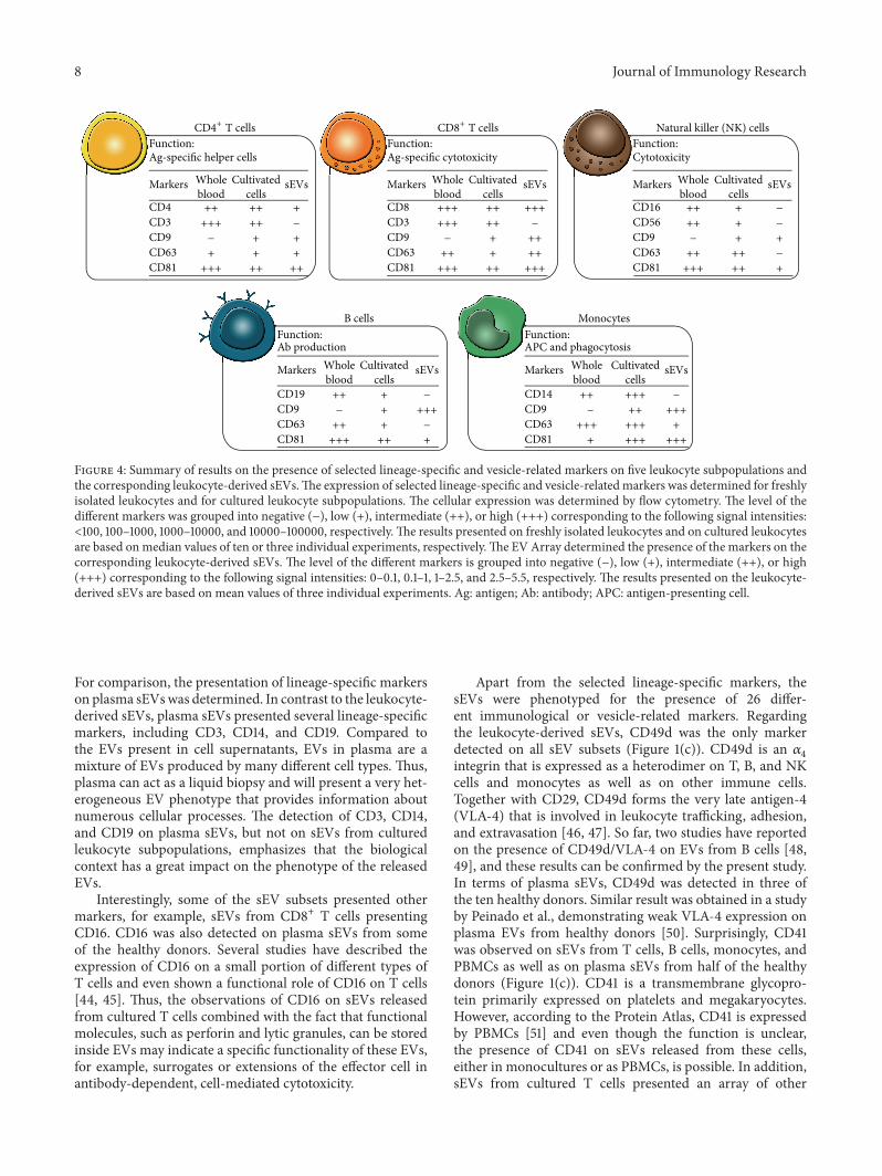
3.3. Cellular versus Vesicular Presentation of Markers. Inorder to investigate the phenotypic homogeneity between thesEVs and the parent cells, the cellular expression patternsof selected lineage-specific and vesicle-related markers werecompared to the vesicular presentation of these markers(Figure 4). In relation to the selected lineage-specificmarkers,CD3, CD14, CD16, and CD19 could not be detected on sEVsfrom cultured T cells, monocytes, NK cells, and B cells,respectively, indicating that not all cell surface moleculesare transferred to the sEV surface. For CD9, CD63, andCD81, the majority of the subpopulations presented vesicularlevels that overall correlated with the cellular expression.Exceptions were observed with CD63, in which case thecellular expression on monocytes was high, but the vesicularpresentation was low. Similarly, the cellular expression ofCD63 was intermediate or low for both NK cells and Bcells, but no CD63 could be detected on the correspondingsEVs.
4. Discussion
Several studies have reported that EVs are released fromdifferent leukocyte subsets; however, a thorough simultane-ous investigation of the major leukocyte subsets with focuson lineage-specific markers has not yet been described. Inthe current study the phenotype of five different subsets ofleukocyte-derived sEVs was determined and related to boththe phenotype of sEVs present in plasma and the cellularphenotype of the leukocyte subpopulations. The EV Array,used for the investigation of the phenotype of sEVs, is aprotein microarray technique that provides the opportunityto detect and characterize sEVs for up to 60markers in a high-throughput manner and with high sensitivity [37, 38]. Onemajor advantage of the EV Array is the ability to phenotypesEVs from unpurified material without a requirement forpreanalytical purification. Isolation of specific EV subsets isin many cases highly warranted, but for phenotyping of EVs
using the EV Array, crude plasma or cell-free supernatantsare applicable.
In order to investigate whether lineage-specific proteinspresent on leukocytes could be detected on the correspond-ing sEVs, leukocytes were phenotyped for selected lineage-specific markers using flow cytometry, while the sEVs werephenotyped using the EVArray.The results showed that noneof the included lineage-specific markers could be detected onthe sEVs released from B cells, NK cells, and monocytes inmonocultures (CD19, CD56, and CD14, resp.). In contrast,sEVs from monocultures of CD4+ and CD8+ T cells didpresent CD4 andCD45 or CD8 andCD45, respectively. Noneof the T cell-derived sEVs presented CD3. A study by Korneket al. investigated EVs from Jurkat T cells activated withphytohemagglutinin (PHA) and observed that the fraction oflarge EVs (sedimented at 10.000×g) only presented low levelsof CD3, while the fraction of sEVs (sedimented at 100.000×g)presented high levels of CD3 [39]. Regarding CD19, a studyby Admyre and coworkers from 2007 showed that EVs froma B cell line presented CD19 [40], which differs from theobservations obtained in the study at hand. Similarly, anotherstudy has described that EVs from resting NK cells presenttypical NK cell markers like CD56 and Fas-L, but not CD16[20]. In terms of CD14, a study by Aharon et al. observedthis marker on large EVs (MVs) released from a monocyticcell line [41]. No CD14 could be detected on sEVs fromcultured monocytes in the current study. The discrepancybetween some of the results may be due to investigationsof different preparations of EVs and/or may be explainedby the fact that the EV-producing cells were different, forexample, cells isolated fromperipheral blood versus cell lines.In addition, differences in the activation state of the EV-producing cells are of importance for the outcome. Uponstimulation, cells change their phenotype and this phenotypicchange depends on the specific stimulus given to the cells[28, 42]. In the present study, leukocyte subpopulations wereisolated and cultured without any activation signal, whileseveral of the other studies investigated the phenotype of

Journal of Immunology Research 7
CD9
Monocytes
Isotype control
B cells
NK cells
0
CD63
CD81
Who
le b
lood
Who
le b
lood
Who
le b
lood
CD8+ T cells
CD4+ T cells
Monocytes
Isotype control
B cells
NK cells
CD8+ T cells
CD4+ T cells
Monocytes
Isotype control
B cells
NK cells
CD8+ T cells
CD4+ T cells
−103
103
104
105
0−103
103
104
105
0−103
103
104
105
MFI
2563
10031
10333
13627
10165
(min–max)
(9074–17125)
(6718–18589)
(5462–16268)
(7852–19807)
(2010–3547)
MFI
79
23
(min–max)
−177
−115
−246
(−522–33)
(−448–170)
(−901–3)
(−320–507)
(−544–245)
MFI
13278
1103
2883
1571
481
(min–max)
(267–779)
(835–2068)
(1518–4692)
(650–1456)
(8336–15856)
(a)
Monocytes
Isotype control
Isotype control
B cells
NK cells
CD9
CD63
CD81
Culti
vate
d ce
llsCu
ltiva
ted
cells
Culti
vate
d ce
lls
CD8+ T cells
CD4+ T cells
Monocytes
Isotype control
Isotype control
B cells
NK cells
CD8+ T cells
CD4+ T cells
Monocytes
Isotype control
Isotype control
B cells
NK cells
CD8+ T cells
CD4+ T cells
0−103
103
104
105
0−103
103
104
105
0−103
103
104
105
MFI
19165
5180
3942
7237
8658
(min–max)
(8600–11020)
(6957–7446)
(2815–4662)
(4228–5925)
(12815–49319)
MFI
4934
544
359
413
371
MFI
20911
642
1030
844
424
(min–max)
(min–max)
(339–453)
(402–423)
(278–363)
(494–657)
(3701–8697)
(395–434)
(786–964)
(708–1280)
(506–779)
(19438–24334)
(b)
Figure 3: Cellular expression of CD9, CD63, and CD81 on five leukocyte subpopulations. The cellular expression pattern of CD9, CD63, andCD81 was investigated for leukocytes present in freshly drawn whole blood (a) as well as for isolated subpopulations of leukocytes followingtwo days of culture (b). For each of the subpopulations, the expression pattern was determined by flow cytometry. The expression from tworepresentative donors is illustrated in histograms. Isotype controls were included for every leukocyte subpopulation and when the obtainedsignals were similar, only one isotype control is depicted. Regarding the cultivated cells, a representative lymphocyte isotype control is shown.Due to differences in the level of the isotype control between the lymphocytes and the monocytes, the monocyte isotype control is alsoshown. The interdonor variation in the expression levels of the different markers is displayed next to each histogram. Data are presented asmedian fluorescence intensities (MFI) with min and max indicated in parenthesis from the ten (whole blood) or three donors (cultivatedcells), respectively.
the EVs released upon adding a stimulus to the cells. Thepresented results obtained with sEVs released from culturedPBMCs support this phenomenon as these sEVs apparentlydisplayed fewer markers than sEVs from monocultures.Overall, these data indicate that the phenotype of the vesiclesubset is context-dependent. However, during the current
conditions, several of the leukocyte-derived sEVs did notpresent the lineage-specific markers found on the parentcell type (Figure 1(a)). Variations in the molecular contentbetween cells and EVs were also observed in a study byHunter et al. that reported significant differences in thepresence of miRNAs between plasma EVs and PBMCs [43].
8 Journal of Immunology Research
Natural killer (NK) cells
MonocytesB cells
CD8+ T cellsCD4+ T cellsFunction:
Function: Function:
Function: Function:
cellssEVsMarkers
CD4CD3CD9CD63CD81
blood
Ag-specific helper cells
Ab production APC and phagocytosis
CytotoxicityAg-specific cytotoxicity
Whole Cultivated
+
+
+++
+
++++
++
++++
+++
+++
−
−
CD8CD3CD9CD63CD81
+++
+
+++++
++
+++++
++
+++++
+++
+++
−
−
cellssEVsMarkers
bloodWhole Cultivated
cellssEVsMarkers
bloodWhole Cultivated
CD16CD56CD9CD63CD81
−
+
−++++
+
+++
+
+++
++
+++
−
−
CD14CD9CD63CD81
CD19CD9CD63CD81
−
+++
+++++++
+
++
+++++
− +++
+++
−
+
++++++
−
+
+++
− +++
++
cellssEVsMarkers
bloodWhole Cultivated
cellssEVsMarkers
bloodWhole Cultivated
Figure 4: Summary of results on the presence of selected lineage-specific and vesicle-related markers on five leukocyte subpopulations andthe corresponding leukocyte-derived sEVs.The expression of selected lineage-specific and vesicle-relatedmarkers was determined for freshlyisolated leukocytes and for cultured leukocyte subpopulations. The cellular expression was determined by flow cytometry. The level of thedifferent markers was grouped into negative (−), low (+), intermediate (++), or high (+++) corresponding to the following signal intensities:<100, 100–1000, 1000–10000, and 10000–100000, respectively.The results presented on freshly isolated leukocytes and on cultured leukocytesare based onmedian values of ten or three individual experiments, respectively.The EVArray determined the presence of the markers on thecorresponding leukocyte-derived sEVs. The level of the different markers is grouped into negative (−), low (+), intermediate (++), or high(+++) corresponding to the following signal intensities: 0–0.1, 0.1–1, 1–2.5, and 2.5–5.5, respectively. The results presented on the leukocyte-derived sEVs are based on mean values of three individual experiments. Ag: antigen; Ab: antibody; APC: antigen-presenting cell.
For comparison, the presentation of lineage-specific markerson plasma sEVswas determined. In contrast to the leukocyte-derived sEVs, plasma sEVs presented several lineage-specificmarkers, including CD3, CD14, and CD19. Compared tothe EVs present in cell supernatants, EVs in plasma are amixture of EVs produced by many different cell types. Thus,plasma can act as a liquid biopsy and will present a very het-erogeneous EV phenotype that provides information aboutnumerous cellular processes. The detection of CD3, CD14,and CD19 on plasma sEVs, but not on sEVs from culturedleukocyte subpopulations, emphasizes that the biologicalcontext has a great impact on the phenotype of the releasedEVs.
Interestingly, some of the sEV subsets presented othermarkers, for example, sEVs from CD8+ T cells presentingCD16. CD16 was also detected on plasma sEVs from someof the healthy donors. Several studies have described theexpression of CD16 on a small portion of different types ofT cells and even shown a functional role of CD16 on T cells[44, 45]. Thus, the observations of CD16 on sEVs releasedfrom cultured T cells combined with the fact that functionalmolecules, such as perforin and lytic granules, can be storedinside EVs may indicate a specific functionality of these EVs,for example, surrogates or extensions of the effector cell inantibody-dependent, cell-mediated cytotoxicity.
Apart from the selected lineage-specific markers, thesEVs were phenotyped for the presence of 26 differ-ent immunological or vesicle-related markers. Regardingthe leukocyte-derived sEVs, CD49d was the only markerdetected on all sEV subsets (Figure 1(c)). CD49d is an 𝛼
4
integrin that is expressed as a heterodimer on T, B, and NKcells and monocytes as well as on other immune cells.Together with CD29, CD49d forms the very late antigen-4(VLA-4) that is involved in leukocyte trafficking, adhesion,and extravasation [46, 47]. So far, two studies have reportedon the presence of CD49d/VLA-4 on EVs from B cells [48,49], and these results can be confirmed by the present study.In terms of plasma sEVs, CD49d was detected in three ofthe ten healthy donors. Similar result was obtained in a studyby Peinado et al., demonstrating weak VLA-4 expression onplasma EVs from healthy donors [50]. Surprisingly, CD41was observed on sEVs from T cells, B cells, monocytes, andPBMCs as well as on plasma sEVs from half of the healthydonors (Figure 1(c)). CD41 is a transmembrane glycopro-tein primarily expressed on platelets and megakaryocytes.However, according to the Protein Atlas, CD41 is expressedby PBMCs [51] and even though the function is unclear,the presence of CD41 on sEVs released from these cells,either in monocultures or as PBMCs, is possible. In addition,sEVs from cultured T cells presented an array of other

Journal of Immunology Research 9
immunologicalmarkers, ranging frommore generalmarkers,like CD45, to specific markers like CD28, and the TLR3.Moreover, sEVs from CD8+ T cells presented CTLA-4 andthe Fas-L. Even though the mechanisms are quite different,both of these molecules play a role in downregulation ofan immune response, either by transmitting an inhibitorysignal in activated T cells or by inducing activation-inducedcell death, respectively. The presence of such regulatorymolecules on EVs suggests that T cell-derived EVs can beinvolved in immune regulation. The presence of Fas-L onEVs from T cells has also been observed in other studies[52–54], but to the best of our knowledge, studies observingCTLA-4 on EVs from leukocytes have not previously beenpublished. Upon evaluating the presence of markers onEVs, it is important to consider the natural variation in themolecular composition of the EV pool that exists betweenhealthy individuals as visualized by the ten plasma samples.Thus, the present results are a snapshot providing insightabout the current vesicular presentation of selected markers.
The sEVs investigated in this study were identified bythe presence of CD9, CD63, and/or CD81. Thus, in orderto investigate the similarity of the leukocytes and the cor-responding sEVs, the cellular expression patterns of CD9,CD63, and CD81 were determined. The expression patternwas determined for both leukocytes present in whole bloodand cultured leukocyte subpopulations. The results demon-strated a lack of CD9 on the surface of leukocytes presentin freshly drawn whole blood (Figure 3(a)). In contrast, CD9was present on every leukocyte subpopulation following twodays of culture. In accordance with the present results, arecently published study also observed CD9 on the surfaceof isolated populations of CD3+, CD14+, andCD19+ cells [55].The expression patterns of CD63 andCD81 were quite similarbetween the two cell preparations but with minor differencesin the expressions levels. Overall, these findings are inagreement with previously described results [56–61]. Whenlooking at both the cellular and the vesicular presentationof these markers, it is clear that, for the majority of thecultured subpopulations, the presentation of these markerswas comparable. However, for the NK cells, the B cells, andthe monocytes, the cellular expression of CD63 was verydifferent from the presentation observed on sEVs. A compar-ison between the presentation of CD9, CD63, and CD81 onwhole blood leukocytes and plasma EVs is a more complexprocedure that needs to take into account that plasma EVsare a very heterogeneous group of EVs that emanate frommultiple cell types. The tetraspanin signals observed on theplasma sEVs represent all tetraspanin-positive sEVs irrespec-tive of their origin. Thus, the presentation of tetraspaninson plasma sEVs cannot be expected to correlate with theexpression of tetraspanin on leukocytes. Nevertheless, it isclear that the CD63 signals in plasma generally were low,while the cellular expression of CD63 for several of the leuko-cyte subpopulations is medium to high, indicating that thepresentation of CD63 is different from sEVs to cells. Overall,the vesicular signal intensities for CD63 were much lowerthan the intensities observed for CD9 and CD81, suggestingthat CD63 might be a poor marker for sEVs in general,which has also been observed in other studies [38, 62–64].
The results underline the importance of detecting EVs with acocktail of antibodies against tetraspanins, as detection witha single marker may overlook some subsets of EVs.
5. Conclusion
Surface molecules on EVs are responsible for the biodis-tribution and the ligation to target cells. Thus, the proteincomposition is related to the functionality of EVs, whyphenotyping of EVs can be used to gain knowledge aboutthe functionality. In summary, the presented data regardingthe lineage-specific markers and the tetraspanins supportthe accumulating observations suggesting that the transferof molecular cargo into EVs is tightly controlled. A tightlyregulated sampling ofmolecules to EVswouldmatchwith thefact that EVs play an important role as systemic regulators,traveling through tissues providing key intercellular commu-nication as well as transfer of biologically active components.
Abbreviations
APC: Antigen-presenting cellCTL: Cytotoxic T-lymphocyteCTLA-4: Cytotoxic T-lymphocyte associated protein-4DCs: Dendritic cellsEVs: Extracellular vesiclesFas-L: Fas LigandICAM-1: Intercellular adhesion molecule 1MFI: Median fluorescence intensityMHC-II: Major histocompatibility complex class IIMIC A/B: Major histocompatibility complex class I-related
chain A/BMVs: MicrovesiclesNK: Natural killerNKG2D: Natural killer group 2, member DPBMCs: Peripheral blood mononuclear cellsPHA: PhytohemagglutininsEVs: Small EVsTLR: Toll-like receptorTNF RI: Tumor necrosis factor receptor-1TSG101: Tumor susceptibility gene 101VLA-4: Very late antigen-4.
Ethical Approval
All research on human subjects presented in this paper wasconducted in accordance with the local ethics legislation.
Consent
Each human subject signed a written consent form, allowingfor the use of his or her blood for research purposes.
Competing Interests
The authors declare that there is no conflict of interestsregarding publication of this paper.

10 Journal of Immunology Research
Authors’ Contributions
Lotte Hatting Pugholm was responsible for the study design,planned and contributed to the cellular work, performedall work related to flow cytometry, and drafted the paper.Rikke Bæk performed all experimental work related to theEV Array and revised the paper. Evo Kristina LinderssonSøndergaard andAnneLouise Schacht Revenfeld contributedto the cellular work, discussed the data, and revised thepaper. Malene Møller Jørgensen analyzed the EV Array data,discussed the data, and revised the paper. Kim Varmingdiscussed the data and revised the paper.
Acknowledgments
The authors thank laboratory technician Anne Elbæk,Department of Clinical Immunology, Aalborg UniversityHospital, for technical assistance.
References
[1] A. L. S. Revenfeld, R. Bæk, M. H. Nielsen, A. Stensballe,K. Varming, and M. Jørgensen, “Diagnostic and prognosticpotential of extracellular vesicles in peripheral blood,” ClinicalTherapeutics, vol. 36, no. 6, pp. 830–846, 2014.
[2] M. Yanez-Mo, P. R.-M. Siljander, Z. Andreu et al., “Biologicalproperties of extracellular vesicles and their physiological func-tions,” Journal of Extracellular Vesicles, vol. 4, Article ID 27066,2015.
[3] C. Thery, L. Zitvogel, and S. Amigorena, “Exosomes: composi-tion, biogenesis and function,”Nature Reviews Immunology, vol.2, no. 8, pp. 569–579, 2002.
[4] S. J. Gould andG. Raposo, “Aswewait: copingwith an imperfectnomenclature for extracellular vesicles,” Journal of ExtracellularVesicles, vol. 2, Article ID 20389, 2013.
[5] H. Valadi, K. Ekstrom, A. Bossios, M. Sjostrand, J. J. Lee,and J. O. Lotvall, “Exosome-mediated transfer of mRNAs andmicroRNAs is a novel mechanism of genetic exchange betweencells,” Nature Cell Biology, vol. 9, no. 6, pp. 654–659, 2007.
[6] K. Denzer, M. J. Kleijmeer, H. F. G. Heijnen, W. Stoorvogel,and H. J. Geuze, “Exosome: from internal vesicle of themultivesicular body to intercellular signaling device,” Journal ofCell Science, vol. 113, part 19, pp. 3365–3374, 2000.
[7] C. Thery, M. Ostrowski, and E. Segura, “Membrane vesicles asconveyors of immune responses,” Nature Reviews Immunology,vol. 9, no. 8, pp. 581–593, 2009.
[8] V. Huber, P. Filipazzi, M. Iero, S. Fais, and L. Rivoltini, “Moreinsights into the immunosuppressive potential of tumor exo-somes,” Journal of TranslationalMedicine, vol. 6, article 63, 2008.
[9] M. Iero, R. Valenti, V. Huber et al., “Tumour-released exosomesand their implications in cancer immunity,” Cell Death andDifferentiation, vol. 15, no. 1, pp. 80–88, 2008.
[10] M. P. Oksvold, A. Kullmann, L. Forfang et al., “Expression ofB-Cell surface antigens in subpopulations of exosomes releasedfrom B-cell lymphoma cells,” Clinical Therapeutics, vol. 36, no.6, pp. 847.e1–862.e1, 2014.
[11] V. Muralidharan-Chari, J. W. Clancy, A. Sedgwick, and C.D’Souza-Schorey, “Microvesicles: mediators of extracellularcommunication during cancer progression,” Journal of CellScience, vol. 123, no. 10, pp. 1603–1611, 2010.
[12] R. Valenti, V. Huber, P. Filipazzi et al., “Human tumor-releasedmicrovesicles promote the differentiation of myeloid cells withtransforming growth factor-𝛽-mediated suppressive activity onT lymphocytes,”Cancer Research, vol. 66, no. 18, pp. 9290–9298,2006.
[13] D. D. Taylor, C. Gercel-Taylor, K. S. Lyons, J. Stanson, andT. L. Whiteside, “T-cell apoptosis and suppression of T-cellreceptor/CD3-zeta by Fas ligand-containingmembrane vesiclesshed from ovarian tumors,” Clinical Cancer Research, vol. 9, no.14, pp. 5113–5119, 2003.
[14] M. Mack, A. Kleinschmidt, H. Bruhl et al., “Transfer of thechemokine receptor CCR5 between cells by membrane-derivedmicroparticles: a mechanism for cellular human immunodefi-ciency virus 1 infection,”Nature Medicine, vol. 6, no. 7, pp. 769–775, 2000.
[15] T. Wurdinger, N. N. Gatson, L. Balaj, B. Kaur, X. O. Breakefield,and D. M. Pegtel, “Extracellular vesicles and their convergencewith viral pathways,” Advances in Virology, vol. 2012, Article ID767694, 12 pages, 2012.
[16] M. Mittelbrunn, C. Gutierrez-Vazquez, C. Villarroya-Beltri etal., “Unidirectional transfer of microRNA-loaded exosomesfrom T cells to antigen-presenting cells,” Nature Communica-tions, vol. 2, no. 1, article 282, 2011.
[17] C. Admyre, S. M. Johansson, S. Paulie, and S. Gabrielsson,“Direct exosome stimulation of peripheral human T cellsdetected by ELISPOT,”European Journal of Immunology, vol. 36,no. 7, pp. 1772–1781, 2006.
[18] M. Eldh, K. Ekstrom, H. Valadi et al., “Exosomes communicateprotective messages during oxidative stress; possible role ofexosomal shuttle RNA,” PLoS ONE, vol. 5, no. 12, Article IDe15353, 2010.
[19] S. C. Saunderson, P. C. Schuberth, A. C. Dunn et al., “Inductionof exosome release in primary B cells stimulated via CD40 andthe IL-4 receptor,” The Journal of Immunology, vol. 180, no. 12,pp. 8146–8152, 2008.
[20] L. Lugini, S. Cecchetti, V. Huber et al., “Immune surveillanceproperties of human NK cell-derived exosomes,” Journal ofImmunology, vol. 189, no. 6, pp. 2833–2842, 2012.
[21] Y. Xie, X. Zhang, T. Zhao, W. Li, and J. Xiang, “NaturalCD8+25+ regulatory T cell-secreted exosomes capable of sup-pressing cytotoxic T lymphocyte-mediated immunity againstB16 melanoma,” Biochemical and Biophysical Research Commu-nications, vol. 438, no. 1, pp. 152–155, 2013.
[22] S. Bhatnagar, K. Shinagawa, F. J. Castellino, and J. S. Schorey,“Exosomes released from macrophages infected with intracel-lular pathogens stimulate a proinflammatory response in vitroand in vivo,” Blood, vol. 110, no. 9, pp. 3234–3244, 2007.
[23] E. N. M. Nolte-’t Hoen, S. I. Buschow, S. M. Anderton, W.Stoorvogel, and M. H. M. Wauben, “Activated T cells recruitexosomes secreted by dendritic cells via LFA-1,” Blood, vol. 113,no. 9, pp. 1977–1981, 2009.
[24] L. Garzetti, R. Menon, A. Finardi et al., “Activated macrophagesrelease microvesicles containing polarized M1 or M2 mRNAs,”Journal of Leukocyte Biology, vol. 95, no. 5, pp. 817–825, 2014.
[25] L. Zitvogel, A. Regnault, A. Lozier et al., “Eradication ofestablished murine tumors using a novel cell-free vaccine:dendritic cell-derived exosomes,” Nature Medicine, vol. 4, no.5, pp. 594–600, 1998.
[26] P. Rialland, D. Lankar, G. Raposo, C. Bonnerot, and P. Hubert,“BCR-bound antigen is targeted to exosomes in human follic-ular lymphoma B-cells,” Biology of the Cell, vol. 98, no. 8, pp.491–501, 2006.

Journal of Immunology Research 11
[27] A. Muntasell, A. C. Berger, and P. A. Roche, “T cell-inducedsecretion of MHC class II-peptide complexes on B cell exo-somes,” EMBO Journal, vol. 26, no. 19, pp. 4263–4272, 2007.
[28] E. Segura, S. Amigorena, and C. Thery, “Mature dendriticcells secrete exosomes with strong ability to induce antigen-specific effector immune responses,” Blood Cells, Molecules, andDiseases, vol. 35, no. 2, pp. 89–93, 2005.
[29] S. Viaud, M. Terme, C. Flament et al., “Dendritic cell-derivedexosomes promote natural killer cell activation and prolifera-tion: a role for NKG2D ligands and IL-15R𝛼,” PLoS ONE, vol. 4,no. 3, Article ID e4942, 2009.
[30] S. I. Buschow, E. N. M. Nolte-’t Hoen, G. van Niel et al., “MHCII in dendritic cells is targeted to lysosomes or T cell-inducedexosomes via distinct multivesicular body pathways,” Traffic,vol. 10, no. 10, pp. 1528–1542, 2009.
[31] S. Hao, J. Yuan, and J. Xiang, “Nonspecific CD4+ T cellswith uptake of antigen-specific dendritic cell-released exosomesstimulate antigen-specific CD8+ CTL responses and long-termT cell memory,” Journal of Leukocyte Biology, vol. 82, no. 4, pp.829–838, 2007.
[32] H. Zhang, Y. Xie,W. Li, R. Chibbar, S. Xiong, and J. Xiang, “CD4T cell-released exosomes inhibit CD8 cytotoxic T-lymphocyteresponses and antitumor immunity,” Cellular and MolecularImmunology, vol. 8, no. 1, pp. 23–30, 2011.
[33] O. Ashiru, P. Boutet, L. Fernandez-Messina et al., “Naturalkiller cell cytotoxicity is suppressed by exposure to the humanNKG2D ligand MICA∗008 that is shed by tumor cells inexosomes,” Cancer Research, vol. 70, no. 2, pp. 481–489, 2010.
[34] X. Li, J.-J. Li, J.-Y. Yang et al., “Tolerance induction by exosomesfrom immature dendritic cells and rapamycin in a mousecardiac allograft model,” PLoS ONE, vol. 7, no. 8, Article IDe44045, 2012.
[35] X. Yang, S. Meng, H. Jiang, C. Zhu, and W. Wu, “Exosomesderived from immature bone marrow dendritic cells inducetolerogenicity of intestinal transplantation in rats,” Journal ofSurgical Research, vol. 171, no. 2, pp. 826–832, 2011.
[36] H. Peche, M. Heslan, C. Usal, S. Amigorena, and M. C.Cuturi, “Presentation of donor major histocompatibility com-plex antigens by bone marrow dendritic cell-derived exosomesmodulates allograft rejection,” Transplantation, vol. 76, no. 10,pp. 1503–1510, 2003.
[37] M. Jørgensen, R. Bæk, S. Pedersen, E. K. Søndergaard, S. R.Kristensen, and K. Varming, “Extracellular Vesicle (EV) Array:microarray capturing of exosomes and other extracellularvesicles for multiplexed phenotyping,” Journal of ExtracellularVesicles, vol. 2, pp. 1–9, 2013.
[38] M. M. Jørgensen, R. Bæk, and K. Varming, “Potentials andcapabilities of the Extracellular Vesicle (EV) Array,” Journal ofExtracellular Vesicles, vol. 4, Article ID 26048, 2015.
[39] M. Kornek, Y. Popov, T. A. Libermann, N. H. Afdhal, and D.Schuppan, “Human T cell microparticles circulate in blood ofhepatitis patients and induce fibrolytic activation of hepaticstellate cells,” Hepatology, vol. 53, no. 1, pp. 230–242, 2011.
[40] C. Admyre, B. Bohle, S. M. Johansson et al., “B cell-derivedexosomes can present allergen peptides and activate allergen-specific T cells to proliferate and produce TH2-like cytokines,”Journal of Allergy and Clinical Immunology, vol. 120, no. 6, pp.1418–1424, 2007.
[41] A. Aharon, T. Tamari, and B. Brenner, “Monocyte-derivedmicroparticles and exosomes induce procoagulant and apop-totic effects on endothelial cells,” Thrombosis and Haemostasis,vol. 100, no. 5, pp. 878–885, 2008.
[42] E. van der Vlist, G. J. A. Arkesteijn, C. H. A. van de Lest, W.Stoorvogel, E. N. M. Nolte-’t Hoen, and M. H. M. Wauben,“CD4+ T cell activation promotes the differential release of dis-tinct populations of nanosized vesicles,” Journal of ExtracellularVesicles, vol. 1, Article ID 18364, 2012.
[43] M. P.Hunter,N. Ismail, X. Zhang et al., “Detection ofmicroRNAexpression in human peripheral blood microvesicles,” PLoSONE, vol. 3, no. 11, Article ID e3694, 2008.
[44] B. Clemenceau, R. Vivien, E. Debeaupuis et al., “Fc𝛾RIIIa(CD16) induction on human t lymphocytes and CD16pos T-lymphocyte amplification,” Journal of Immunotherapy, vol. 34,no. 7, pp. 542–549, 2011.
[45] N. K. Bjorkstrom, V. D. Gonzalez, K.-J. Malmberg et al.,“Elevated numbers of Fc gamma RIIIA+ (CD16+) effector CD8T cells with NK cell-like function in chronic hepatitis C virusinfection,”The Journal of Immunology, vol. 181, no. 6, pp. 4219–4228, 2008.
[46] D.M. Rose, R. Alon, andM. H. Ginsberg, “Integrinmodulationand signaling in leukocyte adhesion and migration,” Immuno-logical Reviews, vol. 218, no. 1, pp. 126–134, 2007.
[47] H. Yusuf-Makagiansar, M. E. Anderson, T. V. Yakovleva, J. S.Murray, and T. J. Siahaan, “Inhibition of LFA-1/ICAM-1 andVLA-4/VCAM-1 as a therapeutic approach to inflammationand autoimmune diseases,”Medicinal Research Reviews, vol. 22,no. 2, pp. 146–167, 2002.
[48] A. Clayton, A. Turkes, S. Dewitt, R. Steadman, M. D. Mason,and M. B. Hallett, “Adhesion and signaling by B cell-derivedexosomes: the role of integrins,”The FASEB Journal, vol. 18, no.9, pp. 977–979, 2004.
[49] R.Wubbolts, R. S. Leckie, P. T. M. Veenhuizen et al., “Proteomicand biochemical analyses of human B cell-derived exosomes:potential implications for their function and multivesicularbody formation,” The Journal of Biological Chemistry, vol. 278,no. 13, pp. 10963–10972, 2003.
[50] H. Peinado, M. Aleckovic, S. Lavotshkin et al., “Melanomaexosomes educate bone marrow progenitor cells toward a pro-metastatic phenotype through MET,” Nature Medicine, vol. 18,no. 6, pp. 883–891, 2012.
[51] M. Uhlen, P. Oksvold, L. Fagerberg et al., “Towards aknowledge-based Human Protein Atlas,” Nature Biotechnology,vol. 28, no. 12, pp. 1248–1250, 2010.
[52] M. J. Martınez-Lorenzo, A. Anel, S. Gamen et al., “Activatedhuman T cells release bioactive Fas ligand and APO2 ligand inmicrovesicles,” Journal of Immunology, vol. 163, no. 3, pp. 1274–1281, 1999.
[53] I. Monleon, M. J. Martınez-Lorenzo, L. Monteagudo et al.,“Differential secretion of Fas ligand- or APO2 ligand/TNF-related apoptosis-inducing ligand-carrying microvesicles dur-ing activation-induced death of human T cells,” Journal ofImmunology, vol. 167, no. 12, pp. 6736–6744, 2001.
[54] R. Alonso, C. Mazzeo, M. C. Rodriguez et al., “Diacylglycerolkinase 𝛼 regulates the formation and polarisation of maturemultivesicular bodies involved in the secretion of Fas ligand-containing exosomes in T lymphocytes,” Cell Death and Differ-entiation, vol. 18, no. 7, pp. 1161–1173, 2011.
[55] S. Tsukamoto, M. Takeuchi, T. Kawaguchi et al., “TetraspaninCD9 modulates ADAM17-mediated shedding of LR11 in leuko-cytes,”Experimental andMolecularMedicine, vol. 46, article e89,2014.
[56] J. M. Tarrant, L. Robb, A. B. van Spriel, and M. D. Wright,“Tetraspanins: molecular organisers of the leukocyte surface,”Trends in Immunology, vol. 24, no. 11, pp. 610–617, 2003.

12 Journal of Immunology Research
[57] V. Rocha-Perugini, M. Zamai, J. M. Gonzalez-Granado et al.,“CD81 controls sustained T cell activation signaling and definesthe maturation stages of cognate immunological synapses,”Molecular and Cellular Biology, vol. 33, no. 18, pp. 3644–3658,2013.
[58] S. C. Todd, S. G. Lipps, L. Crisa, D. R. Salomon, and C.D. Tsoukas, “CD81 expressed on human thymocytes mediatesintegrin activation and interleukin 2-dependent proliferation,”The Journal of Experimental Medicine, vol. 184, no. 5, pp. 2055–2060, 1996.
[59] H. T.Maecker,M.-S. Do, and S. Levy, “CD81 on B cells promotesinterleukin 4 secretion and antibody production during Thelper type 2 immune responses,” Proceedings of the NationalAcademy of Sciences of the United States of America, vol. 95, no.5, pp. 2458–2462, 1998.
[60] A. Gilsanz, L. Sanchez-Martın, M. D. Gutierrez-Lopez etal., “ALCAM/CD166 adhesive function is regulated by thetetraspanin CD9,” Cellular and Molecular Life Sciences, vol. 70,no. 3, pp. 475–493, 2013.
[61] E. Tippett, P. U. Cameron, M. Marsh, and S. M. Crowe,“Characterization of tetraspanins CD9, CD53, CD63, and CD81in monocytes and macrophages in HIV-1 infection,” Journal ofLeukocyte Biology, vol. 93, no. 6, pp. 913–920, 2013.
[62] A. L. Schacht Revenfeld, E. K. Lindersson Søndergaard, A.Stensballe, R. Baek, M. Møller Jørgensen, and K. Varming,“Characterization of a cell-culturing system for the studyof contact-independent extracellular vesicle communication,”Journal of Circulating Biomarkers, 2016.
[63] M.He, J. Crow,M. Roth, Y. Zeng, andA.K.Godwin, “Integratedimmunoisolation and protein analysis of circulating exosomesusingmicrofluidic technology,” Lab on a Chip—Miniaturisationfor Chemistry and Biology, vol. 14, no. 19, pp. 3773–3780, 2014.
[64] J. Kowal, G. Arras, M. Colombo et al., “Proteomic comparisondefines novel markers to characterize heterogeneous popu-lations of extracellular vesicle subtypes,” Proceedings of theNational Academy of Sciences, vol. 113, no. 8, pp. E968–E977,2016.

Submit your manuscripts athttp://www.hindawi.com
Stem CellsInternational
Hindawi Publishing Corporationhttp://www.hindawi.com Volume 2014
Hindawi Publishing Corporationhttp://www.hindawi.com Volume 2014
MEDIATORSINFLAMMATION
of
Hindawi Publishing Corporationhttp://www.hindawi.com Volume 2014
Behavioural Neurology
EndocrinologyInternational Journal of
Hindawi Publishing Corporationhttp://www.hindawi.com Volume 2014
Hindawi Publishing Corporationhttp://www.hindawi.com Volume 2014
Disease Markers
Hindawi Publishing Corporationhttp://www.hindawi.com Volume 2014
BioMed Research International
OncologyJournal of
Hindawi Publishing Corporationhttp://www.hindawi.com Volume 2014
Hindawi Publishing Corporationhttp://www.hindawi.com Volume 2014
Oxidative Medicine and Cellular Longevity
Hindawi Publishing Corporationhttp://www.hindawi.com Volume 2014
PPAR Research
The Scientific World JournalHindawi Publishing Corporation http://www.hindawi.com Volume 2014
Immunology ResearchHindawi Publishing Corporationhttp://www.hindawi.com Volume 2014
Journal of
ObesityJournal of
Hindawi Publishing Corporationhttp://www.hindawi.com Volume 2014
Hindawi Publishing Corporationhttp://www.hindawi.com Volume 2014
Computational and Mathematical Methods in Medicine
OphthalmologyJournal of
Hindawi Publishing Corporationhttp://www.hindawi.com Volume 2014
Diabetes ResearchJournal of
Hindawi Publishing Corporationhttp://www.hindawi.com Volume 2014
Hindawi Publishing Corporationhttp://www.hindawi.com Volume 2014
Research and TreatmentAIDS
Hindawi Publishing Corporationhttp://www.hindawi.com Volume 2014
Gastroenterology Research and Practice
Hindawi Publishing Corporationhttp://www.hindawi.com Volume 2014
Parkinson’s Disease
Evidence-Based Complementary and Alternative Medicine
Volume 2014Hindawi Publishing Corporationhttp://www.hindawi.com