Embed Size (px)
Citation preview
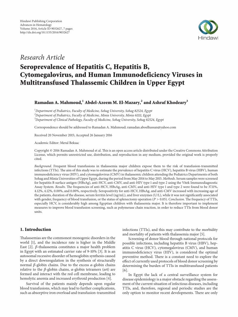
Research ArticleSeroprevalence of Hepatitis C, Hepatitis B,Cytomegalovirus, and Human Immunodeficiency Viruses inMultitransfused Thalassemic Children in Upper Egypt
Ramadan A. Mahmoud,1 Abdel-Azeem M. El-Mazary,2 and Ashraf Khodeary3
1Department of Pediatrics, Faculty of Medicine, Sohag University, Sohag 82524, Egypt2Department of Pediatrics, Faculty of Medicine, Minia University, Minia 61111, Egypt3Department of Clinical Pathology, Faculty of Medicine, Sohag University, Sohag 82524, Egypt
Correspondence should be addressed to Ramadan A. Mahmoud; [email protected]
Received 20 November 2015; Accepted 26 January 2016
Academic Editor: Meral Beksac
Copyright © 2016 Ramadan A. Mahmoud et al.This is an open access article distributed under the Creative Commons AttributionLicense, which permits unrestricted use, distribution, and reproduction in any medium, provided the original work is properlycited.
Background. Frequent blood transfusions in thalassemia major children expose them to the risk of transfusion-transmittedinfections (TTIs). The aim of this study was to estimate the prevalence of hepatitis C virus (HCV), hepatitis B virus (HBV), humanimmunodeficiency virus (HIV), and cytomegalovirus (CMV) in thalassemic children attending the Pediatrics Departments of bothSohag andMiniaUniversities ofUpper Egypt, during the period fromMay 2014 toMay 2015.Methods. Serum sampleswere screenedfor hepatitis B surface antigen (HBsAg), anti-HCV, anti-CMV, and anti-HIV type 1 and type 2 using the Vitek ImmunodiagnosticAssay System. Results. The frequencies of anti-HCV, HBsAg, anti-CMV, and anti-HIV type 1 and type 2 were found to be 37.11%,4.12%, 4.12%, 0.00%, and 0.00%, respectively. Seropositivity for anti-HCV, HBsAg, and anti-CMV increased with increasing age ofthe patients, duration of the disease, serum ferritin level (ng/mL), and liver enzymes (U/L), while it was not significantly associatedwith gender, frequency of blood transfusion, or the status of splenectomy operation (𝑃 > 0.05). Conclusion. The frequency of TTIs,especially HCV, is considerably high among Egyptian children with thalassemia major. It is therefore important to implementmeasures to improve blood transfusion screening, such as polymerase chain reaction, in order to reduce TTIs from blood donorunits.
1. Introduction
Thalassemias are the commonest monogenic disorders in theworld [1], and the incidence rate is higher in the MiddleEast [2]. 𝛽-thalassemia constitutes a major health problemin Egypt with an estimated carrier rate of 9-10% [3]. It is anautosomal recessive disorder of hemoglobin synthesis causedby a direct downregulation in the synthesis of structurallynormal 𝛽-globin chains. Due to the excess 𝛼-globin chainsrelative to the 𝛽-globin chains, 𝛼-globin tetramers (𝛼4) areformed and interact with the red cell membrane, leading tohemolytic anemia and increased erythroid production [4].
Survival of the patients mainly depends upon regularblood transfusions, which may lead to further complications,such as absorptive iron overload and transfusion-transmitted
infections (TTIs), and this may contribute to the morbidityand mortality of patients with thalassemia major [5].
Screening of donor blood through national protocols forpossible infections, including hepatitis B virus (HBV), hep-atitis C virus (HCV), cytomegalovirus (CMV), and humanimmunodeficiency virus (HIV), is considered the optimalpreventive method. There is a constant need to explore theeffect of currently used protocols of blood donor screening bydetermining the burden of TTIs in multitransfused patients[6].
In Egypt the lack of a central surveillance system fordisease epidemiology is amajor obstacle regarding the assess-ment of the current situation of infectious diseases, includingTTIs, and, therefore, regional and periodic studies are theonly option to monitor recent developments. There are only
Hindawi Publishing CorporationAdvances in HematologyVolume 2016, Article ID 9032627, 7 pageshttp://dx.doi.org/10.1155/2016/9032627
2 Advances in Hematology
a few studies about TTIs in thalassemic children in Egypt [3,7–9]. To our knowledge, no study has described the situationof TTIs in thalassemic children in Upper Egypt. These areashad low social economic income compared to other parts inEgypt [10]. The objective of this study was to evaluate theseroprevalence of HBV, HCV, CMV, and HIV. Furthermore,TTI-associated clinical and laboratory risk factors wereinvestigated in this study including gender, family history,duration of the illness, frequency of blood transfusions,splenectomy, hemoglobin, ferritin level, creatinine levels, andliver enzymes.
2. Materials and Methods
In this cross-sectional study, we analyzed blood samplestaken from 97 transfusion-dependent thalassemic childrenduring the period from May 2014 to May 2015 in the Pedi-atrics Departments, Faculty of Medicine, in both Sohag andMinia Universities of Upper Egypt. Ethical approval for thestudy was obtained from the Research Committee of theMedical Faculty, Sohag and Minia Universities, and writteninformed consents were obtained from all guardians/parentsof the children prior to data and sample collection.
Inclusion criteria included all known cases of 𝛽-thalas-semia major according to hemoglobin electrophoresis data.Complete information regarding clinical profile and familyhistory of patients was also mandatory for inclusion criteria.
Hemophilic children, as well as children with other typesof hemolytic anemias, such as 𝛼-thalassemia, sickle cellanemia, and spherocytosis were excluded from the study.
Thalassemic children were subjected to a detailed historyand thorough clinical examination. Special emphasis wasgiven on personal history (age, gender, and location), familyhistory (parent consanguinity, and family history of similarconditions), clinical data (age of diagnosis and number ofblood transfusions), and clinical examination (pallor, jaun-dice, hepatomegaly, cirrhotic manifestations, splenomegaly,splenectomy, and murmur on the heart) and laboratory data(hemoglobin, serum ferritin levels, serum creatinine, andliver enzymes) were recorded.
For laboratory assessments, 7mLs of blood was obtainedby venipuncture with strict sterile measures into a sterileplane tube and pediatric EDTA vacationer tube. In childrenreceiving blood transfusions, samples were drawn beforepacked-RBC transfusion. Serum was collected in two tubes:one for serological testing and the other for the HCV-RT-PCR.The serumwas kept at −20∘C until the time of the assay.
For the serological tests, HBsAg, HCV antibodies, CMVIgM antibodies, and HIV type 1 and type 2 antibodies weremeasured by the Vitek Immunodiagnostic Assay System(VIDAS-BioMerieux, France) which is an enzyme-linked flu-orescent assay, and it was performed according to the manu-facturer’s instructions. Briefly, the instrument uses a dispos-able pipette tip called the solid-phase receptacle, which iscoated with antigens and also acts as a pipetting device. Allthe ready-to-use reagents are contained in a sealed strip. Thespecimen (serum or plasma) is added to the reagent strip,and all the following steps of the test are done automatically,without any further manipulation.
The positive HCV cases were confirmed and tested forviral load by reverse transcriptase polymerase chain reaction(RT-PCR) [11]. RNA extraction was carried out using theQiagen RNA extraction kit according to the kit manual. PCRwas performed using the Applied Biosystems® TaqMan® Uni-versal PCRmastermix and the Applied Biosystems StepOneTM
Real-Time PCR System according to the manufacturer’sinstructions.
A complete blood count (CBC) was performed with allsamples using aCeltic autocounter after calibration. Pretrans-fusion samples were considered for patients requiring bloodtransfusions at the time of study. Serum ferritin levels, alaninetransaminase level (ALT), aspartate aminotransferase (AST),and creatinine levels were measured using VIDAS.
2.1. Statistical Analysis. Datawas analyzed using SPSS (Statis-tical Package of Social Science) version 16. Quantitative dataare represented as the mean (standard deviation) or median(range). Data were analyzed using the Mann-Whitney test asthey were not normally distributed. Qualitative data are pre-sented as number and percentage and were compared usingeither theChi-square test or the Fisher exact test. Graphswereproduced using Excel software. The 𝑃 value was consideredsignificant at 𝑃 < 0.05.
3. Results
In the study with a total of 97 thalassemic children, 36.08% ofpatients were female and 63.92% were male. The mean age atthe time of study was 8.89 (5.07) years (range: 6–18 years).The mean age at diagnosis was 11.40 (13.18) months. Themean duration of the disease was 7.94 (4.90) years. The meanferritin level was 2875 (1764) (ng/mL). Complete history andclinical examination was done for all the children included inthe study as shown in Table 1.
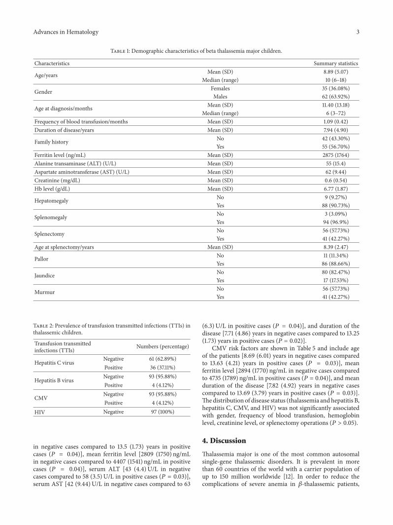
A total of 36 patients (37.11%) were found to be anti-HCVantibody- (anti-HCV-) positive (Table 2). All positive caseswere confirmed by HCV-RT-PCR.There were only 4 (4.12%)patients that were found to be positive for HBsAg and anti-CMV IgM. There were no cases that were positive for anti-HIV type 1 or type 2. Three patients had two infectious dis-eases simultaneously (two had HBV and HCV, and one hadHCV and CMV).
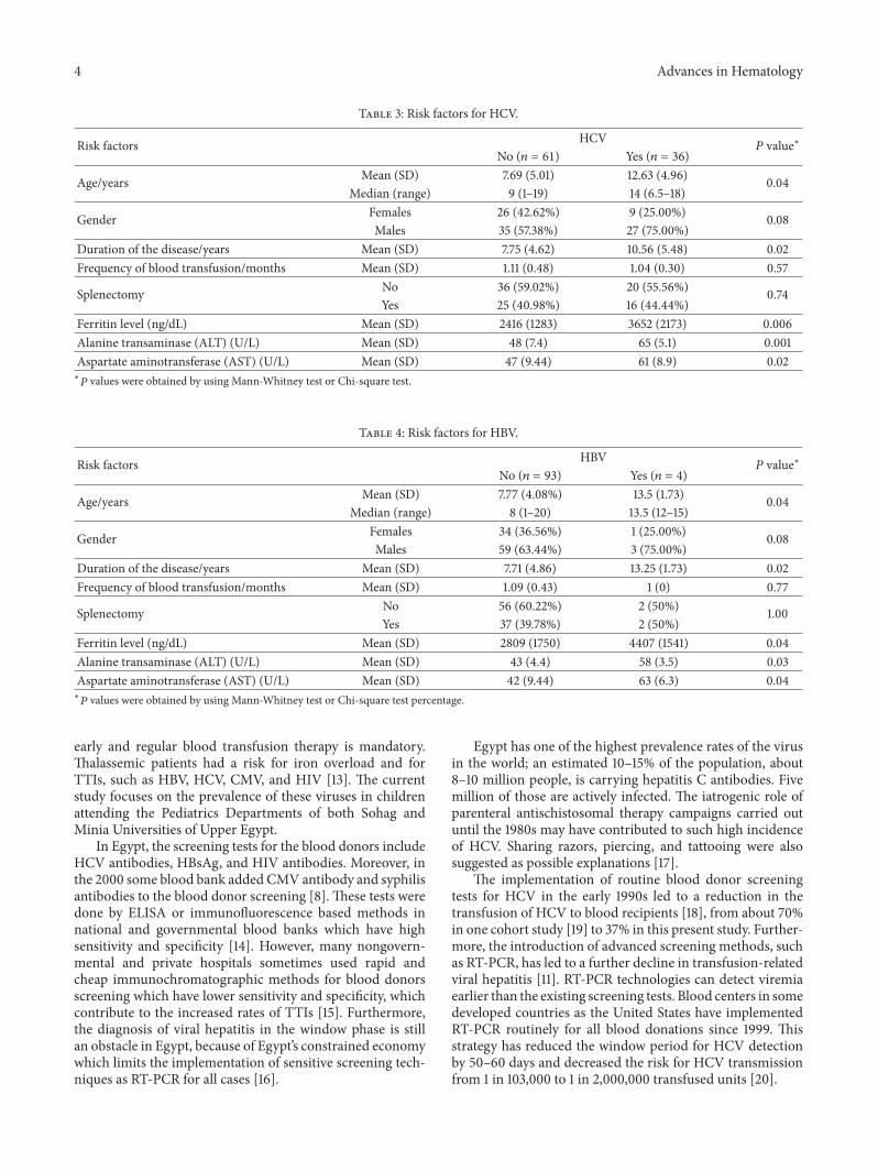
The risk factors for hepatitis C virus are shown in Table 3and include age of the patients [7.69 (5.01) years in negativecases compared to 12.63 (4.96) years in positive cases (𝑃 =0.04)], mean ferritin level [2416 (1283) ng/mL in negativecases compared to 3652 (2173) ng/mL in positive cases (𝑃 =0.006)], serum ALT [48 (7.4)U/L in negative cases comparedto 65 (5.1) U/L in positive cases (𝑃 = 0.001)], serum AST [47(9.44)U/L in negative cases compared to 61 (8.9) U/L in posi-tive cases (𝑃 = 0.02)], and duration of the disease [7.75 (4.62)years in negative cases compared to 10.56 (5.48) years forpositive cases (𝑃 = 0.02)].With a total of 97 patients includedin the study there were 6 patients (6.18%) who hadmanifesta-tions of liver cirrhosis, two were HBV and HCV positive, twowereHCVpositive, and twowere serologically free of viruses.
For hepatitis B virus infection, risk factors are shown inTable 4 and included the age of patients [7.77 (4.08%) years
Advances in Hematology 3
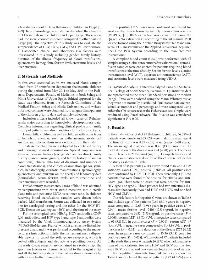
Table 1: Demographic characteristics of beta thalassemia major children.
Characteristics Summary statistics
Age/years Mean (SD) 8.89 (5.07)Median (range) 10 (6–18)
Gender Females 35 (36.08%)Males 62 (63.92%)
Age at diagnosis/months Mean (SD) 11.40 (13.18)Median (range) 6 (3–72)
Frequency of blood transfusion/months Mean (SD) 1.09 (0.42)Duration of disease/years Mean (SD) 7.94 (4.90)
Family history No 42 (43.30%)Yes 55 (56.70%)
Ferritin level (ng/mL) Mean (SD) 2875 (1764)Alanine transaminase (ALT) (U/L) Mean (SD) 55 (15.4)Aspartate aminotransferase (AST) (U/L) Mean (SD) 62 (9.44)Creatinine (mg/dL) Mean (SD) 0.6 (0.54)Hb level (g/dL) Mean (SD) 6.77 (1.87)
Hepatomegaly No 9 (9.27%)Yes 88 (90.73%)
Splenomegaly No 3 (3.09%)Yes 94 (96.9%)
Splenectomy No 56 (57.73%)Yes 41 (42.27%)
Age at splenectomy/years Mean (SD) 8.39 (2.47)
Pallor No 11 (11.34%)Yes 86 (88.66%)
Jaundice No 80 (82.47%)Yes 17 (17.53%)
Murmur No 56 (57.73%)Yes 41 (42.27%)
Table 2: Prevalence of transfusion transmitted infections (TTIs) inthalassemic children.
Transfusion transmittedinfections (TTIs) Numbers (percentage)
Hepatitis C virus Negative 61 (62.89%)Positive 36 (37.11%)
Hepatitis B virus Negative 93 (95.88%)Positive 4 (4.12%)
CMV Negative 93 (95.88%)Positive 4 (4.12%)
HIV Negative 97 (100%)
in negative cases compared to 13.5 (1.73) years in positivecases (𝑃 = 0.04)], mean ferritin level [2809 (1750) ng/mLin negative cases compared to 4407 (1541) ng/mL in positivecases (𝑃 = 0.04)], serum ALT [43 (4.4)U/L in negativecases compared to 58 (3.5) U/L in positive cases (𝑃 = 0.03)],serum AST [42 (9.44)U/L in negative cases compared to 63
(6.3) U/L in positive cases (𝑃 = 0.04)], and duration of thedisease [7.71 (4.86) years in negative cases compared to 13.25(1.73) years in positive cases (𝑃 = 0.02)].
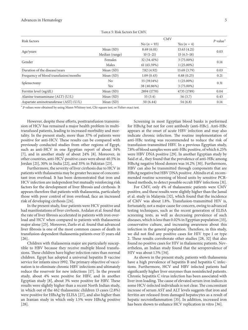
CMV risk factors are shown in Table 5 and include ageof the patients [8.69 (6.01) years in negative cases comparedto 13.63 (4.21) years in positive cases (𝑃 = 0.03)], meanferritin level [2894 (1770) ng/mL in negative cases comparedto 4735 (1789) ng/mL in positive cases (𝑃 = 0.04)], and meanduration of the disease [7.82 (4.92) years in negative casescompared to 13.69 (3.79) years in positive cases (𝑃 = 0.03)].Thedistribution of disease status (thalassemia andhepatitis B,hepatitis C, CMV, and HIV) was not significantly associatedwith gender, frequency of blood transfusion, hemoglobinlevel, creatinine level, or splenectomy operations (𝑃 > 0.05).
4. Discussion
Thalassemia major is one of the most common autosomalsingle-gene thalassemic disorders. It is prevalent in morethan 60 countries of the world with a carrier population ofup to 150 million worldwide [12]. In order to reduce thecomplications of severe anemia in 𝛽-thalassemic patients,
4 Advances in Hematology
Table 3: Risk factors for HCV.
Risk factors HCV𝑃 value∗
No (𝑛 = 61) Yes (𝑛 = 36)
Age/years Mean (SD) 7.69 (5.01) 12.63 (4.96) 0.04Median (range) 9 (1–19) 14 (6.5–18)
Gender Females 26 (42.62%) 9 (25.00%) 0.08Males 35 (57.38%) 27 (75.00%)
Duration of the disease/years Mean (SD) 7.75 (4.62) 10.56 (5.48) 0.02Frequency of blood transfusion/months Mean (SD) 1.11 (0.48) 1.04 (0.30) 0.57
Splenectomy No 36 (59.02%) 20 (55.56%) 0.74Yes 25 (40.98%) 16 (44.44%)
Ferritin level (ng/dL) Mean (SD) 2416 (1283) 3652 (2173) 0.006Alanine transaminase (ALT) (U/L) Mean (SD) 48 (7.4) 65 (5.1) 0.001Aspartate aminotransferase (AST) (U/L) Mean (SD) 47 (9.44) 61 (8.9) 0.02∗
𝑃 values were obtained by using Mann-Whitney test or Chi-square test.
Table 4: Risk factors for HBV.
Risk factors HBV𝑃 value∗
No (𝑛 = 93) Yes (𝑛 = 4)
Age/years Mean (SD) 7.77 (4.08%) 13.5 (1.73) 0.04Median (range) 8 (1–20) 13.5 (12–15)
Gender Females 34 (36.56%) 1 (25.00%) 0.08Males 59 (63.44%) 3 (75.00%)
Duration of the disease/years Mean (SD) 7.71 (4.86) 13.25 (1.73) 0.02Frequency of blood transfusion/months Mean (SD) 1.09 (0.43) 1 (0) 0.77
Splenectomy No 56 (60.22%) 2 (50%) 1.00Yes 37 (39.78%) 2 (50%)
Ferritin level (ng/dL) Mean (SD) 2809 (1750) 4407 (1541) 0.04Alanine transaminase (ALT) (U/L) Mean (SD) 43 (4.4) 58 (3.5) 0.03Aspartate aminotransferase (AST) (U/L) Mean (SD) 42 (9.44) 63 (6.3) 0.04∗
𝑃 values were obtained by using Mann-Whitney test or Chi-square test percentage.
early and regular blood transfusion therapy is mandatory.Thalassemic patients had a risk for iron overload and forTTIs, such as HBV, HCV, CMV, and HIV [13]. The currentstudy focuses on the prevalence of these viruses in childrenattending the Pediatrics Departments of both Sohag andMinia Universities of Upper Egypt.
In Egypt, the screening tests for the blood donors includeHCV antibodies, HBsAg, and HIV antibodies. Moreover, inthe 2000 some blood bank added CMV antibody and syphilisantibodies to the blood donor screening [8]. These tests weredone by ELISA or immunofluorescence based methods innational and governmental blood banks which have highsensitivity and specificity [14]. However, many nongovern-mental and private hospitals sometimes used rapid andcheap immunochromatographic methods for blood donorsscreening which have lower sensitivity and specificity, whichcontribute to the increased rates of TTIs [15]. Furthermore,the diagnosis of viral hepatitis in the window phase is stillan obstacle in Egypt, because of Egypt’s constrained economywhich limits the implementation of sensitive screening tech-niques as RT-PCR for all cases [16].
Egypt has one of the highest prevalence rates of the virusin the world; an estimated 10–15% of the population, about8–10 million people, is carrying hepatitis C antibodies. Fivemillion of those are actively infected. The iatrogenic role ofparenteral antischistosomal therapy campaigns carried outuntil the 1980s may have contributed to such high incidenceof HCV. Sharing razors, piercing, and tattooing were alsosuggested as possible explanations [17].
The implementation of routine blood donor screeningtests for HCV in the early 1990s led to a reduction in thetransfusion of HCV to blood recipients [18], from about 70%in one cohort study [19] to 37% in this present study. Further-more, the introduction of advanced screening methods, suchas RT-PCR, has led to a further decline in transfusion-relatedviral hepatitis [11]. RT-PCR technologies can detect viremiaearlier than the existing screening tests. Blood centers in somedeveloped countries as the United States have implementedRT-PCR routinely for all blood donations since 1999. Thisstrategy has reduced the window period for HCV detectionby 50–60 days and decreased the risk for HCV transmissionfrom 1 in 103,000 to 1 in 2,000,000 transfused units [20].
Advances in Hematology 5
Table 5: Risk factors for CMV.
Risk factors CMV𝑃 value∗
No (𝑛 = 93) Yes (𝑛 = 4)
Age/years Mean (SD) 8.69 (6.01) 13.63 (4.21) 0.03Median (range) 10 (1–21) 15 (6.5–18)
Gender Females 32 (34.41%) 3 (75.00%) 0.14Males 61 (65.59%) 1 (25.00%)
Duration of the disease/years Mean (SD) 7.82 (4.92) 13.69 (3.79) 0.03Frequency of blood transfusion/months Mean (SD) 1.09 (0.43) 0.88 (0.25) 0.21
Splenectomy No 55 (59.14%) 1 (25.00%) 0.31Yes 38 (40.86%) 3 (75.00%)
Ferritin level (ng/dL) Mean (SD) 2894 (1770) 4735 (1789) 0.04Alanine transaminase (ALT) (U/L) Mean (SD) 55 (3.4) 56 (3.7) 0.43Aspartate aminotransferase (AST) (U/L) Mean (SD) 50 (6.44) 54 (6.8) 0.14∗
𝑃 values were obtained by using Mann-Whitney test, Chi-square test, or Fisher exact test.
However, despite these efforts, posttransfusion transmis-sion of HCV has remained a major health problem in multi-transfused patients, leading to increased morbidity and mor-tality. In the present study, more than 37% of patients werepositive for anti-HCV. These results can be compared withpreviously conducted studies from other regions of Egypt,such as anti-HCV in one Egyptian report of about 34%[7], and in another study of about 24% [8]. Moreover, inother countries, anti-HCV-positive cases were about 40.5% inJordan [21], 30% in India [22], and 35% in Pakistan [23].
Furthermore, the severity of liver cirrhosis due toHCV inpatients with thalassemiamay be greater because of concomi-tant iron overload. It has been demonstrated that iron andHCV infection are independent but mutually reinforcing riskfactors for the development of liver fibrosis and cirrhosis. Itappears therefore that patients with thalassemia, particularlythose with poor control of iron overload, face an increasedrisk of developing cirrhosis [24].
In the present study, four patients were HCV positive andhadmanifestations of liver cirrhosis. Ardalan et al. found thatthe rate of liver fibrosis accelerated in patients with iron over-load and HCV when compared to patients with thalassemiamajor alone [25]. Moreover, Zurlo et al. found that advancedliver fibrosis is one of the most common causes of death intransfusion-dependent thalassemia patients over 15 years old[26].
Children with thalassemia major are particularly suscep-tible to HBV because they receive multiple blood transfu-sions.These children have higher infection rates than normalchildren. Egypt has adopted a universal hepatitis B vaccineservice for infants since 1992.The primary objective of vacci-nation is to eliminate chronic HBV infections and ultimatelyreduce the reservoir for new infections [17]. In the presentstudy, about 4% were positive for HBV, and in anotherEgyptian study [8], about 3% were positive for HBV. Theseresults were slightly higher than a recent North Indian study,in which out of the 462 thalassemic children 13 cases (2.8%)were positive for HBsAg by ELISA [27], and also higher thanan Iranian study in which only 1.5% were HBsAg positive[28].
Screening in most Egyptian blood banks is performedfor HBsAg but not for core antibody (anti-HBc). Anti-HBcappears at the onset of acute HBV infection and may alsoindicate chronic infection. The routine implementation ofanti-HBc testing was recommended to reduce the risk oftransfusion-transmitted HBV. In a previous Egyptian study,7.8% of blood samples were anti-HBc positive, of which 6.25%were HBV DNA positive [29]. In another Egyptian study bySaid et al., they found that the prevalence of anti-HBc amongHBsAg negative blood donors was 14.2% [30]. Furthermore,HBV can also be transmitted through components that areHBsAgnegative butHBVDNApositive. Altindis et al. recom-mended routine screening of blood units by sensitive PCR-basedmethods, to detect possible occult HBV infections [31].
For CMV, only 4% of thalassemic patients were CMVpositive, and these results were slightly higher than the Jamalet al. study in Malaysia [32], which found that the incidenceof CMV was about 1.8%. Transfusion-transmitted HIV is,fortunately, not amajor cause for concern, owing to advancedtesting techniques, such as the recent generation of ELISAscreening tests, as well as decreasing prevalence of suchdiseases, which is less than 0.02% inEgyptian population [33],conservative culture, and increasing awareness about thisinfection in the general population. Therefore, in this study,we did not find any positive cases for HIV type 1 or type2. These results corroborate other studies [28, 32] that alsofound no positive cases for HIV in thalassemic patients. Nev-ertheless, an Indian study found that the seroprevalence ofHIV was about 1.5% [34].
As shown in the present study, patients with thalassemiahave a high prevalence of hepatitis B and hepatitis C infec-tions. Furthermore, HCV and HBV infected patients hadsignificantly higher liver enzymes than noninfected patients.Chronic hepatitis C virus infection has been associated withliver iron loading.The cause of elevated serum iron indices insomeHCV-infected individuals is not clear.The concomitantincrease of serum AST and ALT levels suggests that iron andferritin are released from damaged hepatocytes as a result ofhepatic necroinflammation [35]. In addition, increased ironhas been shown to enhance HCV replication in vitro [36].
6 Advances in Hematology
In the current study, splenomegaly was observed in 96.9%of patients, whereas 90.73% of patients had hepatomegaly.The distribution of HCV, HBV, CMV, and HIV was studiedby considering different clinical parameters related to tha-lassemia. In this study, we found that older age of the patients,longer duration of the disease in years, elevated liver enzymes(U/L), and increasing ferritin level (ng/mL) are associatedwith higher seroprevalence of TTIs. Similar findings werereported by other studies [37–39]. Though the standardizedblood screening procedures were outlined in the 1990s forblood-related products and were subsequently implementedin various countries, higher prevalence of TTIs, especiallyHCV, among thalassemic patients requires greater attentionfrom a public health perspective. Screening of blood donorsby advanced ELISA screening tests and RT-PCR could detectHCV in early stages of diseases andwill provide better oppor-tunities for risk assessment.
5. Conclusion
A total of 97 homozygous 𝛽-thalassemia patients were inclu-ded in this study.The seroprevalence of HCVwas the highestTTI in thalassemic children, with a lower percentage forHBVand CMV cases but there were noHIV cases.These seroprev-alence-positive cases were significantly associated with olderage, longer duration of diseases, increased liver enzymes, andhigher ferritin levels. Recent solid-blood screening programsincluding advanced ELISA screening tests, liver enzymes, andRT-PCRwill hopefully reduce the risk of TTIs associatedwithblood transfusions.
Conflict of Interests
The authors had no conflict of interests.
Acknowledgment
The authors thank proof-reading-services.com for linguisticediting.
References
[1] S. L. Thein, “Genetic modifiers of 𝛽-thalassemia,” Haematolog-ica, vol. 90, no. 5, pp. 649–660, 2005.
[2] S. H. Ansari, T. S. Shamsi, M. Ashraf et al., “Molecular epidemi-ology of 𝛽-thalassemia in Pakistan: far reaching implications,”International Journal of Molecular Epidemiology and Genetics,vol. 2, no. 4, pp. 403–408, 2011.
[3] A. El-Beshlawy, N. Kaddah, A. Moustafa, G. Mouktar, and I.Youssry, “Screening for 𝛽-thalassaemia carriers in Egypt: sig-nificance of the osmotic fragility test,” Eastern MediterraneanHealth Journal, vol. 13, no. 4, pp. 780–786, 2007.
[4] S. L. Schrier, “Pathophysiology of thalassemia,”Current Opinionin Hematology, vol. 9, no. 2, pp. 123–126, 2002.
[5] C. Skarmoutsou, I. Papassotiriou, J. Traeger-Synodinos et al.,“Erythroid bone marrow activity and red cell hemoglobiniza-tion in iron-sufficient 𝛽-thalassemia heterozygotes as reflectedby soluble transferrin receptor and reticulocyte hemoglobin
content. Correlation with genotypes andHBA2 levels,”Haema-tologica, vol. 88, no. 6, pp. 631–636, 2003.
[6] S. Aziz, J. Rajper, and W. Noorulain, “Treatment outcome ofHCV infected paediatric patients and young adults at Karachi,Pakistan,” Journal of Ayub Medical College, Abbottabad, vol. 24,no. 3-4, pp. 56–58, 2012.
[7] F. Said, A. E. Beshlawy, M. Hamdy et al., “Intrafamilial trans-mission of hepatitis C infection in Egyptian multitransfusedthalassemia patients,” Journal of Tropical Pediatrics, vol. 59, no.4, pp. 309–313, 2013.
[8] E. Hussein, “Evaluation of infectious disease markers in multi-transfused Egyptian children with thalassemia,” Annals ofClinical and Laboratory Science, vol. 44, no. 1, pp. 62–66, 2014.
[9] A. A. Adly and F. S. Ebeid, “Cultural preferences and limitedpublic resources influence the spectrum of thalassemia inEgypt,” Journal of Pediatric Hematology/Oncology, vol. 37, no. 4,pp. 281–284, 2015.
[10] B. S. Buckner andE. B. Buckner, “Post-revolutionEgypt: theRoyadaptation model in community,” Nursing Science Quarterly,vol. 28, no. 4, pp. 300–307, 2015.
[11] C. Velati, L. Romano, L. Fomiatti et al., “Impact of nucleicacid testing for hepatitis B virus, hepatitis C virus, and humanimmunodeficiency virus on the safety of blood supply in Italy: a6-year survey,” Transfusion, vol. 48, no. 10, pp. 2205–2213, 2008.
[12] L. A.Quratul-Ain,M.Hassan, S.M. Rana, and F. Jabeen, “Preva-lence of 𝛽-thalassemic patients associated with consanguinityand anti-HCV-antibody positivity—a cross sectional study,”Pakistan Journal of Zoology, vol. 43, no. 1, pp. 29–36, 2011.
[13] A.H.Mollah,N.Nahar, A. Siddique, K. S. Anwar, T.Hassan, andG. Azam, “Common transfusion-transmitted infectious agentsamong thalassaemic children in Bangladesh,” Journal of HealthPopulation and Nutrition, vol. 21, no. 1, pp. 67–71, 2003.
[14] J. M. Barrera, B. Francis, G. Ercilla et al., “Improved detectionof anti-HCV in post-transfusion hepatitis by a third-generationELISA,” Vox Sanguinis, vol. 68, no. 1, pp. 15–18, 1995.
[15] A. A. Adeyemi, O. A. Omolade, and R. R. Raheem-Ademola,“Immunochromatographic testing method for hepatitis B, C inblood donors,” Journal of Antivirals & Antiretrovirals, vol. 3, no.10, pp. 4172–4175, 2013.
[16] H. Zaghloul and M. El-Shahat, “Recombinase polymeraseamplification as a promising tool in hepatitis C virus diagnosis,”World Journal of Hepatology, vol. 6, no. 12, pp. 916–922, 2014.
[17] E. Hussein, “Blood donor recruitment strategies and theirimpact on blood safety in Egypt,” Transfusion and ApheresisScience, vol. 50, no. 1, pp. 63–67, 2014.
[18] J. G. Donahue, A.Munoz, P.M.Ness et al., “The declining risk ofpost-transfusion hepatitis C virus infection,” The New EnglandJournal of Medicine, vol. 327, no. 6, pp. 369–373, 1992.
[19] K. Al-Naamani, I. Al-Zakwani, S. Al-Sinani, F. Wasim, and S.Daar, “Prevalence of hepatitis C among multi-transfused tha-lassaemic patients in Oman, single centre experience,” SultanQaboos University Medical Journal, vol. 15, no. 1, pp. e46–e51,2015.
[20] S. L. Stramer, S. A. Glynn, S. H. Kleinman et al., “Nationalheart, lung, and blood institute nucleic acid test study group.Detection of HIV-1 and HCV infections among antibodynegative blood donors by nucleic acid-amplification testing,”The New England Journal of Medicine, vol. 351, no. 8, pp. 760–768, 2004.
[21] M. Al-Sheyyab, A. Batieha, andM. El-Khateeb, “The prevalenceof hepatitis B, hepatitis C and human immune deficiency
Advances in Hematology 7
virus markers in multi-transfused patients,” Journal of TropicalPediatrics, vol. 47, no. 4, pp. 239–242, 2001.
[22] M. Irshad and S. Peter, “Spectrum of viral hepatitis in tha-lassemic children receivingmultiple blood transfusions,” IndianJournal of Gastroenterology, vol. 21, no. 5, pp. 183–184, 2002.
[23] M. Rahman and Y. Lodhi, “Prospects and future of conservativemanagement of beta thalassemia major in a developing coun-try,” Pakistan Journal of Medical Sciences, vol. 20, no. 2, pp. 105–112, 2004.
[24] E. Angelucci, P. Muretto, A. Nicolucci et al., “Effects of ironoverload and hepatitis C virus positivity in determining pro-gression of liver fibrosis in thalassemia following bone marrowtransplantation,” Blood, vol. 100, no. 1, pp. 17–21, 2002.
[25] F. A. Ardalan, M. R. F. Osquei, M. N. Toosi, and G. Irvanloo,“Synergic effect of chronic hepatitis C infection and betathalassemia major with marked hepatic iron overload on liverfibrosis: a retrospective cross-sectional study,” BMC Gastroen-terology, vol. 4, article 17, 2004.
[26] M. Zurlo, P. De Stefano, C. Borgna-Pignatti et al., “Survival andcauses of death in thalassaemia major,”The Lancet, vol. 334, no.8653, pp. 27–30, 1989.
[27] R.N.Makroo, J. S. Arora,M.Chowdhry,A. Bhatia,U.K.Thakur,and A. Minimol, “Red cell alloimmunization and infectiousmarker status (human immunodeficiency virus, hepatitis Bvirus and hepatitis C virus) in multiply transfused thalassemiapatients of North India,” Indian Journal of Pathology andMicrobiology, vol. 56, no. 4, pp. 378–383, 2013.
[28] S. Mirmomen, S.-M. Alavian, B. Hajarizadeh et al., “Epidemiol-ogy of hepatitis B, hepatitis C, and human immunodeficiencyvirus infections in patients with beta-thalassemia in Iran: amulticenter study,” Archives of Iranian Medicine, vol. 9, no. 4,pp. 319–323, 2006.
[29] W. Antar, M. H. El-Shokry, W. A. Abd El Hamid, and M. F.Helmy, “Significance of detecting anti-HBc among Egyptianmale blood donors negative for HBsAg,” Transfusion Medicine,vol. 20, no. 6, pp. 409–413, 2010.
[30] Z. N. Said, M. H. El Sayed, I. I. Salama et al., “Occult hepatitisB virus infection among Egyptian blood donors,”World Journalof Hepatology, vol. 5, no. 2, pp. 64–73, 2013.
[31] M. Altindis, I. Uslan, Z. Cetinkaya et al., “Investigation ofhemodialysis patients in terms of the presence of occult hep-atitis B,”Mikrobiyoloji Bulteni, vol. 41, no. 2, pp. 227–233, 2007.
[32] R. Jamal, G. Fadzillah, S. Z. Zulkifli, and M. Yasmin, “Sero-prevalence of hepatitis B, hepatitis C, CMVandHIV inmultiplytransfused thalassemia patients: results from a thalassemia daycare center in Malaysia,” Southeast Asian Journal of TropicalMedicine and Public Health, vol. 29, no. 4, pp. 792–804, 1998.
[33] D. Oraby, “Harm reduction approach in Egypt: the insight ofinjecting drug users,”Harm Reduction Journal, vol. 10, article 17,2013.
[34] S. Manisha, K. Sanjeev, N. Seema, C. Dilip, and D. Rashmi, “Across-sectional study on burden of hepatitis C, hepatitis B, HIVand syphilis in multi-transfused thalassemia major patientsreporting to a Government Hospital of Central India,” IndianJournal of Hematology and Blood Transfusion, vol. 31, no. 3, pp.367–373, 2015.
[35] J. E. Nelson and K. V. Kowdley, “Iron and hepatitis C,” CurrentHepatitis Reports, vol. 3, no. 4, pp. 140–147, 2004.
[36] S. Kakizaki, H. Takagi, N. Horiguchi et al., “Iron enhanceshepatitis C virus replication in cultured human hepatocytes,”Liver, vol. 20, no. 2, pp. 125–128, 2000.
[37] H. Hussain, R. Iqbal, M. H. Khan et al., “Prevalence of hepatitisC in 𝛽 thalassaemia major,” Gomal Journal of Medical Sciences,vol. 6, no. 2, pp. 87–90, 2008.
[38] M. A. Shah, M. T. Khan, Z. Ullah, and Y. Ashfaq, “Prevalenceof hepatitis B and C virus infection in multiple transfusedthalassemic patients in NorthWest Frontier Province,” PakistanJournal of Medical Sciences, vol. 21, no. 4, pp. 281–283, 2005.
[39] M. R. Uddin, M. Rana, M. Islam et al., “Seroprevalence ofhepatitis C virus in thalassemic patients,” Journal of DhakaMedical College, vol. 18, no. 2, pp. 115–119, 2009.

Submit your manuscripts athttp://www.hindawi.com
Stem CellsInternational
Hindawi Publishing Corporationhttp://www.hindawi.com Volume 2014
Hindawi Publishing Corporationhttp://www.hindawi.com Volume 2014
MEDIATORSINFLAMMATION
of
Hindawi Publishing Corporationhttp://www.hindawi.com Volume 2014
Behavioural Neurology
EndocrinologyInternational Journal of
Hindawi Publishing Corporationhttp://www.hindawi.com Volume 2014
Hindawi Publishing Corporationhttp://www.hindawi.com Volume 2014
Disease Markers
Hindawi Publishing Corporationhttp://www.hindawi.com Volume 2014
BioMed Research International
OncologyJournal of
Hindawi Publishing Corporationhttp://www.hindawi.com Volume 2014
Hindawi Publishing Corporationhttp://www.hindawi.com Volume 2014
Oxidative Medicine and Cellular Longevity
Hindawi Publishing Corporationhttp://www.hindawi.com Volume 2014
PPAR Research
The Scientific World JournalHindawi Publishing Corporation http://www.hindawi.com Volume 2014
Immunology ResearchHindawi Publishing Corporationhttp://www.hindawi.com Volume 2014
Journal of
ObesityJournal of
Hindawi Publishing Corporationhttp://www.hindawi.com Volume 2014
Hindawi Publishing Corporationhttp://www.hindawi.com Volume 2014
Computational and Mathematical Methods in Medicine
OphthalmologyJournal of
Hindawi Publishing Corporationhttp://www.hindawi.com Volume 2014
Diabetes ResearchJournal of
Hindawi Publishing Corporationhttp://www.hindawi.com Volume 2014
Hindawi Publishing Corporationhttp://www.hindawi.com Volume 2014
Research and TreatmentAIDS
Hindawi Publishing Corporationhttp://www.hindawi.com Volume 2014
Gastroenterology Research and Practice
Hindawi Publishing Corporationhttp://www.hindawi.com Volume 2014
Parkinson’s Disease
Evidence-Based Complementary and Alternative Medicine
Volume 2014Hindawi Publishing Corporationhttp://www.hindawi.com