Embed Size (px)
Citation preview

Research ArticleThyroid Cytopathology Reporting by the Bethesda System:A Two-Year Prospective Study in an Academic Institution
Payal Mehra and Anand Kumar Verma
Department Of Pathology, Employees State Insurance (ESI) Postgraduate Institute of Medical Sciences and Research andESI Model Hospital, Basai Darapur, New Delhi 110015, India
Correspondence should be addressed to Anand Kumar Verma; [email protected]
Received 3 August 2014; Revised 16 December 2014; Accepted 24 December 2014
Academic Editor: Marco Volante
Copyright © 2015 P. Mehra and A. K. Verma. This is an open access article distributed under the Creative Commons AttributionLicense, which permits unrestricted use, distribution, and reproduction in any medium, provided the original work is properlycited.
Background. The Bethesda System for Reporting Thyroid Cytopathology (TBSRTC) has attempted to standardize reporting andcytological criteria in aspiration smears. Aims. The objective of this study was to analyze the thyroid cytology smears by TBSRTC,to determine the distribution of diagnostic categories and subcategories, to analyze cytological features, and to correlate thecytopathology with histopathology, wherever surgery was done. Materials and Methods. This was a prospective study of 225 fineneedle aspirations (FNA) of thyroid nodules. All fine needle aspiration cytology (FNAC) diagnoses were classified according tothe features given in the monograph of TBSRTC into nondiagnostic/unsatisfactory (ND/UNS), benign, atypia of undeterminedsignificance/follicular lesion of undetermined significance (AUS/FLUS), follicular neoplasm/suspicious of a follicular neoplasm(FN/SFN), suspicious for malignancy (SFM), and malignant. Cytohistological correlation was done, when surgical material wasavailable. Results. The distribution of various categories from 225 evaluated thyroid nodules was as follows: 7.2% ND/UNS, 80.0%benign, 4.9% AUS/FLUS, 2.2% FN, 3.5% SFM, and 2.2% malignant. Sensitivity, specificity, positive predictive value, and negativepredictive value were calculated. Conclusions. TBSRTC is an excellent reporting system for thyroid FNA. It also provides clearmanagement guidelines to clinicians to go for follow-up FNA or surgery and also the extent of surgery.
1. Introduction
Fine needle aspiration cytology (FNAC) of thyroid occupiesan extremely important role worldwide. This minimallyinvasive and cost-effective technique is extremely useful inidentifying a substantial proportion of thyroid nodules asbenign and reducing unnecessary surgery for patients withbenign disease.
However, terminology of reporting thyroid FNACs hasvariedmarkedly. Various reporting formats of thyroid FNACshave been used varying from two category schemes to six ormore category schemes [1].While some of them tried to diag-nose various lesions using histology-equivalent categories,other formats had categories like equivocal, inconclusive,indeterminate, atypical, suspicious, uncertain, malignancysuspicious, possibly neoplastic, possiblymalignant, and prob-ably malignant to report thyroid aspirates that fell betweenbenign and malignant diagnostic categories [1]. It made itdifficult for clinicians to interpret the reports. To address
terminology and other issues related to thyroid FNACs, theNational Cancer Institute (NCI) hosted “The NCI ThyroidFine Needle Aspiration State of the Science Conference” atBethesda, Maryland. There were six committees which dealtwith different areas regarding thyroid cytology. Commit-tee IV dealt with diagnostic terminology and morphologiccriteria for cytological diagnosis of thyroid lesions. Its rec-ommendations were widely published [2, 3]. Subsequentlya monograph “The Bethesda System for Reporting ThyroidCytopathology” (TBSRTC) which includes definitions, diag-nostic/morphologic criteria, explanatory notes, and a briefmanagement plan for each diagnostic category was published[4]. TBSRTC is a six-category scheme of thyroid cytopathol-ogy reporting (Table 1). Each category has an implied cancerrisk, which ranges from 0% to 3% for the “benign” categoryto virtually 100% for the “malignant” category. It uses threecategories, AUS/FLUS, SFN/Hurthle cell neoplasm, and SFM,to report thyroid aspirates that fall between benign andmalignant. As a function of these risk associations, each
Hindawi Publishing CorporationPathology Research InternationalVolume 2015, Article ID 240505, 11 pageshttp://dx.doi.org/10.1155/2015/240505

2 Pathology Research International
Table 1: The Bethesda System for reporting thyroid cytopathology: recommended diagnostic categories, implied risk of malignancy, andrecommended clinical management.
Diagnostic category Risk of malignancy (%) Usual managementa
(I) Nondiagnostic or unsatisfactory (ND/UNS) Repeat FNA withultrasound guidance
Cyst fluid onlyVirtually acellular specimenOther (obscuring blood, clotting artifact, etc.)(II) Benign 0–3 Clinical follow-upConsistent with a benign follicular nodule (includes adenomatoidnodule,colloid nodule etc.)Consistent with lymphocytic (Hashimoto) thyroiditis in the properclinical contextConsistent with granulomatous (subacute) thyroiditisOther(III) Atypia of undetermined significance or follicular lesion ofundetermined significance (AUS/FLUS) 5–15b Repeat FNA
(IV) Follicular neoplasm or suspicious for follicularneoplasm (FN/SFN) 15–30 Surgical lobectomy
-specify if Hurthle cell (oncocytic) type
(V) Suspicious for malignancy (SFM) 60–75 Near-total thyroidectomyor surgical lobectomyc
Suspicious for papillary carcinomaSuspicious for medullary carcinomaSuspicious for metastatic carcinomaSuspicious for lymphomaOther(VI) Malignant 97–99 Near-total thyroidectomyc
Papillary thyroid carcinomaPoorly differentiated carcinomaMedullary thyroid carcinomaUndifferentiated (anaplastic) carcinomaSquamous cell carcinomaCarcinoma with mixed features (specify)Metastatic carcinomaNon-Hodgkin lymphomaOtheraActual management may depend on other factors (e.g., clinical and sonographic) besides the FNA interpretation.bEstimate extrapolated from histopathologic data from patients with “repeated atypicals” [7, 26].cIn the case of “suspicious for metastatic tumor” or a “malignant” interpretation indicatingmetastatic tumor rather than a primary thyroidmalignancy, surgerymay not be indicated.
category is linked to evidence based clinical managementguidelines.
The objective of the present prospective study, done inan Indian hospital, was to report thyroid cytology smearsby TBSRTC into various diagnostic categories, analyze theircytological features using TBSRTC monograph, conveyingbrief management plan to the clinicians, and correlate withhistology of surgical specimens received.
2. Materials and Methods
This was a prospective study of all successive cases withthyroid swelling referred to the Department of Pathology,
Employees State Insurance (ESI) Postgraduate Institute ofMedical Sciences and Research and ESI Model Hospital,Basaidarapur, New Delhi, for FNAC during the period fromMarch 2010 to February 2012. Relevant clinical history wastaken and examination done. Pre-FNAC requirements asrecommended byCommittee I of theNCI State of the ScienceConference, Bethesda, were followed [5]. All patients weresubjected to FNA sampling under ultrasound guidance byone of the two authors using Zajdela technique 4-5 times ran-domly in different areas [6]. The smears were prepared usingconventional methods and stained with Giemsa and Papan-icolaou stains. The cytological features were evaluated andthe reporting was done according to TBSRTC (Table 1). The

Pathology Research International 3
Table 2: Number of cases in various diagnostic categories and subcategories according to the Bethesda System for Reporting ThyroidCytopathology (TBSRTC).
S.number Cytological categories Subcategories Number of
casesTotal number of cases in
each categoryCyst fluid only 16
1 Nondiagnostic/unsatisfactory(ND/UNS) Virtually acellular specimen 0 16 (7.2)
Other (obscuring blood, clottingartifact, etc.) 0
Consistent with benign follicularnodule (includes adenomatoidnodule, colloid nodule, etc.)
138
2 BenignConsistent with lymphocytic(Hashimoto) thyroiditis in the
proper clinical context36 180 (80)
Consistent with granulomatous(subacute) thyroiditis 02
Other 043 Atypia of undetermined significance/follicular lesion of undetermined significance (AUS/FLUS) 11 (4.9)4 Follicular neoplasm/suspicious for a follicular neoplasm (FN/SFN) 5 (2.2)
Suspicious for papillary carcinoma 06Suspicious for medullary carcinoma 0
5 Suspicious for malignancy (SFM) Suspicious for metastatic carcinoma 0 8 (3.6)Suspicious for lymphoma 01
Other 01Papillary thyroid carcinoma 04
Poorly differentiated carcinoma 0Medullary thyroid carcinoma 01Undifferentiated (anaplastic)
carcinoma 0
6 Malignant Squamous cell carcinoma 0 5 (2.2)Carcinoma with mixed features 0
Metastatic carcinoma 0Non-Hodgkin lymphoma 0
Other 0Total 225 (100)
(1) Figures in parentheses indicate percentages.(2) “Other” subcategory in benign category consisted of cases of chronic nonspecific abscess.(3) “Other” subcategory in suspicious for malignancy category consisted of a case of suspicious for malignancy, not otherwise specified.
morphological criteria given in the monograph of TBSRTCwere used [4].The clinicianswere communicated implied riskofmalignancy and recommended clinical management alongwith the report.Histopathological specimens, wherever avail-able, were processed as per standard methods. Sensitivity,specificity, positive predictive value, and negative predictivevalue were calculated using histopathology diagnosis as goldstandard. For calculating statistical parameters ND/UNS andAUS/FLUS cases were excluded as nondefinitive diagnosisand categories “SFM” and “malignant” were put together. Allthe parameters were calculated either excluding FN/SFN orincluding it with either benign or malignant diagnosis tohighlight the effect on statistical values.
3. Results
The distribution of 225 cases is shown in Table 2. Benigncategory was the largest (80%) followed byND/UNS category(7.2%). Malignant and SFM categories constituted 2.2% and3.6%, respectively, making a total of 5.7%. AUS/FLUS consti-tuted 4.9% cases, while FN/SFN had 2.2% cases.
In the ND/UNS category, all cases were subcategorizedas cyst fluid only. There was no case in subcategory virtuallyacellular specimen or other (obscuring blood, clotting arti-fact, etc.).
In benign category 76.7% of total cases were consistentwith benign follicular nodule (BFN). It had follicular cells

4 Pathology Research International
(a)
(b) (c)
Figure 1: (a) Benign follicular nodule. Photomicrograph showing monolayer sheets of evenly spaced follicular cells having a honeycomb-like arrangement (arrow) (Smear, Giemsa, 400x magnification). (b) Benign follicular nodule. Photomicrograph showing globular mass ofcolloid with superimposed follicular cells (thick arrow) mixed with monolayer sheet of follicular cells (thin arrow) against the backgroundof colloid and blood (Smear, Giemsa, 400x magnification). (c) Benign follicular nodule. Photomicrograph showing follicular cells arrangedin sheets (honeycomb-like) (thin arrow) mixed with macrophages (thick arrow) against the background of colloid (Smear, Giemsa, 400xmagnification).
which were arranged predominantly in monolayer sheetsor were occasionally in intact, 3-dimensional, variably sizedballs/spheres (Figures 1(a) and 1(b)). Rare microfollicles werepresent. Anisonucleosis was seen in some cases but there wasno significant pleomorphism or nuclear atypia. Cellularitywas low to moderate; low cellularity was seen in 52.2% casesand moderate cellularity in 47.8% cases. High cellularity wasnot seen. Pleomorphismwas present only in 2.9% cases out of138 cases. Hurthle cells were present only in 2.2% cases. Foamcells (macrophages) were present in 21.7% cases (Figure 1(c)).
Background was blood-mixed with colloid in 55.8% casesand was only colloid in 44.2% cases. Fire flare or spindle cellwas not seen in any case of benign follicular nodule in thisstudy.
The subcategory consistent with lymphocytic thyroiditis(LT) had 20% cases in benign category. All specimens weremoderately cellular. The lymphoid population was moderatein amount in 88.9% cases. The lymphoid cells were polymor-phic (Figure 2(a)). Intact lymphoid follicles and lymphohisti-ocytic (Figure 2(b)) aggregates were also seen. Hurthle cells(oncocytes) were present in all cases. Multinucleated giantcells were found in 27.8% cases and epithelioid cells in 11.1%cases.
In subcategory consistent with granulomatous thyroiditis(GT), there were 1.1% cases, which showed hypocellular
smears with clusters of epithelioid histiocytes (Figure 3(a)),that is, granulomas along with many multinucleated giantcells (Figure 3(b)), lymphocytes, macrophages, and scantdegenerated follicular cells. Neutrophils were present in bothcases.
The subcategory “other” included cases of chronicabscess.The pus obtainedwas negative for acid fast bacilli andfungus.
In this study, category AUS/FLUS constituted 4.9%. 81.8%of these prominently showed microfollicles in some butnot all the moderately cellular smears (Figure 4(a)), 9.1%prominently showed microfollicles with sparsely cellularsmear with scant colloid (Figure 4(b)), and 9.1% showedpredominantly benign appearing smear with focal featuresof papillary thyroid carcinoma (PTC) including nucleargrooves, enlarged nuclei with pale chromatin, and alterationsin nuclear contour and shape.
There were 2.2% cases in category FN/SFN (Figure 5).There was no case of FN, Hurthle cell type.
In the category SFM, 75% were suspicious for papillarycarcinoma and 12.5% were suspected for lymphoma and12.5% were SFM, not otherwise specified (SFM, NOS). 66.6%cases of suspicious for papillary carcinoma were of PatternA (patchy nuclear change), 16.7% cases were of Pattern B(incomplete nuclear change) and 16.7% cases were of Pattern

Pathology Research International 5
(a) (b)
Figure 2: (a) Lymphocytic (Hashimoto) thyroiditis. Photomicrograph showing polymorphous lymphoid population (Smear, Giemsa,400x magnification). (b) Lymphocytic (Hashimoto) thyroiditis. Photomicrograph showing lymphohistiocytic aggregates in lymphocytic(Hashimoto) thyroiditis (Smear, Giemsa, 400x magnification).
(a) (b)
Figure 3: (a) Granulomatous (subacute) thyroiditis. Photomicrograph showing clusters of epithelioid histiocytes (thick arrow) mixedwith benign follicular cells (thin arrow) (Smear, Giemsa, 400x magnification). (b) Granulomatous (subacute) thyroiditis. Photomicrographshowing many multinucleated giant cells against the background of colloid and blood (Smear, Giemsa, 400x magnification).
(a) (b)
Figure 4: (a) Atypia of undetermined significance. Photomicrograph showing prominent microfollicles in a moderately cellular specimen(Smear, Giemsa, 400x magnification). (b) Atypia of undetermined significance. Photomicrograph showing sparsely cellular specimen with apredominance of microfollicles (Smear, Giemsa, 400x magnification).
D (cystic degeneration). There was no case of Pattern C(sparsely cellular specimen). 1 (12.5%) case of subcategorysuspicious for lymphoma cytologically showed cellular smearcomposed of numerous monomorphic small to intermediatesized lymphoid cells (Figure 6). In the subcategory SFM,NOS, there was only one case showing cytological featuressuggestive of malignancy, but they were not enough tocategorize the type of malignancy.
Category malignant included, 5(2.2%) cases. The maxi-mum number of cases 4 (80%) were of PTC (Figures 7(a)–7(e)) and 1 (20%) case ofmedullary thyroid carcinoma (MTC)(Figures 8(a) and 8(b)).
Out of 225 cases that were cytologically studied, histo-pathological specimens of 40 (17.8%) cases were received andstudied (Table 3).
Surgical specimens of 2 cases, 23 cases, and 1 case,respectively, for categories ND/UNS, benign, and AUS/FLUSamounting to 12.5%, 12.8%, and 9% of cases aspirated werereceived.
Of 18 cases of FN/SFN, SFM, and malignant category,only 15 were surgically resected. Out of 4 cases of FN/SFN,3 were benign (follicular adenoma) and 1 was malignant(PTC). Histopathology was received for 6 cases of SFM. Fiveof them were reported cytologically as being suspicious forpapillary carcinoma. Histopathologically, 3 of them turnedout to be papillary thyroid carcinoma, but 2were lymphocytic(Hashimoto) thyroiditis.
In total, 27 out of 40, that is, 67.5%, cases in this studywith a subsequent tissue diagnosis had a definitive cytologicdiagnosis of being benign or malignant (Table 4). Twenty

6 Pathology Research International
Figure 5: Follicular neoplasm/suspicious for a follicular neoplasm.Photomicrograph showing a highly cellular aspirate composed ofuniform follicular cells arranged in crowded clusters and microfol-licles (Smear, Giemsa, 400x magnification).
Figure 6: Suspicious for lymphoma. Photomicrograph showing ahemodiluted sample comprising exclusively lymphoid cells (Smear,Giemsa, 400x magnification).
cases were benign by both cytopathology and histopathol-ogy. Four cases were malignant by both cytopathology andhistopathology. None of the cases with a malignant diagnosison cytology proved to be benign on biopsy and 3 cases outof 23 that were benign on cytology proved to be malignantlesion on examination of tissue specimen.
The results of various statistical parameters are summa-rized in Table 5.
If FN/SFN is included in malignant group, the sensitivityincreases but the specificity decreases. There is markeddecrease in positive predictive value also.
4. Discussion
This paper shows the two-year experience in reporting thy-roid aspirations byTBSRTC in an Indian academic institutionas well as response of clinicians to the brief managementplan suggested. TBSRTC does not recommend surgery forND/UNS, benign and AUS/FLUS categories. In the FN/SFN,SFM, and malignant categories, we expected excision ofnodules or partial/complete thyroidectomy in all cases as perTBSRTC recommendations.
The present study had 16 (7.2%) cases in ND/UNScategory. Other recent studies had 1.2% to 16.4% cases in thisgroup [7–14]. The guidelines for this category are very clearin TBSRTC. All of them were advised to be reaspirated aftera minimum period of 3 months. The 3-month interval wasrecommended to prevent false positive interpretations due toreactive or reparative changes, as recommended by Commit-tee VI (Post-FNA Technique and Treatment Options) [15].The number of cases in this category is dependent on theaspirator’s experience.The recommendations of Committee I
on indications of thyroid FNA and pre-FNA and CommitteeII on training and credentialing are likely to bring down thenumber in this category in future studies [5, 16]. TBSRTCdoes not provide the implied risk of malignancy for thiscategory. However a recent study found a rate of 8.9% inND/UNS category [10]. Two histopathological specimenswere received and bothwere nodular goiter. Clinicianwas notcomfortable with the term ND/UNS and was not willing towait for 3 months, thus preferred to go for surgery.
The benign category had 180 cases (80%) with BFN beingthe predominant group followed by LT.The “benign” categoryhad a range of 34% to 87.5% in recent studies [7–14]. However,only one study had a percentage less than 50 and this was duea high incidence of AUS/FLUS and FN/SFN. The diagnosticcriteria of all the subcategories are well characterized inTBSRTC monograph. However, TBSRTC recommendationon diagnostic terminology andmorphologic criteria does notmention giant cells and epithelioid cells in LT previouslydescribed in the literature [17, 18]. 23 histopathological spec-imens from category diagnosed as “benign” were received.All of them were operated because of cosmetic reasonsor pressure symptoms. 18 were nodular goiter, 2 follicularadenoma, and 3 PTC. The cytological appearance of nodulargoiter can overlap with those of follicular adenoma andcytological criteria alone cannot reliably distinguish betweenthe two in certain cases [19]. These 2 cases had on cytologyabundant colloid in addition to follicular cells and hencewerediagnosed as BFN. There were 3 cases of PTC which wereincidental findings in thyroid specimen and were less than1 cm in size and thus these nodules were not aspirated. Therewere no lymph nodes in these cases and ultrasound featureswere not suspicious.These “incidentalomas” remain indolentin most cases as implied by 30% prevalence in a study byHarach et al. in 1985 in an autopsy study [20]. One study doesnot consider incidental papillary carcinoma as malignant foranalysis of their data [10].
The classification of “indeterminate” lesions (those notclearly benign or malignant) in thyroid cytopathology haslong been a source of confusion for both pathologists andclinicians. The general category AUS/FLUS is reserved forspecimens that contained cells (follicular, lymphoid, or other)with architectural and/or nuclear atypia that is not sufficientto be classified as suspicious for a follicular neoplasm orsuspicious for malignancy. The atypia is more marked thancan be ascribed confidently to benign changes.
We had 11 cases in group AUS/FLUS. An AUS result hasbeen reported in 3.2–29% of thyroid cases [7–14]. TBSRTCsuggests that the frequency of AUS interpretations should bein the range of approximately 7% of all thyroid FNA inter-pretations. This is a category of last resort and should not beused indiscriminately. Not much data exists in the literatureto support the recommendation that the category shouldnot exceed 7% of all thyroid categories [21]. The incidencealso varies with experience and training of cytopathologists.The recommended management for an initial AUS/FLUSinterpretation is the clinical correlation and, for most cases, arepeat FNA at an appropriate interval. A repeat FNA usuallyresults in a more definitive interpretation; only about 20–25% of nodules are repeatedly AUS. One case of AUS/FLUS
Pathology Research International 7
(a) (b)
(c) (d) (e)
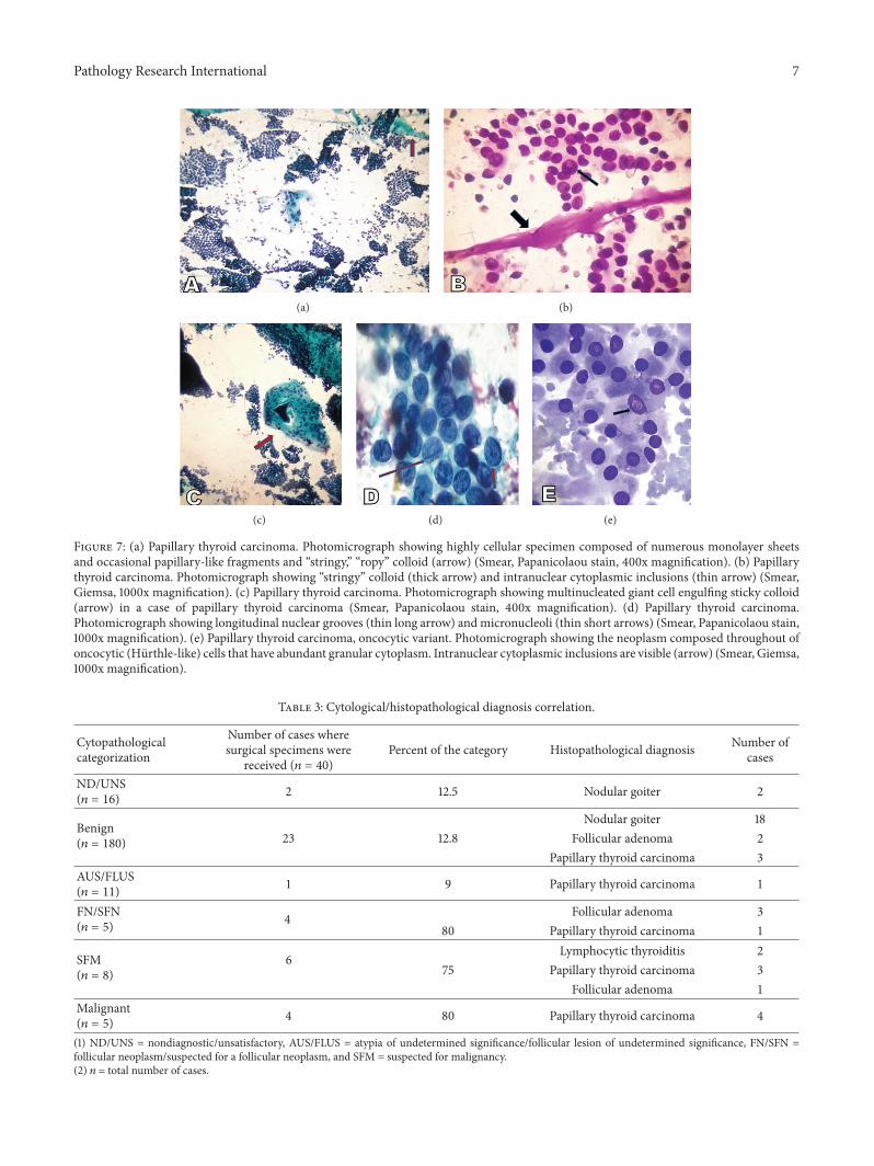
Figure 7: (a) Papillary thyroid carcinoma. Photomicrograph showing highly cellular specimen composed of numerous monolayer sheetsand occasional papillary-like fragments and “stringy,” “ropy” colloid (arrow) (Smear, Papanicolaou stain, 400x magnification). (b) Papillarythyroid carcinoma. Photomicrograph showing “stringy” colloid (thick arrow) and intranuclear cytoplasmic inclusions (thin arrow) (Smear,Giemsa, 1000x magnification). (c) Papillary thyroid carcinoma. Photomicrograph showing multinucleated giant cell engulfing sticky colloid(arrow) in a case of papillary thyroid carcinoma (Smear, Papanicolaou stain, 400x magnification). (d) Papillary thyroid carcinoma.Photomicrograph showing longitudinal nuclear grooves (thin long arrow) andmicronucleoli (thin short arrows) (Smear, Papanicolaou stain,1000x magnification). (e) Papillary thyroid carcinoma, oncocytic variant. Photomicrograph showing the neoplasm composed throughout ofoncocytic (Hurthle-like) cells that have abundant granular cytoplasm. Intranuclear cytoplasmic inclusions are visible (arrow) (Smear, Giemsa,1000x magnification).
Table 3: Cytological/histopathological diagnosis correlation.
Cytopathologicalcategorization
Number of cases wheresurgical specimens were
received (𝑛 = 40)Percent of the category Histopathological diagnosis Number of
cases
ND/UNS(𝑛 = 16) 2 12.5 Nodular goiter 2
Benign(𝑛 = 180)
Nodular goiter 1823 12.8 Follicular adenoma 2
Papillary thyroid carcinoma 3AUS/FLUS(𝑛 = 11) 1 9 Papillary thyroid carcinoma 1
FN/SFN(𝑛 = 5) 4 Follicular adenoma 3
80 Papillary thyroid carcinoma 1
SFM(𝑛 = 8)
6Lymphocytic thyroiditis 2
75 Papillary thyroid carcinoma 3Follicular adenoma 1
Malignant(𝑛 = 5) 4 80 Papillary thyroid carcinoma 4
(1) ND/UNS = nondiagnostic/unsatisfactory, AUS/FLUS = atypia of undetermined significance/follicular lesion of undetermined significance, FN/SFN =follicular neoplasm/suspected for a follicular neoplasm, and SFM = suspected for malignancy.(2) 𝑛 = total number of cases.

8 Pathology Research International
(a) (b)
Figure 8: (a) (100x magnification) and (b) (1000x magnification) Medullary thyroid carcinoma. Photomicrographs showing predominantlycohesive, syncytial-like clusters with few isolated plasmacytoid cells (Smear, Giemsa stain).
Table 4: Cytological/histopathological correlation with benign andmalignant cases.
Cytodiagnosis Histologic diagnosisBenign Malignant
ND/UNS (𝑛 = 2) 2 0Benign (𝑛 = 23) 20 3AUS/FLUS (𝑛 = 1) 0 1FN/SFN (𝑛 = 4) 3 1SFM (𝑛 = 6) 3 3Malignant (𝑛 = 4) 0 4(1)ND/UNS=nondiagnostic/unsatisfactory, AUS/FLUS= atypia of undeter-mined significance/follicular lesion of undetermined significance, FN/SFN= follicular neoplasm/suspected for a follicular neoplasm, and SFM =suspected for malignancy.(2) 𝑛 = total number of cases.
on cytology proved to be malignant on histology and wasPTC. On cytology, this case showed predominantly benignappearing smear with focal features of PTC in only one of thesmears and thus was put under this category.
FN/SFN category had 8 cases (2.2%). TBSRTC providesclear guidelines for this category. Recent studies have shown2.2–16.1% cases in this group [7–14]. Four specimens werereceived, 1 of which was follicular variant of papillary carci-noma. Smears of the latter had predominant follicular patternbut the classic nuclear features of PTCwere not present in thecytological smears.
SFM category had 8 cases (3.6%), 6 of which weresuspicious for papillary carcinoma, one was suspicious for
lymphoma, and one was SFM, NOS. Histopathology wasreceived for 6 cases of SFM. It varies from 1.3 to 10% in recentstudies [7–14]. Five of them were reported cytologically assuspicious for papillary carcinoma. Histopathologically 3 ofthem turned out to be papillary thyroid carcinoma, but 2were lymphocytic (Hashimoto) thyroiditis. The latter wereon cytology diagnosed as suspicious for papillary carcinomabecause of high proportion of follicular cells and presenceof intranuclear cytoplasmic inclusions (INCIs) in rare cells.INCIs are not specific for papillary thyroid carcinoma as theymay be seen focally in benign thyroid nodules. Moreover, anincreased incidence of PTC is well known in LT and hencea diagnosis of suspicious for papillary carcinoma was givenso as not to miss out malignancy. SFM, NOS, had very highcellularity with 3D clusters of follicular cells in all the smearsbut could not be typed cytologically. Histologically, it turnedout to be follicular adenoma.
Committee V of the NCIThyroid Fine Needle AspirationState of the Science Conference has provided guidelines forindications of ancillary studies, specific ancillary studies to beperformed, and sample preparation for each study. Immuno-histochemistry panels have been suggested for suspiciousmalignancies which include medullary carcinoma (calci-tonin, thyroglobulin, CEA, and chromogranin), anaplas-tic carcinoma (pan-cytokeratin), and metastatic carcinoma(TTF-1). These are to be done on cell block from FNA,preferably including at least one dedicated pass for the study.For suspicious lymphoma, flow cytometric immunopheno-typing is suggested. Dedicated passes are also needed forstudies to detect genetic alterations such as BRAF muta-tion or RET/PTC chromosomal rearrangements, which are

Pathology Research International 9
Table 5: Statistical parameters when FN/SFN cases are excluded or are included with either benign or malignant cases.
Statistical Parameters FN/SFN cases excluded FN/SFN cases included with benign cases(%)
FN/SFN cases included with malignantcases (%)
Sensitivity 76.92 (46.20–94.69) 73.33 (44.91–92.05) 78.57 (49.21–95.09)Specificity 88.46 (69.82–97.42) 89.66 (72.62–97.69) 81.25 (63.55–92.75)Positive predictive value 76.92 (46.20–94.69) 78.57 (49.21–95.09) 64.71 (38.35–85.70)Negative predictive value 88.46 (69.82–97.42) 86.67 (69.26–96.16) 89.66 (72.62–97.69)Figures in parentheses show 95% confidence interval.
Table 6: Comparison of results of the present study and random studies over the last 30 years.
Study Number Sensitivity % Specificity % PPV % NPV %Al-Sayer et al. [27] 70 86 93 80 96Silverman et al. [28] 309 93 96.5 88.9 96.5Cusick et al. [29] 283 76 58 72 64Altavilla et al. [30] 257 71.4 100 100 94.4Bouvet et al. [31] 78 93.5 75 85.3 88.2Ko et al. [32] 207 78.4 98.2 99 66.3Kessler et al. [33] 170 79 98.5 98.7 76.6Handa et al. [34] 66 97 100 96 100Gupta et al. [35] 75 80 86.6 80 86.6Present study (FN/SFN excluded) 40 76.92 88.46 76.92 88.46Present study (FN/SFN included as benign) 40 73.33 89.66 78.57 86.67Present study (FN/SFN included as malignant) 40 78.57 81.25 64.71 89.66PPV = positive predictive value and NPV = negative predictive value.
very promising for the diagnosis of papillary carcinoma.Immunocytochemistry on cytospin, direct smear, or prefixedmonolayer may also be utilized, but protocols should becarefully validated [22].
The category malignant had a range of 2.9% to 11% inrecent studies [7–14]. The present study had 5 (2.2%) casesin the malignant category. We received 4 specimens fromthe category diagnosed as “malignant” cytologically. All ofthem were diagnosed as PTC both histopathologically andcytologically.
Table 6 shows a comparison of statistical parameters ofour study and other studies over the last 30 years.
The method of data analysis can alter the results of statis-tical parameters. If suspicious lesions are considered positive,the sensitivity increases while the specificity decreases. Ifsuspicious lesions are excluded, then the sensitivity decreasesand the false negative rates increase. For statistical purposewe had put categories “SFM” and “malignant” in one group.“Unsatisfactory” smears were likely to be malignant orbenign and putting them into a single diagnostic categoryautomatically presents a false picture. All the parameterswere calculated either excluding FN/SFN or including it witheither benign or malignant diagnosis to highlight the effecton statistical values.
Articles implementing TBSRTChave started appearing inthe literature [14, 23–25]. TBSRTC is a relatively recent six-category scheme to classify thyroid cytology smears. It needsto be validated by more prospective studies on larger numberof cases with histopathological correlation. There is need forconsensus amongst institutions in various countries to utilize
TBSRTC to facilitate easy sharing of data across the world forsurveys and research.
5. Conclusions
This study is a prospective analysis of reporting of thyroidaspiration smears by TBSRTC using the Bethesda mono-graph. It was found that the monograph is succinctly writtenin an easy-to-read format and has useful color imageswhich help in making the diagnosis. The clinicians are alsobenefitted because of the management plan it suggests. Casesof AUS/FLUS are followed by repeat FNAC, thus reducing theincidence of surgery in our series (1/11). However, the exactincidence of malignancy in this heterogeneous category isdifficult to predict as most of these cases are unlikely to beoperated if the Bethesda recommendations are to be followed.There is a need for a large study with histopathologicalcorrelation for this category.
Conflict of Interests
The authors declare that there is no conflict of interestsregarding the publication of this paper.
Acknowledgments
The authors acknowledge the help provided by Dr. K. S.Charak, former Head of the department of surgery, and Dr.S. Kohli, Head of the department of radiodiagnosis, for thisstudy.

10 Pathology Research International
References
[1] H. H. Wang, “Reporting thyroid fine-needle aspiration: Litera-ture review and a proposal,” Diagnostic Cytopathology, vol. 34,no. 1, pp. 67–76, 2006.
[2] E. S. Cibas and M. A. Sanchez, “The national cancer institutethyroid fine-needle aspiration state-of-the-science conference:inspiration for a uniform terminology linked to managementguidelines,” Cancer Cytopathology, vol. 114, no. 2, pp. 71–73,2008.
[3] Z. W. Baloch, V. A. LiVolsi, S. L. Asa et al., “Diagnosticterminology and morphologic criteria for cytologic diagnosisof thyroid lesions: a synopsis of the national cancer institutethyroid fine-needle aspiration state of the science conference,”Diagnostic Cytopathology, vol. 36, no. 6, pp. 425–437, 2008.
[4] S. Z. Ali and E. S. Cibas, Eds.,The Bethesda System for ReportingThyroid Cytopathology. Definitions, Criteria and ExplanatoryNotes, Springer, New York, NY, USA, 2010.
[5] E. S. Cibas, E. K. Alexander, C. B. Benson et al., “Indicationsfor thyroid FNA and pre-FNA requirements: a synopsis of theNational Cancer Institute thyroid fine-needle aspiration state ofthe science conference,”Diagnostic Cytopathology, vol. 36, no. 6,pp. 390–399, 2008.
[6] A. Zajdela, M. A. de Maublanc, P. Schlienger, and C. Haye,“Cytologic diagnosis of orbital and periorbital palpable tumorsusing fine-needle sampling without aspiration,” DiagnosticCytopathology, vol. 2, no. 1, pp. 17–20, 1986.
[7] J. Yang, V. Schnadig, R. Logrono, and P. G. Wasserman, “Fine-needle aspiration of thyroid nodules: a study of 4703 patientswith histologic and clinical correlations.,” Cancer, vol. 111, no. 5,pp. 306–315, 2007.
[8] R. Nayar and M. Ivanovic, “The indeterminate thyroid fine-needle aspiration: Experience from an academic center usingterminology similar to that proposed in the 2007 nationalcancer institute thyroid fine needle aspiration state of thescience conference,” Cancer Cytopathology, vol. 117, no. 3, pp.195–202, 2009.
[9] C. G. A.Theoharis, K. M. Schofield, L. Hammers, R. Udelsman,and D. C. Chhieng, “The bethesda thyroid fine-needle aspira-tion classification system: year 1 at an academic institution,”Thyroid, vol. 19, no. 11, pp. 1215–1223, 2009.
[10] V. Y. Jo, E. B. Stelow, S. M. Dustin, and K. Z. Hanley, “Malig-nancy risk for fine-needle aspiration of thyroid lesions accord-ing to the Bethesda system for reporting thyroid cytopathology,”The American Journal of Clinical Pathology, vol. 134, no. 3, pp.450–456, 2010.
[11] M. Bonzanini, P. Amadori, L. Morelli et al., “Subclassification ofthe ‘grey zone’ of thyroid cytology; a retrospective descriptivestudy with clinical, cytological, and histological correlation,”Journal ofThyroid Research, vol. 2011, Article ID 251680, 8 pages,2011.
[12] J. T. Broome and C. C. Solorzano, “The impact ofatypia/follicular lesion of undetermined significance onthe rate of malignancy in thyroid fine-needle aspiration:evaluation of the Bethesda system for reporting thyroidcytopathology,” Surgery, vol. 150, no. 6, pp. 1234–1241, 2011.
[13] M. M. Al-Shraim, O. M. Kaood, M. R. Hussein et al., “Assess-ment of malignancy rate in thyroid nodules according to theBethesda systemof fine-needle aspiration: report froma tertiarycenter in the Southwestern region of Saudi Arabia,” SaudiMedical Journal, vol. 33, no. 2, pp. 167–171, 2012.
[14] S. K.Mondal, S. Sinha, B. Basak, D.N. Roy, and S. K. Sinha, “TheBethesda system for reporting thyroid fine needle aspirates: acytologic study with histologic follow-up,” Journal of Cytology,vol. 30, no. 2, pp. 94–99, 2013.
[15] L. J. Layfield, J. Abrams, B. Cochand-Priollet et al., “Post-thyroid FNA testing and treatment options: a synopsis of thenational cancer institute thyroid fine needle aspiration state ofthe science conference,”Diagnostic Cytopathology, vol. 36, no. 6,pp. 442–448, 2008.
[16] B.-M. E. Ljung, J. Langer, E. L. Mazzaferri, Y. C. Oertel, S.A. Wells, and J. Waisman, “Training, credentialing and re-credentialing for the performance of a thyroid FNA: a synopsisof the National Cancer Institute thyroid fine-needle aspirationstate of the science conference,” Diagnostic Cytopathology, vol.36, no. 6, pp. 400–406, 2008.
[17] P. S. Persson, “Cytodiagnosis of thyroiditis. A comparative studyof cytological, histological, immunological and clinical findingsin thyroiditis, particularly in diffuse lymphoid thyroiditis,” ActaMedica Scandinavica, vol. 483, pp. 7–100, 1968.
[18] R. Bhalotra and G. Jayaram, “Overlapping morphology inthyroiditis (Hashimoto’s and subacute) and Grave’s disease,”Cytopathology, vol. 1, no. 6, pp. 371–372, 1990.
[19] S. R. Orell, G. F. Sterrett, and D.Whitaker, “Chapter 6.Thyroid,”in Fine Needle Aspiration Cytology, pp. 125–164, Elsevier, Syd-ney, Australia, 2005.
[20] H. R. Harach, K. O. Franssila, and V.-M. Wasenius, “Occultpapillary carcinoma of the thyroid. A ‘normal’ finding inFinland. A systematic autopsy study,” Cancer, vol. 56, no. 3, pp.531–538, 1985.
[21] L. J. Layfield, M. J. Morton, H. M. Cramer, and S. Hirschowitz,“Implications of the proposed thyroid fine-needle aspirationcategory of follicular lesion of undetermined significance: a five-yearmulti-institutional analysis,”Diagnostic Cytopathology, vol.37, no. 10, pp. 710–714, 2009.
[22] A. C. Filie, S. L. Asa, K. R. Geisinger et al., “Utilization ofancillary studies in thyroid fine needle aspirates: a synopsis ofthe national cancer institute thyroid fine needle aspiration stateof the science conference,”Diagnostic Cytopathology, vol. 36, no.6, pp. 438–441, 2008.
[23] L. Q. Wong and Z. W. Baloch, “Analysis of the Bethesda Systemfor reporting thyroid cytopathology and similar precursor thy-roid cytopathology reporting schemes,” Advances in AnatomicPathology, vol. 19, no. 5, pp. 313–319, 2012.
[24] M. Bongiovanni, A. Spitale, W. C. Faquin, L. Mazzucchelli,and Z. W. Baloch, “The Bethesda system for reporting thyroidcytopathology: a meta-analysis,” Acta Cytologica, vol. 56, no. 4,pp. 333–339, 2012.
[25] B. K. Richmond, B. A. O’Brien, W. Mangano, S. Thompson,and S. Kemper, “The impact of implementation of the Bethesdasystem for reporting thyroid cytopathology on the surgicaltreatment of thyroid nodules,” The American Surgeon, vol. 78,no. 6, pp. 706–710, 2012.
[26] L. Yassa, E. S. Cibas, C. B. Benson et al., “Long-term assessmentof a multidisciplinary approach to thyroid nodule diagnosticevaluation,” Cancer, vol. 111, no. 6, pp. 508–516, 2007.
[27] H. M. Al-Sayer, Z. H. Krukowski, V. M. M. Williams, and N. A.Matheson, “Fine needle aspiration cytology in isolated thyroidswellings: a prospective two year evaluation,” British MedicalJournal, vol. 290, no. 6480, pp. 1490–1492, 1985.
[28] J. F. Silverman, R. L. West, and E. W. Larkin, “The roleof fine-needle aspiration biopsy in the rapid diagnosis and

Pathology Research International 11
management of thyroid neoplasm,” Cancer, vol. 57, no. 6, pp.1164–1170, 1986.
[29] E. L. Cusick, C. A. MacIntosh, Z. H. Krukowski, V. M. M.Williams, S. W. B. Ewen, and N. A. Matheson, “Managementof isolated thyroid swellings: a prospective six year study of fineneedle aspiration cytology in diagnosis,”BritishMedical Journal,vol. 301, no. 6747, pp. 318–321, 1990.
[30] G. Altavilla, M. Pascale, and I. Nenci, “Fine needle aspirationcytology of thyroid gland diseases,” Acta Cytologica, vol. 34, no.2, pp. 251–256, 1990.
[31] M. Bouvet, J. I. Feldman, G. N. Gill et al., “Surgical managementof the thyroid nodule: patient selection based on the results offine-needle aspiration cytology,”The Laryngoscope, vol. 102, no.12 I, pp. 1353–1356, 1992.
[32] H.-M. Ko, I.-K. Jhu, S.-H. Yang et al., “Clinicopathologic anal-ysis of fine needle aspiration cytology of the thyroid: a reviewof 1,613 cases and correlation with histopathologic diagnoses,”Acta Cytologica, vol. 47, no. 5, pp. 727–732, 2003.
[33] A. Kessler, H. Gavriel, S. Zahav et al., “Accuracy and consistencyof fine-needle aspiration biopsy in the diagnosis and manage-ment of solitary thyroid nodules,” Israel Medical AssociationJournal, vol. 7, no. 6, pp. 371–373, 2005.
[34] U. Handa, S. Garg, H. Mohan, and N. Nagarkar, “Role of fineneedle aspiration cytology in diagnosis and management ofthyroid lesions: a study on 434 patients,” Journal of Cytology, vol.25, no. 1, pp. 13–17, 2008.
[35] M. Gupta, S. Gupta, and V. B. Gupta, “Correlation of fine needleaspiration cytology with histopathology in the diagnosis ofsolitary thyroid nodule,” Journal of Thyroid Research, vol. 2010,Article ID 379051, 5 pages, 2010.

Submit your manuscripts athttp://www.hindawi.com
Stem CellsInternational
Hindawi Publishing Corporationhttp://www.hindawi.com Volume 2014
Hindawi Publishing Corporationhttp://www.hindawi.com Volume 2014
MEDIATORSINFLAMMATION
of
Hindawi Publishing Corporationhttp://www.hindawi.com Volume 2014
Behavioural Neurology
EndocrinologyInternational Journal of
Hindawi Publishing Corporationhttp://www.hindawi.com Volume 2014
Hindawi Publishing Corporationhttp://www.hindawi.com Volume 2014
Disease Markers
Hindawi Publishing Corporationhttp://www.hindawi.com Volume 2014
BioMed Research International
OncologyJournal of
Hindawi Publishing Corporationhttp://www.hindawi.com Volume 2014
Hindawi Publishing Corporationhttp://www.hindawi.com Volume 2014
Oxidative Medicine and Cellular Longevity
Hindawi Publishing Corporationhttp://www.hindawi.com Volume 2014
PPAR Research
The Scientific World JournalHindawi Publishing Corporation http://www.hindawi.com Volume 2014
Immunology ResearchHindawi Publishing Corporationhttp://www.hindawi.com Volume 2014
Journal of
ObesityJournal of
Hindawi Publishing Corporationhttp://www.hindawi.com Volume 2014
Hindawi Publishing Corporationhttp://www.hindawi.com Volume 2014
Computational and Mathematical Methods in Medicine
OphthalmologyJournal of
Hindawi Publishing Corporationhttp://www.hindawi.com Volume 2014
Diabetes ResearchJournal of
Hindawi Publishing Corporationhttp://www.hindawi.com Volume 2014
Hindawi Publishing Corporationhttp://www.hindawi.com Volume 2014
Research and TreatmentAIDS
Hindawi Publishing Corporationhttp://www.hindawi.com Volume 2014
Gastroenterology Research and Practice
Hindawi Publishing Corporationhttp://www.hindawi.com Volume 2014
Parkinson’s Disease
Evidence-Based Complementary and Alternative Medicine
Volume 2014Hindawi Publishing Corporationhttp://www.hindawi.com