Embed Size (px)
Citation preview
RESEARCH ARTICLE
Towards Engineering Hormone-BindingGlobulins as Drug Delivery AgentsWee Lee Chan1, Aiwu Zhou2, Randy J. Read1*
1. Department of Haematology, University of Cambridge, Cambridge Institute for Medical Research,Addenbrooke’s Hospital, Cambridge, United Kingdom, 2. Key Laboratory of Cell Differentiation and Apoptosisof Ministry of Education of China, Shanghai Jiao Tong University, School of Medicine, Shanghai, People’sRepublic of China
Abstract
The treatment of many diseases such as cancer requires the use of drugs that can
cause severe side effects. Off-target toxicity can often be reduced simply by directing
the drugs specifically to sites of diseases. Amidst increasingly sophisticated methods
of targeted drug delivery, we observed that Nature has already evolved elegant
means of sending biological molecules to where they are needed. One such example
is corticosteroid binding globulin (CBG), the major carrier of the anti-inflammatory
hormone, cortisol. Targeted release of cortisol is triggered by cleavage of CBG’s
reactive centre loop by elastase, a protease released by neutrophils in inflamed
tissues. This work aimed to establish the feasibility of exploiting this mechanism to
carry therapeutic agents to defined locations. The reactive centre loop of CBG was
altered with site-directed mutagenesis to favour cleavage by other proteases, to alter
the sites at which it would release its cargo. Mutagenesis succeeded in making CBG
a substrate for either prostate specific antigen (PSA), a prostate-specific serine
protease, or thrombin, a key protease in the blood coagulation cascade. PSA is
conspicuously overproduced in prostatic hyperplasia and is, therefore, a good way of
targeting hyperplastic prostate tissues. Thrombin is released during clotting and
consequently is ideal for conferring specificity to thrombotic sites. Using
fluorescence-based titration assays, we also showed that CBG can be engineered to
bind a new compound, thyroxine-6-carboxyfluorescein, instead of its physiological
ligand, cortisol, thereby demonstrating that it is possible to tailor the hormone binding
site to deliver a therapeutic drug. In addition, we proved that the efficiency with which
CBG releases bound ligand can be increased by introducing some well-placed
mutations. This proof-of-concept study has raised the prospect of a novel means of
targeted drug delivery, using the serpin conformational change to combat the
problem of off-target effects in the treatment of diseases.
OPEN ACCESS
Citation: Chan WL, Zhou A, ReadRJ (2014) Towards Engineering Hormone-BindingGlobulins as Drug Delivery Agents. PLoSONE 9(11): e113402. doi:10.1371/journal.pone.0113402
Editor: Ashley M. Buckle, Monash University,Australia
Received: June 10, 2014
Accepted: October 24, 2014
Published: November 26, 2014
Copyright: � 2014 Chan et al. This is an open-access article distributed under the terms of theCreative Commons Attribution License, whichpermits unrestricted use, distribution, and repro-duction in any medium, provided the original authorand source are credited.
Data Availability: The authors confirm that all dataunderlying the findings are fully available withoutrestriction. The crystal structure and diffraction datareported in this manuscript have been depositedand released as entry 4C41 at the Protein DataBank.
Funding: The research was funded by theWellcome Trust (http://www.wellcome.ac.uk/) grantno. 082961/Z/07/Z to RJR and was facilitated by aWellcome Trust Strategic Award to the CambridgeInstitute for Medical Research. WLC was supportedby the Singapore government’s Agency forScience, Technology and Research (http://www.a-star.edu.sg/). AZ was supported by a SeniorResearch Fellowship from the British HeartFoundation (http://www.bhf.org.uk). RJR is sup-ported by a Principal Research Fellowship from theWellcome Trust. The funders had no role in studydesign, data collection and analysis, decision topublish, or preparation of the manuscript.
Competing Interests: The authors have declaredthat no competing interests exist.
PLOS ONE | DOI:10.1371/journal.pone.0113402 November 26, 2014 1 / 21
Introduction
Since antiquity, Man has sought to use compounds extracted from plants to treat
diseases such as cancer, often with little success. It was not until the interbellum
years that the modern era of cancer chemotherapy really took hold, when toxic
chemicals were found to be effective against this hitherto incurable disease. Nearly
seventy years after the first real effective drug against cancer, nitrogen mustard
(Bis(2-choroethyl)methylamine hydrochloride), was first introduced into clinical
use [1], the vast majority of anti-cancer drugs are still highly toxic. These drugs
need to kill tumour cells using overwhelming cytotoxicity while remaining
innocuous to healthy tissue. Unfortunately, with little to differentiate between the
two, it has frequently been found that the benefits in terms of overall survival are
often marginal, while the risks of off-target toxicity, whether lethal or chronic, are
high [2]. As a result, chemotherapy often causes a plethora of side-effects ranging
from emesis, stomatitis and alopecia that reduce the patient’s quality of life, to
leukopenia, febrile neutropenia and sepsis that are potentially debilitating [3, 4].
To reduce the incidence of these off-target toxicities, chemotherapeutic agents
need to be targeted specifically to tumours. One of the earliest breakthroughs in
attaining site-specificity in cancer therapy was the formulation of a styrene-maleic
acid copolymer-conjugated neocarzinostatin (SMANCS), where the potent anti-
tumour agent, neocarzinostatin, was attached to a lipophilic polymer, allowing it
to penetrate more efficiently into solid tumours than intramuscular injection of
the non-derivatised drug [5–7]. However, SMANCS and other similar polymer
conjugates were found to be limited in their effectiveness in cases involving
hypovascular tumours such as those seen in prostatic and pancreatic cancers [8].
Since then, a new class of antibody-based anti-cancer drugs has emerged. Some of
these comprise antibodies conjugated to therapeutic drugs. The antibody
recognises certain antigens specific to the tumour tissue, thus selectively directing
the drug to the target. ‘Naked’ antibodies have also been used to target tumour
tissues directly for destruction by the body’s immune system. There are currently
three antibody-drug conjugates and 11 ‘naked’ monoclonal antibodies approved
by the Food and Drug Administration for the treatment of cancers [9, 10].
Another form of immunotherapy that has gained traction in recent years is
therapeutic vaccination. One such vaccine, Sipuleucel-T, has recently been
approved for use on prostate cancer. This involves extracting the patient’s
antigen-presenting cells and activating them in vitro with the tumour-associated
antigen, prostate acid phosphatase, before injecting them back into the patient to
prime a T cell response against the tumour [11, 12].
Despite numerous advances in the field, many of these new targeting methods
are still not sufficiently specific to prevent off-target effects. Lipophilic conjugates
were found to accumulate in neovasculature regardless of the tissue type [8], while
many of the antibody conjugates and antibodies developed for cancer therapy
target entire classes of proteins such as epithelial growth factor receptors (EGFRs)
and kinases, which are found in both tumour and healthy tissues. As a
consequence, some drugs such as cetuximab and gefitinib can cause serious side
Towards Engineering Hormone-Binding Globulins as Drug Delivery Agents
PLOS ONE | DOI:10.1371/journal.pone.0113402 November 26, 2014 2 / 21
effects [4]. Therapeutic vaccines appear to be fairly specific, but as they need to be
tailored specifically for the individual patient, they are extremely costly, with a full
course of Sipuleucel-T treatment currently priced at $93,000 [13].
While ever more sophisticated means of delivering medicine to specific
locations in the body are being developed to overcome this problem of non-
specificity, we observed that Nature has already evolved elegant ways of sending
biological molecules to where they are needed. The hormone binding globulins,
corticosteroid binding globulin (CBG) and thyroxine binding globulin (TBG), are
examples of naturally occurring site-specific carriers. These hormone carriers
release their respective ligands, cortisol in the case of CBG and thyroxine in the
case of TBG, when their reactive centre loops are cleaved by elastase, a protease
released by neutrophils at sites of inflammation [14], taking advantage of the site
and temporal specificity dictated by neutrophil elastase secretion. Despite not
possessing any inhibitory activity, CBG and TBG belong to a superfamily of
proteins known as serine protease inhibitors (SERPINS) [15, 16]. Upon cleavage
of their reactive centre loops, both hormone binding globulins undergo the
canonical serpin conformational change that involves the incorporation of the
cleaved reactive centre loop into the central b-sheet A as a new b-strand. This
pronounced conformational change is the basis for the inhibitory mechanism of
inhibitory serpins [17], and in CBG and TBG gives rise to a change in ligand
binding affinity, allowing the release of hormones at inflammatory loci [18–20].
In this work, we used CBG as a case study. We report that we have successfully
proved the concept that hormone binding globulins can, in principle, be engineered
to carry a non-physiological ligand to defined locations in the body. By optimising
the various physical characteristics of the engineered protein in the future, it could
potentially be employed as a drug delivery agent in cancer chemotherapy.
Materials and Methods
Recombinant CBG
Wild type and engineered human CBG were expressed in the BL21star (DE3) strain
of Escherichia coli using the pSUMO3 expression system and purified from the
bacterial lysate using fast protein liquid chromatography, as previously described
[21]. Purified samples of the protein were stored as 1 mg/ml solutions in 10 mM
Tris-HCl, pH 7.4, 150 mM NaCl, 1 mM EDTA at 280 C until they were used.
Fluorimetric determination of binding affinity
CBG is known to bind cortisol with a one-to-one stoichiometry [18, 19].
Therefore, the dissociation constant, which is a measure of binding affinity, is
given by the following equation:
Kd~ E½ �f L½ �f=½E:L� ð1Þ
Towards Engineering Hormone-Binding Globulins as Drug Delivery Agents
PLOS ONE | DOI:10.1371/journal.pone.0113402 November 26, 2014 3 / 21
where [E]f is the concentration of free CBG, [L]f is the concentration of free
ligand, and [E?L] is the concentration of the CBG-ligand complex.
Fluorescence spectroscopy was used to study the CBG-ligand interactions.
Experiments were performed on an LS55 120V fluorescence spectrometer (Perkin
Elmer), and the data were read and recorded using their proprietary FL WinLab
software. For binding studies of CBG with cortisol, the excitation wavelength was
set at 280 nm and the emission was detected at 350 nm, with a 315 nm cut-off
filter used to prevent the excitation laser from leaching into the emission channel.
For the binding studies of engineered CBG with L-T4-thyroxine-6-carboxy-
fluorescein, the excitation wavelength was set at 495 nm and emission was
recorded at 520 nm whilst applying a 515 nm cut-off filter.
The cortisol stock solution was prepared by dissolving lyophilised cortisol
(Sigma Aldrich) in 80% ethanol to make a 500 mM solution, which was then
diluted with water to make solutions of 20, 40 and 80 mM, with water being used
as a negative control. Aliquots of the ligand solutions were then titrated into
800 ml of a 200 nM solution of CBG made by diluting the protein stock with a
buffer containing 10 mM Tris-HCl, pH 7.4, 150 mM NaCl, 1 mM EDTA, 0.1%
v/v PEG 8000, and the decrease in fluorescence due to quenching was measured.
L-T4-thyroxine-6-carboxyfluorescein was previously synthesised and purified as
a 1.09 mM solution with acetonitrile as its solvent [20]. This was diluted into a 5 nM
solution with a buffer containing 10 mM Tris-HCl, pH 7.4, 150 mM NaCl, 1 mM
EDTA, 0.1% v/v PEG 8000. Aliquots of the engineered CBG were titrated into
800 ml of this solution, and increments in fluorescence signal were monitored.
In both cases, the data were fitted using Prism 5 (GraphPad Software) to the
following equation to obtain the dissociation constant, Kd:
DF~DFM L½ �tz E½ �tzKd� �
��
L½ �tz E½ �tzKd� �2�4 L½ �t E½ �t� �1=2
�= 2 E½ �t� � ð2Þ
where DF is the fluorescence change, DFM is the maximum change in fluorescence
signal, [L]t is the total concentration of ligand added and [E]t is the total protein
concentration.
Biolayer interferometry
Biolayer interferometry was carried out on a ForteBio Octet Red apparatus
(ForteBio, Menlo Park, California, USA) to determine binding affinity where
fluorescence quenching experiments would not work due to mutation of Trp 371,
which is the main source of intrinsic fluorescence in the binding pocket. The system
uses a change in interference pattern of reflected light to detect the change in optical
thickness of the sensor tip. Cortisol was immobilised on the biosensor tip surface by
forming an adduct of hydrocortisone 21-hemisuccinate and a pentylamine-biotin
linker through an amide bond, then allowing the biotin moiety on this cortisol
adduct to bind to the streptavidin-conjugated biosensor tip. The cortisol-
Towards Engineering Hormone-Binding Globulins as Drug Delivery Agents
PLOS ONE | DOI:10.1371/journal.pone.0113402 November 26, 2014 4 / 21
conjugated sensors were immersed into wells of a shaking microtitre plate
containing solutions of CBG at 0, 200, 500, 1000, 2500, 5000 and 10000 nM
concentrations in a 10 mM Tris-HCl, pH 7.4, 150 mM NaCl, 1 mM EDTA,
0.1 mg/ml bovine serum albumin buffer, and analysed against an internal reference.
Protease cleavage assay
To determine if a particular target protease is able to cleave the reactive centre
loop of an engineered CBG variant, the protease was incubated with CBG at a
molar ratio of 1:100 at 37 C for half an hour unless otherwise specified. Reactive
loop cleavage is marked by a characteristic 4 kDa change in molecular weight
following release of a peptide when the R-state CBG is unfolded, such as when it is
subjected to the denaturing conditions of an SDS-PAGE [14, 22]. We used this as
a convenient means by which to check the engineered protein’s protease
specificity and, as a corollary, its site specificity.
Crystallisation and data collection
The structure of CBG-S100C-V236C-T349R was determined experimentally by X-
ray crystallography. The protein was expressed and purified from E.coli. It was
then concentrated to 5 mg/ml in 10 mM Tris-HCl, pH 7.4, 150 mM NaCl, 1 mM
EDTA and used to set up sitting drops containing 2 ml protein and 2 ml reservoir
solution (12% PEG 3350, 50 mM MES, pH 5.3, 200 mM NaCl). Initial attempts
gave no crystals, but cross-seeding the drops with fragments of crystals of reactive
loop-cleaved chimeric CBG-AAT yielded crystal clusters after incubating for three
days at 20 C. Single CBG-S100C-V236C-T349R crystals were eventually obtained
by macroseeding sitting drops containing 2 ml protein and 2 ml reservoir solution
(18% PEG 3350, 50 mM MES, pH 5.3, 200 mM NaCl) with fragments of crystals
from the clusters. It is worth noting that the initial seed crystals were a different
crystal form from the resultant CBG-S100C-V236C-T349R crystals. The CBG-
AAT seed crystals had P1 crystallographic symmetry, while CBG-S100C-V236C-
T349R crystallised in space group P212121.
The crystals were soaked in a cryoprotectant (12% PEG 3350, 50 mM MES,
pH 5.3, 200 mM NaCl, 20% ethylene glycol) and then cryocooled in liquid
nitrogen. A 1.8 A data set was collected at station I03 of the Diamond Light
Source. The data were processed with Mosflm [23] and scaled with Aimless [24].
The structure was then solved by molecular replacement with Phaser [25], using
the coordinates of cleaved CBG-AAT chimera (PDB 2VDY [19]) as the search
model. Prior to molecular replacement, the search model was first prepared using
Sculptor [26] to downweight unreliable and non-identical parts of the structure.
The atomic model was then rebuilt in Coot [27] and refined using Phenix. refine
[28]. One copy of the protein was found in the asymmetric unit, and no external
model restraints were used for refinement. The structure was refined with good
geometry and validated using MolProbity [29]. Final refinement statistics are
listed in Table 1. The atomic coordinates and structure factors have been
Towards Engineering Hormone-Binding Globulins as Drug Delivery Agents
PLOS ONE | DOI:10.1371/journal.pone.0113402 November 26, 2014 5 / 21

deposited in the Protein Data Bank as entry 4C41. Diagrams of the models were
generated using the open source molecular graphics software, PyMol (The
PyMOL Molecular Graphics System, Version 1.3 Schrodinger LLC).
Results and Discussion
Altering site specificity
CBG is secreted in the ‘‘stressed’’ (S) conformation, in which the reactive centre
loop is intact. In the body, cleavage of the reactive centre loop by neutrophil
elastase causes it to undergo a conformational change to the ‘‘relaxed’’ (R)
Table 1. Crystallographic statistics.
PDB 4C41
(cleaved CBG Ser100Cys-Val236Cys)
Wavelength (A) 0.9763
Resolution range (A) 40.0–1.80 (1.864–1.80)*
Space group P212121
Unit cell a542.13 b573.39 c5126.69
Total reflections 249310 (16711)
Unique reflections 31823 (2165)
Multiplicity 7.9 (7.7)
Completeness (%) 86.0 (98.4)
Mean I/sigma(I) 8.4 (2.6)
Wilson B-factor (A2) 16.3
R-merge 0.148 (0.810)
R-meas 0.160
CC1/2 0.995 (0.644)
CC* 0.999 (0.886)
R-work 0.176 (0.254)
R-free 0.206 (0.261)
Number of atoms 3115
– macromolecules 2820
– solvent 295
Protein residues 367
RMS bonds (A) 0.003
RMS angles (#) 0.80
Ramachandran favoured (%) 98.9
Ramachandran outliers (%) 0.3
Clashscore 0.51
Average B-factor (A2) 20.6
– macromolecules 19.3
– solvent 32.0
*high resolution shell in parentheses.
doi:10.1371/journal.pone.0113402.t001
Towards Engineering Hormone-Binding Globulins as Drug Delivery Agents
PLOS ONE | DOI:10.1371/journal.pone.0113402 November 26, 2014 6 / 21
conformation, resulting in the release of its ligand, cortisol [14]. It is therefore
evident that when and where CBG acts in the body is controlled to a large extent
by the proteolytic cleavage of the protein’s reactive centre loop. Consequently, the
primary determinants of a hormone binding globulin’s site specificity are the
amino acid sequence of its reactive centre loop, which decides its target serine
protease, and the localisation of this protease.
The reactive centre loop of CBG is a 17 residue region linking s3A of b-sheet A
on its N-terminal end to s1C of b-sheet C on its C-terminal end [17, 30]. X-ray
structures of protease-serpin complexes [31, 32] show that there are extensive
interactions between the serpin’s reactive centre loop from residue P4’ to P4
(Schechter-Berger nomenclature [33]) and the binding pockets of the serine
protease, suggesting that one or more of these residues are important for
recognition by the protease.
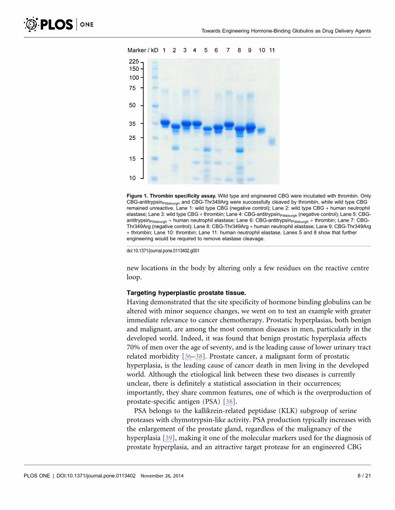
Targeting sites of thrombosis.
Thrombin, also known as Factor IIa, is a key component of the blood coagulation
cascade. It is a serine protease, and its interaction with various substrates and
cofactors in the coagulation network is vital to the maintenance of haemostasis. In
the event of vascular injury, activation of the coagulation cascade causes the
zymogen, prothrombin, to be cleaved to produce enzymatically-active thrombin,
which then goes on to cleave fibrinogen to fibrin. Fibrin polymerises to form the
beginning of a haemostatic plug [34]. The activity of thrombin is typically
regulated via both the coagulation cascade and the action of antithrombin, a
serpin that functions as a thrombin-specific suicide inhibitor. Dysregulation of the
coagulation network, and consequently thrombin function, can result in a
number of pathologies ranging from coagulopathic bleeding on one extreme to
atherothrombotic disease on the other.
We have previously shown that by substituting the P15 to P19 amino acid
residues of wild type human CBG (GVDTAGSTGVTLNLTS) with those of the
Pittsburgh variant of a1-antitrypsin (GTEAAGAMFLEAIPRS), we could make
CBG susceptible to cleavage by thrombin [19]. This variant of a1-antitrypsin was
first discovered in 1983 in a patient who died from an episodic bleeding disorder
[35], and bore a methionine to arginine substitution in the P1 position of the
reactive centre loop. This mutation alone turned a1-antitrypsin into the
functional equivalent of antithrombin. Although exosites such as the heparin
binding site in antithrombin contribute to protease recognition, this result
suggested that the identity of the residue at the P1 position is the single most
important factor for conferring specificity in serpins, and that changing the P1
residue to arginine can be sufficient to make a serpin susceptible to proteolysis by
thrombin. Therefore, we made a single point mutation in wild type CBG,
mutating the threonine in the P1 position of the reactive centre loop to an
arginine by PCR-based site-directed mutagenesis. This CBG-T349R variant was
found to have the same susceptibility to cleavage by thrombin as the CBG-
antitrypsinPittsburgh chimera previously described (Figure 1). This confirms the
feasibility of retargeting hormone binding globulins to deliver small molecules to
Towards Engineering Hormone-Binding Globulins as Drug Delivery Agents
PLOS ONE | DOI:10.1371/journal.pone.0113402 November 26, 2014 7 / 21
new locations in the body by altering only a few residues on the reactive centre
loop.
Targeting hyperplastic prostate tissue.
Having demonstrated that the site specificity of hormone binding globulins can be
altered with minor sequence changes, we went on to test an example with greater
immediate relevance to cancer chemotherapy. Prostatic hyperplasias, both benign
and malignant, are among the most common diseases in men, particularly in the
developed world. Indeed, it was found that benign prostatic hyperplasia affects
70% of men over the age of seventy, and is the leading cause of lower urinary tract
related morbidity [36–38]. Prostate cancer, a malignant form of prostatic
hyperplasia, is the leading cause of cancer death in men living in the developed
world. Although the etiological link between these two diseases is currently
unclear, there is definitely a statistical association in their occurrences;
importantly, they share common features, one of which is the overproduction of
prostate-specific antigen (PSA) [38].
PSA belongs to the kallikrein-related peptidase (KLK) subgroup of serine
proteases with chymotrypsin-like activity. PSA production typically increases with
the enlargement of the prostate gland, regardless of the malignancy of the
hyperplasia [39], making it one of the molecular markers used for the diagnosis of
prostate hyperplasia, and an attractive target protease for an engineered CBG
Figure 1. Thrombin specificity assay. Wild type and engineered CBG were incubated with thrombin. OnlyCBG-antitrypsinPittsburgh and CBG-Thr349Arg were successfully cleaved by thrombin, while wild type CBGremained unreactive. Lane 1: wild type CBG (negative control); Lane 2: wild type CBG + human neutrophilelastase; Lane 3: wild type CBG + thrombin; Lane 4: CBG-antitrypsinPittsburgh (negative control); Lane 5: CBG-antitrypsinPittsburgh + human neutrophil elastase; Lane 6: CBG-antitrypsinPittsburgh + thrombin; Lane 7: CBG-Thr349Arg (negative control); Lane 8: CBG-Thr349Arg + human neutrophil elastase; Lane 9: CBG-Thr349Arg+ thrombin; Lane 10: thrombin; Lane 11: human neutrophil elastase. Lanes 5 and 8 show that furtherengineering would be required to remove elastase cleavage.
doi:10.1371/journal.pone.0113402.g001
Towards Engineering Hormone-Binding Globulins as Drug Delivery Agents
PLOS ONE | DOI:10.1371/journal.pone.0113402 November 26, 2014 8 / 21
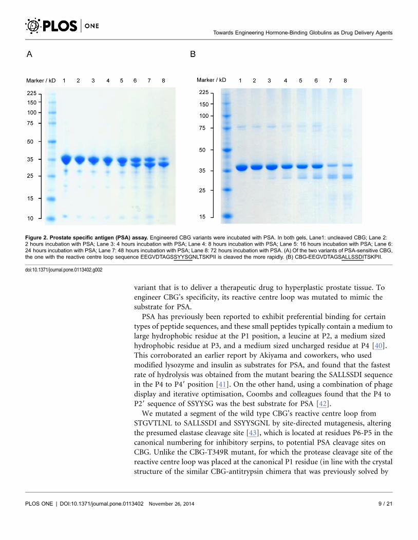
variant that is to deliver a therapeutic drug to hyperplastic prostate tissue. To
engineer CBG’s specificity, its reactive centre loop was mutated to mimic the
substrate for PSA.
PSA has previously been reported to exhibit preferential binding for certain
types of peptide sequences, and these small peptides typically contain a medium to
large hydrophobic residue at the P1 position, a leucine at P2, a medium sized
hydrophobic residue at P3, and a medium sized uncharged residue at P4 [40].
This corroborated an earlier report by Akiyama and coworkers, who used
modified lysozyme and insulin as substrates for PSA, and found that the fastest
rate of hydrolysis was obtained from the mutant bearing the SALLSSDI sequence
in the P4 to P49 position [41]. On the other hand, using a combination of phage
display and iterative optimisation, Coombs and colleagues found that the P4 to
P29 sequence of SSYYSG was the best substrate for PSA [42].
We mutated a segment of the wild type CBG’s reactive centre loop from
STGVTLNL to SALLSSDI and SSYYSGNL by site-directed mutagenesis, altering
the presumed elastase cleavage site [43], which is located at residues P6-P5 in the
canonical numbering for inhibitory serpins, to potential PSA cleavage sites on
CBG. Unlike the CBG-T349R mutant, for which the protease cleavage site of the
reactive centre loop was placed at the canonical P1 residue (in line with the crystal
structure of the similar CBG-antitrypsin chimera that was previously solved by
Figure 2. Prostate specific antigen (PSA) assay. Engineered CBG variants were incubated with PSA. In both gels, Lane1: uncleaved CBG; Lane 2:2 hours incubation with PSA; Lane 3: 4 hours incubation with PSA; Lane 4: 8 hours incubation with PSA; Lane 5: 16 hours incubation with PSA; Lane 6:24 hours incubation with PSA; Lane 7: 48 hours incubation with PSA; Lane 8: 72 hours incubation with PSA. (A) Of the two variants of PSA-sensitive CBG,the one with the reactive centre loop sequence EEGVDTAGSSYYSGNLTSKPII is cleaved the more rapidly. (B) CBG-EEGVDTAGSALLSSDITSKPII.
doi:10.1371/journal.pone.0113402.g002
Towards Engineering Hormone-Binding Globulins as Drug Delivery Agents
PLOS ONE | DOI:10.1371/journal.pone.0113402 November 26, 2014 9 / 21

our group [19]) we had no a priori knowledge of the effect of inserting a PSA-
specific sequence on the structure and biochemical behavior of CBG. The
predicted PSA cleavage sites were therefore chosen to align with the main elastase
cleavage site in wild type CBG, in order to keep the length of the reactive centre
loop the same as the wild type in both the cleaved and intact forms. We then
demonstrated that incubating the engineered CBG variants with PSA resulted in
the cleavage of their respective reactive centre loops as shown in Figure 2, with the
variant bearing the SSYYSGNL motif the better substrate for PSA. Inspection of
this region in the structure of cleaved CBG shows that the change of the residues
Figure 3. Ligand binding pocket of the hormone binding globulins. (A) The ligand binding pocket (solidsurface, PDB entry 2VDY [19]) is formed by Helix A (yellow), Helix D (blue), b-sheet B (red), the s2B/s3B loop(orange) and the s4B/s5B loop (green). (B) R-state human CBG (PDB 2VDY [19], chain A) showing theinteractions of pocket residues with cortisol. (C) Corticosteroid binding globulin (PDB 2VDY, chain A), and (D)Thyroxine binding globulin (PDB 2CEO [54], chain A). Electrostatic map: negative potential (red); positivepotential (blue), uncharged/hydrophobic (white). (C) and (D) (inset). Orientation represented in theelectrostatic maps, with the key secondary structural elements coloured as in (A).
doi:10.1371/journal.pone.0113402.g003
Towards Engineering Hormone-Binding Globulins as Drug Delivery Agents
PLOS ONE | DOI:10.1371/journal.pone.0113402 November 26, 2014 10 / 21
GV in native CBG to LL or particularly YY in the two PSA substrate mutants is
likely to create steric strain on loop insertion. This would lead to a loss of stability
and may render the cleaved mutant proteins more susceptible to further
proteolysis, explaining the apparent reduction in total cleaved CBG (Figure 2).
As a control, we incubated wild type CBG with PSA for 72 hours and found no
evidence of reactive centre loop cleavage (data not shown). As a consequence of
the mutations, it is now possible for CBG to discharge its ligand where there are
high levels of PSA, such as in hyperplastic prostate glands. Nevertheless, the
current rate of reactive centre loop cleavage by PSA would need to be further
optimised to a timescale suitable for drug delivery, as substantial cleavage was only
seen after incubation for greater than 24 hours.
Altering ligand specificity
CBG is particularly promising in the creation of a prostatic hyperplasia specific
drug delivery system because of the properties of its ligand binding pocket. CBG’s
physiological ligands are steroid hormones, and it has a moderately high affinity
for progesterone [44]. Since the 1980s, the synthetic progestin, megestrol acetate,
has been used in the treatment of some forms of prostatic hyperplasia because it
lowers the levels of testosterone, luteinising hormone and follicle-stimulating
hormone, which are responsible for stimulating the unwanted proliferation of
tissue in the prostate gland [45–49]. It also functions as an androgen receptor
antagonist, starving prostate carcinomas of the testosterone and dihydrotestos-
terone required for their growth and maintenance [45, 48]. However, endocri-
nological treatment of prostate cancer is becoming less commonplace due to its
reportedly limited efficacy in many cases [50]. Currently, the most common
chemotherapeutic treatment for malignant metastatic prostate cancer is docetaxel,
a mitotic poison [51]. In the next part of this proof-of-concept experiment, we
sought to show that it is possible to alter the physicochemical characteristics of
CBG’s binding pocket sufficiently to bind a non-steroid compound.
Designing CBG to bind a thyroxine derivative.
CBG and its sister protein, TBG, have adapted to the function of hormone
transport by developing a deep hydrophobic cleft in the region bounded by helix
A, helix H, b-sheet B and the s2B/s3B and s4B/s5B loops, as shown in Figure 3A.
This cleft serves as the binding pocket for both physiological and non-
physiological ligands. As a carrier of glucocorticoid hormones, this pocket in CBG
is lined primarily with uncharged amino acid residues, which complement the
mainly hydrophobic surface of its steroid ligands. In fact, the main interaction
between cortisol and CBG is thought to be the p-p stacking between the indole side
chain of Trp 371 and the A, B and C cycloalkene rings of cortisol [18, 52]. A host of
other hydrophobic interactions between the pocket residues and cortisol help
contribute to the high affinity of binding, while other residues form a hydrogen
bond network contributing to the selectivity of CBG for biologically-active C-21
Towards Engineering Hormone-Binding Globulins as Drug Delivery Agents
PLOS ONE | DOI:10.1371/journal.pone.0113402 November 26, 2014 11 / 21
steroids such as glucocorticoids [18, 19, 53]. These interactions are illustrated in
Figure 3B.
TBG, on the other hand, binds thyroxine, which is a more polar molecule than
cortisol. This is evident from the physical properties of the TBG binding pocket,
which is considerably more polar and positively charged than that of CBG, as seen
in Figures 3C and D. Arg 381 in TBG, like its analogous residue in CBG, Trp 371,
appears to play an instrumental role in the protein-ligand interface. The
positively-charged guanidium side chain of Arg 381 is believed to contribute to a
cation-p interaction with thyroxine and its fluorescent derivatives [20, 54]. In this
part of the study, we attempted to redesign the pocket of CBG to bind the
thyroxine derivative, L-thyroxine-6-carboxyfluorescein.
To establish the importance of Trp 371 in CBG’s interaction with cortisol, we
first mutated it to alanine. The resultant CBG-Trp371Ala protein had no
detectable affinity for cortisol when measured using the ForteBio Octet Red
biolayer interferometry system. This confirmed previous inferences from
biochemical [55] and structural [18, 19] studies about the crucial role of Trp 371
in the function of CBG. We then mutated Trp 371 to arginine, so that the binding
site of CBG now begins to resemble that of TBG. Again, using biolayer
interferometery, we found that CBG-Trp371Arg has no measurable affinity for
cortisol. However, unlike wild type CBG, which showed no affinity for L-
thyroxine-6-carboxyfluorescein, the CBG-Trp371Arg mutant was able to bind the
thyroxine derivative, albeit at a lower affinity than TBG (CBG-Trp371Arg: Kd at
25 C 5840 nM, TBG: Kd at 25 C 50.73 nM). This experiment has demonstrated
the principle that the binding pocket of CBG can be redesigned by means of
carefully chosen mutations to carry a non-physiological ligand. This same
principle can be applied in the future to design CBG to carry other therapeutic
compounds such as docetaxel to hyperplastic prostate tissue, or indeed any
number of combinations of drugs and targets in the body.
Increasing the amount of ligand released at the target
Next, we explored the fine-tuning of this drug-delivery vehicle to discharge a
greater fraction of the bound ligand at the target site. We previously reported that
recombinantly expressed wild type CBG undergoes a 2.6-fold decrease in affinity
for cortisol upon cleavage of its reactive centre loop [21]. However, unlike
cortisol, where a relatively small change in binding affinity to CBG is enough to
perturb an existing equilibrium to result in a physiologically relevant change in
free levels of the hormone, a much greater specificity of release is needed for
therapeutic agents used for chemotherapy of solid tumours. In order to deliver a
sufficiently high concentration of these drugs to the tumour by means of a protein
carrier-based deliver system, a large proportion of the drug bound to the carrier
needs to be released without affecting tissues outside the target.
For a protein carrier that binds the drug with a one-to-one stoichiometry,
equation (1) describes the binding equilibrium. It can be seen from this equation
that the concentration of free ligand is determined by the value of Kd, and to
Towards Engineering Hormone-Binding Globulins as Drug Delivery Agents
PLOS ONE | DOI:10.1371/journal.pone.0113402 November 26, 2014 12 / 21

achieve the dual aims of releasing biologically active amounts of therapeutic drug
whilst avoiding off-target effects, the protein carrier would ideally bind its ligand
with very high affinity (small Kd) in the S-state, and experience a large decrease in
binding affinity (large Kd) upon reactive centre loop cleavage to release most of
the bound compound at the target site in the R-state.
Figure 4. Reactive centre loop P14 residue. The P14 residue in CBG (PDB entry 2VDY [19]), Val 336,shown in yellow spheres, is believed to displace Tyr 235 (blue spheres) on the s2B/s3B loop when thereactive centre loop is inserted into the central b-sheet A (red), leading to conformational changes in thebinding pocket (pale green). CBG’s interaction with cortisol is provided by a network of side chains (brightgreen ball-and-stick representation), which can be perturbed by any slight movement in the pocket and itssurrounding regions.
doi:10.1371/journal.pone.0113402.g004
Table 2. Comparing the changes in cortisol binding affinity of CBG mutants.
CBG variant Conformation Kd (nM) Fold change in Kd
wild-type S (uncleaved) 279¡18
R (cleaved) 734¡48 2.6
P1-Arg + V336I S (uncleaved) 326¡46
R (cleaved) 3420¡440 10
P1-Arg + V336L S (uncleaved) 295¡44
R (cleaved) 2420¡200 8.2
P1-Arg + S100C-V236C S (uncleaved) 278¡64
R (cleaved) 1800¡49 6.5
P1-Arg + S100C-V236C-V336I S (uncleaved) 297¡40
R (cleaved) 2260¡320 7.6
P1-Arg + S100C-V236C-V336L S (uncleaved) 252¡17
R (cleaved) 1600¡320 6.4
Dissociation constants of various forms of CBG in the S- and R-states. Each pair of results for a given CBGvariant represents the Kd of the intact S-state and the reactive centre loop-cleaved R-state (larger Kd).
doi:10.1371/journal.pone.0113402.t002
Towards Engineering Hormone-Binding Globulins as Drug Delivery Agents
PLOS ONE | DOI:10.1371/journal.pone.0113402 November 26, 2014 13 / 21
Replacing the P14 residue with a bulkier amino acid.
It has previously been reported for TBG that the P14 threonine on the reactive
centre loop is a key component of the mechanism by which thyroxine is reversibly
released by the S-state protein. The residue is believed to displace Tyr 241 on the
neighbouring b-sheet B when the reactive centre loop of intact TBG is reversibly
and partially inserted into b-sheet A, thus disrupting the hydrogen bond network
that stabilises the steroid binding pocket [54, 56]. The same is believed to happen
during the irreversible S-to-R conformational change, when the reactive centre
loop is cleaved and inserted, this time irreversibly, into b-sheet A to form a novel
fourth b-strand. Due to the structural homology of TBG and CBG, we inferred
that the P14 valine in CBG might play a similarly important role (Figure 4).
By mutating this residue to amino acids with slightly bulkier side chains and
similar chemical characteristics, we were able to amplify the effect of loop
insertion on the adjacent hormone binding pocket without making it so sterically
hindered as to prevent the process altogether. The two aliphatic natural amino
acids chosen were leucine and isoleucine, with the mutations being made on a
T349R background to allow cleavage by thrombin. These substitutions resulted in
greater disruption to the ligand binding pocket upon reactive centre loop cleavage
than valine in the wild type protein, and as a result, at physiological temperature,
the binding affinity of CBG-V336L-T349R decreased by 8.2 times, while that of
CBG-V336I-T349R decreased by 10 times following cleavage of its reactive centre
loop (Table 2).
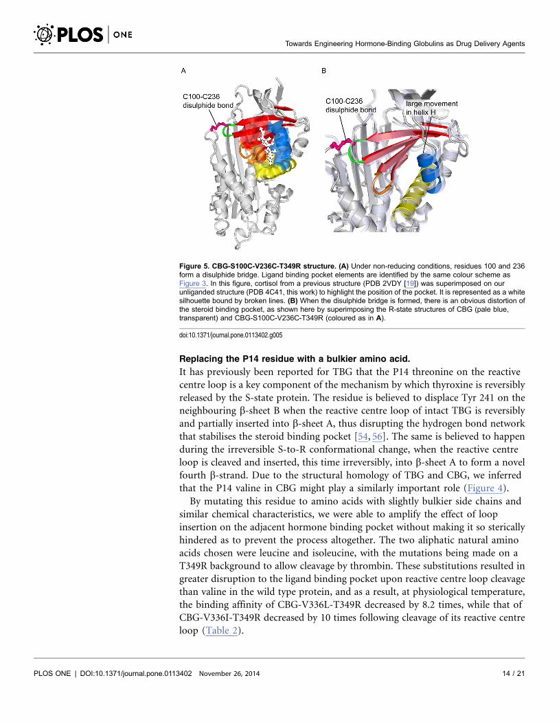
Figure 5. CBG-S100C-V236C-T349R structure. (A) Under non-reducing conditions, residues 100 and 236form a disulphide bridge. Ligand binding pocket elements are identified by the same colour scheme asFigure 3. In this figure, cortisol from a previous structure (PDB 2VDY [19]) was superimposed on ourunliganded structure (PDB 4C41, this work) to highlight the position of the pocket. It is represented as a whitesilhouette bound by broken lines. (B) When the disulphide bridge is formed, there is an obvious distortion ofthe steroid binding pocket, as shown here by superimposing the R-state structures of CBG (pale blue,transparent) and CBG-S100C-V236C-T349R (coloured as in A).
doi:10.1371/journal.pone.0113402.g005
Towards Engineering Hormone-Binding Globulins as Drug Delivery Agents
PLOS ONE | DOI:10.1371/journal.pone.0113402 November 26, 2014 14 / 21
Covalently linking s2A/hD loop to binding pocket amplifies change in binding
affinity.
Based on our previous structures of CBG (PDB 2VDX, 2VDY [19]), we designed a
mutant form of CBG that directly couples changes in helix D to the steroid
binding pocket. Ser 100 on the s2A/hD loop and Val 236 on the s2B/3B loop of b-
sheet B were mutated to cysteines so that under non-reducing conditions, they
may be induced to form a disulphide bridge. This forms a direct covalent bond
between the s2A/hD loop at the top of helix D and b-sheet B, which along with
helices A and H make up the steroid binding pocket. We predicted that by
covalently linking the s2A/hD loop to the binding pocket, any effect caused by the
movement of the loop as a result of the S-to-R conformational change would be
amplified.
The disulphide mutations were made on the T349R background to allow
cleavage by thrombin. When binding assays were performed at 37 C in the
presence of the reducing agent, TCEP, the change in binding affinity of CBG-
S100C-V236C-T349R after reactive loop cleavage was very similar to that of wild-
type CBG. However, in the absence of TCEP, cleavage of the reactive centre loop
resulted in a 6.5-fold decrease in binding affinity for cortisol, as shown in Table 2.
We went on to obtain the crystal structure of reactive centre loop-cleaved CBG-
S100C-V236C-T349R, which revealed that the binding pocket was indeed
perturbed by the presence of the disulphide bridge (Figure 5). Cleavage after
Arg349 was supported by clear density for this residue, located at the bottom of
sheet A after insertion of the RCL, and for Pro352, located more than 60A away at
the other end of the molecule, though density for Ser350 and Lys351 could not be
interpreted unambiguously. The X-ray data also showed that the disulphide
linkage has an occupancy of only about half, which suggests either that the
disulphide bond is broken by radiation damage during data collection or, more
likely, that even under non-reducing conditons, only about half of a given
population of the CBG-S100C-V236C-T349R molecules have the desired
disulphide bond at a given time. This could explain the relatively small effect of
these mutations. Nonetheless, this amplified change in binding affinity is a
definite step towards the rational optimisation of ligand release by CBG.
Combining mutations at Ser100Cys, Val236Cys and P14 residue of reactive
centre loop.
Having observed the augmented changes in cortisol binding affinities upon S-to-R
conformational change either by covalently linking the s2A/hD loop with the
steroid binding pocket, or by substituting the P14 valine with a bulkier residue, we
tested if the effect of combining the two would be cumulative. It was found that
the change in binding affinity of CBG-S100C-V236C-V336L-T349R was, in fact,
diminished from the CBG-V336L-T349R mutant, and was only 6.4-fold.
Similarly, the CBG-S100C-V236C-V336I-T349R mutant proved to have a mere
7.6-fold decrease in cortisol binding affinity following reactive centre loop
cleavage (Table 2).
Towards Engineering Hormone-Binding Globulins as Drug Delivery Agents
PLOS ONE | DOI:10.1371/journal.pone.0113402 November 26, 2014 15 / 21
Conclusions
In this study we have shown that, by means of targeted mutations, CBG can be
engineered to bind and release compounds other than its physiological ligands,
glucocorticoid hormones. In principle, this can be designed to occur at locations
other than its physiological target, inflamed tissues. We hope that by proving the
feasibility of this concept, the scope of future developments in targeted drug
delivery can be expanded to include similar carrier proteins. Mutations made at
and around the P1 residue of the reactive centre loop gave two CBG variants that
had altered protease specificities, becoming susceptible to cleavage by either
thrombin or prostate specific antigen. Since cleavage of the reactive centre loop
triggers ligand release, there is a direct correlation between the protease specificity
and CBG’s site of action, and by targeting these engineered mutants to proteases
that are localised to certain parts of the body, one can then direct the ligand to be
released at very defined loci. Therapeutically, such a strategy could allow a lower
total dose of the drug to be used while maintaining a higher local concentration of
the compound at the desired site than current methods employed in
chemotherapy. Moreover, since the drug is sequestered by the carrier protein until
it is released at the target tissue, the risk of off-target effects would be minimised.
We also showed that, by carrying out site-directed mutagenesis of selected
residues in the hormone binding pocket, CBG can be redesigned to carry a ligand
of our choosing. By changing a binding pocket tryptophan normally involved in
interaction with cortisol to an arginine, the ligand specificity of CBG was altered
such that it now binds a thyroxine adduct, L-thyroxine-6-carboxyfluorescein,
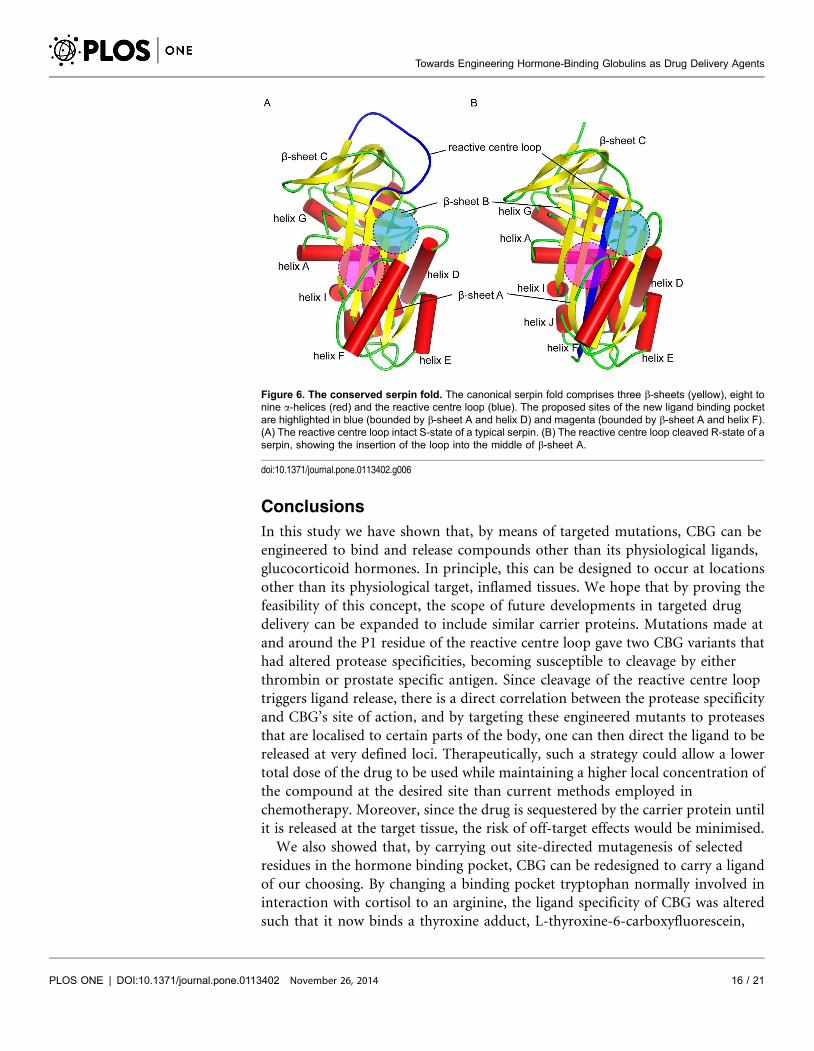
Figure 6. The conserved serpin fold. The canonical serpin fold comprises three b-sheets (yellow), eight tonine a-helices (red) and the reactive centre loop (blue). The proposed sites of the new ligand binding pocketare highlighted in blue (bounded by b-sheet A and helix D) and magenta (bounded by b-sheet A and helix F).(A) The reactive centre loop intact S-state of a typical serpin. (B) The reactive centre loop cleaved R-state of aserpin, showing the insertion of the loop into the middle of b-sheet A.
doi:10.1371/journal.pone.0113402.g006
Towards Engineering Hormone-Binding Globulins as Drug Delivery Agents
PLOS ONE | DOI:10.1371/journal.pone.0113402 November 26, 2014 16 / 21
instead of cortisol. Using this same principle, one can potentially mutate the
pocket to suit the chemistry of a wider variety of ligands. There are various
computational tools to help in identification and redesign of residues involved in
ligand binding so as to alter the ligand specificity more dramatically. Among
them, the Rosetta protein modelling software suite has had some notable success
in recent years [57, 58].
To increase the efficiency of ligand carriage and release, as well as to ensure that
the amount of ligand released by the carrier whilst in circulation is negligible, the
change in the dissociation constant, Kd, upon reactive centre loop cleavage needs
to be amplified. By mutating the P14 residue of the reactive centre loop to leucine
and isoleucine, we have increased the change in binding affinity post-cleavage to
about eight to ten-fold from the 2.6-fold difference seen in the recombinant wild
type CBG. Covalently linking the s2A/hD loop to b-sheet B of the binding pocket
with a disulphide bridge also resulted in a greater (6.6 times) loss in binding
affinity, although the effects of the two alterations were not additive.
In the course of this project, we have come to appreciate that the release of
hormone from the hormone-binding globulins is more subtle and modulated
than was previously believed [21], and it has proven to be more difficult than
anticipated to enhance the effect of cleavage. Perhaps if the ligand binding pocket
were moved to a site where the conformation changes on cleavage are more
pronounced, such as the region bounded by b-sheet A and helix D, or the region
bounded by b-sheet A and helix F (Figure 6), the effects of S-to-R conformational
change would have a significantly larger effect. Engineering a binding pocket ab
initio, instead of making use of an existing site, would be a challenging task, but
significant advances have been made by the Baker group in the development of
Rosetta towards the computational design of proteins from scratch. In particular,
they have demonstrated the rational design of novel proteins with small molecule
binding sites in silico that have high affinities and specificities for their target
ligands/substrates [57, 59, 60]. It is therefore reasonable to expect that the same
technique can be used to build a high affinity binding site for a pre-specified
ligand into an existing protein.
One possible risk associated with using a modified form of an endogenous
protein as a drug carrier is the small but potentially dangerous possibility of
raising an immune response against the mutant protein, which could affect the
pharmacokinetics of the protein and/or result in hypersensitivity, as is observed in
a small number of lysosomal storage disease patients receiving enzyme
replacement therapy [61]. Just as worrying is the prospect of triggering an auto-
immune response against endogenous CBG. However, we expect that by using a
human protein as the starting scaffold, with only a small number of point
mutations, most of which are not solvent exposed, the danger of developing an
immune response against the drug carrier can be kept low. In the context of T
cell-mediated responses, epitopes are typically defined by nonameric peptides
[62], while B cell epitopes seem to involve about eight residues around an
‘‘epitope core’’ [63]. Moreover, seminal work on humanising mouse antibodies
has demonstrated that retaining the mouse variable region, which makes up about
Towards Engineering Hormone-Binding Globulins as Drug Delivery Agents
PLOS ONE | DOI:10.1371/journal.pone.0113402 November 26, 2014 17 / 21
10% of an immunoglobulin, does not pose a problem with immunogenicity [64].
In general, it appears that it is often easier to destroy antigenicity than to create it,
as a single point mutation has been known to destroy an epitope and result in
immune evasion [65]. This combination of reasons suggests that in vivo
administration of engineered CBG would not necessarily give rise to an immune
response.
In summary, we have demonstrated in this study that by making targeted
mutations to various structural features of CBG, we can, in principle, engineer the
protein to carry a therapeutic drug and to release it upon cleavage of its reactive
centre loop by site-specific serine proteases. Future work will need to strive
towards optimising the efficiency of ligand release, and to test out other potential
payload and protease specificities if this drug delivery system were to be
successfully employed in vivo. Nonetheless, as a proof of principle, this study has
opened the prospect of yet another means of targeted drug delivery to combat the
persistent problem of off-target side effects in cancer chemotherapy.
Acknowledgments
The authors would like to thank Robin W. Carrell (Emeritus Professor,
Department of Haematology, University of Cambridge) for edifying discussions
throughout the course of this study. We are also grateful to Francois Loiseau,
Lech-Gustav Milroy, Rebecca M. Myers and Steven V. Ley for their generous gift
of thyroxine-6-carboxyfluorescein with which we carried out some of the binding
assays. In addition, we thank Diamond Light Source for access to beamline I03
(proposal number MX6641) that contributed to the results presented here.
Author ContributionsConceived and designed the experiments: WLC AZ RJR. Performed the
experiments: WLC. Analyzed the data: WLC AZ RJR. Wrote the paper: WLC AZ
RJR.
References
1. Gilman A, Philips FS (1946) The Biological Actions and Therapeutic Applications of the B-ChloroethylAmines and Sulfides. Science 103: 409–436.
2. Fojo T, Parkinson DR (2010) Biologically targeted cancer therapy and marginal benefits: are we makingtoo much of too little or are we achieving too little by giving too much? Clin Cancer Res 16: 5972–5980.
3. Jaiyesimi IA, Buzdar AU, Decker DA, Hortobagyi GN (1995) Use of tamoxifen for breast cancer:twenty-eight years later. J Clin Oncol 13: 513–529.
4. Ricciardi S, Tomao S, de Marinis F (2009) Toxicity of targeted therapy in non-small-cell lung cancermanagement. Clin Lung Cancer 10: 28–35.
5. Maeda H, Takeshita J, Kanamaru R (1979) A lipophilic derivative of neocarzinostatin. A polymerconjugation of an antitumor protein antibiotic. Int J Pept Protein Res 14: 81–87.
Towards Engineering Hormone-Binding Globulins as Drug Delivery Agents
PLOS ONE | DOI:10.1371/journal.pone.0113402 November 26, 2014 18 / 21
6. Matsumura Y, Maeda H (1986) A new concept for macromolecular therapeutics in cancerchemotherapy: mechanism of tumoritropic accumulation of proteins and the antitumor agent smancs.Cancer Res 46: 6387–6392.
7. Maeda H, Bharate GY, Daruwalla J (2009) Polymeric drugs for efficient tumor-targeted drug deliverybased on EPR-effect. Eur J Pharm Biopharm 71: 409–419.
8. Maeda H, Sawa T, Konno T (2001) Mechanism of tumor-targeted delivery of macromolecular drugs,including the EPR effect in solid tumor and clinical overview of the prototype polymeric drug SMANCS.J Control Release 74: 47–61.
9. Reichert JM, Rosensweig CJ, Faden LB, Dewitz MC (2005) Monoclonal antibody successes in theclinic. Nat Biotechnol 23: 1073–1078.
10. Litvak-Greenfeld D, Benhar I (2012) Risks and untoward toxicities of antibody-basedimmunoconjugates. Adv Drug Deliv Rev 64: 1782–1799.
11. Kantoff PW, Higano CS, Shore ND, Berger ER, Small EJ, et al. (2010) Sipuleucel-T immunotherapyfor castration-resistant prostate cancer. N Engl J Med 363: 411–422.
12. May KF, Gulley JL, Drake CG, Dranoff G, Kantoff PW (2011) Prostate cancer immunotherapy. ClinCancer Res 17: 5233–5238.
13. Chambers JD, Neumann PJ (2011) Listening to Provenge—what a costly cancer treatment says aboutfuture Medicare policy. N Engl J Med 364: 1687–1689.
14. Pemberton PA, Stein PE, Pepys MB, Potter JM, Carrell RW (1988) Hormone binding globulinsundergo serpin conformational change in inflammation. Nature 336: 257–258.
15. Carrell R, Travis J (1985) a 1-Antitrypsin and the serpins: variation and countervariation. TrendsBiochem Sci 10: 20–24.
16. Hammond GL, Smith CL, Goping IS, Underhill DA, Harley MJ, et al. (1987) Primary structure ofhuman corticosteroid binding globulin, deduced from hepatic and pulmonary cDNAs, exhibits homologywith serine protease inhibitors. Proc Natl Acad Sci USA 84: 5153–5157.
17. Gettins PG (2002) Serpin structure, mechanism, and function. Chem Rev 102: 4751–4804.
18. Klieber MA, Underhill C, Hammond GL, Muller YA (2007) Corticosteroid-binding globulin, a structuralbasis for steroid transport and proteinase-triggered release. J Biol Chem 282: 29594–29603.
19. Zhou A, Wei Z, Stanley PLD, Read RJ, Stein PE, et al. (2008) The S-to-R transition of corticosteroid-binding globulin and the mechanism of hormone release. Journal of Molecular Biology 380: 244–251.
20. Qi X, Loiseau F, Chan WL, Yan Y, Wei Z, et al. (2011) Allosteric modulation of hormone release fromthyroxine and corticosteroid-binding globulins. Journal of Biological Chemistry 286: 16163–16173.
21. Chan WL, Carrell RW, Zhou A, Read RJ (2013) How Changes in Affinity of Corticosteroid-bindingGlobulin Modulate Free Cortisol Concentration. J Clin Endocrinol Metab 98: 3315–3322.
22. Salvesen GS, Catanese JJ, Kress LF, Travis J (1985) Primary structure of the reactive site of humanC1-inhibitor. J Biol Chem 260: 2432–2436.
23. Leslie AGW (2007) Processing diffraction data with mosflm. In:, Read RJ, Sussman JL, , editors.,Evolving Methods for Macromolecular Crystallography. Springer Netherlands, Vol. 245.pp. 41–51.
24. Evans PR, Murshudov GN (2013) How good are my data and what is the resolution? ActaCrystallogr D Biol Crystallogr 69: 1204–1214.
25. McCoy AJ, Grosse-Kunstleve RW, Adams PD, Winn MD, Storoni LC, et al. (2007) Phasercrystallographic software. J Appl Crystallogr 40: 658–674.
26. Bunkoczi G, Read RJ (2011) Improvement of molecular-replacement models with Sculptor. ActaCrystallogr D Biol Crystallogr 67: 303–312.
27. Emsley P, Cowtan K (2004) Coot: model-building tools for molecular graphics. Acta Crystallogr D BiolCrystallogr 60: 2126–2132.
28. Afonine PV, Grosse-Kunstleve RW, Echols N, Headd JJ, Moriarty NW, et al. (2012) Towardsautomated crystallographic structure refinement with phenix. refine. Acta Crystallogr D Biol Crystallogr68: 352–367.
Towards Engineering Hormone-Binding Globulins as Drug Delivery Agents
PLOS ONE | DOI:10.1371/journal.pone.0113402 November 26, 2014 19 / 21
29. Chen VB, Arendall WB, Headd JJ, Keedy DA, Immormino RM, et al. (2010) MolProbity: all-atomstructure validation for macromolecular crystallography. Acta Crystallogr D Biol Crystallogr 66: 12–21.
30. Lin HY, Muller YA, Hammond GL (2010) Molecular and structural basis of steroid hormone binding andrelease from corticosteroid-binding globulin. Molecular and Cellular Endocrinology 316: 3–12.
31. Ye S, Goldsmith EJ (2001) Serpins and other covalent protease inhibitors. Curr Opin Struct Biol 11:740–745.
32. Baglin TP, Carrell RW, Church FC, Esmon CT, Huntington JA (2002) Crystal structures of native andthrombin-complexed heparin cofactor II reveal a multistep allosteric mechanism. Proc Natl Acad Sci USA99: 11079–11084.
33. Schechter I, Berger A (1967) On the size of the active site in proteases. I. Papain. Biochem BiophysRes Commun 27: 157–162.
34. Davie EW, Fujikawa K, Kisiel W (1991) The coagulation cascade: initiation, maintenance, andregulation. Biochemistry 30: 10363–10370.
35. Owen MC, Brennan SO, Lewis JH, Carrell RW (1983) Mutation of Antitrypsin to Antithrombin.N Engl J Med 309: 694–698.
36. McVary K (2006) Lower urinary tract symptoms and sexual dysfunction: epidemiology andpathophysiology. BJU Int 97 Suppl 2: 23–8
37. Nicholson TM, Ricke WA (2011) Androgens and estrogens in benign prostatic hyperplasia Past,present and future. Differentiation 82: 184–199.
38. Ørsted DD, Bojesen SE (2013) The link between benign prostatic hyperplasia and prostate cancer. NatRev Urol 10: 49–54.
39. Balk SP, Ko YJ, Bubley GJ (2003) Biology of prostate-specific antigen. J Clin Oncol 21: 383–391.
40. Kumar V, Hassan MI, Singh AK, Dey S, Singh TP, et al. (2009) Strategy for sensitive and specificdetection of molecular forms of PSA based on 2DE and kinetic analysis: A step towards diagnosis ofprostate cancer. Clin Chim Acta 403: 17–22.
41. Akiyama K, Nakamura T, Iwanaga S, Hara M (1987) The chymotrypsin-like activity of human prostate-specific antigen, gamma-seminoprotein. FEBS Lett 225: 168–172.
42. Coombs GS, Bergstrom RC, Pellequer JL, Baker SI, Navre M, et al. (1998) Substrate specificity ofprostate-specific antigen (PSA). Chem Biol 5: 475–488.
43. Lewis JG, Elder PA (2011) Corticosteroid-binding globulin reactive centre loop antibodies recogniseonly the intact natured protein: Elastase cleaved and uncleaved CBG may coexist in circulation. J.Steroid Biochem Mol Biol 127: 289–294.
44. Gardill BR, Vogl MR, Lin HY, Hammond GL, Muller YA (2012) Corticosteroid-binding globulin:structure-function implications from species differences. PLoS ONE 7: e52759.
45. Geller J, Albert J, Yen SS (1978) Treatment of advanced cancer of prostate with megestrol acetate.Urology 12: 537–541.
46. Geller J, Albert J, Geller S (1982) Acute therapy with megestrol acetate decreases nuclear and cytosolandrogen receptors in human BPH tissue. Prostate 3: 11–15.
47. Schacter L, Rozencweig M, Canetta R, Kelley S, Nicaise C, et al. (1989) Megestrol acetate: clinicalexperience. Cancer Treatment Reviews 16: 49–63.
48. Dawson NA, Conaway M, Halabi S, Winer EP, Small EJ, et al. (2000) A randomized study comparingstandard versus moderately high dose megestrol acetate for patients with advanced prostate carcinoma:cancer and leukemia group B study 9181. Cancer 88: 825–834.
49. Kaore SN, Langade DK, Yadav VK, Sharma P, Thawani VR, et al. (2012) Novel actions ofprogesterone: what we know today and what will be the scenario in the future? J Pharm Pharmacol 64:1040–1062.
50. Heidenreich A, Aus G, Abbou CC, Bolla M, Joniau S, et al. (2008) Guidelines on Prostate Cancer: 1–114.
51. Tannock IF, de Wit R, Berry WR, Horti J, Pluzanska A, et al. (2004) Docetaxel plus prednisone ormitoxantrone plus prednisone for advanced prostate cancer. N Engl J Med 351: 1502–1512.
Towards Engineering Hormone-Binding Globulins as Drug Delivery Agents
PLOS ONE | DOI:10.1371/journal.pone.0113402 November 26, 2014 20 / 21
52. Stroupe SD, Gray RD, Westphal U (1978) Steroid-protein interactions. Kinetics of binding of cortisoland progesterone to human corticosteroid-binding globulin. FEBS Lett 86: 61–64.
53. Westphal U (1983) Corticosteroid-binding globulin. A review of some recent aspects. Mol Cell Biochem55: 145–157.
54. Zhou A, Wei Z, Read RJ, Carrell RW (2006) Structural mechanism for the carriage and release ofthyroxine in the blood. Proc Natl Acad Sci USA 103: 13321–13326.
55. Avvakumov GV, Hammond GL (1994) Substitutions of tryptophan residues in human corticosteroid-binding globulin: impact on steroid binding and glycosylation. J. Steroid Biochem. Mol. Biol. 49: 191–194.
56. Grasberger H, Golcher HMB, Fingerhut A, Janssen OE (2002) Loop variants of the serpin thyroxine-binding globulin: implications for hormone release upon limited proteolysis. Biochem J 365: 311–316.
57. Morin A, Kaufmann KW, Fortenberry C, Harp JM, Mizoue LS, et al. (2011) Computational design ofan endo-1, 4-beta-xylanase ligand binding site. Protein Eng Des Sel 24: 503–516.
58. Combs SA, DeLuca SL, DeLuca SH, Lemmon GH, Nannemann DP, et al. (2013) Small-moleculeligand docking into comparative models with Rosetta. Nat Protoc 8: 1277–1298.
59. Richter F, Leaver-Fay A, Khare SD, Bjelic S, Baker D (2011) De novo enzyme design using Rosetta3.PLoS ONE 6: e19230.
60. Tinberg CE, Khare SD, Dou J, Doyle L, Nelson JW, et al. (2013) Computational design of ligand-binding proteins with high affinity and selectivity. Nature 501: 212–216.
61. Brooks DA, Kakavanos R, Hopwood JJ (2003) Significance of immune response to enzyme-replacement therapy for patients with a lysosomal storage disorder. Trends in Molecular Medicine 9:450–453.
62. Bernett MJ, Karki S, Moore GL, Leung IWL, Chen H, et al. (2010) Engineering Fully HumanMonoclonal Antibodies from Murine Variable Regions. Journal of Molecular Biology 396: 1474–1490.
63. Nagata S, Pastan I (2009) Removal of B cell epitopes as a practical approach for reducing theimmunogenicity of foreign protein-based therapeutics. Adv Drug Deliv Rev 61: 977–985.
64. Morrison SL, Johnson MJ, Herzenberg LA, Oi VT (1984) Chimeric human antibody molecules: mouseantigen-binding domains with human constant region domains. Proc Natl Acad Sci USA 81: 6851–6855.
65. Lemmermann NAW, Kropp KA, Seckert CK, Grzimek NKA, Reddehase MJ (2011) Reverse GeneticsModification of Cytomegalovirus Antigenicity and Immunogenicity by CD8 T-Cell Epitope Deletion andInsertion. Journal of Biomedicine and Biotechnology 2011: 1–15.
Towards Engineering Hormone-Binding Globulins as Drug Delivery Agents
PLOS ONE | DOI:10.1371/journal.pone.0113402 November 26, 2014 21 / 21


![(OBSTETRICS AND GYNAECOLOGY)repository-tnmgrmu.ac.in/11558/7/270603819padmavathi.pdf · decreased concentrations of sex hormone binding globulin [SHBG]. The anovulation is associated](https://img.pdfslide.net/doc/110x75/5f4f933c0282880c247287be/obstetrics-and-gynaecologyrepository-decreased-concentrations-of-sex-hormone.jpg)