Embed Size (px)
Citation preview
Alabdali et al. Behavioral and Brain Functions 2014, 10:14http://www.behavioralandbrainfunctions.com/content/10/1/14
RESEARCH Open Access
A key role for an impaired detoxificationmechanism in the etiology and severity of autismspectrum disordersAltaf Alabdali1, Laila Al-Ayadhi2,3,4 and Afaf El-Ansary1,2,3,5*
Abstract
Background: Autism Spectrum Disorders (ASD) is a syndrome with a number of etiologies and differentmechanisms that lead to abnormal development. The identification of autism biomarkers in patients with differentdegrees of clinical presentation (i.e., mild, moderate and severe) will give greater insight into the pathogenesis ofthis disease and will enable effective early diagnostic strategies and treatments for this disorder.
Methods: In this study, the concentration of two toxic heavy metals, lead (Pb) and mercury (Hg), were measured inred blood cells, while glutathione-s-transferase (GST) and vitamin E, as enzymatic and non-enzymatic antioxidants,respectively, were measured in the plasma of subgroups of autistic patients with different Social Responsiveness Scale(SRS) and Childhood Autism Rating Scale (CARS) scores. The results were compared to age- and gender-matchedhealthy controls.
Results: The obtained data showed that the patients with autism spectrum disorder had significantly higher Pb andHg levels and lower GST activity and vitamin E concentrations compared with the controls. The levels of heavy metals(Hg and Pb), GST and vitamin E were correlated with the severity of the social and cognitive impairment measures(SRS and CARS). Receiver Operating Characteristics (ROC) analysis and predictiveness curves indicated that the fourparameters show satisfactory sensitivity, very high specificity and excellent predictiveness. Multiple regression analysesconfirmed that higher levels of Hg and Pb, together with lower levels of GST and vitamin E, can be used to predictsocial and cognitive impairment in patients with autism spectrum disorders.
Conclusion: This study confirms earlier studies that implicate toxic metal accumulation as a consequence of impaireddetoxification in autism and provides insight into the etiological mechanism of autism.
Keywords: Autism Spectrum Disorder, Social Responsiveness Scale (SRS), Childhood Autism Rating Scale (CARS), Lead,Mercury, Glutathione-s-transferase, Vitamin E
IntroductionAutism is characterized by a set of repetitious behaviorin combination with social, cognitive and communica-tion deficits [1]. An emerging hypothesis states thatautism may result from a combination of genetic suscep-tibility and exposure to environmental toxins at criticalperiods during brain development [2]. Neurotoxins andassociated inflammation of the brain tissue are often thefocus of therapies for patients with autism. However, if
* Correspondence: [email protected] Department, Science College, King Saud University, P.O box22452, Zip code 11495 Riyadh, Saudi Arabia2Autism Research and Treatment Center, Riyadh, Saudi ArabiaFull list of author information is available at the end of the article
© 2014 Alabdali et al.; licensee BioMed CentraCommons Attribution License (http://creativecreproduction in any medium, provided the orDedication waiver (http://creativecommons.orunless otherwise stated.
body detoxification is impaired, neuroinflammation willnot efficiently improve unless the overall body issues areaddressed. Xenobiotics are neurotoxins of external ori-gin, such as chemicals and pollutants in the air, water,food additives and drugs, that can dramatically alter thehealth of the child. An efficient three-phase mechanismis involved in detoxifying these toxins [3]; however,several factors play a role in modulating this mechanism.For example, age, gender and diet are among the variousbiological and non-biological factors that modulate in-dividual susceptibility. Additionally, genetic variabilityplays a critical role in individual susceptibility becausevariable detoxifier phenotypes result from mutations in
l Ltd. This is an Open Access article distributed under the terms of the Creativeommons.org/licenses/by/2.0), which permits unrestricted use, distribution, andiginal work is properly credited. The Creative Commons Public Domaing/publicdomain/zero/1.0/) applies to the data made available in this article,
Alabdali et al. Behavioral and Brain Functions 2014, 10:14 Page 2 of 11http://www.behavioralandbrainfunctions.com/content/10/1/14
the same gene. These range from individuals with regu-lar enzyme and detoxification functions to poor metabo-lizers with low or no enzyme activity [4,5].Glutathione-S-transferase (GST) functions in the de-
toxification of xenobiotics, drugs, toxins, and metabolitesand is also involved in the regulation of mitogen-activatedprotein kinases, which are important during differen-tiation and development. Hg and Pb, however, are twowell-known toxicants that have toxic effects on the body,with the brain being the most susceptible target organ.Exposure to Hg and Pb during pregnancy and early child-hood can cause neurodevelopmental impairment and sub-clinical brain dysfunction because both heavy metals cancross the placenta and the blood–brain-barrier [6]. In thebrain, these metals can affect critical developmental pro-cesses, including cell proliferation, migration, differen-tiation, synaptogenesis, myelination, and apoptosis [6].It is well documented that patients with ASD have many
statistically significant differences in their nutritional andmetabolic status compared with those who do not haveASD. Some of these biomarkers are indicative of vitaminE insufficiency, increased oxidative stress, and poor de-toxification and are associated with the severity of thedisorder [7-10]. A recent study found that impairedxenobiotic detoxification is correlated with increased gutpermeability (leaky gut) and neuroinflammation, two ac-cepted pathological phenomena in ASD [3].These findings prompted us to search for biochemical
correlations related to the detoxification mechanism andneurobiological processes and the severity and socialfunctioning of patients with ASD measured by the CARSand SRS. We selected Hg and Pb as two environmentaltoxicants involved in the etiology of ASD, together withGST and vitamin E as enzymatic and non-enzymatic an-tioxidants with high activity in terminating lipid peroxi-dation and environmental toxicity. We hypothesized thatconfirming the relationship between impaired detoxifica-tion mechanism and severity of ASD could enhance ef-forts at early prevention, diagnosis, and interventionand, thus, may play a role in decreasing the prevalenceof autism.
Material and methodsSubjectsThis cross-sectional study was conducted on 52 autisticmale children who were recruited from the AutismResearch and Treatment Center, Faculty of Medicine, KingSaud University, Riyadh, Saudi Arabia. Of these children,40 were nonverbal, and 12 were verbal. Their ages rangedbetween 3 and 12 years (mean SD= 7.0 ± 2.34 years). Thecontrol group was comprised of 30 age- and sex-matchedapparently healthy children with a mean age of 7.2 ±2.14 years. The patients fulfilled the diagnostic criteria ofautism described in the 4th edition of the Diagnostic and
Statistical Manual of Mental Disorders. The controls werenormally developing, healthy children who were unrelatedto the autistic subjects and did not have any of the exclu-sion criteria. The control children were the healthy oldersiblings of healthy infants who were attending the WellBaby Clinic at King Khalid University Hospital for routinecheck-ups of their growth parameters. They had no clinicalindications of infectious diseases or neuropsychiatric disor-ders. All participants had normal results for urine analysisand sedimentation rate. The local Ethical Committee ofthe Faculty of Medicine, King Saud University, Riyadh,Saudi Arabia, approved this study. In addition, an in-formed written consent of participation for this studywas signed by the parents or the legal guardians ofthe investigated subjects, according to the Helsinkiprinciples. The selected sample of participants wasbased on how convenient and readily available thegroup of participants was (i.e., convenience sampling).Participants were excluded from the study if they hada diagnosis of fragile X syndrome, epileptic seizures,obsessive–compulsive disorder, affective disorders, orany additional psychiatric or neurological diagnoses.
Measurement of Autism severity scales (CARS and SRS)The Childhood Autism Rating Scale (CARS) score, whichis a measurement of the severity of the disease, rates thechild on a scale from one to four in each of fifteen areas(relating to people’s emotional response; imitation; bodyuse; object use; listening response; fear or nervousness;verbal communication; non-verbal communication; acti-vity level; level and reliability of intellectual response;adaptation to change; visual response; taste, smell andtouch response and general impressions). According tothe scale, children who scored 30–36 had mild to mode-rate autism (n = 23), while those with scores ranging bet-ween 37 and 60 points had severe autism (n = 27) [11,12].To calculate a score for the Social Responsiveness Scale,
a questionnaire was completed in 15 to 20 minutes. Atotal score of 76 or higher was considered severe andstrongly associated with a clinical diagnosis of autistic dis-order. A score of 60–75 was interpreted as falling in themild to moderate range of social impairment [13].
Sample collectionAfter an overnight fast, 10-ml blood samples, from bothgroups of children, were collected in test tubes containingsodium heparin as an anticoagulant. The tubes were cen-trifuged at 3500 rpm for 15 minutes at room temperature.Plasma and red blood cells were separated and stored at −80°C until required for analysis.
Biochemical analysisPlasma is a complex bodily fluid containing proteins, pep-tides, lipids and metabolites and can reflect physiological
Alabdali et al. Behavioral and Brain Functions 2014, 10:14 Page 3 of 11http://www.behavioralandbrainfunctions.com/content/10/1/14
activity and pathology in various body organs, includingthe CNS. In humans, approximately 500 ml of CSF isabsorbed into the blood daily, making blood a suitablesource of biomarkers of neurodegenerative or neurodeve-lopmental diseases [14]. All biochemical assays were per-formed in duplicate and blinded to the clinical status ofthe participants.
1- Measurement of mercuryThe concentration of inorganic mercury (Hg) in red bloodcells was determined by the method described by Magos[15] using a flameless atomic-absorption method. The redblood cells were diluted with saline to 20 ml, followed bythe addition of 1 ml of a 1% cysteine solution, 10 ml of8 M H2SO4 and 1 ml of SnCl2 (100 mg/ml). The samplewas subjected to immediate aeration at a constant rate of2.5 l/min through the reaction vessel, and 20 ml of a 45%NaOH was added. The SnCl2 reagent was used to releaseall of the inorganic mercury from the samples. Aerationwas discontinued after the recorder pen had settled backto within a few chart divisions (2 or 3) of its original
Table 1 Mercury (μg/L), lead (μg/dl), GST (μmol/l) and vitamin
Parameters Group
Mercury (μg/L) Control
Patients with autism
Autism (mild to moderate in CARS)
Autism (severe in CARS)
Autism (mild to moderate in SRS)
Autism (severe in SRS)
Lead (μg/dl) Control
Patients with autism
Autism (mild to moderate in CARS)
Autism (severe in CARS)
Autism (mild to moderate in SRS)
Autism (severe in SRS)
GST (μmol/min/ml) Control
Patients with autism
Autism (mild to moderate in CARS)
Autism (severe in CARS)
Autism (mild to moderate in SRS)
Autism (severe in SRS)
Vitamin E (nmol/L) Control
Patients with autism
Autism (mild to moderate in CARS)
Autism (severe in CARS)
Autism (mild to moderate in SRS)
Autism (severe in SRS)aP value between mild to moderate and severe in CARS and SRS.bP value between control and autistic groups.
baseline, which was approximately 1 to 1.5 min, dependingon the actual aeration rate. The concentration of mercurywas measured using a flameless atomic-absorption me-thod, and the concentration was calculated using a stan-dard calibration curve prepared using a standard Hgconcentration.
2- Measurement of PbLead levels were measured in red blood cells using adap-tations of the methods described by Miller et al. [16]and Parsons and Slavin [17]. Briefly, RBCs (0.1 ml) wereresuspended and digested in 3.9 ml of 0.5 N nitric acid.Lead quantification was based on the measurement oflight absorbed at 283.3 nm by the ground-state atoms ofPb from a hollow cathode lamp (HCl) source.
3- Determination of glutathione-S-transferase activity (GST)GST activity was assessed in the plasma using an assaykit (Biovision, USA). The kit is based on a GST-catalyzedreaction between GSH and the GST substrate, CDNB(1-chloro-2, 4-dinitrobenzene). The GST-catalyzed formation
E (nmol/L) levels in the control & autistic groups
N Mean ± S.D. P valuea P valueb
32 5.12 ± 0.83
58 6.99 ± 0.94 0.001
29 6.49 ± 0.650.042
0.001
28 7.31 ± 0.49 0.001
22 7.02 ± 0.850.050
0.001
22 7.46 ± 0.59 0.001
32 4.73 ± 0.67
58 6.79 ± 0.97 0.001
29 6.34 ± 0.82 0.041 0.001
28 6.80 ± 0.83 0.001
22 6.42 ± 0.84 0.040 0.001
22 6.95 ± 0.81 0.001
30 0.61 ± 0.17
30 0.30 ± 0.08 0.001
18 0.31 ± 0.070.043
0.001
12 0.26 ± 0.07 0.001
8 0.32 ± 0.070.027
0.001
18 0.25 ± 0.06 0.001
27 25.44 ± 2.62
28 14.45 ± 2.28 0.001
11 14.56 ± 1.580.048
0.001
17 13.36 ± 1.47 0.001
11 14.85 ± 1.360.044
0.001
12 13.35 ± 1.14 0.001
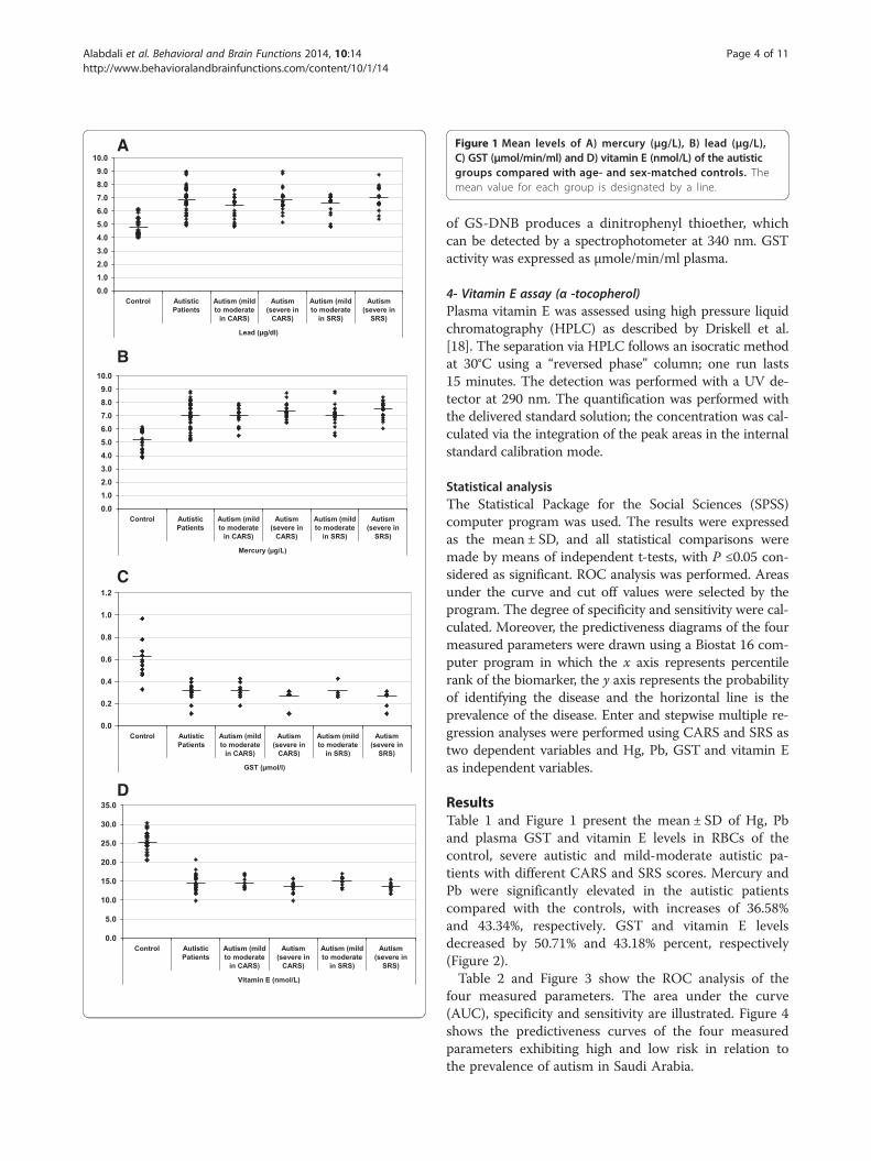
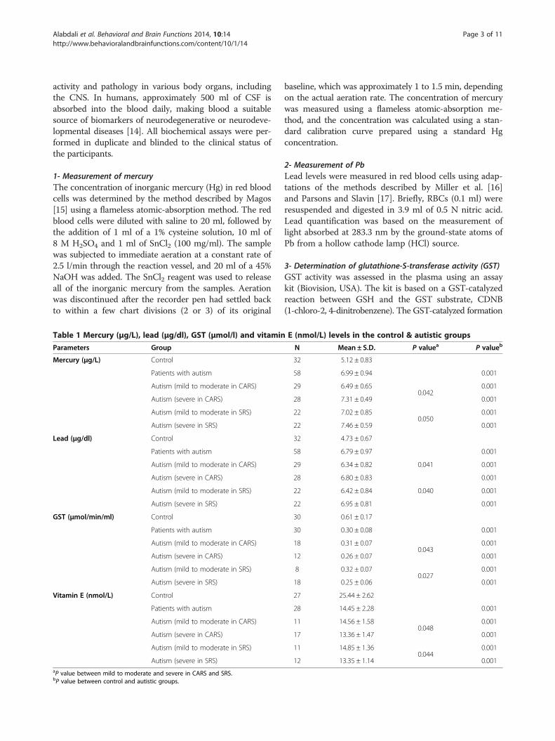
Figure 1 Mean levels of A) mercury (μg/L), B) lead (μg/L),C) GST (μmol/min/ml) and D) vitamin E (nmol/L) of the autisticgroups compared with age- and sex-matched controls. Themean value for each group is designated by a line.
Alabdali et al. Behavioral and Brain Functions 2014, 10:14 Page 4 of 11http://www.behavioralandbrainfunctions.com/content/10/1/14
of GS-DNB produces a dinitrophenyl thioether, whichcan be detected by a spectrophotometer at 340 nm. GSTactivity was expressed as μmole/min/ml plasma.
4- Vitamin E assay (α -tocopherol)Plasma vitamin E was assessed using high pressure liquidchromatography (HPLC) as described by Driskell et al.[18]. The separation via HPLC follows an isocratic methodat 30°C using a “reversed phase” column; one run lasts15 minutes. The detection was performed with a UV de-tector at 290 nm. The quantification was performed withthe delivered standard solution; the concentration was cal-culated via the integration of the peak areas in the internalstandard calibration mode.
Statistical analysisThe Statistical Package for the Social Sciences (SPSS)computer program was used. The results were expressedas the mean ± SD, and all statistical comparisons weremade by means of independent t-tests, with P ≤0.05 con-sidered as significant. ROC analysis was performed. Areasunder the curve and cut off values were selected by theprogram. The degree of specificity and sensitivity were cal-culated. Moreover, the predictiveness diagrams of the fourmeasured parameters were drawn using a Biostat 16 com-puter program in which the x axis represents percentilerank of the biomarker, the y axis represents the probabilityof identifying the disease and the horizontal line is theprevalence of the disease. Enter and stepwise multiple re-gression analyses were performed using CARS and SRS astwo dependent variables and Hg, Pb, GST and vitamin Eas independent variables.
ResultsTable 1 and Figure 1 present the mean ± SD of Hg, Pband plasma GST and vitamin E levels in RBCs of thecontrol, severe autistic and mild-moderate autistic pa-tients with different CARS and SRS scores. Mercury andPb were significantly elevated in the autistic patientscompared with the controls, with increases of 36.58%and 43.34%, respectively. GST and vitamin E levelsdecreased by 50.71% and 43.18% percent, respectively(Figure 2).Table 2 and Figure 3 show the ROC analysis of the
four measured parameters. The area under the curve(AUC), specificity and sensitivity are illustrated. Figure 4shows the predictiveness curves of the four measuredparameters exhibiting high and low risk in relation tothe prevalence of autism in Saudi Arabia.
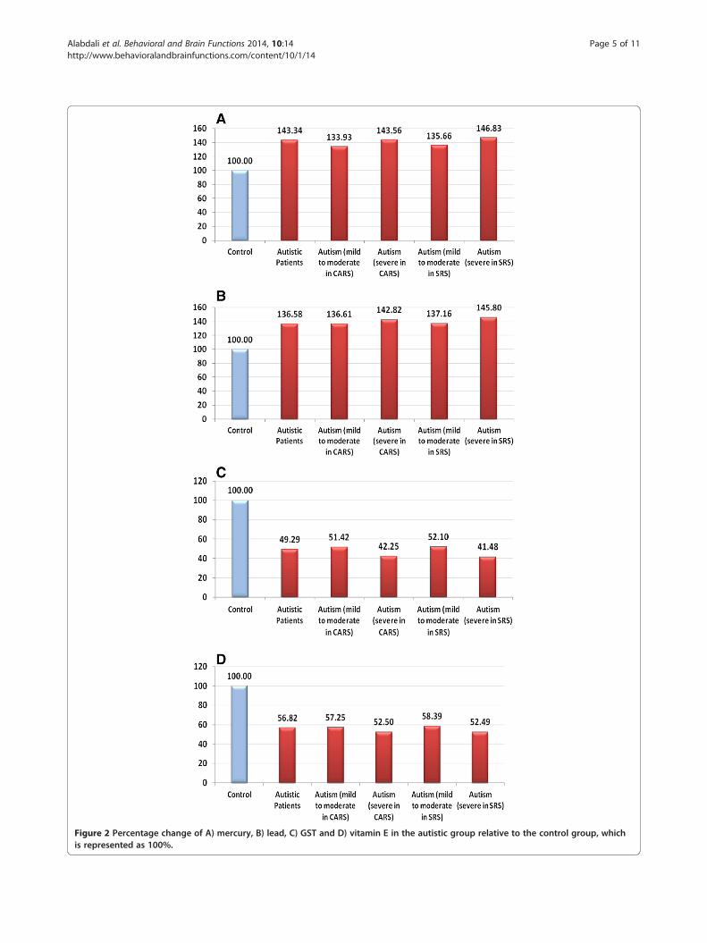
Figure 2 Percentage change of A) mercury, B) lead, C) GST and D) vitamin E in the autistic group relative to the control group, whichis represented as 100%.
Alabdali et al. Behavioral and Brain Functions 2014, 10:14 Page 5 of 11http://www.behavioralandbrainfunctions.com/content/10/1/14
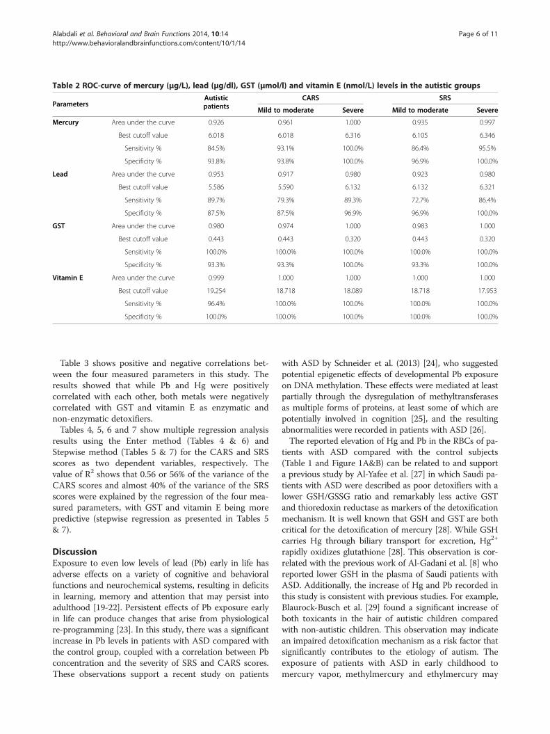
Table 2 ROC-curve of mercury (μg/L), lead (μg/dl), GST (μmol/l) and vitamin E (nmol/L) levels in the autistic groups
ParametersAutisticpatients
CARS SRS
Mild to moderate Severe Mild to moderate Severe
Mercury Area under the curve 0.926 0.961 1.000 0.935 0.997
Best cutoff value 6.018 6.018 6.316 6.105 6.346
Sensitivity % 84.5% 93.1% 100.0% 86.4% 95.5%
Specificity % 93.8% 93.8% 100.0% 96.9% 100.0%
Lead Area under the curve 0.953 0.917 0.980 0.923 0.980
Best cutoff value 5.586 5.590 6.132 6.132 6.321
Sensitivity % 89.7% 79.3% 89.3% 72.7% 86.4%
Specificity % 87.5% 87.5% 96.9% 96.9% 100.0%
GST Area under the curve 0.980 0.974 1.000 0.983 1.000
Best cutoff value 0.443 0.443 0.320 0.443 0.320
Sensitivity % 100.0% 100.0% 100.0% 100.0% 100.0%
Specificity % 93.3% 93.3% 100.0% 93.3% 100.0%
Vitamin E Area under the curve 0.999 1.000 1.000 1.000 1.000
Best cutoff value 19.254 18.718 18.089 18.718 17.953
Sensitivity % 96.4% 100.0% 100.0% 100.0% 100.0%
Specificity % 100.0% 100.0% 100.0% 100.0% 100.0%
Alabdali et al. Behavioral and Brain Functions 2014, 10:14 Page 6 of 11http://www.behavioralandbrainfunctions.com/content/10/1/14
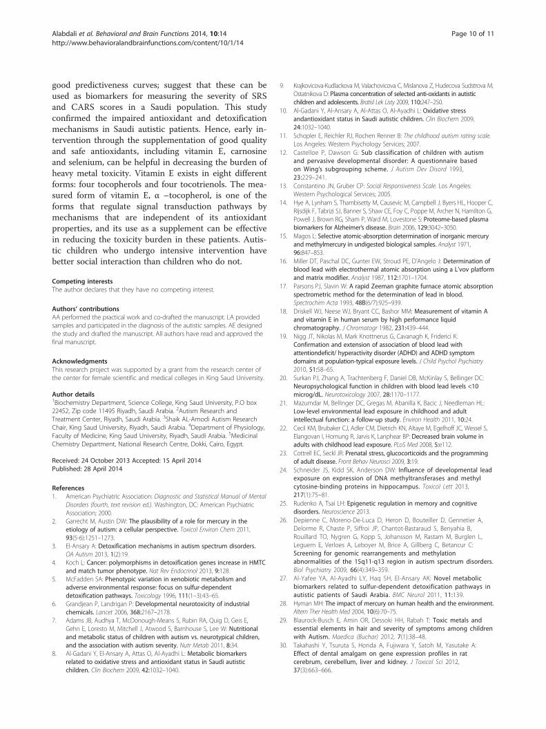
Table 3 shows positive and negative correlations bet-ween the four measured parameters in this study. Theresults showed that while Pb and Hg were positivelycorrelated with each other, both metals were negativelycorrelated with GST and vitamin E as enzymatic andnon-enzymatic detoxifiers.Tables 4, 5, 6 and 7 show multiple regression analysis
results using the Enter method (Tables 4 & 6) andStepwise method (Tables 5 & 7) for the CARS and SRSscores as two dependent variables, respectively. Thevalue of R2 shows that 0.56 or 56% of the variance of theCARS scores and almost 40% of the variance of the SRSscores were explained by the regression of the four mea-sured parameters, with GST and vitamin E being morepredictive (stepwise regression as presented in Tables 5& 7).
DiscussionExposure to even low levels of lead (Pb) early in life hasadverse effects on a variety of cognitive and behavioralfunctions and neurochemical systems, resulting in deficitsin learning, memory and attention that may persist intoadulthood [19-22]. Persistent effects of Pb exposure earlyin life can produce changes that arise from physiologicalre-programming [23]. In this study, there was a significantincrease in Pb levels in patients with ASD compared withthe control group, coupled with a correlation between Pbconcentration and the severity of SRS and CARS scores.These observations support a recent study on patients
with ASD by Schneider et al. (2013) [24], who suggestedpotential epigenetic effects of developmental Pb exposureon DNA methylation. These effects were mediated at leastpartially through the dysregulation of methyltransferasesas multiple forms of proteins, at least some of which arepotentially involved in cognition [25], and the resultingabnormalities were recorded in patients with ASD [26].The reported elevation of Hg and Pb in the RBCs of pa-
tients with ASD compared with the control subjects(Table 1 and Figure 1A&B) can be related to and supporta previous study by Al-Yafee et al. [27] in which Saudi pa-tients with ASD were described as poor detoxifiers with alower GSH/GSSG ratio and remarkably less active GSTand thioredoxin reductase as markers of the detoxificationmechanism. It is well known that GSH and GST are bothcritical for the detoxification of mercury [28]. While GSHcarries Hg through biliary transport for excretion, Hg2+
rapidly oxidizes glutathione [28]. This observation is cor-related with the previous work of Al-Gadani et al. [8] whoreported lower GSH in the plasma of Saudi patients withASD. Additionally, the increase of Hg and Pb recorded inthis study is consistent with previous studies. For example,Blaurock-Busch et al. [29] found a significant increase ofboth toxicants in the hair of autistic children comparedwith non-autistic children. This observation may indicatean impaired detoxification mechanism as a risk factor thatsignificantly contributes to the etiology of autism. Theexposure of patients with ASD in early childhood tomercury vapor, methylmercury and ethylmercury may
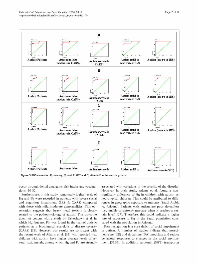
Figure 3 ROC-curves for A) mercury, B) lead, C) GST and D) vitamin E in the autistic groups.
Alabdali et al. Behavioral and Brain Functions 2014, 10:14 Page 7 of 11http://www.behavioralandbrainfunctions.com/content/10/1/14
occur through dental amalgams, fish intake and vaccina-tions [30-32].Furthermore, in this study, remarkably higher levels of
Hg and Pb were recorded in patients with severe socialand cognition impairment (SRS & CARS) comparedwith those with mild-moderate abnormalities. This ob-servation suggests that heavy metal toxicity is closelyrelated to the pathophysiology of autism. This outcomedoes not concur with a study by Elsheshtawy et al. inwhich Hg, but not Pb, was found in the hair of autisticpatients as a biochemical correlate to disease severity(CARS) [33]. However, our results are consistent withthe recent work of Adams et al. [34] who reported thatchildren with autism have higher average levels of se-veral toxic metals, among which Hg and Pb are strongly
associated with variations in the severity of the disorder.However, in their study, Adams et al. found a non-significant difference of Hg in children with autism vs.neurotypical children. This could be attributed to diffe-rences in geographic exposure to mercury (Saudi Arabiavs. Arizona). Patients with autism are poor detoxifiers(i.e., unable to detoxify mercury when it reaches a cer-tain level) [27]. Therefore, this could indicate a higherrate of exposure to Hg in the Saudi population com-pared with the population in Arizona.Face recognition is a core deficit of social impairment
in autism. A number of studies indicate that norepi-nephrine (NE) and dopamine (DA) modulate and reducebehavioral responses to changes in the social environ-ment [35,36]. In addition, serotonin (5HT) transporter
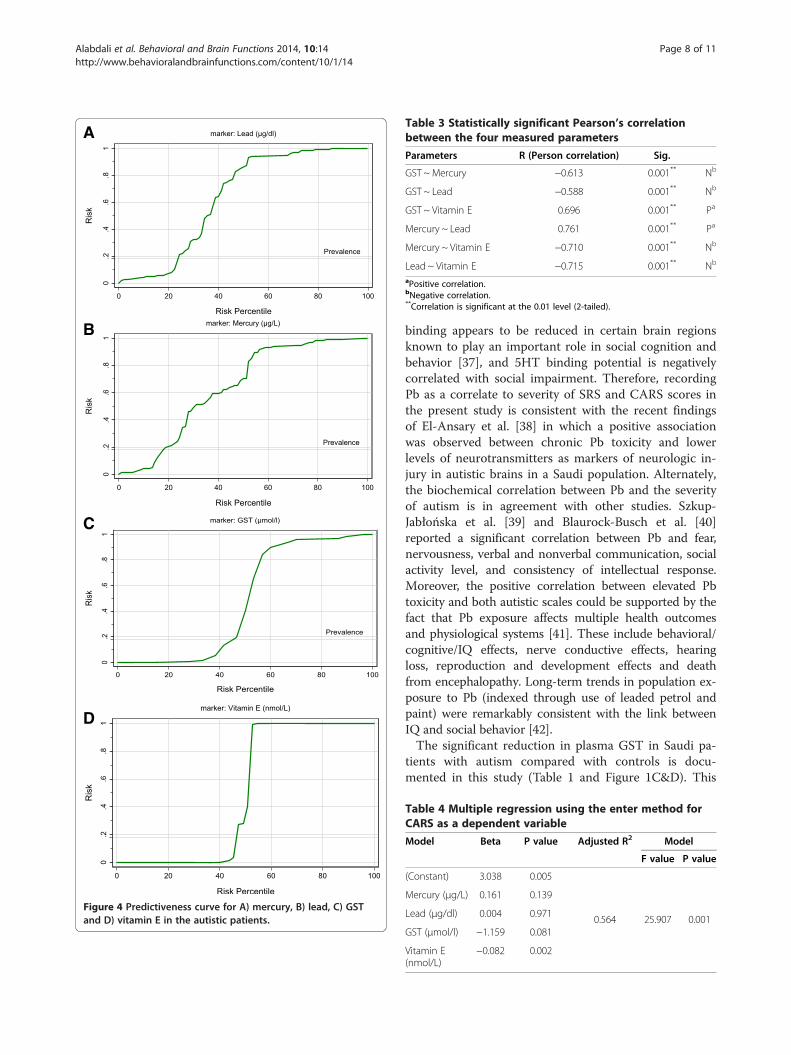
Figure 4 Predictiveness curve for A) mercury, B) lead, C) GSTand D) vitamin E in the autistic patients.
Table 3 Statistically significant Pearson’s correlationbetween the four measured parameters
Parameters R (Person correlation) Sig.
GST ~ Mercury −0.613 0.001** Nb
GST ~ Lead −0.588 0.001** Nb
GST ~ Vitamin E 0.696 0.001** Pa
Mercury ~ Lead 0.761 0.001** Pa
Mercury ~ Vitamin E −0.710 0.001** Nb
Lead ~ Vitamin E −0.715 0.001** Nb
aPositive correlation.bNegative correlation.**Correlation is significant at the 0.01 level (2-tailed).
Alabdali et al. Behavioral and Brain Functions 2014, 10:14 Page 8 of 11http://www.behavioralandbrainfunctions.com/content/10/1/14
binding appears to be reduced in certain brain regionsknown to play an important role in social cognition andbehavior [37], and 5HT binding potential is negativelycorrelated with social impairment. Therefore, recordingPb as a correlate to severity of SRS and CARS scores inthe present study is consistent with the recent findingsof El-Ansary et al. [38] in which a positive associationwas observed between chronic Pb toxicity and lowerlevels of neurotransmitters as markers of neurologic in-jury in autistic brains in a Saudi population. Alternately,the biochemical correlation between Pb and the severityof autism is in agreement with other studies. Szkup-Jabłońska et al. [39] and Blaurock-Busch et al. [40]reported a significant correlation between Pb and fear,nervousness, verbal and nonverbal communication, socialactivity level, and consistency of intellectual response.Moreover, the positive correlation between elevated Pbtoxicity and both autistic scales could be supported by thefact that Pb exposure affects multiple health outcomesand physiological systems [41]. These include behavioral/cognitive/IQ effects, nerve conductive effects, hearingloss, reproduction and development effects and deathfrom encephalopathy. Long-term trends in population ex-posure to Pb (indexed through use of leaded petrol andpaint) were remarkably consistent with the link betweenIQ and social behavior [42].The significant reduction in plasma GST in Saudi pa-
tients with autism compared with controls is docu-mented in this study (Table 1 and Figure 1C&D). This
Table 4 Multiple regression using the enter method forCARS as a dependent variable
Model Beta P value Adjusted R2 Model
F value P value
(Constant) 3.038 0.005
0.564 25.907 0.001
Mercury (μg/L) 0.161 0.139
Lead (μg/dl) 0.004 0.971
GST (μmol/l) −1.159 0.081
Vitamin E(nmol/L)
−0.082 0.002
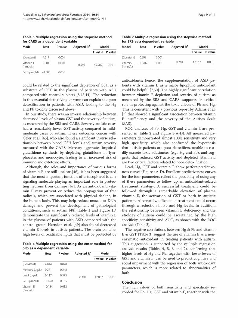
Table 5 Multiple regression using the stepwise methodfor CARS as a dependent variable
Model Beta P value Adjusted R2 Model
F value P value
(Constant) 4.517 0.001
0.560 49.909 0.001Vitamin E(nmol/L)
−0.103 0.001
GST (μmol/l) −1.383 0.035
Table 7 Multiple regression using the stepwise methodfor SRS as a dependent variable
Model Beta P value Adjusted R2 Model
F value P value
(Constant) 6.298 0.001
0.384 47.167 0.001Vitamin E(nmol/L)
−0.202 0.001
Alabdali et al. Behavioral and Brain Functions 2014, 10:14 Page 9 of 11http://www.behavioralandbrainfunctions.com/content/10/1/14
could be related to the significant depletion of GSH as asubstrate of GST in the plasma of patients with ASDcompared with control subjects [8,43,44]. The reductionin this essential detoxifying enzyme can explain the poordetoxification in patients with ASD, leading to the Hgand Pb toxicity discussed above.In our study, there was an inverse relationship between
decreased levels of plasma GST and the severity of autism,as measured by the SRS and CARS. Severely autistic caseshad a remarkably lower GST activity compared to mild-moderate cases of autism. These outcomes concur withGeier et al. [45], who also found a significant inverse rela-tionship between blood GSH levels and autism severitymeasured with the CARS. Mercury aggravates impairedglutathione synthesis by depleting glutathione in lym-phocytes and monocytes, leading to an increased risk ofimmuno and cytotoxic effects.Although, the roles and importance of various forms
of vitamin E are still unclear [46], it has been suggestedthat the most important function of α-tocopherol is as asignaling molecule playing an important role in protec-ting neurons from damage [47]. As an antioxidant, vita-min E may prevent or reduce the propagation of freeradicals, which are associated with physical decline, inthe human body. This may help reduce muscle or DNAdamage and prevent the development of pathologicalconditions, such as autism [48]. Table 1 and Figure 1Ddemonstrate the significantly reduced levels of vitamin Ein the plasma of patients with ASD compared with thecontrol group. Herndon et al. [49] also found decreasedvitamin E levels in autistic patients. The brain containshigh levels of oxidizable lipids that must be protected by
Table 6 Multiple regression using the enter method forSRS as a dependent variable
Model Beta P value Adjusted R2 Model
F value P value
(Constant) 4.844 0.028
0.391 12.867 0.001
Mercury (μg/L) 0.261 0.248
Lead (μg/dl) 0.117 0.575
GST (μmol/l) −1.890 0.185
Vitamin E(nmol/L)
−0.134 0.012
antioxidants; hence, the supplementation of ASD pa-tients with vitamin E as a major lipophilic antioxidantcould be helpful [7,50]. The highly significant correlationbetween vitamin E depletion and severity of autism, asmeasured by the SRS and CARS, supports its criticalrole in protecting against the toxic effects of Pb and Hg.This is consistent with a previous report by Adams et al.[7] that showed a significant association between vitaminE insufficiency and the severity of the Autism Scale(SAS).ROC analyses of Pb, Hg, GST and vitamin E are pre-
sented in Table 2 and Figure 3(A-D). All measured pa-rameters demonstrated almost 100% sensitivity and veryhigh specificity, which also confirmed the hypothesisthat autistic patients are poor detoxifiers, unable to rea-dily excrete toxic substances (e.g., Hg and Pb), and sug-gests that reduced GST activity and depleted vitamin Eare two critical factors related to poor detoxification.Lead, Hg, GST and vitamin E show perfect predictive-
ness curves (Figure 4A-D). Excellent predictiveness curvesfor the four parameters reflect the possibility of using anyof these parameters to follow up an antioxidant-relatedtreatment strategy. A successful treatment could befollowed through a remarkable elevation of plasmavitamin E, the activation of GST or both in autisticpatients. Alternately, efficacious treatment could occurthrough a reduction in Pb and Hg levels. In addition,the relationship between vitamin E deficiency and theetiology of autism could be ascertained by the highspecificity, sensitivity and AUC, as shown with the ROCanalysis (Table 2).The negative correlations between Hg & Pb and vitamin
E & GST (Table 3) suggest the use of vitamin E as a non-enzymatic antioxidant in treating patients with autism.This suggestion is supported by the multiple regressionanalysis results (Tables 4, 5, 6 and 7), confirming thathigher levels of Hg and Pb, together with lower levels ofGST and vitamin E, can be used to predict cognitive andsocial impairment with the regression of both antioxidantparameters, which is more related to abnormalities ofboth.
ConclusionThe high values of both sensitivity and specificity re-corded for Pb, Hg, GST and vitamin E, together with the
Alabdali et al. Behavioral and Brain Functions 2014, 10:14 Page 10 of 11http://www.behavioralandbrainfunctions.com/content/10/1/14
good predictiveness curves; suggest that these can beused as biomarkers for measuring the severity of SRSand CARS scores in a Saudi population. This studyconfirmed the impaired antioxidant and detoxificationmechanisms in Saudi autistic patients. Hence, early in-tervention through the supplementation of good qualityand safe antioxidants, including vitamin E, carnosineand selenium, can be helpful in decreasing the burden ofheavy metal toxicity. Vitamin E exists in eight differentforms: four tocopherols and four tocotrienols. The mea-sured form of vitamin E, α –tocopherol, is one of theforms that regulate signal transduction pathways bymechanisms that are independent of its antioxidantproperties, and its use as a supplement can be effectivein reducing the toxicity burden in these patients. Autis-tic children who undergo intensive intervention havebetter social interaction than children who do not.
Competing interestsThe author declares that they have no competing interest.
Authors’ contributionsAA performed the practical work and co-drafted the manuscript. LA providedsamples and participated in the diagnosis of the autistic samples. AE designedthe study and drafted the manuscript. All authors have read and approved thefinal manuscript.
AcknowledgmentsThis research project was supported by a grant from the research center ofthe center for female scientific and medical colleges in King Saud University.
Author details1Biochemistry Department, Science College, King Saud University, P.O box22452, Zip code 11495 Riyadh, Saudi Arabia. 2Autism Research andTreatment Center, Riyadh, Saudi Arabia. 3Shaik AL-Amodi Autism ResearchChair, King Saud University, Riyadh, Saudi Arabia. 4Department of Physiology,Faculty of Medicine, King Saud University, Riyadh, Saudi Arabia. 5MedicinalChemistry Department, National Research Centre, Dokki, Cairo, Egypt.
Received: 24 October 2013 Accepted: 15 April 2014Published: 28 April 2014
References1. American Psychiatric Association: Diagnostic and Statistical Manual of Mental
Disorders (fourth, text revision ed.). Washington, DC: American PsychiatricAssociation; 2000.
2. Garrecht M, Austin DW: The plausibility of a role for mercury in theetiology of autism: a cellular perspective. Toxicol Environ Chem 2011,93(5-6):1251–1273.
3. El-Ansary A: Detoxification mechanisms in autism spectrum disorders.OA Autism 2013, 1(2):19.
4. Koch L: Cancer: polymorphisms in detoxification genes increase in HMTCand match tumor phenotype. Nat Rev Endocrinol 2013, 9:128.
5. McFadden SA: Phenotypic variation in xenobiotic metabolism andadverse environmental response: focus on sulfur-dependentdetoxification pathways. Toxicology 1996, 111(1–3):43–65.
6. Grandjean P, Landrigan P: Developmental neurotoxicity of industrialchemicals. Lancet 2006, 368:2167–2178.
7. Adams JB, Audhya T, McDonough-Means S, Rubin RA, Quig D, Geis E,Gehn E, Loresto M, Mitchell J, Atwood S, Barnhouse S, Lee W: Nutritionaland metabolic status of children with autism vs. neurotypical children,and the association with autism severity. Nutr Metab 2011, 8:34.
8. Al-Gadani Y, El-Ansary A, Attas O, Al-Ayadhi L: Metabolic biomarkersrelated to oxidative stress and antioxidant status in Saudi autisticchildren. Clin Biochem 2009, 42:1032–1040.
9. Krajkovicova-Kudlackova M, Valachovicova C, Mislanova Z, Hudecova Sudstrova M,Ostatnikova D: Plasma concentration of selected anti-oxidants in autisticchildren and adolescents. Bratisl Lek Listy 2009, 110:247–250.
10. Al-Gadani Y, Al-Ansary A, Al-Attas O, Al-Ayadhi L: Oxidative stressandantioxidant status in Saudi autistic children. Clin Biochem 2009,24:1032–1040.
11. Schopler E, Reichler RJ, Rochen Renner B: The childhood autism rating scale.Los Angeles: Western Psychology Services; 2007.
12. Castelloe P, Dawson G: Sub classification of children with autismand pervasive developmental disorder: A questionnaire basedon Wing’s subgrouping scheme. J Autism Dev Disord 1993,23:229–241.
13. Constantino JN, Gruber CP: Social Responsiveness Scale. Los Angeles:Western Psychological Services; 2005.
14. Hye A, Lynham S, Thambisetty M, Causevic M, Campbell J, Byers HL, Hooper C,Rijsdijk F, Tabrizi SJ, Banner S, Shaw CE, Foy C, Poppe M, Archer N, Hamilton G,Powell J, Brown RG, Sham P, Ward M, Lovestone S: Proteome-based plasmabiomarkers for Alzheimer’s disease. Brain 2006, 129:3042–3050.
15. Magos L: Selective atomic-absorption determination of inorganic mercuryand methylmercury in undigested biological samples. Analyst 1971,96:847–853.
16. Miller DT, Paschal DC, Gunter EW, Stroud PE, D’Angelo J: Determination ofblood lead with electrothermal atomic absorption using a L'vov platformand matrix modifier. Analyst 1987, 112:1701–1704.
17. Parsons PJ, Slavin W: A rapid Zeeman graphite furnace atomic absorptionspectrometric method for the determination of lead in blood.Spectrochim Acta 1993, 48B(6/7):925–939.
18. Driskell WJ, Neese WJ, Bryant CC, Bashor MM: Measurement of vitamin Aand vitamin E in human serum by high performance liquidchromatography. J Chromatogr 1982, 231:439–444.
19. Nigg JT, Nikolas M, Mark Knottnerus G, Cavanagh K, Friderici K:Confirmation and extension of association of blood lead withattentiondeficit/ hyperactivity disorder (ADHD) and ADHD symptomdomains at population-typical exposure levels. J Child Psychol Psychiatry2010, 51:58–65.
20. Surkan PJ, Zhang A, Trachtenberg F, Daniel DB, McKinlay S, Bellinger DC:Neuropsychological function in children with blood lead levels <10microg/dL. Neurotoxicology 2007, 28:1170–1177.
21. Mazumdar M, Bellinger DC, Gregas M, Abanilla K, Bacic J, Needleman HL:Low-level environmental lead exposure in childhood and adultintellectual function: a follow-up study. Environ Health 2011, 10:24.
22. Cecil KM, Brubaker CJ, Adler CM, Dietrich KN, Altaye M, Egelhoff JC, Wessel S,Elangovan I, Hornung R, Jarvis K, Lanphear BP: Decreased brain volume inadults with childhood lead exposure. PLoS Med 2008, 5:e112.
23. Cottrell EC, Seckl JR: Prenatal stress, glucocorticoids and the programmingof adult disease. Front Behav Neurosci 2009, 3:19.
24. Schneider JS, Kidd SK, Anderson DW: Influence of developmental leadexposure on expression of DNA methyltransferases and methylcytosine-binding proteins in hippocampus. Toxicol Lett 2013,217(1):75–81.
25. Rudenko A, Tsai LH: Epigenetic regulation in memory and cognitivedisorders. Neuroscience 2013.
26. Depienne C, Moreno-De-Luca D, Heron D, Bouteiller D, Gennetier A,Delorme R, Chaste P, Siffroi JP, Chantot-Bastaraud S, Benyahia B,Rouillard TO, Nygren G, Kopp S, Johansson M, Rastam M, Burglen L,Leguern E, Verloes A, Leboyer M, Brice A, Gillberg C, Betancur C:Screening for genomic rearrangements and methylationabnormalities of the 15q11-q13 region in autism spectrum disorders.Biol Psychiatry 2009, 66(4):349–359.
27. Al-Yafee YA, Al-Ayadhi LY, Haq SH, El-Ansary AK: Novel metabolicbiomarkers related to sulfur-dependent detoxification pathways inautistic patients of Saudi Arabia. BMC Neurol 2011, 11:139.
28. Hyman MH: The impact of mercury on human health and the environment.Altern Ther Health Med 2004, 10(6):70–75.
29. Blaurock-Busch E, Amin OR, Dessoki HH, Rabah T: Toxic metals andessential elements in hair and severity of symptoms among childrenwith Autism. Maedica (Buchar) 2012, 7(1):38–48.
30. Takahashi Y, Tsuruta S, Honda A, Fujiwara Y, Satoh M, Yasutake A:Effect of dental amalgam on gene expression profiles in ratcerebrum, cerebellum, liver and kidney. J Toxicol Sci 2012,37(3):663–666.
Alabdali et al. Behavioral and Brain Functions 2014, 10:14 Page 11 of 11http://www.behavioralandbrainfunctions.com/content/10/1/14
31. Mania M, Wojciechowska-Mazurek M, Starska K, Rebeniak M, Postupolski J:Fish and seafood as a source of human exposure to methylmercury.Rocz Panstw Zakl Hig 2012, 63(3):257–264.
32. Aschner M, Ceccatelli S: Are neuropathological conditions relevant toethyl mercury exposure? Neurotox Res 2010, 18(1):59–68.
33. Elsheshtawy E, Tobar S, Sherra K, Atallah S, Elkasaby R: Study of somebiomarkers in hair of children with autism. Middle East Curr Psychiatry 2011,18:6–10.
34. Adams JB, Audhya T, McDonough-Means S, Rubin RA, Quig D, Geis E, Gehn E,Loresto M, Mitchell J, Atwood S, Barnhouse S, Lee W: Toxicological status ofchildren with autism vs. neurotypical children and the association withautism severity. Biol Trace Elem Res 2013, 151(2):171–180.
35. Marino MD, Bourdelat-Parks BN, Liles LC, Weinshenker D: Genetic reductionof noradrenergic function alters social memory and reduces aggressionin mice. Behav Brain Res 2005, 161:197–203.
36. Stone JM, Morrison PD, Pilowsky LS: Glutamate and dopaminedysregulation in schizophrenia-a synthesis and selective review.J Psychopharmacol 2007, 21:440–452.
37. Makkonen I, Riikonen R, Kokki H, Airaksinen MM, Kuikka JT: Serotonin anddopamine transporter binding in children with autism determined bySPECT. Dev Med Child Neurol 2008, 50:593–597.
38. El-Ansary AK, Bacha AB, Ayahdi LY: Relationship between chronic leadtoxicity and plasma neurotransmitters in autistic patients fromSaudi Arabia. Clin Biochem 2011, 44:1116–1120.
39. Szkup-Jabłońska M, Karakiewicz B, Grochans E, Jurczak A, Zaremba-Pechmann L,Rotter I, Nowak-Starz G, Samochowiec J: The effects of lead level in the bloodon social functioning in children with developmental disabilities.Psychiatr Pol 2011, 45(5):713–722.
40. Blaurock-Busch E, Amin OR, Rabah T: Heavy metals and trace elements inhair and urine of a sample of arab children with autistic spectrumdisorder. Maedica 2011, 6:247–257.
41. ATSDR: Toxicological Profile for Lead (update). (Agency for Toxic Substancesand Disease Registry). Atlanta, GA: U.S. Department of Health and HumanServices; 1999.
42. Nevin R: How lead exposure relates to temporal changes in IQ, violentcrime, and unwed pregnancy. Environ Res 2000, 83:1–22.
43. Geier DA, King PG, Sykes LK, Geier MR: A comprehensive review ofmercury provoked autism. Indian J Med Res 2008, 128:383–411.
44. Vojdani A, Mumper E, Granpeesheh D, Mielke L, Traver D, Bock K, Hirani K,Neubrander J, Woeller KN, O'Hara N, Usman A, Schneider C, Hebroni F,Berookhim J, McCandless J: Low natural killer cell cytotoxic activity inautism: the role of glutathione, IL-2 and IL-15. J Neuroimmunol 2008,205(1–2):148–154.
45. Geier DA, Kern J, Geier MA: A prospective study of oxidative stressbiomarkers in autistic disorders. Electron J Appl Psychol Innov Autism 2009,5:2–10.
46. Atkinson A, Epand RF, Epand RM: Tocopherols and tocotrienols inmembranes: a critical review. Free Radic Biol Med 2007, 44:739–764.
47. Zingg JM, Azzi A: Non-antioxidant activities of vitamin E. Curr ChemMed 2004, 11:1113–1133.
48. Azzi A: Molecular mechanism of alpha-tocopherol action. Free RadicBiol Med 2007, 43(1):16–21.
49. Herndon AC, Diguiseppi C, Johndon SL: Dose nutritional intake differsbetween children with autism spectrum disorder and children withtypical development? Autism Dev Disord 2008, 6:341–349.
50. Kontush KO, Schekatolina A: Vitamin E in neurodegenerative disorder:Alzheimers disease. Ann N Y Acad Sci 2004, 1031:249–262.
doi:10.1186/1744-9081-10-14Cite this article as: Alabdali et al.: A key role for an impaireddetoxification mechanism in the etiology and severity of autismspectrum disorders. Behavioral and Brain Functions 2014 10:14.
Submit your next manuscript to BioMed Centraland take full advantage of:
• Convenient online submission
• Thorough peer review
• No space constraints or color figure charges
• Immediate publication on acceptance
• Inclusion in PubMed, CAS, Scopus and Google Scholar
• Research which is freely available for redistribution
Submit your manuscript at www.biomedcentral.com/submit