Embed Size (px)
Citation preview

RESEARCH Open Access
Advanced 3D printed model of middlecerebral artery aneurysms for neurosurgerysimulationRuth G. Nagassa1* , Paul G. McMenamin1, Justin W. Adams1, Michelle R. Quayle1 and Jeffrey V. Rosenfeld2,3,4
Abstract
Background: Neurosurgical residents are finding it more difficult to obtain experience as the primary operator inaneurysm surgery. The present study aimed to replicate patient-derived cranial anatomy, pathology and humantissue properties relevant to cerebral aneurysm intervention through 3D printing and 3D print-driven castingtechniques. The final simulator was designed to provide accurate simulation of a human head with a middlecerebral artery (MCA) aneurysm.
Methods: This study utilized living human and cadaver-derived medical imaging data including CT angiographyand MRI scans. Computer-aided design (CAD) models and pre-existing computational 3D models were alsoincorporated in the development of the simulator. The design was based on including anatomical componentsvital to the surgery of MCA aneurysms while focusing on reproducibility, adaptability and functionality of thesimulator. Various methods of 3D printing were utilized for the direct development of anatomical replicas andmoulds for casting components that optimized the bio-mimicry and mechanical properties of human tissues.Synthetic materials including various types of silicone and ballistics gelatin were cast in these moulds. A noveltechnique utilizing water-soluble wax and silicone was used to establish hollow patient-derived cerebrovascularmodels.
Results: A patient-derived 3D aneurysm model was constructed for a MCA aneurysm. Multiple cerebral aneurysmmodels, patient-derived and CAD, were replicated as hollow high-fidelity models. The final assembled simulatorintegrated six anatomical components relevant to the treatment of cerebral aneurysms of the Circle of Willis in theleft cerebral hemisphere. These included models of the cerebral vasculature, cranial nerves, brain, meninges, skulland skin. The cerebral circulation was modeled through the patient-derived vasculature within the brain model.Linear and volumetric measurements of specific physical modular components were repeated, averaged andcompared to the original 3D meshes generated from the medical imaging data. Calculation of the concordancecorrelation coefficient (ρc: 90.2%–99.0%) and percentage difference (≤0.4%) confirmed the accuracy of the models.
Conclusions: A multi-disciplinary approach involving 3D printing and casting techniques was used to successfullyconstruct a multi-component cerebral aneurysm surgery simulator. Further study is planned to demonstrate theeducational value of the proposed simulator for neurosurgery residents.
Keywords: Neurosurgical training, Anatomical models, Aneurysm, 3D printing, Simulation
© The Author(s). 2019 Open Access This article is distributed under the terms of the Creative Commons Attribution 4.0International License (http://creativecommons.org/licenses/by/4.0/), which permits unrestricted use, distribution, andreproduction in any medium, provided you give appropriate credit to the original author(s) and the source, provide a link tothe Creative Commons license, and indicate if changes were made.
* Correspondence: [email protected] of Anatomy and Developmental Biology, Monash University,Clayton, VIC, AustraliaFull list of author information is available at the end of the article
Nagassa et al. 3D Printing in Medicine (2019) 5:11 https://doi.org/10.1186/s41205-019-0048-9
BackgroundNeurosurgery trainees are finding it increasingly difficultto obtain operative experience as the primary operatorin aneurysm surgery [1]. Good quality cadaver dissectionopportunities are also not readily available for neurosur-gery residents. Simulation is emerging as a useful train-ing aid for neurosurgery [2]. The treatment of cerebralaneurysms requires specialized skill development andproficient use of micro-instruments. Furthermore, anyadvance in neurosurgical training methods is of potentialvalue to both neurosurgeons and patients [3]. Operativecaseload of neurosurgical trainees forms an extensiverole in training, alongside surgical simulation in the de-velopment of primary neurosurgical techniques [4].Current simulation methods include the use of humancadavers, large animal models, medical manikins and vir-tual simulation with haptic feedback [5, 6]. However,these models rarely simulate the entire procedure orprovide realistic haptic feedback [7, 8]. A 3D modelmimicking human tissue would allow trainees to gothrough the basic operative steps of specific proceduresand navigate through anatomical landmarks enabling ef-fective training with the supervision of superiors in asafe environment [9]. Simulation-based training has beenshown to improve non-technical skills including cogni-tive and interpersonal skills that can be overlooked insurgical training [10, 11]. Such improvements in thecontext of neurosurgical trainees would decrease med-ical error and potentially improve patient outcomes [12].3D printed models have been demonstrated to accur-ately replicate patient-specific vascular structures [11].Such models have been shown to be valuable in endo-vascular coiling simulation where anatomical complex-ities are detected through medical imaging and therefore
require the determination of a preoperative tactile ap-proach [13]. They can assist in improving the under-standing of spatial anatomy configuration, particularly incases of challenging vasculature [13].In many cases, 3D printed simulation models have not
replicated the cerebral aneurysm together with adjacentarteries [14], vessel lumen [15] and haptic properties.The ideal model for cranial surgery simulation shouldinclude: head anatomy with surface landmarks; ability tobe positioned in a three point head clamp; realistic scalp,bone, dura and brain; and accurate representation ofpathology [16].Here we describe our development of a cerebral
aneurysm simulator that includes the entire human headand relevant surgical anatomy for complete simulationof cerebrovascular surgery.
MethodsTo improve the reproducibility and cost efficacy, the leftside of the brain was designed with the surgical ap-proach and realistic pathology of a middle cerebral ar-tery (MCA) aneurysm at the M1/M2 junction. Due tothe inability to obtain all necessary components of acomprehensive simulator from a singular patient dataset,each component was developed from various patientmedical imaging and computational 3D models that ob-tained optimal clarity of the desired anatomy.Non-anatomical geometries were designed on the 3D
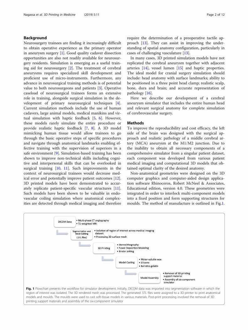
computer graphics and computer-aided design applica-tion software Rhinoceros, Robert McNeel & Associates,Educational edition, version 4.0. These geometries wereintegrated in order to interlock multi-component modelsinto a fixed position and form supporting structures formoulds. The method of manufacture is outlined in Fig.1.
Fig. 1 Flowchart presents the workflow for simulator development. Initially, DICOM data was imported into segmentation software in which theregion of interest was isolated. The 3D rendered mesh was processed. The generated .STL files were assigned to a 3D printer to print anatomicalmodels and moulds. The moulds were used to cast soft-tissue models in various materials. Post-print processing involved the removal of 3Dprinting support materials and assembly of the six-component simulator
Nagassa et al. 3D Printing in Medicine (2019) 5:11 Page 2 of 12

The parameters of each 3D printed model and proper-ties of the 3D print-driven casts are listed in Tables 1and 2 respectively.
Developing vascular models with LuminaThis study made use of CT angiograms (multiphaseCT2) of patients who presented to the Alfred Hospital(Melbourne, Victoria) with cerebral aneurysms in 2017.Data selection was restricted to cases of aneurysms thatoccurred at the MCA, with a defined aneurysm neck,aneurysm size greater than 5 mm and cases that weresurgically clipped. A single dataset was chosen that metthese requirements. The data belonged to a male patientwho presented with a saccular aneurysm at the M1/M2junction. CT angiography was performed on a SiemensAxiom Artis Angio System at a slice thickness of 0.36mm (512 slices) at the Alfred Health Radiology Service(Melbourne, Victoria). For reconstructive purposes thepatient aneurysm dataset was merged with a pre-existing3D computational model derived from a patient CTangiogram obtained from the Centre of Human Anat-omy Education (CHAE), Department of Anatomy andDevelopmental Biology (ADB), Monash University,which contained a clear visual of the internal carotid ar-tery (ICA).The medical image files (IMA files) were converted to
Digital Imaging and Communications in Medicine(DICOM) data using the free access software 3D Slicer(version 4.6). The DICOM data was then imported intosegmentation software Mimics, Materialise Software,version 19.0 Leuven, Belgium to isolate the cerebral vas-culature across all slices of the scan. A semi-automaticthresholding process reported the Hounsfield units (HU)and tissue with a density ranging 2848 to 12807 HUwere segmented. Following segmentation, the geometrieswere rendered as a 3D mesh and converted to a Binary,Little Endian standard tessellation language (.STL file).The files were edited on Geomagic Control, 3D Systemswhere the left ICA, MCA and anterior cerebral artery(ACA) were collectively partitioned from the Circle ofWillis. The models were printed on the Formlabs Form2 high definition desktop 3D printer in photopolymerresin (V3 FLGPWH03). A two-part mould of the model
was created using Dalchem Mould Making Silicone Rub-ber SRT-30®. A negative of the model was achieved andthe hollow cavity was filled with Sol-U-Carv Directionswater-soluble wax. The wax model was then brush-coated with Smooth-On™ Dragon skin 10® platinum curesilicone rubber with Smooth-On™ pigment additive SilcPig in the color Blood (PMS 7421C). Two or three layersof the silicone was applied to achieve a vessel wall (Fig.2a). The model was placed in water allowing the wax todissolve through evacuation points to establish a vessellumen. Vessel wall thickness was measured using theMitutoyo Sliding Caliper (Absolute, 500-171-30).
Computer-aided design aneurysmAn aneurysm was constructed on Geomagic Controlusing the Feature tool to form a sphere 10mm in diam-eter. To construct an aneurysmal neck a cylinder with adiameter of 5 mm was constructed in correspondencewith the width of the parent vasculature. The CADaneurysm was merged using the Combine tool at theMCA. Following the same method, the model wasprinted, moulded and cast. In order to replicate the sen-sitivity of an aneurysmal vascular wall, the aneurysm wascoated in a single layer of silicone and two layers werecoated on the remaining vasculature (Fig. 2b-d).
Patient aneurysmThe aneurysm and parent vessel geometries were de-signed separately to facilitate the alteration of aneurysmmodels and allow easier post-printing manipulation. OnGeomagic Control the aneurysm was partitioned withthe Trim With Plane tool to segment the MCA fromM1 to M2. The isolated patient aneurysm and secondaryvasculature model were merged using the Combine toolto visualize the appropriate orientation to be achievedpost-printing. Following the same method, the modelwas printed, moulded and cast in wax. Melted Sol-U-Carv wax facilitated the adhesion of the patientaneurysm to the vasculature model. One layer of siliconewas coated on the aneurysm and two layers were coatedon the remaining vasculature.
Table 1 Parameters of each 3D printed model
Vasculature Model Cranial NerveModel
Brain Mould(Left and Right)
Skull Model(Two-components)
Skin Mould
3D Printer Formlabs Form 2 MakerBotReplicator 2X
3D Systems ZPrinter650
3D Systems ZPrinter650
3D Systems ZPrinter650
Substrate Photopolymer resin (V3FLGPWH03)
PolymakerPolyflex™
Zp131 Powder, Zb61Binder
Zp131 Powder, Zb61Binder
Zp131 Powder, Zb61Binder
Printer Resolution(mm)
0.05 0.15 0.10 0.10 0.10
Print Time (hours) 3.5 0.8 13.5 11.5 20.5
Nagassa et al. 3D Printing in Medicine (2019) 5:11 Page 3 of 12

Circulatory systemThe Waterpik® Waterflosser Ultra (WP100) was used toreplicate the cerebral circulation. The tip of the appar-atus that projects fluid in a pulsatile manner was intro-duced to the vascular model and tubes used to extendthe terminal branches to form an output. The tubingwas connected to the water reservoir of the Waterpik toform a circuit. Smooth-On Ultimate Blood® Base wasadded to the water reservoir to mimic the color ofblood. The circulatory system projected through thepatient-derived vasculature within the brain model.
Cranial nerve modelThe optic chiasm, optic nerve and olfactory tract wereincluded to serve as anatomical landmarks for intraoper-ative navigation. Pre-existing data of a dissected speci-men (MP1670 Head and Visceral Column of the Neck)from the CHAE, Department of ADB, Monash
University, was used to isolate this desired anatomy. The.STL file was imported into Geomagic Control where abox developed on Rhinoceros was used to eliminatestructures of the cranial cavity using the Boolean (Sub-tract One) tool, until the optic chiasm, optic nerve andolfactory tract remained. The open surfaces were re-solved with the Fill All (Flat) tool. The model wasprinted on the MakerBot Replicator 2X desktop 3Dprinter in thermoplastic elastomer Polymaker Polyflex™(Fig. 3). The print was adhered to the cranial base of theskull model using super glue.
Brain modelThe brain model was developed from a patient T1-weighted MRI scan obtained from The Cancer ImagingArchive (TCIA) at www.cancerimagingarchive.net. Themedical imaging data was chosen based on the numberof slices (512 coronal; 512 sagittal; 212 transverse) and
Table 2 Mechanical and physical properties of 3D print-driven casts
Mechanical and Physical Properties Dragon Skin™ 10Fast
Ecoflex™ 00–10 Clear BallisticsMedical Gel #4
Product Type Silicone Rubber –Platinum Cure
Silicone Rubber –Platinum Cure
SyntheticGelatin
Mix Ratio By Volume 1A: 1B 1A: 1B –
Mix Ration By Weight 1A: 1B 1A: 1B –
Pot Life (minutes) 8 30 –
Cure Time (hours) 1.25 4 3
Hardness 10 Shore A 00–10 Shore A 3.3 Shore OO
Density (kg/m3) – – 834
Melting Point (°C) – – 125
Tensile Strength (psi) 475 120 –
100% Modulus (psi) 22 8 –
Cost US $16.11/ lb US $16.11/ lb US $31.81/ lb
Fig. 2 a Terminal branches of the patient-derived vasculature lumen (left MCA) in which two layers of silicone (line) produced a thinner, moreaccurate representation of vessel wall thickness than that of three layers (double sided arrow). b 3D print of CAD aneurysm model used todevelop c a wax cast and d hollow silicone model
Nagassa et al. 3D Printing in Medicine (2019) 5:11 Page 4 of 12

slice thickness (slice increment 1.0 mm; pixel size 0.51mm) for optimal data extraction. Data selection was re-stricted to MRI scans that lacked superficial pathologicalabnormalities that could impact the overall interface ofthe brain.The data was imported into Aviso Lite (FEI Soft-
ware, version 9.0.1.) accessed through Multi-modalAustralian Sciences Imaging and Visualisation Envir-onment (MASSIVE). Segmentation of the brain wasperformed using Magic Wand to select data withinthe threshold range of 1245 to 1284 HU. Precise iso-lation of boundaries involved manually tracing thecomplete geometry of the brain using the Lasso toolacross all planes. Following segmentation, the GenerateSurface tool was used to construct a 3D surface modelthrough Unconstrained Smoothing. The final surfacemodel was imported into Geomagic Wrap® (2015.1.1).Using Trim With Plane tool, the brain was trimmed
along the longitudinal fissure to separate the two cere-bral hemispheres. To achieve a dissectible Sylvian fissurethat could be mechanically manipulated during simula-tion, the brain mesh was trimmed at tunnels formed atthe anterior ramus and posterior ramus of the lateral
sulcus. The dimensions of the Sylvian fissure were takenusing the Analysis Measure Distance tool.Each hemisphere was inverted using the Invert Planes
tool in order to have the desired anatomy on the exteriorsurface. A box developed on Rhinoceros was mergedwith each hemisphere using the Combine tool to de-velop a block mould. The moulds were printed on theZPrinter 650, 3D Systems powder-based printer. Themould of the left hemisphere was used to cast a replicain Clear Ballistics medical gel #4 with the lowest densityto achieve haptic properties of brain tissue. The rightwas cast in Clear Ballistics gel 10% to provide a highershore hardness surface to prevent slumping of the lefthemisphere within the simulator. Both gels were com-bined with Silc Pig® pigment additives in the color FleshTone and White (PMS White) to replicate the color andconsistency of brain tissue (Fig. 4).
Meninges modelDura materTwo successive layers of Smooth-On Dragon Skin 10combined with Silc Pig® pigment additives (PMS 488C,PMS107C and PMS White) was coated onto the deep
Fig. 3 a Computational model of dissected specimen (MP1670) with the optic chiasm, optic nerve and olfactory tract (between arrows) that wereisolated b as a separate mesh and c 3D printed through FDM technology to achieve a flexible 3D printed model
Fig. 4 a Right cerebral hemisphere cast in Clear Ballistics gel 10% with firmer material properties to that of the b left cerebral hemisphere cast inClear Ballistics medical gel #4
Nagassa et al. 3D Printing in Medicine (2019) 5:11 Page 5 of 12

surface of the replaceable bone component of the skullmodel until an even distribution of silicone wasachieved. Once cured, the silicone was peeled off theskull and the thickness measured using the slidingcalipers. The silicone was re-adhered to the skullthrough the application of Smooth-On Sil-Poxy Siliconeadhesive (Fig. 5a).
Pia materOne layer of Smooth-On Dragon Skin 10 combined withSilc Pig® White (PMS White) was brush coated onto thesurface of the brain model (left hemisphere) (Fig. 5b-c).
Skull modelThe simulator skull was produced using two pre-existingcomputational models derived from CT scans of acadaveric adult cranium obtained from the CHAE, De-partment of ADB, Monash University. The two datasetscontained anatomical omissions that were rectifiedthrough merging the two models using Geomagic Con-trol. The two models were aligned using Manual Regis-tration then trimmed along the same plane superior tothe zygomatic process of the frontal bone using TrimWith Plane. The subsequent superior and inferior re-gions were merged using Combine and connected usingFill Single (Flat) tools. The Defeature tool was used tosmooth the joined sites. A cube developed in Rhinoceroswas used to partition the cranium. The cube was posi-tioned in order for the replaceable component to includeall critical anatomy common to pterional and orbitozy-gomatic craniotomies. The Boolean (Subtract 1/ Inter-sect) tool was used to produce the cranium andreplaceable bone flap component. The two-componentskull model was printed on the ZPrinter 650 in a gyp-sum powder composite material previously reported assimilar to that of bone (Fig. 6) [15].
Skin modelA full size human head model was downloaded from afree access site (Thingiverse; www.thingiverse.com) as a.STL file. The model incorporated facial anatomy thatserved as surgical landmarks and improved the fidelityof the simulator. The .STL file was scaled down to cor-respond with the size of the skull model. A four-partmould was developed using Invert Planes/Flip Normals,Trim With Plane (Intersect Plane) and Combine tools.The mould was printed on the ZPrinter 650, assembled,sealed and cast in Smooth-On Eco-flex 10® with Silc Pig®pigment (PMS 488C) and Surface Tension Diffuser to fa-cilitate demoulding (Fig. 7).
Accuracy validationLinear and volumetric measurements of specific physicalmodular components were repeated, averaged and com-pared to the computational 3D meshes generated fromthe medical imaging data. Mean measurements were re-corded at defined points of the 3D printed model, castmodel and 3D rendered mesh. Measurements includedthat of nine anthropometric landmarks of the facial skel-eton and external surface of the cranial base of the skullmodel as defined by Aiello and Dean (1990) [17]. Fivelinear measurements at anatomical sites of the vascula-ture were measured across the wax and silicone models.All measurements were taken using the sliding calipers.Volumetric measurement of the brain models was deter-mined from the moulds of the left and right cerebralhemisphere. The corresponding measurements were re-peated on the 3D meshes using the Geomagic ControlAnalysis tools. The average of the consecutive measure-ments was recorded for further calculations.Calculation of the concordance correlation coefficient
was used to measure bivariate pairs of observation, spe-cifically of the 3D mesh and physical models. The statis-tical analysis was performed on IBM SPSS Statistics 23.
Fig. 5 a Dura mater model pulled off the calvaria roof demonstrating the thickness and color consistency of the model achieved throughsilicone application. b Brain model (left hemisphere) with pia mater attached and dissected at the Sylvian fissure. c Amplified view demonstratingthe thin transparent silicone layer consistent with the characteristic of real tissue
Nagassa et al. 3D Printing in Medicine (2019) 5:11 Page 6 of 12

A p-value less than 0.05 was considered statisticallysignificant. Additional evaluation involved the calcula-tion of the percentage difference between the meanmeasurements of each mesh and their physical repro-ductions. These quantitative results are presented inTables 3, 4 and 5.Additional quality assurance measures involved refer-
ence to the guidelines for medical 3D printing recom-mended by the Radiological Society of North America(RSNA) 3D printing Special Interest Group (SIG) [18].
ResultsVasculature modelSilicone application of two and three layers resulted in avessel wall thickness of 0.52 mm and 1.40 mm respect-ively. Two layers, or 0.52 mm, of silicone ensured
minimal variance from the true cerebral vascular wallthickness of 0.5 to 0.7 mm at the MCA (Fig. 2a) [19].Therefore, a two-layer application process was deemedmost appropriate.
CAD aneurysmHeterogeneity in vascular wall thickness was achieved inwhich the aneurysm (single layer of silicone) obtained athinner vascular wall relative to adjacent vasculature(two layers of silicone). The 3D print and wax secondaryachieved a defined aneurysm dome and neck followingsilicone casting (Fig. 2b-d).
Patient aneurysmThe left ICA, ACA, MCA and the patient-derivedaneurysm at the M2 bifurcating branches were
Fig. 6 a Inferior view and b lateral view of the skull model with the vasculature model introduced through the carotid canal across the two skullcomponents (dashed red line)
Fig. 7 a Frontal view of the silicone skin model and b angled view permitting visualization of the left ear that serves as a guide for scalp incisionduring surgical intervention
Nagassa et al. 3D Printing in Medicine (2019) 5:11 Page 7 of 12

replicated in a hollow, flexible, watertight silicone of anappropriate color (Fig. 8c).
Circulatory system modelThe Waterpik® successfully projected fluid through thevascular model in a pulsatile manner that could be al-tered by the pressure setting of the apparatus. It permit-ted visualization of the blood model through thevasculature and slight dilation in the aneurysm domeupon filling of the space with fluid.
Cranial nerve modelThe Polymaker PolyFlex™ produced a replica of the opticchiasm, optic nerve and olfactory tract that was of anappropriate size and color (Fig. 3c).
Brain modelThe medical imaging data lacked the definition requiredfor an effective mould needed to produce a mechanicalSylvian fissure. Editing of the brain mesh achieved a leftcerebral hemisphere with a retractable Sylvian fissureand an insular apex in which the M2 aneurysm modelcould be orientated. Measurements of the Sylvian fissurelength and the distance of the insular apex to the surfaceof the Sylvian fissure, 80.62 mm and 20.30 mm, wereclose to known average values of 89.40 mm and 19.26mm respectively [20, 21]. Both hemispheres obtained aneven and appropriate color representation when com-bined with pigment additives (Fig. 4). Both moulds de-veloped a malleable brain model, in particular the leftcerebral hemisphere that obtained optimal hapticproperties.
Meninges modelDura materThe dura mater thickness was measured to be 1.48 mmat the midsagittal edge, 1.43 mm at the coronal edge and0.99 mm at the inferior edge (Fig. 5a).
Pia materBrush-coating the silicone onto the brain model pro-duced a replica that adhered to the brain surface andfollowed the contours of the gyri. The pia mater mea-sured at 0.02 mm in thickness at the Sylvian fissure. Themodel mimicked the glistening characteristic of piamater (Fig. 5b-c).
Skull modelModelling achieved a replaceable component with rele-vant anatomy for a craniotomy including portions of thelesser and greater wing of sphenoid, frontal, temporaland parietal bone (Fig. 6). The design facilitated theinterchange of interior models. The carotid canalallowed for the introduction of the silicone vascularmodel through the skull (Fig. 6b). The introduction oftwo pins allowed for the two components to lock intothe correct position.
Skin modelSilicone and pigment additives produced a cast thatretained the facial features of the head mould (Fig. 7).
Assembly and surgical clipping simulationThe final configuration of the cranial simulator consistedof interchangeable vascular models, optic chiasm, opticnerves, olfactory tract, brain, meninges, skull and skin.
Table 3 Mean linear measurements of the computational 3D mesh, wax and silicone vasculature model. The percentage differenceof the 3D mesh and physical models is presented for each measurement
Wax Model Silicone Model
Computational MeshDiameter (mm)
Physical ModelDiameter (mm)
PercentageDifference (%)
Computational MeshDiameter (mm)
Physical ModelDiameter (mm)
PercentageDifference (%)
ACA Lumen 1.82 2.55 0.34 1.82 2.66 0.37
MCA Lumen 2.13 2.66 0.22 2.13 2.24 0.05
ICA Lumen 5.13 6.02 0.16 5.13 6.11 0.17
AneurysmDome
13.1 13.4 0.03 – – –
AneurysmNeck
5.96 6.13 0.03 – – –
ICA to ACA 86.4 87.1 0.01 – – –
Table 4 Mean volumetric measurement of the computational 3D mesh and the physical brain models (volume of left and rightcerebral hemispheres combined). The percentage difference of the 3D mesh and physical model is presented
Computational Mesh Volume (ml) Physical Model Volume (ml) Percentage Difference (%)
Brain (left and right cerebral hemisphere) 1297.1 1293.2 0.0030
Nagassa et al. 3D Printing in Medicine (2019) 5:11 Page 8 of 12

A single surgical clip was applied at the neck of thepatient-derived MCA aneurysm model as demonstratedin Fig. 8c. Additionally, the cerebral circulation modelwas pumped through the vasculature within the brainmodel and a surgical clip was applied demonstrating theocclusion of the aneurysm from the cerebral circulation.
Accuracy validationThe measurements of the skull model were within 0.04%deviation of the original computational mesh (Table 5).A statistically significant, almost perfect correlation wasdetermined (ρc> 0.99, p-value < 0.01). Volumetricmeasurement of the physical brain model presentedwithin 0.003% deviation from the original mesh data(Table 4). Aneurysm dimensions and distance of ICA toACA could not be measured on the silicone model. Theenclosure of the aneurysmal lumen within the silicone andthe flexible nature of the silicone material prevented theaccurate measurement of vessel length. The concordance
correlation coefficient for the wax vasculature model indi-cated an almost perfect correlation (ρc > 0.99, p-value <0.01) (Table 3). A moderate concordance (ρc = 0.902,p-value > 0.05) was determined for the silicone model,however, this lacked statistical support. Measurementsof both the wax and silicone model were within a0.4% variance from the original computational mesh.Quantitative measures confirmed the 3D printed
models were an accurate replication of input data. Thestudy also adhered to the SIG guidelines. The surfaceaccuracy of the region of interest, specifically thepatient-derived aneurysm, was routinely verified usingMaterialise Mimics. The contours of the final mesh wasrevealed over the original DICOM images using the soft-ware function Contour Visible. The vasculature modelincluding the patient aneurysm consisted of 27,344 tri-angles, which the study accepted as an appropriate num-ber for the model to adequately represent the imagingdata. The resolution of the 3D printers used was
Table 5 Mean linear measurements of anthropometric landmarks from the computational 3D mesh and the 3D printed skull model.The percentage difference of the 3D mesh and 3D printed model is presented for each measurement
Anthropometric Parameter Computational MeshLength (mm)
3D Printed ModelLength (mm)
PercentageDifference (%)
Nasal Aperture 32.6 32.4 0.01
Nasospinale to Prosthion 16.6 17.2 0.04
Alveolare to Opisthion 139 139 0.00
Basion to Opisthion 41.8 41.6 0.01
Occipital Condyle 20.6 20.3 0.01
Mastoid Process 99.9 100 0.00
Styloid Process 80.5 80.3 0.00
Pterygoid Hamulus 37.4 37.7 0.01
Foramen Lacerum 32.4 32.2 0.00
Fig. 8 a Removal of replaceable bone flap with adhered dura mater model on the interior surface, exposing b the left cerebral hemisphere withadhered pia mater model. c Retraction of the Sylvian fissure of the brain model and application of a surgical clip (arrow) at the aneurysm neck ofthe vasculature model. Visualization of the aneurysm dome (triangle) and the neighboring M2 branch (double-sided arrow) of the patient-derivedaneurysm model
Nagassa et al. 3D Printing in Medicine (2019) 5:11 Page 9 of 12

superior to that of the medical imaging. All 3D printersmanufactured models at a layer thickness of 0.05–0.15mm, within the SIG recommendation of less than one-third of a millimetre. Post-processing printed models in-volved the removal of all printing support material andall casting was done in accordance with manufacturerrecommendations. The moulding and casting processdid not alter the intended morphology of the modelswith minimal variation in dimensional accuracy. Thebenefits of casting materials with a reduced shore hard-ness improved soft tissue representation.
DiscussionAdvances in additive manufacturing technology enablethe production of full color, dimensionally accurate, low-cost 3D prints. This technology is being implemented ina range of medical fields [22]. Whilst a variety of sub-strates are available in 3D printing [23], the haptic prop-erties are currently limited. In addition, limitations in3D printing technology complicate the manufacture ofhollow flexible models and the replication of soft tissues[24]. The present study overcame these limitationsthrough 3D print-driven moulds which allowed us tocast materials that mimicked real tissues. Material selec-tion was based on producing anatomically accurateitems with optimal haptic properties.The unique method of developing a vascular wall
through applying various layers of silicone to the waxmodel was a valuable way of altering vessel wall thick-ness. The method of brush-coating sequential layers ofsilicone allowed the representation of aneurysms with athinner vascular wall than that of the remaining vasculartissue. However, as previously published, hollow siliconemodels proved difficult to produce [14]. Studies havesuggested a rotisserie method of manufacture in whichthe model is rotated while the silicone cures to ensurean even distribution of material [25, 26].The final simulator integrated six anatomical models
relevant to the treatment of cerebral aneurysms of theCircle of Willis in the left cerebral hemisphere. The col-lective representation of these anatomical modelsallowed the simulation of the surgical steps during cere-bral aneurysm clipping which included: skin incision andraising the scalp flap; drilling of the burr holes; fashion-ing and the removal of the bone flap; drilling of lesserwing of the sphenoid bone; durotomy; dissection of thearachnoid plane; opening of the Sylvian fissure; retrac-tion of the left frontal lobe; visualization of the olfactorytract, left optic nerve and optic chiasm; location of theICA, M1 and aneurysmal neck; and application of surgi-cal clip. The inclusion of these models provided a morecomplete representation of the entire surgical procedure,broadening the surgical and technical steps that can besimulated. Optimal head positioning in a 3-pin headrest
could also be included. As these models were constructedbased on patient medical imaging or cadaver-derived com-putational 3D models they provided a realistic representa-tion of surgically relevant anatomically accurate features.Variability in surgical techniques including the size of thebone flap removed and scalp incision is dependent on sur-geon experience and preference. The simulator was de-signed to cater for this variability with the development ofreplaceable skull component that expanded beyond therange of standard surgical incisions. The skin also mod-eled the ears to accommodate for the common perform-ance of a skin incision beginning in front of the ear. Thecerebral circulation was demonstrated through the vascu-lature within the brain model alone to avoid the introduc-tion of input and output tubing extremities that transversethrough the head phantom. Future designs are aimed atincorporating the circulation through the entire head.Critically, our decision to follow a modular design
rather than printing via multi-material printing of asingle set of nestled files allows for the rapid replace-ment of worn or damaged components and the po-tential for models to be interchanged to representpathological variants. In addition, multiple hollowaneurysm models were developed demonstrating theability for the single simulator to model various clin-ical aneurysm cases.The overall material cost of manufacture was calcu-
lated as approximately US $1,280, with a total manu-facturing time of 40 h. The majority of the expenseswere attributed to 3D printing the cranial base of theskull model and the moulds of the brain model. How-ever, these components are reusable and thereforehave a one-time cost of manufacture. Models that aredamaged by surgery during simulation cost less thanUS $22 to replace. Major advantages of the simulatorinclude design adaptability and interchangeable com-ponents reducing the cost and time of manufacture.Quantitative evaluation demonstrated relatively minor
variation from the original computational mesh and thephysical model counterparts. The vasculature modelsobtained the greatest percentage difference in compari-son to the brain and skull assessments. This increasedvariation may be accounted for by caliper measurementsobtaining greater error at smaller sizes [27]. Likewise,the calculated concordance correlation coefficients indi-cated moderate to almost perfect correlations. The lim-ited data available on the mechanical properties ofhuman tissues on an equivalent scale complicate thecomparison of our models to their respective tissue type.Currently, there are no models that precisely replicatethe biomechanical qualities of human tissue [24]. TheSIG quality assurance measures were maintained inorder to uphold mechanical and geometric integrity. Thegap in the literature highlights the demand for such
Nagassa et al. 3D Printing in Medicine (2019) 5:11 Page 10 of 12

comparative research particularly as the field of medical3D printing expands.Previous studies have tested 3D printed models
through the simulation of procedures by neurosur-geons and trainees [11, 15, 28, 29]. These studies areoften qualitatively validated by a Likert questionnaireprovided to participants following simulation [15, 30].Further study, involving a more complex evaluationmethod would provide more rigorous validation aboutthe fidelity, educational value and clinical feasibility ofthe presented simulator.The vascular model presented here provides a
means of simulating endovascular interventions as thevascular lumen represents accurate parameters as itwas derived from patient medical imaging, as well as,the inclusion of part of the cerebral circulation. It isfeasible for endovascular instruments to be introducedto the vasculature model, however, we have notassessed it for interventional radiology. Further as-sessment of our simulator with neurosurgical traineeswill be essential to assess the endovascular applica-tions of the simulator. Our simulator could easily beadapted for aneurysms in other sites such as the an-terior communicating artery.Hurdles still exist before the bespoke ‘bedside’ applica-
tion of additive manufacturing is broadly applied in pre-surgical training particularly in cases where multipletissue-types are represented. The proposed manufactur-ing method required extensive time to extract radio-graphic data, create 3D digital models, 3D print andfinally assemble the simulator. Such a method wouldonly be applicable for surgical cases that allow for appro-priate preparation time.The incorporation of 3D printing laboratories is in-
creasingly occurring in clinical medicine and surgeryin a variety of ways. The use of 3D printing for pre-operative planning is being more broadly adopted inhospital-based care. The present study demonstrates afurther area in which 3D printing may impact patientcare by allowing the production of more accuratesimulation devices. The design flexibility and modulardesign of a standardized simulator overcomes manyof the manufacturing limitations.
ConclusionsThe applications of 3D printing in medicine are enhancedwhen integrated with real patient medical imaging data. Inthis study 3D printing was complemented with the castingof synthetic materials to achieve bio-mimicry properties.The design adaptability we have shown is a major advan-tage as it allows the development of modular customizedsimulators to meet a range of teaching and trainingscenarios.
AcknowledgementsNot applicable.
Authors’ contributionsEvery author listed above has been involved in conceptual design, datacollection, data analysis and interpretation, as well as, manuscript draftingand editing. All authors read and approved the final manuscript.
FundingNo funding sources to declare for this study.
Availability of data and materialsThe datasets used and/or analyzed during the current study are availablefrom the corresponding author on reasonable request.
Ethics approval and consent to participateNot applicable.
Consent for publicationNot applicable.
Competing interestsThe authors declare that they have no competing interests.
Author details1Department of Anatomy and Developmental Biology, Monash University,Clayton, VIC, Australia. 2Monash Institute of Medical Engineering, MonashUniversity, Clayton, VIC, Australia. 3Department of Neurosurgery, The AlfredHospital, Melbourne, VIC, Australia. 4Department of Surgery, F. Edward HébertSchool of Medicine, Uniformed Services University, Bethesda, MD, USA.
Received: 12 March 2019 Accepted: 12 July 2019
References1. Lai L, Morgan MK. The impact of changing intracranial aneurysm practice
on the education of cerebrovascular neurosurgeons. J Clin Neurosci. 2012;19(1):81–4.
2. Rehder R, Abd-El-Barr M, Hooten K, Weinstock P, Madsen JR, Cohen AR. Therole of simulation in neurosurgery. Childs Nerv Syst. 2016;32(1):43–54.
3. Alaraj AT, Charbel FP, Birk DP, Tobin MP, Luciano CP, Banerjee PP, et al. Roleof cranial and spinal virtual and augmented reality simulation usingimmersive touch modules in neurosurgical training. Neurosurgery. 2013;72(1):115–23.
4. Shakir HJ, Shallwani H, Levy EI. Editorial: see one, do one, teach one? Paradigmshift with three-dimensional printing. Neurosurgery. 2017;80(1):3–5.
5. Alaraj A, Luciano C, Bailey D, Elsenousi A, Roitberg B, Bernardo A, et al.Virtual reality cerebral aneurysm clipping simulation with real-time hapticfeedback. Neurosurgery. 2015;11(1):52–8.
6. Waran V, Narayanan V, Karuppiah R, Pancharatnam D, Chandran H, Raman R,et al. Injecting realism in surgical training- initial simulation experience withcustom 3D models. J Surg Educ. 2014;71(2):193–7.
7. Pacione D, Tanweer O, Berman P, Harter DH. The utility of a multimaterial3D printed model for surgical planning of complex deformity of the skullbase and craniovertebral junction. J Neurosurg. 2016;125(5):1194–7.
8. Weinstock P, Rehder R, Prabhu SP, Forbes PW, Roussin CJ, Cohen AR.Creation of a novel simulator for minimally invasive neurosurgery: fusion of3D printing and special effects. J Neurosurg Pediatr. 2017;20(1):1–9.
9. Li C, Cheung T, Fan V, Sin K, Wong C, Leung G. Applications of three-dimensional printing in surgery. Surg Innov. 2017;24(1):82–8.
10. Vakharia VN, Vakharia NN, Hill CS. Review of 3-dimensional printing oncranial neurosurgery simulation training. World Neurosurg. 2016;88:188–98.
11. Wang L, Ye X, Hao Q, Ma L, Chen X, Wang H, et al. Three-dimensionalintracranial middle cerebral artery aneurysm models for aneurysm surgeryand training. J Clin Neurosci. 2018;50:77–82.
12. Mayfeld S, Nesbitt C, McCaslin J, Bagnall A, Davey P, Bose P, et al. Three-dimensional (3D) printed endovascular simulation models: a feasibilitystudy. Ann Transl Med. 2017;5(3):42.
13. Govsa F, Yagdi T, Ozer MA, Eraslan C, Alagoz AK. Building 3D anatomicalmodel of coiling of the internal carotid artery derived from CT angiographicdata. Eur Arch Otorhinolaryngol. 2017;274(2):1097–102.
Nagassa et al. 3D Printing in Medicine (2019) 5:11 Page 11 of 12

14. Mashiko T, Otani K, Kawano R, Konno T, Kaneko N, Ito Y, et al. Developmentof three-dimensional hollow elastic model for cerebral aneurysm clippingsimulation enabling rapid and low cost prototyping. World Neurosurg. 2015;83(3):351–61.
15. Ryan JR, Almefty KK, Nakaji P, Frakes DH. Cerebral aneurysm clipping surgerysimulation using patient-specific 3D printing and silicone casting. WorldNeurosurg. 2016;88:175–81.
16. Zhang L, Kamaly I, Luthra P, Whitfield P. Simulation in neurosurgical training:a blueprint and national approach to implementation for initial yearstrainees. Br J Neurosurg. 2016;30(5):577–81.
17. Aiello L, Dean C. An introduction to human evolutionary anatomy. London:Academic; 1990. p. 33–53.
18. Chepelev L, Wake N, Ryan J, Althobaity W, Gupta A, Arribas E, et al.Radiological Society of North America (RSNA) 3D printing special interestgroup (SIG): guidelines for medical 3D printing and appropriateness forclinical scenarios. 3D Print Med. 2018;4(11):1–38.
19. Havenon A, Chung L, Park M, Mossa-Basha M. Intracranial vessel wall MRI: areview of current indications and applications. Neurovasc Imaging. 2016;2(10):1–13.
20. Idowu OE, Soyemi S, Atobatele K. Morphometry, asymmetry and variationsof the sylvian fissure and sulci bordering and within the pars triangularisand pars operculum: an autopsy study. J Clin Diagn Res. 2014;8(11):11–4.
21. Zhang Y, Wang H, Qian M, Qian D, Tong D. Anatomical study of insula andits relationship with the adjacent structures. J Craniofac Surg. 2014;25(5):1895–7.
22. Kitsakis K, Albabey P, Kechagias J, Vaxevandis N. A study of the dimensionalaccuracy obtained by low cost 3D printing for possible application inmedicine. Mater Sci Eng. 2016;161:12–25.
23. Russ M, O’Hara R, Setlur Nagesh SV, Mokin M, Jimenez C, Siddiqui A, etal. Treatment planning for image-guided neuro-vascular interventionsusing patient-specific 3D printed phantoms. Proc SPIE Int Soc Opt Eng.2015;9417:1–18.
24. Ratinam R, Quayle M, Crock J, Lazarus M, Fogg Q, McMenamin P.Challenges in creating dissectible anatomical 3D prints for surgical teaching.J Anat. 2019;234(4):419–37.
25. Cheung CL, Looi T, Lendvay TS, Drake JM, Farhat WA. Use of 3-dimensionalprinting technology and silicone modeling in surgical simulation:development and face validation in pediatric laparoscopic pyeloplasty. JSurg Educ. 2014;71(5):762–7.
26. Joe PS, Shum PC, Brown DW, Lungu CT. A novel method for designing andfabricating low-cost facepiece prototypes. J Occup Environ Hyg. 2014;11(10):665–71.
27. McMenamin PG, Quayle MR, McHenry CR, Adams JW. The production ofanatomical teaching resources using three-dimensional (3D) printingtechnology. Anat Sci Educ. 2014;7(6):479–86.
28. Waran V, Narayanan V, Karuppiah R, Owen SL, Aziz T. Utility of mulitmaterial3D printers in creating models with pathological entities to enhance thetraining experience of neurosurgeons. J Neurosurg. 2014;120(2):489–92.
29. Liu Y, Gao Q, Du S, Chen Z, Fu J, Chen B, et al. Fabrication of cerebralaneurysm simulator with a desktop 3D printer. Sci Rep. 2017;7:1–13.
30. Pacioni A, Carbone M, Freschi C, Viglialoro R, Ferrari V, Ferrari M. Patient-specific ultrasound liver phantom: materials and fabrication method. Int JComput Assist Radiol Surg. 2015;10(7):1065–75.
Publisher’s NoteSpringer Nature remains neutral with regard to jurisdictional claims inpublished maps and institutional affiliations.
Nagassa et al. 3D Printing in Medicine (2019) 5:11 Page 12 of 12