Embed Size (px)
Citation preview
RESEARCH Open Access
b-TrCP is dispensable for Vpu’s ability to overcomethe CD317/Tetherin-imposed restriction to HIV-1releaseHanna-Mari Tervo1, Stefanie Homann1,2, Ina Ambiel1, Joëlle V Fritz1, Oliver T Fackler1*, Oliver T Keppler1*
Abstract
Background: The cellular transmembrane protein CD317/BST-2/HM1.24/Tetherin restricts HIV-1 infection byphysically tethering mature virions to the surface of infected cells. HIV-1 counteracts this restriction by expressingthe accessory protein Vpu, yet the mechanism of this antagonism is incompletely understood. b-TrCP is thesubstrate recognition domain of an E3 ubiquitin ligase complex that interacts with the di-serine motif S52/S56 inthe cytoplasmic tail of Vpu to target the CD4 receptor for proteasomal degradation. Recently, it has beensuggested that b-TrCP is also critically involved in Vpu’s ability to overcome the CD317-mediated virion releaseblock.
Results: To test this model, we analyzed the consequences of several experimental strategies to interfere with theVpu-b-TrCP protein-protein interaction. Under these conditions, we studied effects of Vpu on expression andlocalization of CD317 and CD4, as well as on its ability to promote HIV-1 release. Our results demonstrate a strictrequirement for Vpu’s di-serine motif for degradation of CD4 and also CD317, reduction of cell surface exposure ofCD317, and HIV-1 release enhancement. We further show a critical role of b-TrCP2, but not of the structurally relatedb-TrCP1 isoform, for Vpu-mediated degradation of both receptors. Most importantly, Vpu remained active indownregulating CD317 from the cell surface and in overcoming the HIV-1 release restriction in b-TrCP-depleted cells.
Conclusions: These results demonstrate that b-TrCP is not strictly required for Vpu’s ability to counteract theCD317-imposed virion release block and support the relevance of cell surface down-modulation of the restrictionfactor as a central mechanism of Vpu antagonism. Moreover, we propose the existence of a critical, yet to beidentified cellular factor that interacts with Vpu via its di-serine motif to alter the trafficking of the restriction factor.
BackgroundHIV-1 infection and replication occur in a complexenvironment. The host cell deploys restriction factors tostop the spread of the virus, and the virus uses its owncountermeasures to promote infection. CD317 (BST-2/HM1.24/Tetherin) is a recently discovered restrictionfactor that can limit virus replication. It blocks therelease of a diverse spectrum of enveloped viruses,including primate lentiviruses, simple retroviruses, filo-viruses, arenaviruses and rhabdoviruses [1-8]. CD317causes mature virus particles to be retained at the cell
surface (also referred to as “virion tethering”) [2,9].CD317 dimers apparently connect the virion and plasmamembrane without the physical involvement of otherhost cell factors [10].To overcome the restriction by CD317, HIV-1
expresses the Vpu protein, which in cis or trans rescuesparticle release [2,9]. However, the mechanisms and cel-lular pathways underlying Vpu’s antagonistic activityhave not been fully elucidated. Vpu antagonism mightinvolve direct binding to the restriction factor, targetingCD317 for degradation, selective downregulation of sur-face-exposed CD317, its exclusion from virion incor-poration or altered subcellular trafficking of therestriction factor [10-18].Vpu also mediates degradation of the CD4 receptor
and inhibits activation of the transcription factor
* Correspondence: [email protected]; [email protected] of Infectious Diseases, Virology, University of Heidelberg,Heidelberg, GermanyFull list of author information is available at the end of the article
Tervo et al. Retrovirology 2011, 8:9http://www.retrovirology.com/content/8/1/9
© 2011 Tervo et al; licensee BioMed Central Ltd. This is an Open Access article distributed under the terms of the Creative CommonsAttribution License (http://creativecommons.org/licenses/by/2.0), which permits unrestricted use, distribution, and reproduction inany medium, provided the original work is properly cited.
NF-�B [19,20]. Via the substrate recognition unit beta-transducin repeat-containing protein (b-TrCP), Vpurecruits the multi-subunit SCF (Skp1/Cullin/F-box pro-tein)-E3 ubiquitin ligase complex to the endoplasmicreticulum and bridges it to the targeted host cellprotein [21]. The Vpu-b-TrCP interaction requires acanonical di-serine DS52GxxS56 motif in the cytoplasmictail of Vpu. Of note, human cells encode two structu-rally related b-TrCP isoforms. b-TrCP1 and b-TrCP2function redundantly to regulate I�Ba and b-cateninhomeostasis [22,23] and possibly also Vpu-mediateddegradation and surface-downregulation of CD4 [24].Based on these known interactions, several laboratories
have searched for a role for b-TrCP in the interaction ofVpu and CD317. However, a number of critical issues areunresolved. First, the functional importance of the Vpudi-serine motif is controversial. Mutant analyses wereinconclusive: some reported a complete loss of Vpuantagonism [12,14,25], and others observed substantialvirion release rescue [15,16,26,27]. Second, althoughb-TrCP has been implicated in Vpu’s capacity to reversethe release block, the proposed mechanisms differ con-siderably. Vpu might co-opt the b-TrCP/SCF-E3 ubiqui-tin ligase complex to induce endo-lysosomal traffickingand non-proteasomal degradation to remove CD317from the cell surface [15,16], the presumed site of itsactivity as a virion-tethering factor. Alternatively, Vpumight orchestrate a b-TrCP2-dependent proteasomaldegradation of CD317 [14]. Finally, b-TrCP2 might becritical for both the Vpu-mediated surface downregula-tion and degradation of CD317, yet only in part responsi-ble for enhancing virion release [27]. Moreover, in arecent study, we showed that the abilities of Vpu todegrade CD317 and to enhance virion release can begenetically uncoupled [17], questioning the importanceof b-TrCP-dependent degradation for Vpu antagonism.These controversies prompted us to reassess the role
of the Vpu-b-TrCP interaction for HIV-1. We used aset of Vpu serine mutants, an E3 ubiquitin ligase-binding-deficient b-TrCP deletion mutant that functionsas a di-serine motif-specific Vpu inhibitor, and smallinterfering RNA (siRNA)-depletion of individual b-TrCPisoforms to dissect the functional importance of thisprotein-protein interaction. We investigated the conse-quences of these specific manipulations, in parallel, onthe Vpu-mediated rescue of virus release and on surfaceand intracellular levels of CD4 and CD317.
ResultsVpu requires its di-serine motif to deplete CD4 andCD317 and promote virus releaseAs a first experimental approach, we constructed indivi-dual and double alanine substitutions of serine residues52 and 56 in the cytoplasmic tail of Vpu to study the
role of this motif for Vpu activities. To assess multipleVpu functions in one experimental set-up, 293T cellsthat constitutively express CD4 (293TCD4 cells) wereco-transfected with HIV-1ΔvpuΔnef GFP proviral DNA(an infectious provirus that carries an IRES-driven gfpcassette), an expression construct for CD317 carrying anN-terminal HA-tag (pHA-CD317), and expression plas-mids encoding Vpu wild-type (wt) or Vpu mutants.nef-defective proviruses were used to allow parallelquantification of the well-established effects of Vpu onCD4 surface exposure in the absence of an additionalNef-mediated internalization of the receptor. Two dayslater, supernatants were analyzed for the release ofinfectious HIV-1 in a luminometric infectivity assay onTZM-bl reporter cells (Figure 1A), and cells were pro-cessed for Western blotting (Figure 1B), confocal micro-scopy (Figure 1C,D) and flow cytometry (Figure 1E-G).Expectedly, when HA-CD317 was present during virus
production, significantly less Vpu-defective HIV-1 wasreleased (Figure 1A). Imaging by confocal microcopyshowed large p24CA-positive aggregates in up to 60% ofthese producer cells (Figure 1C (upper row, control);quantification in Figure 1D, open bar). These aggregatesmost likely correspond to accumulations of tethered vir-ions present at the cell surface or intracellularly, follow-ing their internalization [2,12]. Trans-expression of Vpuwt, although only barely detectable by Western blotting,readily overcame the release restriction for HIV-1ΔvpuΔnef GFP (Figure 1A), and this effect on particlerelease was paralleled by a marked reduction in the frac-tion of cells that displayed Gag aggregates (Figure 1C(upper row image, +Vpu); Figure 1D, open bar).Furthermore and consistent with recent reports [12,13],Vpu wt expression led to a significant reduction of cell-associated levels of HA-CD317 as assessed by Westernblotting (Figure 1A; quantification in Figure 1B). Similarresults were obtained when scoring for the frequency ofcells that displayed reduced CD317 expression levels byconfocal microscopy (Figure 1C (lower row image,+Vpu), quantification in Figure 1D, filled bar), anapproach that, due to its inherently lower sensitivity, fre-quently rendered CD317 undetecable in Vpu-expressingcells. In contrast to the wt protein, trans-expression ofthe VpuS52A, VpuS56A, and VpuS52/S56A mutants failed toaugment release of Vpu-defective HIV-1 (Figure 1A).For the di-serine mutant of Vpu, this inability to sup-port release was confirmed in the context of a recombi-nant full-length HIV-1 provirus in TZM-bl cellsexpressing endogenous CD317 (see Additional File 1;Figure S1). In contrast to a recent report [28], no majoreffects of HA-CD317, Vpu, or its serine mutants, wereobserved for the expression levels of HIV-1 Gag or itsmaturation in virus-producing cells (Figure 1A, see alsoAdditional File 2; Figure S2). Also, all three Vpu
Tervo et al. Retrovirology 2011, 8:9http://www.retrovirology.com/content/8/1/9
Page 2 of 15
104
100
100 104 100 104 100 104 100 104
CD
4-A
PC
GFP100 104 100 104
104
100CD
317-
660
HA-CD317+ Vpu + VpuS52A + VpuS56A + VpuS52/56A
2397 2859 1380 2751 692 2012 1282962 2557 1475 2918 1672
32 35 45 192 21 45 36 185 17737 19452
E
Rel
ativ
e C
D4
Surf
ace
Leve
ls (
% C
ontr
ol )
F 120
100
0
80
60
40
20
Rel
ativ
e C
D31
7 Su
rfac
e Le
vels
( %
Con
trol
)
G120
100
0
80
60
40
20
140
**
Vpu
VpuS
52A
Con
trol
VpuS
56A
VpuS
52/5
6A
Vpu
Vpu S
52A
Con
trol
VpuS
56A
VpuS
52/5
6A
ControlControl
**
R2 R3
C80
60
0
20
% C
ells
Con
trol
Vpu
VpuS
52A
VpuS
56A
VpuS
52/5
6A
D
40
p24CA+ AggregatesGFP+/HA-CD317+GFP+/CD4+
H V-1 nef vpu GFP + HA-CD317+ Vpu + VpuS52A + VpuS56A + VpuS52/56AControl
CD
4H
A-C
D31
7p2
4CA
BA
Rel
ativ
e H
A-C
D31
7 Le
vels 6
4
2
0
Vpu
VpuS
52A
Con
trol
VpuS
56A
VpuS
52/5
6A
Rel
ease
of I
nfec
tious
HIV
-1
(% C
ontr
ol)
0
100
300
Vpuanti-HA
p24CAMAPK
Cel
ls
HA-CD317HIV-1 nef vpu GFP
Con
trol
Con
trol
Vpu
Vpu S
52A
VpuS
56A
VpuS
52/5
6A
200
HIV-1 nef vpu GFP
pr55Gag
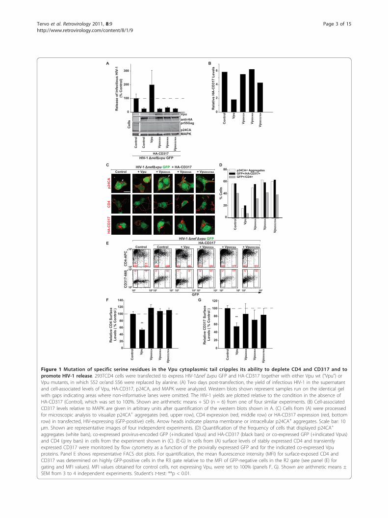
Figure 1 Mutation of specific serine residues in the Vpu cytoplasmic tail cripples its ability to deplete CD4 and CD317 and topromote HIV-1 release. 293TCD4 cells were transfected to express HIV-1Δnef Δvpu GFP and HA-CD317 together with either Vpu wt ("Vpu”) orVpu mutants, in which S52 or/and S56 were replaced by alanine. (A) Two days post-transfection, the yield of infectious HIV-1 in the supernatantand cell-associated levels of Vpu, HA-CD317, p24CA, and MAPK were analyzed. Western blots shown represent samples run on the identical gelwith gaps indicating areas where non-informative lanes were omitted. The HIV-1 yields are plotted relative to the condition in the absence ofHA-CD317 (Control), which was set to 100%. Shown are arithmetic means + SD (n = 6) from one of four similar experiments. (B) Cell-associatedCD317 levels relative to MAPK are given in arbitrary units after quantification of the western blots shown in A. (C) Cells from (A) were processedfor microscopic analysis to visualize p24CA+ aggregates (red, upper row), CD4 expression (red, middle row) or HA-CD317 expression (red, bottomrow) in transfected, HIV-expressing (GFP-positive) cells. Arrow heads indicate plasma membrane or intracellular p24CA+ aggregates. Scale bar: 10μm. Shown are representative images of four independent experiments. (D) Quantification of the frequency of cells that displayed p24CA+
aggregates (white bars), co-expressed provirus-encoded GFP (+indicated Vpus) and HA-CD317 (black bars) or co-expressed GFP (+indicated Vpus)and CD4 (grey bars) in cells from the experiment shown in (C). (E-G) In cells from (A) surface levels of stably expressed CD4 and transientlyexpressed CD317 were monitored by flow cytometry as a function of the provirally expressed GFP and for the indicated co-expressed Vpuproteins. Panel E shows representative FACS dot plots. For quantification, the mean fluorescence intensity (MFI) for surface-exposed CD4 andCD317 was determined on highly GFP-positive cells in the R3 gate relative to the MFI of GFP-negative cells in the R2 gate (see panel (E) forgating and MFI values). MFI values obtained for control cells, not expressing Vpu, were set to 100% (panels F, G). Shown are arithmetic means ±SEM from 3 to 4 independent experiments. Student’s t-test: **p < 0.01.
Tervo et al. Retrovirology 2011, 8:9http://www.retrovirology.com/content/8/1/9
Page 3 of 15
mutants did not significantly impair HA-CD317 expres-sion (Figure 1A (Western blot); quantification in Figure1B; Figure 1C (lower row images, + Vpu mutants),quantification in Figure 1D, filled bars), even thoughthey displayed slightly increased steady-state levels rela-tive to Vpu wt due to their higher stability [29]. Parallelanalysis for expression of CD4 revealed the expected[19] and marked reduction of the virus binding receptorin the presence of Vpu wt, but not of its serine mutants(Figure 1C (middle row images), quantification in Figure1D, grey bars).Next, we assessed surface levels of CD4 and CD317 on
293TCD4 cells, which had been transiently co-transfectedwith HIV-1ΔvpuΔnef GFP, pHA-CD317 and expressionplasmids for the indicated Vpu proteins (Figure 1E-G). Inthis set-up, the provirus-driven GFP expression served asa marker for the transfection level of individual cells. Thesurface exposure of the stably expressed CD4 was reducedby approximately 45% on cells expressing high levels ofVpu wt (Figure 1E, upper row, third FACS plot from theleft; quantification in Figure 1F), as assessed by flow cyto-metry. The dependence of this effect on the integrity ofthe di-serine motif was demonstrated by the trans-expression of the three Vpu mutants, none of whichdemonstrated downregulation of CD4 (Figure 1F).In control 293TCD4 cells transiently transfected with
pHA-CD317, an increase of surface levels of the restric-tion factor relative to the transfection level of these cellswas noted (Figure 1E, lower row, second FACS plotfrom the left). Similar to what was observed for CD4 onthe same cells, co-expression of Vpu wt prevented sur-face exposure of CD317 in a di-serine motif-dependentfashion (Figure 1E, lower row; quantification in Figure1G). To rule out that co-expression of two cellular Vputargets, CD4 and CD317, on the same cell affected HIVparticle release or effects of Vpu on CD317 expression,we repeated the assay in parental 293T cells in theabsence of CD4 and obtained comparable results (datanot shown). Taken together, the Vpu di-serine motif isessential for the ability of the accessory protein toenhance HIV-1 release, to reduce surface levels of CD4and CD317, and to trigger the depletion of both ofthese receptors in infected cells.
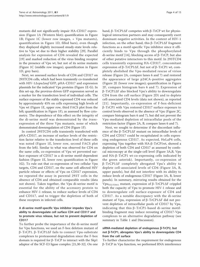
A di-serine motif-specific Vpu inhibitor impedes Vpu’sability to downregulate cell surface CD4 and CD317 andto promote virus release, but not to prevent depletion ofCD317To further probe the importance of the di-serine motiffor Vpu functions, we used an F-box deletion mutant ofb-TrCP1. b-TrCP1ΔF fails to connect Vpu-substratecomplexes to proteasomal degradation since the F-boxdomain is required for b-TrCP to interact with the Skp1adaptor of the SCF-E3 ligase complex [21,30-32]. On one
hand, b-TrCP1ΔF competes with b-TrCP wt for physio-logical interaction partners and may consequently exertdominant-negative activities. In the context of HIV-1infection, on the other hand, this b-TrCP1ΔF fragmentfunctions as a motif-specific Vpu inhibitor since it effi-ciently binds to Vpu through the phosphorylateddi-serine motif [14], blocking access of b-TrCP, but alsoof other putative interactors to this motif. In 293TCD4cells transiently expressing HA-CD317, concomitantexpression of b-TrCP1ΔF, but not of b-TrCP1 wt, com-pletely abolished the Vpu-mediated rescue of virionrelease (Figure 2A, compare lanes 6 and 7) and restoredthe appearance of large p24CA-positive aggregates(Figure 2E (lower row images); quantification in Figure2F, compare histogram bars 6 and 7). Expression ofb-TrCP1ΔF also blocked Vpu’s ability to downregulateCD4 from the cell surface (Figure 2D) and to depletecell-associated CD4 levels (data not shown), as reported[21]. Importantly, co-expression of F-box-deficientb-TrCP1 with Vpu restored CD317 surface exposure tocontrol levels observed in the absence of Vpu (Figure 2C,compare histogram bars 6 and 7), but did not prevent theVpu-mediated depletion of intracellular pools of therestriction factor (Figure 2A, B, compare lanes 6 and 7).Next, we sought to determine if the discordant influ-
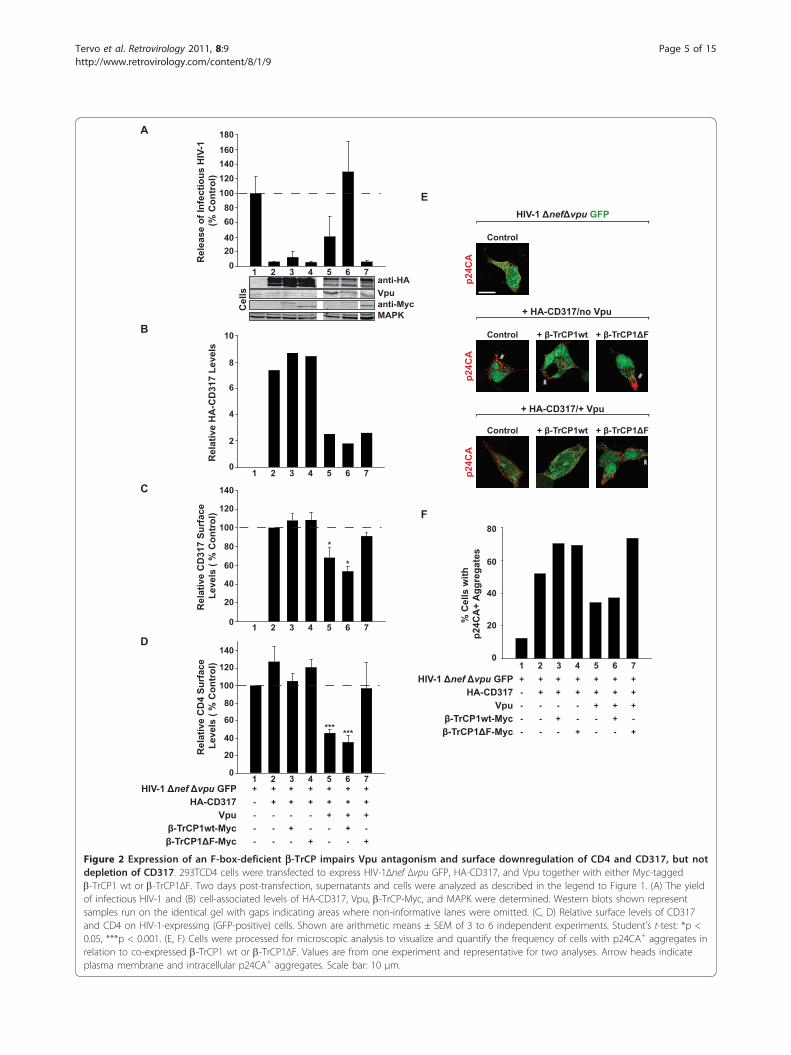
ence of the b-TrCP1ΔF mutant on intracellular levels ofCD4 and CD317 could be recapitulated in cells expres-sing endogenous CD317. As expected, HeLaP4 cells,expressing Vpu together with HA-b-TrCP1wt, showed adepletion of both CD4 and CD317 as assessed by confo-cal microscopy at the single-cell level (Figure 3A,B, Vpuand HA-b-TrCP1 wt-co-expressing cells indicated bythe green asterisk). Importantly, co-expression ofb-TrCP1ΔF completely abrogated Vpu’s ability todeplete cell-associated levels of CD4 (Figure 3A, B,upper panels), but did not interfere with its ability toreduce levels of endogenous CD317 (Figure 3A, B, lowerpanels). In summary, mirroring results obtained for theVpuS52/S56A mutant, expression of b-TrCP1ΔF crippledboth the capacity of Vpu to promote HIV-1 release andto downregulate cell surface-exposure of CD4 andCD317. As a notable discrepancy with the di-serinemutant of Vpu, expression of b-TrCP1ΔF did not pre-vent depletion of intracellular pools of CD317 by Vpu,indicating that this b-TrCP1-based di-serine motif-binding fragment may induce rerouting of CD317-Vpucomplexes to an alternative degradation pathway (seesummary in Table 1 and Discussion).
siRNA-mediated depletion of endogenous b-TrCP2, butnot b-TrCP1, abrogates Vpu’s ability to downregulate CD4from the cell surfaceTo further characterize the requirement for endogenousb-TrCP in Vpu function, we performed RNA interference
Tervo et al. Retrovirology 2011, 8:9http://www.retrovirology.com/content/8/1/9
Page 4 of 15
20
A
D
Rel
ease
of I
nfec
tious
HIV
-1
(% C
ontr
ol)
Rel
ativ
e C
D4
Surf
ace
Leve
ls (
% C
ontr
ol)
60
40
100
140
0
20
80
120
120
80
160
0
Rel
ativ
e H
A-C
D31
7 Le
vels
8
6
0
4
10
2
anti-HA
anti-MycMAPK
B
Vpu
40
140
60
100
180
1 2 3 4 5 6 7
1 2 3 4 5 6 7
1 2 3 4 5 6 7HIV-1 nef vpu GFP + + + + + + +
Vpu - - - - + + +HA-CD317 - + + + + + +
- rCP1 t-Myc - - + - - + -- rCP1 F-Myc - - - + - - +
E
F
20% C
ells
it
p2
4CA
+ A
ggre
gate
s
80
0
40
60
HIV-1 nef vpu GFP + + + + + + +
Vpu - - - - + + +
1 2 3 4 5 6 7
HA-CD317 - + + + + + +
- rCP1 t-Myc - - + - - + -- rCP1 F-Myc - - - + - - +
Cel
ls
1 2 3 4 5 6 7
C
Rel
ativ
e C
D31
7 Su
rfac
e Le
vels
( %
Con
trol
)
60
40
100
140
0
20
80
120
Control
p24C
A
HIV-1 nef vpu GFP
+ HA-CD317/no Vpu
+ - rCP1 t + - rCP1 FControl
p24C
A
+ - rCP1 t + - rCP1 F
+ HA-CD317/+ Vpu
Control
p24C
A
*
*
*** ***
Figure 2 Expression of an F-box-deficient b-TrCP impairs Vpu antagonism and surface downregulation of CD4 and CD317, but notdepletion of CD317. 293TCD4 cells were transfected to express HIV-1Δnef Δvpu GFP, HA-CD317, and Vpu together with either Myc-taggedb-TrCP1 wt or b-TrCP1ΔF. Two days post-transfection, supernatants and cells were analyzed as described in the legend to Figure 1. (A) The yieldof infectious HIV-1 and (B) cell-associated levels of HA-CD317, Vpu, b-TrCP-Myc, and MAPK were determined. Western blots shown representsamples run on the identical gel with gaps indicating areas where non-informative lanes were omitted. (C, D) Relative surface levels of CD317and CD4 on HIV-1-expressing (GFP-positive) cells. Shown are arithmetic means ± SEM of 3 to 6 independent experiments. Student’s t-test: *p <0.05, ***p < 0.001. (E, F) Cells were processed for microscopic analysis to visualize and quantify the frequency of cells with p24CA+ aggregates inrelation to co-expressed b-TrCP1 wt or b-TrCP1ΔF. Values are from one experiment and representative for two analyses. Arrow heads indicateplasma membrane and intracellular p24CA+ aggregates. Scale bar: 10 μm.
Tervo et al. Retrovirology 2011, 8:9http://www.retrovirology.com/content/8/1/9
Page 5 of 15
studies using siRNAs which specifically target eitherb-TrCP1 or b-TrCP2 mRNA. Because antibodies for reli-able detection of these b-TrCP isoforms are lacking, wefirst validated effectiveness and specificity of these siR-NAs using co-expressed Myc- or Flag-tagged b-TrCP1 orb-TrCP2 constructs (data not shown). In addition, weestablished a real-time PCR-based quantification of the
respective endogenous b-TrCP mRNAs. This assayallowed us to confirm the isoform-specific interferenceby the siRNAs and to evaluate knockdown efficiencies insubsequent functional studies (see below).In a first functional analysis, we tested the effects of
siRNA-mediated depletion of endogenous b-TrCP onthe ability of Vpu to downregulate surface-exposed
*
**
*
**
0
50
% H
A C
ells
with
Red
uced
CD
317
Leve
ls
100
HA C 1wtC
D4
CD
317
HA C 1
% H
A C
ells
with
Red
uced
CD
4 Le
vels
0
50
100
HA C 1wt HA C 1
A B
n.s.
**+
+
*
*
Figure 3 Overexpression of an F-box-deficient b-TrCP abolishes Vpu-mediated depletion of CD4, but not of endogenous CD317.HeLaP4 cells were transfected with expression constructs for Vpu and either HA-tagged b-TrCP1 wt or b-TrCP1ΔF. Two days later, cells wereprocessed for microscopic analysis to (A) visualize and (B) quantify the expression of either CD4 or endogenous CD317 (both in red) in relationto HA-b-TrCP1 wt or the HA-b-TrCP1ΔF mutant (cells co-expressing HA-tagged b-TrCPs and Vpu are labelled by a green asterisk; this fluorescentchannel is not depicted). The quantification in (B) depicts the percentage of cells that displayed markedly reduced levels of CD4 (upper panel) orCD317 (lower panel) in cells co-expressing Vpu together with either HA-tagged HA-b-TrCP1 wt or HA-b-TrCP1ΔF relative to untransfectedneighbouring cells. The arithmetic means ± SEM of three independent experiments are shown. Scale bar: 10 μm. Student’s t-test: **p = 0.0021,n.s. = not significant.
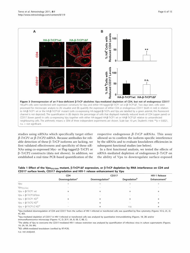
Table 1 Effect of the VpuS52/56A mutant, b-TrCP1ΔF expression, or b-TrCP depletion by RNA interference on CD4 andCD317 surface levels, CD317 degradation and HIV-1 release enhancement by Vpu
CD4 CD317 HIV-1 Release
Downregulationa Downregulationa Degradationb Enhancementc
Vpu + + + +
VpuS52/56A - - - -
Vpu + b-TrCP1 wt + + + +
Vpu + b-TrCP1ΔFbox - - - -
Vpu + b-TrCP1 KDd + + + +
Vpu + b-TrCP2 KDd - + - +
Vpu + b-TrCP1/2 KDd - + n.a. +aVpu-mediated downregulation of CD4 and CD317 from the surface of HIV-1-infected or transfected cells was quantified by flow cytometry (Figures 1E-G; 2C, D;4C; 8D).bVpu-mediated depletion of CD317 in HIV-1-infected or transfected cells was analyzed by quantitative immunoblotting (Figures. 1B; 2B) and/orimmunofluorescence microscopy (Figures 1C, D; 2E-F; 3A, B; 5B, C; 8B, C).cThe ability of Vpu to overcome the CD317-mediated HIV-1 release restriction was analyzed by quantification of infectious virus in culture supernatants (Figures.1A; 2A; 3A, 5A; 8A).dKD: siRNA-mediated knockdown (verified by RT-PCR).
n.a.: not analyzed.
Tervo et al. Retrovirology 2011, 8:9http://www.retrovirology.com/content/8/1/9
Page 6 of 15
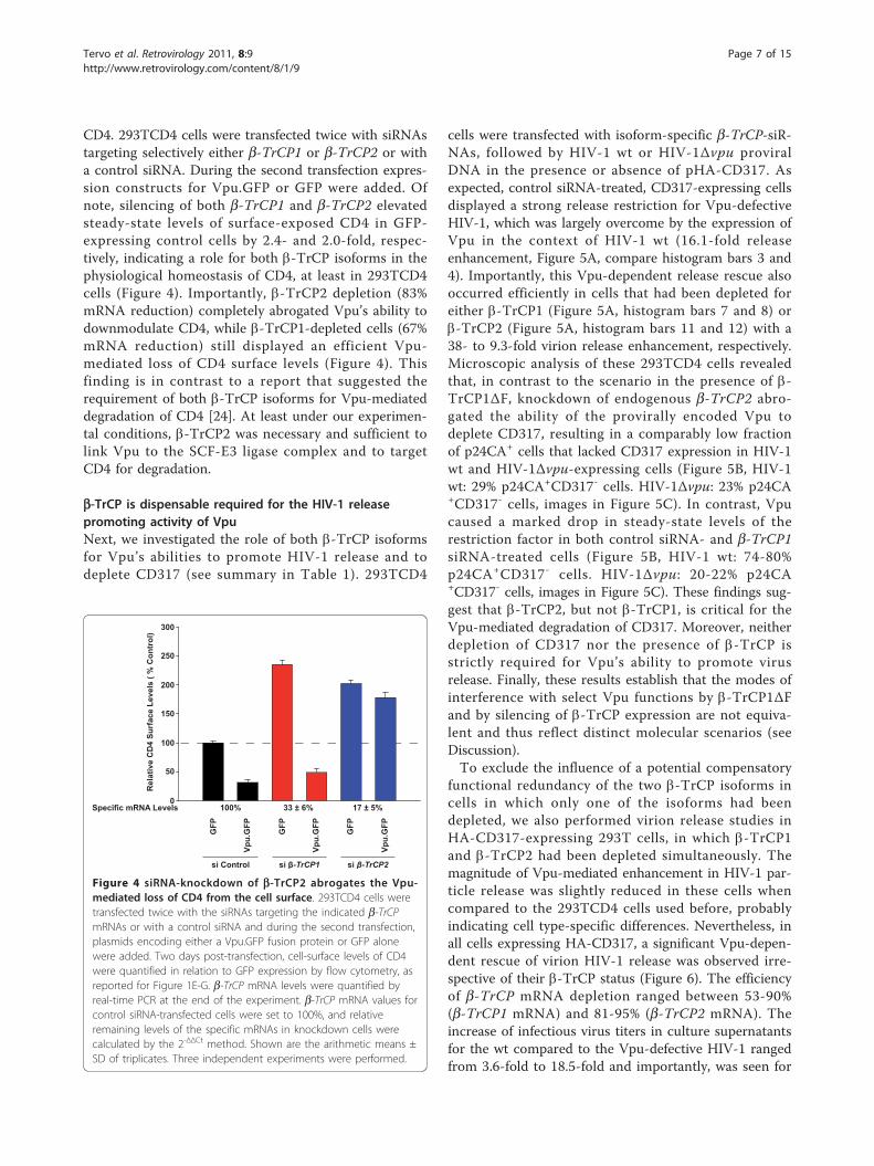
CD4. 293TCD4 cells were transfected twice with siRNAstargeting selectively either b-TrCP1 or b-TrCP2 or witha control siRNA. During the second transfection expres-sion constructs for Vpu.GFP or GFP were added. Ofnote, silencing of both b-TrCP1 and b-TrCP2 elevatedsteady-state levels of surface-exposed CD4 in GFP-expressing control cells by 2.4- and 2.0-fold, respec-tively, indicating a role for both b-TrCP isoforms in thephysiological homeostasis of CD4, at least in 293TCD4cells (Figure 4). Importantly, b-TrCP2 depletion (83%mRNA reduction) completely abrogated Vpu’s ability todownmodulate CD4, while b-TrCP1-depleted cells (67%mRNA reduction) still displayed an efficient Vpu-mediated loss of CD4 surface levels (Figure 4). Thisfinding is in contrast to a report that suggested therequirement of both b-TrCP isoforms for Vpu-mediateddegradation of CD4 [24]. At least under our experimen-tal conditions, b-TrCP2 was necessary and sufficient tolink Vpu to the SCF-E3 ligase complex and to targetCD4 for degradation.
b-TrCP is dispensable required for the HIV-1 releasepromoting activity of VpuNext, we investigated the role of both b-TrCP isoformsfor Vpu’s abilities to promote HIV-1 release and todeplete CD317 (see summary in Table 1). 293TCD4
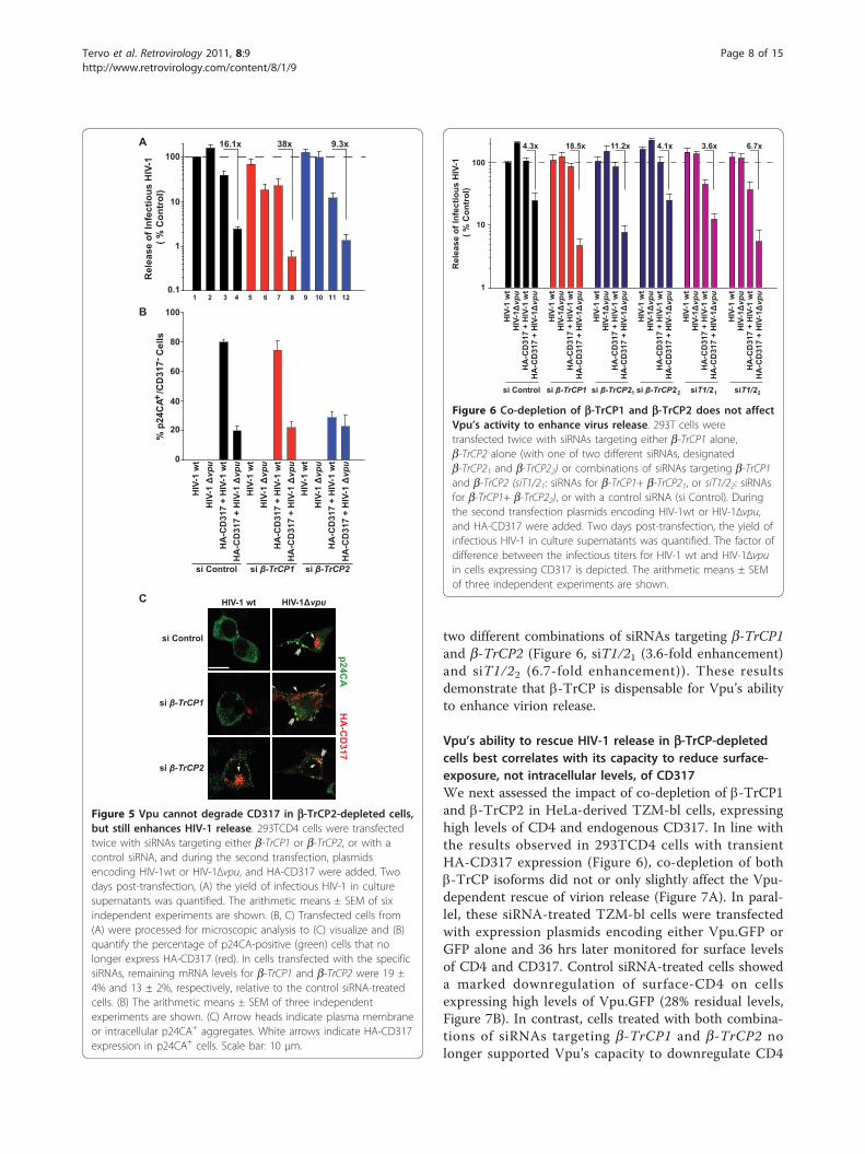
cells were transfected with isoform-specific b-TrCP-siR-NAs, followed by HIV-1 wt or HIV-1Δvpu proviralDNA in the presence or absence of pHA-CD317. Asexpected, control siRNA-treated, CD317-expressing cellsdisplayed a strong release restriction for Vpu-defectiveHIV-1, which was largely overcome by the expression ofVpu in the context of HIV-1 wt (16.1-fold releaseenhancement, Figure 5A, compare histogram bars 3 and4). Importantly, this Vpu-dependent release rescue alsooccurred efficiently in cells that had been depleted foreither b-TrCP1 (Figure 5A, histogram bars 7 and 8) orb-TrCP2 (Figure 5A, histogram bars 11 and 12) with a38- to 9.3-fold virion release enhancement, respectively.Microscopic analysis of these 293TCD4 cells revealedthat, in contrast to the scenario in the presence of b-TrCP1ΔF, knockdown of endogenous b-TrCP2 abro-gated the ability of the provirally encoded Vpu todeplete CD317, resulting in a comparably low fractionof p24CA+ cells that lacked CD317 expression in HIV-1wt and HIV-1Δvpu-expressing cells (Figure 5B, HIV-1wt: 29% p24CA+CD317- cells. HIV-1Δvpu: 23% p24CA+CD317- cells, images in Figure 5C). In contrast, Vpucaused a marked drop in steady-state levels of therestriction factor in both control siRNA- and b-TrCP1siRNA-treated cells (Figure 5B, HIV-1 wt: 74-80%p24CA+CD317- cells. HIV-1Δvpu: 20-22% p24CA+CD317- cells, images in Figure 5C). These findings sug-gest that b-TrCP2, but not b-TrCP1, is critical for theVpu-mediated degradation of CD317. Moreover, neitherdepletion of CD317 nor the presence of b-TrCP isstrictly required for Vpu’s ability to promote virusrelease. Finally, these results establish that the modes ofinterference with select Vpu functions by b-TrCP1ΔFand by silencing of b-TrCP expression are not equiva-lent and thus reflect distinct molecular scenarios (seeDiscussion).To exclude the influence of a potential compensatory
functional redundancy of the two b-TrCP isoforms incells in which only one of the isoforms had beendepleted, we also performed virion release studies inHA-CD317-expressing 293T cells, in which b-TrCP1and b-TrCP2 had been depleted simultaneously. Themagnitude of Vpu-mediated enhancement in HIV-1 par-ticle release was slightly reduced in these cells whencompared to the 293TCD4 cells used before, probablyindicating cell type-specific differences. Nevertheless, inall cells expressing HA-CD317, a significant Vpu-depen-dent rescue of virion HIV-1 release was observed irre-spective of their b-TrCP status (Figure 6). The efficiencyof b-TrCP mRNA depletion ranged between 53-90%(b-TrCP1 mRNA) and 81-95% (b-TrCP2 mRNA). Theincrease of infectious virus titers in culture supernatantsfor the wt compared to the Vpu-defective HIV-1 rangedfrom 3.6-fold to 18.5-fold and importantly, was seen for
100
50
200
300
0
150
250
Rel
ativ
e C
D4
Surf
ace
Leve
ls (
% C
ontr
ol)
Vpu.
GFP
GFP
GFP
Vpu.
GFP
GFP
Vpu.
GFP
si -TrCP1 si -TrCP2si Control
Specific mRNA Levels 100% 33 ± 6% 17 ± 5%
Figure 4 siRNA-knockdown of b-TrCP2 abrogates the Vpu-mediated loss of CD4 from the cell surface. 293TCD4 cells weretransfected twice with the siRNAs targeting the indicated b-TrCPmRNAs or with a control siRNA and during the second transfection,plasmids encoding either a Vpu.GFP fusion protein or GFP alonewere added. Two days post-transfection, cell-surface levels of CD4were quantified in relation to GFP expression by flow cytometry, asreported for Figure 1E-G. b-TrCP mRNA levels were quantified byreal-time PCR at the end of the experiment. b-TrCP mRNA values forcontrol siRNA-transfected cells were set to 100%, and relativeremaining levels of the specific mRNAs in knockdown cells werecalculated by the 2-ΔΔCt method. Shown are the arithmetic means ±SD of triplicates. Three independent experiments were performed.
Tervo et al. Retrovirology 2011, 8:9http://www.retrovirology.com/content/8/1/9
Page 7 of 15
two different combinations of siRNAs targeting b-TrCP1and b-TrCP2 (Figure 6, siT1/21 (3.6-fold enhancement)and siT1/22 (6.7-fold enhancement)). These resultsdemonstrate that b-TrCP is dispensable for Vpu’s abilityto enhance virion release.
Vpu’s ability to rescue HIV-1 release in b-TrCP-depletedcells best correlates with its capacity to reduce surface-exposure, not intracellular levels, of CD317We next assessed the impact of co-depletion of b-TrCP1and b-TrCP2 in HeLa-derived TZM-bl cells, expressinghigh levels of CD4 and endogenous CD317. In line withthe results observed in 293TCD4 cells with transientHA-CD317 expression (Figure 6), co-depletion of bothb-TrCP isoforms did not or only slightly affect the Vpu-dependent rescue of virion release (Figure 7A). In paral-lel, these siRNA-treated TZM-bl cells were transfectedwith expression plasmids encoding either Vpu.GFP orGFP alone and 36 hrs later monitored for surface levelsof CD4 and CD317. Control siRNA-treated cells showeda marked downregulation of surface-CD4 on cellsexpressing high levels of Vpu.GFP (28% residual levels,Figure 7B). In contrast, cells treated with both combina-tions of siRNAs targeting b-TrCP1 and b-TrCP2 nolonger supported Vpu’s capacity to downregulate CD4
Rel
ease
of I
nfec
tious
HIV
-1
( % C
ontr
ol)
10
1
100
HIV
-1vp
uH
IV-1
wt
HA
-CD
317
+ H
IV-1
wt
HA
-CD
317
+ H
IV-1
vpu
si Control si si si si si
4.3x 18.5x 11.2x 4.1x 3.6x 6.7x
1 1 2 2
HIV
-1vp
uH
IV-1
wt
HA
-CD
317
+ H
IV-1
wt
HA
-CD
317
+ H
IV-1
vpu
HIV
-1vp
uH
IV-1
wt
HA
-CD
317
+ H
IV-1
wt
HA
-CD
317
+ H
IV-1
vpu
HIV
-1vp
uH
IV-1
wt
HA
-CD
317
+ H
IV-1
wt
HA
-CD
317
+ H
IV-1
vpu
HIV
-1vp
uH
IV-1
wt
HA
-CD
317
+ H
IV-1
wt
HA
-CD
317
+ H
IV-1
vpu
HIV
-1vp
uH
IV-1
wt
HA
-CD
317
+ H
IV-1
wt
HA
-CD
317
+ H
IV-1
vpu
Figure 6 Co-depletion of b-TrCP1 and b-TrCP2 does not affectVpu’s activity to enhance virus release. 293T cells weretransfected twice with siRNAs targeting either b-TrCP1 alone,b-TrCP2 alone (with one of two different siRNAs, designatedb-TrCP21 and b-TrCP22) or combinations of siRNAs targeting b-TrCP1and b-TrCP2 (siT1/21: siRNAs for b-TrCP1+ b-TrCP21, or siT1/22: siRNAsfor b-TrCP1+ b-TrCP22), or with a control siRNA (si Control). Duringthe second transfection plasmids encoding HIV-1wt or HIV-1Δvpu,and HA-CD317 were added. Two days post-transfection, the yield ofinfectious HIV-1 in culture supernatants was quantified. The factor ofdifference between the infectious titers for HIV-1 wt and HIV-1Δvpuin cells expressing CD317 is depicted. The arithmetic means ± SEMof three independent experiments are shown.
A
B
C
HIV
vpu
HIV
-1 w
t
HA
-CD
317
+ H
IV-1
wt
si -TrCP1 si -TrCP2si Control
Rel
ease
of I
nfec
tious
HIV
-1
( % C
ontr
ol)
60
40
100
0
20
80
% p
24C
A /C
D31
7 C
ells
10
1
100
0.1
+-
si Control
si -TrCP1
si -TrCP2
HIV-1 wt HIV-1 vpu
HA
-CD
317p24C
A
HA
-CD
317
+ H
IV-1
vp
u
HIV
-1
vpu
HIV
-1 w
t
HA
-CD
317
+ H
IV-1
wt
HA
-CD
317
+ H
IV-1
vp
u
HIV
-1
vpu
HIV
-1 w
t
HA
-CD
317
+ H
IV-1
wt
HA
-CD
317
+ H
IV-1
vp
u
16.1x 38x 9.3x
1 2 3 4 5 6 7 8 9 10 11 12
Figure 5 Vpu cannot degrade CD317 in b-TrCP2-depleted cells,but still enhances HIV-1 release. 293TCD4 cells were transfectedtwice with siRNAs targeting either b-TrCP1 or b-TrCP2, or with acontrol siRNA, and during the second transfection, plasmidsencoding HIV-1wt or HIV-1Δvpu, and HA-CD317 were added. Twodays post-transfection, (A) the yield of infectious HIV-1 in culturesupernatants was quantified. The arithmetic means ± SEM of sixindependent experiments are shown. (B, C) Transfected cells from(A) were processed for microscopic analysis to (C) visualize and (B)quantify the percentage of p24CA-positive (green) cells that nolonger express HA-CD317 (red). In cells transfected with the specificsiRNAs, remaining mRNA levels for b-TrCP1 and b-TrCP2 were 19 ±4% and 13 ± 2%, respectively, relative to the control siRNA-treatedcells. (B) The arithmetic means ± SEM of three independentexperiments are shown. (C) Arrow heads indicate plasma membraneor intracellular p24CA+ aggregates. White arrows indicate HA-CD317expression in p24CA+ cells. Scale bar: 10 μm.
Tervo et al. Retrovirology 2011, 8:9http://www.retrovirology.com/content/8/1/9
Page 8 of 15
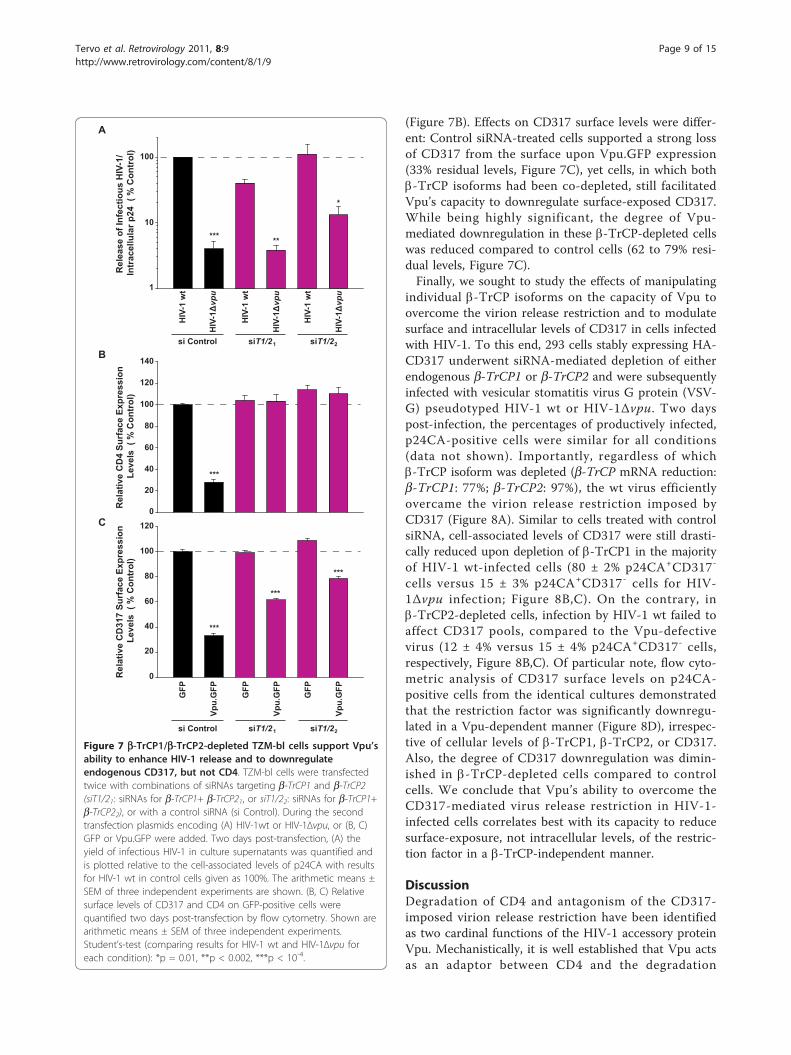
(Figure 7B). Effects on CD317 surface levels were differ-ent: Control siRNA-treated cells supported a strong lossof CD317 from the surface upon Vpu.GFP expression(33% residual levels, Figure 7C), yet cells, in which bothb-TrCP isoforms had been co-depleted, still facilitatedVpu’s capacity to downregulate surface-exposed CD317.While being highly significant, the degree of Vpu-mediated downregulation in these b-TrCP-depleted cellswas reduced compared to control cells (62 to 79% resi-dual levels, Figure 7C).Finally, we sought to study the effects of manipulating
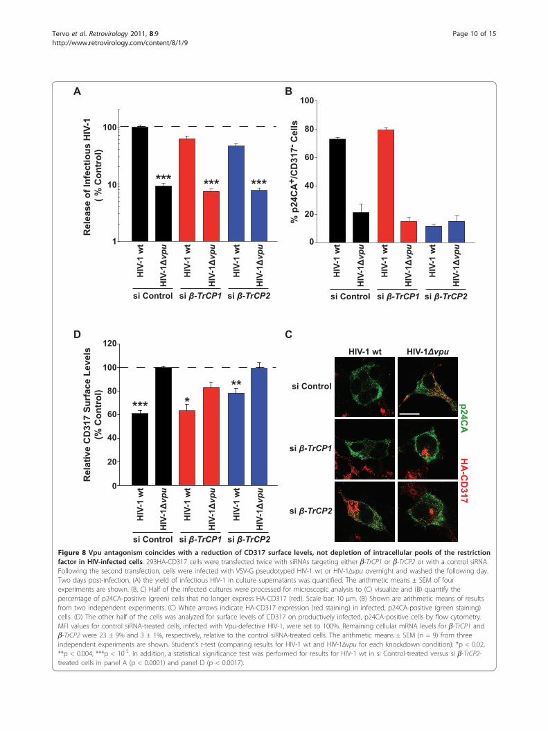
individual b-TrCP isoforms on the capacity of Vpu toovercome the virion release restriction and to modulatesurface and intracellular levels of CD317 in cells infectedwith HIV-1. To this end, 293 cells stably expressing HA-CD317 underwent siRNA-mediated depletion of eitherendogenous b-TrCP1 or b-TrCP2 and were subsequentlyinfected with vesicular stomatitis virus G protein (VSV-G) pseudotyped HIV-1 wt or HIV-1Δvpu. Two dayspost-infection, the percentages of productively infected,p24CA-positive cells were similar for all conditions(data not shown). Importantly, regardless of whichb-TrCP isoform was depleted (b-TrCP mRNA reduction:b-TrCP1: 77%; b-TrCP2: 97%), the wt virus efficientlyovercame the virion release restriction imposed byCD317 (Figure 8A). Similar to cells treated with controlsiRNA, cell-associated levels of CD317 were still drasti-cally reduced upon depletion of b-TrCP1 in the majorityof HIV-1 wt-infected cells (80 ± 2% p24CA+CD317-
cells versus 15 ± 3% p24CA+CD317- cells for HIV-1Δvpu infection; Figure 8B,C). On the contrary, inb-TrCP2-depleted cells, infection by HIV-1 wt failed toaffect CD317 pools, compared to the Vpu-defectivevirus (12 ± 4% versus 15 ± 4% p24CA+CD317- cells,respectively, Figure 8B,C). Of particular note, flow cyto-metric analysis of CD317 surface levels on p24CA-positive cells from the identical cultures demonstratedthat the restriction factor was significantly downregu-lated in a Vpu-dependent manner (Figure 8D), irrespec-tive of cellular levels of b-TrCP1, b-TrCP2, or CD317.Also, the degree of CD317 downregulation was dimin-ished in b-TrCP-depleted cells compared to controlcells. We conclude that Vpu’s ability to overcome theCD317-mediated virus release restriction in HIV-1-infected cells correlates best with its capacity to reducesurface-exposure, not intracellular levels, of the restric-tion factor in a b-TrCP-independent manner.
DiscussionDegradation of CD4 and antagonism of the CD317-imposed virion release restriction have been identifiedas two cardinal functions of the HIV-1 accessory proteinVpu. Mechanistically, it is well established that Vpu actsas an adaptor between CD4 and the degradation
Rel
ease
of I
nfec
tious
HIV
-1/
Intr
acel
lula
r p24
( %
Con
trol
)
10
1
100
HIV
-1vp
u
HIV
-1 w
t
si Control siT1/2 1 siT1/2 2
HIV
-1vp
u
HIV
-1 w
t
HIV
-1vp
u
HIV
-1 w
t
Vpu.
GFP
GFP
si Control siT1/21 siT1/22
Vpu.
GFP
GFP
Vpu.
GFP
GFP
100
120
140
80
60
40
20
0
100
120
80
60
40
20
0
Rel
ativ
e C
D4
Surf
ace
Expr
essi
on L
evel
s (
% C
ontr
ol)
Rel
ativ
e C
D31
7 Su
rfac
e Ex
pres
sion
Lev
els
( %
Con
trol
)A
B
C
***
***
***
***
*** **
*
Figure 7 b-TrCP1/b-TrCP2-depleted TZM-bl cells support Vpu’sability to enhance HIV-1 release and to downregulateendogenous CD317, but not CD4. TZM-bl cells were transfectedtwice with combinations of siRNAs targeting b-TrCP1 and b-TrCP2(siT1/21: siRNAs for b-TrCP1+ b-TrCP21, or siT1/22: siRNAs for b-TrCP1+b-TrCP22), or with a control siRNA (si Control). During the secondtransfection plasmids encoding (A) HIV-1wt or HIV-1Δvpu, or (B, C)GFP or Vpu.GFP were added. Two days post-transfection, (A) theyield of infectious HIV-1 in culture supernatants was quantified andis plotted relative to the cell-associated levels of p24CA with resultsfor HIV-1 wt in control cells given as 100%. The arithmetic means ±SEM of three independent experiments are shown. (B, C) Relativesurface levels of CD317 and CD4 on GFP-positive cells werequantified two days post-transfection by flow cytometry. Shown arearithmetic means ± SEM of three independent experiments.Student’s-test (comparing results for HIV-1 wt and HIV-1Δvpu foreach condition): *p = 0.01, **p < 0.002, ***p < 10-4.
Tervo et al. Retrovirology 2011, 8:9http://www.retrovirology.com/content/8/1/9
Page 9 of 15
A
C
Rel
ease
of I
nfec
tious
HIV
-1
( % C
ontr
ol)
10
1
100
HIV-1 wt HIV-1
si Control
si
si
HIV
-1HIV
-1 w
t
si si si Control
HIV
-1HIV
-1 w
t
HIV
-1HIV
-1 w
t
B
HIV
-1HIV
-1 w
t
si si si Control
HIV
-1HIV
-1 w
t
HIV
-1HIV
-1 w
t
60
40
100
0
20
80
% p
24C
A /C
D31
7 C
ells
+-
D
Rel
ativ
e C
D31
7 Su
rfac
e Le
vels
(% C
ontr
ol)
60
40
100
0
20
80
120
HIV
-1HIV
-1 w
t
si si si Control
HIV
-1HIV
-1 w
t
HIV
-1HIV
-1 w
t
*** ***
HA
-CD
317p24C
A
*** *** ***
Figure 8 Vpu antagonism coincides with a reduction of CD317 surface levels, not depletion of intracellular pools of the restrictionfactor in HIV-infected cells. 293HA-CD317 cells were transfected twice with siRNAs targeting either b-TrCP1 or b-TrCP2 or with a control siRNA.Following the second transfection, cells were infected with VSV-G pseudotyped HIV-1 wt or HIV-1Δvpu overnight and washed the following day.Two days post-infection, (A) the yield of infectious HIV-1 in culture supernatants was quantified. The arithmetic means ± SEM of fourexperiments are shown. (B, C) Half of the infected cultures were processed for microscopic analysis to (C) visualize and (B) quantify thepercentage of p24CA-positive (green) cells that no longer express HA-CD317 (red). Scale bar: 10 μm. (B) Shown are arithmetic means of resultsfrom two independent experiments. (C) White arrows indicate HA-CD317 expression (red staining) in infected, p24CA-positive (green staining)cells. (D) The other half of the cells was analyzed for surface levels of CD317 on productively infected, p24CA-positive cells by flow cytometry.MFI values for control siRNA-treated cells, infected with Vpu-defective HIV-1, were set to 100%. Remaining cellular mRNA levels for b-TrCP1 andb-TrCP2 were 23 ± 9% and 3 ± 1%, respectively, relative to the control siRNA-treated cells. The arithmetic means ± SEM (n = 9) from threeindependent experiments are shown. Student’s t-test (comparing results for HIV-1 wt and HIV-1Δvpu for each knockdown condition): *p < 0.02,**p < 0.004, ***p < 10-5. In addition, a statistical significance test was performed for results for HIV-1 wt in si Control-treated versus si b-TrCP2-treated cells in panel A (p < 0.0001) and panel D (p < 0.0017).
Tervo et al. Retrovirology 2011, 8:9http://www.retrovirology.com/content/8/1/9
Page 10 of 15
machinery. While binding to CD4 occurs via a hydro-philic C-terminal domain of Vpu [26,33], Vpu is phos-phorylated at serine residues 52/56 by casein kinase II[33,34], allowing for recruitment of an E3 ubiquitinligase multi-protein complex via the substrate recogni-tion factor b-TrCP [21]. Since it is unclear whether Vpuexploits the same general strategy for antagonizing theCD317 restriction to HIV-1 particle release, we usedseveral experimental approaches to investigate thedependence of both major Vpu activities on the di-serine interaction motif of the accessory protein and onexpression of cellular b-TrCP (see summary of results inTable 1). We found that b-TrCP2, not the structurallyrelated b-TrCP1, is the critical Vpu adapter for the E3ubiquitin ligase complex that targets both CD4 andCD317 for accelerated degradation. In contrast, b-TrCPwas largely dispensable for Vpu-mediated downregula-tion of CD317, but not CD4, from the cell surface. Mostimportantly, b-TrCP was not required for Vpu’s abilityto counteract the release restriction imposed by CD317.In agreement with findings in earlier reports
[12,14,21,25,26,35], we observed that the integrity of thedi-serine motif was strictly required for effects of Vpuon cell-surface exposure and overall expression levels ofCD317 and CD4, as well as its antagonism of the virionrelease block. These results are also in line with a recentreport [25] which demonstrated, through the use of acasein kinase II inhibitor, that Vpu phosphorylation iscritical for its antagonistic activity. A central role ofVpu’s di-serine motif was further supported by findingswith b-TrCP1ΔF. This mutant protein binds to thismotif [14,27] without coupling the Vpu-substrate com-plex to the E3 ubiquitin ligase complex. In the contextof HIV studies, b-TrCP1ΔF should be regarded as aVpu inhibitor, since its expression competitively blocksthe functionality of the di-serine motif to which b-TrCPand other currently unknown cellular proteins maybind. In agreement with its role as a motif-specific Vpuinhibitor, co-expression of Vpu with b-TrCP1ΔFresulted in a loss of Vpu activities, the pattern of whichwas similar to that observed with the VpuS52/56A mutant(Table 1). Notably, overexpression of b-TrCP wt, whichlike b-TrCP1ΔF associates with Vpu [21], did not exertsimilar inhibitory effects. This discrepancy betweenoverexpression of the F-box-deleted fragment, comparedto the full-length protein, possibly reflects different on/off-rates in the interaction of these two molecules withVpu, governed by the inability of the b-TrCP1ΔF tocouple substrate complexes to the E3 ubiquitin ligasecomplex, possibly resulting in a prolonged occupation ofthe di-serine motif. Vpu-mediated CD317 depletion inthe presence of b-TrCP1ΔF posed a noteworthy excep-tion under our experimental conditions. We speculatethat binding of b-TrCP1ΔF specifically impairs the Vpu-
mediated proteasomal degradation of CD317 and CD4.While this appears to be the only degradative pathwayfor CD4, inhibiting this pathway with b-TrCP1ΔF mightresult in a preferential mistrafficking of CD317 to thetrans-Golgi network (TGN) or early endosomes, andsubsequent delivery to the lysosome [15,16,18].siRNA-mediated depletion of endogenous b-TrCP
provides a direct means for probing its role for Vpuactivities. Consistent with two recent reports [14,27], wefind that b-TrCP2, but not b-TrCP1, is critical for thedepletion of cellular pools of CD317. These reportswent on to suggest an inverse correlation of cell-associated CD317 levels and HIV-1 release enhancementby Vpu. To the contrary, we found that depletion ofb-TrCP2 or simultaneous depletion of both b-TrCP iso-forms did not result in a marked impairment of Vpu’sability to promote HIV-1 release in cells with exogenousand endogenous CD317 expression. Of note, underthese conditions, downregulation of CD317 surfacelevels by Vpu, although statistically highly significant,was less pronounced. The discrepancies between theseand our studies may reflect differences in cell lines andexperimental conditions. Moreover, some of these pre-vious reports simplified, at times, the interpretation ofapparent residual effects of Vpu on virion release rescuein b-TrCP-depleted cells as a lack of functionality of theviral protein [14,16,27]. We included the concomitantvalidation of b-TrCP mRNA levels or side-by-sideassessment of Vpu-mediated modulation of CD4 levelsfollowing RNA interference in all our b-TrCP depletionstudies. This validation together with the robustness ofthe observed consequences for Vpu activities allowed usto conclude that both b-TrCP isoforms are not strictlyrequired for Vpu counteraction of CD317 in our experi-mental system and thus are not general Vpu co-factorsfor this activity.Based on these findings, we propose an integrative
model for Vpu antagonism of CD317-mediated virionrelease restriction: Vpu binds directly to CD317 [25],involving the transmembrane domains of both proteins[13,26,36]. While the cellular membranes of their pri-mary contact are still undefined, this interaction mark-edly affects the subcellular distribution of CD317,resulting in a reduction of CD317 surface levels andconcomitant accumulation in the TGN (at least inHeLa-derived cells, in which Vpu-mediated degradationis not very prominent). As a downstream consequenceof reduced cell-surface exposure of CD317 in the pre-sence of Vpu, CD317 can be subject to accelerateddegradation. Two different degradative pathways canapparently be targeted, possibly depending on the speci-fic cellular environment and/or targeted subpopulationsof CD317. Proteasomal degradation of CD317 occurs viab-TrCP2 and the E3 ubiquitin ligase complex.
Tervo et al. Retrovirology 2011, 8:9http://www.retrovirology.com/content/8/1/9
Page 11 of 15
Lysosomal degradation may be a consequence of pro-nounced mistrafficking of CD317 via early endosomes.Importantly, changes in the major intracellular poolsof CD317 do not necessarily feed back into the surfacepopulation of the restriction factor and are dispensablefor Vpu’s antagonistic activity. In line with this sce-nario, Vpu efficiently antagonizes degradation-insensi-tive CD317 variants, and this correlates with apreserved ability to downregulate the restriction factorfrom the cell surface [17]. Thus, Vpu-mediated degra-dation of CD317 can be regarded as a secondary effectof the viral protein on the restriction factor that is notstrictly required for its capacity to promote HIV-1release.Reduction of cell-surface pools of CD317, therefore,
emerges as key activity of Vpu for its role as anantagonist of the virion release restriction. Experimen-tal evidence indicates that Vpu does not enhance theendocytotic rate of surface-exposed CD317 [15], imply-ing that interceptions at the level of anterograde trans-port of newly synthesized CD317 or of recyclingpopulations of CD317 might underlie the Vpu-mediated downregulation of CD317 from the surface.This activity depends on the phosphorylated di-serinemotif in Vpu, but not its bona-fide cellular interactionpartner b-TrCP2, and thus most likely reflects theinteraction of Vpu with another host factor. ReportedVpu-interacting factors include Vpu-binding proteinUBP, also referred to as small glutamine-rich tetratri-copeptide repeat protein (SGT) [37,38], the MHC IIinvariant chain CD74 [39] and the TASK-1 ion chan-nel [40]. Since UBP/SGT was dispensable for CD317antagonism by Vpu (data not shown), most cell typesused for studies of Vpu counteraction lack expressionof CD74, and TASK-1 interferes with, rather thanfacilitates Vpu activity, this putative factor still needsto be identified. This factor should bind to the phos-phorylated di-serine motif of Vpu and is predicted tomediate the trapping of CD317 at the TGN by director indirect mechanisms.Despite increasing insight into the molecular mechan-
isms underlying Vpu’s ability to overcome the CD317-mediated virion release restriction, key aspects remainunresolved and warrant future investigation. These includethe identification of critical cellular Vpu co-factors as wellas the definition of relevant intracellular transport steps orsubpopulations of CD317 that are affected by Vpu. Theemergence of distinct strategies employed by viral factorsother than Vpu for counteraction of CD317 [4,5,41-43]emphasizes the potency of this restriction to limit thespread and pathogenesis of viruses.
MethodsCells and plasmids293T, 293TCD4, 293HA-CD317, TZM-bl, andHeLaP4 cells were maintained in DMEM supplemen-ted with 10% fetal calf serum, 1% penicillin-strepto-mycin and 1% L-glutamine (all from Invitrogen).293TCD4 cells were generated by lentiviral vectortransduction in principle as reported [44]. 293HA-CD317 cells were generated by transfection ofpcDNA3.1-HA-CD317neo [12] and neomycin selec-tion. The HA-tag is fused to the N-terminus ofCD317. Proviral plasmids pHIV-1NL4-3 wt (BH10Env) and pHIV-1NL4-3Δvpu (BH10 Env) were fromValerie Bosch [45] and pBR-NL4-3.IRES.eGFPΔnefand pBR-NL4.3-IRES.eGFPΔnefΔvpu [46] from FrankKirchhoff. The proviral plasmid pHIV-1NL4-3VpuS52NS56N was generated by site-directed muta-genesis. pcDNA-Vphu, expressing a codon-optimized,Rev-independent HIV-1NL4-3 Vpu protein [47] andpcDNA-VphuS52NS56N were from Klaus Strebel.Analogous expression constructs for Vpu mutantsS52A/56A [12], S52A, and S56A were generated bysite-directed mutagenesis. pcDNA-Vphu.GFP, encodinga Vpu.GFP fusion protein, was generated by PCR ofpSynVphu with the forward primer (5’-CGAATTCT-GATGGTGCCCATTATTG-3’) and reverse primer (5’-TGGATCCCGCAGGTGGTCAATG TCCCA-3 ’) togenerate EcoRI and BamHI restriction sites and sub-cloning into pEGFP-N1. HA-tagged and Myc-taggedexpression plasmids for human b-TrCP wt and b-TrCPwt ΔF were from Florence Margottin-Goguet [21] anda Flag-tagged expression plasmid for mouse b-TrCP2wt [48] was from Keiichi Nakayama.
Transfections293T cells, 293TCD4 cells (each 1.2 × 10⁵/well) orHeLaP4 cells (1 × 10⁵/well) were seeded in 12-wellplates 1 day before calcium-phosphate transfection ofproviral DNA (1.2 μg) or empty vector together withpcDNA3.1-HA-CD317neo (0.1 μg) and the indicatedVpu expression constructs (0.12 μg). For co-expressionof epitope-tagged b-TrCP wt or F-box-deleted frag-ments, 1 μg of the respective expression plasmids wasadded. TZM-bl cells (1 × 10⁵/well) were transfectedusing jetPRIME® (PEQLAB Biotechnologie) accordingto the manufacturer’s protocol.
HIV-1 infection293HA-CD317 cells were infected with VSV-G HIV-1NL4-3 wt (BH10 Env) or isogenic Vpu-defective virus ata multiplicity of infection of 0.2.
Tervo et al. Retrovirology 2011, 8:9http://www.retrovirology.com/content/8/1/9
Page 12 of 15
Virus release quantificationThe virion- and cell-associated amount of HIV-1 p24CAantigen was determined by an antigen enzyme-linkedimmunosorbent assay (p24CA ELISA) [49]. The infectiv-ity of HIV-1 in culture supernatants was determined 2days after transfection on TZM-bl reporter cells asreported [50].
Surface-exposed receptor levelsTo quantify CD4 expression at the cell surface, washedcells were stained in PBS with allophycocyanin (APC)-con-jugated mouse-anti human CD4 antibodies (clone RPA-T4; BD Bioscience). To quantify CD317 surface-exposure,cells were stained with mouse anti-HM1.24/CD317 [51] (5μg/ml; Chugai Pharmaceuticals) followed by staining withgoat anti-mouse (Alexa Fluor 660) (Invitrogen). A parallelassessment of the productive infection was performedafter fixing cells with 4% paraformaldehyde (PFA) for 90min by staining for intracellular p24CA antigen withFluoresceinisothiocyanat (FITC)-conjugated KC57 mono-clonal antibodies (Beckman Coulter) in 0.1% Triton X-100/PBS for 30 minutes on ice. A FACS Calibur with BDCellQuest Pro 4.0.2 Software (BD Pharmingen) was usedfor analysis. The MFI for surface-exposed receptors wasquantified in principle as reported [52].
Western blottingWashed cell pellets were lysed in SDS-lysis buffer. Pro-teins were separated on 12.5% SDS-PAGE and blottedto nitrocellulose membranes. Blocked membranes wereprobed with the following primary antibodies: mousemonoclonal anti-HIV-1 p24CA antibody 183, rabbitpolyclonal anti-Vpu antisera (from Ulrich Schubert orKlaus Strebel), anti-MAPK antiserum (Santa Cruz Bio-technology), mouse anti-HA mAb HA.11 (Covance),mouse anti-c-Myc antibody (Santa Cruz Biotechnology)and mouse-anti Flag M2 antibody (Sigma-Aldrich). Sec-ondary antibodies were conjugated either to horseradishperoxidase for ECL-based detection or to Alexa Fluor700/800 fluorescent dyes for detection by Odyssey Infra-red Imaging System (LI-COR Biosciences) and quantifi-cation by Odyssey software (version 2.1).
Immunofluorescence microscopyTransfected or infected cells growing on coverslips werefixed with 4% PFA and permeabilized for 2 min with0.1% Triton X-100 in PBS. Cells were blocked for1 hour with 1% bovine serum albumin in PBS andstained with appropriate primary and secondary antibo-dies for detection of p24CA, HA, CD4, or CD317,respectively. Coverslips were mounted in mountingmedium (Dianova) and analyzed with a Zeiss LSM510
confocal microscope with a 100x PLAN-APO objectivelens. Images were recorded with the Zeiss proprietarysoftware LSM5 and processed with Adobe Photoshop6.0. Gag localization was grouped for individual cellsinto marked accumulation (at the plasma membrane aswell as intracellularly (cells with at least one p24CA+
aggregates) or diffusely cytoplasmic), in principle asreported [2,12,17,43]. Effects of Vpu on cellular expres-sion of CD4 or CD317, respectively, was judged by com-paring to untransfected/uninfected neighbouring cells,scoring cells as displaying reduced expression levelswhen the fluorescent signal was unambiguously lower.
siRNA-mediated b-TrCP depletionSeeded cells were transfected twice on consecutive days bycalcium-phosphate precipitation or jetPRIME with siRNAsspecific for b-TrCP1 mRNA (5’-GAAUUCACUUAGACAGACA-3’), b-TrCP2 mRNA (5’-AGAUUAUCCAG-GAUAUAGA-3’ (b-TrCP21), and 5’-GAAGUAAAUC-GACCG UCAA-3’ [24] (b-TrCP22)), combinations ofsiRNAs specific for b-TrCP1 and b-TrCP2 mRNA, or non-specific control siRNA (5’-AGGUAGUGUAA UCGC-CUUG-3’) (each 50 pmol) and harvested 48 hours afterthe second transfection. Where indicated, proviral DNAsor expression plasmids for CD317, Vpu.GFP or GFP wereco-transfected, or cells were infected with VSV-G HIV-1at the second time-point.
b-TrCP knockdown quantificationTotal RNA was extracted by a standard Trizol-chloroformprotocol and precipitated with isopropyl alcohol. RNA pel-lets were washed with 75% ethanol, dissolved in water, andstored at -80°C until use. After treatment with DNaseDNA-free (Ambion) and cDNA synthesis (NEB), relativequantitative PCR analyses were performed on theABI Prism 7500 sequence detection system (Applied Bio-systems). b-TrCP mRNA levels were quantified with pri-mers specific for b-TrCP1 and b-TrCP2, reported [24],and TaqMan-specific probes for b-TrCP1 (5’-FAM-GCAAGCACTGCTATGAAGAC-TAMRA-3’) and b-TrCP2(5’-FAM-CAGTCTGCACTT TCACCCGT-TAMRA-3’),respectively. b-TrCP mRNA levels were quantified byusing the 2-ΔΔCt method with human RNaseP gene asendogenous reference control. Pooled triplicates were ana-lyzed for each condition. Data analysis was conductedusing the 7500 System Software (Applied Biosystems).
Additional material
Additional file 1: HIV-1-encoded VpuS52NS56N cannot counteractendogenous CD317. The provirally expressed di-serine mutant of Vpu
Tervo et al. Retrovirology 2011, 8:9http://www.retrovirology.com/content/8/1/9
Page 13 of 15
cannot overcome the CD317-imposed virion release restriction in TZM-blcells.
Additional file 2: Expression of HA-CD317, Vpu wt, or its serinemutants, does not affect expression or maturation of HIV-1 Gag.Expression neither of CD317/Tetherin nor Vpu considerably alters theexpression levels or processing of the Gag polyprotein of HIV-1 in 293Tcells.
AcknowledgementsWe thank Hans-Georg Kräusslich for support and reagents. We are gratefulto Valerie Bosch, Frank Kirchhoff, Masaki Matsumoto, Florence Margottin-Goguet, Ulrich Schubert, Klaus Strebel, and Chugai Pharmaceuticals for thegift of reagents. We thank Nadine Tibroni for technical assistance andmembers of the Fackler and Keppler laboratories for comments on themanuscript. We are grateful to Gary Howard for editorial assistance. Thiswork was in part funded by the Deutsche Forschungsgemeinschaft (KE742/4-1) (to O.T.K. and O.T.F). O.T. F and O.T.K. are members of the CellNetworksCluster of Excellence EXC81.
Author details1Department of Infectious Diseases, Virology, University of Heidelberg,Heidelberg, Germany. 2Department of Medicine, University of California SanDiego, La Jolla, California, USA.
Authors’ contributionsHMT, SH, OTF and OTK designed the study. HMT, SH, JVF and IA conductedthe experiments. OTF and OTK wrote the paper. All authors commented onand approved the final manuscript.
Competing interestsThe authors declare that they have no competing interests.
Received: 20 December 2010 Accepted: 10 February 2011Published: 10 February 2011
References1. Jouvenet N, Neil SJ, Zhadina M, Zang T, Kratovac Z, Lee Y, McNatt M,
Hatziioannou T, Bieniasz PD: Broad-spectrum inhibition of retroviral andfiloviral particle release by tetherin. J Virol 2009, 83(4):1837-1844.
2. Neil SJ, Zang T, Bieniasz PD: Tetherin inhibits retrovirus release and isantagonized by HIV-1 Vpu. Nature 2008, 451(7177):425-430.
3. Sakuma T, Noda T, Urata S, Kawaoka Y, Yasuda J: Inhibition of Lassa andMarburg virus production by tetherin. J Virol 2009, 83(5):2382-2385.
4. Kaletsky RL, Francica JR, Agrawal-Gamse C, Bates P: Tetherin-mediatedrestriction of filovirus budding is antagonized by the Ebola glycoprotein.Proc Natl Acad Sci USA 2009, 106(8):2886-2891.
5. Mansouri M, Viswanathan K, Douglas JL, Hines J, Gustin J, Moses AV, Fruh K:Molecular mechanism of BST2/tetherin downregulation by K5/MIR2 ofKaposi’s sarcoma-associated herpesvirus. J Virol 2009, 83(19):9672-9681.
6. Pardieu C, Vigan R, Wilson SJ, Calvi A, Zang T, Bieniasz P, Kellam P,Towers GJ, Neil SJ: The RING-CH ligase K5 antagonizes restriction ofKSHV and HIV-1 particle release by mediating ubiquitin-dependentendosomal degradation of tetherin. PLoS Pathog 2010, 6(4):e1000843.
7. Radoshitzky SR, Dong L, Chi X, Clester JC, Retterer C, Spurgers K, Kuhn JH,Sandwick S, Ruthel G, Kota K, Boltz D, Warren T, Kranzusch PJ, Whelan SP,Bavari S: Infectious Lassa virus but not filoviruses is restricted by BST-2/tetherin. J Virol 2010, 84(20):10569-10580.
8. Weidner JM, Jiang D, Pan XB, Chang J, Block TM, Guo JT: Interferon-induced cell membrane proteins, IFITM3 and tetherin, inhibit vesicularstomatitis virus infection via distinct mechanisms. J Virol 2010,84(24):12646-12657.
9. Van Damme N, Goff D, Katsura C, Jorgenson RL, Mitchell R, Johnson MC,Stephens EB, Guatelli J: The interferon-induced protein BST-2 restrictsHIV-1 release and is downregulated from the cell surface by the viralVpu protein. Cell Host Microbe 2008, 3(4):245-252.
10. Perez-Caballero D, Zang T, Ebrahimi A, McNatt MW, Gregory DA,Johnson MC, Bieniasz PD: Tetherin inhibits HIV-1 release by directlytethering virions to cells. Cell 2009, 139(3):499-511.
11. Bartee E, McCormack A, Fruh K: Quantitative membrane proteomicsreveals new cellular targets of viral immune modulators. PLoS Pathog2006, 2(10):e107.
12. Goffinet C, Allespach I, Homann S, Tervo HM, Habermann A, Rupp D,Oberbremer L, Kern C, Tibroni N, Welsch S, Krijnse-Locker J, Banting G,Kräusslich HG, Fackler OT, Keppler OT: HIV-1 antagonism of CD317 isspecies specific and involves Vpu-mediated proteasomal degradation ofthe restriction factor. Cell Host Microbe 2009, 5(3):285-297.
13. Gupta RK, Hue S, Schaller T, Verschoor E, Pillay D, Towers GJ: Mutation of asingle residue renders human tetherin resistant to HIV-1 Vpu-mediateddepletion. PLoS Pathog 2009, 5(5):e1000443.
14. Mangeat B, Gers-Huber G, Lehmann M, Zufferey M, Luban J, Piguet V: HIV-1Vpu neutralizes the antiviral factor Tetherin/BST-2 by binding it anddirecting its beta-TrCP2-dependent degradation. PLoS Pathog 2009, 5(9):e1000574.
15. Mitchell RS, Katsura C, Skasko MA, Fitzpatrick K, Lau D, Ruiz A, Stephens EB,Margottin-Goguet F, Benarous R, Guatelli JC: Vpu antagonizes BST-2-mediated restriction of HIV-1 release via beta-TrCP and endo-lysosomaltrafficking. PLoS Pathog 2009, 5(5):e1000450.
16. Iwabu Y, Fujita H, Kinomoto M, Kaneko K, Ishizaka Y, Tanaka Y, Sata T,Tokunaga K: HIV-1 accessory protein Vpu internalizes cell-surface BST-2/tetherin through transmembrane interactions leading to lysosomes. JBiol Chem 2009, 284(50):35060-35072.
17. Goffinet C, Homann S, Ambiel I, Tibroni N, Rupp D, Keppler OT, Fackler OT:Antagonism of CD317 restriction of human immunodeficiency virus type1 (HIV-1) particle release and depletion of CD317 are separable activitiesof HIV-1 Vpu. J Virol 2010, 84(8):4089-4094.
18. Habermann A, Krijnse-Locker J, Oberwinkler H, Eckhardt M, Homann S,Andrew A, Strebel K, Krausslich HG: CD317/tetherin is enriched in the HIV-1 envelope and downregulated from the plasma membrane upon virusinfection. J Virol 2010, 84(9):4646-4658.
19. Willey RL, Maldarelli F, Martin MA, Strebel K: Human immunodeficiencyvirus type 1 Vpu protein regulates the formation of intracellular gp160-CD4 complexes. J Virol 1992, 66(1):226-234.
20. Bour S, Perrin C, Akari H, Strebel K: The human immunodeficiency virustype 1 Vpu protein inhibits NF-kappa B activation by interfering withbeta TrCP-mediated degradation of Ikappa B. J Biol Chem 2001,276(19):15920-15928.
21. Margottin F, Bour SP, Durand H, Selig L, Benichou S, Richard V, Thomas D,Strebel K, Benarous R: A novel human WD protein, h-beta TrCp, thatinteracts with HIV-1 Vpu connects CD4 to the ER degradation pathwaythrough an F-box motif. Mol Cell 1998, 1(4):565-574.
22. Guardavaccaro D, Kudo Y, Boulaire J, Barchi M, Busino L, Donzelli M,Margottin-Goguet F, Jackson PK, Yamasaki L, Pagano M: Control of meioticand mitotic progression by the F box protein beta-Trcp1 in vivo. Dev Cell2003, 4(6):799-812.
23. Nakayama K, Hatakeyama S, Maruyama S, Kikuchi A, Onoe K, Good RA,Nakayama KI: Impaired degradation of inhibitory subunit of NF-kappa B(I kappa B) and beta-catenin as a result of targeted disruption of thebeta-TrCP1 gene. Proc Natl Acad Sci USA 2003, 100(15):8752-8757.
24. Butticaz C, Michielin O, Wyniger J, Telenti A, Rothenberger S: Silencing ofboth beta-TrCP1 and HOS (beta-TrCP2) is required to suppress humanimmunodeficiency virus type 1 Vpu-mediated CD4 down-modulation. JVirol 2007, 81(3):1502-1505.
25. Schindler M, Rajan D, Banning C, Wimmer P, Koppensteiner H, Iwanski A,Specht A, Sauter D, Dobner T, Kirchhoff F: Vpu serine 52 dependentcounteraction of tetherin is required for HIV-1 replication in macrophages,but not in ex vivo human lymphoid tissue. Retrovirology 2010, 7(1):1.
26. Schubert U, Bour S, Ferrer-Montiel AV, Montal M, Maldarell F, Strebel K: Thetwo biological activities of human immunodeficiency virus type 1 Vpuprotein involve two separable structural domains. J Virol 1996,70(2):809-819.
27. Douglas JL, Viswanathan K, McCarroll MN, Gustin JK, Fruh K, Moses AV: Vpudirects the degradation of the human immunodeficiency virusrestriction factor BST-2/Tetherin via a {beta}TrCP-dependent mechanism.J Virol 2009, 83(16):7931-7947.
28. Zhang J, Liang C: BST-2 diminishes HIV-1 infectivity. J Virol 2010,84(23):12336-12343.
29. Belaidouni N, Marchal C, Benarous R, Besnard-Guerin C: Involvement of thebetaTrCP in the ubiquitination and stability of the HIV-1 Vpu protein.Biochem Biophys Res Commun 2007, 357(3):688-693.
Tervo et al. Retrovirology 2011, 8:9http://www.retrovirology.com/content/8/1/9
Page 14 of 15
30. Hattori K, Hatakeyama S, Shirane M, Matsumoto M, Nakayama K: Moleculardissection of the interactions among IkappaBalpha, FWD1, and Skp1required for ubiquitin-mediated proteolysis of IkappaBalpha. J Biol Chem1999, 274(42):29641-29647.
31. Li Y, Gazdoiu S, Pan ZQ, Fuchs SY: Stability of homologue of Slimb F-boxprotein is regulated by availability of its substrate. J Biol Chem 2004,279(12):11074-11080.
32. Fuchs SY, Chen A, Xiong Y, Pan ZQ, Ronai Z: HOS, a human homolog ofSlimb, forms an SCF complex with Skp1 and Cullin1 and targets thephosphorylation-dependent degradation of IkappaB and beta-catenin.Oncogene 1999, 18(12):2039-2046.
33. Schubert U, Henklein P, Boldyreff B, Wingender E, Strebel K, Porstmann T:The human immunodeficiency virus type 1 encoded Vpu protein isphosphorylated by casein kinase-2 (CK-2) at positions Ser52 and Ser56within a predicted alpha-helix-turn-alpha-helix-motif. J Mol Biol 1994,236(1):16-25.
34. Schubert U, Schneider T, Henklein P, Hoffmann K, Berthold E, Hauser H,Pauli G, Porstmann T: Human-immunodeficiency-virus-type-1-encodedVpu protein is phosphorylated by casein kinase II. Eur J Biochem 1992,204(2):875-883.
35. Chen MY, Maldarelli F, Karczewski MK, Willey RL, Strebel K: Humanimmunodeficiency virus type 1 Vpu protein induces degradation of CD4in vitro: the cytoplasmic domain of CD4 contributes to Vpu sensitivity. JVirol 1993, 67(7):3877-3884.
36. McNatt MW, Zang T, Hatziioannou T, Bartlett M, Fofana IB, Johnson WE,Neil SJ, Bieniasz PD: Species-specific activity of HIV-1 Vpu and positiveselection of tetherin transmembrane domain variants. PLoS Pathog 2009,5(2):e1000300.
37. Callahan MA, Handley MA, Lee YH, Talbot KJ, Harper JW, Panganiban AT:Functional interaction of human immunodeficiency virus type 1 Vpuand Gag with a novel member of the tetratricopeptide repeat proteinfamily. J Virol 1998, 72(6):5189-5197.
38. Dutta S, Tan YJ: Structural and functional characterization of human SGTand its interaction with Vpu of the human immunodeficiency virus type1. Biochemistry 2008, 47(38):10123-10131.
39. Hussain A, Wesley C, Khalid M, Chaudhry A, Jameel S: Humanimmunodeficiency virus type 1 Vpu protein interacts with CD74 andmodulates major histocompatibility complex class II presentation. J Virol2008, 82(2):893-902.
40. Hsu K, Seharaseyon J, Dong P, Bour S, Marban E: Mutual functionaldestruction of HIV-1 Vpu and host TASK-1 channel. Mol Cell 2004,14(2):259-267.
41. Jia B, Serra-Moreno R, Neidermyer W, Rahmberg A, Mackey J, Fofana IB,Johnson WE, Westmoreland S, Evans DT: Species-specific activity of SIVNef and HIV-1 Vpu in overcoming restriction by tetherin/BST2. PLoSPathog 2009, 5(5):e1000429.
42. Le Tortorec A, Neil SJ: Antagonism to and intracellular sequestration ofhuman tetherin by the human immunodeficiency virus type 2 envelopeglycoprotein. J Virol 2009, 83(22):11966-11978.
43. Zhang F, Wilson SJ, Landford WC, Virgen B, Gregory D, Johnson MC,Munch J, Kirchhoff F, Bieniasz PD, Hatziioannou T: Nef proteins fromsimian immunodeficiency viruses are tetherin antagonists. Cell HostMicrobe 2009, 6(1):54-67.
44. Keppler OT, Yonemoto W, Welte FJ, Patton KS, Iacovides D, Atchison RE,Ngo T, Hirschberg DL, Speck RF, Goldsmith MA: Susceptibility of rat-derived cells to replication by human immunodeficiency virus type 1.J Virol 2001, 75(17):8063-8073.
45. Pfeiffer T, Pisch T, Devitt G, Holtkotte D, Bosch V: Effects of signal peptideexchange on HIV-1 glycoprotein expression and viral infectivity inmammalian cells. FEBS Lett 2006, 580(15):3775-3778.
46. Wildum S, Schindler M, Munch J, Kirchhoff F: Contribution of Vpu, Env,and Nef to CD4 down-modulation and resistance of humanimmunodeficiency virus type 1-infected T cells to superinfection. J Virol2006, 80(16):8047-8059.
47. Nguyen KL, llano M, Akari H, Miyagi E, Poeschla EM, Strebel K, Bour S:Codon optimization of the HIV-1 vpu and vif genes stabilizes theirmRNA and allows for highly efficient Rev-independent expression.Virology 2004, 319(2):163-175.
48. Kitagawa M, Hatakeyama S, Shirane M, Matsumoto M, Ishida N, Hattori K,Nakamichi I, Kikuchi A, Nakayama K, Nakayama K: An F-box protein, FWD1,
mediates ubiquitin-dependent proteolysis of beta-catenin. Embo J 1999,18(9):2401-2410.
49. Keppler OT, Allespach I, Schuller L, Fenard D, Greene WC, Fackler OT:Rodent cells support key functions of the human immunodeficiencyvirus type 1 pathogenicity factor Nef. J Virol 2005, 79(3):1655-1665.
50. Geuenich S, Goffinet C, Venzke S, Nolkemper S, Baumann I, Plinkert P,Reichling J, Keppler OT: Aqueous extracts from peppermint, sage andlemon balm leaves display potent anti-HIV-1 activity by increasing thevirion density. Retrovirology 2008, 5:27.
51. Ishikawa J, Kaisho T, Tomizawa H, Lee BO, Kobune Y, Inazawa J, Oritani K,Itoh M, Ochi T, Ishihara K, et al: Molecular cloning and chromosomalmapping of a bone marrow stromal cell surface gene, BST2, that maybe involved in pre-B-cell growth. Genomics 1995, 26(3):527-534.
52. Michel N, Allespach I, Venzke S, Fackler OT, Keppler OT: The Nef protein ofhuman immunodeficiency virus establishes superinfection immunity bya dual strategy to downregulate cell-surface CCR5 and CD4. Curr Biol2005, 15(8):714-723.
doi:10.1186/1742-4690-8-9Cite this article as: Tervo et al.: b-TrCP is dispensable for Vpu’s ability toovercome the CD317/Tetherin-imposed restriction to HIV-1 release.Retrovirology 2011 8:9.
Submit your next manuscript to BioMed Centraland take full advantage of:
• Convenient online submission
• Thorough peer review
• No space constraints or color figure charges
• Immediate publication on acceptance
• Inclusion in PubMed, CAS, Scopus and Google Scholar
• Research which is freely available for redistribution
Submit your manuscript at www.biomedcentral.com/submit
Tervo et al. Retrovirology 2011, 8:9http://www.retrovirology.com/content/8/1/9
Page 15 of 15