Embed Size (px)
Citation preview
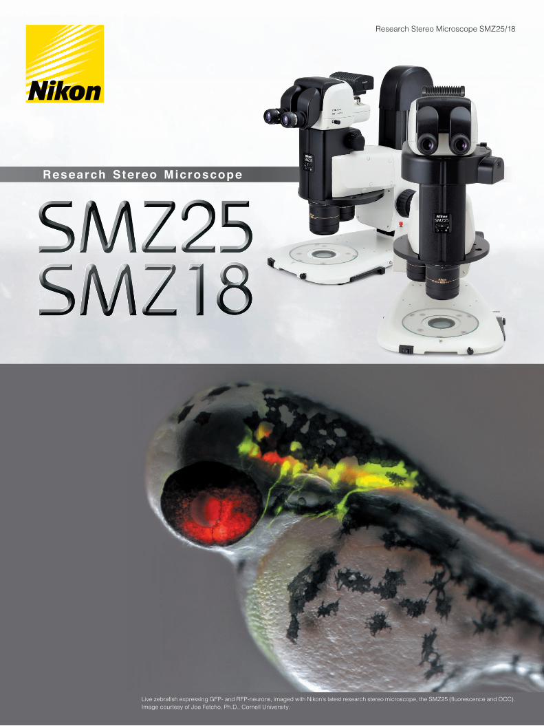
Research Stereo Microscope SMZ25/18
Research Ste reo Microscope
Live zebrafish expressing GFP- and RFP-neurons, imaged with Nikon’s latest research stereo microscope, the SMZ25 (fluorescence and OCC). Image courtesy of Joe Fetcho, Ph.D., Cornell University.
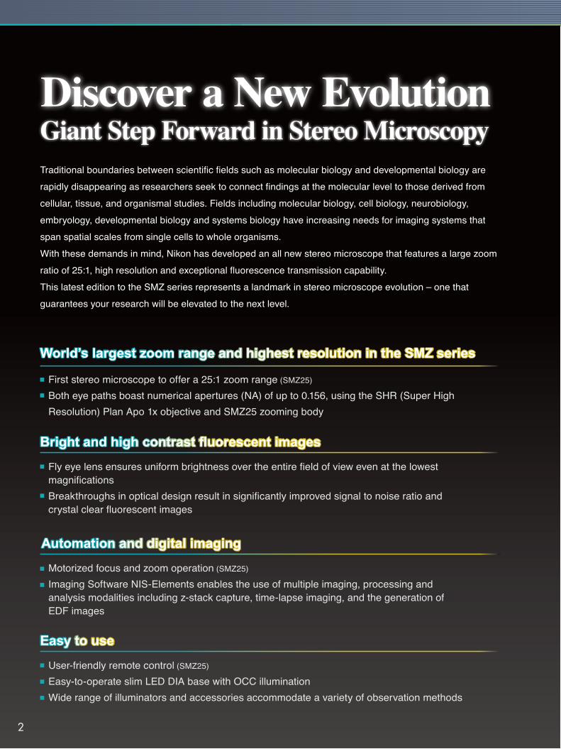
World’s largest zoom range and highest resolution in the SMZ series
First stereo microscope to offer a 25:1 zoom range (SMZ25)
Both eye paths boast numerical apertures (NA) of up to 0.156, using the SHR (Super High Resolution) Plan Apo 1x objective and SMZ25 zooming body
Fly eye lens ensures uniform brightness over the entire field of view even at the lowest magnificationsBreakthroughs in optical design result in significantly improved signal to noise ratio and crystal clear fluorescent images
Motorized focus and zoom operation (SMZ25)
Imaging Software NIS-Elements enables the use of multiple imaging, processing and analysis modalities including z-stack capture, time-lapse imaging, and the generation of EDF images
User-friendly remote control (SMZ25)
Easy-to-operate slim LED DIA base with OCC illuminationWide range of illuminators and accessories accommodate a variety of observation methods
Traditional boundaries between scientific fields such as molecular biology and developmental biology are rapidly disappearing as researchers seek to connect findings at the molecular level to those derived from cellular, tissue, and organismal studies. Fields including molecular biology, cell biology, neurobiology, embryology, developmental biology and systems biology have increasing needs for imaging systems that span spatial scales from single cells to whole organisms. With these demands in mind, Nikon has developed an all new stereo microscope that features a large zoom ratio of 25:1, high resolution and exceptional fluorescence transmission capability.This latest edition to the SMZ series represents a landmark in stereo microscope evolution – one that guarantees your research will be elevated to the next level.
Bright and high contrast fluorescent images
Automation and digital imaging
Easy to use
2
Motorized zoom model with the highest zoom ratio and resolution in the SMZ series
Manual zoom model providing advanced optical performance and incredibly bright fluorescence at an economical cost
Zooming observation
Zoom ratio
Magnification range
Maximum magnification
Maximum FOV
Maximum NA of objective
Motorized zoomBF/DF/FL/Simple polarizing
25:10.63x ~ 15.75x
315x*1
ø70mm*2
0.312*3
Manual zoomBF/DF/FL/Simple polarizing
18:10.75x ~ 13.5x
270x*1
ø59mm*2
0.3*3
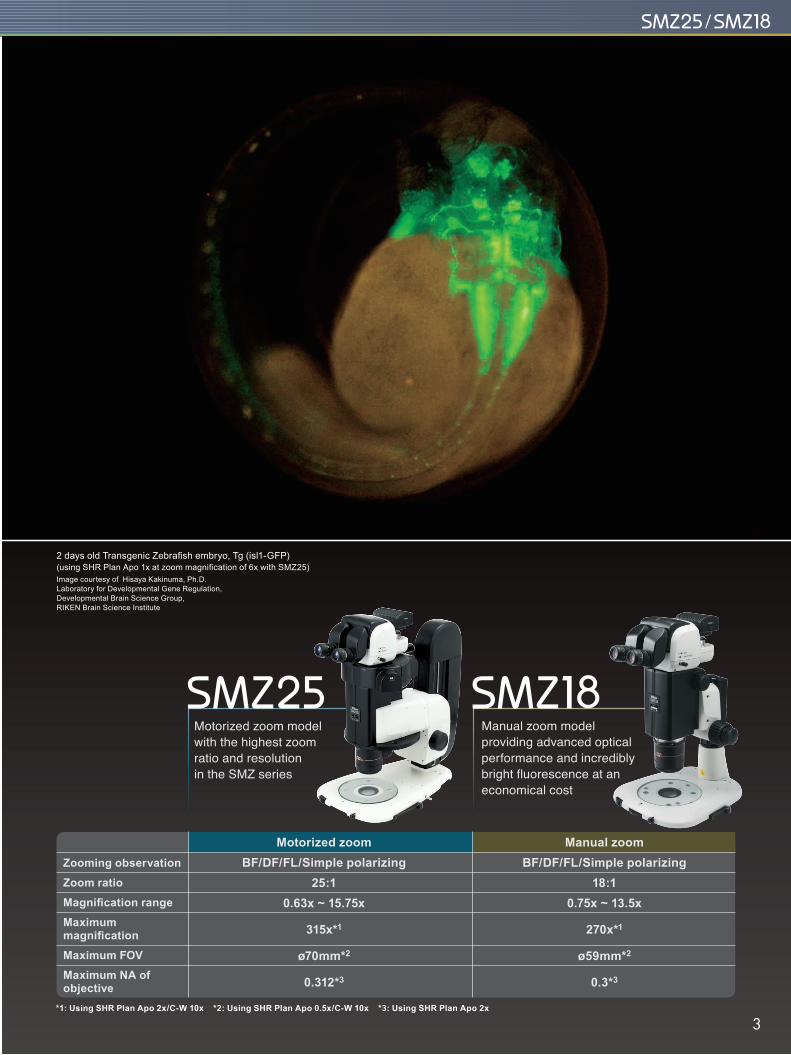
2 days old Transgenic Zebrafish embryo, Tg (isl1-GFP)(using SHR Plan Apo 1x at zoom magnification of 6x with SMZ25)Image courtesy of Hisaya Kakinuma, Ph.D. Laboratory for Developmental Gene Regulation, Developmental Brain Science Group, RIKEN Brain Science Institute
*1: Using SHR Plan Apo 2x/C-W 10x *2: Using SHR Plan Apo 0.5x/C-W 10x *3: Using SHR Plan Apo 2x
3
Offers the highest zoom ratio thanks to Nikon’s Perfect Zoom System
SHR Plan Apo 0.5xzoom 6x
A breakthrough in stereoscope design, Perfect Zoom System dynamically change the distance between the two optical axes as the zoom factor is changed. This change in optical axis distance enables maximization of light entry into the optical system at every magnification. The result is an uncompromised, large zoom range, high resolution in both eye paths, and minimal aberrations over the entire zoom-range. Furthermore, this breakthrough in optical design enables all of these desirable features to be housed in a compact zoom body, resulting in an ergonomic instrument design.
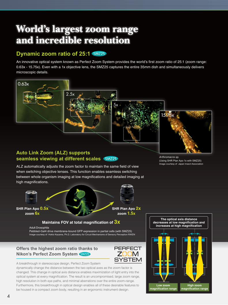
An innovative optical system known as Perfect Zoom System provides the world’s first zoom ratio of 25:1 (zoom range: 0.63x - 15.75x). Even with a 1x objective lens, the SMZ25 captures the entire 35mm dish and simultaneously delivers microscopic details.
Dynamic zoom ratio of 25:1
0.63x
2.5x
5x
15.75x
Arthromacra sp.(Using SHR Plan Apo 1x with SMZ25)Image courtesy of Japan Insect Association
Adult DrosophilaPebbled-Gal4 drive membrane-bound GFP expression in partial cells (with SMZ25)Image courtesy of Hokto Kazama, Ph.D. Laboratory for Circuit Mechanisms of Sensory Perception RIKEN
The optical axis distance decreases at low magnification and
increases at high magnification
ALZ automatically adjusts the zoom factor to maintain the same field of view when switching objective lenses. This function enables seamless switching between whole organism imaging at low magnifications and detailed imaging at high magnifications.
Auto Link Zoom (ALZ) supports seamless viewing at different scales
SHR Plan Apo 2xzoom 1.5x
Maintains FOV at total magnification of 3x
High zoom magnification range
World’s largest zoom range and incredible resolution
Low zoom magnification range
4
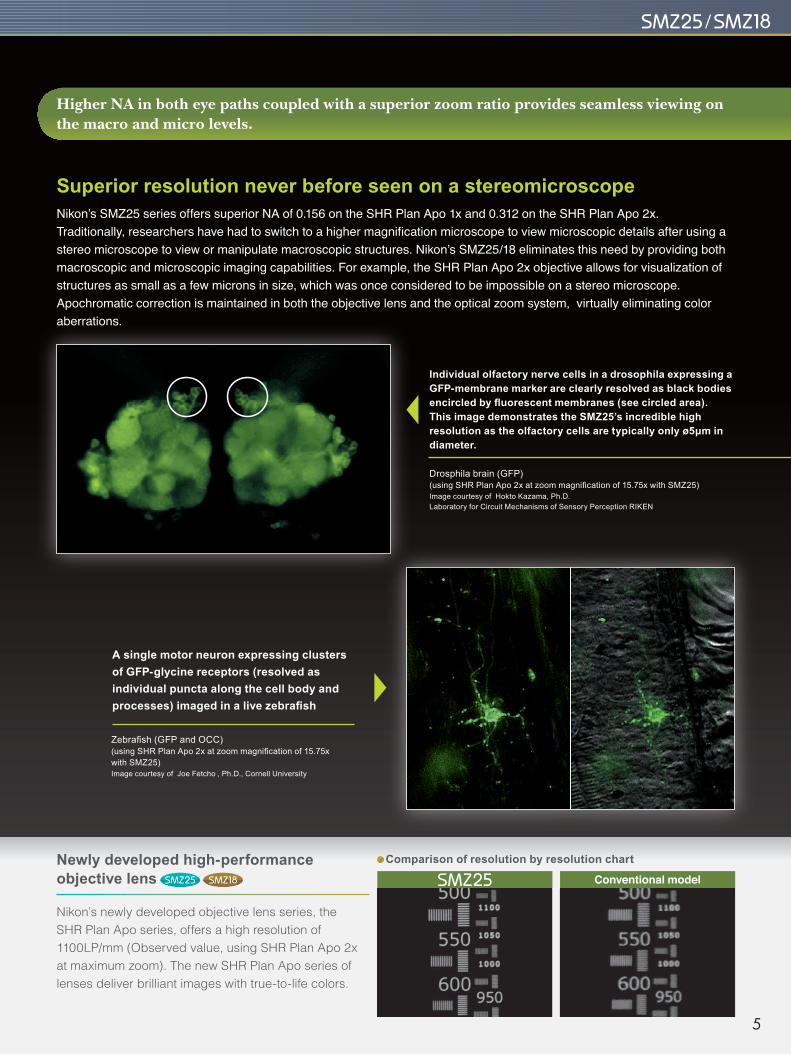
Newly developed high-performance objective lens
Comparison of resolution by resolution chart
Drosphila brain (GFP)(using SHR Plan Apo 2x at zoom magnification of 15.75x with SMZ25)Image courtesy of Hokto Kazama, Ph.D. Laboratory for Circuit Mechanisms of Sensory Perception RIKEN
Zebrafish (GFP and OCC)(using SHR Plan Apo 2x at zoom magnification of 15.75x with SMZ25)Image courtesy of Joe Fetcho , Ph.D., Cornell University
Individual olfactory nerve cells in a drosophila expressing a GFP-membrane marker are clearly resolved as black bodies encircled by fluorescent membranes (see circled area). This image demonstrates the SMZ25’s incredible high resolution as the olfactory cells are typically only ø5μm in diameter.
A single motor neuron expressing clusters of GFP-glycine receptors (resolved as individual puncta along the cell body and processes) imaged in a live zebrafish
Higher NA in both eye paths coupled with a superior zoom ratio provides seamless viewing on the macro and micro levels.
Nikon’s newly developed objective lens series, the SHR Plan Apo series, offers a high resolution of 1100LP/mm (Observed value, using SHR Plan Apo 2x at maximum zoom). The new SHR Plan Apo series of lenses deliver brilliant images with true-to-life colors.
Conventional model
Nikon’s SMZ25 series offers superior NA of 0.156 on the SHR Plan Apo 1x and 0.312 on the SHR Plan Apo 2x. Traditionally, researchers have had to switch to a higher magnification microscope to view microscopic details after using a stereo microscope to view or manipulate macroscopic structures. Nikon’s SMZ25/18 eliminates this need by providing both macroscopic and microscopic imaging capabilities. For example, the SHR Plan Apo 2x objective allows for visualization of structures as small as a few microns in size, which was once considered to be impossible on a stereo microscope. Apochromatic correction is maintained in both the objective lens and the optical zoom system, virtually eliminating color aberrations.
Superior resolution never before seen on a stereomicroscope
5
Time-lapse imaging of developing C. elegans embryos expressing RFP-histones and GFP-membrane markers allows researchers to screen for cytokinesis mutants prior to selection for downstream applications
Fly eye lens ensures uniform brightness over the entire field of view
C. elegans embryos (GFP and RFP; each ovoid is ø30μm in diameter) (using SHR Plan Apo 2x at zoom magnification of 8x with SMZ25)Image courtesy of Julie C. Canman, Ph.D., Columbia University.
12.5 days old mouse embryo, Red: Nucleus(Using SHR Plan Apo 0.5x at zoom magnification 1.30x with SMZ18)Image courtesy of Kazuo Yamagata, Ph.D. Center for Genetic Analysis of Biological Responses, Research Institute for Microbial Diseases, Osaka University
t=0 min t=20 min t=60 min
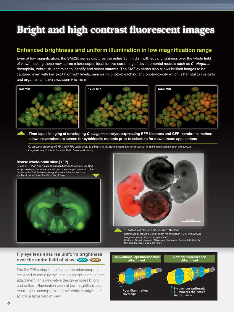
Conventional epi-fluorescence attachment
New epi-fluorescence attachment
Even at low magnification, the SMZ25 series captures the entire 35mm dish with equal brightness over the whole field of view*, making these new stereo microscopes ideal for live screening of developmental models such as C. elegans, drosophila, zebrafish, and mice to identify and select mutants. The SMZ25 series also allows brilliant images to be captured even with low excitation light levels, minimizing photo-bleaching and photo-toxicity which is harmful to live cells and organisms. *Using SMZ25/SHR Plan Apo 1x
Enhanced brightness and uniform illumination in low magnification range
The SMZ25 series is the first stereo microscope in the world to use a fly eye lens on an epi-fluorescence attachment. This innovative design ensures bright and uniform illumination even at low magnifications, resulting in uncompromised uniformity in brightness across a large field of view.
Poor illumination coverage
Fly eye lens uniformly illuminates the entire field of view
Bright and high contrast fluorescent images
Mouse whole-brain slice (YFP)(Using SHR Plan Apo 1x at zoom magnification 0.63x with SMZ25)Image courtesy of Tadatsune Iida, MD., Ph.D. and Shigeo Okabe, M.D., Ph.D.Department of Cellular Neurobiology, Graduate School of Medicine and Faculty of Medicine, the University of Tokyo
6
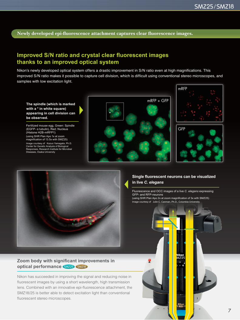
The spindle (which is marked with a * in white square) appearing in cell division can be observed.
Fertilized mouse egg, Green: Spindle (EGFP- α tubulin), Red: Nucleus (Histone H2B-mRFP1)(using SHR Plan Apo 1x at zoom magnification of 13.5x with SMZ25)Image courtesy of Kazuo Yamagata, Ph.D. Center for Genetic Analysis of Biological Responses, Research Institute for Microbial Diseases, Osaka University
Fluorescence and OCC images of a live C. elegans expressing GFP- and RFP-neurons(using SHR Plan Apo 2x at zoom magnification of 3x with SMZ25)Image courtesy of Julie C. Canman, Ph.D., Columbia University
Single fluorescent neurons can be visualized in live C. elegans
mRFP
GFP
mRFP + GFP
Newly developed epi-fluorescence attachment captures clear fluorescence images.
Zoom body with significant improvements in optical performance
Nikon has succeeded in improving the signal and reducing noise in fluorescent images by using a short wavelength, high transmission lens. Combined with an innovative epi-fluorescence attachment, the SMZ18/25 is better able to detect excitation light than conventional fluorescent stereo microscopes.
Nikon’s newly developed optical system offers a drastic improvement in S/N ratio even at high magnifications. This improved S/N ratio makes it possible to capture cell division, which is difficult using conventional stereo microscopes, and samples with low excitation light.
Improved S/N ratio and crystal clear fluorescent images thanks to an improved optical system
7
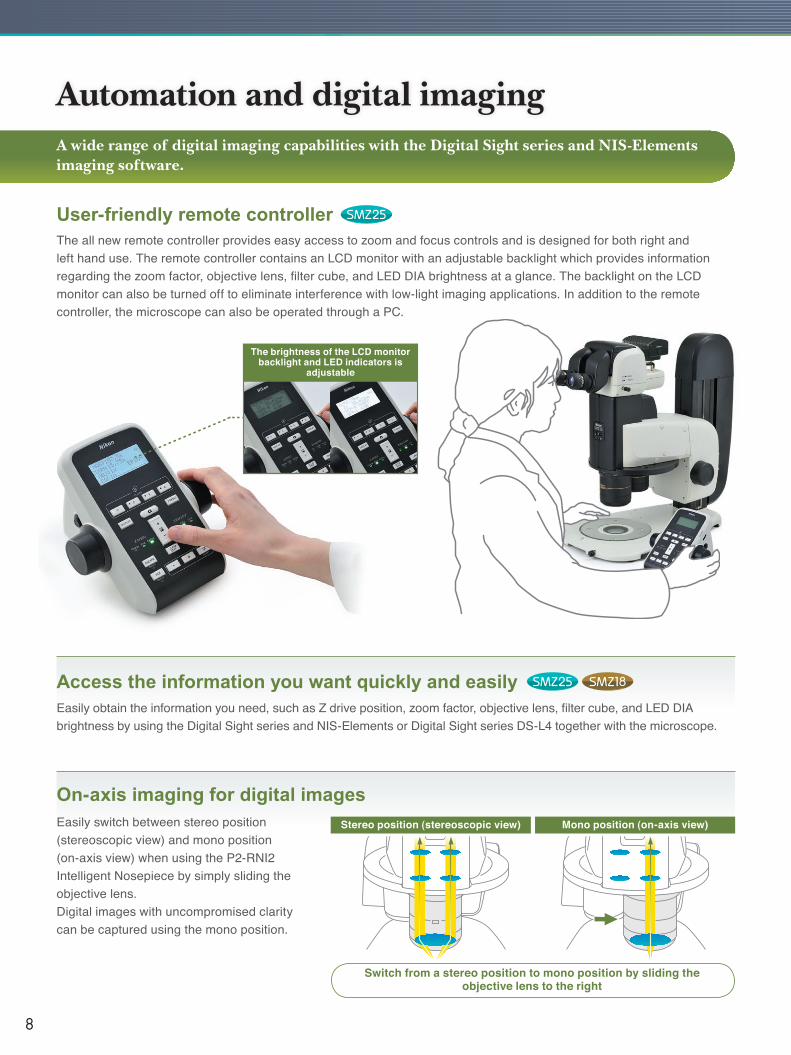
The all new remote controller provides easy access to zoom and focus controls and is designed for both right and left hand use. The remote controller contains an LCD monitor with an adjustable backlight which provides information regarding the zoom factor, objective lens, filter cube, and LED DIA brightness at a glance. The backlight on the LCD monitor can also be turned off to eliminate interference with low-light imaging applications. In addition to the remote controller, the microscope can also be operated through a PC.
User-friendly remote controller
Access the information you want quickly and easilyEasily obtain the information you need, such as Z drive position, zoom factor, objective lens, filter cube, and LED DIA brightness by using the Digital Sight series and NIS-Elements or Digital Sight series DS-L4 together with the microscope.
On-axis imaging for digital imagesEasily switch between stereo position (stereoscopic view) and mono position (on-axis view) when using the P2-RNI2 Intelligent Nosepiece by simply sliding the objective lens.Digital images with uncompromised clarity can be captured using the mono position.
Switch from a stereo position to mono position by sliding the objective lens to the right
A wide range of digital imaging capabilities with the Digital Sight series and NIS-Elements imaging software.
The brightness of the LCD monitor backlight and LED indicators is
adjustable
Stereo position (stereoscopic view) Mono position (on-axis view)
8
Select the perfect camera for your application.
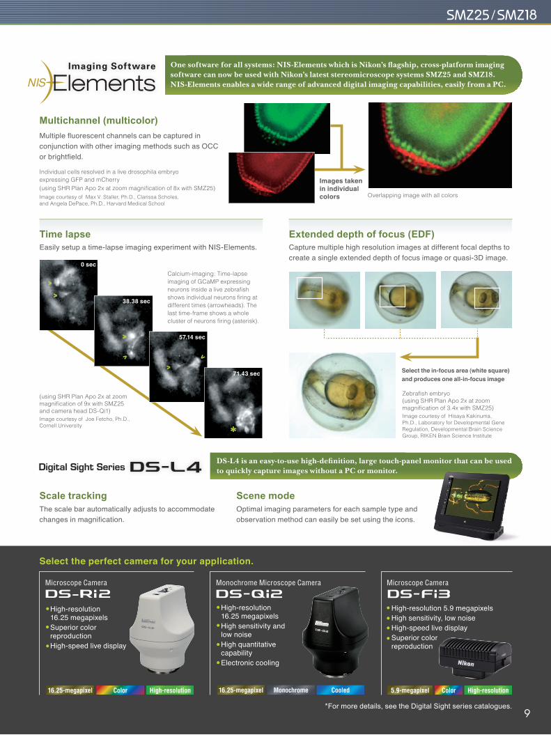
Select the in-focus area (white square) and produces one all-in-focus image
Multichannel (multicolor)Multiple fluorescent channels can be captured in conjunction with other imaging methods such as OCC or brightfield.
Time lapseEasily setup a time-lapse imaging experiment with NIS-Elements.
Images taken in individual colors Overlapping image with all colors
One software for all systems: NIS-Elements which is Nikon’s flagship, cross-platform imaging software can now be used with Nikon’s latest stereomicroscope systems SMZ25 and SMZ18. NIS-Elements enables a wide range of advanced digital imaging capabilities, easily from a PC.
Scale trackingThe scale bar automatically adjusts to accommodate changes in magnification.
DS-L4 is an easy-to-use high-definition, large touch-panel monitor that can be used to quickly capture images without a PC or monitor.
Scene modeOptimal imaging parameters for each sample type and observation method can easily be set using the icons.
Extended depth of focus (EDF)Capture multiple high resolution images at different focal depths to create a single extended depth of focus image or quasi-3D image.
Calcium-imaging: Time-lapse imaging of GCaMP expressing neurons inside a live zebrafish shows individual neurons firing at different times (arrowheads). The last time-frame shows a whole cluster of neurons firing (asterisk).
Zebrafish embryo(using SHR Plan Apo 2x at zoom magnification of 3.4x with SMZ25)Image courtesy of Hisaya Kakinuma, Ph.D., Laboratory for Developmental Gene Regulation, Developmental Brain Science Group, RIKEN Brain Science Institute
*For more details, see the Digital Sight series catalogues.
Microscope Camera
High-resolution 5.9 megapixelsHigh sensitivity, low noiseHigh-speed live displaySuperior color reproduction
Microscope Camera
High-resolution 16.25 megapixelsSuperior color reproductionHigh-speed live display
Monochrome Microscope Camera
High-resolution 16.25 megapixelsHigh sensitivity and low noiseHigh quantitative capabilityElectronic cooling
0 sec
38.38 sec
57.14 sec
71.43 sec
(using SHR Plan Apo 2x at zoom magnification of 9x with SMZ25 and camera head DS-Qi1)Image courtesy of Joe Fetcho, Ph.D., Cornell University
Individual cells resolved in a live drosophila embryo expressing GFP and mCherry(using SHR Plan Apo 2x at zoom magnification of 8x with SMZ25)Image courtesy of Max V. Staller, Ph.D., Clarissa Scholes, and Angela DePace, Ph.D., Harvard Medical School
Monochrome Cooled16.25-megapixelColor High-resolution16.25-megapixel Color High-resolution5.9-megapixel
9
Wide range of available accessories
SMZ18 can be mounted on various compact stands using a focus mount.
Stand / Focus mount
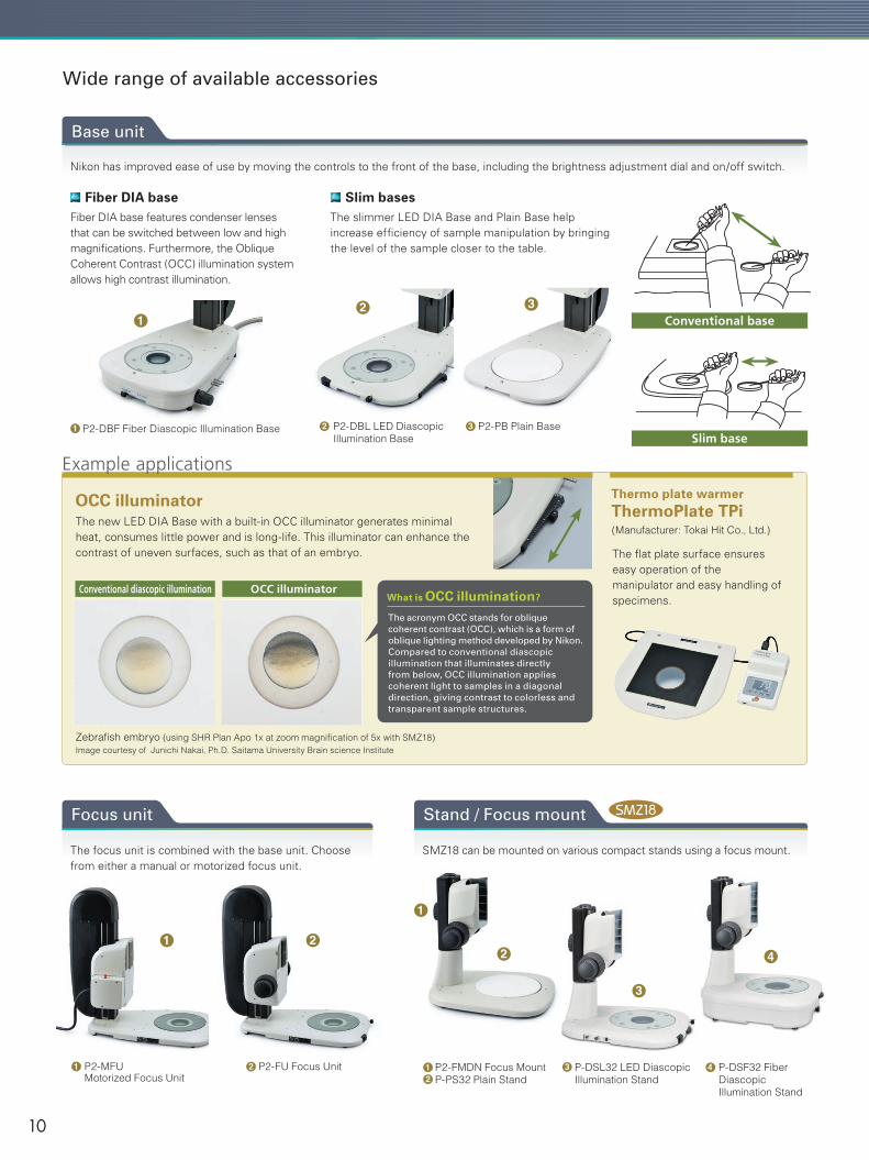
Nikon has improved ease of use by moving the controls to the front of the base, including the brightness adjustment dial and on/off switch.
Base unit
The focus unit is combined with the base unit. Choose from either a manual or motorized focus unit.
Focus unit
P2-MFU Motorized Focus Unit
OCC illuminatorThe new LED DIA Base with a built-in OCC illuminator generates minimal heat, consumes little power and is long-life. This illuminator can enhance the contrast of uneven surfaces, such as that of an embryo.
Thermo plate warmer ThermoPlate TPi(Manufacturer: Tokai Hit Co., Ltd.)
The flat plate surface ensures easy operation of themanipulator and easy handling of specimens.
P2-FMDN Focus MountP-PS32 Plain Stand
What is OCC illumination?
The acronym OCC stands for oblique coherent contrast (OCC), which is a form of oblique lighting method developed by Nikon.Compared to conventional diascopic illumination that illuminates directly from below, OCC illumination applies coherent light to samples in a diagonal direction, giving contrast to colorless and transparent sample structures.
Conventional diascopic illumination OCC illuminator
P2-DBF Fiber Diascopic Illumination Base
P2-FU Focus Unit
P2-PB Plain BaseP2-DBL LED DiascopicIllumination Base
Fiber DIA base features condenser lenses that can be switched between low and high magnifications. Furthermore, the Oblique Coherent Contrast (OCC) illumination system allows high contrast illumination.
The slimmer LED DIA Base and Plain Base help increase efficiency of sample manipulation by bringing the level of the sample closer to the table.
Fiber DIA base Slim bases
Conventional base
Slim base
Zebrafish embryo (using SHR Plan Apo 1x at zoom magnification of 5x with SMZ18) Image courtesy of Junichi Nakai, Ph.D. Saitama University Brain science Institute
2 31
1 2 12
Example applications
P-DSL32 LED Diascopic Illumination Stand
3 4 P-DSF32 Fiber Diascopic Illumination Stand
10
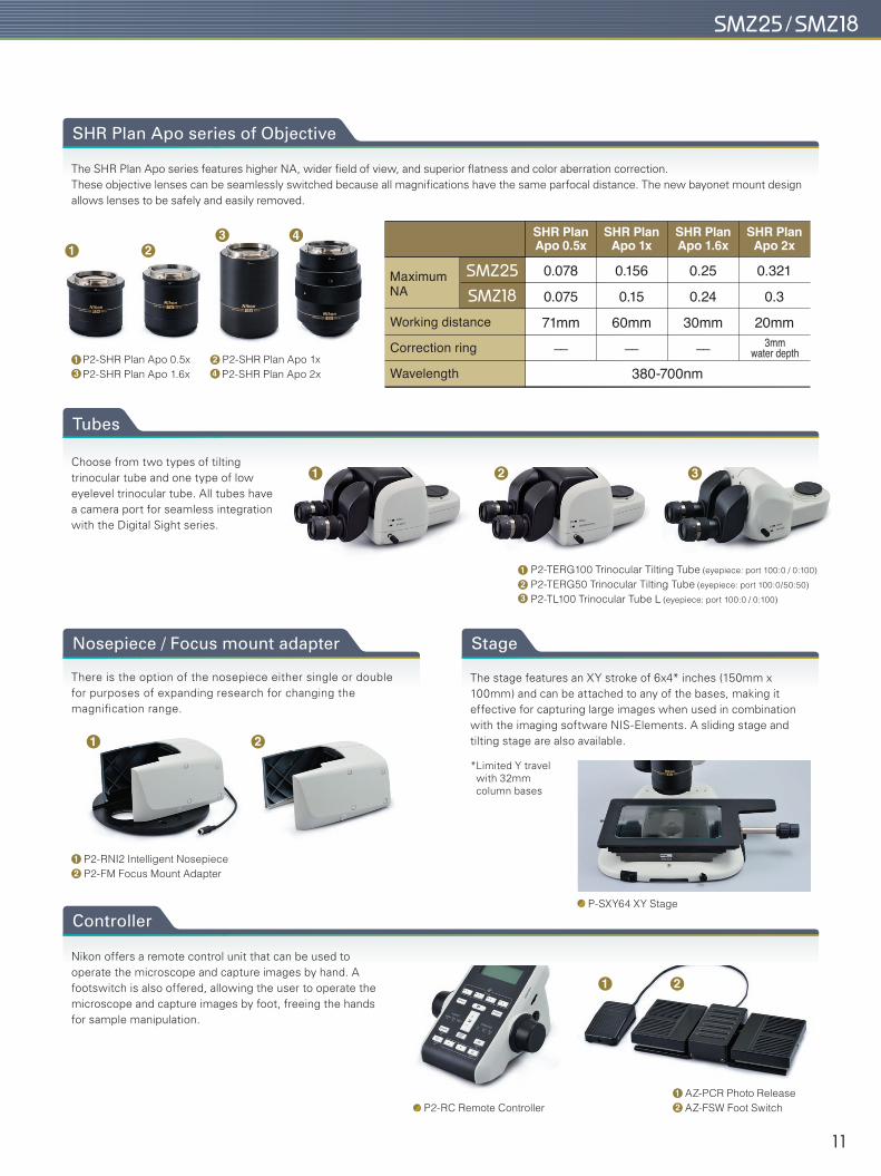
Nikon offers a remote control unit that can be used to operate the microscope and capture images by hand. A footswitch is also offered, allowing the user to operate the microscope and capture images by foot, freeing the hands for sample manipulation.
Controller
P2-RC Remote ControllerAZ-PCR Photo ReleaseAZ-FSW Foot Switch
P2-TERG100 Trinocular Tilting Tube (eyepiece: port 100:0 / 0:100)
P2-TERG50 Trinocular Tilting Tube (eyepiece: port 100:0/50:50)
P2-TL100 Trinocular Tube L (eyepiece: port 100:0 / 0:100)
The SHR Plan Apo series features higher NA, wider field of view, and superior flatness and color aberration correction. These objective lenses can be seamlessly switched because all magnifications have the same parfocal distance. The new bayonet mount design allows lenses to be safely and easily removed.
SHR Plan Apo series of Objective
There is the option of the nosepiece either single or double for purposes of expanding research for changing the magnification range.
Nosepiece / Focus mount adapter
P2-RNI2 Intelligent NosepieceP2-FM Focus Mount Adapter
Choose from two types of tilting trinocular tube and one type of low eyelevel trinocular tube. All tubes have a camera port for seamless integration with the Digital Sight series.
Tubes
The stage features an XY stroke of 6x4* inches (150mm x 100mm) and can be attached to any of the bases, making it effective for capturing large images when used in combination with the imaging software NIS-Elements. A sliding stage and tilting stage are also available.
Stage
P-SXY64 XY Stage
P2-SHR Plan Apo 0.5x P2-SHR Plan Apo 1xP2-SHR Plan Apo 1.6x P2-SHR Plan Apo 2x
Maximum NA
0.25
0.24
30mm
––
SHR Plan Apo 1.6x
380-700nm
Working distance
Correction ring
Wavelength
3mm water depth
0.321
0.3
20mm
SHR Plan Apo 2x
0.156
0.15
60mm
––
SHR Plan Apo 1x
0.078
0.075
71mm
––
SHR Plan Apo 0.5x
*Limited Y travel with 32mm column bases
13
24
123
12
12
11
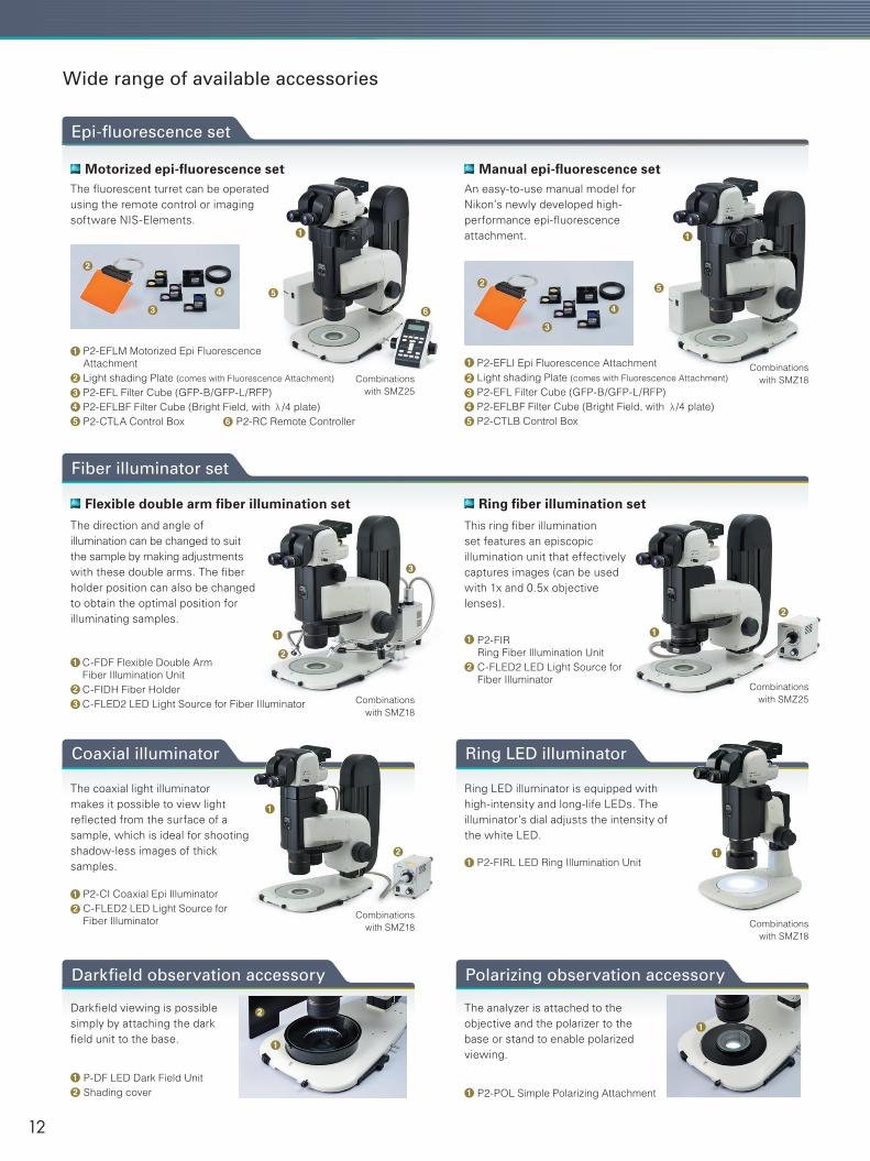
Darkfield viewing is possible simply by attaching the dark field unit to the base.
Darkfield observation accessory
The analyzer is attached to the objective and the polarizer to the base or stand to enable polarized viewing.
Polarizing observation accessory
P2-EFLM Motorized Epi Fluorescence AttachmentLight shading Plate (comes with Fluorescence Attachment)
P2-EFL Filter Cube (GFP-B/GFP-L/RFP)P2-EFLBF Filter Cube (Bright Field, with /4 plate)P2-CTLA Control Box P2-RC Remote Controller
Coaxial illuminator
The coaxial light illuminator makes it possible to view light reflected from the surface of a sample, which is ideal for shooting shadow-less images of thick samples.
Ring LED illuminator
Ring LED illuminator is equipped with high-intensity and long-life LEDs. The illuminator’s dial adjusts the intensity of the white LED.
Combinations with SMZ18
Flexible double arm fiber illumination set
The direction and angle of illumination can be changed to suit the sample by making adjustments with these double arms. The fiber holder position can also be changed to obtain the optimal position for illuminating samples.
Fiber illuminator set
Ring fiber illumination set
This ring fiber illumination set features an episcopic illumination unit that effectively captures images (can be used with 1x and 0.5x objective lenses).
C-FDF Flexible Double ArmFiber Illumination UnitC-FIDH Fiber HolderC-FLED2 LED Light Source for Fiber Illuminator
P-DF LED Dark Field UnitShading cover
P2-FIRL LED Ring Illumination Unit
P2-CI Coaxial Epi IlluminatorC-FLED2 LED Light Source for Fiber Illuminator
P2-POL Simple Polarizing Attachment
P2-FIRRing Fiber Illumination UnitC-FLED2 LED Light Source for Fiber Illuminator
Wide range of available accessories
Epi-fluorescence set
The fluorescent turret can be operated using the remote control or imaging software NIS-Elements.
Motorized epi-fluorescence set
Combinations with SMZ18
Combinations with SMZ18
Combinations with SMZ25
Combinations with SMZ25
Combinations with SMZ18
An easy-to-use manual model for Nikon’s newly developed high-performance epi-fluorescence attachment.
Manual epi-fluorescence set
P2-EFLI Epi Fluorescence AttachmentLight shading Plate (comes with Fluorescence Attachment)
P2-EFL Filter Cube (GFP-B/GFP-L/RFP)P2-EFLBF Filter Cube (Bright Field, with /4 plate)P2-CTLB Control Box
1
32
45 6
1
32
45
1
21
32
12
1
112
12
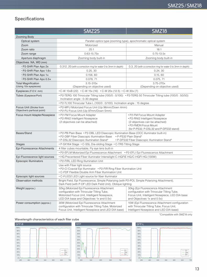
Specifications
Zooming Body Optical system Parallel-optics type (zooming type), apochromatic optical system
Zoom Motorized Manual
Zoom ratio 25:1 18:1
Zoom range 0.63-15.75x 0.75-13.5x
Aperture diaphragm Zooming body built-in Zooming body built-in
Objectives NA, WD (mm) • P2-SHR Plan Apo 2x 0.312, 20 (with a correction ring for water 0 to 3mm in depth) 0.3, 20 (with a correction ring for water 0 to 3mm in depth)
• P2-SHR Plan Apo 1.6× 0.25, 30 0.24, 30
• P2-SHR Plan Apo 1× 0.156, 60 0.15, 60
• P2-SHR Plan Apo 0.5× 0.078, 71 0.075, 71
Total Magnification 3.15-315x 3.75-270x(Using 10x eyepieces) (Depending on objective used) (Depending on objective used)
Eyepieces (F.O.V. mm) • C-W 10xB (22) • C-W 15x (16) • C-W 20x (12.5) • C-W 30x (7)
Tubes (Eyepiece/Port) • P2-TERG 100 Trinocular Tilting tube (100/0 : 0/100) • P2-TERG 50 Trinocular Tilting tube (100/0 : 50/50) Inclination angle : 0-30 degree
• P2-TL100 Trinocular Tube L (100/0 : 0/100) Inclination angle : 15 degree
Focus Unit (Stroke from • P2-MFU Motorized Focus Unit (Up 96mm/Down 4mm)Objective's parfocal point) • P2-FU Focus Unit (Up 97mm/Down 5mm)
Focus mount Adapter/Nosepiece • P2-FM Focus Mount Adapter • P2-FM Focus Mount Adapter • P2-RNI2 Intelligent Nosepiece • P2-RNI2 Intelligent Nosepiece (2 objectives can be attached) (2 objectives can be attached) • P2-FMDN Focus Mount (for P-PS32, P-DSL32 and P-DFS32 stand)
Bases/Stand • P2-PB Plain Base • P2-DBL LED Diascopic Illumination Base (OCC illuminator built-in) • P2-DBF Fiber Diascopic Illumination Base • P-PS32 Plain Stand* • P-DSL32 Diascopic Illumination Stand* • P-DFS32 Fiber Diascopic Illumination Stand*
Stages • P-SXY64 Stage • C-SSL Dia-sliding Stage • C-TRS Tilting Stage
Epi-Fluorescence Attachments 4 filter cubes mountable, Fly eye lens built-in
• P2-EFLM Motorized Epi Fluorescence Attachment • P2-EFLI Epi Fluorescence Attachment
Epi-Fluorescence light sources • HG Precentered Fiber illuminator Intensilight C-HGFIE HG/C-HGFI HG (130W)
Episcopic Illuminators • P2-FIRL LED Ring Illumination Unit
Use with Fiber light source • P2-CI Coaxial Epi Illuminator • P2-FIR Ring Fiber Illumination Unit • C-FDF Flexible Double Arm Fiber Illumination Unit
Episcopic light sources • C-FLED2 LED Light source for fiber illuminator
Observation methods Bright Field, Epi Fluorescence, Simple Polarizing (with P2-POL Simple Polarizing Attachment), Dark Field (with P-DF LED Dark Field Unit), Oblique lighting
Weight (approx.) 32kg (Motorized Epi Fluorescence Attachment 30kg (Epi Fluorescence Attachment configuration with Trinocular Tilting Tube, configuration with Trinocular Tilting Tube, Motorized Focus Unit, Intelligent Nosepiece, Focus Unit, Intelligent Nosepiece, LED DIA base LED DIA base and Objectives 1x and 0.5x) and Objectives 1x and 0.5x)
Power consumption (approx.) 30W (Motorized Epi Fluorescence Attachment 10W (Epi Fluorescence Attachment configuration configuration with Trinocular Tilting Tube, Motorized with Trinocular Tilting Tube, Focus Unit, Focus Unit, Intelligent Nosepiece and LED DIA base) Intelligent Nosepiece and LED DIA base)
400
Wavelength characteristics of each filer cube
450 500 550 600 650 7000%
10%
20%
30%
40%
50%
60%
70%
80%
90%
100%
EXDMBA
500 550 600 650 7500%
10%
20%
30%
40%
50%
60%
70%
80%
90%
100%
400 450 500 550 600 650 7000%
10%
20%
30%
40%
50%
60%
70%
80%
90%
100%
EXDMBA
EXDMBA
GFP-B RFP GFP-L
700
*Compatible with SMZ18 only
13
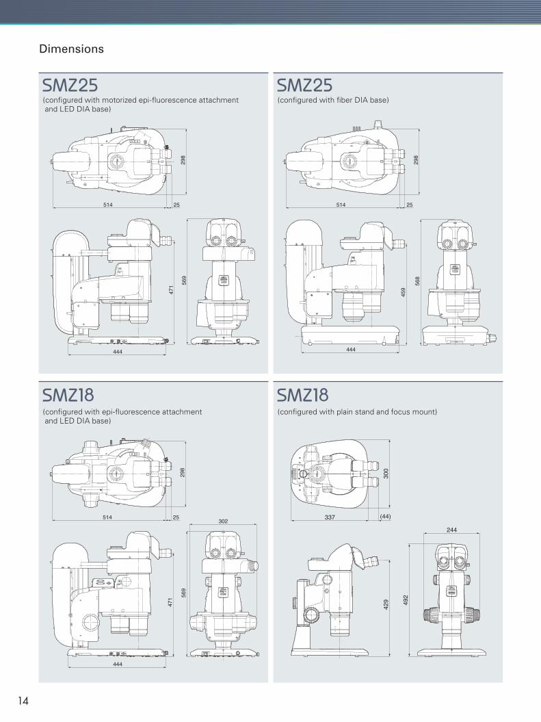
(configured with motorized epi-fluorescence attachment and LED DIA base)
300
337 (44)
244
429 49
2
444
514 25
569
471
298
302
444
514 25
568
459
298
444
514 25
569
471
298
Dimensions
(configured with fiber DIA base)
(configured with epi-fluorescence attachment and LED DIA base)
(configured with plain stand and focus mount)
14
������������
�����������
��������������
���������������������������������
��������������������������������
�������������������������
��������������
��������������������������������������
�������������������������������������
���������������������������
������������������������������
����������������������������
������������������������������������������������������������
����������������������������������������������������������������������
���������������������������� ������������
�����������������
���������������������������
����������������
���������������
����������������������� ��������
�����
����������������������������������
���������������������������������������������
�����������������������������������������������
������������������
��������������������������������
������������������
�������������������
������������������
������������������
�����������������
����������������������
�����������������
���������������������������
��������������������������������
�������������������
������������������
��������������������
��������������������
�����������������������
�������������������������
����������������������������������
������������������
������������������������������ �����
����������������������������������
���������������������
�����������������
�����������������������������������������
���������������������������������������
���������������
������������������
����������������������
����������������������
���������������
��������������
��������������
J
JZ
Z
IH
B
B
B
Y
V X
HI
Y
Y
����������������������������
X
X
X
A
A A A
XXW C
W
Z
D
D
����������������������������������������������
�����������������
�����������������
X
G
IH
GG
GD
���������������������������
�������������������������
������������
�����������
������������
�����������
��������������������
����������������
C
V
IH
G
JIH
GGD
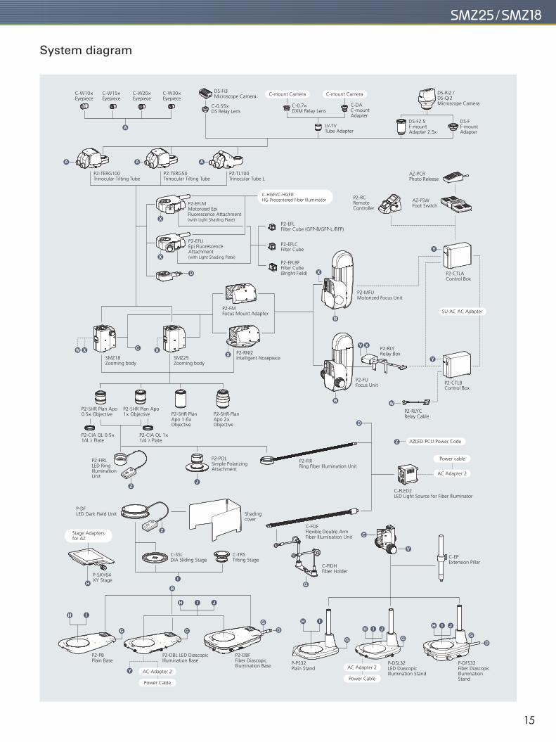
JIH
System diagram
15
Specifications and equipment are subject to change without any notice or obligation on the part of the manufacturer. December 2017 ©2013-2017 NIKON CORPORATIONN.B. Export of the products* in this brochure is controlled under the Japanese Foreign Exchange and Foreign Trade Law. Appropriate export procedures shall be required in case of export from Japan.*Products: Hardware and its technical information (including software)
ISO 14001 Certifiedfor NIKON CORPORATIONNIKON CORPORATION
Shinagawa Intercity Tower C, 2-15-3, Konan, Minato-ku, Tokyo 108-6290, JapanHealthcare Business Unit, phone: +81-3-6433-3705 fax: +81-3-6433-3785Industrial Metrology Business Unit, phone: +81-3-6433-3703 fax: +81-3-6433-3784http://www.nikon.com/products/
Printed in Japan (1712-04) Am/M Code No. 2CE-MPGH-5
NIKON INSTECH CO., LTD.Shinagawa Intercity Tower C, 2-15-3, Konan, Minato-ku, Tokyo 108-6290phone: +81-3-6433-3701 fax: +81-3-6433-3784http://www.nikon-instruments.jp/
NIKON INSTRUMENTS INC.1300 Walt Whitman Road, Melville, N.Y. 11747-3064, U.S.A.phone: +1-631-547-8500; +1-800-52-NIKON (within the U.S.A. only)fax: +1-631-547-0306http://www.nikoninstruments.com/
NIKON METROLOGY, INC.12701 Grand River Avenue, Brighton, MI 48116 U.S.A.phone: +1-810-220-4360 fax: +1-810-220-4300E-mail: [email protected]://www.nikonmetrology.com/
NIKON INSTRUMENTS EUROPE B.V.Tripolis 100, Burgerweeshuispad 101, 1076 ER Amsterdam, The Netherlandsphone: +31-20-7099-000 fax: +31-20-7099-298 http://www.nikoninstruments.eu/
NIKON METROLOGY EUROPE NVGeldenaaksebaan 329, 3001 Leuven, Belgiumphone: +32-16-74-01-00 fax: +32-16-74-01-03 Email: [email protected]://www.nikonmetrology.com/
NIKON INSTRUMENTS (SHANGHAI) CO., LTD.CHINA phone: +86-21-6841-2050 fax: +86-21-6841-2060(Beijing branch) phone: +86-10-5831-2028 fax: +86-10-5831-2026(Guangzhou branch) phone: +86-20-3882-0550 fax: +86-20-3882-0580
NIKON INSTRUMENTS KOREA CO., LTD.KOREA phone: +82-2-2186-8400 fax: +82-2-555-4415
NIKON SINGAPORE PTE LTD.SINGAPORE phone: +65-6559-3651 fax: +65-6559-3668NIKON MALAYSIA SDN. BHD.MALAYSIA phone: +60-3-7809-3688 fax: +60-3-7809-3633PT. NIKON INDONESIAINDONESIA phone: +62-21-574-6262 fax: +62-21-574-6363Nikon Sales (Thailand) Co., Ltd.THAILAND phone: +66-2633-5100 fax: +66-2633-5191NIKON INDIA PRIVATE LIMITEDINDIA phone: +91-124-4688500 fax: +91-124-4688527NIKON CANADA INC.CANADA phone: +1-905-602-9676 fax: +1-905-602-9953NIKON INSTRUMENTS S.p.A.ITALY phone: +39-55-300-96-01 fax: +39-55-30-09-93NIKON GMBH SWITZERLANDSWITZERLAND phone: +41-43-277-28-67 fax: +41-43-277-28-61
NIKON CEE GMBHAUSTRIA phone: +43-1-972-6111-00 fax: +43-1-972-6111-140
NIKON UK LTD.UNITED KINGDOM phone: +44-208-247-1717 fax: +44-208-541-4584
NIKON METROLOGY UK LTD.UNITED KINGDOM phone: +44-1332-811-349 fax: +44-1332-639-881E-mail: [email protected]
NIKON FRANCE S.A.S.FRANCE phone: +33-1-4516-45-16 fax: +33-1-4516-45-55NIKON METROLOGY SARLFRANCE phone: +33-1-60-86-09-76 fax: +33-1-60-86-57-35E-mail: [email protected]
NIKON GMBHGERMANY phone: +49-211-941-42-20 fax:+49-211-941-43-22NIKON METROLOGY GMBHGERMANY phone: +49-6023-91733-0 fax: +49-6023-91733-229E-mail: [email protected]


![[Manual] SHR 1041K](https://img.pdfslide.net/doc/110x75/547ca951b4af9f8a138b45c5/manual-shr-1041k.jpg)