Embed Size (px)
Citation preview
Guo et al. Journal of Translational Medicine 2010, 8:50http://www.translational-medicine.com/content/8/1/50
Open AccessR E S E A R C H
ResearchAssessing agonistic potential of a candidate therapeutic anti-IL21R antibodyYongjing Guo1, Andrew A Hill1, Renee C Ramsey1, Frederick W Immermann2, Christopher Corcoran1, Deborah Young3, Edward R LaVallie1, Mark Ryan3, Theresa Bechard4, Richard Pfeifer5, Garvin Warner6, Marcia Bologna7, Laird Bloom1 and Margot O'Toole*8,9
AbstractBackground: Selective neutralization of the IL21/IL21R signaling pathway is a promising approach for the treatment of a variety of autoimmune diseases. Ab-01 is a human neutralizing anti-IL21R antibody. In order to ensure that the activities of Ab-01 are restricted to neutralization even under in vitro cross-linking and in vivo conditions, a comprehensive assessment of agonistic potential of Ab-01 was undertaken.
Methods: In vitro antibody cross-linking and cell culture protocols reported for studies with a human agonistic antibody, TGN1412, were followed for Ab-01. rhIL21, the agonist ligand of the targeted receptor, and cross-linked anti-CD28 were used as positive controls for signal transduction. In vivo agonistic potential of Ab-01 was assessed by measuring expression levels of cytokine storm-associated and IL21 pathway genes in blood of cynomolgus monkeys before and after IV administration of Ab-01.
Results: Using a comprehensive set of assays that detected multiple activation signals in the presence of the positive control agonists, in vitro Ab-01-dependent activation was not detected in either PBMCs or the rhIL21-responsive cell line Daudi. Furthermore, no difference in gene expression levels was detected in blood before and after in vivo Ab-01 dosing of cynomolgus monkeys.
Conclusions: Despite efforts to intentionally force an agonistic signal from Ab-01, none could be detected.
BackgroundIL21 (interleukin 21) is a type I cytokine produced byactivated CD4+ T cells and natural killer (NK) T cells [1-4]. It promotes both B cell function [2], and growth of theTH17 T cell subset involved in chronic inflammation [5].Involvement of the IL21 pathway has been demonstratedin a variety of pro-inflammatory and autoimmune animalmodels [6-10]. Inhibition of the IL21/IL21R pathwaytherefore represents a promising therapeutic strategy fortreatment of chronic inflammatory and autoimmuneconditions [11]. We have taken the approach of blockingIL21-mediated activation by developing Ab-01, an anti-body that selectively binds the high-affinity alpha chain ofthe IL21 receptor, IL21R. Ab-01 blocks the binding ofIL21 to IL21R and inhibits IL21-mediated activation [12].
Ab-01 was selected based on the property of inhibiting(antagonizing) rhIL21-mediated cell activation [13], (Araiet al. Journal of Translational Medicine, in press). Giventhat the therapeutic goal of Ab-01 is the down-modula-tion of autoimmune disease activity, it was important toensure that Ab-01 cannot deliver an activation signal,even when cross-linked. Since Ab-01 is an antagonisticantibody, we did not expect that agonistic activity wouldbe detected. However due diligence dictated that allefforts should be made to force an agonistic signal so thatpotential risks and biomarkers associated with any ago-nistic activity could be thoroughly assessed and under-stood. The need for such due diligence was highlighted bythe clinical experience with TGN1412, an anti-CD28 can-didate therapeutic antibody that, in stark contrast to Ab-01, was developed based on immune system activatingactivity [14]. Immunotoxicity had not been observed inpreclinical studies done in rhesus monkeys, but withinhours of clinical administration, life-threatening organ
* Correspondence: [email protected] Pfizer, BioTherapeutics Clinical Translational Medicine, Cambridge, MA, USAFull list of author information is available at the end of the article
© 2010 Guo et al; licensee BioMed Central Ltd. This is an Open Access article distributed under the terms of the Creative Commons At-tribution License (http://creativecommons.org/licenses/by/2.0), which permits unrestricted use, distribution, and reproduction in anymedium, provided the original work is properly cited.
Guo et al. Journal of Translational Medicine 2010, 8:50http://www.translational-medicine.com/content/8/1/50
Page 2 of 11
failure associated with cytokine storm was observed in alltreated volunteers [15]. So even though Ab-01 is animmune system antagonist and TGN1412 an immunesystem agonist, we conducted an in-depth search for acti-vation signals induced by in vitro cross-linked Ab-01 andexamined the effects of Ab-01 when administered in vivoto cynomolgus monkeys.
In addition to antagonist activity of Ab-01 versus theagonist activity of TGN1412, there are other importantdistinctions between Ab-01 and TGN1412, particularly inthe preclinical data packages of the two antibodies. Bind-ing of TGN1412 to rhesus T cells had been demonstratedprior to clinical testing [16], but biological activity ofTGN1412 on rhesus cells had not been extensivelyexplored. In contrast, we have shown that Ab-01 blocksrhIL21-mediated cell activation in cynomolgus monkeys,the Ab-01 safety study species [13], (Arai et al. Journal ofTranslational Medicine, in press), and that the IC50 ofAb-01 in cynomolgus monkey is only about 3.5-foldhigher than the IC50 in human (Arai and LaVallie,unpublished data). These studies established that Ab-01activity is very similar in cynomolgus monkeys and inhuman, whereas the activity of TGN1412 in rhesus wasclearly very different from the activity in humans.
The clinical experience with TGN1412 has heightenedconcern within the medical, regulatory and drug develop-ment communities regarding immunotoxic potential ofimmuno-modulatory antibody therapeutic candidates[15-19]. As a result of the review conducted in the after-math of the experience with TGN1412, comprehensiveefforts were undertaken to identify in vitro protocolscapable of revealing the immunotoxic cytokine storm-inducing properties of TGN1412. Using purified PMBCsfrom healthy donors, Stebbins et al. found that TGN1412,when cross-linked in vitro using any of three methods,induced secretion of a set of cytokines that had beenassociated with the cytokine storm syndrome in the clinic[14]. Notably, PBMC from rhesus did not give thisresponse to in vitro cross-linked TGN1412, and this invitro dichotomy between humans and the safety studyspecies mirrored the dichotomy that had been observedin vivo. Two lines of evidence supported the hypothesisthat the in vitro cross-linking assay system was a relevantsurrogate read-out for the observed in vivo immunotoxic-ity of TGN1412: a) there was a concordance in the cytok-ines induced in humans in vivo and in vitro, and b) thecytokine storm-associated response was not observedeither in vivo or in vitro in rhesus.
Our strategy for in vitro testing of the ability of Ab-01to induce an activation signal was to follow and signifi-cantly expand upon the cross-linking protocols reportedby Stebbins et al.[14]. We used two types of positive con-trols: rhIL21 stimulation for signal transduction throughthe therapeutic target IL21R, and in vitro cross-linked
anti-CD28 for induction of cytokines associated withimmunotoxicity. In parallel, we identified biomarkers ofrhIL21 pathway agonism in monkey blood [13], (Arai etal. Journal of Translational Medicine, in press), andreport here on the levels of these biomarkers in bloodfrom monkeys collected before and after high dose IVadministration of Ab-01.
MethodsProtein reagents: rhIL21, Ab-01, anti-CD28, and control antibodiesMost protein reagents used in this study - rhIL21, anti-rhIL21 receptor antibody Ab-01, control antibody humanIgG1α-tetanus triple mutant (IgG1TM), control antibodyhuman IgG1 α-tetanus wildtype (IgG1) and control anti-body human Fc control (IgGFc) - were made by theGlobal Biotherapeutics Technologies Department atPfizer, Cambridge, MA. The three mutations common tothe Fc portion of Ab-01 and IgG1TM reduced theirpotential effector activity. Antibodies with these muta-tions had undetectable activity in ADCC or C1q bindingassays [20,21]. An antibody with severely compromisedeffector function was chosen for development becausethe therapeutic goal is to block the interaction of IL21with IL21R, and therefore minimization of effector func-tion is desirable. rhIL21 was used as a positive control forsignal delivery through IL21R. Anti-CD28 antibodyANC28.1/5D10(Ancell, Bayport, MN) was used as a posi-tive control for cell activation mediated by cross-linkedantibodies. Endotoxin levels in all proteins reagents werebelow 1.0 EU/mg.
Adherence and confirmation of adherence of antibodies to wellsAb-01, anti-CD28, and control immunoglobulins (humanIgG1 α-tetanus triple mutant, human IgG1 α-tetanus wildtype and human Fc control) at 5 ng, 50 ng, 100 ng, 300 ng,1 μg or 10 μg per well were presented to Daudi cells andpurified human PBMC using the conditions for coatingonto plastic wells as previously described [14]. For drycoating, a total volume of 50 μL containing the indicatedconcentration of IgG reagent was applied to each well(96-well polystyrene Corning High Bind plates, Corning,Lowell, MA). Uncovered plates were left to dry in a tissueculture hood at room temperature overnight. For theanti-IgG-coated wells, the indicated concentration of IgGreagent was added in a volume of 100 μL to wells of goatanti-human IgG plates (H+L) (BD Biosciences, Bedford,MA) at room temperature for 1 hour, and then agitatedovernight at 4°C. For the wet coating method, IgGreagents at the indicated concentration were added in 100μL in PBS (PH = 7.2) to wells of 96-well polystyreneCorning High Bind plates (Corning, Lowell, MA). Plateswere agitated at room temperature for 1 hour and incu-
Guo et al. Journal of Translational Medicine 2010, 8:50http://www.translational-medicine.com/content/8/1/50
Page 3 of 11
bated at 4°C overnight. All antibody-coated plates werewashed three times with PBS CMF (PBS free of calcium/magnesium, pH = 7.2) before addition of cells.
Following the completion of cell culture proceduresdescribed below, the persistence of bound immunoglobu-lin on wells was confirmed by ELISA detection of humanIgG. Following cell harvest and 4 washes with 0.03%Tween-20 in PBS, 100 μL/well of 1:2,000 dilution of HRP-coupled mouse anti-human IgG (Southern Biotech, Bir-mingham, AL) was added. Plates were agitated gently for30 min, washed 4 times with 0.03% Tween-20 in PBS and100 μL/well of BioFX TMB HRP Microwell Substrate(BioFX Laboratories, Inc., Owings Mills, MD) added.Color development was allowed to proceed for 5 minutesat room temperature. The reaction was stopped with 50μL/well 0.18N H2SO4. The relative amount of bound anti-body was recorded using a SpectraMax Plus plate reader(Molecular Devices, Sunnyvale, CA). Bound IgG wasconfirmed by ELISA in all wells used in the studiesreported here. ELISA results showed increasing IgG-spe-cific signal between the 0.1 μg/well and 1 μg/well concen-tration, with no difference in signal between the 1 μg/welland 10 μg/well concentrations (data not shown).
Human PBMC isolation and assay for effects of cross-linked antibodyHuman blood was obtained from Research Blood Com-ponents, Brighton, MA. An IRB-approved consent formwas obtained from each donor consenting to blood dona-tion for research purposes. Peripheral blood mononu-clear cells (PBMCs) were isolated from 136-450 mL ofblood using Sodium Citrate CPT Vacutainer tubesaccording to the manufacturer's instructions. PBMCswere washed twice in PBS (pH = 7.2), and differential cellcounts were measured using a Pentra 60C (Horiba ABXDiagnostics, Irving, CA). Cells were resuspended inRPMI with complete supplements and plated at 2.5 × 10⁵
cells in 100 μL/well and cultured for the durations indi-cated. Experiments testing the effects of cross-linked Ab-01 were conducted on PBMCs from 10 different individ-ual human donors, and positive and negative controlswere performed contemporaneously.
Levels of IFNγ, IL1β, IL2, IL4, IL5, IL8, IL10, IL12p70,IL13, and TNF were determined using 10-spot 96 wellMSD plates (MS6000 Human TH1/TH2 10-Plex Kit,Meso Scale Discovery, Gaithersburg, MD) according tothe manufacturer's instructions. In addition, levels of IL6and CCL3 were determined by customized 2 spot MSD96 well plates (Meso Scale Discovery). Tests were run intriplicate and Student's t-tests were used to identify sig-nificant differences. Fold changes were calculated foreach donor by dividing values of cytokine concentrationfrom Ab-01 cross-linked groups by values from controlantibody cross-linked groups. rhIL21-dependent foldchange responses were calculated by dividing values in
the presence of rhIL21 to media control. The rhIL21stimulation controls were included both on plates pre-coated with anti-IgG and on plates that were not pre-coated.
In addition to measuring secreted protein levels, RNAexpression levels were also measured. Following harvestof supernatants, RNA was isolated from cells by additionof 125 μL RLT lysis buffer (Qiagen, Valencia, CA) con-taining 1% β-mecaptoethanol. Total RNA isolation wasperformed using the QIA Shredder and RNeasy Mini kitkits (Qiagen, Valencia, CA) according to the manufac-turer's recommendations. All of the samples were sub-jected to a DNase on-column treatment to removepotential DNA contamination, and then purified usingthe RNeasy Mini kit.
A phenol:chloroform (1:1) extraction was performedsubsequently, and RNA was repurified using the RNeasyMini kit. Eluted RNA was quantified using a ND-1000Spectrophotometer (Nanodrop, Wilmington, DE). RNAwas converted to cDNA using the Applied BiosystemsHigh Capacity cDNA Archive kit with RNase Inhibitor at50 U/sample (Applied Biosystems). cDNA samples werestored at -20°C pending TaqMan® assay.
RNA expression levels in human PBMCs were mea-sured by Human Immune TaqMan® Arrays (Applied Bio-systems) using the ABI 7900HT Sequence detector(Sequence Detector Software 2.2.3) according to manu-facturer's instructions. Relative quantification (RQ) val-ues for all data from the Human Immune TaqMan® Arraywere calculated from ΔΔCt values [22] using theSequence Detection System Software, and further ana-lyzed in a Spotfire-guided application (Spotfire Decision-Site 8™, TIBCO Software Inc. Somerville, MA) developedwithin the Pfizer Global Biotherapeutics TechnologiesBioinformatics Department. The genes used to normalizefor RNA input for samples run on the Human ImmuneTLDA (human PBMC samples) were GusB, TFRC andPGK1. Tests for significance were performed by subject-ing ΔΔCt values for each detector to one-way ANOVAanalysis with respect to time and culture condition. TheBenjamini-Hochberg (BH) - corrected p-value (FDR -False Discovery Rate) was calculated to adjust for multi-plicity of testing [23]. Each sample measured by TLDAwas assayed once. Three factors supported the decisionto run a single TLDA per sample: 1) extensive experiencehas shown extremely small TLDA technical variability, 2)at least 5 biological replicates were tested for each datapoint, and 3) the volume of blood (400 mL) permitted bythe blood donor program usually did not yield sufficientRNA for duplicates.
Test for gene activation in blood from cynomolgus monkeys treated with Ab-01Adult male cynomolgus monkeys (Macaca fascicularis;Covance Research Products, Inc., Alice, TX) weighing 3.0
Guo et al. Journal of Translational Medicine 2010, 8:50http://www.translational-medicine.com/content/8/1/50
Page 4 of 11
to 5.6 kg were singly housed and cared for according tothe American Association for Accreditation of Labora-tory Animal Care guidelines. The internal InstitutionalAnimal Care and Use Committee approved all aspects ofthis study. FACS analysis was performed to compare thebinding of Ab-01 to PBMCs from human and monkey,(see Additional file 1, Figure S1). Animals were adminis-tered a single 100 mg/kg dose of Ab-01 by means of bolusintravenous infusion via saphenous vein catheter (22G 1"Surflo, Terumo Co, Somerset, NJ). Control monkeysreceived no treatment. Blood samples were collectedfrom control monkeys at the indicated time points overthe course of 56 days. Pre-dose blood samples wereobtained from each of two treated monkeys. Post-dosesamples were collected from each of three treated mon-keys at the time points indicated in Additional file 2,Table S1. Post-dose blood samples from two treated mon-keys were collected at 6 hours, and at 2 weeks, and from 3monkeys at 1 day. Immediately upon collection, blood (1mL) was added to tubes containing sodium citrate [0.1M], inverted and then centrifuged at 1200 × g for 5 min-utes to pellet cells. The plasma was removed and 500 μLof RPMI 1640 added to the blood pellet, 2.6 mL of RNAl-ater (Ambion, Austin, TX) added and handled in accor-dance with manufacturer's instructions.
RNA was isolated using the Human RiboPure™-BloodProtocol (Ambion). Cells were lysed in a guanidinium-based solution and initial purification of the RNA by phe-nol/chloroform extraction with final RNA purification bysolid-phase extraction on a glass-fiber filter. The residualgenomic DNA was removed according to the manufac-turer's instructions for DNase treatment using the DNA-free™ reagents provided in the kit. RNA quantity wasdetermined by absorbance at 260 nm with a NanoDrop1000 for all samples. RNA quality was spot-checked usinga 2100 Bioanalyzer (Agilent 2100 expert software versionB.02.05.SI360, Agilent, Palo Alto, CA). The genes used tonormalize samples run on the custom monkey TLDAwere PGK1 and ZNF592. At least one post-dose samplecollected within the first day after dosing was tested foreach of the three treated monkeys. However, some sam-ples from some time-points did not yield RNA of suffi-cient quantity or quality for testing, and therefore therewere less than 3 per group at some time points. Sampleswere stored at -80°C pending cDNA synthesis. 1800 ng oftotal RNA per sample was converted to cDNA.
With the exception of IL2, which was measured in aseparate TaqMan® assay (see below), TaqMan® assays forgenes with a known association with cytokine storm syn-drome and/or an ex vivo rhIL21-dependent bloodresponse in cynomolgus monkeys were measured using acustom TLDA for monkey studies as described previ-ously (Arai et al. Journal of Translational Medicine, inpress). Assays to measure the transcriptional levels of the
following genes were included on the TLDA: CD19,CSF1, GZMB, ICOS, IFNγ, IL2, IL10, IL21R, IL2RA, IL6,IL7, IL8, PRF1, STAT3, TBX21, TNF, CSF2, IL12B. Mon-key IL2 was assayed independently using ABI assayRh02621714_m1 and 7500 Fast Real-Time PCR Systemand TaqMan® Fast Universal PCR Master Mix (2X) Proto-col. Levels of RNA were determined as described above.CT values >36 were considered unreliable and wereexcluded from analysis.
ResultsBreadth of search for Ab-01-dependent activation signalsThe effects of cross-linked Ab-01 on levels of RNAexpression in human PBMCs were tested for the 96 geneson the Human Immune TLDA and on levels of secretionof 10 cytokines associated with cytokine storm and/orpro-inflammatory cascade. Binding of all IgG reagents towells was confirmed by ELISA performed after cell har-vest (data not shown). Negative controls included mediaalone and IgG1TM.
Two additional control Ig reagents were evaluated onPBMCs from two of the donors. IL21 stimulation servedas the positive control for activation through IL21R.Cross-linked anti-CD28 Ab served as a positive controlfor activation of cytokine storm-associated genes by across-linked antibody. Summaries of the conditions andtime points tested on PBMCs from each of 10 donors arelisted in Tables 1 and 2. In an extensive series of experi-ments with cross-linked anti-CD28, the most robustresponses were observed at 20 hours. In total, 675 RNAsamples were assayed on 180 TLDA cards (more than17,000 RNA measurements) and 13,000 MSD cytokinemeasurements were taken. Data are presented belowfrom the 20 hour time-point for cytokine secretion, andfrom the 4 hour and 20 hour time points for RNA mea-surement because the most robust signals were observedin the positive controls at these time points.
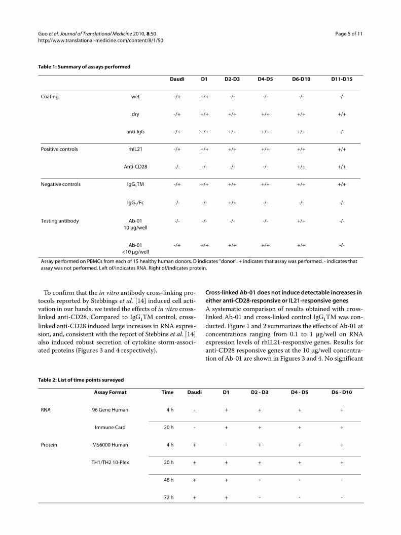
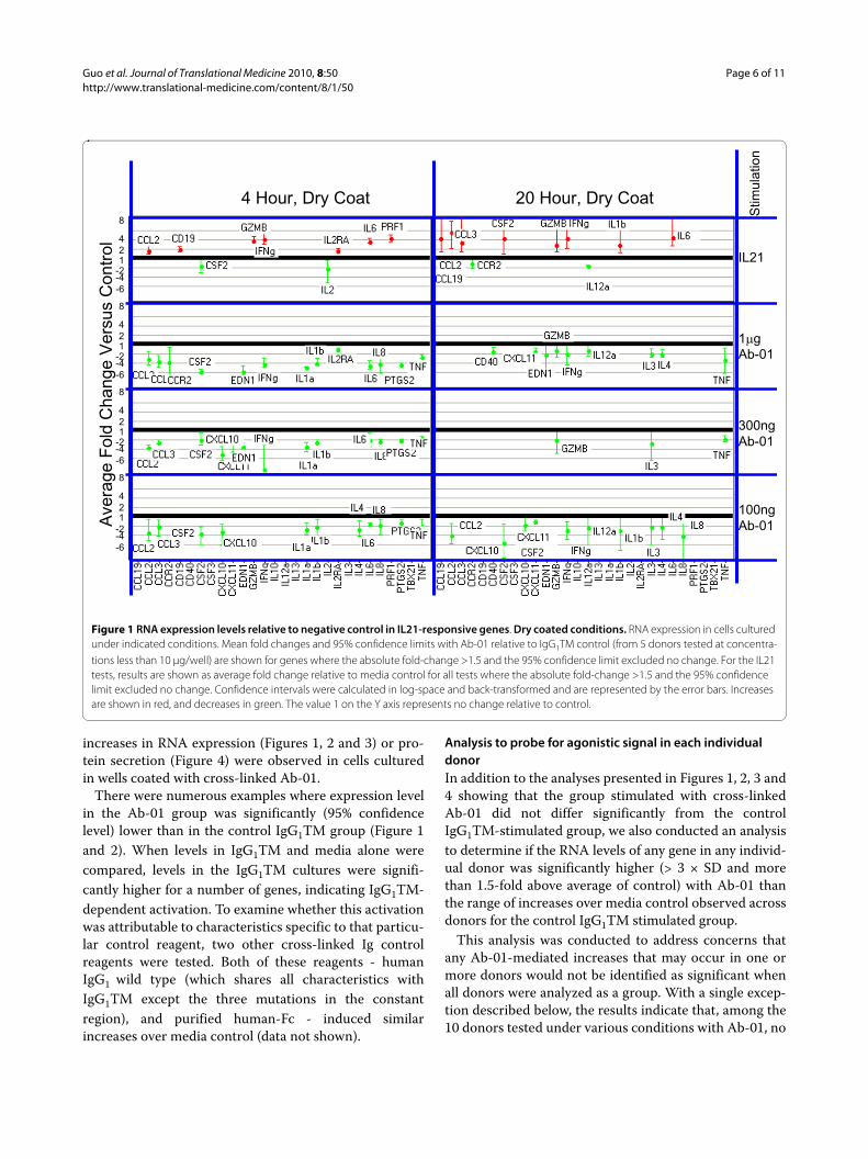
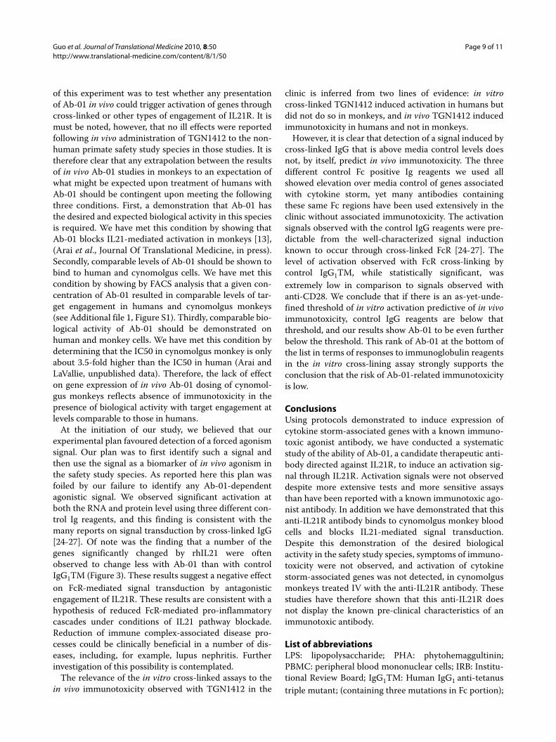
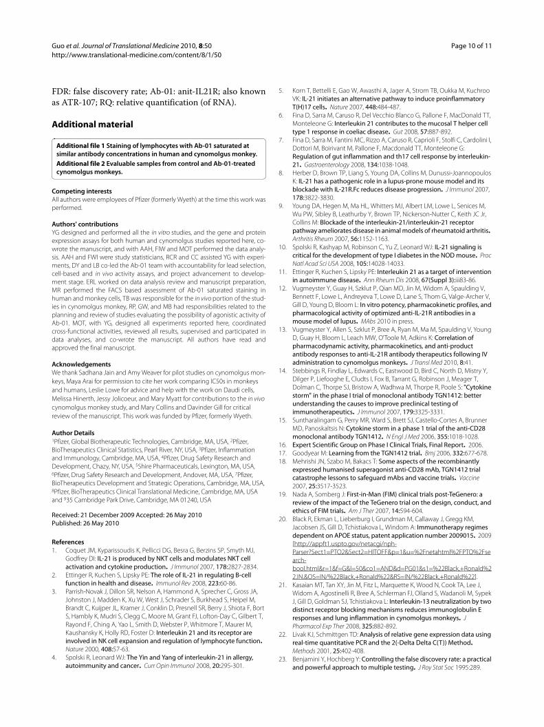
Characterization of positive control responsesTo ensure that activation signals delivered through IL21R(the target of Ab-01) were detectable under the cultureconditions used, rhIL21 was used as a positive control.Responses were detected by measuring the effects of invitro stimulation with rhIL21 on both RNA and secretedcytokine levels in human PBMCs. Of the 96 genes testedfor RNA expression, 21 gave a positive response to IL21(at 95% confidence level) in at least one of the conditionstested (two time points and two plating conditions pertime point). Results are presented in Figure 1 and 2. Themost robust IL21-dependent changes were observed forIFNγ, GZMB, PRF1 and IL6. Significant rhIL21-depen-dent elevation of IFNγ RNA was observed under all con-ditions tested.
Guo et al. Journal of Translational Medicine 2010, 8:50http://www.translational-medicine.com/content/8/1/50
Page 5 of 11
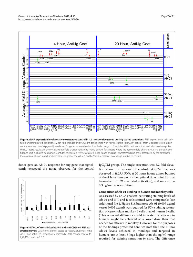
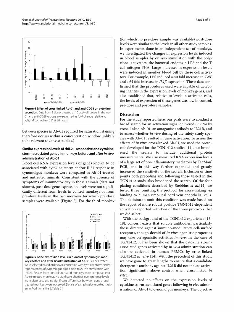
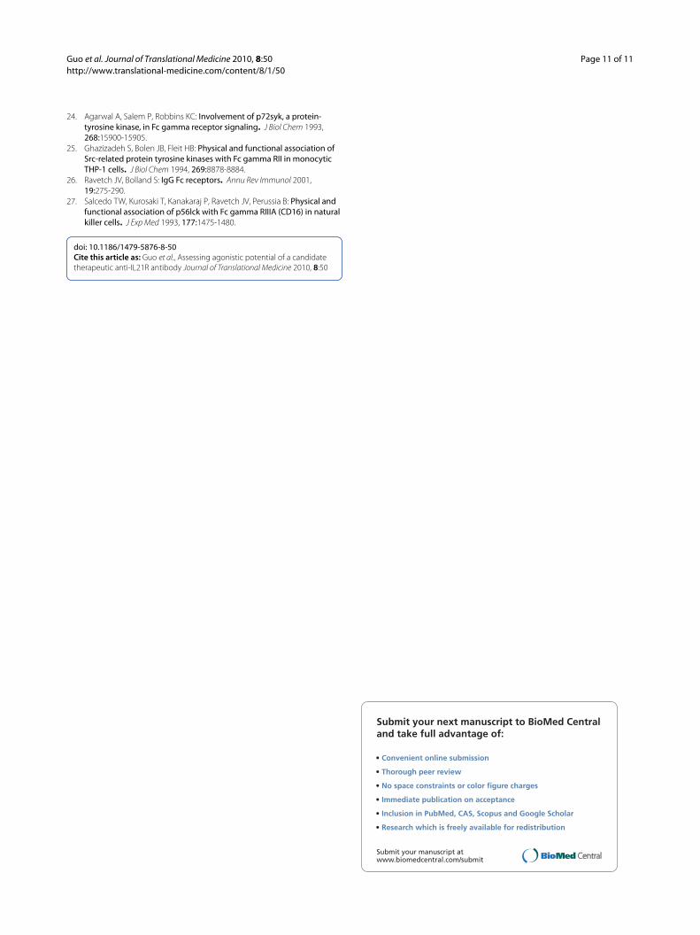
To confirm that the in vitro antibody cross-linking pro-tocols reported by Stebbings et al. [14] induced cell acti-vation in our hands, we tested the effects of in vitro cross-linked anti-CD28. Compared to IgG1TM control, cross-linked anti-CD28 induced large increases in RNA expres-sion, and, consistent with the report of Stebbins et al. [14]also induced robust secretion of cytokine storm-associ-ated proteins (Figures 3 and 4 respectively).
Cross-linked Ab-01 does not induce detectable increases in either anti-CD28-responsive or IL21-responsive genesA systematic comparison of results obtained with cross-linked Ab-01 and cross-linked control IgG1TM was con-ducted. Figure 1 and 2 summarizes the effects of Ab-01 atconcentrations ranging from 0.1 to 1 μg/well on RNAexpression levels of rhIL21-responsive genes. Results foranti-CD28 responsive genes at the 10 μg/well concentra-tion of Ab-01 are shown in Figures 3 and 4. No significant
Table 1: Summary of assays performed
Daudi D1 D2-D3 D4-D5 D6-D10 D11-D15
Coating wet -/+ +/+ -/- -/- -/- -/-
dry -/+ +/+ +/+ +/+ +/+ +/+
anti-IgG -/+ +/+ +/+ +/+ +/+ -/-
Positive controls rhIL21 -/+ +/+ +/+ +/+ +/+ +/+
Anti-CD28 -/- -/- -/- -/- +/+ +/+
Negative controls IgG1TM -/+ +/+ +/+ +/+ +/+ +/+
IgG1/Fc -/- -/- +/+ -/- -/- -/-
Testing antibody Ab-0110 μg/well
-/- -/- -/- -/- +/+ -/-
Ab-01<10 μg/well
-/+ +/+ +/+ +/+ +/+ -/-
Assay performed on PBMCs from each of 15 healthy human donors. D indicates "donor". + indicates that assay was performed. - indicates that assay was not performed. Left of/indicates RNA. Right of/indicates protein.
Table 2: List of time points surveyed
Assay Format Time Daudi D1 D2 - D3 D4 - D5 D6 - D10
RNA 96 Gene Human 4 h - + + + +
Immune Card 20 h - + + + +
Protein MS6000 Human 4 h + - + + +
TH1/TH2 10-Plex 20 h + + + + +
48 h + + - - -
72 h + + - - -
Guo et al. Journal of Translational Medicine 2010, 8:50http://www.translational-medicine.com/content/8/1/50
Page 6 of 11
increases in RNA expression (Figures 1, 2 and 3) or pro-tein secretion (Figure 4) were observed in cells culturedin wells coated with cross-linked Ab-01.
There were numerous examples where expression levelin the Ab-01 group was significantly (95% confidencelevel) lower than in the control IgG1TM group (Figure 1and 2). When levels in IgG1TM and media alone werecompared, levels in the IgG1TM cultures were signifi-cantly higher for a number of genes, indicating IgG1TM-dependent activation. To examine whether this activationwas attributable to characteristics specific to that particu-lar control reagent, two other cross-linked Ig controlreagents were tested. Both of these reagents - humanIgG1 wild type (which shares all characteristics withIgG1TM except the three mutations in the constantregion), and purified human-Fc - induced similarincreases over media control (data not shown).
Analysis to probe for agonistic signal in each individual donorIn addition to the analyses presented in Figures 1, 2, 3 and4 showing that the group stimulated with cross-linkedAb-01 did not differ significantly from the controlIgG1TM-stimulated group, we also conducted an analysisto determine if the RNA levels of any gene in any individ-ual donor was significantly higher (> 3 × SD and morethan 1.5-fold above average of control) with Ab-01 thanthe range of increases over media control observed acrossdonors for the control IgG1TM stimulated group.
This analysis was conducted to address concerns thatany Ab-01-mediated increases that may occur in one ormore donors would not be identified as significant whenall donors were analyzed as a group. With a single excep-tion described below, the results indicate that, among the10 donors tested under various conditions with Ab-01, no
Figure 1 RNA expression levels relative to negative control in IL21-responsive genes. Dry coated conditions. RNA expression in cells cultured under indicated conditions. Mean fold changes and 95% confidence limits with Ab-01 relative to IgG1TM control (from 5 donors tested at concentra-tions less than 10 μg/well) are shown for genes where the absolute fold-change >1.5 and the 95% confidence limit excluded no change. For the IL21 tests, results are shown as average fold change relative to media control for all tests where the absolute fold-change >1.5 and the 95% confidence limit excluded no change. Confidence intervals were calculated in log-space and back-transformed and are represented by the error bars. Increases are shown in red, and decreases in green. The value 1 on the Y axis represents no change relative to control.
1�g
Ab-01
300ng
Ab-01
100ng
Ab-01
Stim
ula
tion
1
4
2
8
1
4
2
8
1
4
2
8
1
4
2
8
-6
-4-2
-6
-4-2
-6
-4-2
-6
-4-2
Avera
ge F
old
Change V
ers
us C
ontr
ol
4 Hour, Dry Coat 20 Hour, Dry Coat
IL21
Guo et al. Journal of Translational Medicine 2010, 8:50http://www.translational-medicine.com/content/8/1/50
Page 7 of 11
donor gave an Ab-01 response for any gene that signifi-cantly exceeded the range observed for the control
IgG1TM group. The single exception was 3.2-fold eleva-tion above the average of control IgG1TM that wasobserved in IL2RA RNA at 20 hours in one donor, but notat the 4 hour time point (the optimal time point for thatbiomarker of IL21-mediated activation), and only at the0.3 μg/well concentration.
Comparison of Ab-01 binding to human and monkey cellsAs assessed by FACS analysis, saturating staining levels ofAb-01 and % T and B cells stained were comparable (seeAdditional file 1, Figure S1), but more Ab-01 (0.009 μg/mlversus 0.006 μg/ml) was required for 50% staining satura-tion of cynomolgus monkey B cells than of human B cells.(This observed difference could indicate that efficacy inhumans might be achieved at a lower dose than thatneeded for efficacy in monkey. However, for the purposesof the findings presented here, we note that, the in vivoAb-01 levels achieved in monkeys and targeted inhumans are at least 3 logs higher than the ng/ml rangerequired for staining saturation in vitro. The difference
Figure 2 RNA expression levels relative to negative control in IL21-responsive genes. Anti-Ig coated conditions. RNA expression in cells cul-tured under indicated conditions. Mean fold changes and 95% confidence limits with Ab-01 relative to IgG1TM control (from 5 donors tested at con-centrations less than 10 μg/well) are shown for genes where the absolute fold-change >1.5 and the 95% confidence limit excluded no change. For the IL21 tests, results are shown as average fold change relative to media control for all tests where the absolute fold-change >1.5 and the 95% con-fidence limit excluded no change. Confidence intervals were calculated in log-space and back-transformed and are represented by the error bars. Increases are shown in red, and decreases in green. The value 1 on the Y axis represents no change relative to control.
Stim
ula
tion
1
4
2
8
1
4
2
8
1
4
2
8
1
4
2
8
-6
-4-2
-6
-4-2
-6
-4-2
-6
-4-2
Avera
ge F
old
Change V
ers
us C
ontr
ol
4 Hour, Anti-Ig Coat 20 Hour, Anti-Ig Coat
IL21
1�g
Ab-01
300ng
Ab-01
100ng
Ab-01
Figure 3 Effect of cross-linked Ab-01 and anti-CD28 on RNA ex-pression levels. Data from 5 donors tested at 10 μg/well. Levels in the Ab-01 and anti-CD28 groups are expressed as fold change relative to IgG1TM control, +/- S.D.
-5
0
5
10
15
20
25
30
CD
40L
GZ
MB
ICO
S
IFN
G
IL10
IL12
B
IL13
IL1B IL
2
IL2R
A
IL4
IL6
IL8
TN
F
Ave
rage
Log
2 F
old
Cha
nge
RN
A E
xpre
ssio
n
anti-CD28/IgG1TM Ab-01/IgG1TM
Guo et al. Journal of Translational Medicine 2010, 8:50http://www.translational-medicine.com/content/8/1/50
Page 8 of 11
between species in Ab-01 required for saturation stainingtherefore occurs within a concentration window unlikelyto be relevant to in vivo studies.)
Similar expression levels of rhIL21-responsive and cytokine storm-associated genes in monkeys before and after in vivo administration of Ab-01Blood cell RNA expression levels of genes known to beassociated with cytokine storm and/or IL21 response incynomolgus monkeys were compared in Ab-01-treatedand untreated animals. Consistent with the absence ofsymptoms of immunotoxicity in these animals (data notshown), post-dose gene expression levels were not signifi-cantly different from levels in control monkeys or frompre-dose levels in the two monkeys for which pre-dosesamples were available (Figure 5). For the third monkey
(for which no pre-dose sample was available) post-doselevels were similar to the levels in all other study samples.In experiments done in an independent set of monkeys,we investigated the changes in expression levels inducedin blood samples by ex vivo stimulation with the poly-clonal activators, the bacterial endotoxin LPS and the Tcell mitogen PHA. Large increases in expre ssion levelswere induced in monkey blood cell by these cell activa-tors. For example, LPS induced a 40 fold increase in TNFand a 64 fold increase in IL1β expression. These data con-firmed that the procedures used were capable of detect-ing changes in the expression levels of monkey genes, andalso established that, relative to levels in activated cells,the levels of expression of these genes was low in control,pre-dose and post-dose samples.
DiscussionFor the study reported here, our goals were to conduct abroad search for an activation signal delivered in vitro bycross-linked Ab-01, an antagonist antibody to IL21R, andto assess whether in vivo dosing of the safety study spe-cies with Ab-01 resulted in gene activation. To assess theeffects of in vitro cross-linked Ab-01, we used the proto-cols developed for the TGN1412 studies [14], but broad-ened the search to include additional proteinmeasurements. We also measured RNA expression levelsof a large set of pro-inflammatory mediators by TaqMan®
PCR, and in this way further expanded and greatlyincreased the sensitivity of the search. Inclusion of timepoints both preceding and following those tested in theTGN1412 study also broadened the search. Of the fourplating conditions described by Stebbins et al.[14] wetested three, omitting the protocol for cross-linking viabinding to human umbilical cord vein endothelial cells.The decision to omit this condition was made based onthe report of more robust positive TGN1412-dependentactivation reported with two of the three protocols thatwe did select.
With the background of the TGN1412 experience [15-19], concern exists that soluble antibodies, particularlythose directed against immuno-modulatory cell-surfacereceptors, though devoid of in vitro agonistic propertiesmay take on agonistic activities in vivo. In the case ofTGN1412, it has been shown that the cytokine storm-associated genes activated by in vivo administration canalso be activated in human PBMCs by cross-linkedTGN1412 in vitro [14]. With the precedent of this study,we have gone to great lengths to ensure that a candidatetherapeutic antibody against IL21R did not induce activa-tion significantly above control when cross-linked invitro.
We detected no effects on the expression levels ofcytokine storm-associated genes following in vivo admin-istration of Ab-01 to cynomolgus monkeys. The objective
Figure 4 Effect of cross-linked Ab-01 and anti-CD28 on cytokine secretion. Data from 5 donors tested at 10 μg/well. Levels in the Ab-01 and anti-CD28 groups are expressed as fold change relative to IgG1TM control +/- S.D at 20 hours.
-4
-2
0
2
4
6
8
10
12
IF
NG
IL10
IL12
B
IL13
IL1B
IL2
IL4
IL5
IL8
TN
F
Ave
rage
Log
2 F
old
Cha
nge
In P
rote
i n
anti-CD28/IgG1TM Ab-01/IgG1TM
Figure 5 Gene expression levels in blood of cynomolgus mon-keys before and after IV administration of Ab-01. Genes tested were selected based on known association with cytokine storm and/or reponsiveness of cynomolgus blood cells to ex vivo stimulation with rhIL21. Results from control untreated monkeys were comparable to Ab-01-treated monkeys. No signficant changes over pre-dose levels were observed, and no significant differences between control and treated monkeys were observed. Details of sampling by monkey is giv-en in Additional file 2, Table S1.
Ave
rage
Rel
ativ
e R
NA
Con
cent
rati
on (
RQ
) +
S.D
.
IFNg IL10 IL2 IL21R IL2RA IL6 IL8 PRF1 TNF
Guo et al. Journal of Translational Medicine 2010, 8:50http://www.translational-medicine.com/content/8/1/50
Page 9 of 11
of this experiment was to test whether any presentationof Ab-01 in vivo could trigger activation of genes throughcross-linked or other types of engagement of IL21R. It ismust be noted, however, that no ill effects were reportedfollowing in vivo administration of TGN1412 to the non-human primate safety study species in those studies. It istherefore clear that any extrapolation between the resultsof in vivo Ab-01 studies in monkeys to an expectation ofwhat might be expected upon treatment of humans withAb-01 should be contingent upon meeting the followingthree conditions. First, a demonstration that Ab-01 hasthe desired and expected biological activity in this speciesis required. We have met this condition by showing thatAb-01 blocks IL21-mediated activation in monkeys [13],(Arai et al., Journal Of Translational Medicine, in press).Secondly, comparable levels of Ab-01 should be shown tobind to human and cynomolgus cells. We have met thiscondition by showing by FACS analysis that a given con-centration of Ab-01 resulted in comparable levels of tar-get engagement in humans and cynomolgus monkeys(see Additional file 1, Figure S1). Thirdly, comparable bio-logical activity of Ab-01 should be demonstrated onhuman and monkey cells. We have met this condition bydetermining that the IC50 in cynomolgus monkey is onlyabout 3.5-fold higher than the IC50 in human (Arai andLaVallie, unpublished data). Therefore, the lack of effecton gene expression of in vivo Ab-01 dosing of cynomol-gus monkeys reflects absence of immunotoxicity in thepresence of biological activity with target engagement atlevels comparable to those in humans.
At the initiation of our study, we believed that ourexperimental plan favoured detection of a forced agonismsignal. Our plan was to first identify such a signal andthen use the signal as a biomarker of in vivo agonism inthe safety study species. As reported here this plan wasfoiled by our failure to identify any Ab-01-dependentagonistic signal. We observed significant activation atboth the RNA and protein level using three different con-trol Ig reagents, and this finding is consistent with themany reports on signal transduction by cross-linked IgG[24-27]. Of note was the finding that a number of thegenes significantly changed by rhIL21 were oftenobserved to change less with Ab-01 than with controlIgG1TM (Figure 3). These results suggest a negative effecton FcR-mediated signal transduction by antagonisticengagement of IL21R. These results are consistent with ahypothesis of reduced FcR-mediated pro-inflammatorycascades under conditions of IL21 pathway blockade.Reduction of immune complex-associated disease pro-cesses could be clinically beneficial in a number of dis-eases, including, for example, lupus nephritis. Furtherinvestigation of this possibility is contemplated.
The relevance of the in vitro cross-linked assays to thein vivo immunotoxicity observed with TGN1412 in the
clinic is inferred from two lines of evidence: in vitrocross-linked TGN1412 induced activation in humans butdid not do so in monkeys, and in vivo TGN1412 inducedimmunotoxicity in humans and not in monkeys.
However, it is clear that detection of a signal induced bycross-linked IgG that is above media control levels doesnot, by itself, predict in vivo immunotoxicity. The threedifferent control Fc positive Ig reagents we used allshowed elevation over media control of genes associatedwith cytokine storm, yet many antibodies containingthese same Fc regions have been used extensively in theclinic without associated immunotoxicity. The activationsignals observed with the control IgG reagents were pre-dictable from the well-characterized signal inductionknown to occur through cross-linked FcR [24-27]. Thelevel of activation observed with FcR cross-linking bycontrol IgG1TM, while statistically significant, wasextremely low in comparison to signals observed withanti-CD28. We conclude that if there is an as-yet-unde-fined threshold of in vitro activation predictive of in vivoimmunotoxicity, control IgG reagents are below thatthreshold, and our results show Ab-01 to be even furtherbelow the threshold. This rank of Ab-01 at the bottom ofthe list in terms of responses to immunoglobulin reagentsin the in vitro cross-lining assay strongly supports theconclusion that the risk of Ab-01-related immunotoxicityis low.
ConclusionsUsing protocols demonstrated to induce expression ofcytokine storm-associated genes with a known immuno-toxic agonist antibody, we have conducted a systematicstudy of the ability of Ab-01, a candidate therapeutic anti-body directed against IL21R, to induce an activation sig-nal through IL21R. Activation signals were not observeddespite more extensive tests and more sensitive assaysthan have been reported with a known immunotoxic ago-nist antibody. In addition we have demonstrated that thisanti-IL21R antibody binds to cynomolgus monkey bloodcells and blocks IL21-mediated signal transduction.Despite this demonstration of the desired biologicalactivity in the safety study species, symptoms of immuno-toxicity were not observed, and activation of cytokinestorm-associated genes was not detected, in cynomolgusmonkeys treated IV with the anti-IL21R antibody. Thesestudies have therefore shown that this anti-IL21R doesnot display the known pre-clinical characteristics of animmunotoxic antibody.
List of abbreviationsLPS: lipopolysaccharide; PHA: phytohemaggultinin;PBMC: peripheral blood mononuclear cells; IRB: Institu-tional Review Board; IgG1TM: Human IgG1 anti-tetanustriple mutant; (containing three mutations in Fc portion);
Guo et al. Journal of Translational Medicine 2010, 8:50http://www.translational-medicine.com/content/8/1/50
Page 10 of 11
FDR: false discovery rate; Ab-01: anit-IL21R; also knownas ATR-107; RQ: relative quantification (of RNA).
Additional material
Competing interestsAll authors were employees of Pfizer (formerly Wyeth) at the time this work wasperformed.
Authors' contributionsYG designed and performed all the in vitro studies, and the gene and proteinexpression assays for both human and cynomolgus studies reported here, co-wrote the manuscript, and with AAH, FIW and MOT performed the data analy-sis. AAH and FWI were study statisticians, RCR and CC assisted YG with experi-ments, DY and LB co-led the Ab-01 team with accountability for lead selection,cell-based and in vivo activity assays, and project advancement to develop-ment stage. ERL worked on data analysis review and manuscript preparation,MR performed the FACS based assessment of Ab-01 saturated staining inhuman and monkey cells, TB was responsible for the in vivo portion of the stud-ies in cynomolgus monkey, RP, GW, and MB had responsibilities related to theplanning and review of studies evaluating the possibility of agonistic activity ofAb-01. MOT, with YG, designed all experiments reported here, coordinatedcross-functional activities, reviewed all results, supervised and participated indata analyses, and co-wrote the manuscript. All authors have read andapproved the final manuscript.
AcknowledgementsWe thank Sadhana Jain and Amy Weaver for pilot studies on cynomolgus mon-keys, Maya Arai for permission to cite her work comparing IC50s in monkeys and humans, Leslie Lowe for advice and help with the work on Daudi cells, Melissa Hinerth, Jessy Jolicoeur, and Mary Myatt for contributions to the in vivo cynomolgus monkey study, and Mary Collins and Davinder Gill for critical review of the manuscript. This work was funded by Pfizer, formerly Wyeth.
Author Details1Pfizer, Global Biotherapeutic Technologies, Cambridge, MA, USA, 2Pfizer, BioTherapeutics Clinical Statistics, Pearl River, NY, USA, 3Pfizer, Inflammation and Immunology, Cambridge, MA, USA, 4Pfizer, Drug Safety Research and Development, Chazy, NY, USA, 5Shire Pharmaceuticals, Lexington, MA, USA, 6Pfizer, Drug Safety Research and Development, Andover, MA, USA, 7Pfizer, BioTherapeutics Development and Strategic Operations, Cambridge, MA, USA, 8Pfizer, BioTherapeutics Clinical Translational Medicine, Cambridge, MA, USA and 935 Cambridge Park Drive, Cambridge, MA 01240, USA
References1. Coquet JM, Kyparissoudis K, Pellicci DG, Besra G, Berzins SP, Smyth MJ,
Godfrey DI: IL-21 is produced by NKT cells and modulates NKT cell activation and cytokine production. J Immunol 2007, 178:2827-2834.
2. Ettinger R, Kuchen S, Lipsky PE: The role of IL-21 in regulating B-cell function in health and disease. Immunol Rev 2008, 223:60-86.
3. Parrish-Novak J, Dillon SR, Nelson A, Hammond A, Sprecher C, Gross JA, Johnston J, Madden K, Xu W, West J, Schrader S, Burkhead S, Heipel M, Brandt C, Kuijper JL, Kramer J, Conklin D, Presnell SR, Berry J, Shiota F, Bort S, Hambly K, Mudri S, Clegg C, Moore M, Grant FJ, Lofton-Day C, Gilbert T, Rayond F, Ching A, Yao L, Smith D, Webster P, Whitmore T, Maurer M, Kaushansky K, Holly RD, Foster D: Interleukin 21 and its receptor are involved in NK cell expansion and regulation of lymphocyte function. Nature 2000, 408:57-63.
4. Spolski R, Leonard WJ: The Yin and Yang of interleukin-21 in allergy, autoimmunity and cancer. Curr Opin Immunol 2008, 20:295-301.
5. Korn T, Bettelli E, Gao W, Awasthi A, Jager A, Strom TB, Oukka M, Kuchroo VK: IL-21 initiates an alternative pathway to induce proinflammatory T(H)17 cells. Nature 2007, 448:484-487.
6. Fina D, Sarra M, Caruso R, Del Vecchio Blanco G, Pallone F, MacDonald TT, Monteleone G: Interleukin 21 contributes to the mucosal T helper cell type 1 response in coeliac disease. Gut 2008, 57:887-892.
7. Fina D, Sarra M, Fantini MC, Rizzo A, Caruso R, Caprioli F, Stolfi C, Cardolini I, Dottori M, Boirivant M, Pallone F, Macdonald TT, Monteleone G: Regulation of gut inflammation and th17 cell response by interleukin-21. Gastroenterology 2008, 134:1038-1048.
8. Herber D, Brown TP, Liang S, Young DA, Collins M, Dunussi-Joannopoulos K: IL-21 has a pathogenic role in a lupus-prone mouse model and its blockade with IL-21R.Fc reduces disease progression. J Immunol 2007, 178:3822-3830.
9. Young DA, Hegen M, Ma HL, Whitters MJ, Albert LM, Lowe L, Senices M, Wu PW, Sibley B, Leathurby Y, Brown TP, Nickerson-Nutter C, Keith JC Jr, Collins M: Blockade of the interleukin-21/interleukin-21 receptor pathway ameliorates disease in animal models of rheumatoid arthritis. Arthritis Rheum 2007, 56:1152-1163.
10. Spolski R, Kashyap M, Robinson C, Yu Z, Leonard WJ: IL-21 signaling is critical for the development of type I diabetes in the NOD mouse. Proc Natl Acad Sci USA 2008, 105:14028-14033.
11. Ettinger R, Kuchen S, Lipsky PE: Interleukin 21 as a target of intervention in autoimmune disease. Ann Rheum Dis 2008, 67(Suppl 3):iii83-86.
12. Vugmeyster Y, Guay H, Szklut P, Qian MD, Jin M, Widom A, Spaulding V, Bennett F, Lowe L, Andreyeva T, Lowe D, Lane S, Thom G, Valge-Archer V, Gill D, Young D, Bloom L: In vitro potency, pharmacokinetic profiles, and pharmacological activity of optimized anti-IL-21R antibodies in a mouse model of lupus. MAbs 2010 in press.
13. Vugmeyster Y, Allen S, Szklut P, Bree A, Ryan M, Ma M, Spaulding V, Young D, Guay H, Bloom L, Leach MW, O'Toole M, Adkins K: Correlation of pharmacodynamic activity, pharmacokinetics, and anti-product antibody responses to anti-IL-21R antibody therapeutics following IV administration to cynomolgus monkeys. J Transl Med 2010, 8:41.
14. Stebbings R, Findlay L, Edwards C, Eastwood D, Bird C, North D, Mistry Y, Dilger P, Liefooghe E, Cludts I, Fox B, Tarrant G, Robinson J, Meager T, Dolman C, Thorpe SJ, Bristow A, Wadhwa M, Thorpe R, Poole S: "Cytokine storm" in the phase I trial of monoclonal antibody TGN1412: better understanding the causes to improve preclinical testing of immunotherapeutics. J Immunol 2007, 179:3325-3331.
15. Suntharalingam G, Perry MR, Ward S, Brett SJ, Castello-Cortes A, Brunner MD, Panoskaltsis N: Cytokine storm in a phase 1 trial of the anti-CD28 monoclonal antibody TGN1412. N Engl J Med 2006, 355:1018-1028.
16. Expert Scientific Group on Phase I Clinical Trials, Final Report. 2006.17. Goodyear M: Learning from the TGN1412 trial. Bmj 2006, 332:677-678.18. Mehrishi JN, Szabo M, Bakacs T: Some aspects of the recombinantly
expressed humanised superagonist anti-CD28 mAb, TGN1412 trial catastrophe lessons to safeguard mAbs and vaccine trials. Vaccine 2007, 25:3517-3523.
19. Nada A, Somberg J: First-in-Man (FIM) clinical trials post-TeGenero: a review of the impact of the TeGenero trial on the design, conduct, and ethics of FIM trials. Am J Ther 2007, 14:594-604.
20. Black R, Ekman L, Lieberburg I, Grundman M, Callaway J, Gregg KM, Jacobsen JS, Gill D, Tchistiakova L, Windom A: Immunotherapy regimes dependent on APOE status, patent application number 2009015. 2009 [http://appft1.uspto.gov/netacgi/nph-Parser?Sect1=PTO2&Sect2=HITOFF&p=1&u=%2Fnetahtml%2FPTO%2Fsearch-bool.html&r=1&f=G&l=50&co1=AND&d=PG01&s1=%22Black,+Ronald%22.IN.&OS=IN/%22Black,+Ronald%22&RS=IN/%22Black,+Ronald%22].
21. Kasaian MT, Tan XY, Jin M, Fitz L, Marquette K, Wood N, Cook TA, Lee J, Widom A, Agostinelli R, Bree A, Schlerman FJ, Olland S, Wadanoli M, Sypek J, Gill D, Goldman SJ, Tchistiakova L: Interleukin-13 neutralization by two distinct receptor blocking mechanisms reduces immunoglobulin E responses and lung inflammation in cynomolgus monkeys. J Pharmacol Exp Ther 2008, 325:882-892.
22. Livak KJ, Schmittgen TD: Analysis of relative gene expression data using real-time quantitative PCR and the 2(-Delta Delta C(T)) Method. Methods 2001, 25:402-408.
23. Benjamini Y, Hochberg Y: Controlling the false discovery rate: a practical and powerful approach to multiple testing. J Roy Stat Soc 1995:289.
Additional file 1 Staining of lymphocytes with Ab-01 saturated at similar antibody concentrations in human and cynomolgus monkey.Additional file 2 Evaluable samples from control and Ab-01-treated cynomolgus monkeys.
Received: 21 December 2009 Accepted: 26 May 2010 Published: 26 May 2010This article is available from: http://www.translational-medicine.com/content/8/1/50© 2010 Guo et al; licensee BioMed Central Ltd. This is an Open Access article distributed under the terms of the Creative Commons Attribution License (http://creativecommons.org/licenses/by/2.0), which permits unrestricted use, distribution, and reproduction in any medium, provided the original work is properly cited.Journal of Translational Medicine 2010, 8:50
Guo et al. Journal of Translational Medicine 2010, 8:50http://www.translational-medicine.com/content/8/1/50
Page 11 of 11
24. Agarwal A, Salem P, Robbins KC: Involvement of p72syk, a protein-tyrosine kinase, in Fc gamma receptor signaling. J Biol Chem 1993, 268:15900-15905.
25. Ghazizadeh S, Bolen JB, Fleit HB: Physical and functional association of Src-related protein tyrosine kinases with Fc gamma RII in monocytic THP-1 cells. J Biol Chem 1994, 269:8878-8884.
26. Ravetch JV, Bolland S: IgG Fc receptors. Annu Rev Immunol 2001, 19:275-290.
27. Salcedo TW, Kurosaki T, Kanakaraj P, Ravetch JV, Perussia B: Physical and functional association of p56lck with Fc gamma RIIIA (CD16) in natural killer cells. J Exp Med 1993, 177:1475-1480.
doi: 10.1186/1479-5876-8-50Cite this article as: Guo et al., Assessing agonistic potential of a candidate therapeutic anti-IL21R antibody Journal of Translational Medicine 2010, 8:50