Embed Size (px)
Citation preview
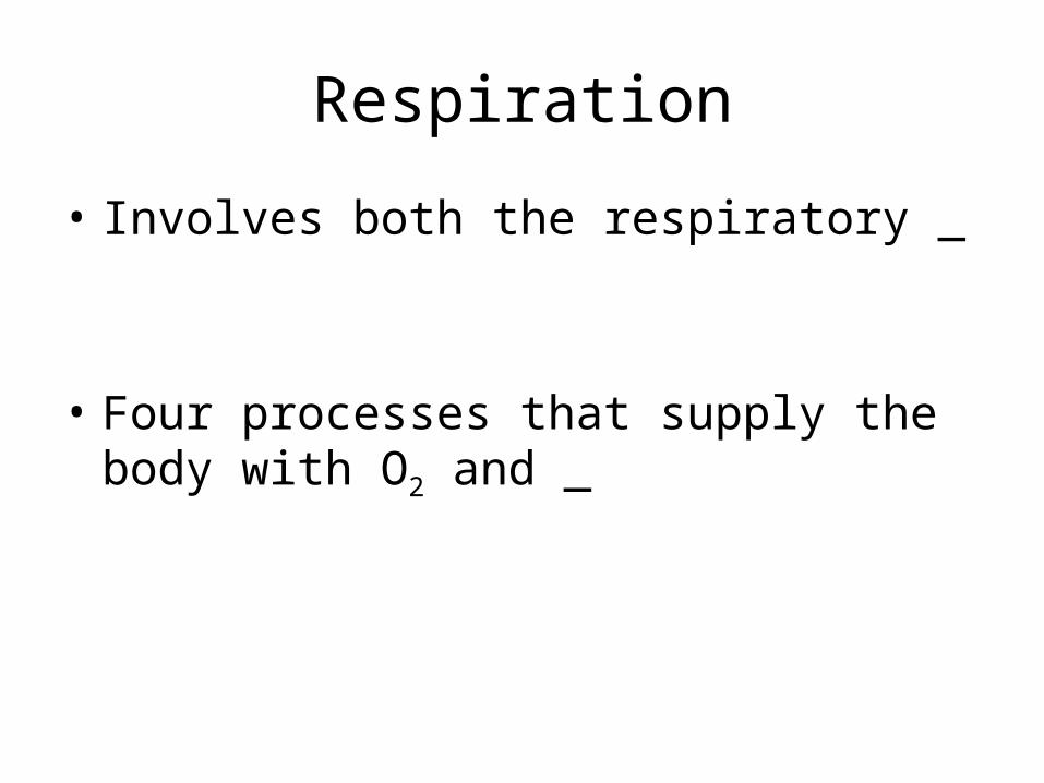
Respiration
• Involves both the respiratory _
• Four processes that supply the body with O2 and _

Respiration• _________________________________(breathing):
movement of air into and outof the lungs
• – O2 and CO2
exchange between the lungsand the blood
• Transport: – O2 and CO2 in the blood
• – O2 and CO2
exchange between systemic bloodvessels and tissues

Respiratory System: Functional Anatomy
• Major organs– Nose, nasal cavity, and paranasal sinuses– – Larynx– – Bronchi and their branches–

Functional Anatomy
• Respiratory zone: – site of _
– Microscopic structures:• respiratory bronchioles, alveolar ducts, and alveoli
• – conduits to gas exchange sites– Includes all other respiratory structures
• Respiratory muscles: – ________________________________________ and other
muscles that promote ventilation

The Nose
• Functions– Provides an _
– _________________________________________ and warms the entering air
– _____________________________________ and cleans inspired air
– Serves as a resonating chamber for speech
– Houses _
The Nose
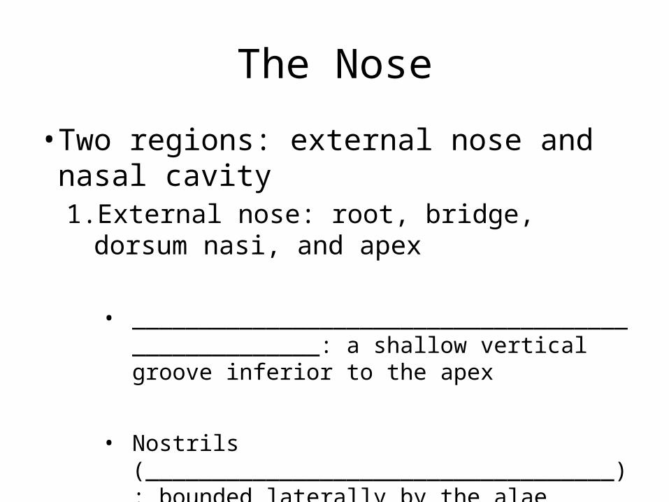
• Two regions: external nose and nasal cavity1. External nose: root, bridge, dorsum nasi, and apex
• ___________________________________________________: a shallow vertical groove inferior to the apex
• Nostrils (___________________________________): bounded laterally by the alae

The Nose
2. Nasal cavity: in and __________________________________ to the external nose• Divided by a midline _
• Posterior _________________________________________ (choanae) open into the nasal pharynx
• ____________________________________________: ethmoid and sphenoid bones
• ____________________________________________: hard and soft palates

Nasal Cavity
• Vestibule: – nasal cavity _
– Vibrissae • • ___________________________________________ coarse
particles from inspired air
• Olfactory mucosa– Lines the _– Contains _____________________________ receptors

Nasal Cavity
• Respiratory mucosa–
– Mucous and serous secretions contain lysozyme and defensins
– _______________________________________ move contaminated mucus posteriorly to throat
– Inspired air is warmed by _
– Sensory nerve endings triggers sneezing

Nasal Cavity
• Superior, middle, and inferior nasal conchae
– Protrude from _
– Increase mucosal area
– Enhance _

Functions of the Nasal Mucosa and Conchae
• During inhalation, the conchae and nasal mucosa–
• During exhalation these structures– ________________________________________
heat and moisture

Paranasal Sinuses
• In frontal, sphenoid, ethmoid, and maxillary bones
• _____________________________________ the skull and help to warm and moisten the air

Pharynx
• Muscular tube that connects to the– _________________________________________
____ superiorly– Larynx and esophagus inferiorly
• From the ____________________________________ to the level of the sixth cervical vertebra

Nasopharynx
• Air passageway posterior to the nasal cavity
• Lining– pseudostratified columnar epithelium
• – close nasopharynx during swallowing

Nasopharynx
• Pharyngeal tonsil – also called _– Located on _
• Pharyngotympanic tubes – Also called _– open into the _

Oropharynx• Passageway for food and air from the level of the soft palate to
the epiglottis
• Lining is ______________________________________________ epithelium
• _________ _______________________________ tonsils in the lateral walls
• _________________________________________ tonsil on the posterior surface of the tongue

Laryngopharynx
• Passageway for food and air
• Posterior to the _
• Extends to the larynx, where it is also continuous with the _

Larynx
• Attaches to the ___________________________ and opens into the laryngopharynx
• Continuous with the _
• Functions1. Provides an _
2. Routes air and food into proper channels
3.

Larynx• Cartilages of the larynx
– ____________________________________ cartilage except for the epiglottis
– ___________________________________________ with laryngeal prominence (Adam’s apple)
– Ring-shaped _
– Paired arytenoid, cuneiform, and corniculate cartilages
• : – ________________________________________ cartilage;
covers the laryngeal inlet during swallowing

Larynx
• Vocal ligaments
– Contain _
– Form the core of ________________________________ (true vocal cords)• Opening between them is the _
• Folds vibrate to produce sound as air rushes up from the lungs

Larynx
• Vestibular _
– Superior to the vocal folds
–
– Help to close the glottis during swallowing

Voice Production• – intermittent release of expired air while opening and closing the glottis
• – determined by the length and tension of the vocal cords
• – depends upon the force of air
• Chambers of pharynx, oral, nasal, and sinus cavities ___________________________________________ sound quality
• Sound is “shaped” into language by muscles of the pharynx, tongue, soft palate, and lips

Larynx
• Vocal folds may act as a __________________________________ to prevent air passage
• Example: – _________________________________________ closes to
prevent exhalation– _________________________________________ muscles
contract– Intra-abdominal pressure rises – Helps to _________________________________________ or
stabilizes the trunk during heavy lifting

Trachea
•Windpipe: –from the larynx into the mediastinum
•Wall composed of three layers1. • ciliated pseudostratified epithelium with _
2. : • connective tissue with seromucous glands
3. Adventitia: • outermost layer made of
______________________________________________ that encases the C-shaped rings of hyaline cartilage

Trachea
• Trachealis muscle– Connects posterior parts of cartilage rings–
• Carina– Last _– Point where trachea branches into _

Bronchi and Subdivisions• Air passages undergo 23 orders of branching
• Branching pattern called the _

Conducting Zone Structures
• Trachea right and left _
• Each main bronchus enters the _________________________ of one lung– ______________________________ main
bronchus is wider, shorter, and more vertical than the left
• Each main bronchus branches into lobar (secondary) bronchi (three right, two left)– Each lobar bronchus supplies _

Conducting Zone Structures
• Each lobar bronchus branches into __________________________________ (tertiary) bronchi– Segmental bronchi divide repeatedly
• Bronchioles are less than 1 mm in diameter
• Terminal bronchioles are the ________________________________ , less than 0.5 mm diameter

Conducting Zone Structures
• From bronchi through bronchioles, structural changes occur– Cartilage rings give way to _• cartilage is _
– Epithelium changes from pseudostratified columnar to _• _____________________________________________
___________ become infrequent
– Relative amount of smooth muscle _

Respiratory Zone
• Respiratory bronchioles, _________________________________ , alveolar sacs (clusters of alveoli)
• ~300 million alveoli account for most of the lungs’ volume and are the _

Respiratory Membrane
• Alveolar and capillary walls and _
• Alveolar walls– Single layer of
___________________________________________ (type I cells)
• Scattered type II _____________________________ secrete _____________________________________ and antimicrobial proteins

Alveoli
• Surrounded by _
• Contain open _________________________ that
– Connect adjacent alveoli
– Allow ______________________________________ throughout the lung to be equalized
• House alveolar _____________________________ that keep alveolar surfaces sterile

Lungs
• Occupy ______________________________________ except the mediastinum
• – site of vascular and bronchial attachments
• – anterior, lateral, and posterior surfaces

Lungs
• Apex: –
• Base: – inferior surface that rests on _
• – on mediastinal surface; site for attachment of
blood vessels, bronchi, lymphatic vessels, and nerves
• Cardiac notch of left lung: – concavity that _

Lungs
• Left lung is smaller, separated into two lobes by an _
• Right lung has ___________________________ separated by _
• Bronchopulmonary segments (10 right, 8–9 left)
• Lobules are the smallest subdivisions; served by bronchioles and their branches

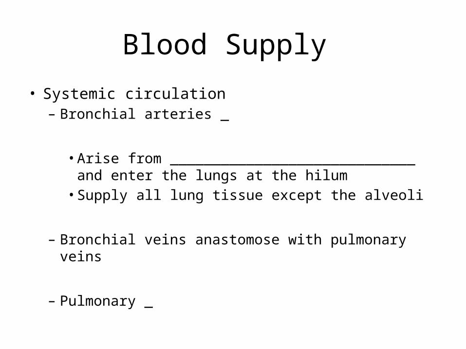
Blood Supply
• Pulmonary circulation
– Pulmonary _______________________________ deliver systemic _• Branch profusely, along with bronchi• Feed into the _
– Pulmonary ______________________ carry ______________________________________________ from respiratory zones to the heart
Blood Supply
• Systemic circulation– Bronchial arteries _
• Arise from _____________________________ and enter the lungs at the hilum• Supply all lung tissue except the alveoli
– Bronchial veins anastomose with pulmonary veins
– Pulmonary _

Pleurae
• Thin, double-layered serosa
• __________________________________________ on thoracic wall and superior face of diaphragm
• __________________________________________ on external lung surface
• Pleural fluid fills the slitlike pleural cavity– Provides _

Mechanics of Breathing
• Pulmonary ventilation consists of two phases
1. Inspiration:
2. ___________________________________________: gases exit the lungs

Pressure Relationships in the Thoracic Cavity
• Atmospheric pressure (Patm)– Pressure exerted by the air surrounding the body – 760 mm Hg at sea level
• Respiratory pressures are described _
– _______________________________________ respiratory pressure is less than Patm
– _______________________________________ respiratory pressure is greater than Patm
– Zero respiratory pressure = Patm