Embed Size (px)
Citation preview

Respiratory Anatomy

Anatomical PlanesSuperior
Inferior
Anterior
PosteriorLateral
Transverse
CoronalSagittal

Anatomical Planes
Transverse
Superior
Inferior
Sagittal
Anterior
Posterior
Coronal
MedialLateral

Respiratory System
• Respiration: exchange of gas between an organism & its environment.
• Inspiration: Inhalation; drawing air into the lungs
• Expiration: The expulsion of air from the lungs
• Alveoli: Minute air sacs within the lung tissue

Nasal Cavity
Oral Cavity
Larynx
Lungs
Diaphragm
Airway

Respiratory System
• Which is true of the lung when stretched out?– Size of your fist.– Size of a tennis court– Size of a kitchen table
• Does blood circulate in the lung?– Why or Why not?
• How does the rate of ventilation change with-– Exercising?– Quiet breathing?– Speech?

Respiratory System• What does the respiratory system do?
– sustain life
– speech secondary• source of all pressures and flows
• What is included in the respiratory system used for speech?
– Rib cage
– Diaphragm
– Abdomen
– Contents of RC & AB
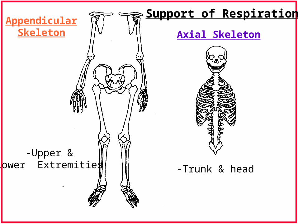
Axial Skeleton
-Trunk & head
AppendicularSkeleton
-Upper &Lower Extremities
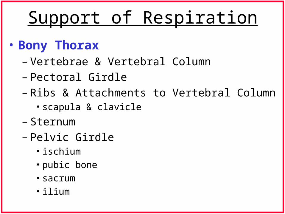
Support of Respiration
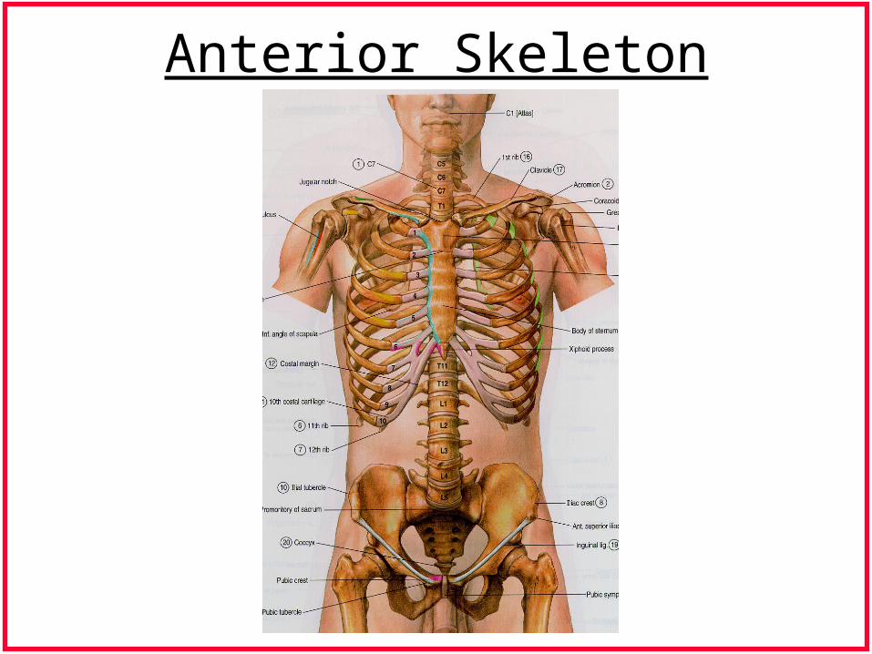
Anterior Skeleton
Support of Respiration
• Bony Thorax– Vertebrae & Vertebral Column– Pectoral Girdle– Ribs & Attachments to Vertebral Column
• scapula & clavicle
– Sternum– Pelvic Girdle
• ischium
• pubic bone
• sacrum
• ilium
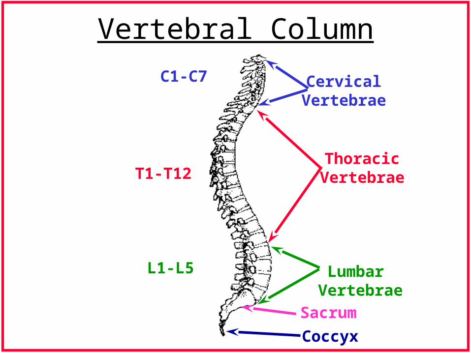
Vertebral Column
CervicalVertebrae
ThoracicVertebrae
Lumbar Vertebrae
Sacrum
Coccyx
C1-C7
T1-T12
L1-L5

Vertebral Column
• 33 segments of bone
• Many fossa & protuberances
• Form depending on location,
attachments & pathway

Anterior Posterior Lateral
Vertebral Column
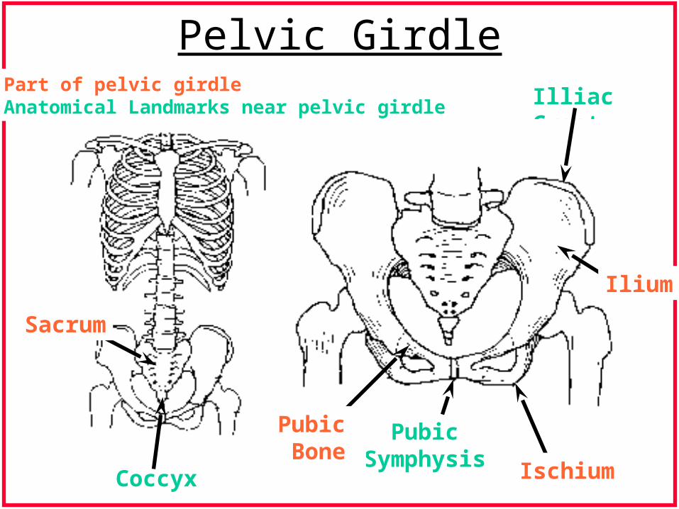
Pelvic GirdleIlliac Crest
Ilium
Ischium
PubicSymphysis
Pubic Bone
Coccyx
Sacrum
1. Part of pelvic girdle2. Anatomical Landmarks near pelvic girdle

Pelvic Girdle
• Vertebral column attaches
• Lower extremity attachment
• Provides distribution of force
• Made up of: ilium, sacrum, pubic bone,
ischium
– ilium-large, wing-like
– sacrum- five fused vertebrae

Pectoral Girdle
Clavicle
Sternum
Scapula
* Pectoral girdle includesonly the scapula & clavicle
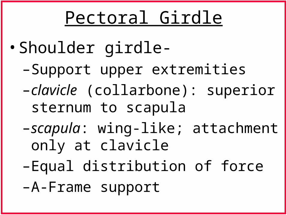
Pectoral Girdle
• Shoulder girdle- – Support upper extremities
– clavicle (collarbone): superior sternum to scapula
– scapula: wing-like; attachment only at clavicle
– Equal distribution of force
– A-Frame support
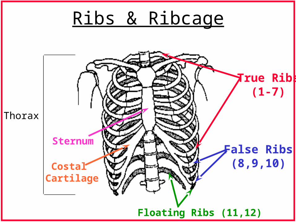
Ribs & Ribcage
True Ribs(1-7)
False Ribs(8,9,10)
Floating Ribs (11,12)
Thorax
Sternum
Costal Cartilage
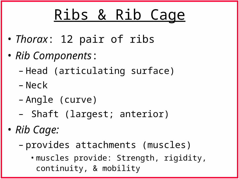
Ribs & Rib Cage
• Thorax: 12 pair of ribs
• Rib Components: – Head (articulating surface)
– Neck
– Angle (curve)
– Shaft (largest; anterior)
• Rib Cage:– provides attachments (muscles)
• muscles provide: Strength, rigidity, continuity, & mobility

Ribs
• Three general classes:– True ribs- upper ribs (1-7), attach to sternum,
cartilaginous attachment
– False ribs- (8,9,10), attach to sternum via cartilage running superior
– Floating ribs- (11,12), articulate with vertebral column only.
• Characteristics:– cartilage (chondral) attachment can be torqued
• strength and movement

Thoracic Expansion
Vertical
Transverse
Anteroposterior

Lateral/ Anteroposterior Thoracic Expansion

Diaphragm
Aponeurosis
Relaxed Expanded
Muscle
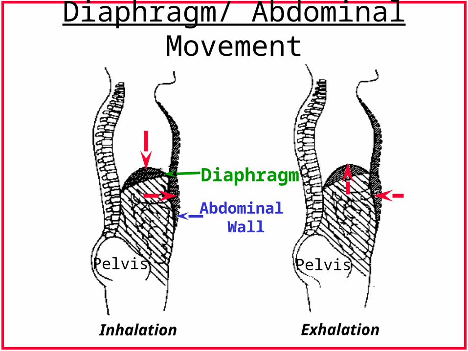
Diaphragm/ Abdominal Movement
Diaphragm
Abdominal Wall
Pelvis Pelvis
Inhalation Exhalation

Respiratory System: Components
Rib Cage
Abdominal Wall
Chest Wall
Viscera
Diaphragm
Mediastinum
Left Bronchus
Trachea
Right Bronchus
AlveolarAir Sacs
Pulmonary System
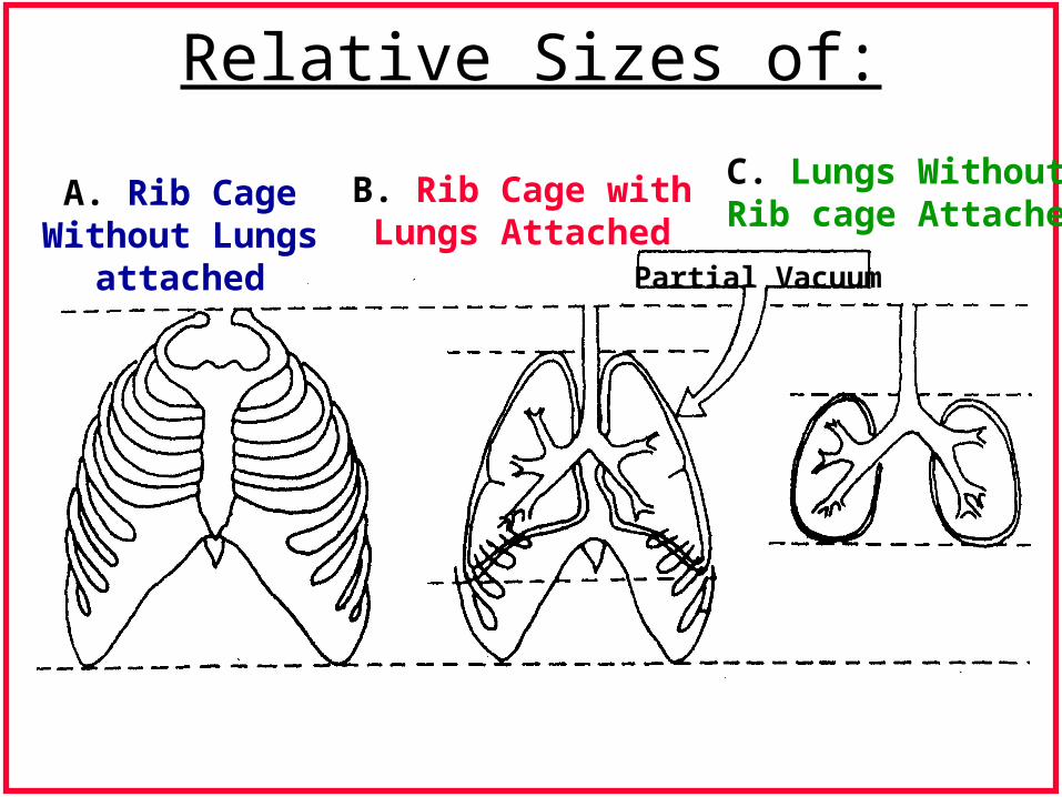
Relative Sizes of:
A. Rib CageWithout Lungs
attached
B. Rib Cage withLungs Attached
Partial Vacuum
C. Lungs WithoutRib cage Attached
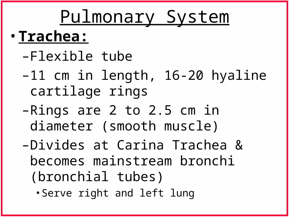
Pulmonary System• Trachea:
– Flexible tube
– 11 cm in length, 16-20 hyaline cartilage rings
– Rings are 2 to 2.5 cm in diameter (smooth muscle)
– Divides at Carina Trachea & becomes mainstream bronchi (bronchial tubes)• Serve right and left lung
Pulmonary System (cont.)• Bronchi:
– Divisions: main stem , secondary (lobar), tertiary (segmental)
– 28 generations of bronchial tree (first 9-”Dead Space”)
• trachea-mainstream bronchi-lobar bronchi-branchings to terminal respiratory bronchioles
– 1 (trachea), 2 (mainstem bronchi), 5 (lobar); 19 (segmental); 38 (subsegmental)...
– Final 7 divisions: respiratory zones- respiratory bronchioles, alveolar ducts & alveoli
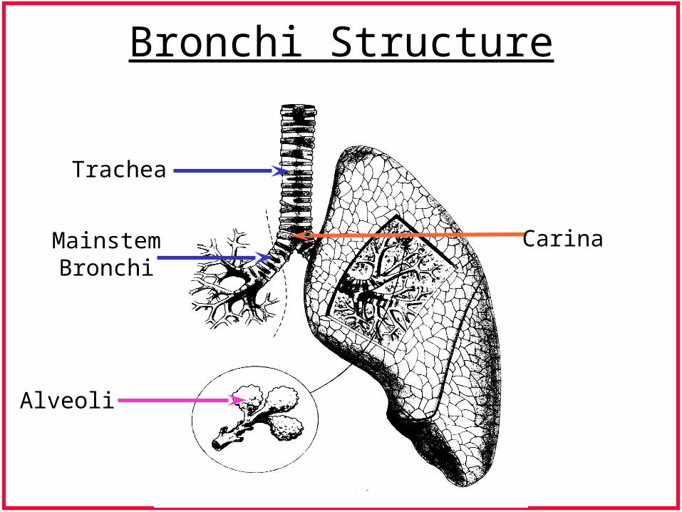
Bronchi Structure
Trachea
MainstemBronchi
Alveoli
Carina
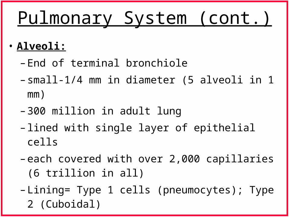
Pulmonary System (cont.)
• Alveoli:
– End of terminal bronchiole
– small-1/4 mm in diameter (5 alveoli in 1 mm)
– 300 million in adult lung
– lined with single layer of epithelial cells
– each covered with over 2,000 capillaries (6 trillion in all)
– Lining= Type 1 cells (pneumocytes); Type 2 (Cuboidal)
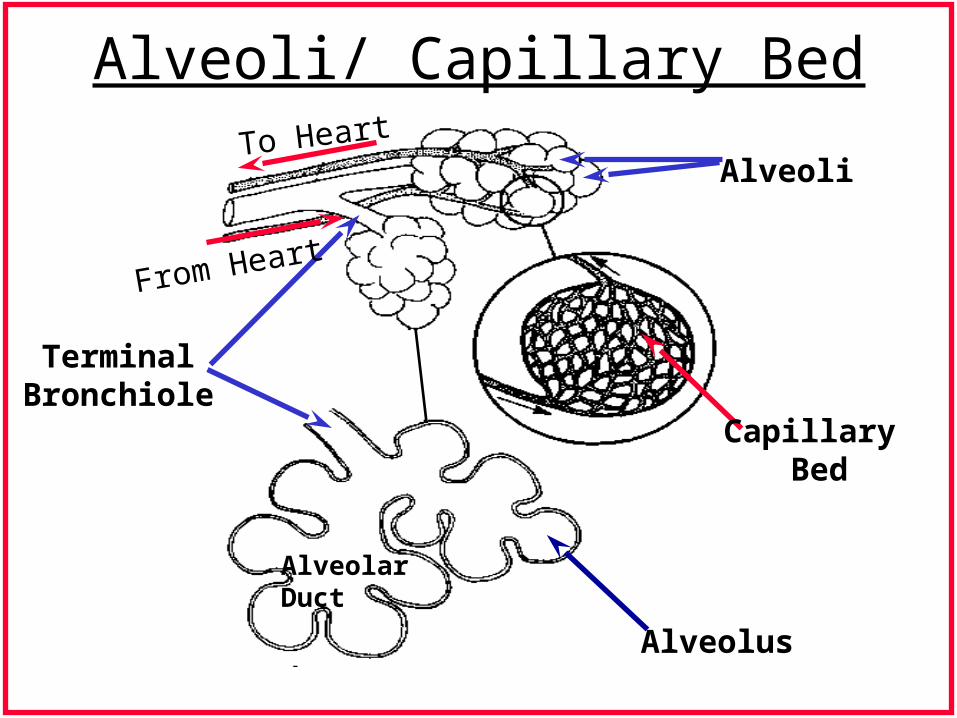
Alveoli/ Capillary Bed
TerminalBronchiole
AlveoliTo Heart
From Heart
Capillary Bed
AlveolarDuct
Alveolus
Readings
• Seikel: Ch. 3 (pgs. 35-76)
• Dickson: Ch. 3 (pgs. 59-84)