Embed Size (px)
Citation preview
Respiratory Physiology
#AnimalPhysio2015

• What is respiration?– Respiration = the series
of exchanges that leads to the uptake of oxygen by the cells, and the release of carbon dioxide to the lungs
Step 1 = ventilation– Inspiration &
expirationStep 2 = exchange
between alveoli (lungs) and pulmonary
capillaries (blood)
– Referred to as External Respiration
Step 3 = transport of gases in blood
Step 4 = exchange between blood and cells
– Referred to as Internal Respiration
– Cellular respiration = use of oxygen in ATP synthesis
CoughSneezeSound

Pulmonary circulation

Schematic View of Respiration

Basics of the Respiratory SystemGeneral Functions
• Exchange of gases• Directionality depends on gradients!
– Atmosphere to blood– Blood to tissues
• Regulation of pH– Dependent on rate of CO2 release
• Protection• Vocalization• Synthesis

• Respiratory neurons in brain stem – sets basic drive of ventilation– descending neural traffic to spinal cord– activation of muscles of respiration
• Ventilation of alveoli coupled with perfusion of pulmonary capillaries
• Exchange of oxygen and carbon dioxide
Basics of the Respiratory SystemControl of Respiration

R esp ira to ry C on tro l S ys tem
P erfu s ion ----->
N erve Im p u lses
N erve Im p u lses
V en tila tion
D iffu s ion
F orce ,d isp lacem en t
P co2 , P o2 , p H
M ech an orecep to rs
B lood
R esp ira to ry m em b ran ce
L u n g & C h es t W all
R esp ira to ry M u sc les
S p in a l C ord
R esp ira to ry cen te r-M ed u lla C h em orecep to rs
C ereb ra l C ortex
Basics of the Respiratory SystemControl of Respiration

• Located in brain stem– Dorsal & Ventral Medullary group– Pneumotaxic & Apneustic centers
• Affect rate and depth of ventilation• Influenced by:
– higher brain centers– peripheral mechanoreceptors– peripheral & central chemoreceptors
Basics of the Respiratory SystemRespiratory centers

Basics of the Respiratory SystemMuscles of Ventilation
• Inspiratory muscles-– increase thoracic cage volume
• Diaphragm, External Intercostals, SCM,• Ant & Post. Sup. Serratus, Scaleni, Levator Costarum
• Expiratory muscles-– decrease thoracic cage volume
• Abdominals, Internal Intercostals, Post Inf. Serratus, Transverse Thoracis, Pyramidal

Basics of the Respiratory SystemVentilation-Inspiration
• Muscles of Inspiration-when contract thoracic cage volume– diaphragm
• drops floor of thoracic cage
– external intercostals– sternocleidomastoid– anterior serratus– scaleni– serratus posterior superior– levator costarum– (all of the above except diaphragm lift rib cage)

Ventilation-expiration• Muscles of expiration when contract pull rib
cage down thoracic cage volume (forced expiration
• rectus abdominus• external and internal obliques• transverse abdominis• internal intercostals• serratus posterior inferior• transversus thoracis• pyramidal
– Under resting conditions expiration is passive and is associated with recoil of the lungs

Movement of air in/out of lungs• Considerations
– Pleural pressure• negative pressure between parietal and visceral pleura
that keeps lung inflated against chest wall• varies between -5 and -7.5 cmH2O (inspiration to
expiration
– Alveolar pressure• subatmospheric during inspiration• supra-atmospheric during expiration
– Transpulmonary pressure• difference between alveolar P & pleural P• measure of the recoil tendency of the lung• peaks at the end of inspiration

Basics of the Respiratory SystemFunctional Anatomy
• What structural aspects must be considered in the process of respiration?– The conduction portion– The exchange portion– The structures involved with
ventilation• Skeletal & musculature• Pleural membranes• Neural pathways
• All divided into– Upper respiratory tract
• Entrance to larynx– Lower respiratory tract
• Larynx to alveoli (trachea to lungs)

Basics of the Respiratory SystemFunctional Anatomy
• Bones, Muscles & Membranes

Basics of the Respiratory SystemFunctional Anatomy
• Function of these Bones, Muscles & Membranes– Create and transmit a pressure gradient
• Relying on – the attachments of the
muscles to the ribs (and overlying tissues)
– The attachment of the diaphragm to the base of the lungs and associated pleural membranes
– The cohesion of the parietal pleural membrane to the visceral pleural membrane
– Expansion & recoil of the lung and therefore alveoli with the movement of the overlying structures


Basics of the Respiratory SystemFunctional Anatomy
• What is the function of the upper respiratory tract?– Warm– Humidify – Filter– Vocalize
Raises incoming air to 37 Celsius
Raises incoming air to 100% humidity
Forms mucociliary escalator

Basics of the Respiratory SystemFunctional Anatomy
• What is the function of the lower respiratory tract?– Exchange of gases …. Due to
• Huge surface area = 1x10⁵ m2 of type I alveolar cells (simple squamous epithelium)
• Associated network of pulmonary capillaries– 80-90% of the space between alveoli is filled with blood in
pulmonary capillary networks• Exchange distance is approx 1 um from alveoli to blood!
– Protection• Free alveolar macrophages (dust cells)• Surfactant produced by type II alveolar cells (septal cells)

Basics of the Respiratory SystemFunctional Anatomy
• Characteristics of exchange membrane– High volume of blood through huge capillary
network results in• Fast circulation through lungs
– Pulmonary circulation = 5L/min through lungs….– Systemic circulation = 5L/min through entire body!
• Blood pressure is low…– Means
» Filtration is not a main theme here, we do not want a net loss of fluid into the lungs as rapidly as the systemic tissues
» Any excess fluid is still returned via lymphatic system

Basics of the Respiratory SystemFunctional Anatomy
• Sum-up of functional anatomy– Ventilation?– Exchange?– Vocalization?– Protection?

Effect of Thoracic Cage on Lung
• Reduces compliance by about 1/2 around functional residual capacity (at the end of a normal expiration)
• Compliance greatly reduced at high or low lung volumes

Work of Breathing
• Compliance work (elastic work)– Accounts for most of the work normally
• Tissue resistance work– viscosity of chest wall and lung
• Airway resistance work• Energy required for ventilation
– 3-5% of total body energy
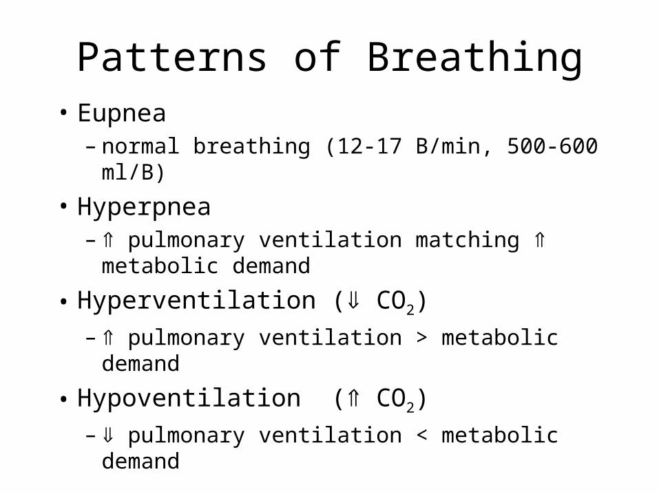
Patterns of Breathing• Eupnea
– normal breathing (12-17 B/min, 500-600 ml/B)
• Hyperpnea– pulmonary ventilation matching metabolic
demand
• Hyperventilation ( CO2)– pulmonary ventilation > metabolic demand
• Hypoventilation ( CO2)– pulmonary ventilation < metabolic demand

Patterns of breathing (cont.)
• Tachypnea– frequency of respiratory rate
• Apnea– Absense of breathing. e.g. Sleep apnea
• Dyspnea– Difficult or labored breathing
• Orthopnea– Dyspnea when recumbent, relieved when
upright. e.g. congestive heart failure, asthma, lung failure

Pleural Pressure
• Lungs have a natural tendency to collapse– surface tension forces 2/3– elastic fibers 1/3
• What keeps lungs against the chest wall?– Held against the chest wall by negative pleural
pressure “suction”

Collapse of the lungs
• If the pleural space communicates with the atmosphere, i.e. pleural P = atmospheric P the lung will collapse
• Causes– Puncture of the parietal pleura
• Sucking chest wound– Erosion of visceral pleura– Also if a major airway is blocked the air trapped distal to
the block will be absorbed by the blood and that segment of the lung will collapse

Pleural Fluid• Thin layer of mucoid fluid
– provides lubrication– transudate (interstitial fluid + protein)– total amount is only a few ml’s
• Excess is removed by lymphatics– mediastinum– superior surface of diaphragm– lateral surfaces of parietal pleural– helps create negative pleural pressure

Pleural Effusion
• Collection of large amounts of free fluid in pleural space
• Edema of pleural cavity• Possible causes:
– blockage of lymphatic drainage– cardiac failure-increased capillary filtration P– reduced plasma colloid osmotic pressure– infection/inflammation of pleural surfaces which
breaks down capillary membranes

Surfactant
• Reduces surface tension forces by forming a monomolecular layer between aqueous fluid lining alveoli and air, preventing a water-air interface
• Produced by type II alveolar epithelial cells• complex mix-phospholipids, proteins, ions
– dipalmitoyl lecithin, surfactant apoproteins, Ca++ ions

Static Lung Volumes• Tidal Volume (500ml)
– amount of air moved in or out each breath
• Inspiratory Reserve Volume (3000ml)– maximum vol. one can inspire above normal
inspiration
• Expiratory Reserve Volume (1100ml)– maximum vol. one can expire below normal
expiration
• Residual Volume (1200 ml)– volume of air left in the lungs after maximum
expiratory effort

Static Lung Capacities
• Functional residual capacity (RV+ERV)– vol. of air left in the lungs after a normal expir.,
balance point of lung recoil & chest wall forces
• Inspiratory capacity (TV+IRV)– max. vol. one can inspire during an insp effort
• Vital capacity (IRV+TV+ERV)– max. vol. one can exchange in a resp. cycle
• Total lung capacity (IRV+TV+ERV+RV)– the air in the lungs at full inflation

Pulmonary Flow Rates
• Compromised with obstructive conditions– decreased air flow
• minute respiratory volume– RR X TV
• Forced Expiratory Volumes (timed)– FEV/VC
• Peak expiratory Flow• Maximum Ventilatory Volume

• 20 generations of branching– Trachea (2 cm2)
– Bronchi • first 11 generations of branching
– Bronchioles (lack cartilage)• Next 5 generations of branching
– Respiratory bronchioles • Last 4 generations of branching
– Alveolar ducts give rise to alveolar sacs which give rise to alveoli
• 300 million with surface area 50-100 M2
Airways in lung
Dead Space• Area where gas exchange cannot occur• Includes most of airway volume• Anatomical dead space (=150 ml)
– Airways
• Physiological dead space– = anatomical + non functional alveoli
• Calculated using a pure O2 inspiration and measuring nitrogen in expired air (fig 37-7)– % area X Ve

Alveolar Volume
• Alveolar volume (2150 ml) = FRC (2300 ml)- dead space (150 ml)
• At the end of a normal expiration most of the FRC is at the level of the alveoli
• Slow turnover of alveolar air (6-7 breaths)• Rate of alveolar ventilation
– Va = RR (Vt-Vd)