Embed Size (px)
Citation preview
Respiratory Physiology Part I
BIO 219Dr. Adam Ross
Napa Valley College
Respiratory System
• Primary functions:• Supply oxygen and eliminate carbon dioxide• Regulate pH of blood


Structure of Respiratory System
• Respiratory tract is made up of two zones• Conducting zone
• No gas exchange• Terminal zone
• Where gas exchange occurs

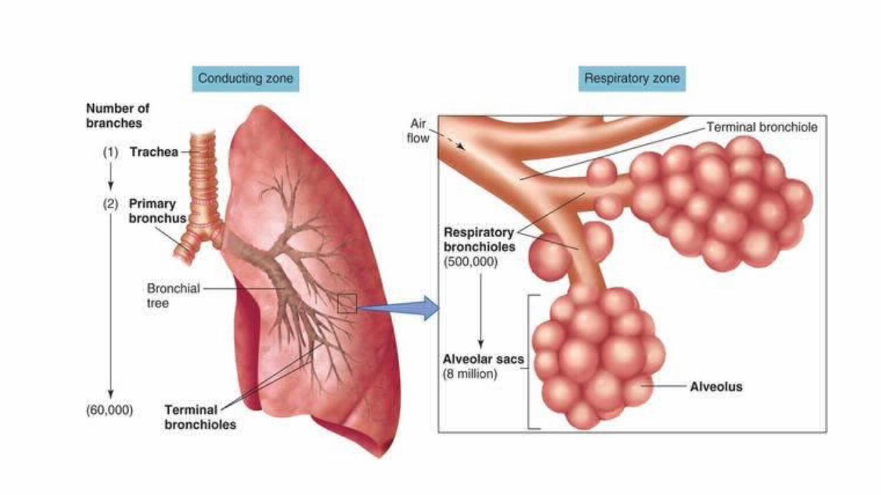

Alveoli
• Primary sites of gas exchange, have huge surface area• Type I cells- simple squamous epithelia• Type II cells- secrete surfactant, helps keep alveoli open• Alveolar macrophages- “dust cells” help keep lungs clean
• Alveoli are surrounded by pulmonary capillaries, exchange gas with blood
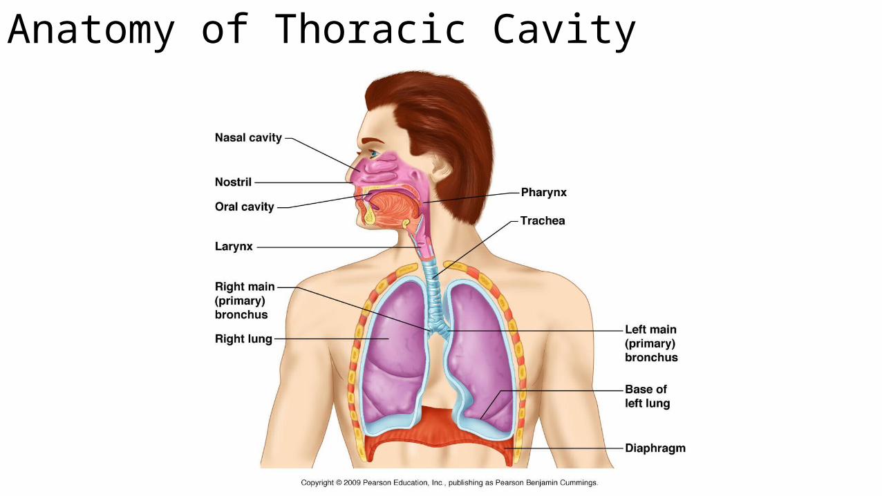
Anatomy of Thoracic Cavity

Thoracic Cavity
• Chest Wall• Surrounds thoracic cavity (ribs, intercostal muscles, etc)
• Diaphragm• Separates thoracic and abdominal cavities
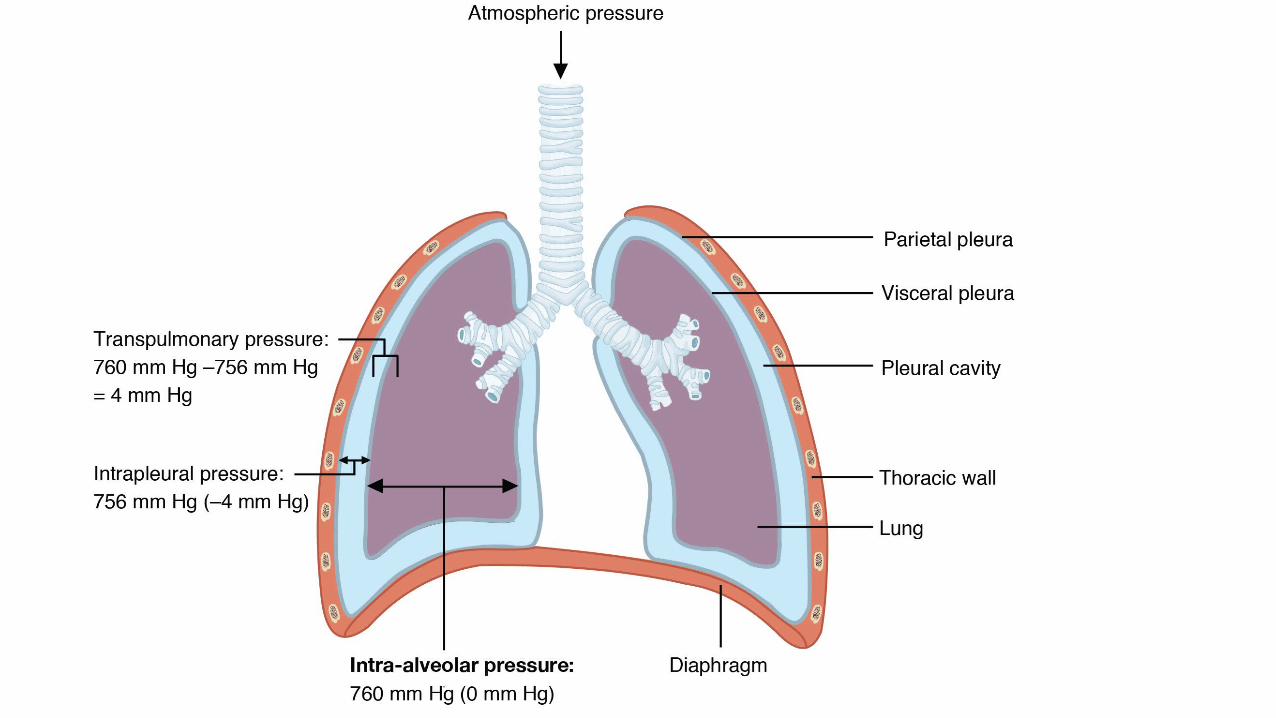
• Pleurae• Serous membranes that surround each lung, form fluid filled pleural sacs• Parietal pleura lines chest wall and diaphragm• Visceral pleura covers lungs
• Intrapleural space• Thin fluid filled space between parietal and visceral pleurae
• Fluid in intrapleural space connects lung to chest wall
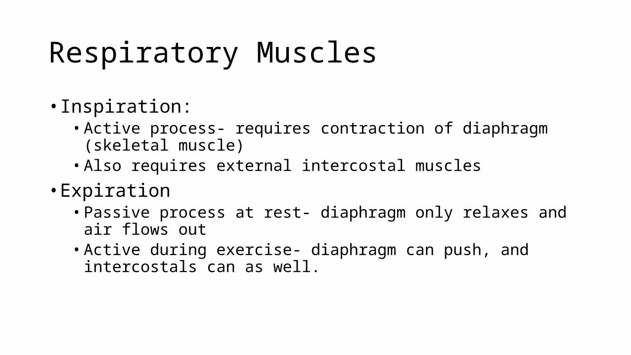
Respiratory Muscles
• Inspiration:• Active process- requires contraction of diaphragm (skeletal muscle)• Also requires external intercostal muscles
• Expiration• Passive process at rest- diaphragm only relaxes and air flows out• Active during exercise- diaphragm can push, and intercostals can as well.

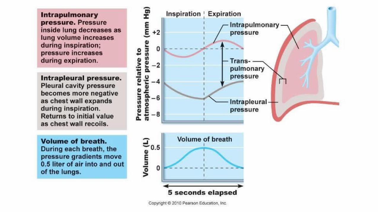
Ventilatory Mechanics• Gas flows in and out of lungs due to pressure gradients between lungs
and environment• If PATM > PAlv. Then gas will flow in to lungs from environment• If PATM < PAlv. Then gas will flow out of lungs into environment

Pressure – Volume Relationship
• Pressure of any gas is inversely proportional to its volume• Boyle’s Law P1V1 = P2V2
• During inspiration, respiratory muscles contract, and thoracic cavity expands (more volume), lowering the pressure in the thoracic cavity• Pressure in lungs is now greater than the pressure in thoracic cavity so they
expand (increase in volume, decrease in pressure)• PATM > Palv Air flows in to lungs.• Negative pressure in intrapleural space allows lungs to stay inflated
• Pneumothorax- air enters IP space and lung collapses
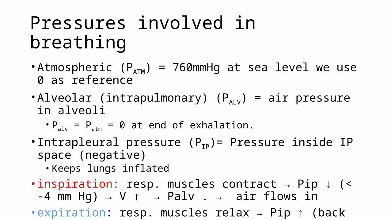
Pressures involved in breathing
• Atmospheric (PATM) = 760mmHg at sea level we use 0 as reference• Alveolar (intrapulmonary) (PALV) = air pressure in alveoli• Palv = Patm = 0 at end of exhalation.
• Intrapleural pressure (PIP)= Pressure inside IP space (negative)• Keeps lungs inflated
• inspiration: resp. muscles contract → Pip ↓ (< -4 mm Hg) → V ↑ → Palv ↓ → air flows in• expiration: resp. muscles relax → Pip ↑ (back to -4 mm Hg) → V ↓ →
Palv ↑ → air flows out
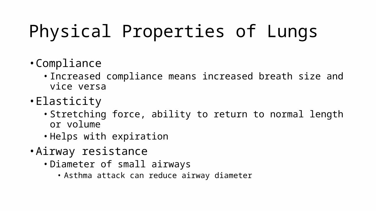
Physical Properties of Lungs
• Compliance• Increased compliance means increased breath size and vice versa
• Elasticity• Stretching force, ability to return to normal length or volume• Helps with expiration
• Airway resistance• Diameter of small airways
• Asthma attack can reduce airway diameter

Surface Tension and Surfactant• Surface Tension• - results from forces between water molecules at air-water interface• - contributes to inward recoil force in lungs, tends to collapse alveoli inward• - greater effect on small alveoli than large alveoli (Law of LaPlace: P = 2T/r)
• pulmonary surfactant - secreted by type II alveolar cells → reduces surface tension• - ↑ compliance, decreases work of breathing• - stabilizes alveoli by reducing surface tension more in small alveoli• respiratory distress syndrome (RDS) in premature infants is due to insufficient
surfactant

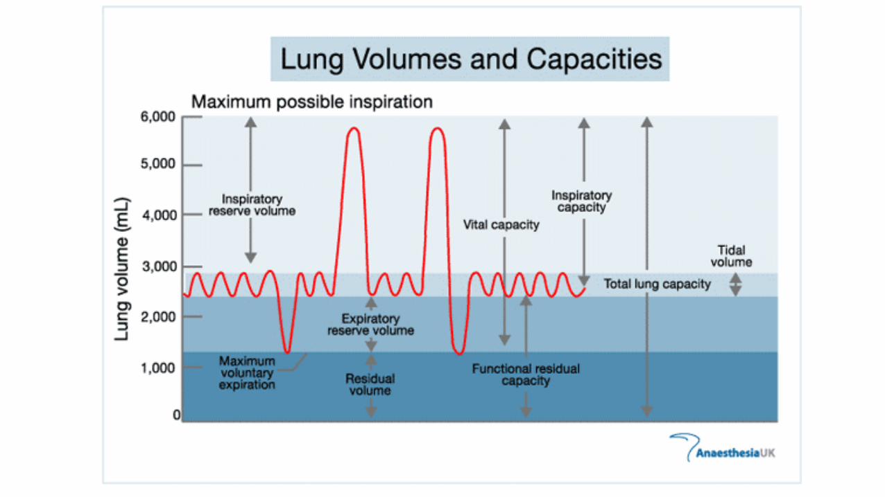
Lung Volumes and Capacities
• Total lung capacity• Total air in lungs at max capacity
• Tidal volume• Volume of 1 normal breath
• Vital capacity• Maximum breathing volume
• Inspiratory reserve volume• Inhalation volume after normal tidal inhalation
• Expiratory reserve volume• Exhalation volume after normal tidal exhalation
• Residual volume• Air in lungs after maximal exhalation
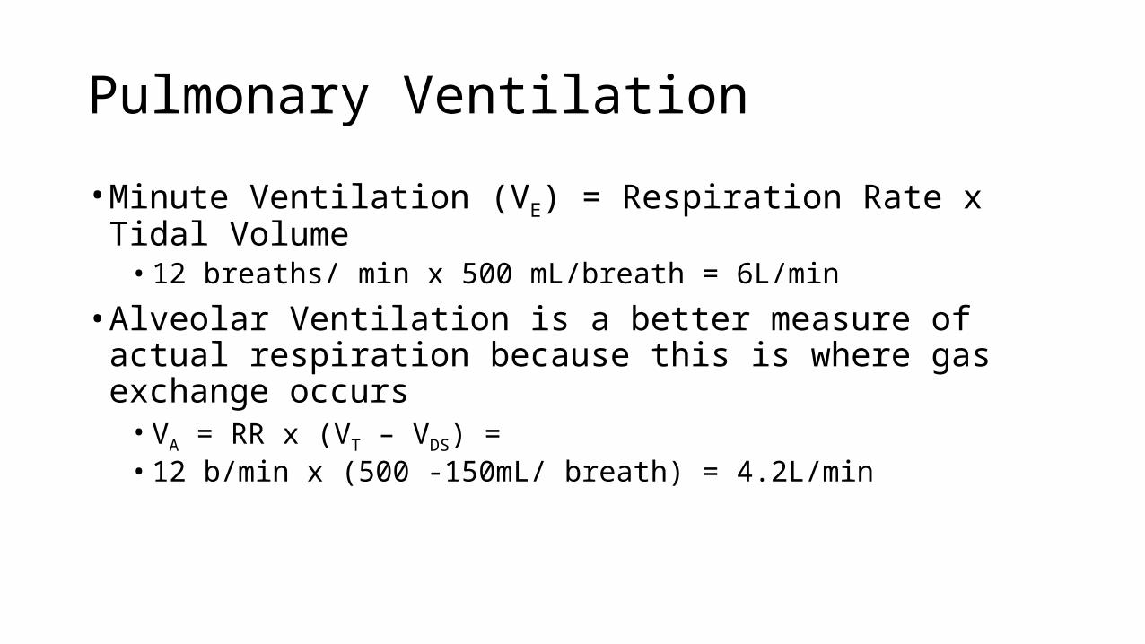
Pulmonary Ventilation
• Minute Ventilation (VE) = Respiration Rate x Tidal Volume• 12 breaths/ min x 500 mL/breath = 6L/min
• Alveolar Ventilation is a better measure of actual respiration because this is where gas exchange occurs• VA = RR x (VT – VDS) = • 12 b/min x (500 -150mL/ breath) = 4.2L/min

Pulmonary Disorders
• restrictive disorders – e.g., pulmonary fibrosis• reduced lung compliance → difficult inspiration, reduced vital capacity• obstructive disorders – e.g., asthma• increased airway resistance → difficult expiration, lower rate of expiration• chronic obstructive pulmonary disease (COPD): emphysema, asthma, chronic
bronchitis• emphysema involves destruction of alveolar tissue• - fewer, larger alveoli → decreased surface area for gas exchange• - reduced elastic recoil of lungs → difficult expiration, small airways collapse
→ air trapping