Embed Size (px)
Citation preview
Respiratory System
Lecture 2
Gas Exchange & Regulation
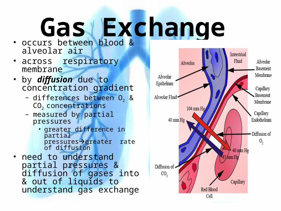
Gas Exchange • occurs between blood &
alveolar air• across respiratory
membrane• by diffusion due to
concentration gradient– differences between O2 & CO2
concentrations– measured by partial
pressures• greater difference in partial
pressuresgreater rate of diffusion
• need to understand partial pressures & diffusion of gases into & out of liquids to understand gas exchange
Dalton’s Law of Partial Pressure• air-mixture of gases & water vapor
• consists of N2-78%-most abundant, O2-20.9%water, CO2, Argon• atmospheric pressure is result of collision of all gas molecules• at any time 78.6% of collisions involve N2 & 20.9% involve O2
• each gas contributes to total pressure in proportion to its relative abundance-Dalton’s Law
• PressureTotal = Pressure1 + Pressure2 ... Pressuren• pressure contributed by one gas is partial pressure• directly proportional to % of gas in mixture• all partial pressures added = total pressure exerted by gas
mixture =760mm Hg• PN2-parital pressure nitrogen = 78.6 X 760 mm Hg-597 mm Hg• PO2 20.9 X 760 = 159 mm Hg
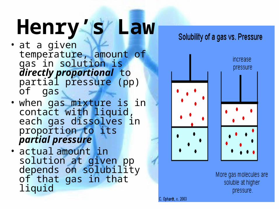
Henry’s Law• at a given temperature,
amount of gas in solution is directly proportional to partial pressure (pp) of gas
• when gas mixture is in contact with liquid, each gas dissolves in proportion to its partial pressure
• actual amount in solution at given pp depends on solubility of that gas in that liquid
Partial Pressures in Alveoli & Alveolar Capillaries
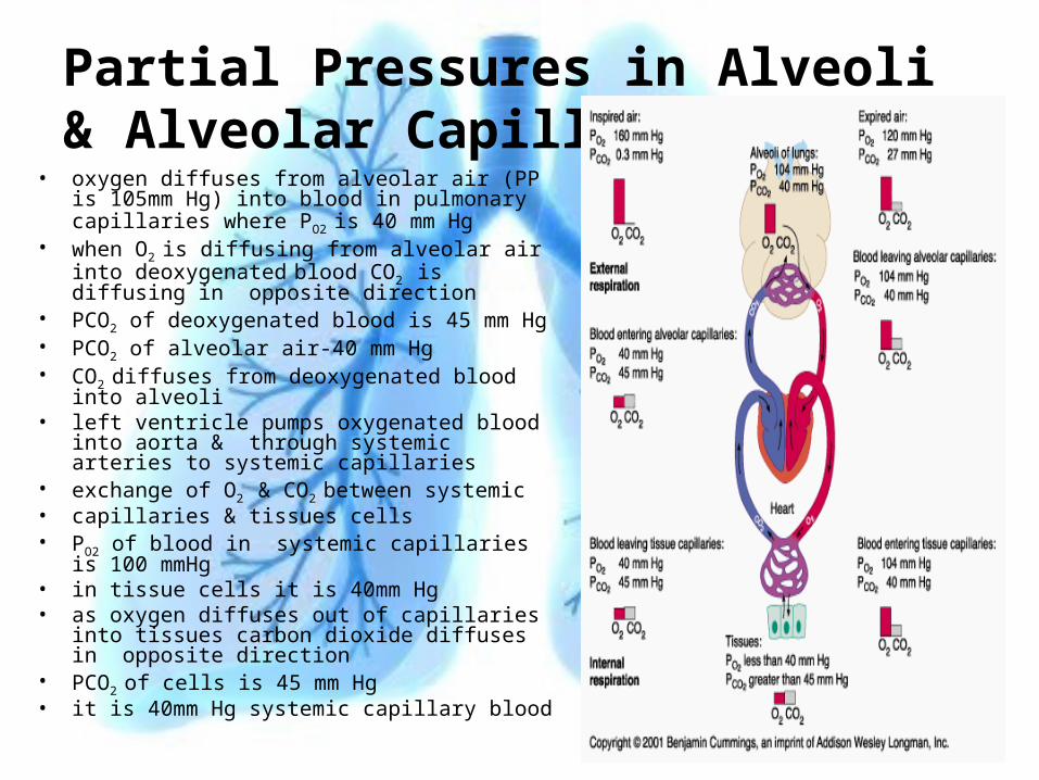
• oxygen diffuses from alveolar air (PP is 105mm Hg) into blood in pulmonary capillaries where PO2 is 40 mm Hg
• when O2 is diffusing from alveolar air into deoxygenated blood CO2 is diffusing in opposite direction
• PCO2 of deoxygenated blood is 45 mm Hg• PCO2 of alveolar air-40 mm Hg• CO2 diffuses from deoxygenated blood into
alveoli• left ventricle pumps oxygenated blood into
aorta & through systemic arteries to systemic capillaries
• exchange of O2 & CO2 between systemic• capillaries & tissues cells• PO2 of blood in systemic capillaries is 100
mmHg • in tissue cells it is 40mm Hg• as oxygen diffuses out of capillaries into
tissues carbon dioxide diffuses in opposite direction
• PCO2 of cells is 45 mm Hg
• it is 40mm Hg systemic capillary blood
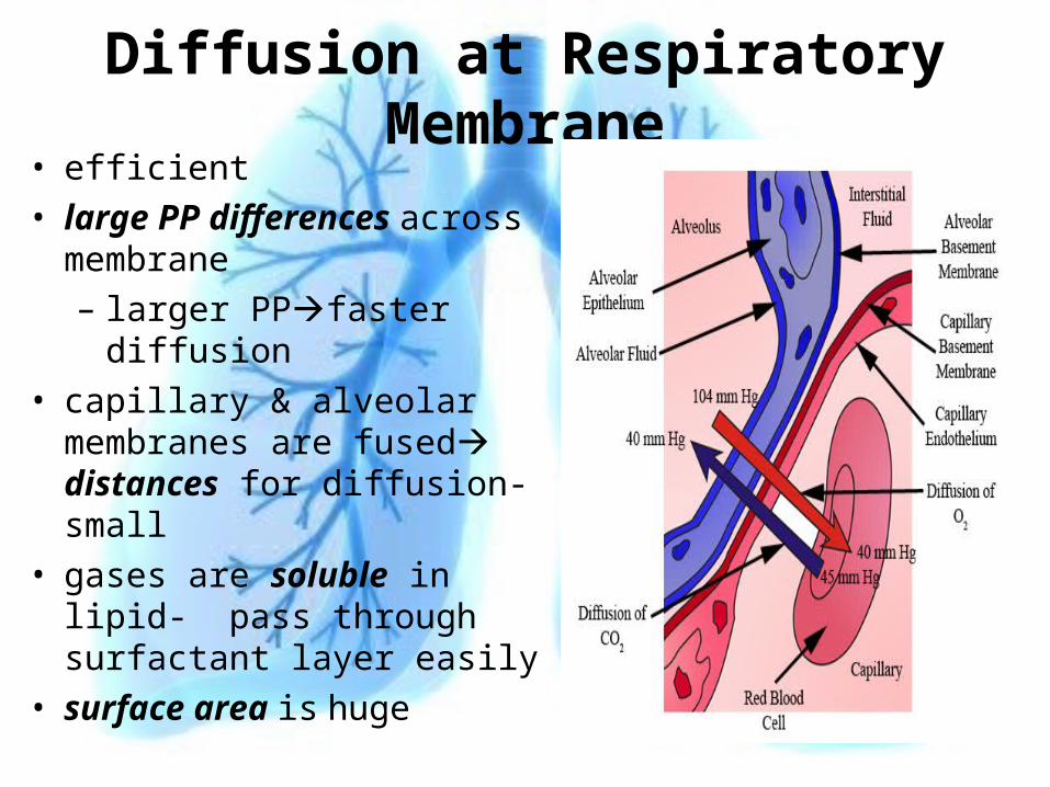
Diffusion at Respiratory Membrane• efficient
• large PP differences across membrane
– larger PPfaster diffusion
• capillary & alveolar membranes are fused distances for diffusion-small
• gases are soluble in lipid- pass through surfactant layer easily
• surface area is huge
Gas Transport• Major function of blood
• O2
• Co2
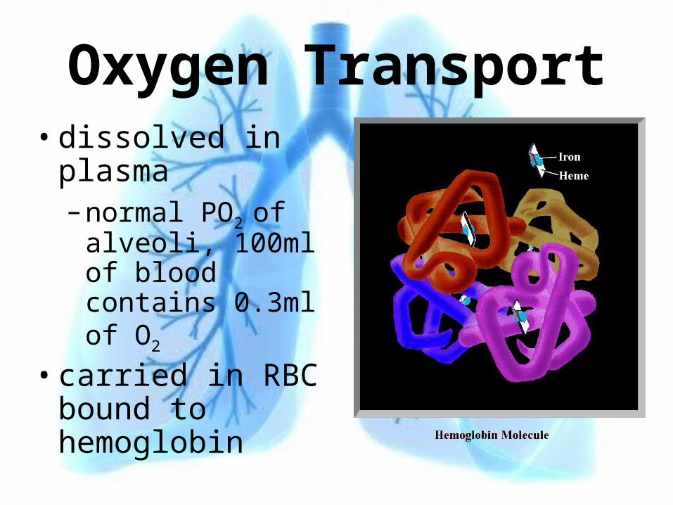
Oxygen Transport• dissolved in
plasma– normal PO2 of
alveoli, 100ml of blood contains 0.3ml of O2
• carried in RBC bound to hemoglobin
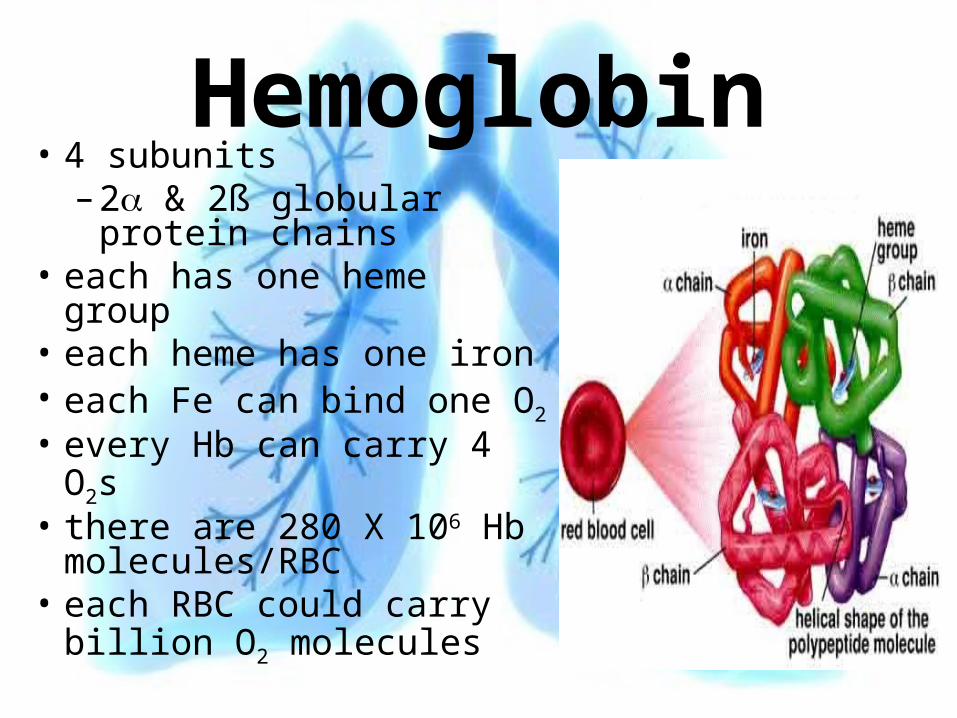
Hemoglobin• 4 subunits
– 2 & 2ß globular protein chains
• each has one heme group
• each heme has one iron• each Fe can bind one O2
• every Hb can carry 4 O2s• there are 280 X 106 Hb
molecules/RBC• each RBC could carry
billion O2 molecules
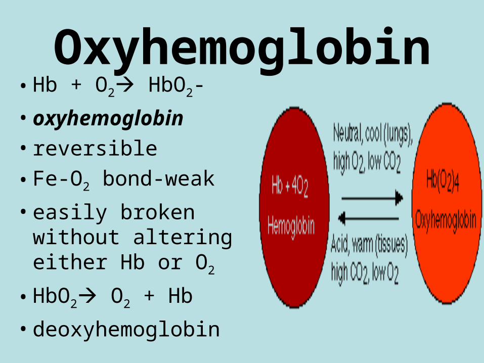
Oxyhemoglobin• Hb + O2 HbO2-
• oxyhemoglobin• reversible
• Fe-O2 bond-weak
• easily broken without altering either Hb or O2
• HbO2 O2 + Hb
• deoxyhemoglobin
Amount of Oxygen Bound to HB• PO2 of plasma
• most important factor determining how much O2 binds to Hb
• actual amount bound/maximum that could bind = % saturation
• all binding sites occupied-100% saturation
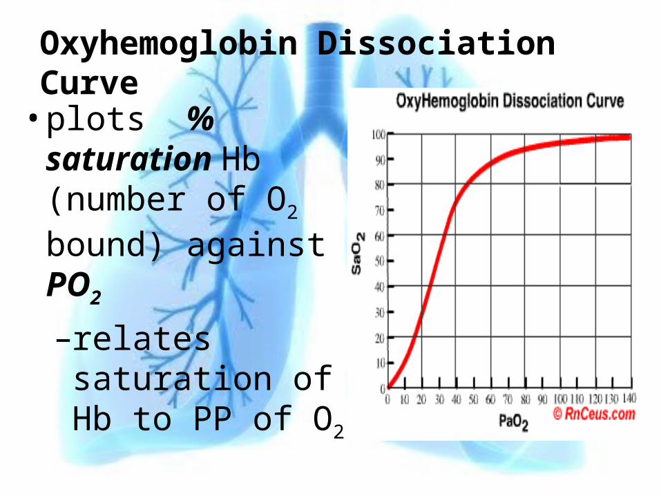
Oxyhemoglobin Dissociation Curve
• plots % saturation Hb (number of O2 bound) against PO2
–relates saturation of Hb to PP of O2
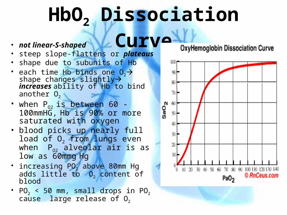
HbO2 Dissociation Curve• not linear-S-shaped• steep slope-flattens or plateaus• shape due to subunits of Hb• each time Hb binds one O2 shape
changes slightly increases ability of Hb to bind another O2
• when PO2 is between 60 -100mmHG, Hb is 90% or more saturated with oxygen
• blood picks up nearly full load of O2 from lungs even when PO2
alveolar air is as low as 60mmg Hg
• increasing PO2 above 80mm Hg adds little to O2 content of blood
• PO2 < 50 mm, small drops in PO2 cause large release of O2
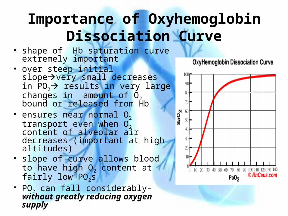
Importance of Oxyhemoglobin Dissociation Curve
• shape of Hb saturation curve extremely important
• over steep initial slopevery small decreases in PO2 results in very large changes in amount of O2 bound or released from Hb
• ensures near normal O2 transport even when O2 content of alveolar air decreases (important at high altitudes)
• slope of curve allows blood to have high O2 content at fairly low PO2s
• PO2 can fall considerably-without greatly reducing oxygen supply
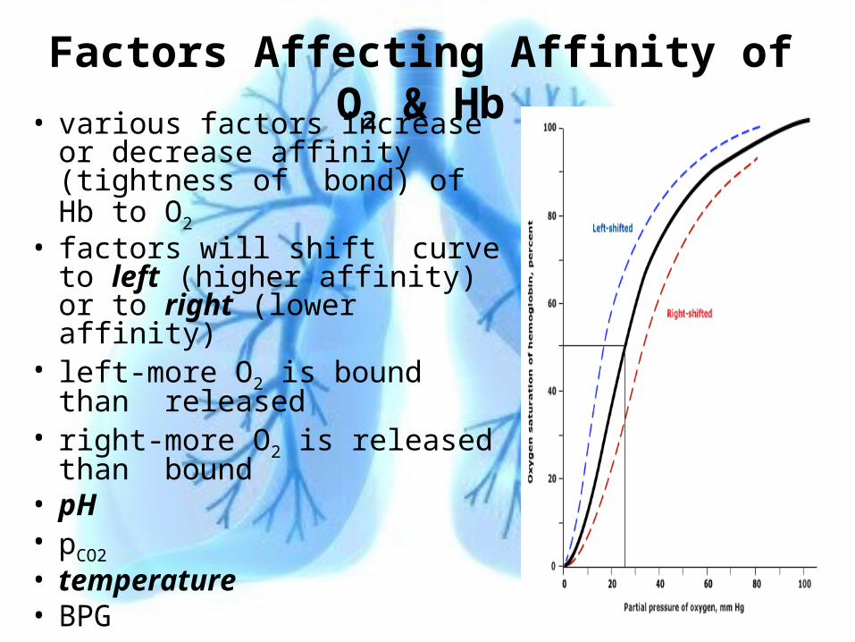
Factors Affecting Affinity of O2 & Hb• various factors increase or
decrease affinity (tightness of bond) of Hb to O2
• factors will shift curve to left (higher affinity) or to right (lower affinity)
• left-more O2 is bound than released
• right-more O2 is released than bound
• pH• pCO2
• temperature• BPG
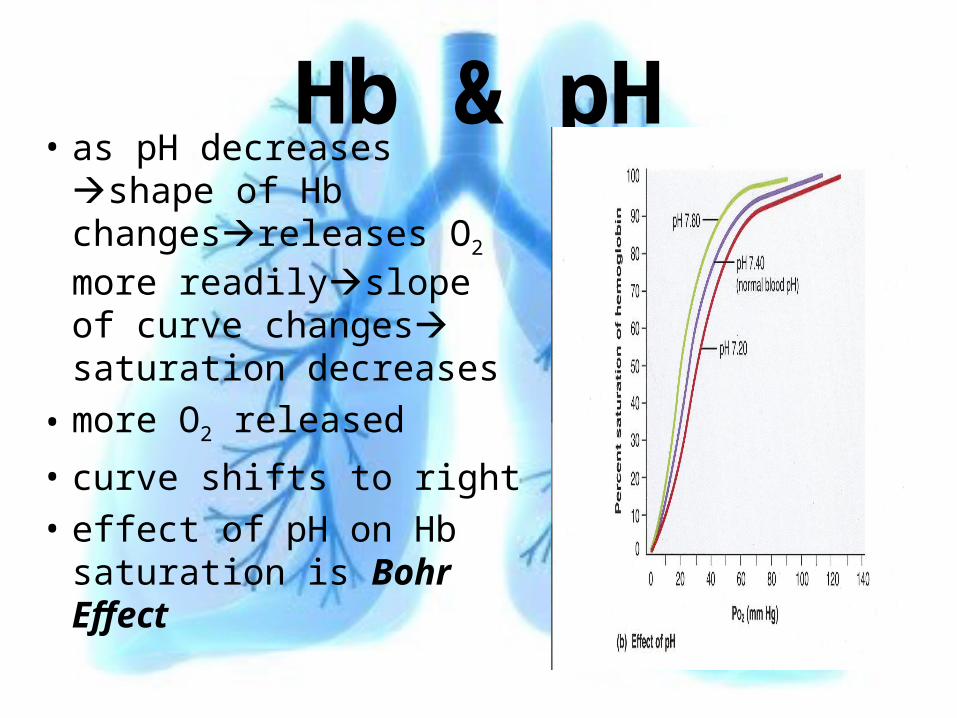
Hb & pH• as pH decreases
shape of Hb changesreleases O2 more readilyslope of curve changes saturation decreases
• more O2 released
• curve shifts to right• effect of pH on Hb
saturation is Bohr Effect
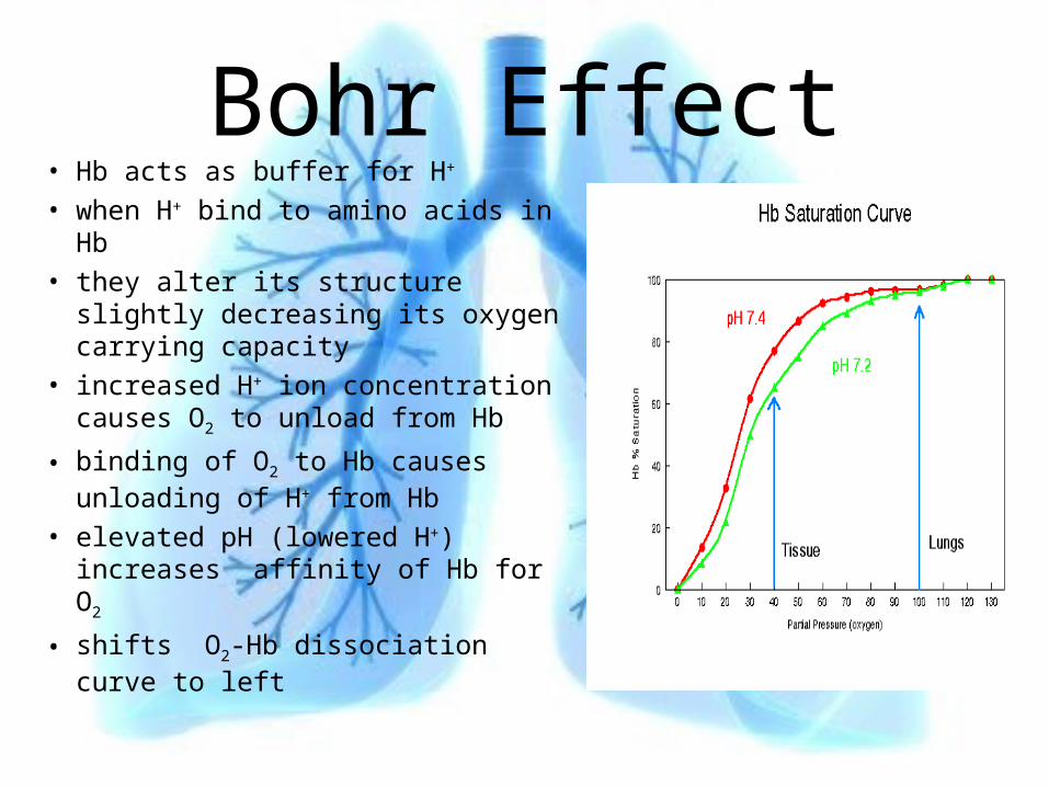
Bohr Effect• Hb acts as buffer for H+
• when H+ bind to amino acids in Hb
• they alter its structure slightly decreasing its oxygen carrying capacity
• increased H+ ion concentration causes O2 to unload from Hb
• binding of O2 to Hb causes unloading of H+ from Hb
• elevated pH (lowered H+) increases affinity of Hb for O2
• shifts O2-Hb dissociation curve to left
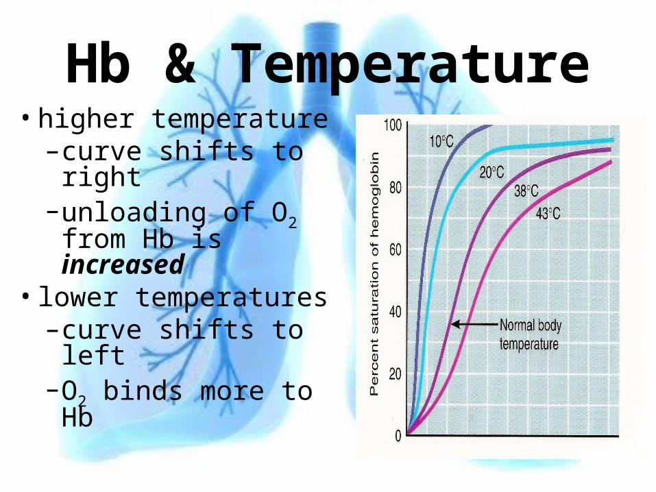
Hb & Temperature• higher temperature
–curve shifts to right
–unloading of O2 from Hb is increased
• lower temperatures–curve shifts to left–O2 binds more to
Hb
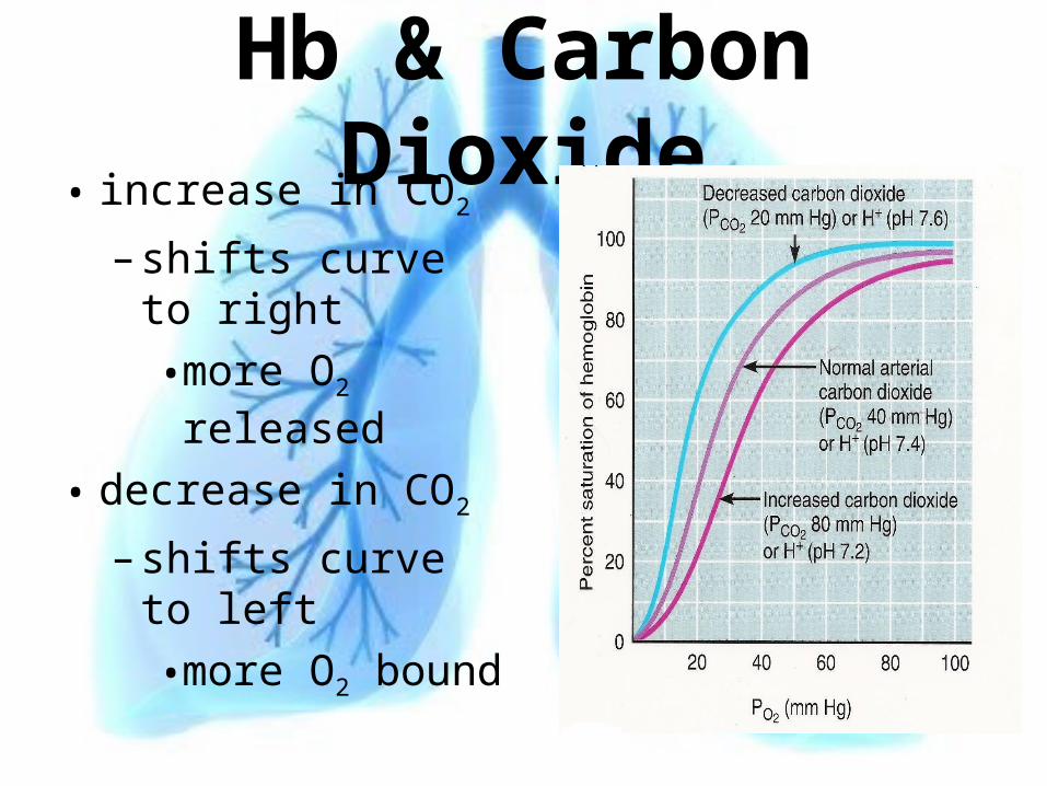
Hb & Carbon Dioxide• increase in CO2
– shifts curve to right
• more O2 released
• decrease in CO2
– shifts curve to left
• more O2 bound
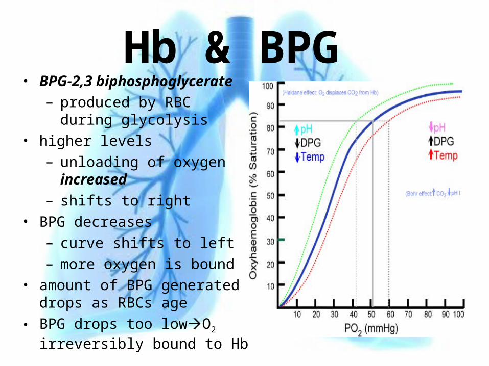
Hb & BPG • BPG-2,3 biphosphoglycerate
– produced by RBC during glycolysis
• higher levels
– unloading of oxygen increased
– shifts to right
• BPG decreases
– curve shifts to left
– more oxygen is bound
• amount of BPG generated drops as RBCs age
• BPG drops too lowO2 irreversibly bound to Hb
CO2 Transport• dissolved in plasma
–7%• transported as HCO3 (bicarbonate ion)
–70%• bound to HB
–23%–attaches to –NH2 groups (amino) of
histidine–Carbaminohemoglobin HB-CO2
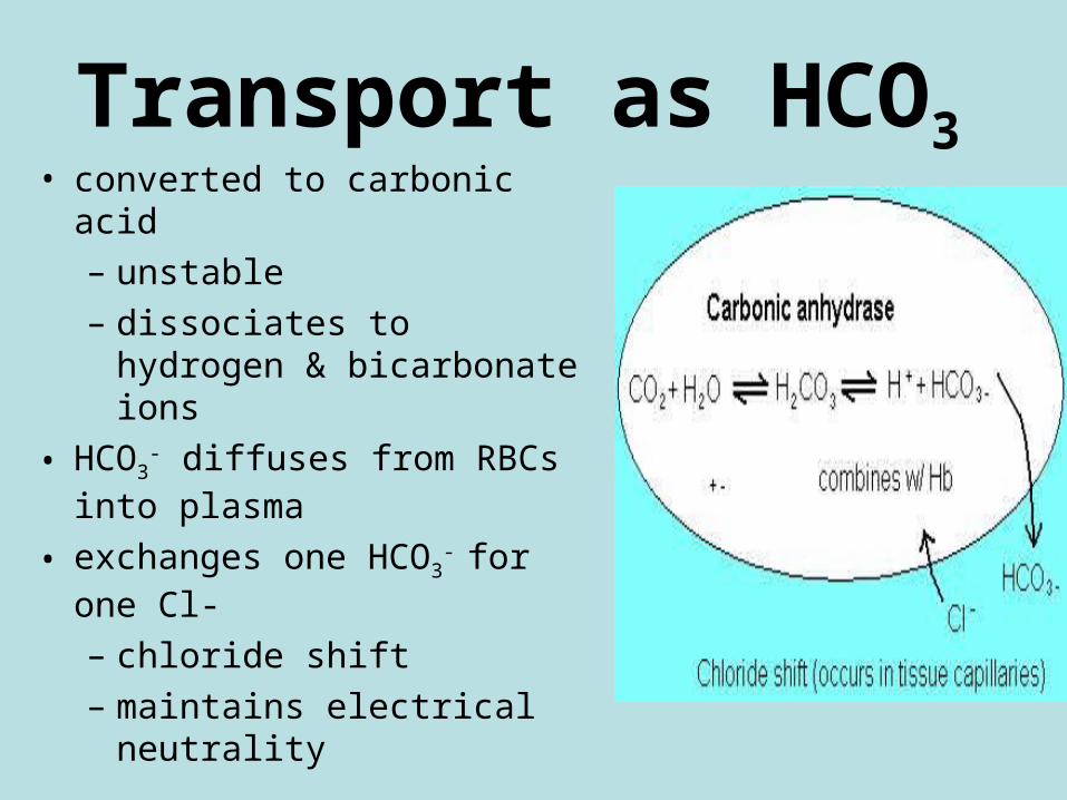
Transport as HCO3 • converted to carbonic acid
– unstable
– dissociates to hydrogen & bicarbonate ions
• HCO3- diffuses from RBCs
into plasma
• exchanges one HCO3- for one
Cl-
– chloride shift
– maintains electrical neutrality

AT LUNG
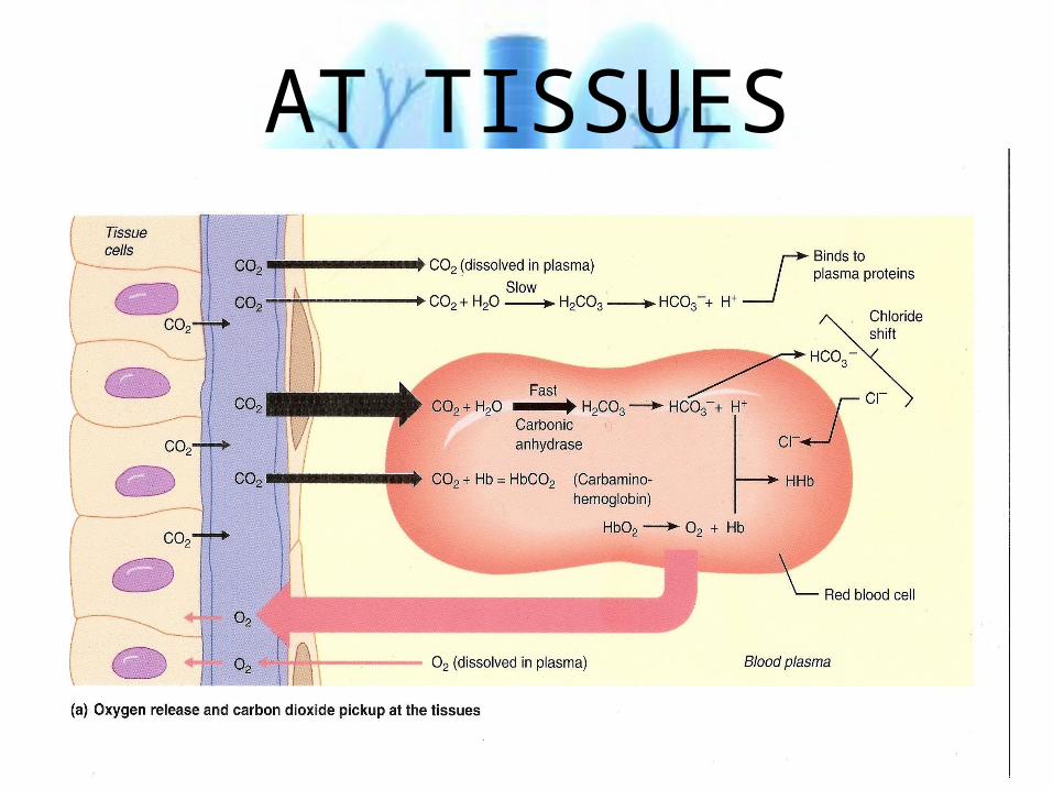
AT TISSUES
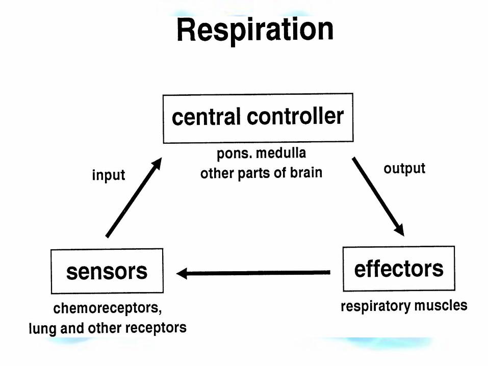
Control of Respiration• normally cellular rates of absorption &
generation of gases are matched by capillary rates of delivery & removal
• rates are identical to rates of O2 absorption & CO2 excretion at lungs
• if absorption & excretion become unbalanced– homeostatic mechanisms restore equilibrium
• changing blood flow & O2 delivery– locally regulated
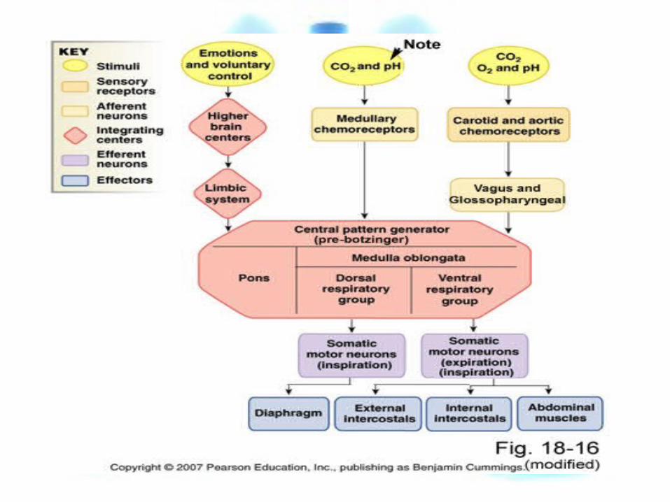
• changing depth & rate of respiration– respiratory centers in brain
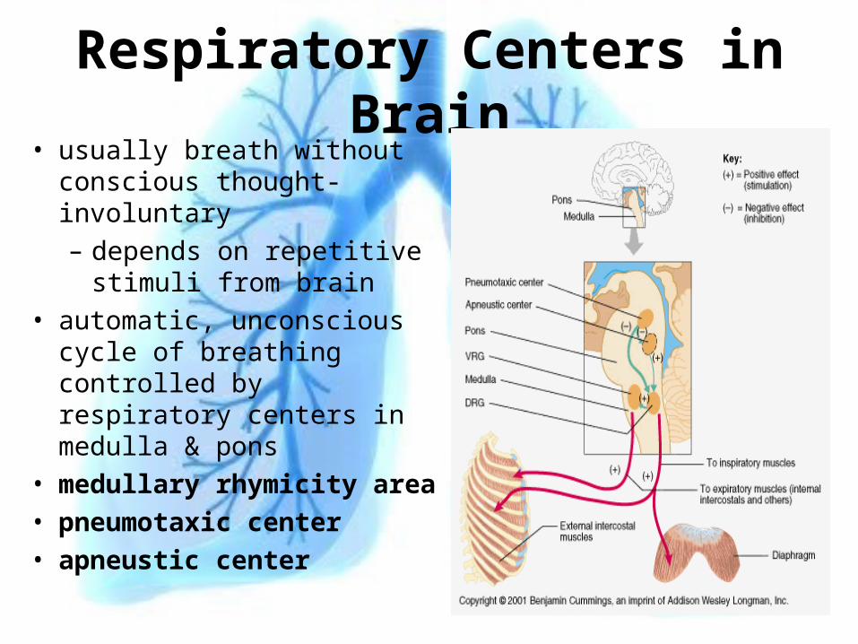
Respiratory Centers in Brain• usually breath without
conscious thought-involuntary– depends on repetitive
stimuli from brain• automatic, unconscious
cycle of breathing controlled by respiratory centers in medulla & pons
• medullary rhymicity area• pneumotaxic center• apneustic center
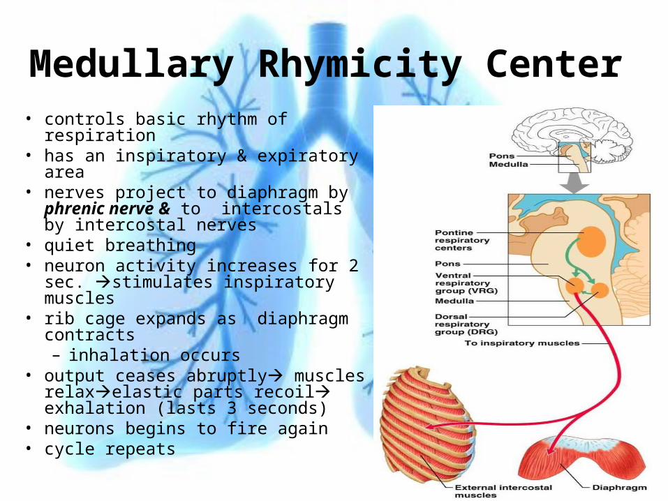
Medullary Rhymicity Center• controls basic rhythm of respiration• has an inspiratory & expiratory area• nerves project to diaphragm by
phrenic nerve & to intercostals by intercostal nerves
• quiet breathing• neuron activity increases for 2 sec.
stimulates inspiratory muscles• rib cage expands as diaphragm
contracts– inhalation occurs
• output ceases abruptly muscles relaxelastic parts recoil exhalation (lasts 3 seconds)
• neurons begins to fire again• cycle repeats
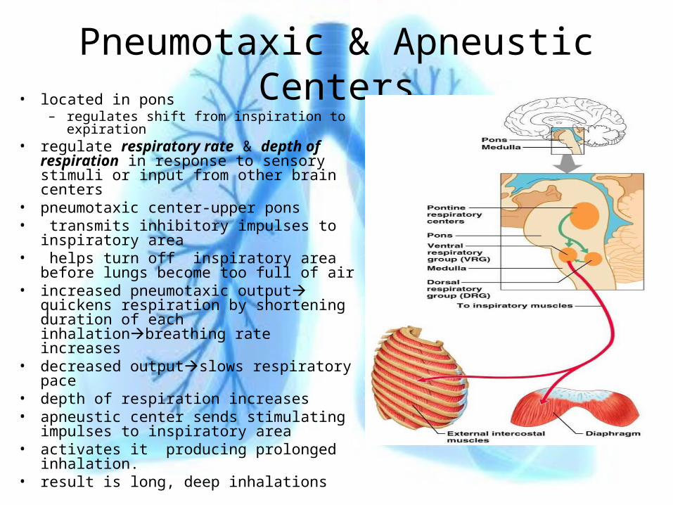
Pneumotaxic & Apneustic Centers• located in pons
– regulates shift from inspiration to expiration• regulate respiratory rate & depth of
respiration in response to sensory stimuli or input from other brain centers
• pneumotaxic center-upper pons• transmits inhibitory impulses to
inspiratory area• helps turn off inspiratory area before
lungs become too full of air• increased pneumotaxic output quickens
respiration by shortening duration of each inhalationbreathing rate increases
• decreased outputslows respiratory pace• depth of respiration increases• apneustic center sends stimulating
impulses to inspiratory area • activates it producing prolonged
inhalation.• result is long, deep inhalations
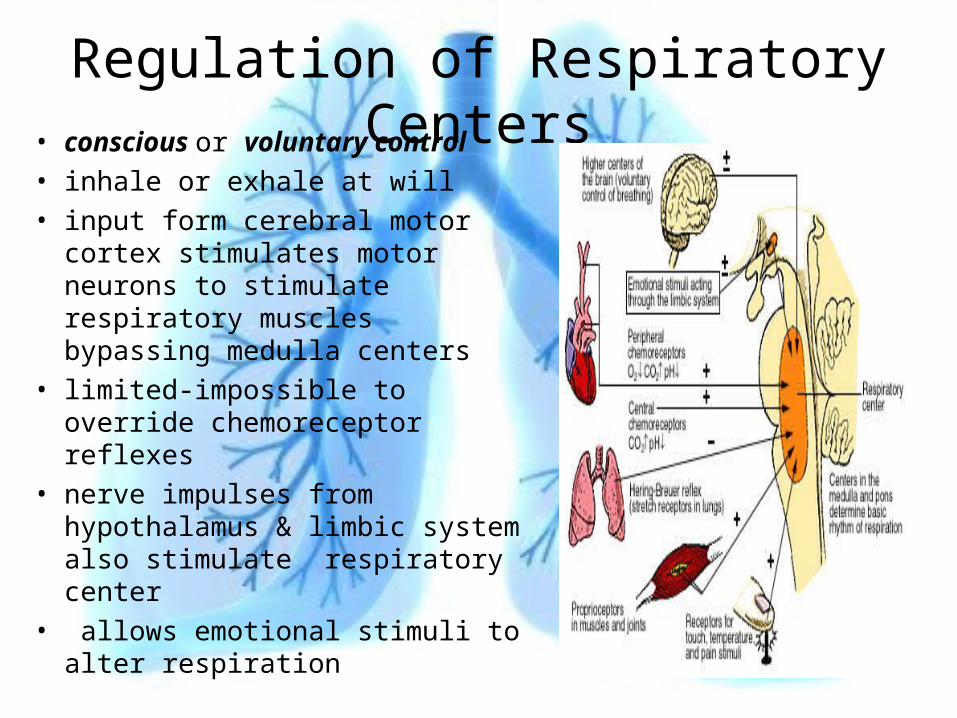
Regulation of Respiratory Centers• conscious or voluntary
control• inhale or exhale at will• input form cerebral motor cortex
stimulates motor neurons to stimulate respiratory muscles bypassing medulla centers
• limited-impossible to override chemoreceptor reflexes
• nerve impulses from hypothalamus & limbic system also stimulate respiratory center
• allows emotional stimuli to alter respiration
Respiratory Reflexes• brain centers regulate respiratory rate & depth
of respiration– in response to sensory stimuli or input from other
brain centers• sensory information comes from• central chemoreceptors• peripheral chemoreceptors• proprioceptors• stretch receptors• information from these alters patterns of
respiration• changes are respiratory reflexes
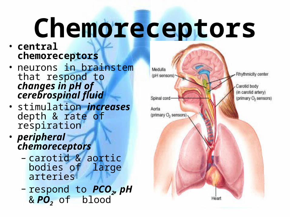
Chemoreceptors• central
chemoreceptors• neurons in brainstem
that respond to changes in pH of cerebrospinal fluid
• stimulation increases depth & rate of respiration
• peripheral chemoreceptors– carotid & aortic bodies
of large arteries– respond to PCO2, pH
& PO2 of blood
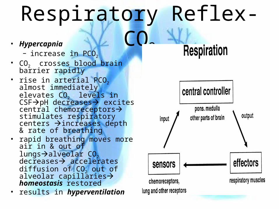
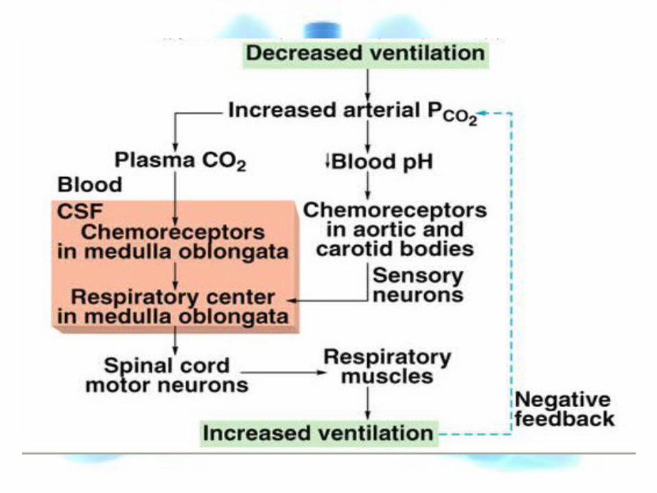
Respiratory Reflex-CO2• Hypercapnia
– increase in PCO2 • CO2 crosses blood brain
barrier rapidly• rise in arterial PCO2 almost
immediately elevates CO2 levels in CSFpH decreases excites central chemoreceptors stimulates respiratory centers increases depth & rate of breathing
• rapid breathing moves more air in & out of lungsalveolar CO2 decreases accelerates diffusion of CO2 out of alveolar capillaries homeostasis restored
• results in hyperventilation
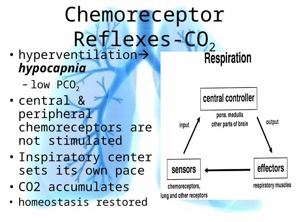
Chemoreceptor Reflexes-CO2
• hyperventilation hypocapnia– low PCO2
• central & peripheral chemoreceptors are not stimulated
• Inspiratory center sets its own pace
• CO2 accumulates• homeostasis restored
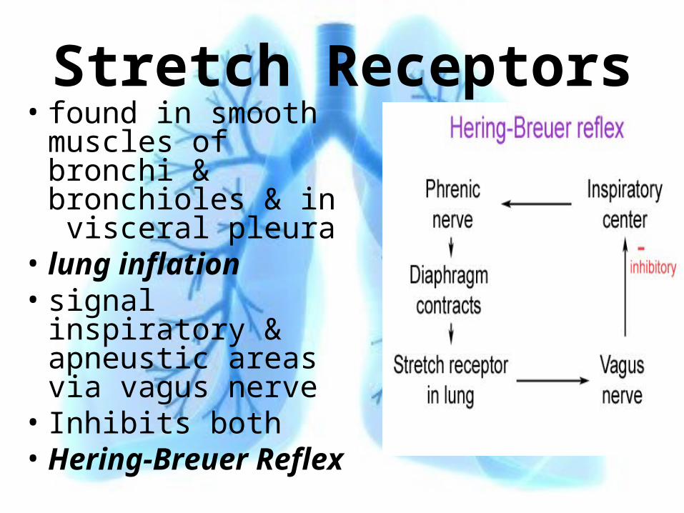
Stretch Receptors• found in smooth
muscles of bronchi & bronchioles & in visceral pleura
• lung inflation• signal inspiratory &
apneustic areas via vagus nerve
• Inhibits both• Hering-Breuer
Reflex