Embed Size (px)
Citation preview
Retarded growth of the medial septum: a major gene effect in acallosal mice
By: Douglas Wahlsten, Barbara Bulman-Fleming
Wahlsten, D., and Bulman-Fleming, B. Retarded growth of the medial septum: A major gene effect in acallosal
mice. Developmental Brain Research, 1994, 77(2), 203-214.
Made available courtesy of Elsevier: http://www.elsevier.com
***Reprinted with permission. No further reproduction is authorized without written permission from
Elsevier. This version of the document is not the version of record. Figures and/or pictures may be
missing from this format of the document.***
Abstract:
Absence of the corpus callosum is a hereditary brain defect that appears with varying severity in four inbred
mouse strains and is the result of more than one major genetic locus. If relatively few, perhaps two or three, loci
are involved in the prenatal ontogeny of the abnormal corpus callosum, it should be possible to identify a
distinct morphological process which shows a major gene effect. Because available evidence suggests the
source of callosal agenesis occurs in the substrates of axon guidance near the midsagittal plane rather than in the
axons themselves, morphometric analysis was done for sagittal sections of the medial septal region in embryos
of normal hybrids and four acallosal strains. The anterodorsal zone of the medial septum subadjacent to the
cavum septi grew much slower in acallosal BALB/c and I/LnJ mice whereas the ventral septal region was
apparently normal. In the Bailey recombinant inbred strains derived from an acallosal BALB/c progenitor, one
recombinant (CXBG/By) closely resembled BALB/c whereas the others resembled the normal C57BL/6 parent
strain. This pattern of results supports a major gene influence on fusion of the cerebral hemispheres near the
region where the corpus callosum first crosses midplane over the dorsal septum.
Key words: Corpus callosum; Anterior commissure; Prenatal development; Inbred strain; Recombinant inbred
strain; Morphometry
Article:
INTRODUCTION
The precision of axon growth over long distances to a remote target requires a suitable substrate or cellular
environment3,9,21
. The formation of the corpus callosum is especially interesting in this regard because axons
from diverse regions of the cerebral cortex converge in a small area at the midsagittal plane just dorsal and
anterior to the hippocampal commissure11,17,21,25
, which crosses midplane prior to the corpus callosum after
following a very different path13,23
. Cells of the subventricular layer emanating from the lateral ventricles are
thought to play an important part in directing callosal axons toward midplane21,32
, which requires an abrupt
change of direction to prevent them from entering the lateral septum. When the subventricular cells do not
extend to the midsagittal region, the corpus callosum may fail to form19,25
.
Four inbred strains of mice lack a corpus callosum in the adult; instead, they have putative callosal axons
assembled in a longitudinal bundle (the Probst bundle) and projecting ipsilaterally. Adult mice of the I/ LnJ
strain never have a corpus callosum12,14
, whereas in the BALB/cWahl and 129 strains about half the mice have
very small or absent corpus callosum24,25
. In the ddN strain the callosal defect is relatively infrequent, and large
samples are required to find enough acallosal mice for research16
. All studies of inheritance find that callosal
defects in adult animals involve more than one recessive gene12,26
. Although the four inbred strains are quite
dissimilar genetically1,14,27
, crosses between them yield many cases of deficient corpus callosum, with the
exception of the BALB/c by 129 cross, which is almost always normal14
. In BALB/c embryos the hippocampal
commissure is also markedly deficient but it eventually recovers and grows to normal adult size25
. In the
embryo the combined size of the hippocampal commissure and the corpus callosum shows a pattern of
inheritance typical of two autosomal, recessive loci29
. In adults of the F2 hybrid cross of BALB/ cWah 1 and
129, a. three-locus model is supported28
. These data suggest that relatively few loci are responsible for callosal
agenesis in mice.
Tracing axons in the embryo with lipophilic dyes demonstrates that callosal axons reach the vicinity of the
midsagittal plane normally and have normal growth cone morphologies until then17
, but they fail to cross to the
opposite hemisphere when they encounter the interhemispheric fissure, For some reason, the dorsal septal
region has not grown far enough to provide an adequate bridge or scaffold to allow axonal traverse of midplane,
If the major cause of the callosal agenesis occurs in the substrate cells, and if the problem arises from only two
major loci, it should be possible to identify some feature of the axonal surroundings which is defective and has
single locus inheritance. Consequently, the present study sought evidence of a specific morphological. defect in
acallosal mice at the midsagittal plane and tested for single locus inheritance with recombinant inbred strains2.
An essential question in understanding a hereditary brain defect is where and when the defect first becomes
manifest, The sequelae of callosal agenesis are relatively well documented, but the crucial events causing the
first axons to divert away from midplane are inadequately understood. In this study a wide range of
morphological ages was examined to assess possible abnormalities prior to the time callosal axons usually
arrive at midplane, relying on genetic and morphometric analysis to localize the source of' the problem. The
degree of abnormality was quantified with reference to a standard F2 hybrid mouse population which has
substantial genetic differences among individuals but never shows outright deficiency of forebrain
commissures29
.
MATERIALS AND METHODS
Mice
The strains and sample sizes are given in Table I, The standard for normal development was based on F2 hybrid
embryos produced by mating B6D2F1 /7 mice purchased from the Jackson Laboratory; parents were the F1
hybrid offspring of a C57B1.16j female mated with a DBA/2.1 male. The acallosal strains 129/5 and 129/ReJ
were also purchased from the Jackson labs, and the I/LnJ mice were a generous donation from Dr. R.L. Collins
of the Jackson labs. Acallosal strains BALB/cWahl and BALB/cWah2 are maintained by full-sib inbreeding at
the University of Alberta. The ddN strain had been inbred for over 35 generations at the Kagawa Medical
School in Japan and was obtained via the Japan Clea Co. of Osaka, courtesy of Dr. H.S. Ozaki who has studied
them extensively. The Bailey recombinant inbred strains and their two progenitors were purchased from the
Jackson labs. Most of the mice were maintained and bred at the University of Waterloo as described
previously24,25
, but all the ddN and several of the 129/Rej and I/LnJ mice were studied at the University of
Alberta under similar conditions, In order to fill in gaps in the body weight. distribution in three important
groups and pro-vide equal sample sizes, some previously stained tissue collected with the same histological
methods was traced anew and measured more extensively for the B6D2F2 /J normal25
and the BALB/cWahl and
2 acallosal groups5.
Matings, litter extractions, and histology
One to three females were mated with one male at about 08.30 h and were then checked for vaginal plugs
around noon and 16.30 h. when the male was removed until the next morning. The time of conception was
defined as the midpoint between detection of a plug and the previous check without a plug, which made
gestation age accurate to within 2 h. In several cases the mice were mated overnight in order to obtain litters
with half-clay gestation ages, which were accurate to only 8 h. The plugged female was weighed and then
housed alone until extraction of the litter. Litters of the normal B6D2F2 /J group were obtained at gestation ages
El4 to E18, whereas those from all other groups were observed at the four ages E1.5 to El8 because inbred mice
are known to develop more slowly than hybricls26,30
. In the CXBD/By group, no litter was obtained at El8
because of very poor breeding, and the body weight range for that group (Table I) was restricted.
Within one hour of the designated gestation age, the pregnant female was deeply anesthetized with sodium
pentobarbital (120 mg/ kg), the uterus was removed surgically and plunged into 4°C saline, and then each
embryo or fetus was removed from the amnion. blotted gently and weighed. The whole mouse was immersed in
Bouin-Duboscq fixative for 48 h and then transferred to several changes of 70% ethanol. Shortly after being
placed in the fixative, the animal had the scalp removed and small slits cut in the skull lateral to midline to aid
penetration of fixative. The skull was left intact near midline to avoid damage to the meninges and blood
vessels in the interhemispheric fissure. Mice were chosen for histology from among a larger group of littermates
such that each group had a wide range of body weights, which yielded good statistical estimates of growth rates
despite modest sample sizes in some groups. Whole heads were embedded in paraffin for sectioning in the
sagittal plane, then serial sections were cut at 10 μm and stained with hematoxylin and eosin. Tracings were
made with a Leitz tracing device and measurements were made with a Numonics 2200 graphics tablet and the
Sigma Scan program from Jandel Scientific.
Correction for artifacts
Dehydration shrinks neural tissue substantially and slicing the paraffin block dorsal to ventral compresses the
section to some extent, which necessarily affects measures of area and thickness. The magnitudes of these
artifacts were estimated for a subset of the brains by piercing the midbrain region of the unfixed fetus with four
tungsten needles in a square array with tips 1.0 mm apart. The distances between holes in the stained tissue
averaged 0.8 mm in the anterior-posterior dimension affected only by shrinkage, whereas in the dorsal-ventral
dimension affected by both shrinkage and compression, the average was 0.7 mm at the University of Waterloo25
and 0.6 mm at the University of Alberta. Distance measures were corrected by multiplying the raw measure by
the appropriate correction factor (1.25 = 1/0.8, etc.), and areas of structures were corrected by multiplying by
1.25 times the appropriate dorsal-ventral factor.
The desired plane of' sectioning was sagittal, but a perfectly sagittal alignment was rare. When the actual plane
of sectioning deviates from the true sagittal plane by an angle θ, the measured area of a right cylindrical
structure is increased by 1/cosθ, and the corrected area can be obtained by multiplying the measured area by
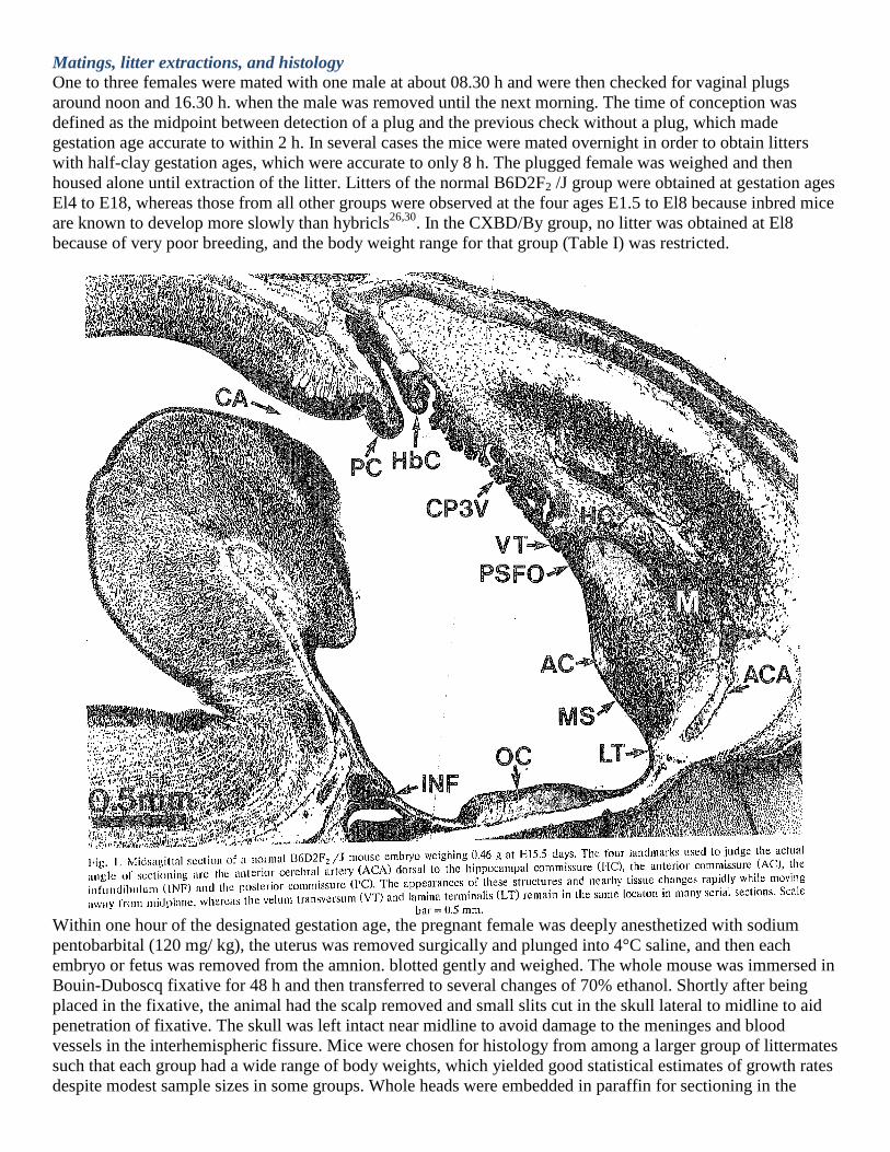
cosθ. To find θ for each brain, four landmarks were. identified (Fig. I) for which reliable judgments could be
made that the midsagittal plane passed through the structure in a particular section; the shape of the structure or
nearby tissue changed abruptly as the sections moved away from midplane. These were (a) the anterior
commissure, (10 the anterior cerebral artery dorsal to medial septum, (c) the posterior commissure dorsal to the
cerebral aqueduct, and (d) the infundibulum. A standard coordinate system was established with the origin
(0,0,0) at the center of the anterior commissure at mid-plane and Y-axis passing along the most caudal portion
of the hippocampal commissure. Distances of structures were measured with respect to these axes and then
corrected for shrinkage and compression. Finally, the value of θ was estimated using analytic geometry. Linear
distances were corrected separately for angular deviations in the y (dorsal-ventral) and z (anterior-posterior)
dimensions, respectively. In most cases the corrections for shrinkage and compression were much greater than
for angle of sectioning.
Sagittal reconstruction
In embryos and fetuses the cerebral hemispheres are separated by a deep and narrow interhemispheric fissure
(Fig. 2), the bottom of which demarcates the surface of the medial septum. In many fetuses the medial septum
extends more than 0.5 mm in the dorsal-ventral dimension. With a section thickness of 0.01 mm, an angle of
cutting of 0.02 radian (1.1°) or more will cause the dorsal and ventral portions of the septum to be at midplane
in different sections. To obtain a valid representation of the midplane section of the medial septum, that section
must be reconstructed front serial sections, each of which contains a portion of the true midplane; the greater the
value of θ, the more sections must be included in the reconstruction. The interhemispheric fissure contains the
meninges lining the fissure as well as a dense plexus of blood vessels emanating from the anterior cerebral
artery and entering the medial septal region. There is also a zone of widely spaced glia-like cells and their
processes just anterior to the HC where the cavum septi later forms in normal mice6,19,20,25
. The medial septum
proper was defined in this study as the contiguous, closely packed neural tissue composed of neurons, glia, their
precursor cells and cells of the neuroepithelium lining the third ventricle but not including meninges or blood
vessels in the fissure. When the angle of sectioning is imperfect, the region of the fissure clearly contains
meninges and blood vessels, and a line can be drawn to demarcate the limit of the medial septum (MS). To
assemble the fragments of the MS into a whole image, landmarks are needed which do not change appreciably
as sections move away from midplane. Three of these were identified which maintained nearly constant
distances from each other in serial sections near midplane: (a) the anterior commissure, (h) the lamina
terminalis (LT) at the ventral limit of the MS, and (c) the velum transversum (VT) which separated the choroid
plexus of the third ventricle from the primordium of the subfornical organ6. Each successive section was aligned
with respect to these three landmarks and the portion at midplane was traced onto the composite. When the
entire midplane region from the VT to the LT had been covered, a reasonably smooth line was drawn to define
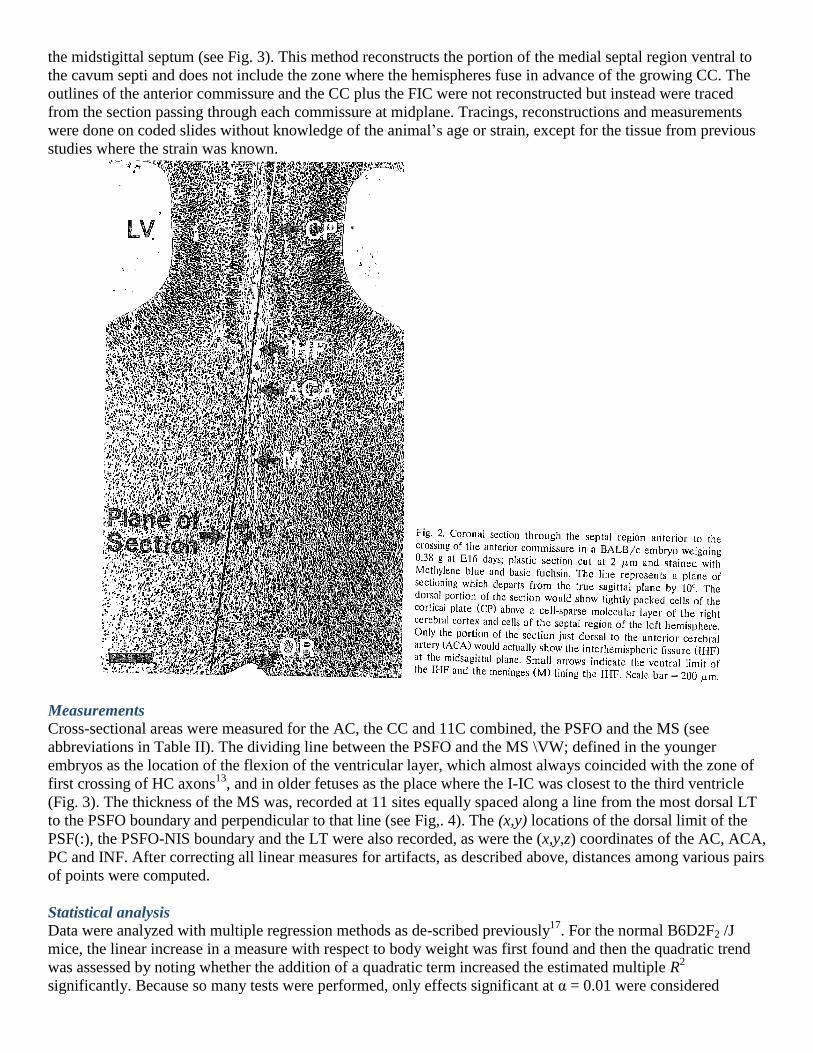
the midstigittal septum (see Fig. 3). This method reconstructs the portion of the medial septal region ventral to
the cavum septi and does not include the zone where the hemispheres fuse in advance of the growing CC. The
outlines of the anterior commissure and the CC plus the FIC were not reconstructed but instead were traced
from the section passing through each commissure at midplane. Tracings, reconstructions and measurements
were done on coded slides without knowledge of the animal’s age or strain, except for the tissue from previous
studies where the strain was known.
Measurements
Cross-sectional areas were measured for the AC, the CC and 11C combined, the PSFO and the MS (see
abbreviations in Table II). The dividing line between the PSFO and the MS \VW; defined in the younger
embryos as the location of the flexion of the ventricular layer, which almost always coincided with the zone of
first crossing of HC axons13
, and in older fetuses as the place where the I-IC was closest to the third ventricle
(Fig. 3). The thickness of the MS was, recorded at 11 sites equally spaced along a line from the most dorsal LT
to the PSFO boundary and perpendicular to that line (see Fig,. 4). The (x,y) locations of the dorsal limit of the
PSF(:), the PSFO-NIS boundary and the LT were also recorded, as were the (x,y,z) coordinates of the AC, ACA,
PC and INF. After correcting all linear measures for artifacts, as described above, distances among various pairs
of points were computed.
Statistical analysis
Data were analyzed with multiple regression methods as de-scribed previously17
. For the normal B6D2F2 /J
mice, the linear increase in a measure with respect to body weight was first found and then the quadratic trend
was assessed by noting whether the addition of a quadratic term increased the estimated multiple R2
significantly. Because so many tests were performed, only effects significant at α = 0.01 were considered
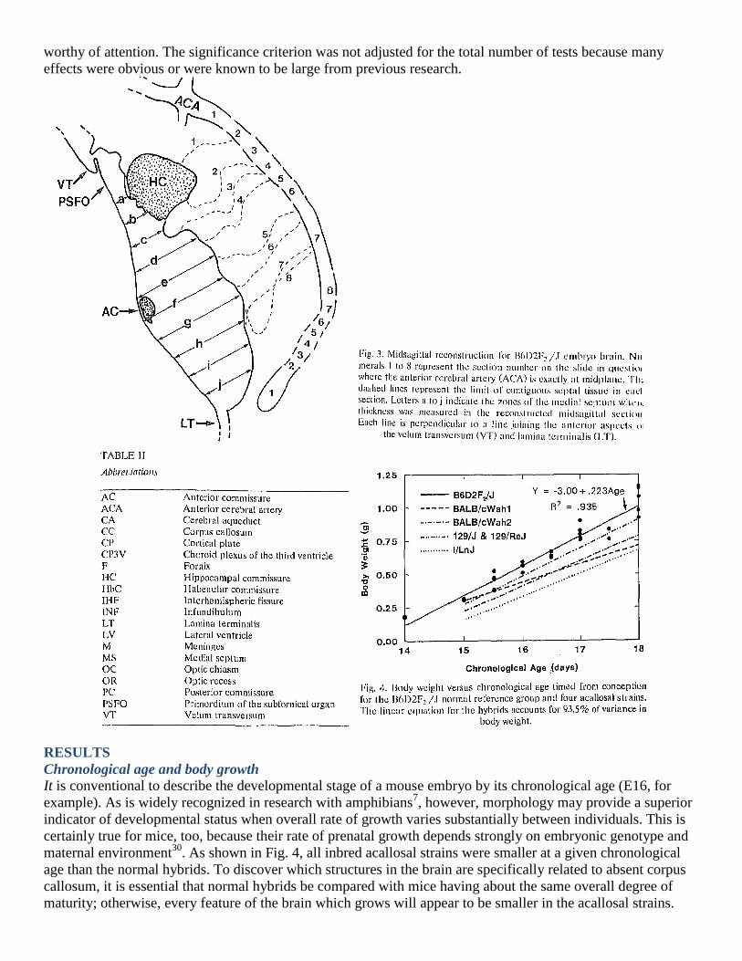
worthy of attention. The significance criterion was not adjusted for the total number of tests because many
effects were obvious or were known to be large from previous research.
RESULTS
Chronological age and body growth
It is conventional to describe the developmental stage of a mouse embryo by its chronological age (E16, for
example). As is widely recognized in research with amphibians7, however, morphology may provide a superior
indicator of developmental status when overall rate of growth varies substantially between individuals. This is
certainly true for mice, too, because their rate of prenatal growth depends strongly on embryonic genotype and
maternal environment30
. As shown in Fig. 4, all inbred acallosal strains were smaller at a given chronological
age than the normal hybrids. To discover which structures in the brain are specifically related to absent corpus
callosum, it is essential that normal hybrids be compared with mice having about the same overall degree of
maturity; otherwise, every feature of the brain which grows will appear to be smaller in the acallosal strains.
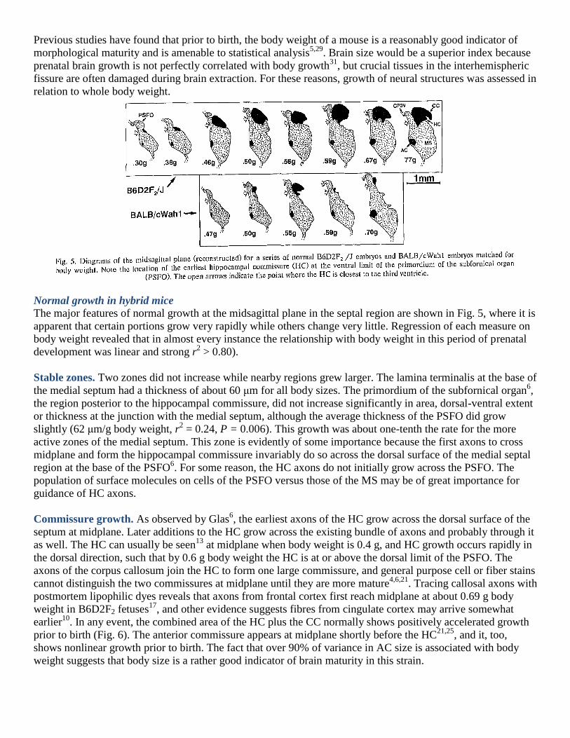
Previous studies have found that prior to birth, the body weight of a mouse is a reasonably good indicator of
morphological maturity and is amenable to statistical analysis5,29
. Brain size would be a superior index because
prenatal brain growth is not perfectly correlated with body growth31
, but crucial tissues in the interhemispheric
fissure are often damaged during brain extraction. For these reasons, growth of neural structures was assessed in
relation to whole body weight.
Normal growth in hybrid mice
The major features of normal growth at the midsagittal plane in the septal region are shown in Fig. 5, where it is
apparent that certain portions grow very rapidly while others change very little. Regression of each measure on
body weight revealed that in almost every instance the relationship with body weight in this period of prenatal
development was linear and strong r2 > 0.80).
Stable zones. Two zones did not increase while nearby regions grew larger. The lamina terminalis at the base of
the medial septum had a thickness of about 60 μm for all body sizes. The primordium of the subfornical organ6,
the region posterior to the hippocampal commissure, did not increase significantly in area, dorsal-ventral extent
or thickness at the junction with the medial septum, although the average thickness of the PSFO did grow
slightly (62 μm/g body weight, r2 = 0.24, P = 0.006). This growth was about one-tenth the rate for the more
active zones of the medial septum. This zone is evidently of some importance because the first axons to cross
midplane and form the hippocampal commissure invariably do so across the dorsal surface of the medial septal
region at the base of the PSFO6. For some reason, the HC axons do not initially grow across the PSFO. The
population of surface molecules on cells of the PSFO versus those of the MS may be of great importance for
guidance of HC axons.
Commissure growth. As observed by Glas6, the earliest axons of the HC grow across the dorsal surface of the
septum at midplane. Later additions to the HC grow across the existing bundle of axons and probably through it
as well. The HC can usually be seen13
at midplane when body weight is 0.4 g, and HC growth occurs rapidly in
the dorsal direction, such that by 0.6 g body weight the HC is at or above the dorsal limit of the PSFO. The
axons of the corpus callosum join the HC to form one large commissure, and general purpose cell or fiber stains
cannot distinguish the two commissures at midplane until they are more mature4,6,21
. Tracing callosal axons with
postmortem lipophilic dyes reveals that axons from frontal cortex first reach midplane at about 0.69 g body
weight in B6D2F2 fetuses17
, and other evidence suggests fibres from cingulate cortex may arrive somewhat
earlier10
. In any event, the combined area of the HC plus the CC normally shows positively accelerated growth
prior to birth (Fig. 6). The anterior commissure appears at midplane shortly before the HC21,25
, and it, too,
shows nonlinear growth prior to birth. The fact that over 90% of variance in AC size is associated with body
weight suggests that body size is a rather good indicator of brain maturity in this strain.
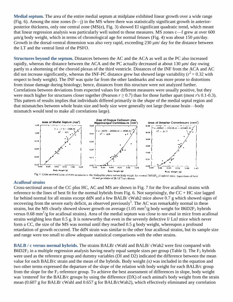
Medial septum. The area of the entire medial septum at midplane exhibited linear growth over a wide range
(Fig. 6). Among the nine zones (b—j) in the MS where there was statistically significant growth in anterior-
posterior thickness, only one central zone (MS(e), Fig. 3) showed El significant quadratic trend, which meant
that linear regression analysis was particularly well suited to those measures. MS zones c—f grew at over 600
μm/g body weight, which in terms of chronological age for normal fetuses (Fig. 4) was about 150 μm/day.
Growth in the dorsal-ventral dimension was also very rapid, exceeding 230 μm/ day for the distance between
the LT and the ventral limit of the PSFO.
Structures beyond the septum. Distances between the AC and the ACA as well as the PC also increased
rapidly, whereas the distance between the ACA and the PC actually decreased at about 130 μm/ day owing
partly to a shortening of the choroid plexus of the third ventricle. Distances of the INF from the ACA and AC
did not increase significantly, whereas the INF-PC distance grew but showed large variability (r2
= 0.32 with
respect to body weight). The INF was quite far from the other landmarks and was more prone to distortions
from tissue damage during histology; hence, distances from this structure were not analyzed further.
Correlations between deviations from expected values for different measures were usually positive, but they
were much higher for structures closer together (Pearson r ≥ 0.7) than for those further apart (most r's 0.1-0.3).
This pattern of results implies that individuals differed primarily in the shape of the medial septal region and
that mismatches between whole brain size and body size were generally not large (because brain—body
mismatch would tend to make all correlations large).
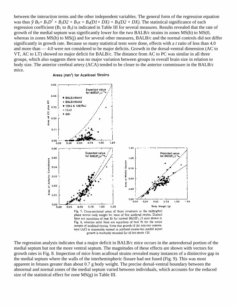
Acallosal strains
Cross-sectional areas of the CC plus HC, AC and MS are shown in Fig. 7 for the five acallosal strains with
reference to the lines of best fit for the normal hybrids from Fig. 6. Not surprisingly, the CC + HC size lagged
far behind normal for all strains except ddN and a few BALB/ cWah2 mice above 0.7 g which showed signs of
recovering from the severe early deficit, as observed previously5. The AC was remarkably normal in these
strains, but the MS clearly showed slower growth on average (1.05 mm2/g body weight for B6D2F2 hybrids
versus 0.68 mm2/g for acallosal strains). Area of the medial septum was close to nor-mal in mice from acallosal
strains weighing less than 0.5 g. It is noteworthy that even in the severely defective I/ LnJ mice which never
form a CC, the size of the MS was normal until they reached 0.5 g body weight, whereupon a profound
retardation of growth occurred. The ddN strain was similar to the other four acallosal strains, but its sample size
and range were too small to allow adequate statistical comparisons with the other strains.
BALB / c versus normal hybrids. The strains BALB/ cWahl and BALB/ cWah2 were first compared with
B6D2F2 in a multiple regression analysis having nearly equal sample sizes per group (Table I). The F2 hybrids
were used as the reference group and dummy variables (DI and D2) indicated the difference between the mean
value for each BALB/c strain and the mean of the hybrids. Body weight (x) was included in the equation and
two other terms expressed the departure of the slope of the relation with body weight for each BALB/c group
from the slope for the F2 reference group. To achieve the best assessment of differences in slope, body weight
was 'centered' for the BALB/c groups by using the difference (DX) of each animal's body weight from the strain
mean (0.607 g for BALB/ cWahl and 0.657 g for BALB/cWah2), which effectively eliminated any correlation
between the interaction terms and the other independent variables. The general form of the regression equation
was thus ̂ B0+ B1D1 + B2D2 + B3x + B4(D1× DX) + B5(D2 × DX). The statistical significance of each
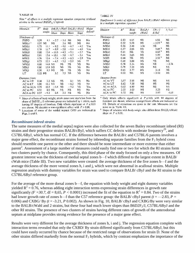
regression coefficient (B1 to B5) is indicated in Table III for several measures. Results revealed that the rate of
growth of the medial septum was significantly lower for the two BALB/c strains in zones MS(b) to MS(0,
whereas in zones MS(h) to MS(j) and for several other measures, BALB/c and the normal controls did not differ
significantly in growth rate. Because so many statistical tests were done, effects with a t ratio of less than 4.0
and more than — 4.0 were not considered to be major deficits. Growth in the dorsal-ventral dimension (AC to
VT, AC to LT) showed no major deficit for BALB/c. The distance from AC to PC was similar in all three
groups, which also suggests there was no major variation between groups in overall brain size in relation to
body size. The anterior cerebral artery (ACA) tended to be closer to the anterior commissure in the BALB/c
mice.
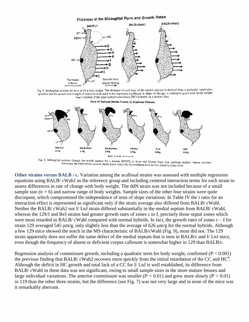
The regression analysis indicates that a major deficit in BALB/c mice occurs in the anterodorsal portion of the
medial septum but not the more ventral septum. The magnitudes of these effects are shown with vectors for
growth rates in Fig. 8. Inspection of mice from acallosal strains revealed many instances of a distinctive gap in
the medial septum where the walls of the interhemispheric fissure had not fused (Fig. 9). This was most
apparent in fetuses greater than about 0.7 g body weight. The precise dorsal-ventral boundary between the
abnormal and normal zones of the medial septum varied between individuals, which accounts for the reduced
size of the statistical effect for zone MS(g) in Table III.
Other strains versus BALB / c. Variation among the acallosal strains was assessed with multiple regression
equations using BALB/ cWah1 as the reference group and including centered interaction terms for each strain to
assess differences in rate of change with body weight. The ddN strain was not included because of a small
sample size (n = 6) and narrow range of body weights. Sample sizes of the other four strains were quite
discrepant, which compromised the independence of tests of slope variations. In Table IV the t ratio for an
interaction effect is represented as significant only if the strain average also differed from BALB/ cWahl.
Neither the BALB/ cWah2 nor I/ LnJ strain differed substantially in the medial septum from BALB/ cWahl,
whereas the 129/J and ReJ strains had greater growth rates of zones c to f, precisely those septal zones which
were most retarded in BALB/ cWahl compared with normal hybrids. In fact, the growth rates of zones c—f for
strain 129 averaged 545 μm/g, only slightly less than the average of 626 μm/g for the normal hybrids. Although
a few 129 mice showed the notch in the MS characteristic of BALB/cWahl (Fig. 9), most did not. The 129
strain apparently does not suffer the same defect of the medial septum that is seen in BALB/c and I/ LnJ mice,
even though the frequency of absent or deficient corpus callosum is somewhat higher in 129 than BALB/c.
Regression analysis of commissure growth, including a quadratic term for body weight, confirmed (P < 0.001)
the previous finding that BALB/ cWah2 recovers more quickly from the initial retardation of the CC and HC4.
Although the deficit in HC growth and total lack of a CC for I/ LnJ is well established, its difference from
BALB/ cWahl in these data was not significant, owing to small sample sizes in the more mature fetuses and
large individual variations. The anterior commissure was smaller (P < 0.01) and grew more slowly (P < 0.01)
in 119 than the other three strains, but the difference (see Fig. 7) was not very large and in none of the mice was
it remarkably aberrant.
Recombinant inbred strains
The same measures of the medial septa] region were also collected for the seven Bailey recombinant inbred (RI)
strains and their progenitor strains BALB/cByJ, which suffers CC defects with moderate frequency29
, and
C57BL/6ByJ, which has normal CC. If the difference between the BALB/c and C57BL/6 parents involves a
major gene effect, the recombinant strains formed by inbreeding separate families from the F2 hybrid cross
should resemble one parent or the other and there should be none intermediate or more extreme than either
parent2. Assessment of a large number of measures could easily find one or two for which the RI strains form
two clusters purely by chance. Consequently, this third phase of the study focused on only a few measures. Of
greatest interest was the thickness of medial septal zones b—f which differed to the largest extent in BALB/
cWah mice (Table III). Two new variables were created: the average thickness of the five zones b—f and the
average thickness of the more ventral zones h, i and j, which were not abnormal in acallosal strains. Multiple
regression analysis with dummy variables for strain was used to compare BALB/ cByJ and the RI strains to the
C57BL/6ByJ reference group.
For the average of the more dorsal zones b—f, the equation with body weight and eight dummy variables
yielded R2 = 0.76, whereas adding eight interaction terms expressing strain differences in growth rate
significantly (F =30.7, df = 8,65, P < 0.0001) increased the fit of the equation to R2 = 0.84. Two of the strains
had lower growth rate of zones b—f than the C57 reference group: the BALB/ cByJ parent (t = —2.83, P =
0.006) and CXBG/ By (t = -3.21, P 0.002). As shown in Fig. 10, BALB/ cByJ and CXBG/By were very similar
to the BALB/cWahl and 2 strains, but these four had much lower slopes than B6D2F2/J, C57BL/6ByJ and the
other RI strains. The presence of two clusters of strains having different rates of growth of the anterodorsal
septum at midplane provides strong evidence for the presence of a major gene effect.
Results were very different for the average thickness of zones h, i and j. The regression equation complete with
interaction terms revealed that only the CXBD/ By strain differed significantly from C57BL/6ByJ, but this
could have easily occurred by chance because of the restricted range of observations for strain D. None of the
other strains differed markedly from the normal F2 hybrids, which by contrast emphasizes the importance of the
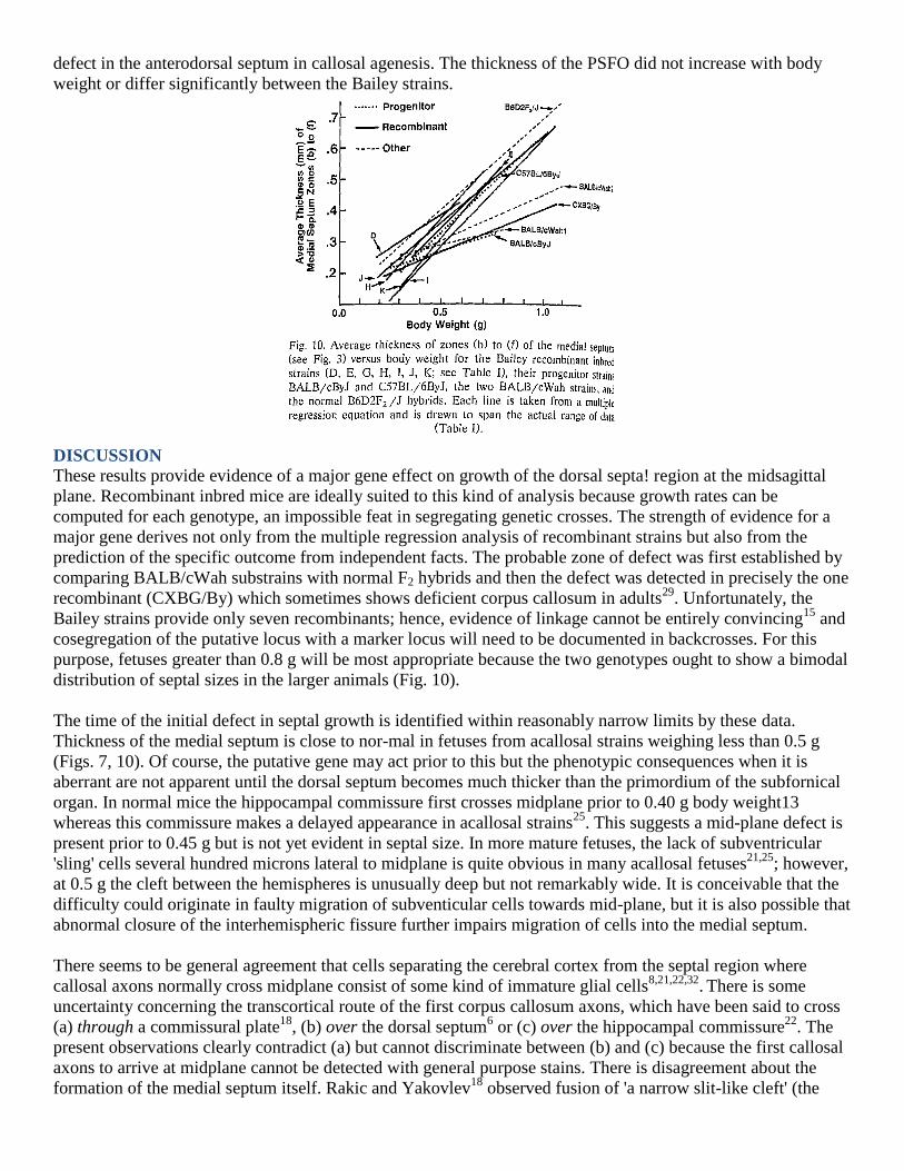
defect in the anterodorsal septum in callosal agenesis. The thickness of the PSFO did not increase with body
weight or differ significantly between the Bailey strains.
DISCUSSION
These results provide evidence of a major gene effect on growth of the dorsal septa! region at the midsagittal
plane. Recombinant inbred mice are ideally suited to this kind of analysis because growth rates can be
computed for each genotype, an impossible feat in segregating genetic crosses. The strength of evidence for a
major gene derives not only from the multiple regression analysis of recombinant strains but also from the
prediction of the specific outcome from independent facts. The probable zone of defect was first established by
comparing BALB/cWah substrains with normal F2 hybrids and then the defect was detected in precisely the one
recombinant (CXBG/By) which sometimes shows deficient corpus callosum in adults29
. Unfortunately, the
Bailey strains provide only seven recombinants; hence, evidence of linkage cannot be entirely convincing15
and
cosegregation of the putative locus with a marker locus will need to be documented in backcrosses. For this
purpose, fetuses greater than 0.8 g will be most appropriate because the two genotypes ought to show a bimodal
distribution of septal sizes in the larger animals (Fig. 10).
The time of the initial defect in septal growth is identified within reasonably narrow limits by these data.
Thickness of the medial septum is close to nor-mal in fetuses from acallosal strains weighing less than 0.5 g
(Figs. 7, 10). Of course, the putative gene may act prior to this but the phenotypic consequences when it is
aberrant are not apparent until the dorsal septum becomes much thicker than the primordium of the subfornical
organ. In normal mice the hippocampal commissure first crosses midplane prior to 0.40 g body weight13
whereas this commissure makes a delayed appearance in acallosal strains25
. This suggests a mid-plane defect is
present prior to 0.45 g but is not yet evident in septal size. In more mature fetuses, the lack of subventricular
'sling' cells several hundred microns lateral to midplane is quite obvious in many acallosal fetuses21,25
; however,
at 0.5 g the cleft between the hemispheres is unusually deep but not remarkably wide. It is conceivable that the
difficulty could originate in faulty migration of subventicular cells towards mid-plane, but it is also possible that
abnormal closure of the interhemispheric fissure further impairs migration of cells into the medial septum.
There seems to be general agreement that cells separating the cerebral cortex from the septal region where
callosal axons normally cross midplane consist of some kind of immature glial cells8,21,22,32
. There is some
uncertainty concerning the transcortical route of the first corpus callosum axons, which have been said to cross
(a) through a commissural plate18
, (b) over the dorsal septum6 or (c) over the hippocampal commissure
22. The
present observations clearly contradict (a) but cannot discriminate between (b) and (c) because the first callosal
axons to arrive at midplane cannot be detected with general purpose stains. There is disagreement about the
formation of the medial septum itself. Rakic and Yakovlev18
observed fusion of 'a narrow slit-like cleft' (the
sulcus medianus telencephali medii) but not of the medial hemispheres themselves in human tissue, and Zaki32
argued against fusion of murine tissue separated by mesenchymc. Others studying rodents attributed the medial
septum to fusion of the hemispheres19,21,25
. The present data clearly indicate a steady growth of the medial septal
region, but they cannot reveal whether this occurs via a genuine fusion of the medial hemispheres, enlargement
by migration of cells into the region which thereby pushes the meninges further anterior, or both processes. It
can be stated with confidence, however, that the corpus callosum does not cross in the zone of rapidly growing
septal tissue identified in this study; rather, it crosses near the dorsal limit of the hippocampal commissure,
which itself crosses over the dorsal surface of the medial septum and rapidly fills the region anterior to the
primordium of the subfornical organ. Discovering a distinct septal notch in acallosal strains does not imply that
this is the only defective midplane zone. On the contrary, previous data clearly implicate the glial sling, and the
zipper-like formation that precedes the anterior extension of the CC20
may also be malformed in acallosal mice.
The presence of the septal notch in acallosal strains is one of several processes leading to absence of the adult
corpus callosum. The adult defect clearly involves problems at several loci. If one of these loci yields a 'septa'
notch,' there could be just one other major locus or several minor loci combining to yield a permanent structural
abnormality. To distinguish these possibilities will require a much larger sample of recombinant strains. It is
evident that a mouse can have the homozygous recessive genotype and yet have a normal adult corpus
callosum. Almost all fetuses of the strains BALB/ cWah2 and CXBG/ By show the septal notch, yet most adults
of these strains have a corpus callosum of normal size29
. BALB/ cWah1 and BALB/ cWah2 suffer similar
degrees of retarded septal growth, yet the former has a much higher frequency of severe callosal defects in the
adult26
. Presence of the septa] notch apparently acts as a risk factor which can be overcome through recovery
processes which allow callosal axons to cross over the hippocampal commissure, except in the I/ LnJ strain
where growth of the hippocampal commissure itself is so retarded that callosal axons cannot utilize it as a
bridge. There can also be failure of corpus callosum formation when there is no septal notch, as often occurs in
the 129 strain. Two strains can express a similar anatomical error for different genetic and developmental
reasons. The hybrid cross of BALB/c and 129 has a normal corpus callosum14
. This evidence of the
involvement of different genetic loci in these strains is consistent with the present observations indicating that
severe septal notch is prevalent only in BALB/c.
REFERENCES
1 Atchley, W.R. and Fitch, W.M., Gene trees and the origins of inbred strains of mice, Science, 254 (1991) 554-
558.
2 Bailey, D.W., Recombinant inbred strains and bilineal congenic strains. In H.L. Foster, J.D. Small and J.G.
Fox (Eds.), The Mouse in Biomedical Research, Vol. I, Academic, New York, 1981, pp, 223-239.
3 Belanger, M.-C., Auclair, F., Bertrand, L. and Marchand, R., The disposition of early-generated neurons in the
rat embryo predicts the pattern of major axonal tracts, Brain Res. Bull., 30 (1993) 273-279.
4 Berbel, P. and Innocenti, G.M., The development of the corpus callosum in cats: a light- and electron-
microscopic study, J. Comp. Neural., 278 (1988) 132-156.
5 Bulman-Fleming, B. and Wahlsten, D., The effects of intrauterine position on the degree of corpus callosum
deficiency in two substrains of BALB/c mice, Den PsychobioL, 24 (1991) 395-412.
6 Glas, P. Onderzoek naar de vroege ontwikkeling van de commissuren in het mediane gebied van het
telencephalon btj de witte nutis, Drukkerij Van Denderen B.V., Groningen, 1975.
7 Hamburger, V. and Flamilton, H.L., A series of normal stages in the development of the chick embryo, J.
MorphoL, 88 0951) 49-92.
8 Hankin, M.H. and Silver, J., Development of intersecting CNS fiber tracts: the corpus callosum and its
perforating fiber path-way, J. Comp. Neurol., 272 (1988) 177-190.
9 Katz, M.J., Lasek, R.J. and Nauta, H.J.W., Ontogeny of substrate pathways and the origin of the neural circuit
pattern, Neuroscience, 5 (1980) 821-833.
10 Koester, S.E. and O'Leary, D.D.M., Subplate cells in medial cortex send the first axons across the corpus
callosum, Soc. Neurosci. Abstr., 17 (1991) 41.
11 Lent, R., Hedin-Pereira, C., Menezes, J.R.L. and Jhaveri, S., Neurogenesis and development of callosal and
intracortical con-nections in the hamster, Neurosci., 38 (1990) 21-37.
12 Lipp, 11.-P., Waanders, R., Ricceri, L., Wolfer, D.P. and Scheffrahn, II., A new mouse model of complete
and partial agenesis of the corpus callosum; crosses involving the I strain, Eur. Neurosci., in press.
13 Livy, D., Formation of the hippocampal commissure in normal mouse embryo. Soc. Neurosci. Abstr., 18
(1992) 221.
14 Livy, D.J. anti Wah!sten, D., Tests of genetic allelism between four inbred mouse strains with absent corpus
callosum, J. Hered., 82 ( 1991) 459-464.
15 Neumann, P.E., Two-locus linkage analysis using recombinant inbred Sifilit1S and Bayes' theorem,
Genetics, 126 (1990) 277-284.
16 Ozaki, H.S., Murakami, T.H., Toyoshima, T. and Shimada, M., Agenesis the corpus callosum in ddN strain
mouse associated with unusual facial appearance (fiat face), Neurosci. Res., 1 (1984) 81-87.
17 Ozaki, H.S. and Wahlsten, D., Cortical axon trajectories and growth conmorphologies in fetuses of acallosal
mouse strains, J. Comp. Neural., 336 (1993) 595-604.
18 Rakic, P. and Yakovlev, P.I., Development of the corpus callosum and cavum septi in man, J. Comp.
Neurol., 132 (1968)45-72,
19 Schneider, B.F. and Silver, J., Failure of the subcallosal sling to develop after embryonic X-irradiation is
correlated with absence of the cavum septi, J. Comp. Neurol., 299 (1990) 462-469.
20 Silver, J., Edwards, M.A. and Levitt, P., Immunocytochemical demonstration of early appearing astroglial
structures that form boundaries and pathways along axon tracts in the fetal brain,J, Comp. Neurol., 328 (1993)
415-436.
21 Silver, J., Lorenz, S.E., Wahlsten, D. and Coughlin, J., Axonal guidance during development of the great
cerebral commissures: descriptive and experimental studies, in vivo, on the role of preformed glial pathways, J.
Comp. Neurol., 210 (1982)10-29.
22 Valentino, K.L. and Jones, E.G., The early formation of the corpus callosum: a light and electron
microscopic study in foetal and neonatal rats, J. Neurocytol., 11 (1982) 583-609,
23 Wahlsten, D., Prenatal schedule of appearance of mouse brain commissures, Dev. Brain Res., 1 (1981) 461-
473.
24 Wahlsten, D., Deficiency of corpus callosum varies with strain and supplier of the mice, Brain Res., 239
(1982) 329-347.
25 Wahlsten, D., Defects of the fetal forebrain in mice with hereditary agenesis of the corpus callosum, J.
Comp. Neurol., 262(1987) 227-241.
26 Wahlsten, D., Genetic and developmental defects of the mouse corpus callosum, Experientia, 45 (1989) 828-
838.
27 Wahlsten, D., Ozaki, H.S. and Livy, D., Deficient corpus din-sum in hybrids between ddN and three other
abnormal mouse strains, Neurosci. Lett., 136 (1992) 99-101.
28 Wahlsten, D. and Schalomon, M.S., A new hybrid mouse model for agenesis of the corpus callosum, Behav.
Brain Res., in press.
29 Wahlsten, D. and Smith, G., Inheritance of retarded forebrain commissure development in fetal mice: results
from classical crosses and recombinant inbred strains, J. Hered., 80 (1989) 11-16.
30 Wahlsten, D. and Wainwright, P., Application of a morphological time scale to hereditary differences in
prenatal mouse development, J. Emblyol. Exp. Morphol., 42 (1977) 79-92.
31 Wainwright, P. and Deeks, S., A comparison of corpus callosum development in the BALB/cCF and
C57BL/6J inbred mouse strains, Growth, 48 (1984) 192-197.
32 Zaki, W., Le processus degneratif au cours du developpement du corps calleux, Arch. Anat. Micr. Morphol.
Exp., 74 (19851 133-149.
![Medial septum lesions disrupt exploratory trip ... · septohippocampal involvement in dead reckoning ... cholinergic and GABAergic projections to the hippocampus [16,17]. Second,](https://img.pdfslide.net/doc/110x75/5fa6e449750b7f31bc09c35f/medial-septum-lesions-disrupt-exploratory-trip-septohippocampal-involvement.jpg)