Embed Size (px)
Citation preview

Case Report
512
Retroperitoneoscopic Nephrectomy for a Horseshoe Kidney with Unilateral Severe Hydronephrosis and Ureteral Hypoplasia
Jung Woo Lee Seung Hyun You Dong Yup Han Hee Jong Jeong Doo Young Choi1 Yeon Kyun Oh1
From the Departments of Urology 1Pediatrics Wonkwang University School of Medicine Iksan Korea
A horseshoe kidney is the most common renal fusion anomaly It is well known that horseshoe kidneys may be associated with many urological problems including calculi vesicoureteral reflux and ureteropelvic junc-tion obstruction However a horseshoe kidney with unilateral severe hydronephrosis and ureteral hypoplasia is very rare We report an 11- year-old female who underwent a retroperitoneoscopic nephrectomy for a horseshoe kidney with severe hydronephrosis and unilateral ureteral hypoplasia (Korean J Urol 200950512-515)985103985103985103985103985103985103985103985103985103985103985103985103985103985103985103985103985103985103985103985103Key Words Hydronephrosis Ureter
Korean Journal of Urology Vol 50 No 5 512-515 May 2009
DOI 104111kju2009505512ReceivedDecember 17 2008AcceptedFebruary 24 2009
Correspondence to Hee Jong JeongDepartment of Urology Wonkwang University School of Medicine and Hospital 344-2 Shinyong-dong Iksan 570-711 KoreaTEL 063-859-1332FAX 063-842-1455E-mail uro94cwonkwangackr
This work was supported by Wonkwang University in 2008
The Korean Urological Association 2009
A horseshoe kidney is the most common renal fusion
anomaly Several associated anomalies including ureteropelvic
junction obstruction vesicoureteral reflux and a duplicated
ureter can occur in patients with this anomaly The common
presenting symptoms are urolithiasis infection and hydro-
nephrosis1 However a horseshoe kidney with unilateral severe
hydronephrosis and ureteral hypoplasia is very rare Here we
report an 11-year-old female who underwent a retroperitoneo-
scopic nephrectomy for a horseshoe kidney with severe
hydronephrosis and unilateral ureteral hypoplasia
CASE REPORT
An 11-year-old female presented with left flank pain and
gross hematuria after a fall Her past medical and family history
were nonspecific There was a distended palpable mass and
direct tenderness in the left upper quadrant on the physical
examination There were no abnormal findings on the blood
tests however there were many red blood cells on the
urinalysis Computed tomography imaging showed a horseshoe
kidney with severe hydronephrosis and renal cortical thinning
of the left kidney (Fig 1A B) A MAG3 renal scan confirmed
the left kidney function to have a 24 decrease in uptake (Fig
1C) The cystoscopy findings were normal One day before
surgery we performed a percutaneous nephrostomy for reduc-
tion of the hydronephrosis and 4000 ml of brownish fluid was
drained (Fig 1D)
The patient was placed in the lateral position and a lateral
incision was made longitudinally at 2 cm below the left 12th
rib the nephroscope was inserted with an attached surgical
glove at the tip 800 ml of saline was injected and the required
space was secured for the surgical procedure After inserting
a 12 mm Hasson trocar CO2 gas was perfused to form a
pneumoperitoneum and then a flexible camera was inserted In
addition 5 mm trocars were inserted at the anterior and
posterior axillary lines at the level of the umbilicus after
examination of the surrounding structures The position of the
previously placed nephrostomy catheter was confirmed and we
then opened Gerotarsquos fascia dissected the kidney from the
surrounding tissues and then dissected by the psoas muscle
noting the thread-like hypoplastic ureter (Fig 2A-C) A total
of 3 arteries and veins were clipped and the isthmus was
incised by using electrocautery and the Sonosurg (Olympus
Japan) for the resection of the left kidney (Fig 2D E) The
resected left kidney was removed from the body in a LapBag
(Sejong Medical Korea) through a 12 mm Hasson trocar (Fig
Jung Woo Lee et alRetroperitoneoscopic Nephrectomy for a Horseshoe Kidney 513
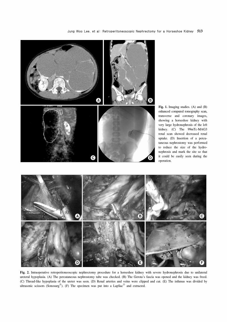
Fig 1 Imaging studies (A) and (B)
enhanced computed tomography scan
transverse and coronary images
showing a horseshoe kidney with
very large hydronephrosis of the left
kidney (C) The 99mTc-MAG3
renal scan showed decreased renal
uptake (D) Insertion of a percu-
taneous nephrostomy was performed
to reduce the size of the hydro-
nephrosis and mark the site so that
it could be easily seen during the
operation
Fig 2 Intraoperative retroperitoneoscopic nephrectomy procedure for a horseshoe kidney with severe hydronephrosis due to unilateral
ureteral hypoplasia (A) The percutaneous nephrostomy tube was checked (B) The Gerotas fascia was opened and the kidney was freed
(C) Thread-like hypoplasia of the ureter was seen (D) Renal arteries and veins were clipped and cut (E) The isthmus was divided by
ultrasonic scissors (Sonosurg) (F) The specimen was put into a LapSac and extracted
514 Korean Journal of Urology vol 50 512-515 May 2009
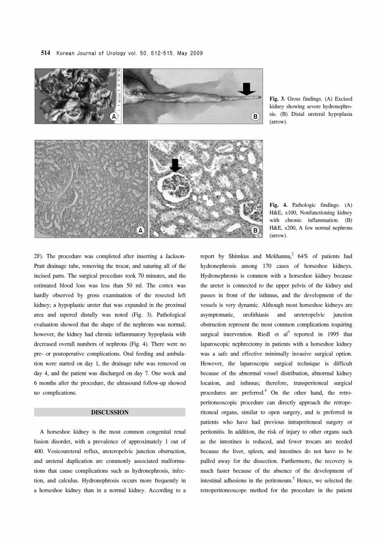
Fig 3 Gross findings (A) Excised
kidney showing severe hydronephro-
sis (B) Distal ureteral hypoplasia
(arrow)
Fig 4 Pathologic findings (A)
HampE x100 Nonfunctioning kidney
with chronic inflammation (B)
HampE x200 A few normal nephrons
(arrow)
2F) The procedure was completed after inserting a Jackson-
Pratt drainage tube removing the trocar and suturing all of the
incised parts The surgical procedure took 70 minutes and the
estimated blood loss was less than 50 ml The cortex was
hardly observed by gross examination of the resected left
kidney a hypoplastic ureter that was expanded in the proximal
area and tapered distally was noted (Fig 3) Pathological
evaluation showed that the shape of the nephrons was normal
however the kidney had chronic inflammatory hypoplasia with
decreased overall numbers of nephrons (Fig 4) There were no
pre- or postoperative complications Oral feeding and ambula-
tion were started on day 1 the drainage tube was removed on
day 4 and the patient was discharged on day 7 One week and
6 months after the procedure the ultrasound follow-up showed
no complications
DISCUSSION
A horseshoe kidney is the most common congenital renal
fusion disorder with a prevalence of approximately 1 out of
400 Vesicoureteral reflux ureteropelvic junction obstruction
and ureteral duplication are commonly associated malforma-
tions that cause complications such as hydronephrosis infec-
tion and calculus Hydronephrosis occurs more frequently in
a horseshoe kidney than in a normal kidney According to a
report by Shimkus and Mekhanna2 64 of patients had
hydronephrosis among 170 cases of horseshoe kidneys
Hydronephrosis is common with a horseshoe kidney because
the ureter is connected to the upper pelvis of the kidney and
passes in front of the isthmus and the development of the
vessels is very dynamic Although most horseshoe kidneys are
asymptomatic urolithiasis and ureteropelvic junction
obstruction represent the most common complications requiring
surgical intervention Riedl et al3 reported in 1995 that
laparoscopic nephrectomy in patients with a horseshoe kidney
was a safe and effective minimally invasive surgical option
However the laparoscopic surgical technique is difficult
because of the abnormal vessel distribution abnormal kidney
location and isthmus therefore transperitoneal surgical
procedures are preferred4 On the other hand the retro-
peritoneoscopic procedure can directly approach the retrope-
ritoneal organs similar to open surgery and is preferred in
patients who have had previous intraperitoneal surgery or
peritonitis In addition the risk of injury to other organs such
as the intestines is reduced and fewer trocars are needed
because the liver spleen and intestines do not have to be
pulled away for the dissection Furthermore the recovery is
much faster because of the absence of the development of
intestinal adhesions in the peritoneum5 Hence we selected the
retroperitoneoscope method for the procedure in the patient
Jung Woo Lee et alRetroperitoneoscopic Nephrectomy for a Horseshoe Kidney 515
reported here The surgery was performed with only 3 trocars
and the isthmus could be managed with electrocautery and
Sonosurg (Olympus Japan) without bleeding The patient
recovered quickly and started an oral diet and ambulation on
day 1 and the drainage tube was removed on day 4
The presence of a hypoplastic ureter is very rare and on the
developmental spectrum is considered to be between
ureteropelvic junction obstruction and polycystic dysplastic
kidneys In patients with a hypoplastic ureter the ureter does
not transport urine which is the main function of the ureter
urine obstruction occurs during early development in the fetus
The extent and timing of obstruction are critical factors in
determining the function of the kidneys For example the
occurrence of obstruction in earlier stages of development in
the fetus tends to be associated with dysplastic kidneys
whereas the occurrence during later stages results in simple
fetal hydronephrosis67 Therefore a hypoplastic ureter may be
associated with a wide range of renal pathologies Frozen
section biopsy of the kidney might help to determine the
associated renal pathology however in most cases a nephrec-
tomy will be the best option8
In a case report by Allen and Husmann8 on 3 children with
ureteropelvic junction obstruction and hypoplastic ureter all
had fiber-like thin hypoplastic ureters an Anderson-Hynes
pyeloplasty was performed after expansion of the ureter with
a lacrimal probe and insertion of a thin silicon ureteral stent
In addition saline was injected from time to time to facilitate
peristalsis of the ureter However peristalsis of the ureter was
not present in these cases and the obstruction persisted
resulting in a nephrectomy
In conclusion a horseshoe kidney with severe hydrone-
phrosis was found in the patient reported here however the
computed tomography did not detect the hypoplastic ureter The
function of the left kidney was decreased by 24 on the renal
scan Therefore a retroperitoneoscopic nephrectomy was
successfully performed
REFERENCES
1 Bauer SB Anomalies of the upper urinary tract In Wein AJ
Kavoussi LR Novick AC Partin AW Peters CA editors
Campbell-Walsh urology 9th ed Philadelphia Saunders
20073269-304
2 Shimkus EM Mekhanna I Hydronephrosis in a horseshoe
kidney Urol Nefrol (Mosk) 1993348-51
3 Riedl CR Huebner WA Schramek P Pflueger H Laparo-
scopic hemi-nephrectomy in a horseshoe kidney Br J Urol
199576140-1
4 Yohannes P Smith AD The endourological management of
complications associated with horseshoe kidney J Urol 2002
1685-8
5 Pearle MS Nakada SY Laparoscopic nephrectomy retroperi-
toneal approach Semin Laparosc Surg 1996375-83
6 Beck AD The effect of intra-uterine urinary obstruction upon
the development of the fetal kidney J Urol 1971105784-9
7 Glick PL Harrison MR Noall RA Villa RL Correction of
congenital hydronephrosis in utero III Early mid-trimester
ureteral obstruction produces renal dysplasia J Pediatr Surg
198318681-7
8 Allen TD Husmann DA Ureteropelvic junction obstruction
associated with ureteral hypoplasia J Urol 1989142353-5
Jung Woo Lee et alRetroperitoneoscopic Nephrectomy for a Horseshoe Kidney 513
Fig 1 Imaging studies (A) and (B)
enhanced computed tomography scan
transverse and coronary images
showing a horseshoe kidney with
very large hydronephrosis of the left
kidney (C) The 99mTc-MAG3
renal scan showed decreased renal
uptake (D) Insertion of a percu-
taneous nephrostomy was performed
to reduce the size of the hydro-
nephrosis and mark the site so that
it could be easily seen during the
operation
Fig 2 Intraoperative retroperitoneoscopic nephrectomy procedure for a horseshoe kidney with severe hydronephrosis due to unilateral
ureteral hypoplasia (A) The percutaneous nephrostomy tube was checked (B) The Gerotas fascia was opened and the kidney was freed
(C) Thread-like hypoplasia of the ureter was seen (D) Renal arteries and veins were clipped and cut (E) The isthmus was divided by
ultrasonic scissors (Sonosurg) (F) The specimen was put into a LapSac and extracted
514 Korean Journal of Urology vol 50 512-515 May 2009
Fig 3 Gross findings (A) Excised
kidney showing severe hydronephro-
sis (B) Distal ureteral hypoplasia
(arrow)
Fig 4 Pathologic findings (A)
HampE x100 Nonfunctioning kidney
with chronic inflammation (B)
HampE x200 A few normal nephrons
(arrow)
2F) The procedure was completed after inserting a Jackson-
Pratt drainage tube removing the trocar and suturing all of the
incised parts The surgical procedure took 70 minutes and the
estimated blood loss was less than 50 ml The cortex was
hardly observed by gross examination of the resected left
kidney a hypoplastic ureter that was expanded in the proximal
area and tapered distally was noted (Fig 3) Pathological
evaluation showed that the shape of the nephrons was normal
however the kidney had chronic inflammatory hypoplasia with
decreased overall numbers of nephrons (Fig 4) There were no
pre- or postoperative complications Oral feeding and ambula-
tion were started on day 1 the drainage tube was removed on
day 4 and the patient was discharged on day 7 One week and
6 months after the procedure the ultrasound follow-up showed
no complications
DISCUSSION
A horseshoe kidney is the most common congenital renal
fusion disorder with a prevalence of approximately 1 out of
400 Vesicoureteral reflux ureteropelvic junction obstruction
and ureteral duplication are commonly associated malforma-
tions that cause complications such as hydronephrosis infec-
tion and calculus Hydronephrosis occurs more frequently in
a horseshoe kidney than in a normal kidney According to a
report by Shimkus and Mekhanna2 64 of patients had
hydronephrosis among 170 cases of horseshoe kidneys
Hydronephrosis is common with a horseshoe kidney because
the ureter is connected to the upper pelvis of the kidney and
passes in front of the isthmus and the development of the
vessels is very dynamic Although most horseshoe kidneys are
asymptomatic urolithiasis and ureteropelvic junction
obstruction represent the most common complications requiring
surgical intervention Riedl et al3 reported in 1995 that
laparoscopic nephrectomy in patients with a horseshoe kidney
was a safe and effective minimally invasive surgical option
However the laparoscopic surgical technique is difficult
because of the abnormal vessel distribution abnormal kidney
location and isthmus therefore transperitoneal surgical
procedures are preferred4 On the other hand the retro-
peritoneoscopic procedure can directly approach the retrope-
ritoneal organs similar to open surgery and is preferred in
patients who have had previous intraperitoneal surgery or
peritonitis In addition the risk of injury to other organs such
as the intestines is reduced and fewer trocars are needed
because the liver spleen and intestines do not have to be
pulled away for the dissection Furthermore the recovery is
much faster because of the absence of the development of
intestinal adhesions in the peritoneum5 Hence we selected the
retroperitoneoscope method for the procedure in the patient
Jung Woo Lee et alRetroperitoneoscopic Nephrectomy for a Horseshoe Kidney 515
reported here The surgery was performed with only 3 trocars
and the isthmus could be managed with electrocautery and
Sonosurg (Olympus Japan) without bleeding The patient
recovered quickly and started an oral diet and ambulation on
day 1 and the drainage tube was removed on day 4
The presence of a hypoplastic ureter is very rare and on the
developmental spectrum is considered to be between
ureteropelvic junction obstruction and polycystic dysplastic
kidneys In patients with a hypoplastic ureter the ureter does
not transport urine which is the main function of the ureter
urine obstruction occurs during early development in the fetus
The extent and timing of obstruction are critical factors in
determining the function of the kidneys For example the
occurrence of obstruction in earlier stages of development in
the fetus tends to be associated with dysplastic kidneys
whereas the occurrence during later stages results in simple
fetal hydronephrosis67 Therefore a hypoplastic ureter may be
associated with a wide range of renal pathologies Frozen
section biopsy of the kidney might help to determine the
associated renal pathology however in most cases a nephrec-
tomy will be the best option8
In a case report by Allen and Husmann8 on 3 children with
ureteropelvic junction obstruction and hypoplastic ureter all
had fiber-like thin hypoplastic ureters an Anderson-Hynes
pyeloplasty was performed after expansion of the ureter with
a lacrimal probe and insertion of a thin silicon ureteral stent
In addition saline was injected from time to time to facilitate
peristalsis of the ureter However peristalsis of the ureter was
not present in these cases and the obstruction persisted
resulting in a nephrectomy
In conclusion a horseshoe kidney with severe hydrone-
phrosis was found in the patient reported here however the
computed tomography did not detect the hypoplastic ureter The
function of the left kidney was decreased by 24 on the renal
scan Therefore a retroperitoneoscopic nephrectomy was
successfully performed
REFERENCES
1 Bauer SB Anomalies of the upper urinary tract In Wein AJ
Kavoussi LR Novick AC Partin AW Peters CA editors
Campbell-Walsh urology 9th ed Philadelphia Saunders
20073269-304
2 Shimkus EM Mekhanna I Hydronephrosis in a horseshoe
kidney Urol Nefrol (Mosk) 1993348-51
3 Riedl CR Huebner WA Schramek P Pflueger H Laparo-
scopic hemi-nephrectomy in a horseshoe kidney Br J Urol
199576140-1
4 Yohannes P Smith AD The endourological management of
complications associated with horseshoe kidney J Urol 2002
1685-8
5 Pearle MS Nakada SY Laparoscopic nephrectomy retroperi-
toneal approach Semin Laparosc Surg 1996375-83
6 Beck AD The effect of intra-uterine urinary obstruction upon
the development of the fetal kidney J Urol 1971105784-9
7 Glick PL Harrison MR Noall RA Villa RL Correction of
congenital hydronephrosis in utero III Early mid-trimester
ureteral obstruction produces renal dysplasia J Pediatr Surg
198318681-7
8 Allen TD Husmann DA Ureteropelvic junction obstruction
associated with ureteral hypoplasia J Urol 1989142353-5