Embed Size (px)
Citation preview

ORIGINAL RESEARCH
Curcumin improves the therapeutic efficacy ofListeriaat-Mage-b vaccine in correlation with improvedT-cell responses in blood of a triple-negative breastcancer model 4T1Manisha Singh1, Ilyssa Ramos1, Denise Asafu-Adjei1, Wilber Quispe-Tintaya1, Dinesh Chandra1,Arthee Jahangir1, Xingxing Zang1, Bharat B. Aggarwal2 & Claudia Gravekamp1
1Department of Microbiology and Immunology, Albert Einstein College of Medicine, 1300 Morris Park Avenue, Bronx, New York 104612Cytokine Research Laboratory, Department of Experimental Therapeutics, The University of Texas M. D. Anderson Cancer Center, Houston,
Texas 77054
Keywords
Cancer vaccines, curcumin, metastases,
T cells, triple-negative breast cancer
Correspondence
Claudia Gravekamp, Department of
Microbiology and Immunology, Albert
Einstein College of Medicine, 1300 Morris
Park Avenue, Forchheimer Bldg, Room 407A,
Bronx, NY 10461. Tel: 718-430-4048(office)/
4067 (lab); Fax: 718-430-8711;
E-mail: [email protected]
Funding Information
No funding information provided.
Received: 8 October 2012; Revised: 4 April
2013; Accepted: 23 April 2013
Cancer Medicine 2013; 2(4): 571–582
doi: 10.1002/cam4.94
Abstract
Success of cancer vaccination is strongly hampered by immune suppression in
the tumor microenvironment (TME). Interleukin (IL)-6 is particularly and
highly produced by triple-negative breast cancer (TNBC) cells, and has been
considered as an important contributor to immune suppression in the TME.
Therefore, we hypothesized that IL-6 reduction may improve efficacy of vacci-
nation against TNBC cancer through improved T-cell responses. To prove this
hypothesis, we investigated the effect of curcumin, an inhibitor of IL-6 produc-
tion, on vaccination of a highly attenuated Listeria monocytogenes (Listeriaat),
encoding tumor-associated antigens (TAA) Mage-b in a TNBC model 4T1.
Two therapeutic vaccination strategies with Listeriaat-Mage-b and curcumin
were tested. The first immunization strategy involved all Listeriaat-Mage-b vac-
cinations and curcumin after tumor development. As curcumin has been con-
sumed all over the world, the second immunization strategy involved curcumin
before and all therapeutic vaccinations with Listeriaat-Mage-b after tumor devel-
opment. Here, we demonstrate that curcumin significantly improves therapeutic
efficacy of Listeriaat-Mage-b with both immunization strategies particularly
against metastases in a TNBC model (4T1). The combination therapy was
slightly but significantly more effective against the metastases when curcumin
was administered before compared to after tumor development. With curcumin
before tumor development in the combination therapy, the production of IL-6
was significantly decreased and IL-12 increased by myeloid-derived suppressor
cells (MDSC), in correlation with improved CD4 and CD8 T-cell responses in
blood. Our study suggests that curcumin improves the efficacy of Listeriaat-
Mage-b vaccine against metastases in TNBC model 4T1 through reversal of
tumor-induced immune suppression.
Introduction
Triple-negative breast cancer (TNBC), defined as tumors
lacking estrogen receptor (ER), progesterone receptor (PR),
and HER2/neu accounts for about 20% of all breast cancers,
and is particularly increased in black women [1]. Women
with TNBC represent high-grade tumors that are large and
commonly associated with regional node metastases, and
recur at distant sites, especially within the first 5 years of
diagnosis [2]. The absence of any specific targeted therapy
for TNBC or basal subtype limits the therapeutic options
to cytotoxic therapy [3, 4], indicating the need for new
therapeutic approaches. Immunotherapy may be our best
and most benign option for preventing or curing TNBC.
However, immune suppression in the tumor micro-
environment (TME) remains as a potential limitation to
ª 2013 The Authors. Cancer Medicine published by John Wiley & Sons Ltd. This is an open access article under the terms of
the Creative Commons Attribution License, which permits use, distribution and reproduction in any medium,
provided the original work is properly cited.
571
Cancer MedicineOpen Access

immunotherapy. Myeloid-derived suppressor cells
(MDSC) are one of the most important players in
mediating TME-associated immune suppression, with
tumor-associated macrophages (TAM), Tregs, and M2
macrophages also playing a role [5–8]. Interleukin (IL)-6
is one of such immune suppressive cytokines that is fre-
quently and highly produced by metastatic breast cancers
in humans and mice, and particularly by TNBC [9–11].TNBC are enriched for stem-like breast cancer cells
(CD44+/CD24�/low), which are typically aggressive and
highly resistant to current therapies [12–15]. These stem-
like breast cancer cells produce high levels of IL-6, and
have the capacity to metastasize [16]. Moreover, IL-6 is
capable of converting dormant breast cancer cells into an
actively growing tumor.
IL-6 is a potent regulator of dendritic cell (DC) differ-
entiation in vivo, and is able to turn on the expression of
signal transducer and activator of transcription (STAT)3
in DC [17]. However, high levels of STAT3 can prevent
DC from maturation and subsequent presentation of anti-
gens [18]. This in turn may lead to T-cell unresponsive-
ness. In a previous study, we found high levels of IL-6
produced by breast cancer cells and by immune cells in
their TME in an aggressive TNBC mouse model 4T1
[19]. This IL-6 strongly reduced T-cell responses to
Mage-b, but elimination of IL-6 using anti-IL-6 anti-
bodies restored T-cell responses to Mage-b in vitro [20].
Agents that are able to inhibit IL-6 are of great value
for immunotherapies against TNBC and other IL-6-pro-
ducing cancers. One such agent could be curcumin. Curc-
umin (diferuloylmethane), a polyphenol derived from the
plant Curcumina longa, commonly called turmeric, has a
broad anticancer effect through downregulating transcrip-
tion factor NFkB thereby affecting downstream genes
such as c-myc, Bcl-2, COX-2, NOS, Cyclin D1, TNFa,and MMP9 [21]. Curcumin is also known for reducing
immune suppressive cytokines such as IL-6 through the
NFkB pathway [22]. It has been shown that curcumin
improves therapeutic efficacy of doxorubicin or of B16-R
lysate against B16-R melanoma in mice, and that curcu-
min prevents tumor-induced T cell apoptosis in mice [23,
24]. In a previous study, we developed a Listeriaat-based
vaccine expressing tumor-associated antigen (TAA) Mage-
b [20]. Mage-b is homologous to Mage-a [25], and its
human homologue MAGE-A is expressed in 26% of the
TNBC [26]. Vaccination with Listeriaat-Mage-b showed to
be highly effective against metastatic breast cancer in a
TNBC model 4T1 in a preventive setting [20]. However,
Listeriaat-Mage-b was less effective in a therapeutic setting
because of immune suppression in the TME. Here, we
demonstrate that curcumin improved therapeutic efficacy
of Listeriaat-Mage-b by reducing the production of IL-6
and increasing the production of IL-12, in correlation
with improved T-cell responses in blood of the TNBC
4T1 model. Most important, we found a dramatic effect
of the combination therapy on the metastases without
having side effects. The results of this study may provide
new opportunities to improve efficacy of other types of
vaccines and/or against other IL-6-producing cancers.
Material and Methods
Mice
Normal female BALB/c (3-month-old) mice were
obtained from Charles River and maintained in the ani-
mal husbandry facility Albert Einstein College of Medi-
cine according to the Association and Accreditation of
Laboratory Animal Care (AACAC) guidelines. All mice
were kept under Bsl-2 condition as required for Listeria
vaccinations.
Cells and cell culture
The TNBC 4T1 cell line, derived from a spontaneous
mammary carcinoma in a BALB/c mouse [27], was cul-
tured in Dulbecco’s modified Eagle’s medium (DMEM)
supplemented with 10% fetal bovine serum (FBS),
1 mmol/L mixed nonessential amino acids, 2 mmol/L
L-glutamine, insulin (0.5 USP units/mL), penicillin (100
units/mL), and streptomycin (100 lg/mL).
Listeriaat-based vaccine
In this study, a highly attenuated Listeria monocytogenes
(Listeriaat) has been used for the delivery of TAA Mage-b
in vivo, as described previously [20]. The Listeriaat plas-
mid pGG-34, expresses the positive regulatory factor A
(prfA) and one of the virulence genes Listeriolysin O
(LLO) [28]. The coding region for the C-terminal part of
the LLO (cytolytic domain that binds cholesterol in the
membranes) protein in the plasmid has been deleted, but
Listeriaat is still able to escape the vacuole [29]. Mutations
have been introduced into the prfA gene and in the LLO,
which further reduced the pathogenicity of the Listeriaat
[28]. The background strain XFL-7 lacks the prFA gene
and retains the plasmid in vitro and in vivo [29]. Listeriaat-
Mage-b, expressing nucleotide fragment 311–660 of
mouse Mage-b, was developed earlier in our laboratory
[20].
Curcumin
As indicated in the text below, a dose of curcumin (95%
curcuminoid) (Alfa Aesar, Ward Hill, MA) of 0.8 or 2 g/kg
(20 or 50 mg/mouse) in olive oil was administered orally.
572 ª 2013 The Authors. Cancer Medicine published by John Wiley & Sons Ltd.
Curcumin Improves Therapeutic Efficacy of Listeriaat-Mage-b M. Singh et al.

Piperine (black pepper) of 20 mg/kg (0.48 mg/mouse) was
added to the olive oil in all studies with curcumin. Piperine
improves the bioavailability with 2000%, and has been suc-
cessfully used in humans and animals [30]. Piperine is a
known inhibitor of hepatic and intestinal glucuronidation,
a process that breaks down curcumin in vivo [31, 32].
Immunization and tumor challenge
In this study, two different immunization protocols were
tested. The first immunization protocol consisted of three
therapeutic immunizations with Listeriaat-Mage-b and
curcumin. Briefly, mice received 0.5 9 105 4T1 tumor cells
in the mammary fat pad on day 0, then 0.5 9 107 CFU
of Listeriaat-Mage-b, or Listeriaat or saline intraperitone-
ally (ip) on days 2, 9, and 16, and finally curcumin orally
(50 mg curcumin + 0.48 mg black pepper in olive oil/
mouse) on days 4, 5, 6, 11, 12, and 13 (Immunization
protocol A). All mice were euthanized on day 17 and ana-
lyzed for the number of metastases and tumor growth.
All untreated 4T1 mice developed a primary tumor in the
mammary fat pad that extended to the chest cavity lining
and metastasized predominantly to the mesenteric lymph
nodes (MLN), and less frequently to the diaphragm, por-
tal liver, spleen, and kidneys within 14 days (metastases
were visible as nodules and counted by eye) as described
previously [20].The second immunization protocol consisted of three
therapeutic immunizations with Listeriaat-Mage-b, but
curcumin was administered before tumor development.
Briefly, mice received curcumin orally (50 mg curcumin
+ 0.48 mg black pepper in olive oil/mouse) on days 0,
1, and 2, then 0.5 9 105 4T1 tumor cells in the mam-
mary fat pad on day 5, and finally three therapeutic
immunizations (ip) with 1 9 104 CFU Listeriaat-Mage-b,
Listeriaat, or saline on days 8, 11, and 14 (Immunization
protocol B). All mice were euthanized on day 16 and
analyzed for metastases and tumor growth as described
above.
ELISPOT
Restimulation of spleen cells of vaccinated or control
mice was performed as described previously [20]. Briefly,
29105 spleen cells were transfected with pcDNA3.1-
Mage-b plasmid DNA and pCMV-GM-CSF plasmid
DNA) (1 lg of each plasmid DNA), using the Nucleo-
fector kit of AMAXA (Gaithersburg, MD). Two days
later, the frequency of IFNc-producing cells was deter-
mined by ELISPOT for both restimulation assays accord-
ing to standard protocols (Pharmingen, San Diego, CA),
using an ELISPOT reader (CTL Immunospot S4 analyzer,
Cellular Technology Ltd, Cleveland, OH). Spleen cells
were depleted for CD8 T cells using magnetic bead
depletion techniques according to the manufacturer’s
instructions (Miltenyi Biotec Inc, Auburn, CA). FACS
analysis demonstrated that ≥90% of all CD8 T cells were
depleted.
Flow cytometry analysis
Cells were isolated from spleen and blood as described
previously [33]. Briefly, red blood or spleen cells were
lysed according to standard protocols, and the remaining
leukocyte population was used for analysis. Single cell
suspensions were also obtained from primary tumors
using GentleMacs combined with a mild treatment of the
cells using Collagenase, Dispase, and DNAse I, according
to the manufacturer’s instructions (Miltenyi Biotec Inc,
Auburn, CA).
Cells were first incubated with an Fc blocker (anti-
CD16), and subsequently with the antibodies for the
identification of different cell types. For MDSC,
anti-CD11b and -Gr1 antibodies were used. CD11b+Gr1low
represents monocytic MDSC (mMDSC), and
CD11b+Gr1high granulocytic MDSC (gMDSC). Anti-CD8
antibodies were used to identify CD8 T cells and anti-CD4
to identify CD4 T cells. Anti-CD45 antibodies were used
to identify the leukocyte population in the primary
tumors. To detect the production of intracellular lympho-
kines the cytofix/cytoperm kit from Pharmingen (San
Diego, CA) according to the manufacturer’s instructions,
and antibodies to IL-6, IL-12, and IFNc were used. Appro-priate isotype controls were used for each sample.
Depending on the sample size, 10,000–500,000 cells were
acquired by scanning using a Fluorescence Activated Cell
sorter (flow cytometry) (BD-FACS-Calibur, Franklin
Lakes, NJ), and analyzed using Flojo software as described
previously [33]. Cell debris and dead cells were excluded
from the analysis based on scatter signals and use of Fix-
able Blue or Green Live/Dead Cell Stain Kit (Invitrogen,
Grand Island, NY). In blood and spleens, MDSC were
analyzed in the total live gated leukocyte population, and
T cells in the total live gated lymphocyte population. In
the tumor cell suspension, MDSC and T cells were ana-
lyzed in the total live gated CD45+ (leukocyte) population.
All antibodies were purchased from BD Biosciences (San
Diego, CA) Pharmingen.
Cell proliferation, mitotic index, andapoptosis
Cell proliferation
4T1 cells (2000 cells in 0.1 mL) were cultured with differ-
ent doses of curcumin in dimethyl sulfoxide (DMSO) for
ª 2013 The Authors. Cancer Medicine published by John Wiley & Sons Ltd. 573
M. Singh et al. Curcumin Improves Therapeutic Efficacy of Listeriaat-Mage-b

72 h, then cell viability was analyzed by 3-(4, 5-dimethyl-
thiazolyl-2)-2, 5-diphenyltetrazolium bromide (MTT)
method using a microtiter plate reader at a wave length
of 570 nm.
Mitotic index
Sections of 1 mm thick of primary tumors of mice trea-
ted with Listeriaat-Mage-b and curcumin or with saline
were stained with hematoxylin and eosin (H and E) and
subsequently analyzed for the number of cells in mitosis
by light microscopy.
Apoptosis
Early and late apoptosis was analyzed by Annexin-V and
TUNEL assay, respectively. For the Annexin-V assay, 4T1
tumor cells were cultured with or without 100 lmol/L of
curcumin for 24 h, and subsequently incubated with
Annexin-V antibodies (BD Biosciences), for the detection
of apoptosis. For the TUNEL Assay, the ApoTag� In Situ
Apoptosis detection (Millipore, Billerica, MA) was used.
Briefly, slides were deparaffinized through graded alco-
hols to PBS. TUNEL staining was performed using
ApopTag� In Situ Apoptosis Detection Kit (Millipore).
Briefly, samples were Proteinase K digested (20 lg/mL)
for 15 min at room temperature. Endogenous peroxidas-
es were blocked using 3% H2O2 for 5 min at RT. Sam-
ples were washed and placed in Equilibration Buffer for
10 sec followed by TdT enzyme incubation in reaction
buffer for 1 h at 37°C. Samples were incubated in the
Anti-Digoxigenin, washed and developed using DAB
(3, 3′-diaminobenzidine). Slides were briefly counter-
stained in hematoxylin and mounted using Permount
(Fisher Scientific, Pittsburgh, PA). From each tissue, two
sections were analyzed, and from each section the
number of apoptotic cells in 10 fields were counted by
light microscopy. The TUNEL assay and Mitotic Index
analyses were performed in the Laboratory of Dr. Rani
Sellers, Director of Histology and Comparative Pathology
Core Facility, Albert Einstein College of Medicine.
Pathological examination
All pathological analyses were performed by Dr. Rani Sell-
ers, Director of Histology and Comparative Pathology
Core Facility, Albert Einstein College of Medicine. Briefly,
normal tissues such as kidneys, heart, lungs, liver, and
spleen were fixed in 10% formaldehyde for 48 h and then
kept in 70% ethanol until use. Sections of 1 mm thick
were stained with H and E, and analyzed for pathological
damage by light microscopy.
Results
Curcumin administered after tumordevelopment significantly improvedtherapeutic effect of Listeriaat-Mage-b inthe 4T1 model
Here, we tested whether curcumin could improve the effi-
cacy of Listeriaat-Mage-b vaccination in the model 4T1.
Listeriaat-Mage-b and curcumin were alternately adminis-
tered after tumor development (Immunization protocol
A). As shown in Figure 1A, the number of metastases in
the mice that received Listeriaat-Mage-b and curcumin
was significantly lower compared to all control groups.
Also the tumor weight in the mice that received Liste-
riaat-Mage-b and curcumin was significantly lower than
in the mice that received Listeriaat or curcumin alone, but
not compared to the mice that received Listeriaat-Mage-b
alone (Fig. 1B). Curcumin alone had no significant effect
on the tumor weight or metastases compared to the saline
group.
Curcumin administered before tumordevelopment also significantly improvedtherapeutic effect of Listeriaat-Mage-b inthe 4T1 model
As curcumin is frequently used in food all over the world,
we tested whether curcumin could improve therapeutic
vaccine efficacy of Listeriaat-Mage-b when consumed
before tumor development (Immunization protocol B).
B A
* *
* *
ns
ns ns
*** **
** **
Figure 1. Significant reduction in the number of metastases by
therapeutic immunizations with Listeriaat-Mage-b and curcumin in
4T1 tumor-bearing mice. BALB/c mice were immunized with
Listeriaat-Mage-b and treated with curcumin after tumor
development (Immunization protocol A), and analyzed for the
frequency of metastases (A) and tumor weight (B). This experiment
was performed two times with five mice per group. Average of
two experiments. Mann–Whitney P < 0.05 is significant. *P < 0.05,
** P < 0.01, *** P < 0.001, ****P < 0.0001; ns, not significant. All
groups were compared to LM-Mb+Curc. In addition, curcumin
alone was compared to the saline group.
574 ª 2013 The Authors. Cancer Medicine published by John Wiley & Sons Ltd.
Curcumin Improves Therapeutic Efficacy of Listeriaat-Mage-b M. Singh et al.

Here, we used a low dose of Listeriaat-Mage-b (104 CFU)
at a high frequency (every 3 days; four times totally) in
order to obtain a continuous delivery of Listeriaat-Mage-b
in vivo without having side effects. Using this immuniza-
tion protocol, the number of metastases in the mice that
received Listeriaat-Mage-b and curcumin was significantly
decreased compared to all control groups (Fig. 2A). Also
the tumor weight in the mice that received Listeriaat-
Mage-b and curcumin was significantly lower compared
to all control groups (Fig. 2B). Curcumin alone had also
a significant effect on the metastases and primary tumors
compared to the saline group (Fig. 2B). The growth
kinetics of the primary tumors was analyzed as well in
mice that received Listeriaat-Mage-b and curcumin, and
confirmed the results shown in Figure 2B; that is, on day
14 the tumor size in mice that received Listeriaat-Mage-b
and curcumin was significantly lower compared to all
other control groups (Fig. S1).
The combination therapy with curcumin before and
Listeriaat-Mage-b after tumor development was slightly
but significantly more effective against the metastases than
curcumin and Listeriaat-Mage-b both after tumor devel-
opment (Fig. 3A and B); that is, the number of metastases
in the combination therapy with curcumin before tumor
development was 4 � 1, and after tumor development
31 � 12 (Mann–Whitney P = 0.0017).
The effects of Listeriaat-Mage-b andcurcumin on MDSC in vivo
As MDSC strongly contributes to immune suppression in
the TME, we analyzed the effect of the combination ther-
apy on MDSC in blood and primary tumors of mice
immunized according to immunization protocol B. In
total blood, the percentage of MDSC was extremely high
(~80%) (Fig. 3A). This percentage was strongly reduced to
~20% by the combination of Listeriaat-Mage-b and curcu-
min compared to the saline group, but was also signifi-
cantly lower compared to all other control groups
(Fig. 3A). More detailed analysis showed that gMDSC was
predominantly responsible for the strong decrease in per-
centage of MDSC (Fig. 3B and C). In the primary tumors,
the percentage of MDSC was much lower than in blood
(~12%), and the effect of Listeriaat-Mage-b and curcumin on
MDSC was much less robust than in blood. The combina-
tion therapy slightly but significantly reduced the percentage
of MDSC and gMDSC (but not of mMDSC) compared to
the saline or curcumin groups only (Fig. 3D–F).
Curcumin reduced the production of IL-6 inprimary tumors and in MDSC
Here, we analyzed the effect of curcumin on the produc-
tion of IL-6 in total tumor cell lysates, in MDSC of pri-
mary tumors and blood, and in serum of the 4T1 model.
In the tumor cell lysates, we found that the curcumin sig-
nificantly reduced IL-6 levels compared to the saline
group (Fig. 4A). In the primary tumor, the IL-6 produc-
tion in mMDSC was significantly reduced by curcumin
compared to Listeriaat-Mage-b (Fig. 4C), but IL-6 was
not produced by gMDSC (Fig. 4B). In blood, the IL-6
production in gMDSC (Fig. 4D) and mMDSC (Fig. 4E)
was significantly reduced by curcumin compared to the
Listeriaat-Mage-b group. In serum, IL-6 was undetectable
and therefore not shown.
Also Listeriaat-Mage-b reduced IL-6 levels in the primary
tumors (tumor cell lysates) (Fig. 4A), but not in MDSC in
blood and primary tumors (Fig. 4B–E). Moreover, Liste-
riaat-Mage-b significantly increased the production of IL-6
in sub populations of the MDSC (with an exception of
gMDSC in tumors), probably to protect themselves from
immune clearance, but as mentioned above curcumin
strongly reduced the IL-6 production in both types of
MDSC in blood and primary tumor (Fig. 4C–E).
Curcumin administered before andListeriaat-Mage-b after tumor developmentimproved the IL-12 production by MDSC andT-cell responses to Mage-b
Here, we analyzed the IL-12 production in subpopula-
tions of gMDSC and mMDSC in blood of mice that
received the combination of curcumin before and Listeriaat-
Mage-b after tumor development. A significant increase
was found in the percentage of IL-12-producing gMDSC
B A
*
** ** **
*
* * * *
* *** ***
****
***
Figure 2. Significant reduction in the number of metastases by
preventive administration of curcumin followed by therapeutic
immunization with Listeriaat-Mage-b in 4T1 tumor-bearing mice.
BALB/c mice were treated with curcumin before tumor development
and immunized with Listeriaat-Mage-b after tumor development
(Immunization protocol B), and analyzed for the frequency of
metastases (A) and tumor weight (B). This experiment was performed
three times with five mice per group. Average of three experiments.
Mann–Whitney P < 0.05 is significant. *P < 0.05, **P < 0.01,
***P < 0.001, ****P < 0.0001; ns, not significant. All groups were
compared to LM-Mb+Curc. In addition, curcumin alone was
compared to the saline group.
ª 2013 The Authors. Cancer Medicine published by John Wiley & Sons Ltd. 575
M. Singh et al. Curcumin Improves Therapeutic Efficacy of Listeriaat-Mage-b

and mMDSC in the combination group compared to all
other groups (Fig. 5A and B), but not in the primary
tumor (data not shown). These results raised the ques-
tion whether the lower number of MDSC (Fig. 3), the
decreased IL-6 levels (Fig. 4) and increased IL-12 pro-
duction (Fig. 5A and B) induced by Listeriaat-Mage-b
and curcumin, could improve T-cell responses in vivo.
For this purpose, we analyzed the production of IFNcby CD4 and CD8 T cells in blood and primary tumors
in vaccinated and control mice by flow cytometry. IFNcis a marker for T-cell activation. The cells were ana-
lyzed in all groups without restimulation in order to
determine whether the T cells were activated in vivo by
the combination therapy compared to the control
A C DB
nsns
**
***
****
IL-6
in tu
mor
cel
lly
sate
s (p
g/m
L)
Saline LM
LM-M
b
LM-M
b+CurcCurc
Saline LM
LM-M
b
LM-M
b+CurcCurc
Saline LM
LM-M
b
LM-M
b+CurcCurc
Saline LM
LM-M
b
LM-M
b+CurcCurc
Saline LM
LM-M
b
LM-M
b+CurcCurc
0
50
100
E
% o
f gM
DSC
pro
duci
ngIL
-6 in
Blo
od
% o
f gM
DSC
pro
duci
ngIL
-6 in
Blo
od
0.00.51.01.52.02.5
0.0
0.5
1.0
1.5
% o
f gM
DSC
pro
duci
ngIL
-6 in
Tum
or
0
2
4
6
% o
f mM
DSC
pro
duci
ngIL
-6 in
Tum
or
0
2
4
6*
*
Figure 4. Effects of Listeriaat-Mage-b and curcumin on IL-6 in 4T1 tumor-bearing mice. Curcumin treatment before tumor development followed
by immunizations with Listeriaat-Mage-b after tumor development (Immunization protocol B), significantly reduced IL-6 levels in primary tumors as
shown here by ELISA (A), and the intracellular production of IL-6 by mMDSC in primary tumors (B, C) and by gMDSC and mMDSC in blood (D, E)
as shown here by flow cytometry. In A, the curcumin-containing groups were compared to the saline group, while in B, C and E, D, the
curcumin-containing groups were compared to Lm-Mb. These experiments were repeated three times with five mice per group, and the results
were averaged. Mann–Whitney P < 0.05 is significant *P < 0.05, **P < 0.01, ***P < 0.001, ****P < 0.0001; ns, not significant.
CBA
*
*
**
** **
*
ED
ns *
nsns
ns
ns
F
nsns
*****
**
% o
f MD
SC
in T
umor
0
5
10
15
% o
f gM
DS
C in
Tum
or
0
5
10
15
**
% o
f mM
DS
C in
Tum
or
0
5
10
15
ns
ns
ns
ns
ns ns
% o
f MD
SC
in B
lood
Saline LM
LM-M
b
LM-M
b+Curc Curc
Saline LM
LM-M
b
LM-M
b+Curc Curc
Saline LM
LM-M
b
LM-M
b+Curc Curc
Saline LM
LM-M
b
LM-M
b+Curc Curc
Saline LM
LM-M
b
LM-M
b+Curc Curc
Saline LM
LM-M
b
LM-M
b+Curc Curc
0
20
40
60
80
100
% o
f gM
DS
C in
Blo
od
0
20
40
60
80
% o
f mM
DS
C in
Blo
od
0
20
40
60
80
Figure 3. The effect of Listeriaat-Mage-b and curcumin on MDSC in 4T1 tumor-bearing mice. BALB/c mice were treated with curcumin
before tumor development and immunized with Listeriaat-Mage-b after tumor development (Immunization protocol B), and analyzed for
MDSC (CD11b+Gr1+) (A), gMDSC (CD11b+Gr1high) (B), and mMDSC (CD11b+Gr1low) (C) in blood and for MDSC (D), gMDSC (E), and mMDSC
(F) in primary tumors using flow cytometry. All groups were compared to Lm-Mb+Curc. Flow cytometry profiles of MDSC of each group
(saline, Listeriaat, Listeriaat-Mage-b, Listeriaat-Mage-b and curcumin, curcumin) are presented in Figure S3. This experiment was performed
three times with five mice per group. Average of three experiments. Mann–Whitney P < 0.05 is significant. *P < 0.05, **P < 0.01,
***P < 0.001, **** P < 0.0001; ns, not significant.
576 ª 2013 The Authors. Cancer Medicine published by John Wiley & Sons Ltd.
Curcumin Improves Therapeutic Efficacy of Listeriaat-Mage-b M. Singh et al.
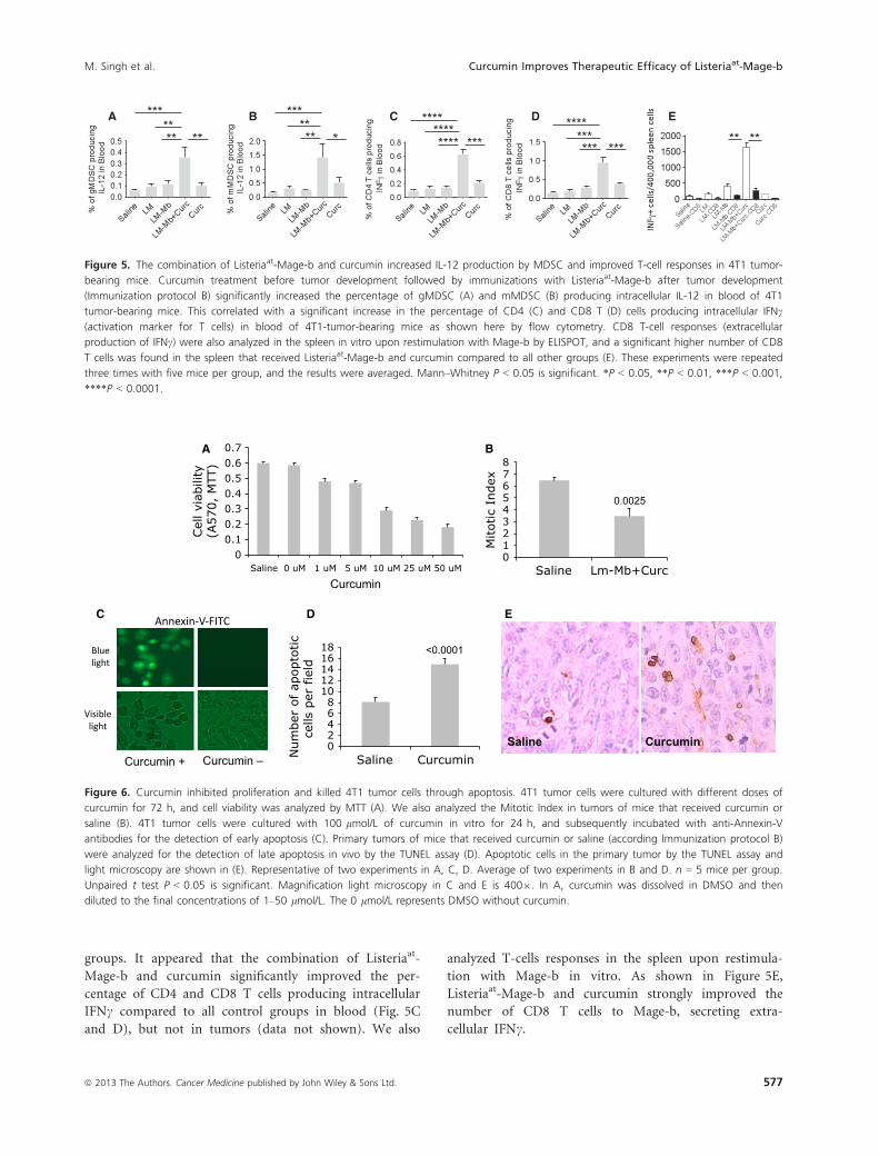
groups. It appeared that the combination of Listeriaat-
Mage-b and curcumin significantly improved the per-
centage of CD4 and CD8 T cells producing intracellular
IFNc compared to all control groups in blood (Fig. 5C
and D), but not in tumors (data not shown). We also
analyzed T-cells responses in the spleen upon restimula-
tion with Mage-b in vitro. As shown in Figure 5E,
Listeriaat-Mage-b and curcumin strongly improved the
number of CD8 T cells to Mage-b, secreting extra-
cellular IFNc.
012345678
Saline Lm-Mb+Curc
Mitotic
Index
0.0025
Saline Curcumin 02468
1012141618
Saline CurcuminNum
ber
of ap
opto
tic
cells
per
fie
ld <0.0001
00.10.20.30.40.50.60.7
Saline 0 uM 1 uM 5 uM 10 uM 25 uM 50 uM
Cel
l vi
abili
ty(A
570,
MTT
)
Curcumin
Curcumin + Curcumin –
A B
C D E
Figure 6. Curcumin inhibited proliferation and killed 4T1 tumor cells through apoptosis. 4T1 tumor cells were cultured with different doses of
curcumin for 72 h, and cell viability was analyzed by MTT (A). We also analyzed the Mitotic Index in tumors of mice that received curcumin or
saline (B). 4T1 tumor cells were cultured with 100 lmol/L of curcumin in vitro for 24 h, and subsequently incubated with anti-Annexin-V
antibodies for the detection of early apoptosis (C). Primary tumors of mice that received curcumin or saline (according Immunization protocol B)
were analyzed for the detection of late apoptosis in vivo by the TUNEL assay (D). Apoptotic cells in the primary tumor by the TUNEL assay and
light microscopy are shown in (E). Representative of two experiments in A, C, D. Average of two experiments in B and D. n = 5 mice per group.
Unpaired t test P < 0.05 is significant. Magnification light microscopy in C and E is 4009. In A, curcumin was dissolved in DMSO and then
diluted to the final concentrations of 1–50 lmol/L. The 0 lmol/L represents DMSO without curcumin.
A EDCB
*
***
** **
**
*** ** **
**** **** **** ***
**** *** *** ***
** **
Figure 5. The combination of Listeriaat-Mage-b and curcumin increased IL-12 production by MDSC and improved T-cell responses in 4T1 tumor-
bearing mice. Curcumin treatment before tumor development followed by immunizations with Listeriaat-Mage-b after tumor development
(Immunization protocol B) significantly increased the percentage of gMDSC (A) and mMDSC (B) producing intracellular IL-12 in blood of 4T1
tumor-bearing mice. This correlated with a significant increase in the percentage of CD4 (C) and CD8 T (D) cells producing intracellular IFNc
(activation marker for T cells) in blood of 4T1-tumor-bearing mice as shown here by flow cytometry. CD8 T-cell responses (extracellular
production of IFNc) were also analyzed in the spleen in vitro upon restimulation with Mage-b by ELISPOT, and a significant higher number of CD8
T cells was found in the spleen that received Listeriaat-Mage-b and curcumin compared to all other groups (E). These experiments were repeated
three times with five mice per group, and the results were averaged. Mann–Whitney P < 0.05 is significant. *P < 0.05, **P < 0.01, ***P < 0.001,
****P < 0.0001.
ª 2013 The Authors. Cancer Medicine published by John Wiley & Sons Ltd. 577
M. Singh et al. Curcumin Improves Therapeutic Efficacy of Listeriaat-Mage-b

Curcumin inhibited proliferation of tumorcells and killed tumor cells throughapoptosis
Several reports describe that curcumin inhibits prolifera-
tion and kills tumor cells through apoptosis, including
breast tumor cells [34–36]. We found that curcumin inhib-
ited the growth of 4T1 tumor cells in vitro (Fig. 6A), and
mitosis of the tumor cells in vivo (Fig. 6B). In addition,
we found that curcumin killed tumor cells through
apoptosis in vitro as shown by Annexin-V (early apopto-
sis) (Fig. 6C), and in the primary tumors in vivo as
shown by the TUNEL assay (late apoptosis) (Fig. 6D). A
representative example of apoptotic cells by the TUNEL
assay is shown in Figure 6E.
Listeriaat-Mage-b is nonpathogenic andcurcumin is nontoxic
In a previous study we have shown that Listeriaat-Mage-b
is nonpathogenic [37], while curcumin, consumed through
food all over the world, is nontoxic [32]. However, the
combination of Listeriaat-Mage-b and curcumin has never
been tested. Here, we demonstrate by pathological exami-
nation of various normal tissues (as kidney, heart, lungs,
liver, and spleen) in tumor-bearing mice that the combi-
nation of Listeriaat-Mage-b and curcumin is nonpatho-
genic and nontoxic, but primarily activated the innate
immune system. Most obvious was the increased extra-
medullary myeloid hematopoiesis in the spleen and liver of
mice that received Listeriaat-Mage-b and curcumin com-
pared to the saline group. An example of extramedullary
myeloid hematopoiesis in the liver is shown in Figure S2.
An overview of pathological analysis of normal tissues of
tumor-bearing mice that received Listeriaat-Mage-b and
curcumin is shown in Table S1.
Discussion
Patients with TNBC have the poorest prognosis. One of
the main problems of current therapies against TNBC is
their inability to target metastases and their high toxicity.
They do not respond to therapies that target ER, PR, and
HER2/neu because their tumors lack the expression of
these receptors/molecules, and other types of therapies
such as tyrosine kinase inhibitor sunitinib, targeting vas-
cular endothelial growth factor (VEGF), or therapies tar-
geting c-kit or Flt2, or bevacizumab, a human antibody
to VGEF [38–42], are under investigation but with mod-
erate success. In the study presented here, we developed
two nontoxic vaccination strategies in a preclinical TNBC
mouse model 4T1. We demonstrated that three therapeu-
tic vaccinations with a highly attenuated nonpathogenic
Listeriaat-based vaccine, expressing TAA Mage-b, and
nontoxic curcumin significantly reduced the number of
metastases compared to Listeriaat-Mage-b or curcumin
alone. Curcumin alone had no significant effect on the
primary tumors or metastases. Others described that
curcumin killed tumor cells in vitro [43–47]. However,
tumor cells may react differently to curcumin in vitro
than in vivo because in vitro bioavailability and the
immune system do not play a role and higher concentra-
tions can be obtained in vitro compared to the in vivo
situation. Also, the time point of administering curcumin,
the concentration of curcumin, and the type of cancer
may determine the antitumor effect of curcumin. For
instance, others reported that in the Lewis Lung model,
curcumin was not effective against metastases and that
the time point of administration of curcumin was impor-
tant [48].
We also tested three administrations of curcumin
before tumor development followed by three immuniza-
tions with Listeriaat-Mage-b after tumor development.
This immunization protocol was slightly but significantly
more effective against the metastases compared to
Listeriaat-Mage-b and curcumin both after tumor develop-
ment. Most interestingly, curcumin alone significantly
reduced the number of metastases and tumor growth, in
contrast to administering curcumin after tumor develop-
ment. These results suggest that consuming curcumin
before cancer develops may provide an advantage over
consuming curcumin after cancer develops in the battle
against metastatic breast cancer.
Curcumin is known for reducing the production of IL-6
[49, 50]. Here, we demonstrate that curcumin significantly
reduced the production of IL-6 in vivo in the primary
tumors (tumor cell lysates), and in MDSC of blood and
primary tumors. Also Listeriaat-Mage-b reduced the
production of IL-6 significantly in the tumor primary
tumors, but IL-6 production was even more reduced by the
combination of Listeriaat-Mage-b and curcumin.
MDSC are important regulators of the immune system
in the TME [5, 6], and therefore became one of our
most important targets in this study. As mentioned
above, curcumin reduced the production of IL-6 signifi-
cantly in MDSC in blood and primary tumors. To our
surprise, the combination of Listeriaat-Mage-b and
curcumin significantly increased the production of IL-12
in gMDSC and mMDSC in blood (but not in tumors).
It has been reported that IL-12 activates na€ıve and
mature CD4 and CD8 T cells [51, 52], which may have
happened in this study as well. An interesting observa-
tion was that the combination of Listeriaat-Mage-b and
curcumin significantly reduced the number of MDSC
(predominantly gMDSC) in blood of the TNBC model
4T1. Various factors may have played a role here. It is
578 ª 2013 The Authors. Cancer Medicine published by John Wiley & Sons Ltd.
Curcumin Improves Therapeutic Efficacy of Listeriaat-Mage-b M. Singh et al.

possible that MDCS infected with Listeriaat-Mage-b
became a target for Listeriaat- and Mage-b-specific T-cell
and perhaps NK-cell responses because the combination
therapy improved these immune responses to Listeriaat
and Mage-b by reducing IL-6, and increasing IL-12 pro-
duction. As Listeriaat [37] and curcumin kill 4T1 tumor
cells directly (this study), it is also possible that the
combination therapy prevented the tumor cells from
growing in the early phase of treatment, and conse-
quently prevented migration of the MDSC to the TME.
We found that curcumin alone decreased the percentage
of MDSC in blood (although this effect was much stron-
ger when Listeriaat-Mage-b was combined with curcu-
min). Reduction in the percentage of MDSC by
curcumin was also found by others in a xenograft model
of colon cancer [53]. They concluded that reduction in
IL-6 production by curcumin reduced the mobilization
of MDSC to the primary tumors. Others found that
activated T cells might express Fas ligand and induce
apoptosis of Fas+ MDSC [54]. In conclusion, various
pathways may lead to the reduction in MDSC and more
analysis is required.
The decrease in IL-6 and increase in IL-12 production,
the improved CD4 and CD8 T-cell responses in blood
and spleen, and the dramatic reduction in the number of
metastases by the combination therapy strongly suggest
that T-cell responses contributed to the effect on the
metastases. However, this strong reduction by the combi-
nation therapy is not only an effect of Mage-b-specific
T-cell responses. As shown previously, Listeriaat exhibits
several pathways to kill tumor cells; that is, Listeriaat
infects tumor cells in vivo and in vitro, and kills tumor
cells directly through high levels of reactive oxygen spe-
cies (ROS) [37]. Moreover, we have shown that Listeriaat-
activated CD8 T cells eliminated Listeriaat-infected tumor
cells in vivo [37]. In addition, we have shown that curcu-
min kills 4T1 tumor cells through apoptosis (this study).
Therefore, it is most likely that the synergistic effects of the
multiple pathways of Listeriaat-Mage-b and curcumin as
described above, are responsible for the overall strong thera-
peutic effect on the metastases in this TNBC model 4T1.
The therapeutic effect of the combination therapy was
strong but less pronounced on the primary tumors com-
pared to the metastases. It is possible that the production
of IL-6 was not sufficiently reduced in the primary
tumors (IL-6 was reduced in the tumor cell lysates by
~65% by Listeriaat-Mage-b and curcumin treatment), and
other inhibitory cytokines likes TGFb, which is highly
produced by 4T1 tumor cells [55], may play here a role
as well. However, most primary tumors can be removed
by surgery, radiation, or chemotherapy, while metastases
are unresectable and usually chemoresistant despite
aggressive and toxic follow-up [56].
The highly attenuated Listeriaat of this study is non-
pathogenic, and are naturally cleared by the immune
system within 3–5 days [37], which is different from
wild type Listeriaat that multiplies in hepatocytes in the
liver or epithelial cells of the gastrointestinal tract [57,
58]. Moreover, the side effects of the combination ther-
apy of Listeriaat-Mage-b and curcumin in the 4T1 model
were minimal; that is, primarily induction of inflamma-
tory responses in the liver and spleen and no significant
findings were observed in other normal tissues such as
heart, lungs, and kidneys. Therefore, Listeriaat-Mage-b
and curcumin may be of value as a nontoxic adjuvant
therapy, to prevent the development of metastases in
TNBC patients that produce IL-6 and express MAGE.
This study may be a platform for improvement of other
cancer vaccines by curcumin and against other IL-6-pro-
ducing cancers.
Acknowledgments
This work was supported by NCI grant 1R21CA129470-
01A1, NIA/NCI grant 1RO1 AG023096-01, and Reliable
Cancer Therapies (RCT). We like to thank Lydie Tesfa of
the FACS Facility Core of Einstein and Lisa Scandiuzzi of
Xingxing Zang’s laboratory for their excellent assistance
with the flow cytometry.
Conflict of Interest
Bharat Aggarwal has filed several patents with respect to
curcumin. US classification: 514/679; International classi-
fication: A61K3112; issued 6 April 1999.
References
1. Stead, L. A., T. L. Lash, J. E. Sobieraj, D. D. Chi, J. L.
Westrup, M. Charlot, et al. 2009. Triple-negative
breast cancers are increased in black women
regardless of age or body mass index. Breast Cancer Res.
11:R18.
2. Dent, R., M. Traudeau, K. I. Pritchard, W. H. Hanna,
H. K. Kahn, C. A. Sawka, et al. 2007. Triple-negative
breast cancer: clinical features and patterns of recurrence.
Clin. Cancer Res. 13:4429–4434.
3. Pegram, M. D., A. Lipton, D. F. Hayes, B. L. Weber,
J. M. Baselga, D. Tripathy, et al. 1998. Phase II study
of receptor-enhanced chemosensitivity using
recombinant humanized anti-p185(HER2/neu)
monoclonal antibody plus cisplatin in patients with
HER2/neu-overexpressing metastatic breast cancer
refractory to chemotherapy treatment. J. Clin. Oncol.
16:2659–2671.
4. Lehmann, B. D., J. A. Bauer, X. Chen, M. E. Sanders, A. B.
Chakravarthy, Y. Shyr, et al. 2011. Identification of human
ª 2013 The Authors. Cancer Medicine published by John Wiley & Sons Ltd. 579
M. Singh et al. Curcumin Improves Therapeutic Efficacy of Listeriaat-Mage-b

triple-negative breast cancer subtypes and preclinical
models for selection of targeted therapies. J. Clin. Invest.
121:2750–2767.
5. Gabrilovich, D. I., and S. Nagaraj. 2009. Myeloid-derived
suppressor cells as regulators of the immune system. Nat.
Rev. Immunol. 9:162–174.
6. Ostrand-Rosenberg, S., and P. Sinha. 2009. Myeloid-
derived suppressor cells: linking inflammation and cancer.
J. Immunol. 182:4499–4506.
7. Gajewski, T. F., Y. Meng, and H. Harlin. 2006. Immune
suppression in the tumor microenvironment. J.
Immunother. 29:233–240.
8. Curiel, T. J. 2007. Tregs and rethinking cancer
immunotherapy. J. Clin. Invest. 117:1167–1174.
9. Suarez-Cuervo, C., K. W. Harris, L. Kallman, H. K.
Vaananen, and K. S. Selander. 2003. Tumor necrosis
factor-alpha induces interleukin-6 production via
extracellular-regulated kinase 1 activation in breast cancer
cells. Breast Cancer Res. Treat. 80:71–78.
10. Berishaj, M., S. P. Gao, S. Ahmed, K. Leslie, H. Al-
Ahmadle, W. L. Gerald, et al. 2007. Stat3 is tyrosine-
phosphorylated through the interleukin-6/glycoprotein
130/Janus kinase pathway in breast cancer. Breast Cancer
Res. 9:R32.
11. Deng, X. S., S. Wang, A. Deng, B. Liu, S. M. Edgerton,
S. E. Lind, et al. 2012. Metformin targets Stat3 to inhibit
cell growth and induce apoptosis in triple-negative breast
cancers. Cell Cycle 11:367–376.
12. Idowu, M. O., M. Kmieciak, C. Dunur, R. S. Burton, M. M.
Grimes, C. N. Powers, et al. 2012. CD44(+)/CD24(�/low)
cancer stem/progenitor cells are more abundant in triple-
negative invasive breast carcinoma phenotype and are
associated with poor outcome. Hum. Pathol. 43:364–373.
13. Liu, S., and M. S. Wicha. 2010. Targeting breast cancer
stem cells. J. Clin. Oncol. 28:4006–4012.
14. Rosen, J. M., and C. T. Jordan. 2009. The increasing
complexity of the cancer stem cell paradigm. Science
324:1670–1673.
15. Frank, N. Y., T. Schatton, and M. H. Frank. 2010. The
therapeutic promise of the cancer stem cell concept. J.
Clin. Invest. 120:41–50.
16. Marotta, L. L. C., V. Almendo, A. Marusyk, M. Shipitsin,
J. Schemme, S. R. Walker, et al. 2011. The JAK2/STAT3
signaling pathway is required for growth of CD44(+)CD24(�) stem cell-like breast cancer cells in human tumors.
J. Clin. Invest. 121:2723–2735.
17. Park, S. J., T. Nakagawa, H. Kitamura, T. Atsumi, H.
Kamon, S. I. Sawa, et al. 2004. IL-6 regulates in vivo
dendritic cell differentiation through STAT3 activation.
J. Immunol. 173:3844–3854.
18. Xie, J., J. Qian, S. Wang, M. E., III Freeman, J. Epstein,
and Q. Yi. 2003. Novel and detrimental effects of
lipopolysaccharide on in vitro generation of immature
dendritic cells: involvement of mitogen-activated protein
kinase p38. J. Immunol. 171:4792–4800.
19. Gravekamp, C., B. Leal, A. Denny, R. Bahar, S. Lampkin,
S. H. Kim, et al. 2008. In vivo responses to vaccination
with Mage-b, GM-CSF and thioglycollate in a highly
metastatic mouse breast tumor model, 4T1. Cancer
Immunol. Immunother. 57:1067–1077.
20. Kim, S. H., F. Castro, Y. Paterson, and C. Gravekamp.
2008. Mage-b vaccine delivered by recombinant Listeria
monocytogenes is highly effective against breast cancer
metastases. Br. J. Cancer 99:741–749.
21. Wilken, R., M. S. Veena, M. B. Wang, and E. S. Srivatsan.
2011. Curcumin: a review of anti-cancer properties and
therapeutic activity in head and neck squamous cell
carcinoma. Mol. Cancer 10:12.
22. Aggarwal, B. B., S. Shishodia, Y. Takada, S. Banerjee, R. A.
Newman, C. E. Bueso-Ramos, et al. 2005. Curcumin
suppresses the paclitaxel-induced nuclear factor-kappaB
pathway in breast cancer cells and inhibits lung metastasis
of human breast cancer in nude mice. Clin. Cancer Res.
11:7490–7498.
23. Odot, J., P. Albert, A. Carlier, M. Tarpin, and C.
Madoulet. 2004. In vitro and in vivo anti-tumoral effect of
curcumin against melanoma cells. Int. J. Cancer 111:381–
387.
24. Bhattacharyya, S., D. Mandal, B. Saha, G. S. Sen, T. Das,
and G. Sa. 2007. Curcumin prevents tumor-induced T cell
apoptosis through Stat-5a-mediated Bcl-2 induction. J.
Biol. Chem. 282:15954–15964.
25. De Backer, O., A. M. Verheyden, B. Martin, D. Godelaine,
E. de Plean, R. Brasseur, et al. 1995. Structure,
chromosomal location, and expression pattern of three
mouse genes homologous to the human MAGE genes.
Genomics 28:74–83.
26. Curigliano, G., G. Viale, M. Ghioni, A. A. Jungbluth, V.
Bagnardi, G. C. Spagnoli, et al. 2011. Cancer-testis antigen
expression in triple-negative breast cancer. Ann. Oncol.
22:98–103.
27. Aslakson, C. J., and F. R. Miller. 1992. Selective events in
the metastatic process defined by analysis of the sequential
dissemination of subpopulations of a mouse mammary-
tumor. Cancer Res. 52:1399–1405.
28. Gunn, G., A. Zubair, C. Peters, Z. K. Pan, T. C. Wu,
and Y. Paterson. 2001. Two Listeria monocytogenes
vaccine vectors that express different molecular forms of
human papilloma virus-16 (HPV-16) E7 induce
qualitatively different T cell immunity that correlates
with their ability to induce regression of established
tumors immortalized by HPV-16. J. Immunol. 167:6471–
6479.
29. Singh, R., M. E. Dominiecki, E. M. Jaffee, and Y. Paterson.
2005. Fusion to Listeriolysin O and delivery by Listeria
monocytogenes enhances the immunogenicity of HER-2/
580 ª 2013 The Authors. Cancer Medicine published by John Wiley & Sons Ltd.
Curcumin Improves Therapeutic Efficacy of Listeriaat-Mage-b M. Singh et al.

neu and reveals subdominant epitopes in the FVB/N
mouse. J. Immunol. 175:3663–3673.
30. Shoba, G., D. Joy, T. Joseph, M. Majeed, R. Rajendran,
and P. S. S. R. Srinivas. 1998. Influence of piperine on the
pharmacokinetics of curcumin in animals and human
volunteers. Planta Med. 64:353–356.
31. Atal, C. K., R. K. Dubey, and J. Singh. 1985. Biochemical
basis of enhanced drug bioavailability by piperine –
evidence that piperine is a potent inhibitor of drug-
metabolism. J. Pharmacol. Exp. Ther. 232:258–262.
32. Anand, P., A. B. Kunnumakkara, R. A. Newman, and B. B.
Aggarwal. 2007. Bioavailability of curcumin: problems and
promises. Mol. Pharm. 4:807–818.
33. Castro, F., B. Leal, A. Denny, R. Bahar, S. Lampkin, R.
Reddick, et al. 2009. Vaccination with Mage-b DNA
induces CD8 T-cell responses at young but not old age in
mice with metastatic breast cancer. Br. J. Cancer 101:1329–
1337.
34. Choudhuri, T., S. Pal, M. L. Agwarwal, T. Das, and G. Sa.
2002. Curcumin induces apoptosis in human breast cancer
cells through p53-dependent Bax induction. FEBS Lett.
512:334–340.
35. Karunagaran, D., R. Rashmi, and T. R. Kumar. 2005.
Induction of apoptosis by curcumin and its implications
for cancer therapy. Curr. Cancer Drug Targets 5:117–129.
36. Anto, R. J., A. Mukhopadhyay, K. Denning, and B. B.
Aggarwal. 2002. Curcumin (diferuloylmethane) induces
apoptosis through activation of caspase-8, BID cleavage
and cytochrome c release: its suppression by ectopic
expression of Bcl-2 and Bcl-xl. Carcinogenesis 23:143–150.
37. Kim, S. H., F. Castro, Y. Paterson, and C. Gravekamp. 2009.
High efficacy of a Listeria-based vaccine against metastatic
breast cancer reveals a dual mode of action. Cancer Res.
69:5860–5866.
38. Mukai, H. 2010. Targeted therapy in breast cancer: current
status and future directions. Jpn. J. Clin. Oncol. 40:711–716.
39. Bernard-Marty, C., F. Lebrun, A. Awada, and M. J.
Piccart. 2006. Monoclonal antibody-based targeted therapy
in breast cancer: current status and future directions.
Drugs 66:1577–1591.
40. Agus, D. B., M. S. Gordon, C. Taylor, R. B. Natale, B.
Karlan, D. S. Lieberman, et al. 2005. Phase I clinical study
of pertuzumab, a novel HER dimerization inhibitor, in
patients with advanced cancer. J. Clin. Oncol. 23:2534–
2543.
41. Lin, N. U., L. A. Minetta, J. Younger, S. E. Come, M.
Ewend, G. J. Gordon, et al. 2008. Phase II trial of lapatinib
for brain metastases in patients with human epidermal
growth factor receptor 2-positive breast cancer. J. Clin.
Oncol. 26:1993–1999.
42. Barrios, C. H., M. C. Liu, S. H. Lee, L. Vanlemmens, J. M.
Ferrero, T. Tabei, et al. 2010. Phase III randomized trial of
sunitinib versus capecitabine in patients with previously
treated HER2-negative advanced breast cancer. Breast
Cancer Res. Treat. 121:121–131.
43. Kim, M. S., H. J. Kang, and A. Moon. 2001. Inhibition of
invasion and induction of apoptosis by curcumin in
H-ras-transformed MCF10A human breast epithelial cells.
Arch. Pharm. Res. 24:349–354.
44. Shao, Z. M., Z. Z. Shen, C. H. Liu, M. R. Sartippour, V.
L. Go, and M. Nguyen. 2002. Curcumin exerts multiple
suppressive effects on human breast carcinoma cells. Int. J.
Cancer 98:234–240.
45. Ramachandran, C., S. Rodriguez, R. Ramachandran, P. K.
R. Nair, H. Fonseca, Z. Khatib, et al. 2005. Expression
profiles of apoptotic genes induced by curcumin in human
breast cancer and mammary epithelial cell lines.
Anticancer Res. 25:3293–3302.
46. Bachmeier, B. E., I. V. Mohrenz, V. Mirisola, E. Schleicher,
F. Romeo, C. Hohneke, et al. 2008. Curcumin
downregulates the inflammatory cytokines CXCL1 and -2
in breast cancer cells via NFkappaB. Carcinogenesis
29:779–789.
47. Zong, H., F. Wang, Q. X. Fan, and L. X. Wang. 2012.
Curcumin inhibits metastatic progression of breast cancer
cell through suppression of urokinase-type plasminogen
activator by NF-kappa B signaling pathways. Mol. Biol.
Rep. 39:4803–4808.
48. Yan, L. 2013. Dietary supplementation with curcumin
enhances metastatic growth of Lewis lung carcinoma in
mice. Int. J. Cancer 132:269–275.
49. Chakravarti, N., J. N. Myers, and B. B. Aggarwal. 2006.
Targeting constitutive and interleukin-6-inducible signal
transducers and activators of transcription 3 pathway in
head and neck squamous cell carcinoma cells by
curcumin (diferuloylmethane). Int. J. Cancer 119:1268–
1275.
50. Bharti, A. C., N. Donato, and B. B. Aggarwal. 2003.
Curcumin (diferuloylmethane) inhibits constitutive and
IL-6-inducible STAT3 phosphorylation in human multiple
myeloma cells. J. Immunol. 171:3863–3871.
51. Wolf, S. F., D. Sieburth, and J. Sypek. 1994. Interleukin
12: a key modulator of immune function. Stem Cells
12:154–168.
52. Valenzuela, J., C. Schmidt, and M. Mescher. 2002. The
roles of IL-12 in providing a third signal for clonal
expansion of naive CD8 T cells. J. Immunol. 169:6842–
6849.
53. Tu, S. P., H. Jin, J. D. Shi, L. M. Zhu, Y. Suo, G. Lu, et al.
2012. Curcumin induces the differentiation of myeloid-
derived suppressor cells and inhibits their interaction with
cancer cells and related tumor growth. Cancer Prev. Res.
5:205–215.
54. Sinha, P., O. Chornoguz, V. K. Clements, K. A.
Artemenko, R. A. Zubarev, and S. Ostrand-Rosenberg.
2011. Myeloid-derived suppressor cells express the death
ª 2013 The Authors. Cancer Medicine published by John Wiley & Sons Ltd. 581
M. Singh et al. Curcumin Improves Therapeutic Efficacy of Listeriaat-Mage-b

receptor Fas and apoptose in response to T cell-expressed
FasL. Blood 117:5381–5390.
55. Chakrabarti, R., V. Subramaniam, S. Abdalla, S. Jothy,
and G. J. Prud’homme. 2009. Tranilast inhibits the
growth and metastasis of mammary carcinoma.
Anticancer Drugs 20:334–345.
56. Pardal, R., M. F. Clarke, and S. J. Morrison. 2003.
Applying the principles of stem-cell biology to cancer. Nat.
Rev. Cancer 3:895–902.
57. Racz, P., K. Tenner, and E. Mero. 1972. Experimental
Listeria enteritis. I. An electron microscopic study of the
epithelial phase in experimental listeria infection. Lab.
Invest. 26:694–700.
58. Rosen, H., and S. Gordon. 1987. Monoclonal antibody
to the murine type 3 complement receptor inhibits
adhesion of myelomonocytic cells in vitro and
inflammatory cell recruitment in vivo. J. Exp. Med.
166:1685–1701.
Supporting Information
Additional Supporting Information may be found in the
online version of this article:
Figure S1. Significant reduction in the tumor size by
preventive administration of curcumin followed by thera-
peutic immunization with Listeriaat-Mage-b in 4T1
tumor-bearing mice.
Figure S2. The combination therapy of Listeriaat-Mage-b
and curcumin is non-pathogenic and non-toxic.
Figure S3. The effect of Listeriaat-Mage-b and curcumin
on MDSC in 4T1 tumor-bearing mice (Flow cytometry
profile).
Table S1. Histological examination of tissues after thera-
peutic treatment with Listeriaat-Mage-b and curcumin.
582 ª 2013 The Authors. Cancer Medicine published by John Wiley & Sons Ltd.
Curcumin Improves Therapeutic Efficacy of Listeriaat-Mage-b M. Singh et al.


![Myeloid derived-suppressor cells: their role in cancer and ...digitalmeasures.umbc.edu/dmeasures/xn95525... · [2,3,4 ,5], so only a brief overview of MDSC character-istics and function](https://img.pdfslide.net/doc/110x75/5f27de4697630f5cc77dd3c2/myeloid-derived-suppressor-cells-their-role-in-cancer-and-234-5-so-only.jpg)