Embed Size (px)
Citation preview

Hindawi Publishing CorporationOxidative Medicine and Cellular LongevityVolume 2012, Article ID 232464, 10 pagesdoi:10.1155/2012/232464
Review Article
Cell Stress Proteins in Atherothrombosis
Julio Madrigal-Matute, Roxana Martinez-Pinna,Carlos Ernesto Fernandez-Garcia, Priscila Ramos-Mozo, Elena Burillo,Jesus Egido, Luis Miguel Blanco-Colio, and Jose Luis Martin-Ventura
Vascular Research Laboratory, IIS-Fundacion Jimenez Dıaz, Universidad Autonoma de Madrid, 28040 Madrid, Spain
Correspondence should be addressed to Julio Madrigal-Matute, [email protected]
Received 30 March 2012; Accepted 14 May 2012
Academic Editor: Ana Fortuno
Copyright © 2012 Julio Madrigal-Matute et al. This is an open access article distributed under the Creative Commons AttributionLicense, which permits unrestricted use, distribution, and reproduction in any medium, provided the original work is properlycited.
Cell stress proteins (CSPs) are a large and heterogenous family of proteins, sharing two main characteristics: their levels and/orlocation are modified under stress and most of them can exert a chaperon function inside the cells. Nonetheless, they are alsoinvolved in the modulation of several mechanisms, both at the intracellular and the extracellular compartments. There are morethan 100 proteins belonging to the CSPs family, among them the thioredoxin (TRX) system, which is the focus of the presentpaper. TRX system is composed of several proteins such as TRX and peroxiredoxin (PRDX), two thiol-containing enzymes thatare key players in redox homeostasis due to their ability to scavenge potential harmful reactive oxygen species. In addition to theirmain role as antioxidants, recent data highlights their function in several processes such as cell signalling, immune inflammatoryresponses, or apoptosis, all of them key mechanisms involved in atherothrombosis. Moreover, since TRX and PRDX are present inthe pathological vascular wall and can be secreted under prooxidative conditions to the circulation, several studies have addressedtheir role as diagnostic, prognostic, and therapeutic biomarkers of cardiovascular diseases (CVDs).
1. Introduction
The vast majority of the proteins require further assistancefor acquiring proper maturation and stability; this processdescribed in the late 80s is facilitated through the activityof a family of proteins called “molecular chaperones” or cellstress proteins (CSPs) [1]. This large and diverse group ofproteins composed by more than 20 families of proteins andmore than 100 proteins includes the heat shock proteins(HSPs) and the thioredoxin (TRX) system. The role ofHSPs in cardiovascular diseases (CVDs) has been thoroughlyreviewed [2]. In the present paper, we will focus on therole of the TRX system, specifically TRX and peroxiredoxins(PRDXs), in atherothrombosis. For this purpose, we havefollowed the PRISMA chart displayed in SupplementaryFigure 1 (see Suppplementary Figure 1 in supplementarymaterial available online at doi:10.1155/2012/232464).
2. The Thioredoxin System
The TRX system mainly comprises TRX, TRX reductase,TRX interacting protein ((TXNIP), vitamin D3-upregulated
protein-1 ((VDUP)-1) or TRX-binding protein (TRXBP)),and the PRDXs [3]. The TRX system is involved in proteinassembly and plays a key role in cellular redox maintenance.
Under physiological conditions, intra- and extracellularreactive oxygen species (ROS) modulate metabolic, signal-ing, and transcriptional processes within the cell. However,pathological dysregulation of the redox balance could con-tribute to CVDs [4, 5]. Cellular redox homeostasis is tightlyregulated by the coordinated action of NADPH oxidases, theTRX system and glutathione (GSH) [6], among others. TheTRX system and GSH are thiol reduction systems with akey role in the defense against excessive ROS production,as well as in the modulation of signaling processes suchas inflammation, cellular proliferation, and apoptosis [7–9].These molecules maintain the intracellular milieu in areduced state. GSH is used by the GSH peroxidase to reduceperoxides, producing oxidized GSH (GSSG) while GSHreductase reduces this oxidized form to GSH. The antiox-idant properties of TRX result from PRDX action, whichrecycle H2O2 through reduction of several hydroperoxidesinto water and alcohol (Figure 1).
2 Oxidative Medicine and Cellular Longevity
TXNIP
PRDXox PRDXred
NADPH
GSH
GR
GSSG
SOD
NADPH
Extracellular medium
Cytoplasm
Plasma membrane
TRXR
GPx
NADPHoxidase
O2
·O2− H2O2
H2O + O2
H2O + O2
H+
NADP+
NADP+ NADP+ H+H+
TRX-SH2 TRX-S2
Figure 1: Schematic diagram showing the maintenance of the cellular redox homeostasis by the NADPH oxidase, GSH, and the TRXsystem. PRDXox-oxidized PRDX; PRDXred-reduced PRDX; TRX-S2-oxidized TRX; TRX-SH2-reduced TRX; GR-GSH reductase; GPx-GSHperoxidase.
Depending on their cellular location, TRX/PRDX mayexert different functions than their known chaperone andantioxidant activities. This process can be described withthe so-called “Moonlighting proteins” theory [10]. Thisidea supports the notion that one gene = one protein =one function is simple and old-fashioned because largenumber of proteins have two or more functions. This theorymight not apply to every protein but it seems to be rightfor TRX/PRDX. Under certain circumstances, mainly pro-oxidative conditions, TRX and PRDX could be released tothe extracellular milieu [11, 12] although their traffickingmechanisms are not yet fully described.
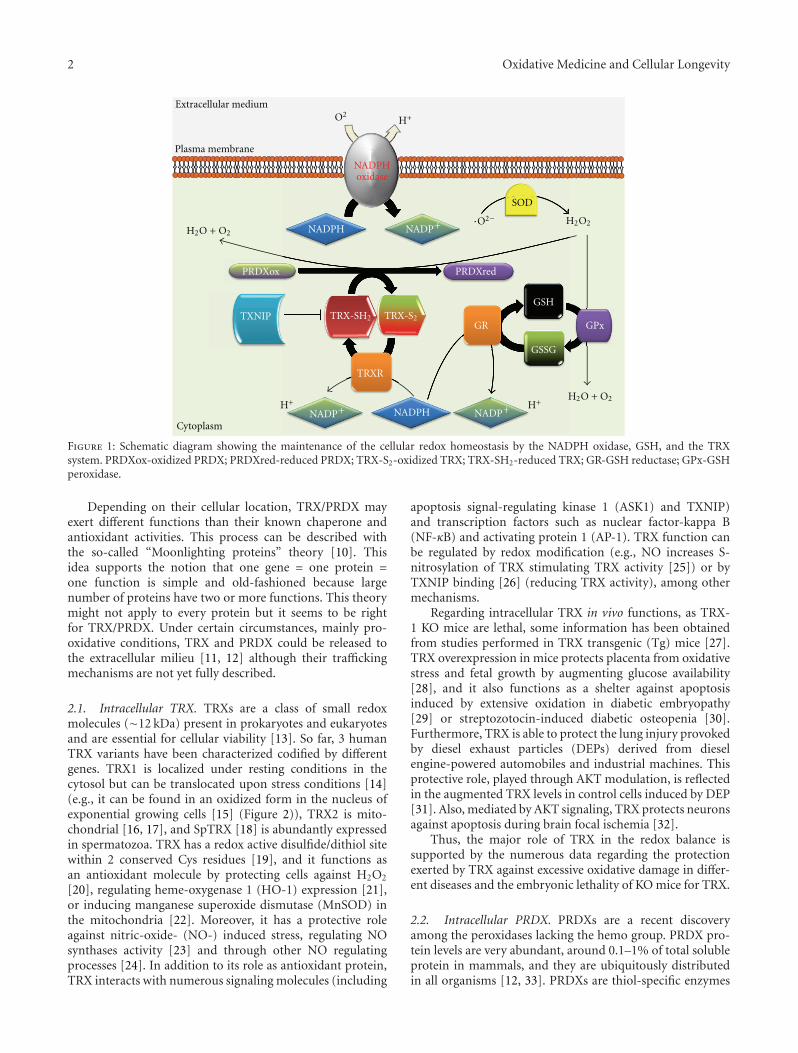
2.1. Intracellular TRX. TRXs are a class of small redoxmolecules (∼12 kDa) present in prokaryotes and eukaryotesand are essential for cellular viability [13]. So far, 3 humanTRX variants have been characterized codified by differentgenes. TRX1 is localized under resting conditions in thecytosol but can be translocated upon stress conditions [14](e.g., it can be found in an oxidized form in the nucleus ofexponential growing cells [15] (Figure 2)), TRX2 is mito-chondrial [16, 17], and SpTRX [18] is abundantly expressedin spermatozoa. TRX has a redox active disulfide/dithiol sitewithin 2 conserved Cys residues [19], and it functions asan antioxidant molecule by protecting cells against H2O2
[20], regulating heme-oxygenase 1 (HO-1) expression [21],or inducing manganese superoxide dismutase (MnSOD) inthe mitochondria [22]. Moreover, it has a protective roleagainst nitric-oxide- (NO-) induced stress, regulating NOsynthases activity [23] and through other NO regulatingprocesses [24]. In addition to its role as antioxidant protein,TRX interacts with numerous signaling molecules (including
apoptosis signal-regulating kinase 1 (ASK1) and TXNIP)and transcription factors such as nuclear factor-kappa B(NF-κB) and activating protein 1 (AP-1). TRX function canbe regulated by redox modification (e.g., NO increases S-nitrosylation of TRX stimulating TRX activity [25]) or byTXNIP binding [26] (reducing TRX activity), among othermechanisms.
Regarding intracellular TRX in vivo functions, as TRX-1 KO mice are lethal, some information has been obtainedfrom studies performed in TRX transgenic (Tg) mice [27].TRX overexpression in mice protects placenta from oxidativestress and fetal growth by augmenting glucose availability[28], and it also functions as a shelter against apoptosisinduced by extensive oxidation in diabetic embryopathy[29] or streptozotocin-induced diabetic osteopenia [30].Furthermore, TRX is able to protect the lung injury provokedby diesel exhaust particles (DEPs) derived from dieselengine-powered automobiles and industrial machines. Thisprotective role, played through AKT modulation, is reflectedin the augmented TRX levels in control cells induced by DEP[31]. Also, mediated by AKT signaling, TRX protects neuronsagainst apoptosis during brain focal ischemia [32].
Thus, the major role of TRX in the redox balance issupported by the numerous data regarding the protectionexerted by TRX against excessive oxidative damage in differ-ent diseases and the embryonic lethality of KO mice for TRX.
2.2. Intracellular PRDX. PRDXs are a recent discoveryamong the peroxidases lacking the hemo group. PRDX pro-tein levels are very abundant, around 0.1–1% of total solubleprotein in mammals, and they are ubiquitously distributedin all organisms [12, 33]. PRDXs are thiol-specific enzymes

Oxidative Medicine and Cellular Longevity 3
Cytoplasm
TXNIP
PRDX
TRX
Exosomes
Stress
TR
X80
Protease ?
Unstressed conditions
Cytoplasm
PRDX
TRX
NucleusNucleus
PRDX
PRDX
TRX
TRX
TRX
TRXTXNIP
?
TRX
?
Extracellular medium
TRXTRX
PRDX
TXNIP
Plasma membrane
PRDX
TRX
TR
X80
TRXTXNIP
Figure 2: Schematic diagram showing PRDX1/TRX main cellular location under nonstress conditions (left), under oxidative stress (right)and trafficking mechanisms (right).
lacking selenium, and they use their active sites to reduceperoxides and scavenge ROS [12]. Mammal cells express6 PRDX isoforms (PRDX 1–6): 1, 2, and 6 are cytosolic,and 3 and 5 are mitochondrial, while 4 is described as asecretory protein in most tissues [34]. Every isoform containsa conserved Cys residue, which is the primary site for H2O2
oxidation, and they can be further classified according totheir Cys residues (PRDX1-4 belongs to the 2-Cys subfamily,PRDX5 or atypical 2-Cys and PRDX6 or 1-Cys PRDX) [35].
PRDXs scavenge H2O2 more efficiently than other sys-tems such as catalase due to its higher affinity for H2O2
[36]. PRDX can modulate NADPH oxidase activity throughH2O2 inactivation [37]. In addition, PRDX can also reduceperoxynitrites levels through peroxynitrites reductases [38].Among the mechanisms modifying PRDX functions, severalposttranslational modifications have been described, suchas nitrosylation [39], glutathionylation [40], phosphoryla-tion, and the hyperoxidation of its active site [41], whichstimulates its chaperone activity [42]. For example, underlow H2O2 concentrations produced in conditions of cellularhomeostasis PRDX forms low-molecular-weight oligomers,exerting peroxidase activity. However, under significantchanges in H2O2 concentration, PRDX experiences struc-tural changes and forms high-molecular-weight oligomersand acquires chaperone activity [43, 44]. Similarly to TRX,PRDX interacts with several proteins (e.g., cyclophilin A,macrophage inhibitory factor, etc.) and can modulate thefunction of these binding proteins, in a dependent or inde-pendent manner of the PRX redox status [35].
Mice lacking PRDX1 are viable with a phenotype char-acterized by hemolytic anemia caused by an increased ROS
production by red blood cells (RBCs). Furthermore, PRDX1has tumor suppressor properties as PRDX1 KO mice show anincreased rate in malignant tumors as they age, which can beexplained by the excessive accumulation of damaged tissuedue to extreme ROS production [45]. Similarly to PRDX1KO mice, targeted disruption of PRDX2 causes cysteineoxidation of several proteins on RBCs membranes, whichfinally results in augmented levels of denatured protein,cell toxification, and hemolysis [46]. PRDX2 also has beenidentified as a tumor suppressor gene [47]. Accordingly,PRDX3 (also known as MER5) KO mice are characterizedby increased ROS production in macrophages and developmore severe lung injury upon Lipopolisaccharide (LPS)induction, possibly due to an excessive DNA and proteinoxidative damage and inflammatory cell infiltration [48]. Infact, it has been calculated that almost 90% of H2O2 targetsPRDX3 within the mitochondrial matrix, playing a majorrole in redox signaling in the mitochondria [49], protectingthe cells against apoptosis induced by excessive damageto mitochondrial macromolecules [50]. PRDX3 absence isalso involved in mitochondrial dysregulation associated withobesity, through increased protein carbonylation and ROSproduction [51]. It is noteworthy that PRDX3 KO adipocytesaccumulate more fat than wild type due to hypertrophy anddefects in the levels of enzymes implicated in glucose/lipidmetabolism [51]. PRDX4 is mostly a secretory protein, whileit is attached to the endoplasmic reticulum (ER) membraneof spermatogenic cells in mature testes. In these cells PRDX4protects from cell death through its antioxidant properties,nonetheless PRDX4 KO spermatozoa shows normal fertiliza-tion [52]. To our knowledge there is no PRDX5 KO strain,
4 Oxidative Medicine and Cellular Longevity
although it has been described that PRDX5 overexpressionprevents ROS production and p53-dependent apoptosis [53].Nevertheless, KO mice for PRDX6, the only 1-Cys member ofthe peroxiredoxin family, were more vulnerable to ischemicreperfusion injury as shown by increased infarct size andhigher amount oxidative stress [54]. In agreement, PRDX6overexpression functions as a shelter in mouse lungs againsttoxicity of hyperoxia [55].
On the whole, the plethora of data regarding thedifferent isoforms of PRDX demonstrate that every subunit,independently of its location, is a member of one of the majorcellular systems in charge of scavenging prooxidant speciesand thus in the maintenance of cellular redox status.
2.3. Extracellular TRX. TRX expression can be augmentedvery fast under stress and is secreted by normal and tumorcells although its secretion does not seem to follow a classicalGolgi apparatus pathway [11]. TRX location is regulatedby TXNIP binding [26] and facilitates TRX transport fromcytoplasm to the membrane under oxidative stress [14].Another mechanism that can be involved in TRX activesecretion is the exosomal pathway. Exosomes are smallmicroparticles released by cells upon activation or apoptosis.These vesicles have been implicated in thrombosis, diabetes,inflammation, atherosclerosis, and vascular cell proliferation[56]. Proteomic studies have described the presence of TRXin exosomes in B cells [57], bladder cancer cells [58],colorectal cancer cells [59], and urine [60] (Figure 2).
There is also a truncated form of TRX that correspondsto the last 80–84 amino acids from the N-terminal end,named TRX80, and it is present in plasma where it was firstlyidentified as a stimulating factor of eosinophils cytotoxicity[61]. It is possibly a result of protease activity but this processis still unknown (Figure 2). Recombinant TRX80 is a potentmitogenic cytokine for peripheral blood mononuclear cells(PBMCs), an effect not shared by TRX [62]. TRX80 differsfrom TRX because it forms a dimer lacking reductase activityand its activity is independent of the Cys residues fromthe TRX active site. The main cellular target for TRX80are PBMCs in which it induces a Th1 response via IL12production [63].
Extracellular TRX is present in the circulation and itslevels are increased under oxidative stress or inflammation[64] (Figure 2). TRX has been pointed out as a biomarkerin numerous oxidative and inflammatory diseases such asrheumatoid arthritis (RA) in which plasma TRX levels ofnormal subjects were significantly lower than those of RApatients and correlated with RA disease activity and C-reactive protein [65]. TRX levels were increased in patientswith systemic inflammatory stress syndrome (SIRS)/sepsiscompared to control subjects [66].
2.4. Extracellular PRDX. PRDX1 can be found inside theGolgi apparatus on endothelial cells (ECs) [67], and underphorbol 12-myristate 13-acetate (PMA) stimulation PRDX1is translocated to the cellular membrane [68], as alsoshowed for PRDX6 in polymorphonuclear cells (PMNs) [69](Figure 2). PRDX might also be secreted by lung cancer cells
through a nonclassical pathway [70, 71]. Nonetheless, theextracellular function of PRDX is still unknown. Unlike thewell-described function of intracellular PRDX1, membranePRDX6 helps in the maintenance of an optimal NADPHoxidase activity [69]. A number of chaperones, includingTRX and HSPs, are released by stressed or dying cells, actingas an endogenous warning system through binding of thesesignals to receptors on the outer membrane [72–75]. Most ofthese signals are recognized by Toll-like receptor 4 (TLR4)[74, 75]. Accordingly, PRDX1 binds TLR4 and stimulatesproinflammatory cytokine production in macrophages anddendritic cells, which suggests that PRDX could be actingas damage-associated molecular-pattern molecule (DAMP).Its trafficking seems to be dependent on PRDX binding toprotein kinase C (PKC) through microvesicles [76]. In fact,exosomes can be participating in active transport of PRDXsince proteomic studies have described PRDX in exosomesin B cells [57], bladder cancer cells [58], breast cancer cells[77], breast milk [78], colorectal cancer cells [59], and saliva[79] (Figure 2).
Thus, it is tempting to speculate that extracellular levelsof PRDX/TRX result from a cellular response to high oxidantconditions in the outer milieu.
3. TRX/PRDX in Atherothrombosis
Atherothrombosis is an immune-inflammatory disease, orig-inated by the subendothelial accumulation of LDLs, that canbe oxidized by ROS. Oxidative stress is involved not only inthe first stages of atherogenesis by modifying LDLs or NO,but also in later stages of atherothrombosis by modulatingthe expression of proteases that weakens the fibrous cap[80, 81]. ROS overproduction also produces direct damageto macromolecules such as lipids, nucleic acids, and proteins[82]. Furthermore, ROS can act as signaling molecules byinducing the activation of several cells from the vasculature.For example, through LDL oxydation and/or direct celltargeting, ROS can induce endothelial dysfunction andfurther leucocyte activation, deposition, and extravasation.In addition, ROS are clearly involved in the activation ofvascular smooth muscle cells (VSMCs) from the lesion andsustain foam cell formation. Thus, pathological ROS over-production is a main feature in atherogenesis and plaquerupture, which finally results in almost 70% of the clinicalevents [83].
Among the different systems involved in the redoxmaintenance in the vasculature, one of the most active is theTRX system. It is present in ECs [14, 84], VSMCs [85, 86],monocytes/macrophages [87, 88], RBCs [89, 90], and PMNs[90, 91]. Since TRX and PRDX are present in the atheroscle-rotic plaque and they can modulate different mechanismsinvolved in CVDs, several studies have addressed their roleas diagnostic, prognostic, and therapeutic biomarkers.
3.1. TRX. TRX is abundantly expressed in the vasculature,and its levels are increased under oxidative stress, possiblyas a response mechanism to augmented ROS production[19]. Besides, TRX expression in the endothelium and
Oxidative Medicine and Cellular Longevity 5
in macrophages is augmented in atherosclerotic patients[92] and in arteries damaged by the balloon model [93].More recently, TRX has been pointed as a possible markerfor unstable atherosclerotic plaques due to its associationwith oxidative stress and intraplaque hemorrhage in culpritlesions [94]. TRX reductase is as well overexpressed in ath-erosclerotic plaques, maybe synthesized by macrophagesengulfing oxLDLs [95].
The antioxidant effects of TRX are shown when overex-pressed in mouse hearts, protecting them from myocardialinfarction and displaying significantly improved postis-chemic ventricular recovery [96]. The positive effects of TRXin myocardial infarction are also due to its neoangiogenicproperties as shown in different murine models [97, 98].
On the other hand, TRX can function as a signalingmolecule by decreasing pressure-overload cardiac hypertro-phy [99], maybe through upregulation of miR-98 [100].However, there is a controversy about this matter becausealmost at the same time it was published another articlein which the authors described that activation of TRXparticipates in the development of pressure-overload cardiachypertrophy. In this respect, animals overexpressing TXNIPdeveloped less hypertrophy [101]. Furthermore, transverseaortic constriction increased TRX activity accompanied bya 40% reduction in TXNIP levels [101]. Whether variationsin TXNIP and TRX levels/activity reflect an increase inoxidative stress or they act as signaling molecules is still amatter to elucidate [102].
Recent studies have shown that extracellular TRX caninhibit interleukin-1 expression stimulated by LPS in mono-cyte-derived macrophages [103]. Besides, TRX1 administra-tion has beneficial effects on myosin-induced autoimmunemyocarditis through inhibition of inflammatory mediatorsand macrophage infiltration [104] and TRX1 also protectsfrom reperfusion-induced arrhythmias [105]. Furthermore,TRX administration has been also shown to be benefi-cial in cerebral ischemia/reperfusion injury reducing theinfarcted area through its antioxidant properties [106]. Asan additional support for the beneficial effects of TRX intherapeutics, it is to note that TRX-1 gene delivery protectshypertensive rats from myocardial infarction through HO-1/B-cell lymphoma 2 (BCL-2) [107].
Regarding cardiovascular diseases, TRX levels are ele-vated in plasma from atherothrombotic patients [108, 109],and high homocysteine plus low TRX is related to CADseverity [110]. Furthermore, TRX was elevated in patientsfollowing angioplasty, and there was an association withincreased TRX levels and decreased rate of restenosis atfollow-up angiography (6 months) [111]. We have recentlyreported an increase in serum TRX from abdominal aorticaneurysm (AAA) patients compared with control subjects.Besides, TRX correlates with AAA size and expansion ratewhich suggests that TRX could be a good biomarker of AAAevolution [91].
3.2. PRDX. PRDX expression can be modified by proox-idative stimulus such as LPS or the phorbol ester 12-O-tet-radecanoylphorbol- 13-acetate (TPA) [88, 112]. Attention to
PRDX as a major regulator of H2O2 homeostasis is growing[34]. In cells stimulated with platelet-derived growth factor(PDGF) or tumor necrosis factor alpha, PRDX overexpres-sion or silencing provoked, respectively, reduction or in-crease of H2O2 levels [113]. Moreover, through H2O2 scav-enging, PRDX can inhibit the NF-κB pathway and conse-quently the inflammatory response [114]. Different PRDXisoforms seem to modulate different cellular responses. Forexample, transfection of VSMCs from rat pulmonary arterywith an expression plasmid for PRDX1 increases pro-liferation rate significantly [115]. PRDX1 also diminishesleucocytes activation and adhesion to vascular endothelium.Double KO mice for PRDX1 and ApoE chow fed showedlarger atherosclerotic lesions and macrophages enrichedthan ApoE KO mice [116]. KO mice for PRDX2/ApoEshowed exacerbated atherosclerotic lesion formation depen-dent both on vascular and hematopoietic cells. Besides,immune cells accumulation in the atherosclerotic lesions isincreased due to PRDX2 absence and also redox-dependentsignaling PRDX2 [117]. Moreover, PRDX2 modulates PDGFsignaling, inhibiting thereby cell proliferation and migration[113, 118]. Using different in vivo models, it was shownthat CD36 KO mice produce lower levels of ROS, alongwith an increase in heme-oxygenase1 (HO-1) and PRDX2.Furthermore, NF-E2-related factor-2 (Nrf2), a transcriptionfactor in charge of the transcriptional regulation of HO-1and PRDX2, knockdown led to increased ROS productionand a prothrombotic phenotype under FeCl3 treatment, asimilar phenotype to that of PRDX2 KO mice [119]. Re-garding PRDX2 and vascular diseases, it has been recentlyshown that PRDX2 is overexpressed in ruptured AAA tissuecompared with nonruptured [120]. This could be associatedwith the increased oxidative stress observed in AAA tissue,which produces 2.5 times higher superoxide than adjacentnon-AAA tissue and 10 times higher than nonpathologicalaorta [121].
Besides, PRDX3 overexpression prevents ventricular re-modeling and cardiac failure after myocardial infarctionin mice [122]. As mentioned above, PRDX6 protectsmice against ischemic reperfusion injury [54]. Although,little is known about circulating levels of PRDX, we haverecently described high PRDX1 levels in serum from AAApatients [90]. Besides, PRDX1 levels correlated positivelywith size and expansion rate of AAA, suggesting its po-tential role as diagnostic and prognostic biomarker forAAA.
4. Conclusion
On the whole, we have summarized several findings thatdemonstrate the major role of the TRX system in themaintenance of the redox status in CVDs. Furthermore, theextracellular levels of PRDX/TRX seem to be related with aprooxidative scenario and there is growing data suggestingtheir potential role as biomarkers for oxidative-stress-relateddiseases. Finally, their value as useful therapeutic tools isbeing tested and future studies are necessary to validate thierprospective beneficial effects in CVDs.
6 Oxidative Medicine and Cellular Longevity
Acknowledgments
This work was supported by the Spanish Ministerio de Cien-cia y Tecnologıa (SAF 2010-21852), Ministerio de Sanidady Consumo, Instituto de Salud Carlos III, Redes RECAVA(RD06/0014/0035), Ministerio de Sanidad y Consumo, Insti-tuto de Salud Carlos III (PI10/00072), and Sociedad Espanolade Arterosclerosis and Fundacion Lilly.
References
[1] S. M. Hemmingsen, C. Woolford, S. M. Van der Vies et al.,“Homologous plant and bacterial proteins chaperone oli-gomeric protein assembly,” Nature, vol. 333, no. 6171, pp.330–334, 1988.
[2] J. Madrigal-Matute, J. L. Martin-Ventura, L. M. Blanco-Colio, J. Egido, J. B. Michel, and O. Meilhac, “Heat-shockproteins in cardiovascular disease,” Advances in ClinicalChemistry, vol. 54, pp. 1–43, 2011.
[3] G. Powis, M. Briehl, and J. Oblong, “Redox signalling and thecontrol of cell growth and death,” Pharmacology and Ther-apeutics, vol. 68, no. 1, pp. 149–173, 1995.
[4] R. Albertini, R. Moratti, and G. De Luca, “Oxidation of low-density lipoprotein in atherosclerosis from basic biochem-istry to clinical studies,” Current Molecular Medicine, vol. 2,no. 6, pp. 579–592, 2002.
[5] Y. Lavrovsky, B. Chatterjee, R. A. Clark, and A. K. Roy, “Roleof redox-regulated transcription factors in inflammation,aging and age-related diseases,” Experimental Gerontology,vol. 35, no. 5, pp. 521–532, 2000.
[6] C. J. World, H. Yamawaki, and B. C. Berk, “Thioredoxin inthe cardiovascular system,” Journal of Molecular Medicine,vol. 84, no. 12, pp. 997–1003, 2006.
[7] A. Holmgren, “Antioxidant function of thioredoxin and glu-taredoxin systems,” Antioxidants and Redox Signaling, vol. 2,no. 4, pp. 811–820, 2000.
[8] G. Powis and W. R. Montfort, “Properties and biologicalactivities of thioredoxins,” Annual Review of Biophysics & Bio-molecular Structure, vol. 30, pp. 421–455, 2001.
[9] Z. A. Wood, L. B. Poole, and P. A. Karplus, “Peroxiredoxinevolution and the regulation of hydrogen peroxide signaling,”Science, vol. 300, no. 5619, pp. 650–653, 2003.
[10] C. J. Jeffery, “Moonlighting proteins,” Trends in BiochemicalSciences, vol. 24, no. 1, pp. 8–11, 1999.
[11] A. Rubartelli, A. Bajetto, G. Allavena, E. Wollman, and R.Sitia, “Secretion of thioredoxin by normal and neoplasticcells through a leaderless secretory pathway,” Journal of Bio-logical Chemistry, vol. 267, no. 34, pp. 24161–24164, 1992.
[12] S. G. Rhee, H. Z. Chae, and K. Kim, “Peroxiredoxins: a histor-ical overview and speculative preview of novel mechanismsand emerging concepts in cell signaling,” Free Radical Biologyand Medicine, vol. 38, no. 12, pp. 1543–1552, 2005.
[13] I. Slaby and A. Holmgren, “Thioredoxin reductase-depend-ent insulin disulfide reduction by phage T7 DNA polymerasereflects dissociation of the enzyme into subunits,” Journal ofBiological Chemistry, vol. 264, no. 28, pp. 16502–16506, 1989.
[14] C. World, O. N. Spindel, and B. C. Berk, “Thioredoxin-interacting protein mediates TRX1 translocation to theplasma membrane in response to tumor necrosis factor-α: a key mechanism for vascular endothelial growth factorreceptor-2 transactivation by reactive oxygen species,” Arte-riosclerosis, Thrombosis, and Vascular Biology, vol. 31, no. 8,pp. 1890–1897, 2011.
[15] J. C. Spielberger, A. D. Moody, and W. H. Watson, “Oxidationand nuclear localization of thioredoxin-1 in sparse cellcultures,” Journal of Cellular Biochemistry, vol. 104, no. 5, pp.1879–1889, 2008.
[16] G. Spyrou, E. Enmark, A. Miranda-Vizuete, and J. A.Gustafsson, “Cloning and expression of a novel mammalianthioredoxin,” Journal of Biological Chemistry, vol. 272, no. 5,pp. 2936–2941, 1997.
[17] T. Tanaka, F. Hosoi, Y. Yamaguchi-Iwai et al., “Thioredoxin-2 (TRX-2) is an essential gene regulating mitochondria-de-pendent apoptosis,” EMBO Journal, vol. 21, no. 7, pp. 1695–1703, 2002.
[18] A. Miranda-Vizuete, J. Ljung, A. E. Damdimopoulos et al.,“Characterization of sptrx, a novel member of the thiore-doxin family specifically expressed in human spermatozoa,”Journal of Biological Chemistry, vol. 276, no. 34, pp. 31567–31574, 2001.
[19] H. Nakamura, K. Nakamura, and J. Yodoi, “Redox regulationof cellular activation,” Annual Review of Immunology, vol. 15,pp. 351–369, 1997.
[20] H. Nakamura, M. Matsuda, K. Furuke et al., “Adult T cellleukemia-derived factor/human thioredoxin protects endo-thelial F-2 cell injury caused by activated neutrophils orhydrogen peroxide,” Immunology Letters, vol. 42, no. 1-2, pp.75–80, 1994.
[21] W. L. Trigona, I. K. Mullarky, Y. Cao, and L. M. Sordillo,“Thioredoxin reductase regulates the induction of haemoxygenase-1 expression in aortic endothelial cells,” Biochem-ical Journal, vol. 394, no. 1, pp. 207–216, 2006.
[22] K. C. Das, Y. Lewis-Molock, and C. W. White, “Elevation ofmanganese superoxide dismutase gene expression by thiore-doxin,” American Journal of Respiratory Cell and MolecularBiology, vol. 17, no. 6, pp. 713–726, 1997.
[23] J. Zhang, Y. D. Li, J. M. Patel, and E. R. Block, “Thioredoxinoverexpressin prevents NO-induced reduction of NO syn-thase activity in lung endothelial cells,” American Journal ofPhysiology, Lung Cellular and Molecular Physiology, vol. 275,no. 2, pp. L288–L293, 1998.
[24] L. E. Shao, T. Tanaka, R. Gribi, and J. Yu, “Thioredoxin-related regulation of NO/NOS activities,” Annals of the NewYork Academy of Sciences, vol. 962, pp. 140–150, 2002.
[25] J. Haendeler, J. Hoffmann, A. M. Zeiher, and S. Dimmeler,“Antioxidant effects of statins via S-nitrosylation and acti-vation of thioredoxin in endothelial cells: a novel vasculo-protective function of statins,” Circulation, vol. 110, no. 7, pp.856–861, 2004.
[26] E. Junn, S. H. Han, J. Y. Im et al., “Vitamin D3 up-regulatedprotein 1 mediates oxidative stress via suppressing the thiore-doxin function,” Journal of Immunology, vol. 164, no. 12, pp.6287–6295, 2000.
[27] M. Matsui, M. Oshima, H. Oshima et al., “Early embryoniclethality caused by targeted disruption of the mouse thiore-doxin gene,” Developmental Biology, vol. 178, no. 1, pp. 179–185, 1996.
[28] T. Umekawa, T. Sugiyama, T. Kihira et al., “Overexpression ofthioredoxin-1 reduces oxidative stress in the placenta oftransgenic mice and promotes fetal growth via glucose meta-bolism,” Endocrinology, vol. 149, no. 8, pp. 3980–3988, 2008.
[29] Y. Kamimoto, T. Sugiyama, T. Kihira et al., “Transgenic miceoverproducing human thioredoxin-1, an antioxidative andanti-apoptotic protein, prevents diabetic embryopathy,” Dia-betologia, vol. 53, no. 9, pp. 2046–2055, 2010.
[30] Y. Hamada, H. Fujii, R. Kitazawa, J. Yodoi, S. Kitazawa, andM. Fukagawa, “Thioredoxin-1 overexpression in transgenic
Oxidative Medicine and Cellular Longevity 7
mice attenuates streptozotocin-induced diabetic osteopenia:a novel role of oxidative stress and therapeutic implications,”Bone, vol. 44, no. 5, pp. 936–941, 2009.
[31] M. Kaimul Ahsan, H. Nakamura, M. Tanito, K. Yamada, H.Utsumi, and J. Yodoi, “Thioredoxin-1 suppresses lung injuryand apoptosis induced by diesel exhaust particles (DEP) byscavenging reactive oxygen species and by inhibiting DEP-induced downregulation of Akt,” Free Radical Biology andMedicine, vol. 39, no. 12, pp. 1549–1559, 2005.
[32] F. Zhou, M. Gomi, M. Fujimoto et al., “Attenuation of neu-ronal degeneration in thioredoxin-1 overexpressing miceafter mild focal ischemia,” Brain Research, vol. 1272, pp. 62–70, 2009.
[33] Z. A. Wood, E. Schroder, J. R. Harris, and L. B. Poole,“Structure, mechanism and regulation of peroxiredoxins,”Trends in Biochemical Sciences, vol. 28, no. 1, pp. 32–40, 2003.
[34] H. Z. Chae, H. J. Kim, S. W. Kang, and S. G. Rhee, “Character-ization of three isoforms of mammalian peroxiredoxin thatreduce peroxides in the presence of thioredoxin,” DiabetesResearch and Clinical Practice, vol. 45, no. 2-3, pp. 101–112,1999.
[35] S. G. Rhee and H. A. Woo, “Multiple functions of peroxire-doxins: peroxidases, sensors and regulators of the intracellu-lar messenger H2O2, and protein chaperones,” Antioxidantsand Redox Signaling, vol. 15, no. 3, pp. 781–794, 2011.
[36] A. V. Peskin, F. M. Low, L. N. Paton, G. J. Maghzal, M. B.Hampton, and C. C. Winterbourn, “The high reactivity ofperoxiredoxin 2 with H2O2 is not reflected in its reactionwith other oxidants and thiol reagents,” Journal of BiologicalChemistry, vol. 282, no. 16, pp. 11885–11892, 2007.
[37] P. J. Leavey, C. Gonzalez-Aller, G. Thurman et al., “A29-kDa protein associated with p67phox expresses bothperoxiredoxin and phospholipase A2 activity and enhancessuperoxide anion production by a cell-free system of NADPHoxidase activity,” Journal of Biological Chemistry, vol. 277, no.47, pp. 45181–45187, 2002.
[38] J. Uwayama, A. Hirayama, T. Yanagawa et al., “TissuePrx I in the protection against Fe-NTA and the reductionof nitroxyl radicals,” Biochemical and Biophysical ResearchCommunications, vol. 339, no. 1, pp. 226–231, 2006.
[39] J. Fang, T. Nakamura, D. H. Cho, Z. Gu, and S. A. Lipton, “S-nitrosylation of peroxiredoxin 2 promotes oxidative stress-induced neuronal cell death in Parkinson’s disease,” Proceed-ings of the National Academy of Sciences of the United States ofAmerica, vol. 104, no. 47, pp. 18742–18747, 2007.
[40] H. Z. Chae, H. Oubrahim, J. W. Park, S. G. Rhee, and P.B. Chock, “Protein glutathionylation in the regulation ofperoxiredoxins: a family of thiol-specific peroxidases thatfunction as antioxidants, molecular chaperones, and signalmodulators,” Antioxidants & Redox Signaling, vol. 16, no. 6,pp. 506–523, 2012.
[41] K. S. Yang, S. W. Kang, H. A. Woo et al., “Inactivation of hu-man peroxiredoxin I during catalysis as the result of the oxi-dation of the catalytic site cysteine to cysteine-sulfinic acid,”Journal of Biological Chemistry, vol. 277, no. 41, pp. 38029–38036, 2002.
[42] S. Barranco-Medina, J. J. Lazaro, and K. J. Dietz, “The oli-gomeric conformation of peroxiredoxins links redox state tofunction,” FEBS Letters, vol. 583, no. 12, pp. 1809–1816, 2009.
[43] J. C. Lim, H. I. Choi, Y. S. Park et al., “Irreversible oxidation ofthe active-site cysteine of peroxiredoxin to cysteine sulfonicacid for enhanced molecular chaperone activity,” Journal ofBiological Chemistry, vol. 283, no. 43, pp. 28873–28880, 2008.
[44] S. G. Rhee, W. Jeong, T. S. Chang, and H. A. Woo, “Sul-firedoxin, the cysteine sulfinic acid reductase specific to 2-Cys peroxiredoxin: its discovery, mechanism of action, andbiological significance,” Kidney international. Supplement,no. 106, pp. S3–S8, 2007.
[45] C. A. Neumann, D. S. Krause, C. V. Carman et al., “Essentialrole for the peroxiredoxin Prdx1 in erythrocyte antioxidantdefence and tumour suppression,” Nature, vol. 424, no. 6948,pp. 561–565, 2003.
[46] T. H. Lee, S. U. Kim, S. L. Yu et al., “Peroxiredoxin II is essen-tial for sustaining life span of erythrocytes in mice,” Blood,vol. 101, no. 12, pp. 5033–5038, 2003.
[47] S. Agrawal-Singh, F. Isken, K. Agelopoulos et al., “Genome-wide analysis of histone H3 acetylation patterns in AMLidentifies PRDX2 as an epigenetically silenced tumor sup-pressor gene,” Blood, vol. 119, no. 10, pp. 2346–2357, 2012.
[48] L. Li, W. Shoji, H. Takano et al., “Increased susceptibilityof MER5 (peroxiredoxin III) knockout mice to LPS-inducedoxidative stress,” Biochemical and Biophysical Research Com-munications, vol. 355, no. 3, pp. 715–721, 2007.
[49] A. G. Cox, C. C. Winterbourn, and M. B. Hampton, “Mito-chondrial peroxiredoxin involvement in antioxidant defenceand redox signalling,” Biochemical Journal, vol. 425, no. 2, pp.313–325, 2010.
[50] T. S. Chang, C. S. Cho, S. Park, S. Yu, W. K. Sang, and G.R. Sue, “Peroxiredoxin III, a mitochondrion-specific peroxi-dase, regulates apoptotic signaling by mitochondria,” Journalof Biological Chemistry, vol. 279, no. 40, pp. 41975–41984,2004.
[51] J. Y. Huh, Y. Kim, J. Jeong et al., “Peroxiredoxin 3 is a keymolecule regulating adipocyte oxidative stress, mitochon-drial biogenesis, and adipokine expression,” Antioxidants &Redox Signaling, vol. 16, no. 3, pp. 229–243, 2012.
[52] Y. Iuchi, F. Okada, S. Tsunoda et al., “Peroxiredoxin 4 knock-out results in elevated spermatogenic cell death via oxidativestress,” Biochemical Journal, vol. 419, no. 1, pp. 149–158,2009.
[53] Y. Zhou, K. H. Kok, A. C. S. Chun et al., “Mouse perox-iredoxin V is a thioredoxin peroxidase that inhibits p53-induced apoptosis,” Biochemical and Biophysical ResearchCommunications, vol. 268, no. 3, pp. 921–927, 2000.
[54] N. Nagy, G. Malik, A. B. Fisher, and D. K. Das, “Targeteddisruption of peroxiredoxin 6 gene renders the heart vul-nerable to ischemia-reperfusion injury,” American Journal ofPhysiology, Heart and Circulatory Physiology, vol. 291, no. 6,pp. H2636–H2640, 2006.
[55] Y. Manevich and A. B. Fisher, “Peroxiredoxin 6, a 1-Cysperoxiredoxin, functions in antioxidant defense and lungphospholipid metabolism,” Free Radical Biology and Medi-cine, vol. 38, no. 11, pp. 1422–1432, 2005.
[56] L. C. P. Azevedo, M. A. Pedro, and F. R. M. Laurindo, “Circu-lating microparticles as therapeutic targets in cardiovasculardiseases,” Recent Patents on Cardiovascular Drug Discovery,vol. 2, no. 1, pp. 41–51, 2007.
[57] S. I. Buschow, B. W. M. Van Balkom, M. Aalberts, A. J.R. Heck, M. Wauben, and W. Stoorvogel, “MHC class II-associated proteins in B-cell exosomes and potential func-tional implications for exosome biogenesis,” Immunology andCell Biology, vol. 88, no. 8, pp. 851–856, 2010.
[58] J. L. Welton, S. Khanna, P. J. Giles et al., “Proteomics ana-lysis of bladder cancer exosomes,” Molecular and Cellular Pro-teomics, vol. 9, no. 6, pp. 1324–1338, 2010.
[59] D. S. Choi, J. M. Lee, W. P. Gun et al., “Proteomic analysisof microvesicles derived from human colorectal cancer cells,”

8 Oxidative Medicine and Cellular Longevity
Journal of Proteome Research, vol. 6, no. 12, pp. 4646–4655,2007.
[60] P. A. Gonzales, T. Pisitkun, J. D. Hoffert et al., “Large-scaleproteomics and phosphoproteomics of urinary exosomes,”Journal of the American Society of Nephrology, vol. 20, no. 2,pp. 363–379, 2009.
[61] A. J. Dessein, H. L. Lenzi, and J. C. Bina, “Modulation ofeosinophil cytotoxicity by blood mononuclear cells fromhealthy subjects and patients with chronic schistosomiasismansoni,” Cellular Immunology, vol. 85, no. 1, pp. 100–113,1984.
[62] K. Pekkari, R. Gurunath, E. S. J. Arner, and A. Holmgren,“Truncated thioredoxin is a mitogenic cytokine for restinghuman peripheral blood mononuclear cells and is present inhuman plasma,” Journal of Biological Chemistry, vol. 275, no.48, pp. 37474–37480, 2000.
[63] K. Pekkari, J. Avila-Carino, A. Bengtsson, R. Gurunath,A. Scheynius, and A. Holmgren, “Truncated thioredoxin.(Trx80) induces production of interleukin-12 and enhancesCD14 expression in human monocytes,” Blood, vol. 97, no.10, pp. 3184–3190, 2001.
[64] N. Kondo, Y. Ishii, Y. W. Kwon et al., “Redox-sensing releaseof human thioredoxin from t lymphocytes with negativefeedback loops,” Journal of Immunology, vol. 172, no. 1, pp.442–448, 2004.
[65] T. Jikimoto, Y. Nishikubo, M. Koshiba et al., “Thioredoxin asa biomarker for oxidative stress in patients with rheumatoidarthritis,” Molecular Immunology, vol. 38, no. 10, pp. 765–772, 2002.
[66] S. K. Leaver, N. S. MacCallum, V. Pingle et al., “Increasedplasma thioredoxin levels in patients with sepsis: positiveassociation with macrophage migration inhibitory factor,”Intensive Care Medicine, vol. 36, no. 2, pp. 336–341, 2010.
[67] A. L. Mowbray, D. H. Kang, G. R. Sue, W. K. Sang, and H. Jo,“Laminar shear stress up-regulates peroxiredoxins (PRX) inendothelial cells: PRX 1 as a mechanosensitive antioxidant,”Journal of Biological Chemistry, vol. 283, no. 3, pp. 1622–1627, 2008.
[68] C. Lehel, Z. Olah, G. Petrovics, G. Jakab, and W. B.Anderson, “Influence of various domains of protein kinaseC ∈ on its PMA-induced translocation from the golgi tothe plasma membrane,” Biochemical and Biophysical ResearchCommunications, vol. 223, no. 1, pp. 98–103, 1996.
[69] D. R. Ambruso, M. A. Ellison, G. W. Thurman, and T. L.Leto, “Peroxiredoxin 6 translocates to the plasma membraneduring neutrophil activation and is required for optimalNADPH oxidase activity,” Biochimica et Biophysica Acta, vol.1823, no. 2, pp. 306–315, 2012.
[70] J. W. Chang, S. H. Lee, Y. Lu, and Y. J. Yoo, “Transforminggrowth factor-β1 induces the non-classical secretion ofperoxiredoxin-I in A549 cells,” Biochemical and BiophysicalResearch Communications, vol. 345, no. 1, pp. 118–123, 2006.
[71] W. C. Jong, H. L. Seung, Y. J. Ju et al., “Peroxiredoxin-I isan autoimmunogenic tumor antigen in non-small cell lungcancer,” FEBS Letters, vol. 579, no. 13, pp. 2873–2877, 2005.
[72] S. Yoshida, T. Katoh, T. Tetsuka, K. Uno, N. Matsui, andT. Okamoto, “Involvement of thioredoxin in rheumatoidarthritis: its costimulatory roles in the TNF-α-induced pro-duction of IL-6 and IL-8 from cultured synovial fibroblasts,”Journal of Immunology, vol. 163, no. 1, pp. 351–358, 1999.
[73] C. Hunter-Lavin, E. L. Davies, M. M. F. V. G. Bacelar,M. J. Marshall, S. M. Andrew, and J. H. H. Williams,“Hsp70 release from peripheral blood mononuclear cells,”
Biochemical and Biophysical Research Communications, vol.324, no. 2, pp. 511–517, 2004.
[74] A. Asea, M. Rehli, E. Kabingu et al., “Novel signal transduc-tion pathway utilized by extracellular HSP70. Role of toll-likereceptor (TLR) 2 and TLR4,” Journal of Biological Chemistry,vol. 277, no. 17, pp. 15028–15034, 2002.
[75] M. T. Lotze, H. J. Zeh, A. Rubartelli et al., “The grateful dead:damage-associated molecular pattern molecules and reduc-tion/oxidation regulate immunity,” Immunological Reviews,vol. 220, no. 1, pp. 60–81, 2007.
[76] P. Westermann, M. Knoblich, O. Maier, C. Lindschau, andH. Haller, “Protein kinase C bound to the Golgi apparatussupports the formation of constitutive transport vesicles,”Biochemical Journal, vol. 320, no. 2, pp. 651–658, 1996.
[77] S. Staubach, H. Razawi, and F. G. Hanisch, “Proteomicsof MUC1-containing lipid rafts from plasma membranesand exosomes of human breast carcinoma cells MCF-7,”Proteomics, vol. 9, no. 10, pp. 2820–2835, 2009.
[78] C. Admyre, S. M. Johansson, K. R. Qazi et al., “Exosomeswith immune modulatory features are present in humanbreast milk,” Journal of Immunology, vol. 179, no. 3, pp. 1969–1978, 2007.
[79] M. Gonzalez-Begne, B. Lu, X. Han et al., “Proteomic analysisof human parotid gland exosomes by multidimensionalprotein identification technology (MudPIT),” Journal ofProteome Research, vol. 8, no. 3, pp. 1304–1314, 2009.
[80] D. Harrison, K. K. Griendling, U. Landmesser, B. Hornig,and H. Drexler, “Role of oxidative stress in atherosclerosis,”American Journal of Cardiology, vol. 91, no. 3, pp. A7–A11,2003.
[81] Y. Lu and L. M. Wahl, “Oxidative stress augments theproduction of matrix metalloproteinase-1, cyclooxygenase-2, and prostaglandin E2 through enhancement of NF-κB activity in lipopolysaccharide-activated human primarymonocytes,” Journal of Immunology, vol. 175, no. 8, pp. 5423–5429, 2005.
[82] J. Blumberg, “Use of biomarkers of oxidative stress in re-search studies,” Journal of Nutrition, vol. 134, no. 11, pp.3188S–3189S, 2004.
[83] R. Virmani, A. P. Burke, A. Farb, and F. D. Kolodgie, “Path-ology of the Vulnerable Plaque,” Journal of the AmericanCollege of Cardiology, vol. 47, no. 8, supplement, pp. C13–C18, 2006.
[84] H. Jo, H. Song, and A. Mowbray, “Role of NADPH oxidasesin disturbed flow- and BMP4-induced inflammation andatherosclerosis,” Antioxidants and Redox Signaling, vol. 8, no.9-10, pp. 1609–1619, 2006.
[85] A. Qu, C. Jiang, M. Xu et al., “PGC-1α attenuates neointimalformation via inhibition of vascular smooth muscle cellmigration in the injured rat carotid artery,” American Journalof Physiology, Cell Physiology, vol. 297, no. 3, pp. C645–C653,2009.
[86] D. A. Popowich, A. K. Vavra, C. P. Walsh et al., “Regulation ofreactive oxygen species by p53: implications for nitric oxide-mediated apoptosis,” American Journal of Physiology, Heartand Circulatory Physiology, vol. 298, no. 6, pp. H2192–H2200,2010.
[87] B. Sahaf and A. Rosen, “Secretion of 10-kDa and 12-kDathioredoxin species from blood monocytes and transformedleukocytes,” Antioxidants and Redox Signaling, vol. 2, no. 4,pp. 717–726, 2000.
[88] A. Hess, N. Wijayanti, A. P. Neuschafer-Rube, N. Katz, T.Kietzmann, and S. Immenschuh, “Phorbol ester-dependentactivation of peroxiredoxin i gene expression via a protein
Oxidative Medicine and Cellular Longevity 9
kinase C, Ras, p38 mitogen-activated protein kinase signalingpathway,” Journal of Biological Chemistry, vol. 278, no. 46, pp.45419–45434, 2003.
[89] M. K. Cha and I. H. Kim, “Thioredoxin-linked peroxidasefrom human red blood cell: evidence for the existence ofthioredoxin and thioredoxin reductase in human red bloodcell,” Biochemical and Biophysical Research Communications,vol. 217, no. 3, pp. 900–907, 1995.
[90] R. Martinez-Pinna, P. Ramos-Mozo, J. Madrigal-Matute etal., “Identification of peroxiredoxin-1 as a novel biomarkerof abdominal aortic aneurysm,” Arteriosclerosis, Thrombosis,and Vascular Biology, vol. 31, no. 4, pp. 935–943, 2011.
[91] R. Martinez-Pinna, J. S. Lindholt, L. M. Blanco-Colio et al.,“Increased levels of thioredoxin in patients with abdominalaortic aneurysms (AAAs). A potential link of oxidative stresswith AAA evolution,” Atherosclerosis, vol. 212, no. 1, pp. 333–338, 2010.
[92] M. Okuda, N. Inoue, H. Azumi et al., “Expression of glu-taredoxin in human coronary arteries: its potential role inantioxidant protection against atherosclerosis,” Arteriosclero-sis, Thrombosis, and Vascular Biology, vol. 21, no. 9, pp. 1483–1487, 2001.
[93] Y. Takagi, Y. Gon, T. Todaka et al., “Expression of thioredoxinis enhanced in atherosclerotic plaques and during neointimaformation in rat arteries,” Laboratory Investigation, vol. 78,no. 8, pp. 957–966, 1998.
[94] K. Nishihira, A. Yamashita, T. Imamura et al., “Thioredoxinin coronary culprit lesions: possible relationship to oxidativestress and intraplaque hemorrhage,” Atherosclerosis, vol. 201,no. 2, pp. 360–367, 2008.
[95] C. Furman, A. K. Rundlof, G. Larigauderie et al., “Thiore-doxin reductase 1 is upregulated in atherosclerotic plaques:specific induction of the promoter in human macrophages byoxidized low-density lipoproteins,” Free Radical Biology andMedicine, vol. 37, no. 1, pp. 71–85, 2004.
[96] T. Turoczi, V. W. H. Chang, R. M. Engelman, N. Maulik, Y.S. Ho, and D. K. Das, “Thioredoxin redox signaling in theischemic heart: an insight with transgenic mice overexpress-ing Trx1,” Journal of Molecular and Cellular Cardiology, vol.35, no. 6, pp. 695–704, 2003.
[97] S. M. Samuel, M. Thirunavukkarasu, S. V. Penumathsa et al.,“Thioredoxin-1 gene therapy enhances angiogenic signalingand reduces ventricular remodeling in infarcted myocardiumof diabetic rats,” Circulation, vol. 121, no. 10, pp. 1244–1255,2010.
[98] R. S. Adluri, M. Thirunavukkarasu, L. Zhan et al., “Thiore-doxin 1 enhances neovascularization and reduces ventricularremodeling during chronic myocardial infarction: a studyusing thioredoxin 1 transgenic mice,” Journal of Molecularand Cellular Cardiology, vol. 50, no. 1, pp. 239–247, 2011.
[99] M. Yamamoto, G. Yang, C. Hong et al., “Inhibition of endo-genous thioredoxin in the heart increases oxidative stress andcardiac hypertrophy,” Journal of Clinical Investigation, vol.112, no. 9, pp. 1395–1406, 2003.
[100] Y. Yang, T. Ago, P. Zhai, M. Abdellatif, and J. Sadoshima,“Thioredoxin 1 negatively regulates angiotensin II-Inducedcardiac hypertrophy through upregulation of miR-98/let-7,”Circulation Research, vol. 108, no. 3, pp. 305–313, 2011.
[101] J. Yoshioka, P. C. Schulze, M. Cupesi et al., “Thioredoxin-interacting protein controls cardiac hypertrophy throughregulation of thioredoxin activity,” Circulation, vol. 109, no.21, pp. 2581–2586, 2004.
[102] C. J. Lowenstein, “Exogenous thioredoxin reduces inflamma-tion in autoimmune myocarditis,” Circulation, vol. 110, no.10, pp. 1178–1179, 2004.
[103] L. Billiet, C. Furman, G. Larigauderie et al., “Extracellularhuman thioredoxin-1 inhibits lipopolysaccharide-inducedinterleukin-1β expression in human monocyte-derivedmacrophages,” Journal of Biological Chemistry, vol. 280, no.48, pp. 40310–40318, 2005.
[104] W. Liu, H. Nakamura, K. Shioji et al., “Thioredoxin-1ameliorates myosin-induced autoimmune myocarditis bysuppressing chemokine expressions and leukocyte chemo-taxis in mice,” Circulation, vol. 110, no. 10, pp. 1276–1283,2004.
[105] M. Aota, K. Matsuda, N. Isowa, H. Wada, J. Yodoi, and T.Ban, “Protection against reperfusion-induced arrhythmiasby human thioredoxin,” Journal of Cardiovascular Pharma-cology, vol. 27, no. 5, pp. 727–732, 1996.
[106] I. Hattori, Y. Takagi, H. Nakamura et al., “Intravenousadministration of thioredoxin decreases brain damage fol-lowing transient focal cerebral ischemia in mice,” Antioxi-dants and Redox Signaling, vol. 6, no. 1, pp. 81–87, 2004.
[107] S. Koneru, S. V. Penumathsa, M. Thirunavukkarasu, L.Zhan, and N. Maulik, “Thioredoxin-1 gene delivery inducesheme oxygenase-1 mediated myocardial preservation afterchronic infarction in hypertensive rats,” American Journal ofHypertension, vol. 22, no. 2, pp. 183–190, 2009.
[108] S. Miyamoto, T. Sakamoto, H. Soejima et al., “Plasmathioredoxin levels and platelet aggregability in patients withacute myocardial infarction,” American Heart Journal, vol.146, no. 3, pp. 465–471, 2003.
[109] J. Hokamaki, H. Kawano, H. Soejima et al., “Plasma thiore-doxin levels in patients with unstable angina,” InternationalJournal of Cardiology, vol. 99, no. 2, pp. 225–231, 2005.
[110] Y. Wu, L. Yang, and L. Zhong, “Decreased serum levels ofthioredoxin in patients with coronary artery disease plushyperhomocysteinemia is strongly associated with the dis-ease severity,” Atherosclerosis, vol. 212, no. 1, pp. 351–355,2010.
[111] C. M. Wahlgren and K. Pekkari, “Elevated thioredoxin afterangioplasty in peripheral arterial disease,” European Journalof Vascular and Endovascular Surgery, vol. 29, no. 3, pp. 281–286, 2005.
[112] N. Wijayanti, S. Naidu, T. Kietzmann, and S. Immenschuh,“Inhibition of phorbol ester-dependent peroxiredoxin I geneactivation by lipopolysaccharide via phosphorylation ofRelA/p65 at serine 276 in monocytes,” Free Radical Biologyand Medicine, vol. 44, no. 4, pp. 699–710, 2008.
[113] M. H. Choi, I. K. Lee, G. W. Kim et al., “Regulation ofPDGF signalling and vascular remodelling by peroxiredoxinII,” Nature, vol. 435, no. 7040, pp. 347–353, 2005.
[114] S. G. Rhee, S. W. Kang, W. Jeong, T. S. Chang, K. S. Yang,and H. A. Woo, “Intracellular messenger function of hydro-gen peroxide and its regulation by peroxiredoxins,” CurrentOpinion in Cell Biology, vol. 17, no. 2, pp. 183–189, 2005.
[115] K. Ihida-Stansbury, D. M. McKean, S. A. Gebb et al., “Re-gulation and functions of the paired-related homeobox genePRX1 in pulmonary vascular development and disease,”Chest, vol. 128, no. 6, supplement, p. S591, 2005.
[116] J. Kisucka, A. K. Chauhan, I. S. Patten et al., “Peroxire-doxin1 prevents excessive endothelial activation and earlyatherosclerosis,” Circulation Research, vol. 103, no. 6, pp. 598–605, 2008.
10 Oxidative Medicine and Cellular Longevity
[117] J.-G. Park, J.-Y. Yoo, S.-J. Jeong et al., “Peroxiredoxin 2deficiency exacerbates atherosclerosis in apolipoprotein E-deficient mice,” Circulation Research, vol. 109, no. 7, pp. 739–749, 2011.
[118] S. W. Kang, S. G. Rhee, T. S. Chang, W. Jeong, and M. H.Choi, “2-Cys peroxiredoxin function in intracellular signaltransduction: therapeutic implications,” Trends in MolecularMedicine, vol. 11, no. 12, pp. 571–578, 2005.
[119] W. Li, M. Febbraio, S. P. Reddy, D. Y. Yu, M. Yamamoto, andR. L. Silverstein, “CD36 participates in a signaling pathwaythat regulates ROS formation in murine VSMCs,” Journal ofClinical Investigation, vol. 120, no. 11, pp. 3996–4006, 2010.
[120] S. Urbonavicius, J. S. Lindholt, H. Vorum, G. Urbonaviciene,E. W. Henneberg, and B. Honore, “Proteomic identifica-tion of differentially expressed proteins in aortic wall ofpatients with ruptured and nonruptured abdominal aorticaneurysms,” Journal of Vascular Surgery, vol. 49, no. 2, pp.455–463, 2009.
[121] F. J. Miller Jr., W. J. Sharp, X. Fang, L. W. Oberley, T. D.Oberley, and N. L. Weintraub, “Oxidative stress in humanabdominal aortic aneurysms: a potential mediator of aneu-rysmal remodeling,” Arteriosclerosis, Thrombosis, and Vascu-lar Biology, vol. 22, no. 4, pp. 560–565, 2002.
[122] S. Matsushima, T. Ide, M. Yamato et al., “Overexpressionof mitochondrial peroxiredoxin-3 prevents left ventricularremodeling and failure after myocardial infarction in mice,”Circulation, vol. 113, no. 14, pp. 1779–1786, 2006.

Submit your manuscripts athttp://www.hindawi.com
Stem CellsInternational
Hindawi Publishing Corporationhttp://www.hindawi.com Volume 2014
Hindawi Publishing Corporationhttp://www.hindawi.com Volume 2014
MEDIATORSINFLAMMATION
of
Hindawi Publishing Corporationhttp://www.hindawi.com Volume 2014
Behavioural Neurology
EndocrinologyInternational Journal of
Hindawi Publishing Corporationhttp://www.hindawi.com Volume 2014
Hindawi Publishing Corporationhttp://www.hindawi.com Volume 2014
Disease Markers
Hindawi Publishing Corporationhttp://www.hindawi.com Volume 2014
BioMed Research International
OncologyJournal of
Hindawi Publishing Corporationhttp://www.hindawi.com Volume 2014
Hindawi Publishing Corporationhttp://www.hindawi.com Volume 2014
Oxidative Medicine and Cellular Longevity
Hindawi Publishing Corporationhttp://www.hindawi.com Volume 2014
PPAR Research
The Scientific World JournalHindawi Publishing Corporation http://www.hindawi.com Volume 2014
Immunology ResearchHindawi Publishing Corporationhttp://www.hindawi.com Volume 2014
Journal of
ObesityJournal of
Hindawi Publishing Corporationhttp://www.hindawi.com Volume 2014
Hindawi Publishing Corporationhttp://www.hindawi.com Volume 2014
Computational and Mathematical Methods in Medicine
OphthalmologyJournal of
Hindawi Publishing Corporationhttp://www.hindawi.com Volume 2014
Diabetes ResearchJournal of
Hindawi Publishing Corporationhttp://www.hindawi.com Volume 2014
Hindawi Publishing Corporationhttp://www.hindawi.com Volume 2014
Research and TreatmentAIDS
Hindawi Publishing Corporationhttp://www.hindawi.com Volume 2014
Gastroenterology Research and Practice
Hindawi Publishing Corporationhttp://www.hindawi.com Volume 2014
Parkinson’s Disease
Evidence-Based Complementary and Alternative Medicine
Volume 2014Hindawi Publishing Corporationhttp://www.hindawi.com