Embed Size (px)
Citation preview
Int J Clin Exp Med 2019;12(8):9522-9532www.ijcem.com /ISSN:1940-5901/IJCEM0088342
Review ArticleCT perfusion guided intravenous thrombolytic therapy for acute ischemic stroke: a systematic review and meta-analysis
Jiantong Shen1*, Xiaodong Wang2*, Guihua Tian3, Xianglian Li4, Fang Fu5, Xuhui Shen1, Jinlan Yao1, Peng Qiao1
1School of Medicine, Huzhou University, Huzhou Central Hospital, 759 Erhuan Rd, Huzhou 313000, Zhejiang, PR China; 2Institute of Hospital Management, West China Hospital, Sichuan University, Chengdu 610041, PR China; 3Dongzhimen Hospital Beijing University of Chinese Medicine, Beijing 100700, PR China; 4Nephrology Depart-ment, Chengdu Military General Hospital, Chengdu 610083, PR China; 5Ophthalmology Department, Chengdu Aviation Medical Center, Chengdu 610202, PR China. *Co-first authors.
Received November 13, 2018; Accepted March 13, 2019; Epub August 15, 2019; Published August 30, 2019
Abstract: Background: The effectiveness of CT perfusion guided to select acute ischemic stroke patients who are eligible for thrombolytic therapy is unclear. This meta-analysis studied the effect of CT perfusion guided intravenous thrombolytic therapy for acute ischemic stroke on clinical outcomes. Methods: PubMed, EMBASE, CENTRAL, Co-chrane Library, Health Technology Assessment database were searched in June 2017 and screened reference lists of included studies. The Mantel-Haenszel method were used in pooled Odds Ratio of favorable outcome, which were used random effects model, while the SICH, ICH and mortality data were pooled by using Peto’s methods. Addition-ally, the random effects model was used in pooled adjusted estimates. Results: Eight experimental and observa-tional studies with 9900 samples were included. The pooled OR of favorable outcome (mRS score ≤ 2) in patients who received CTP guided thrombolysis versus time guided thrombolysis was 1.12 (95% CI, 0.79-1.58). The pooled Peto ORs of SICH, ICH and Mortality were 0.81 (0.39-1.68), 1.28 (0.76-2.17) and 1.07 (0.89-1.28) respectively. Conclusions: Functional outcome, SICH, and mortality following thrombolysis of AIS patients beyond time window using CTP are comparable to those for patient thrombolysed at time window using NCCT alone. However, because of the limited number of the studies and low level evidences, these estimates should be regarded with caution.
Keywords: Systematic review, CT perfusion, intravenous thrombolytic, stroke
Background
Stroke is the leading cause of death and dis-ability in the world [1, 2]. There were 11 931.1 thousands incident stroke cases, 104 178.7 thousands prevalent stroke cases, 6167.3 th- ousands stroke deaths, caused 113 million DALYs lost in 2017 [2, 3]. The estimated global lifetime risk of stroke from the age of 25 years onward is 24.9% [4]. According to American Heart Association reporting, about 87% of st- rokes were ischemic strokes [5]. In the early stage of ischemic change, based on non-con-trast CT (NCCT), intravenous thrombolysis is recommended by current clinical guidelines. However, it only had a short time window. Intravenous administration of tissue plasmino-gen activator (tPA) was approved for use within
3 hours of symptom onset, and newer evidence show a potential benefit to 4.5 h [6]. The pro-portion of patients treated within time window was low and one of the most important ap- proaches to solve this problem was to extend the thrombolysis time. CT perfusion could de- termine a patient’s eligibility for thrombolysis according to extent of salvageable brain tissue or ischemic penumbra from symptom onset regardless of the time, which may be useful to select acute ischemic stroke patients who we- re beyond approved time window or unknown symptom onset time to get intravenous throm-bolysis. However, the benefit of this approach in clinical outcome remains debate [7, 8]. The goal of this study was to systematically review the evidence for the effectiveness of CT perfusion versus time guided selection of acute ischemic
CT perfusion guided intravenous thrombolytic therapy for acute ischemic stroke
9523 Int J Clin Exp Med 2019;12(8):9522-9532
stroke patients if they are eligible for thrombo-lytic therapy.
Methods
Criteria for considering studies for this review
A study was eligible if it was a randomized con-trolled trial (RCT), non-randomized controlled clinical trial, cohort study, or case-control study that compared CTP guided intravenous throm-bolysis for acute ischemic stroke against time guided intravenous thrombolysis. The eligible study was also required to have explicitly re- ported the outcome of favorable outcome (mRS score ≤ 2) at 90 days, symptomatic intracere-bral haemorrhage (SICH), intracerebral haemo- rrhage (ICH) or mortality (either reported as raw data or adjusted effect estimates with 95% confidence intervals).
Literature search
A literature search of PubMed, EMBASE, CEN- TRAL, Cochrane Database of Systematic Re- views, Cochrane Controlled Trials Register Da- tabase of Reviews of Effectiveness, Health Te- chnology Assessment database was undertak-en in June 2017. The references of all articles selected for the review for potentially relevant articles were also scanned.
Study process
In order to select articles that met the inclusi- on criteria, two reviewers (S.J.T. and L.X.L.), were trained in research methods, screened all titles/abstracts and full texts independently. They assessed risk of bias and extracted infor-mation for each included study independently too. Disagreements were resolved by con- sensus.
Assessment of methodological quality
The Cochrane risk of bias tool for RCTs have been used in assessed risk of bias of included RCTs [9]. These items included random se- quence generation, allocation concealment, blinding of participants and personnel, adjudi-cation of the outcomes, incomplete outcome data, selective reporting and prognostic bal-ance between treatment groups.
Newcastle-Ottawa Quality Assessment Scale have been used to assess the risk of bias of
cohort studies [10, 11]. These items included representativeness of the exposed cohort, as- certainment of exposure, selection of the non-exposed cohort, demonstration that outcome of interest was not present at start of study, comparability of study controls for important factors, assessment of outcome, follow-up ti- me, incomplete outcome data. According to Cochrane risk of bias tool, the risk of bias have been classed to low risk, unclear risk and high risk during assessment.
Data collection
The information was extracted from each in- cluded study including general study character-istics (author name, year of publication, total number of participants, study design), patient characteristics (baseline NIHSS, time from sym- ptom onset to treatment, age), treatment (CT scanner type, CT perfusion color maps, dose of rtPA). Furthermore, information on data source (e.g. claims data, electronic medical records), and methods used to control confounding (e.g. logistic or cox regression, and control variables) were also documented by researcher. Except raw event data, the adjusted estimates and their associated 95% CIs, as well as the factors adjusted were also collected by researcher.
Statistical analysis and data synthesis
The date of study design were analyzed sepa-rately (i.e. trials and observational studies). RevMan5.3 software was used to analyze the data. A random effects model with Mantel-Haenszel method was used to calculate pooled Odds Ratio (OR) (and 95% confidence interval [CI]) of favorable outcome. Because of the very low event rate, Peto’s methods have been used to pooled the SICH, ICH and mortality data and pooled Peto ORs and associated 95% CIs are reported [12, 13]. Additionally, random effects model has been used to pool the adjusted esti-mates (adjusted OR and 95% CI). Cochran chi-square test and the I-squared statistic were used to examine the heterogeneity among stu- dies. Alternative effect measures (OR vs. risk ratio (RR)), pooling methods (Peto vs. Mantel-Hanszel method), and statistical models regar- ding heterogeneity (random vs. fixed effects) were used to carried out sensitivity analyses.
The standards set by the Meta-analysis of Observational Studies in Epidemiology (MO- OSE) and Preferred Reporting Items for Sy-
CT perfusion guided intravenous thrombolytic therapy for acute ischemic stroke
9524 Int J Clin Exp Med 2019;12(8):9522-9532
stematic Reviews and Meta-Analyses (PRISMA) for the conduct and reporting of the study were been strictly followed [14, 15].
Results
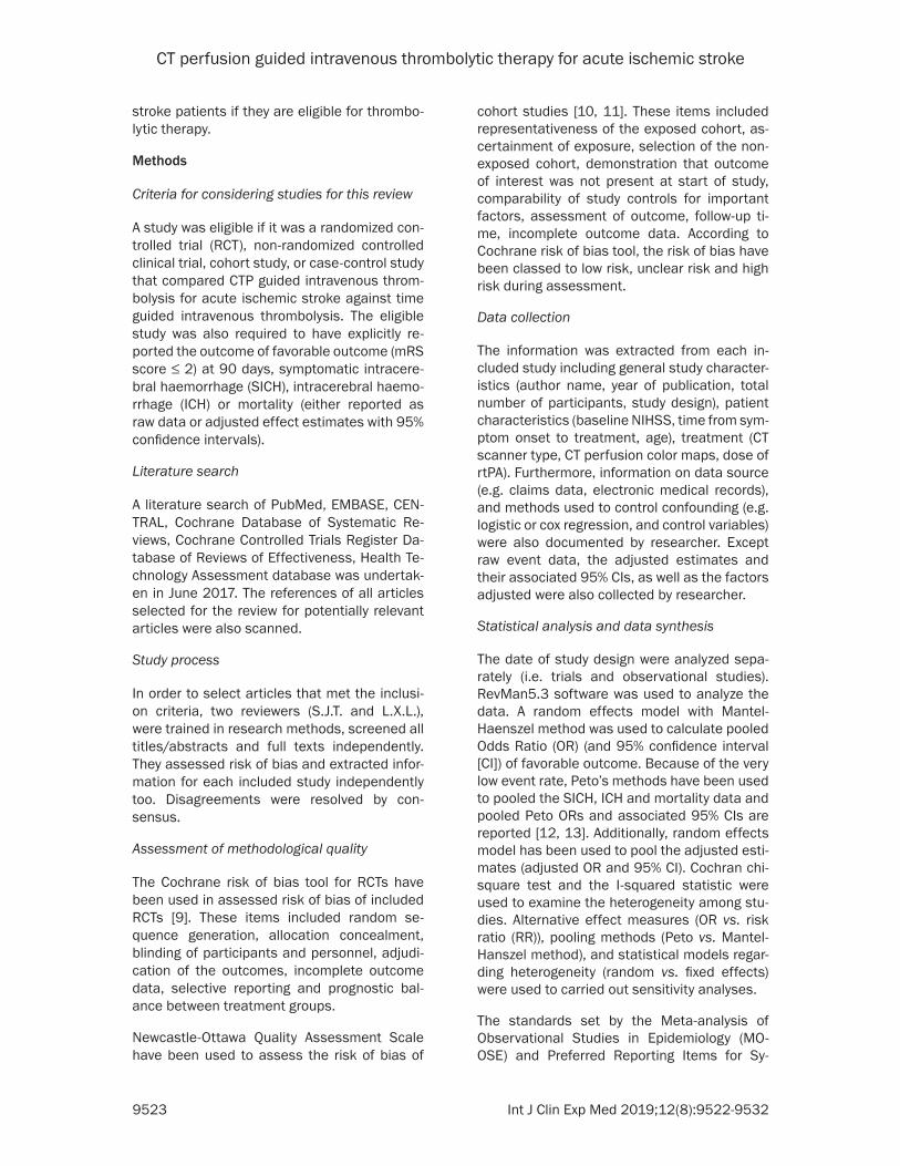
A total of 10217 potentially relevant reports were identified. After a title and abstract sc- reening, there were 206 reports potentially eli-gible; ultimately, eight reports including five prospectively cohort studies, two retrospective cohort studies and one quasi-randomized con-trolled trial are included (Figure 1).
The eight included studies were published between 2011 and 2015 with 9900 samples. These studies were conducted in the US (Gen- tile 2012 [16], McDonald 2014 [17]), Spain (Obach 2011 [18], García-Bermejo 2012 [19]), UK (Sztriha 2011 [20]), Australia (Bivard 2015 [21]), Germany (Eyding 2011 [22]) and China (Huang 2013 [23]). The characteristics of in- cluded studies are shown in Table 1.
Methodological quality of included studies
All studies (except McDonald) were performed in tertiary care centers, patients were thrombo-
at low risk, although the blinding of participants and personnel was incomplete, because out-comes were objective, which were not likely to be influenced by lack of blinding (Table 2).
Favorable outcome
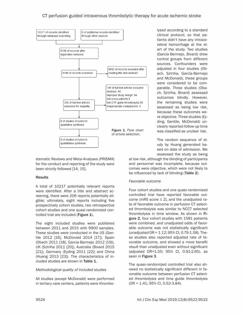
Four cohort studies and one quasi-randomized controlled trial have reported favorable out-come (mRS score ≤ 2), and the unadjusted ra- te of favorable outcome in perfusion CT select-ed thrombolysis was similar to NCCT selected thrombolysis in time window. As shown in Fi- gure 2, four cohort studies with 1581 patients were combined, and unadjusted odds of favor-able outcome was not statistically significant (unadjusted OR = 1.12; 95% CI, 0.79-1.58). The- se studies also reported adjusted rate of fa- vorable outcome, and showed a more benefit result than unadjusted even without significant (adjusted OR=1.55; 95% CI, 0.91-2.65), as seen in Figure 3.
The quasi-randomized controlled trial also sh- owed no statistically significant different in fa- vorable outcome between perfusion CT select-ed thrombolysis and time guide thrombolysis (OR = 1.41; 95% CI, 0.52-3.84).
lysed according to a standard clinical protocol, so that pa- tients didn’t have any intrace-rebral hemorrhage at the st- art of the study. Two studies (García-Bermejo, Bivard) drew control groups from different sources. Confounders were adjusted in four studies (Ob- ach, Sztriha, García-Bermejo and McDonald), these groups were considered to be com- parable. Three studies (Oba- ch, Sztriha, Bivard) assessed outcomes blindly. However, the remaining studies were assessed as being low risk, because these outcomes we- re objective. Three studies (Ey- ding, Gentile, McDonald) un- clearly reported follow-up time was classified as unclear risk.
The random sequence of st- udy by Huang generated ba- sed on date of admission. We assessed the study as being
Figure 1. Flow chart of article selection.

CT perfusion guided intravenous thrombolytic therapy for acute ischemic stroke
9525 Int J Clin Exp Med 2019;12(8):9522-9532
Table 1. Characteristic of included studies
Studies Design Sample size CT scanner type CT perfusion
Colour maps
CT perfusion group NCCT groupRtPA
(alteplase)Time to treatment (h) Age (y) NIHSS Time to
treatment (h) Age (y) NIHSS
Obach 2011 Prospectively cohort 368 64 row, Siemens CBF, CBV, TPP > 3 h (28%) 73 9 > 3 h (16%) 73 10 0.9 mg/kg
Sztriha 2011 Prospectively cohort 254 16-slice, GE CBF, CBV, MTT 3-6 74 13 0-3 74 14 0.9 mg/kg
Eyding 2012 Retrospective cohort 57 64-slice, Siemens CBF, CBV, TTP 3-4.5 71 11 < 3 73 7 0.9 mg/kg
Gentile 2012 Prospectively cohort 25 16/64-slice, Siemens CBF, CBV, TPP < 5 62 NR < 5 64 NR 0.9 mg/kg
García-Bermejo 2012 Prospectively cohort 215 64 rows, GE; 32 rows, Toshiba CBF, CBV, MTT > 4.5 69 9 < 4.5 72 11 mg/kg
Huang 2013 Quasi-randomized controlled trial 66 64 rows, GE MTT, CBV 3-6 58 9 < 4.5 64 10 0.6-0.9mg/kg
McDonald 2014 Retrospective cohort 8153 NR NR NR 73 NR NR 73 NR NR
Bivard 2015 Prospectively cohort 762 64/320 slice, Toshiba NR < 4.5 73 14 < 4.5 74 12 NRNote. NCCT: non-contrast CT; NIHSS = National Institutes of Health Stroke Scale; MTT = mean transit time; CBV = cerebral blood volume; CBF = cerebral blood flow; TTP = time to peak; NR = not report.
Table 2. Risk bias of included studies
Author (year)Representativeness
of the exposed cohort
Ascertainment of exposure
Selection of the non-exposed
cohort
Demonstration that outcome of interest was not present at
start of study
Comparability of study controls for important
factors
Assessment of outcome
Was follow-up long enough for
outcomes to occur
Adequacy of follow up of cohorts
Obach (2011) + + + + + + + +Sztriha (2011) + + + + + + + +Eyding (2012) + + + + - + ? +Gentile (2012) + + + + - + ? +García-Bermejo (2012) + + - + + + + +McDonald (2014) + + + + + + ? +Bivard (2015) + + - + - + + +
Random sequence generation
Allocation concealment
Blinding of participants
and personnel
Blinding of outcome
assessment
Incomplete outcome data
Selective reporting Other bias
Huang (2013) - - + + + + +Note. + = low risk, ? = unclear risk, - = high risk.
CT perfusion guided intravenous thrombolytic therapy for acute ischemic stroke
9526 Int J Clin Exp Med 2019;12(8):9522-9532
SICH
As seen in Figure 4, four cohort studies with 888 patients were included to perform meta-
analyses, which demonstrated that the no sig-nificant differences in outcome of SICH bet- ween CTP group and control group (unadjusted OR = 0.81; 95% CI, 0.39-1.68). One study
Figure 2. Rate of favorable outcome (mRS score ≤ 2) in patients who received CTP guided thrombolysis versus time guided thrombolysis based on raw data.
Figure 3. Rate of favorable outcome (mRS score v2) in patients who received CTP guided thrombolysis versus time guided thrombolysis based on adjusted data.
Figure 4. Risk of SICH in patients who received CTP guide thrombolysis versus time guide thrombolysis based on raw data.

CT perfusion guided intravenous thrombolytic therapy for acute ischemic stroke
9527 Int J Clin Exp Med 2019;12(8):9522-9532
(Sztriha 2014) reported adjusted rate of SICH (unadjusted OR = 0.63; 95% CI, 0.13-3.12) and one quasi-randomized controlled trial (adjust- ed OR = 3.0; 95% CI, 0.59-15.21) are both demonstrated the same result.
ICH
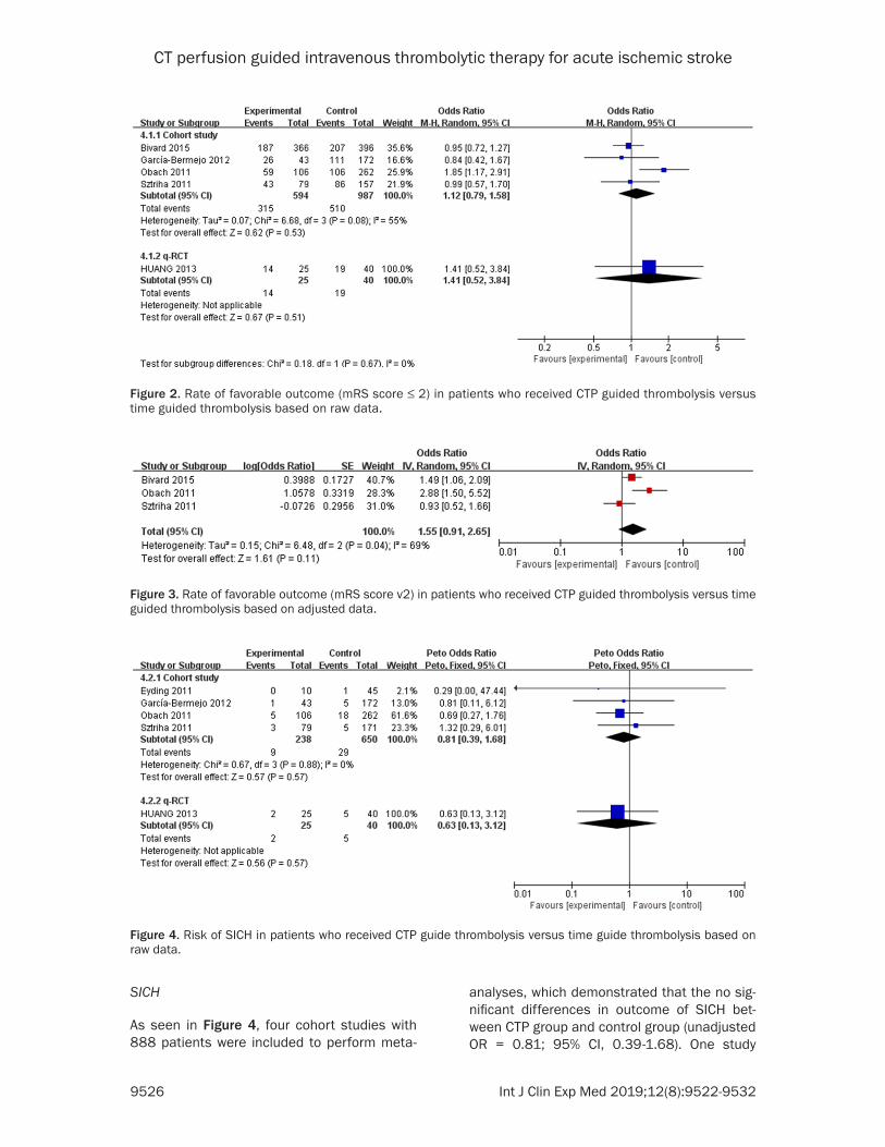
Four cohort studies with 1092 patients were included to perform mete analysis, there sh- owed no significant difference in outcome of
ICH between perfusion CT guided thrombolysis and NCCT guided thrombolysis in time window (unadjusted OR = 1.28; 95% CI, 0.76-2.17), as shown in Figure 5. The pooled result of adjust-ed ICH rate was similar (adjusted OR = 1.53; 95% CI, 0.84-2.80), as shown in Figure 6.
No significant difference was found in the qua-si-randomized controlled trial (unadjusted OR = 0.79; 95% CI, 0.14-4.38).
Figure 5. Risk of ICH in patients who received CTP guide thrombolysis versus time guide thrombolysis based on raw data.
Figure 6. Risk of ICH in patients who received CTP guide thrombolysis versus time guided thrombolysis based on adjusted data.
Figure 7. Risk of mortality in patients who received CTP guide thrombolysis versus time guide thrombolysis based on raw data.

CT perfusion guided intravenous thrombolytic therapy for acute ischemic stroke
9528 Int J Clin Exp Med 2019;12(8):9522-9532
Mortality
Five cohort studies with 9579 patients were included to perform meta-analysis. There were similar unadjusted mortality between perfusion CT selected thrombolysis and NCCT selected thrombolysis in time window (OR = 1.07; 95% CI, 0.89-1.28), as shown in Figure 7. Except the study by McDonald, there was a benefit trend on outcome of mortality in CTP group. The mor-tality was decreased after adjustment (adjust-ed OR = 0.83; 95% CI, 0.48-1.44), as shown in Figure 8.
Sensitivity analysis
Five studies (Bivard 2015, Obach 2011, Sztri- ha 2011, McDonald 2014 and Huang 2013) reported the comparison results of CTP guid- ed thrombolysis beyond time window to NCCT guided thrombolysis in three hours. Compared to NCCT guided thrombolysis in three hours, CTP guided thrombolysis had similar results in favorable outcome and ICH, while benefit in
thrombolysis. It was found that patients treated beyond time window based on CTP had higher rates of favorable outcome and ICH, and lower rate of SICH than patients treated within time window based on NCCT. Although these differ-ences were small and have no statistically sig-nificant different, but these results were similar to adjusted results. Patients treated beyond time window based on CTP had lower rates of adjusted mortality than patients treated within time window based on NCCT, although the dif-ference have no statistically significant differ-ent. Compared to patients thrombolysis within 3 hours based on NCCT, patients treated be- yond 3 hour based on CTP also had higher rates of favorable outcome and ICH, but had a lower rates of SICH and mortality, although the differ-ence have no statistically significant different too. Therefore, the results of review were ro- bust. The outcomes of mRS ≤ 2 at 90 days, SICH and mortality in CTP guided thrombolysis group in this review were 53%, 3.8% and 13% respectively, which were similar to the results
Figure 8. Risk of mortality in patients who received CTP guide thrombolysis versus time guide thrombolysis based on adjusted data.
Table 3. Results of CTP guided thrombolysis versus thromboly-sis in 3 hour based on NCCT
Outcome or subgroup Studies Participants Effect estimate (OR) I2
Favorable outcome* 4 Cohort study 3 1248 1.14 [0.75, 1.74] 52% q-RCT 1 38 1.09 [0.28, 4.19] -ICHΔ 4 Cohort study 3 1067 1.34 [0.79, 2.28] 0% q-RCT 1 38 1.04 [0.09, 12.11] -SICHΔ 4 Cohort study 3 555 0.83 [0.29, 2.40] 0% q-RCT 1 38 0.47 [0.05, 4.02] -MortalityΔ 4 1308 0.78 [0.56, 1.08] 0%NOTE: *Odds Ratio (M-H, Random, 95% CI); ΔPeto Odds Ratio (Peto, Fixed, 95% CI).
SICH and morality, but without statistically significant different (Table 3).
The sensitivity analysis using al- ternative effect measures, statis-tical methods, and analysis mod-els did not show any important changes in the pooled effects (Supplemental Figures 1, 2, 3, 4, 5, 6, 7, 8, 9, 10, 11).
Discussion
This systematic review and me- ta-analysis estimated the pool- ed outcomes from studies where CT perfusion guided thrombolys- is was compared to time guided

CT perfusion guided intravenous thrombolytic therapy for acute ischemic stroke
9529 Int J Clin Exp Med 2019;12(8):9522-9532
of recent mete analysis with results as 59.4%, 3.6% and 10% respectively [24].
Compared with NCCT, CTP is increasingly being used to discriminate extent of salvageable br- ain tissue to optimize acute therapy and pre- dict clinical outcomes. Acute ischemic stroke patients were still get intravenous thrombolysis treatment even they were not meet the tradi-tional thrombolysis criteria (e.g. beyond time window, unknown onset time). Some studies showed that there were no differences in SICH or functional outcome between patients throm-bolysed within 4.5 hours or beyond 4.5 hours [20, 25]. The study by Obach showed that CTP guided thrombolysis yielded superior benefits (adjusted OR = 4.48; 95% CI, 1.68-11.98), and lower rate of SICH (RR = 0.65; 95% CI, 0.09-4.75) and mortality (RR = 0.84; 95% CI, 0.43-1.67) in patients treated beyond 3 hours than treated within 3 hours [18]. The small random-ized control trial show that patients with stroke of unknown onset who get CTP guided throm-bolysis had higher rates of recanalization and favorable functional outcome than patients in placebo group [26]. A high proportion of acute stroke patients with SITS-MOST and ECASS-3 exclusion criteria can be safely and efficacious-ly treated with intravenous thrombolysis using a CTP selection protocol [27].
Findings may be influenced by many factors, such as CTP penumbra and mismatch parame-ters, CT scanners as well as varied brain cover-age, those were important determinants of stroke treatment risk and the validation and standardization of CTP methods. Two included studies restricted their samples to middle cere-bral artery territory patients and the results were similar to other studies. These demon-strated that the results were not influenced by type of artery involved in the stroke, which con-trasts with the conclusions of previous rese- arch.
Two disadvantages of CTP were renal dama- ge from iodinated contrast administration and radiation exposure. However, none of the stud-ies in this review reported these. The risk for contrast induced nephropathy (CIN) in patients with stroke receiving CTP was low, and the long term effects of radiation from CTP stroke imag-ing were not entirely clear yet. Compared to NCCT, CTP was less cost and more incremental benefits used to select patient for thromboly-
sis, because CTP was better able to exclude patients who were at higher risk of secondary intracranial hemorrhage (ICH) and better in- clude patients who could benefit from throm-bolysis beyond 4.5 hours post symptom onset [28-30].
To the best of our knowledge, this is the first review to systematically assess the effective-ness of CT perfusion versus NCCT guided selec-tion of acute ischemic stroke patients who are eligible for thrombolytic therapy. The literature search strategy was thorough, and all relevant evidences have been captured. The analysis of data was thorough and careful. Instead of using grand pooling of the data, several pre specified subgroup analyses were used to explore sourc-es of heterogeneity and both raw data and adjusted data were used in analyses. Although our assessment was low to moderate risk of bias, it was limited by the nature of the observe study which was considered as low quality evi-dence, and this view’s studies were all observed research. Thus, our study does not provide level 1 evidence whether perfusion CT selec-tion leads to better outcomes with rtPA treat-ment than non-contrast CT selection. RCTs needed to be done in the future.
Conclusions
Functional outcome, SICH and mortality follow-ing thrombolysis of AIS patients beyond the time window using CTP are comparable to those for patient thrombolysed at time window using NCCT alone. However, because of the lim-ited number of the studies and low level evi-dences, these estimates should be regarded with caution.
Acknowledgements
This work was supported by the Department of Education of Zhejiang Province, China [Grant numbers Y201635273]. The work also funded by science and technology plans of health and family planning commission of Zhejiang Provin- ce, China. [Grant numbers 2017KY648]. The funders had no role in the study design, data collection and analysis, decision to publish, or preparation of the manuscript. The authors declare that they have no competing interests.
Disclosure of conflict of interest
None.
CT perfusion guided intravenous thrombolytic therapy for acute ischemic stroke
9530 Int J Clin Exp Med 2019;12(8):9522-9532
Abbreviations
AIS, acute ischemic stroke; NCCT, non-contrast CT; SICH, symptomatic intracerebral haemor-rhage; ICH, intracerebral haemorrhage; DALY, Disability Adjusted of Life Years; CIN, contrast induced nephropathy.
Address correspondence to: Jinlan Yao, School of Medicine, Huzhou University, Huzhou Central Hos- pital, 759 Erhuan Road, Huzhou 313000, Zhejiang, PR China. E-mail: [email protected]
References
[1] GBD 2017 DALYs and HALE Collaborators. Global, regional, and national disability-adjust-ed life-years (DALYs) for 359 diseases and inju-ries and healthy life expectancy (HALE) for 195 countries and territories, 1990-2017: a sys-tematic analysis for the Global Burden of Dis-ease Study 2017. Lancet 2018; 392: 1859-1922.
[2] GBD 2017 Causes of Death Collaborators. Global, regional, and national age-sex-specific mortality for 282 causes of death in 195 coun-tries and territories, 1980-2017: a systematic analysis for the Global Burden of Disease Study 2017. Lancet 2018; 392: 1736-1788.
[3] GBD 2017 Disease and Injury Incidence and Prevalence Collaborators. Global, regional, and national incidence, prevalence, and years lived with disability for 354 diseases and inju-ries for 195 countries and territories, 1990-2017: a systematic analysis for the Global Bur-den of Disease Study 2017. Lancet 2018; 392: 1789-1858.
[4] GBD 2016 Lifetime Risk of Stroke Collabora-tors, Feigin VL, Nguyen G, Cercy K, Johnson CO, Alam T, Parmar PG, Abajobir AA, Abate KH, Abd-Allah F, Abejie AN, Abyu GY, Ademi Z, Agar-wal G, Ahmed MB, Akinyemi RO, Al-Raddadi R, Aminde LN, Amlie-Lefond C, Ansari H, Asayesh H, Asgedom SW, Atey TM, Ayele HT, Banach M, Banerjee A, Barac A, Barker-Collo SL, Bär-nighausen T, Barregard L, Basu S, Bedi N, Be-hzadifar M, Béjot Y, Bennett DA, Bensenor IM, Berhe DF, Boneya DJ, Brainin M, Campos-Non-ato IR, Caso V, Castañeda-Orjuela CA, Rivas JC, Catalá-López F, Christensen H, Criqui MH, Damasceno A, Dandona L, Dandona R, Davle-tov K, de Courten B, deVeber G, Dokova K, Edessa D, Endres M, Faraon EJA, Farvid MS, Fischer F, Foreman K, Forouzanfar MH, Gall SL, Gebrehiwot TT, Geleijnse JM, Gillum RF, Giroud M, Goulart AC, Gupta R, Gupta R, Hachinski V, Hamadeh RR, Hankey GJ, Hareri HA, Hav-moeller R, Hay SI, Hegazy MI, Hibstu DT, James SL, Jeemon P, John D, Jonas JB, Jóźwiak J, Ka-
lani R, Kandel A, Kasaeian A, Kengne AP, Khader YS, Khan AR, Khang YH, Khubchanda-ni J, Kim D, Kim YJ, Kivimaki M, Kokubo Y, Kolte D, Kopec JA, Kosen S, Kravchenko M, Krishnamurthi R, Kumar GA, Lafranconi A, La-vados PM, Legesse Y, Li Y, Liang X, Lo WD, Lorkowski S, Lotufo PA, Loy CT, Mackay MT, Abd El Razek HM, Mahdavi M, Majeed A, Male-kzadeh R, Malta DC, Mamun AA, Mantovani LG, Martins SCO, Mate KK, Mazidi M, Mehata S, Meier T, Melaku YA, Mendoza W, Mensah GA, Meretoja A, Mezgebe HB, Miazgowski T, Miller TR, Ibrahim NM, Mohammed S, Mokdad AH, Moosazadeh M, Moran AE, Musa KI, Negoi RI, Nguyen M, Nguyen QL, Nguyen TH, Tran TT, Nguyen TT, Anggraini Ningrum DN, Norrving B, Noubiap JJ, O’Donnell MJ, Olagunju AT, Onuma OK, Owolabi MO, Parsaeian M, Patton GC, Pira-dov M, Pletcher MA, Pourmalek F, Prakash V, Qorbani M, Rahman M, Rahman MA, Rai RK, Ranta A, Rawaf D, Rawaf S, Renzaho AM, Rob-inson SR, Sahathevan R, Sahebkar A, Salomon JA, Santalucia P, Santos IS, Sartorius B, Schutte AE, Sepanlou SG, Shafieesabet A, Shaikh MA, Shamsizadeh M, Sheth KN, Sisay M, Shin MJ, Shiue I, Silva DAS, Sobngwi E, Sol-jak M, Sorensen RJD, Sposato LA, Stranges S, Suliankatchi RA, Tabarés-Seisdedos R, Tanne D, Nguyen CT, Thakur JS, Thrift AG, Tirschwell DL, Topor-Madry R, Tran BX, Nguyen LT, Tru-elsen T, Tsilimparis N, Tyrovolas S, Ukwaja KN, Uthman OA, Varakin Y, Vasankari T, Venketasu-bramanian N, Vlassov VV, Wang W, Werdecker A, Wolfe CDA, Xu G, Yano Y, Yonemoto N, Yu C, Zaidi Z, El Sayed Zaki M, Zhou M, Ziaeian B, Zipkin B, Vos T, Naghavi M, Murray CJL, Roth GA. Global, regional, and country-specific life-time risks of stroke, 1990 and 2016. N Engl J Med 2018; 379: 2429-2437.
[5] Benjamin EJ, Virani SS, Callaway CW, Cham-berlain AM, Chang AR, Cheng S, Chiuve SE, Cushman M, Delling FN, Deo R, de Ferranti SD, Ferguson JF, Fornage M, Gillespie C, Isasi CR, Jiménez MC, Jordan LC, Judd SE, Lackland D, Lichtman JH, Lisabeth L, Liu S, Longenecker CT, Lutsey PL, Mackey JS, Matchar DB, Matsu-shita K, Mussolino ME, Nasir K, O’Flaherty M, Palaniappan LP, Pandey A, Pandey DK, Reeves MJ, Ritchey MD, Rodriguez CJ, Roth GA, Rosa-mond WD, Sampson UKA, Satou GM, Shah SH, Spartano NL, Tirschwell DL, Tsao CW, Voeks JH, Willey JZ, Wilkins JT, Wu JH, Alger HM, Wong SS, Muntner P; American Heart Association Council on Epidemiology and Prevention Sta-tistics Committee and Stroke Statistics Sub-committee. Heart disease and stroke statis-tics-2018 update: a report from the American Heart Association. Circulation 2018; 137: e67-492.
CT perfusion guided intravenous thrombolytic therapy for acute ischemic stroke
9531 Int J Clin Exp Med 2019;12(8):9522-9532
[6] Powers WJ, Rabinstein AA, Ackerson T, Adeoye OM, Bambakidis NC, Becker K, Biller J, Brown M, Demaerschalk BM, Hoh B, Jauch EC, Kidwell CS, Leslie-Mazwi TM, Ovbiagele B, Scott PA, Sheth KN, Southerland AM, Sum-mers DV, Tirschwell DL; American Heart Asso-ciation Stroke Council. 2018 guidelines for the early management of patients with acute isch-emic stroke: a guideline for healthcare profes-sionals from the american heart association/american stroke association. Stroke 2018; 49: e46-e110.
[7] Goyal M, Menon BK and Derdeyn CP. Perfusion imaging in acute ischemic stroke: let us im-prove the science before changing clinical practice. Radiology 2013; 266: 16-21.
[8] Sharma M and Pelz DM. CT perfusion in acute stroke: added value or waste of time? Stroke 2013; 44: e115.
[9] Higgins JP, Altman DG, Gøtzsche PC, Jüni P, Moher D, Oxman AD, Savovic J, Schulz KF, Weeks L and Sterne JA. The Cochrane Collabo-ration’s tool for assessing risk of bias in ran-domised trials. BMJ 2011; 343: d5928.
[10] Stang A. Critical evaluation of the Newcastle-Ottawa scale for the assessment of the quality of nonrandomized studies in meta-analyses. Eur J Epidemiol 2010; 25: 603-605.
[11] Zeng X, Zhang Y, Kwong JS, Zhang C, Li S, Sun F, Niu Y and Du L. The methodological quality assessment tools for preclinical and clinical studies, systematic review and meta-analysis, and clinical practice guideline: a systematic re-view. J Evid Based Med 2015; 8: 2-10.
[12] Bradburn MJ, Deeks JJ, Berlin JA and Russell Localio A. Much ado about nothing: a compari-son of the performance of meta-analytical methods with rare events. Stat Med 2007; 26: 53-77.
[13] Higgins JPT DJ and Altman DG. Special topics in statistics. In: Higgins JPT, Green S, editors. Cochrane handbook for systematic reviews of interventions. Version 5.1.0. Cochrane Collab-oration 2011.
[14] Stroup DF, Berlin JA, Morton SC, Olkin I, Wil-liamson GD, Rennie D, Moher D, Becker BJ, Sipe TA and Thacker SB. Meta-analysis of ob-servational studies in epidemiology: a propos-al for reporting. Meta-analysis of observational studies in epidemiology (MOOSE) group. JAMA 2000; 283: 2008-2012.
[15] Moher D1, Liberati A, Tetzlaff J, Altman DG; PRISMA Group. Preferred reporting items for systematic reviews and meta-analyses: the PRISMA statement. BMJ 2009; 339: b2535.
[16] Gentile NT, Cernetich J, Kanamalla US, Kochan JP, Reimer H, Freeman B and Jungreis C. Expe-dited computed tomography perfusion and an-giography in acute ischemic stroke: a feasibility study. J Emerg Med 2012; 43: 308-315.
[17] McDonald JS, Fan J, Kallmes DF and Cloft HJ. Pretreatment advanced imaging in patients with stroke treated with IV thrombolysis: evalu-ation of a multihospital data base. AJNR Am J Neuroradiol 2014; 35: 478-481.
[18] Macho J, Amaro S, Capurro S, Gomez-Choco M, San Román L, Cervera A, Blasco J, Vargas M, Torres F and Chamorro Á. Multimodal CT-as-sisted thrombolysis in patients with acute stroke: a cohort study. Stroke 2011; 42: 1129-1131.
[19] Garcia-Bermejo P, Calleja AI, Pérez-Fernández S, Cortijo E, del Monte JM, García-Porrero M, Fe Muñoz M, Fernández-Herranz R and Arenil-las JF. Perfusion computed tomography-guided intravenous thrombolysis for acute ischemic stroke beyond 4.5 hours: a case-control study. Cerebrovasc Dis 2012; 34: 31-37.
[20] Sztriha LK, Manawadu D, Jarosz J, Keep J and Kalra L. Safety and clinical outcome of throm-bolysis in ischaemic stroke using a perfusion CT mismatch between 3 and 6 hours. PLoS One 2011; 6: e25796.
[21] Bivard A, Levi C, Krishnamurthy V, McElduff P, Miteff F, Spratt NJ, Bateman G, Donnan G, Da-vis S and Parsons M. Perfusion computed to-mography to assist decision making for stroke thrombolysis. Brain 2015; 138: 1919-1931.
[22] Eyding J, Wiebringhaus R, Klein FG, Skodda S, Schlegel U, Alekseyev A and Heuser L. Multi-modal CT imaging and recanalizing therapy in acute ischemic stroke: retrospective analysis of a one-year single-center experience. Eur Neurol 2012; 67: 193-199.
[23] Huang Q, Wu J, Ma QF, Fan CQ, Jia JP and Lu J. Intravenous thrombolysis directed by whole-brain computed tomographic perfusion: a case-control study. Zhonghua Yi Xue Za Zhi 2013; 93: 3419-3423.
[24] Burton KR, Dhanoa D, Aviv RI, Moody AR, Kapral MK and Laupacis A. Perfusion CT for selecting patients with acute ischemic stroke for intravenous thrombolytic therapy. Radiolo-gy 2015; 274: 103-114.
[25] Cortijo E, García-Bermejo P, Calleja AI, Pérez-Fernández S, Gómez R, del Monte JM, Reyes J and Arenillas JF. Intravenous thrombolysis in ischemic stroke with unknown onset using CT perfusion. Acta Neurol Scand 2014; 129: 178-183.
[26] Schindler C, Bogousslavsky J, Maeder P, Meuli R and Wintermark M. Perfusion-CT guided in-travenous thrombolysis in patients with un-known-onset stroke: a randomized, double-blind, placebo-controlled, pilot feasibility trial. Neuroradiology 2012; 54: 579-588.
[27] Michel P, Ntaios G, Reichhart M, Schindler C, Bogousslavsky J, Maeder P and Wintermark M. Perfusion-CT guided intravenous thrombolysis in patients with unknown-onset stroke: a ran-
CT perfusion guided intravenous thrombolytic therapy for acute ischemic stroke
9532 Int J Clin Exp Med 2019;12(8):9522-9532
domized, double-blind, placebo-controlled, pi-lot feasibility trial. Neuroradiology 2012; 54: 579-588.
[28] Jackson D, Earnshaw SR, Farkouh R and Schwamm L. Cost-effectiveness of CT perfu-sion for selecting patients for intravenous thrombolysis: a US hospital perspective. AJNR Am J Neuroradiol 2010; 31: 1669-1674.
[29] Earnshaw SR, McDade C, Chapman AM, Jack-son D and Schwamm L. Economic impact of using additional diagnostic tests to better se-lect patients with stroke for intravenous throm-bolysis in the united kingdom. Clin Ther 2012; 34: 1544-1558.
[30] Burton KR, Perlis N, Aviv RI, Moody AR, Kapral MK, Krahn MD and Laupacis A. Systematic re-view, critical appraisal, and analysis of the quality of economic evaluations in stroke imag-ing. Stroke 2014; 45: 807-814.
CT perfusion guided intravenous thrombolytic therapy for acute ischemic stroke
1
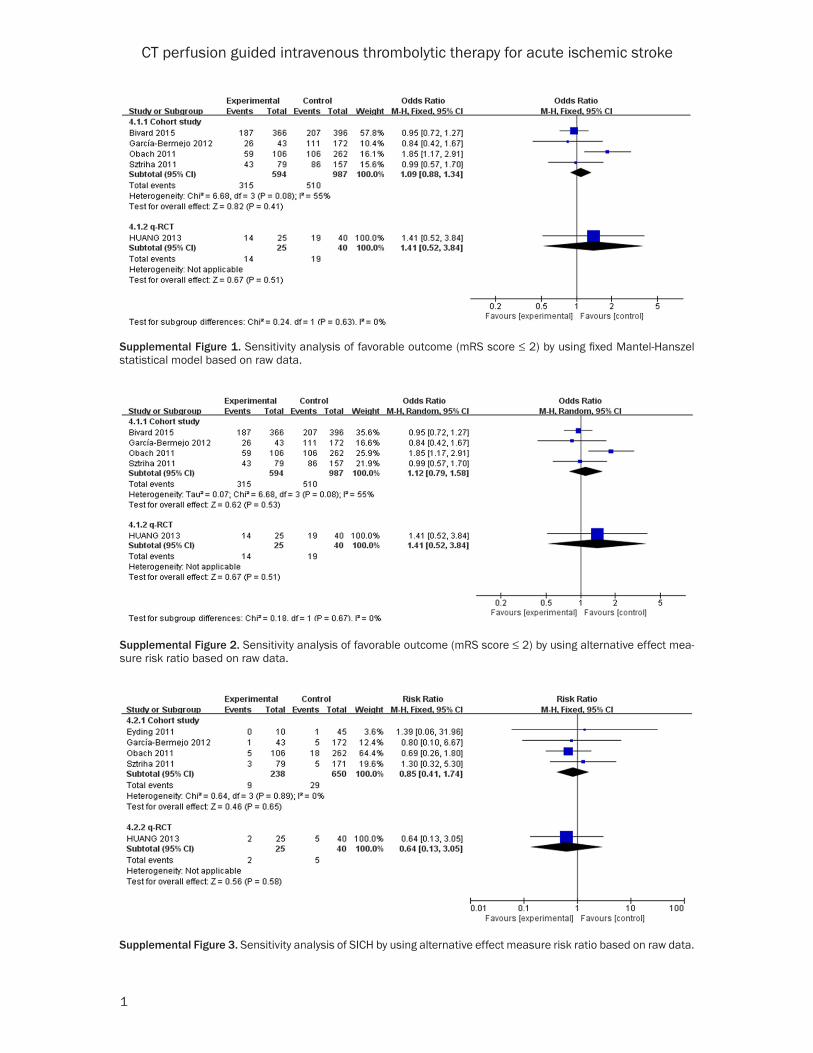
Supplemental Figure 1. Sensitivity analysis of favorable outcome (mRS score ≤ 2) by using fixed Mantel-Hanszel statistical model based on raw data.
Supplemental Figure 2. Sensitivity analysis of favorable outcome (mRS score ≤ 2) by using alternative effect mea-sure risk ratio based on raw data.
Supplemental Figure 3. Sensitivity analysis of SICH by using alternative effect measure risk ratio based on raw data.
CT perfusion guided intravenous thrombolytic therapy for acute ischemic stroke
2
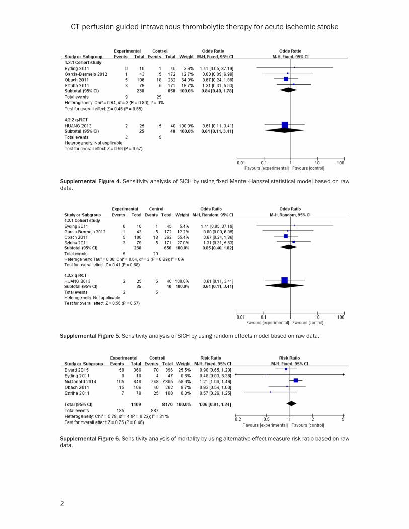
Supplemental Figure 4. Sensitivity analysis of SICH by using fixed Mantel-Hanszel statistical model based on raw data.
Supplemental Figure 5. Sensitivity analysis of SICH by using random effects model based on raw data.
Supplemental Figure 6. Sensitivity analysis of mortality by using alternative effect measure risk ratio based on raw data.

CT perfusion guided intravenous thrombolytic therapy for acute ischemic stroke
3
Supplemental Figure 7. Sensitivity analysis of mortality by using fixed Mantel-Hanszel statistical model based on raw data.
Supplemental Figure 8. Sensitivity analysis of mortality by using random effects model based on raw data.
Supplemental Figure 9. Sensitivity analysis of ICH by using alternative effect measure risk ratio based on raw data.
CT perfusion guided intravenous thrombolytic therapy for acute ischemic stroke
4
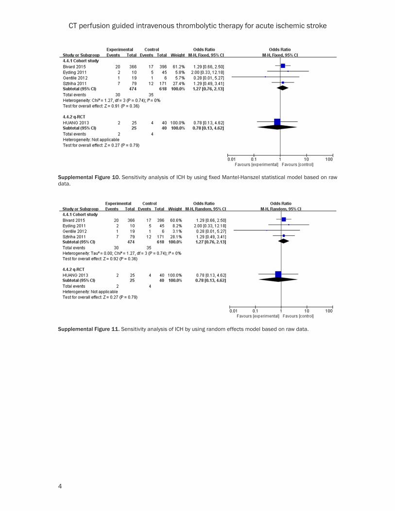
Supplemental Figure 10. Sensitivity analysis of ICH by using fixed Mantel-Hanszel statistical model based on raw data.
Supplemental Figure 11. Sensitivity analysis of ICH by using random effects model based on raw data.