Embed Size (px)
Citation preview
Review ArticleStereotactic Ablative Radiotherapy forOligometastatic Disease in Liver
Myungsoo Kim, Seok Hyun Son, Yong Kyun Won, and Chul Seung Kay
Department of Radiation Oncology, Incheon St Mary’s Hospital, College of Medicine, The Catholic University of Korea,56 Dongsu-ro, Bupyeong-gu, Incheon-si, Seoul 403-720, Republic of Korea
Correspondence should be addressed to Chul Seung Kay; [email protected]
Received 28 January 2014; Accepted 8 April 2014; Published 29 April 2014
Academic Editor: Manousos-Georgios Pramateftakis
Copyright © 2014 Myungsoo Kim et al.This is an open access article distributed under the Creative Commons Attribution License,which permits unrestricted use, distribution, and reproduction in any medium, provided the original work is properly cited.
Liver metastasis in solid tumors, including colorectal cancer, is the most frequent and lethal complication. The development ofsystemic therapy has led to prolonged survival. However, in selected patients with a finite number of discrete lesions in liver, definedas oligometastatic state, additional local therapies such as surgical resection, radiofrequency ablation, cryotherapy, and radiotherapycan lead to permanent local disease control and improve survival. Among these, an advance in radiation therapymade it possible todeliver high dose radiation to the tumor more accurately, without impairing the liver function. In recent years, the introduction ofstereotactic ablative radiotherapy (SABR) has offered evenmore intensive tumor dose escalation in a few fractionswith reduced doseto the adjacent normal liver. Many studies have shown that SABR for oligometastases is effective and safe, with local control rateswidely ranging from 50% to 100% at one or two years. And actuarial survival at one and two years has been reported ranging from72% to 94% and from 30% to 62%, respectively, without severe toxicities. In this paper, we described the definition and technicalaspects of SABR, clinical outcomes including efficacy and toxicity, and related parameters after SABR in liver oligometastases fromcolorectal cancer.
1. Introduction
Colorectal cancer (CRC) is the third most commonly diag-nosed malignancy and the second leading cause of cancer-related death in the USA [1]. Approximately 15%–25% ofCRC patients have liver metastases at the time of diagnosis,and liver metastases develop within 3 years in another 25%–50% of patients with CRC following resection of the primarytumor [2]. The median survival time of these patients rangesfrom 5 to 12 months without treatment [3]. For patients withmetastatic CRC, systemic therapy, including chemotherapyand molecular-targeted agents, is preferred and the devel-opment in systemic therapy has led to prolong survival [4].However, systemic therapy itself rarely eradicates sites ofmacrometastatic disease permanently. For some subset ofpatients with a finite number of discrete lesions in the liver,surgical resection has led to 5-year survival rates of up to 60%in some recent series [5–7]. In this “oligometastatic” setting,local therapy has the potential to cure liver metastases,improving survival. Unfortunately, 80%–90%of patients with
liver metastases are not suitable for surgical resection due tounfavorable tumor factors or medical comorbidities. Non-surgical approaches, such as radiofrequency ablation (RFA),transarterial chemoembolization, and cryotherapy, have beenwidely used as alternative local therapy for unresectable livermetastases. However, each of these techniques has limitationssuch as the size, the location, the number of tumors, and thevariable high recurrence rates [8–10].
Historically, the radiation therapy has had a limited rolein the treatment of liver cancer due to the low toleranceof the liver to radiation dose and it was difficult to deliverthe radiation doses necessary to ablate gross tumors withoutcausing radiation-induced liver disease (RILD) [11]. However,technical developments of radiation therapy such as three-dimensional conformal radiotherapy (3D-CRT) and intensitymodulated radiation therapy have gradually expanded theirrole from a palliative to a curative intent, whereby high-dose radiation can be delivered to the tumor more safelywithout affecting the liver function. In recent years, theintroduction of stereotactic ablative radiotherapy (SABR) has
Hindawi Publishing CorporationBioMed Research InternationalVolume 2014, Article ID 340478, 9 pageshttp://dx.doi.org/10.1155/2014/340478
2 BioMed Research International
offered even more intensive tumor dose escalation using ahypofractionated regimen with reduced dose to the normalliver, usually below the threshold dose above which severeRILD is observed. Early result of SABR for liver metastaseshas promising outcomes and very low complication rates. Inthis review, we will discuss the recent treatment outcome andtoxicity following SABR for livermetastases in CRC.Thedoseconstraint and pattern of failure will also be discussed.
2. Definitions
2.1. Oligometastases. Until fairly recently, distant metastaseshave been considered to occur late in the natural history ofcancer and represent an incurable state, thereby warrantingpalliative care only. It is now recognized that some patients ofdistantmetastases with “oligo,” or few sites ofmetastases, mayhave isolated sites of metastases. Hellman andWeichselbaumhypothesized the intermediate state of metastases betweenpurely localized and widely metastatic disease and coinedthe term “oligometastases” in which metastases are limited innumber and location [12]. For patients with oligometastases,aggressive local therapies might prolong survival, especiallywhen combined with effective systemic therapy to addressoccult micrometastases. Oligometastases are clinically dis-tinguished by two scenarios; the de novo oligometastases, inwhich the tumor early in the evolution of metastatic progres-sion producingmetastasis that are limited in number and site,and the induced oligometastases, which is generated wheneffective systemic chemotherapy eradicates the majority ofmetastatic deposits in a patients with wide spread metastatic2 disease [13]. The induced “oligometastases” group is likelyto have a much less favorable prognosis.
2.2. Stereotactic Ablative Radiotherapy (SABR). SABR, alsocalled stereotactic body radiation therapy (SBRT), is anextension of intracranial stereotactic radiosurgery (SRS) thatencompasses all sites below the cranium. The AmericanSociety of Radiation Oncology defines SABR as externalbeam radiotherapy used to deliver a high dose of radiationextremely precisely to an extracranial target within the body,as a single dose or a small number of fractions [32]. SABR isused and showed efficacy in controlling early stage primaryand oligometastatic disease at a wide range of tumor sites,including lung, liver, pancreas, prostate, spine, and headand neck [33, 34]. In addition to increasing the accuracyof radiation dose delivery, thus reducing irradiated adjacentnormal structures, the high radiation dose per fraction canpotentially ablate all tissues in the treated area. Becauseof the high fractional dose, it is extremely important thatthe treatment organ geometry is as close as possible to theplanning CT data. Immobilization devices or other typesof interfractional motion management is one way to movetowards that goal.The techniques for improving the accuracyand precision can be classified into two broad categories:immobilization and motion reduction techniques and imageguidance. Immobilization with stereotactic body frames aimsto optimize patient fixation, provide external reference sys-tem for stereotactic coordinates, and use a device to reducebreathing mobility. Image guidance technology allows the
guidance of dose delivery with 3D real-time information oftarget localization. These tools serve to reduce patient set-up errors and provide systematic assessment of organmotionand deformation during the course of treatment. Treatmentplanning is usually based on patients’ CT images and includeseithermultiple noncoplanar fixed gantry beams or arcs.Manydifferent treatment devices are commercially available forSABR including Novalis Tx (Brainlab AG, Feldkirchen, Ger-many), TrueBeam (Varian Medical Systems, Palo Alto, CA,USA), Elekta Axesse and Synergy (both Elekta, Stockholm,Sweden), Cyberknife (Accuray, Sunnyvale, CA, USA), andTomoTherapy (Accuray, Madison, WI, USA) (Figure 1).
3. Selection of Patients for SABR
The main goal of SABR is to achieve local control of eacholigometastatic site; however, whether obtaining local controlof the metastasis would translate into clinical or survivalbenefit for the patients depends onmultiple factors, includingage, performance status, medical comorbidities, and histol-ogy of malignancies. Therefore, careful and strict patientselection is needed and the patient’s whole condition shouldbe fully considered. Ideal candidates for SABRmay be definedas follows: a limited number of metastases (one to five), alimited tumor size (<6 cm), a locally controlled primary site,metachronous occurrence of metastatic disease, favorablehistologies (such as CRC and breast cancer), young age, goodperformance status, at least 700mof uninvolved liver volume,and adequate pretreatment baseline liver function. In general,the risk of occult diffuse metastases increases as the numberof metastases increases, and the best results are found inpatients with 3 or fewer metastases, ideally less than 6 cm inmaximum diameter [35, 36]. Figure 2 shows the case of goodcandidate for SABR in liver oligometastases.
4. Clinical Outcomes
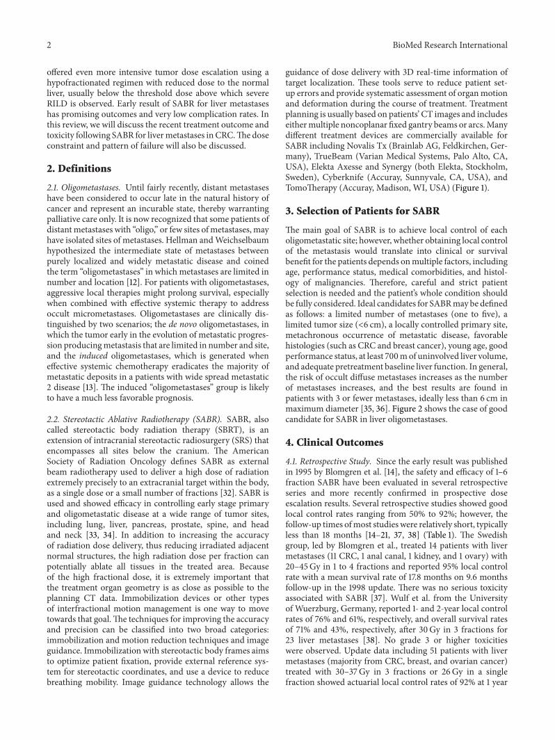
4.1. Retrospective Study. Since the early result was publishedin 1995 by Blomgren et al. [14], the safety and efficacy of 1–6fraction SABR have been evaluated in several retrospectiveseries and more recently confirmed in prospective doseescalation results. Several retrospective studies showed goodlocal control rates ranging from 50% to 92%; however, thefollow-up times ofmost studies were relatively short, typicallyless than 18 months [14–21, 37, 38] (Table 1). The Swedishgroup, led by Blomgren et al., treated 14 patients with livermetastases (11 CRC, 1 anal canal, 1 kidney, and 1 ovary) with20–45Gy in 1 to 4 fractions and reported 95% local controlrate with a mean survival rate of 17.8 months on 9.6 monthsfollow-up in the 1998 update. There was no serious toxicityassociated with SABR [37]. Wulf et al. from the Universityof Wuerzburg, Germany, reported 1- and 2-year local controlrates of 76% and 61%, respectively, and overall survival ratesof 71% and 43%, respectively, after 30Gy in 3 fractions for23 liver metastases [38]. No grade 3 or higher toxicitieswere observed. Update data including 51 patients with livermetastases (majority from CRC, breast, and ovarian cancer)treated with 30–37Gy in 3 fractions or 26Gy in a singlefraction showed actuarial local control rates of 92% at 1 year

BioMed Research International 3
(a) Novalis Tx (b) TrueBeam (c) Elekta Axesse
(d) Elekta Synergy (e) Cyberknife (f) TomoTherapy
Figure 1: Various treatment devices available for stereotactic ablative radiotherapy.
(a) (b) (c)
Figure 2: A 52-year-old male patient had been treated with surgery and postoperative adjuvant chemotherapy for sigmoid colon cancer(adenocarcinoma, T2N2M0). 13months later, livermetastasis developed and hewas then treatedwith salvage chemotherapy; however, follow-up CT scan after the chemotherapy showed progression of liver metastasis (white arrows) (a). We decided to treat him with SABR. Theprescriptive dose to the planning target volume including two metastatic tumor lesions was 40Gy in 4 fractions on consecutive day (b). TheCT scan on 3 months after the completion of SABR showed complete response (c). Radiotherapy related change of increased density aroundthe previous tumor lesions was shown but the patient’s liver function test was normal.
and 66% at 2 years [15]. Higher dose was associated withimproved local control. There was a trend for patients withCRC liver metastases to have worse local control comparedwith other histologies. Katz et al. [16] treated 69 patientswith 174 liver metastases (CRC 20, breast 16, pancreas 9, andlung 5) with a median dose of 48Gy in 2–6 fractions andshowed 76% and 54% of local control rate on 10 months and20months, respectively, with 14.5 months of median survival.There was no grade 3 or higher toxicity.
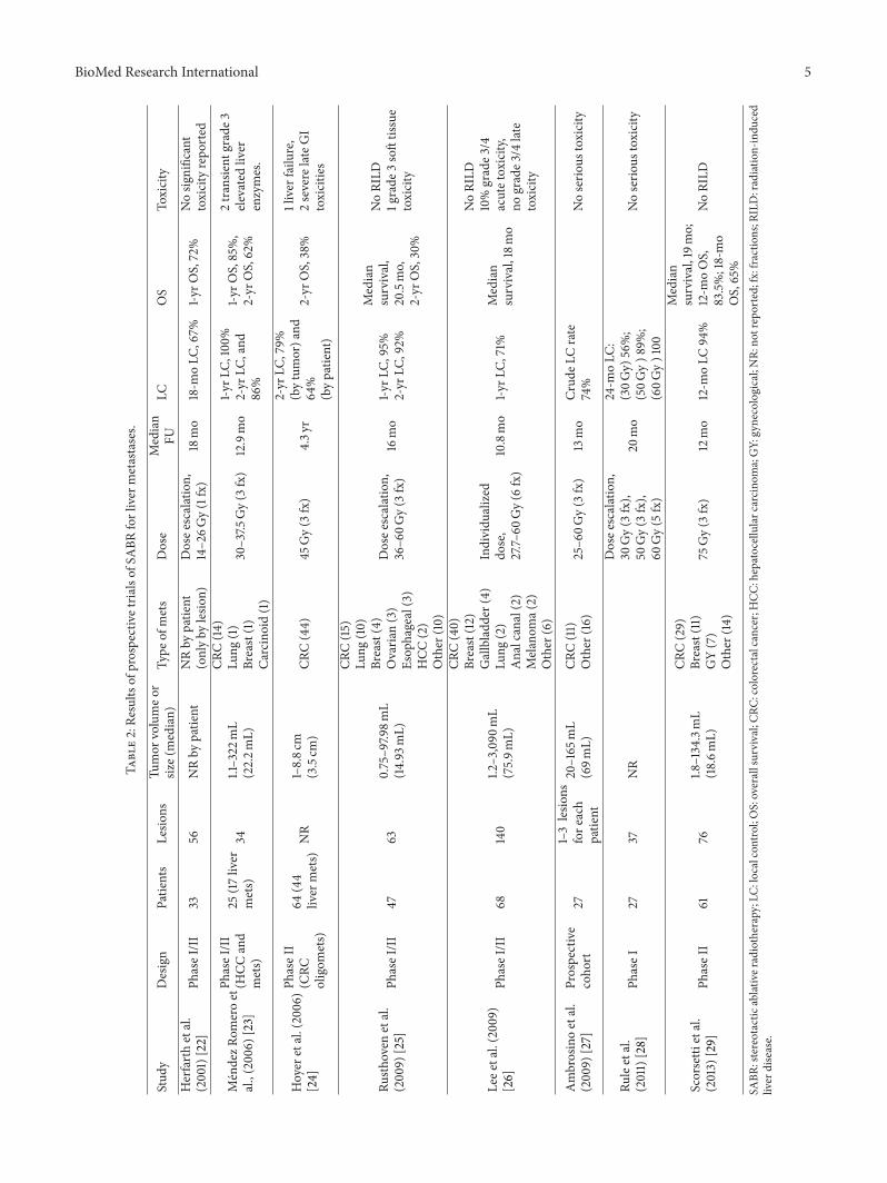
4.2. Prospective Study. There are several prospective trials ofSABR for liver oligometastases in the literature and somestudies have examined the dose escalation in a formal,structured phase I trial [22–29] (Table 2). Herfarth et al.performed a prospective phase I/II study using single doseSBRT from 14–26Gy in 33 patients with 56 liver metastases(majorityCRC, others frombreast cancer, soft tissue sarcoma,lung, pancreas, kidney, ormelanoma) [22].The actuarial localcontrol rates on 6, 12, and 18 months were 75%, 71%, and
4 BioMed Research International
Table1:Re
sults
ofretro
spectiv
etria
lsof
SABR
forliver
metastases.
Stud
yPatie
nts
Lesio
nsTu
mor
volumeo
rsiz
e(median)
Type
ofmets
Dose
Median
FULC
OS
Toxicity
Blom
gren
etal.(1995)[14]14
173–260m
L
CRC(11)
Analcanal(1)
Kidn
ey(1)
Ovaria
n(1)
7.7–4
5Gy
(1–4fx)
9.6mo
NR-50%respon
serate
NR
2caseso
fhemorrhagic
gastr
itis
Wulfetal.(2006)
[15]
44(39liver
mets)
519–
355m
L
CRC(23)
Breast(11)
Ovaria
n(4)
Other
(13)
30–37.5
Gy(3
fx)
26Gy(1fx)
2yr
1-yrL
C,92%,
2-yr
LC,66%
1-yrO
S,72%
2-yr
OS,32%
Nograde2
–4toxicity
Katzetal.(2007)[16]
69174
0.6–
12.5cm
(2.7cm
)
CRC(20)
Breast(16)
Pancreas
(9)
Lung
(5)
Other
(19)
30–55G
y(5–15fx)
14.5mo
10-m
oLC
,76%
,20-m
oLC
,57%
Mediansurvival,
14.5mo,
6-moOS,46
%,
12-m
oOS,24%
Nograde3
/4toxicity
vanderP
ool
etal.(2010)[17]
2031
0.7–6.2c
m(2.3cm
)AllCR
C30–37.5
Gy
(3fx)
26mo
1-yrL
C,100%
,2-yr
LC,74%
Mediansurvival,
34mo,
1-yrO
S,100%
,2-yr
OS,83%
2grade3
late
liver
enzyme
changes,
1grade
2rib
fracture
Changetal.(2011)[18]
65102
0.6–
3088
mL
(30.1m
L)AllCR
C22–6
0Gy
(1–6fx)
14.4mo
1-yrL
C,62%,
2-yr
LC,45%
1-yrO
S,72%,
2-yr
OS,38%
2grade3
gastritis,
2grade3
elevatedliver
enzymes.
Vautravers-D
ewas
etal.
(2011)[19
]42
620.7–10cm
(3.4cm
)40
Gy(4
fx)
45Gy(3
fx)
14.3mo
2-yr
LC,86%
2-yr
OS,48%
Lanciano
etal.(2012)[20]
30(23liver
mets)
412.29–316
mL
(60.9m
L)
CRC(15)
Breast(3)
Esop
hagus(1)
GIST(1)
Pancreas
(1)
NSC
LC(2)
36–6
0Gy(3
fx)
50Gy(5
fx)
22mo
1-yrL
C,92%,
2-yr
LC,56%
1-yrO
S,73%,
2-yr
OS,31%
Nograde3
/4toxicity
Habermehletal.(2013)
[21]90
138
11–333m
L(62m
L)
CRC(70)
Breast(27)
Pancreas
(11)
Ovaria
n(7)
Lung
(6)
Others(16),
bysite
10–30G
y(1fx)
21.7mo
87%,69%
,and
59%
after
6,12,
and18mo
MedianOS
24.3mo;localP
FSwas
87%,70%
,and
59%aft
er6,12,and
18mo,respectiv
ely
NoRILD
SABR
:stereotactic
ablativ
eradiotherapy;LC
:localcontrol;OS:overallsurvival;CR
C:colorectalcancer;G
IST:
gastr
ointestin
alstr
omaltumor;N
SCLC
:non
-smallcell
lung
cancer;N
R:no
treported;
fx:fractions;
RILD
:radiatio
n-indu
cedliver
disease.
BioMed Research International 5
Table2:Re
sults
ofprospectivetria
lsof
SABR
forliver
metastases.
Stud
yDesign
Patie
nts
Lesio
nsTu
mor
volumeo
rsiz
e(median)
Type
ofmets
Dose
Median
FULC
OS
Toxicity
Herfarthetal.
(2001)[22]
PhaseI/II
3356
NRby
patient
NRby
patie
nt(onlyby
lesio
n)Dosee
scalation,
14–26G
y(1fx)
18mo
18-m
oLC
,67%
1-yrO
S,72%
Nosig
nificant
toxicityrepo
rted
MendezR
omeroet
al.,(200
6)[23]
PhaseI/II
(HCC
and
mets)
25(17liver
mets)
341.1–322
mL
(22.2m
L)
CRC(14
)Lu
ng(1)
Breast(1)
Carcinoid(1)
30–37.5
Gy(3
fx)
12.9mo
1-yrL
C,100%
2-yr
LC,and
86%
1-yrO
S,85%,
2-yr
OS,62%
2transie
ntgrade3
elevatedliver
enzymes.
Hoyer
etal.(2006)
[24]
PhaseII
(CRC
oligom
ets)
64(44
liver
mets)
NR
1–8.8c
m(3.5cm
)CR
C(44)
45Gy(3
fx)
4.3y
r
2-yr
LC,79%
(bytumor)a
nd64
%(bypatie
nt)
2-yr
OS,38%
1liver
failu
re,
2severe
lateGI
toxicitie
s
Rusth
oven
etal.
(200
9)[25]
PhaseI/II
4763
0.75–9
7.98m
L(14
.93m
L)
CRC(15)
Lung
(10)
Breast(4)
Ovaria
n(3)
Esop
hageal(3)
HCC
(2)
Other
(10)
Dosee
scalation,
36–6
0Gy(3
fx)
16mo
1-yrL
C,95%
2-yr
LC,92%
Median
survival,
20.5mo,
2-yr
OS,30%
NoRILD
1grade
3softtissue
toxicity
Leee
tal.(200
9)[26]
PhaseI/II
68140
1.2–3,090
mL
(75.9m
L)
CRC(40)
Breast(12)
Gallbladd
er(4)
Lung
(2)
Analcanal(2)
Melanom
a(2)
Other
(6)
Individu
alized
dose,
27.7–
60Gy(6
fx)
10.8mo
1-yrL
C,71%
Median
survival,18m
o
NoRILD
10%grade3
/4acutetoxicity,
nograde3
/4late
toxicity
Ambrosinoetal.
(200
9)[27]
Prospective
coho
rt27
1–3lesio
nsfore
ach
patie
nt
20–165
mL
(69m
L)CR
C(11)
Other
(16)
25–6
0Gy(3
fx)
13mo
Crud
eLCrate
74%
Noserio
ustoxicity
Ruleetal.
(2011)[28]
PhaseI
2737
NR
Dosee
scalation,
30Gy(3
fx),
50Gy(3
fx),
60Gy(5
fx)
20mo
24-m
oLC
:(30G
y)56%;
(50G
y)8
9%;
(60G
y)100
Noserio
ustoxicity
Scorsetti
etal.
(2013)
[29]
PhaseII
6176
1.8–134.3mL
(18.6m
L)
CRC(29)
Breast(11)
GY(7)
Other
(14)
75Gy(3
fx)
12mo
12-m
oLC
94%
Median
survival,19m
o;12-m
oOS,
83.5%;18-mo
OS,65%
NoRILD
SABR
:stereotactic
ablativ
eradiotherapy;LC
:localcontrol;OS:overallsurvival;CR
C:colorectalcancer;H
CC:hepatocellular
carcinom
a;GY:gynecological;NR:
notreported;fx:fractions;R
ILD:radiatio
n-indu
ced
liver
disease.

6 BioMed Research International
67%, respectively. No severe toxicity was observed. In anotherphase I study of 37.5 Gy in 3 fractions in 17 patients with 34liver metastases (CRC 14, lung 1, breast 1, and carcinoid 1),MendezRomero et al. reported 1-year and 2-year local controlrates of 100% and 86%, respectively, and 1- and 2-year survivalrates of 85% and 62%, respectively, after a median follow-up of 12.9 months [23]. Transient grade 3 elevation of liverenzyme levels was observed within 3 months after treatmentin two patients. University of Colorado performed a multi-institutional phase I/II SBRT trial of 47 patients with 63 livermetastases starting with 36Gy in 3 fractions, and the doseswere escalated by 6Gy per dose up to a defined maximumof 60Gy. The 2year local control rate was 92% and a mediansurvival rate was 20.5 months [25]. No hepatic toxicity wasnoted, but grade 3 soft-tissue toxicity was developed inone patient receiving 48Gy in 3 fractions to the anteriorabdominal wall to the region of the subcutaneous tissue. Ruleet al. from the University of Texas Southwestern reported ona phase I study using escalated dose from 30Gy in 3 fractionsto 50Gy in 5 fractions to 60Gy in 5 fractions. In total, 27patients with 37 lesions (one to five liver metastases) wereenrolled. The median follow-up was 20 months. The 2-yearactuarial LC rates were 56%, 89%, and 100% for the cohortstreated with 30Gy, 50Gy, and 60Gy, respectively. There wasa significant difference in local control between the cohortstreated with 60Gy and 30Gy. No grade 4 or 5 toxic effectsor treatment-related grade 3 toxic effects were observed [28].Lee et al. from the Princess Margaret Hospital conductedphase I/II trial using 6 fractions over weeks of SBRT in 68patients with livermetastases of varying sizes (up to 3090mL)to evaluate the safety of SBRT for larger liver metastases [26].Radiotherapeutic dose was individualized based on the livervolume irradiated in order to avoid RILD (range: 24–60Gy).With a median follow-up of 11 months, the 1-year LC rate was71% and the median survival rate was 18 months. There wasno RILD, resulting in a low risk of serious liver toxicity (95%CI, 0 to 5.3%). Grade 2 nontraumatic rib fractures occurredin two patients treated with the maximum doses to 51.8 Gyand 66.2Gy in 6 fractions to 0.5mL of rib. More recently,Scorsetti et al. published a preliminary result from a phase IItrial of high-dose SBRTusing 75Gy in 3 fractions. A total of 61patients with 76 lesions were treated. With a median follow-up of 12 months, the in-field local response rate was 94%,the median survival rate was 19 months, and 1-year actuarialsurvival rate was 83.5%. No RILD was detected [29].
Published reports to date have been small and con-founded by significant heterogeneity concerning the primarysubtype, tumor size and the number of metastases, thenumber of systemic treatments before and after RT, totaldose, and the number of fractions. As a result, it is difficultto evaluate and compare clinical results for liver metastases.Generally, most SABR were performed with 30–60Gy in 1to 6 fractions, for 5 or fewer metastases, with maximumtumor sizes of 6 cm, and the reported 2-year local control andsurvival rates range from 60% to 90% and from 30% to 83%,respectively.Higher dose showed to associatewith better localcontrol and a total prescription dose over 48Gy in 3 fractionswas recommended, whenever possible [35]. Other thanhigher dose, smaller tumor volume, potentially non-CRC
metastases, metachronous liver metastases, and the absenceof previous systemic chemotherapy were prognostic factorsrelated to improved local control. The possible explanationfor better local control in patients with non-CRC metastasesthan CRC metastases may be that most patients with CRCliver metastases have been heavily treated with systemic andother local treatments before SABR [35].
5. Radiation Related Toxicity andDose Volume Constraints
RILD is the most common hepatic toxicity after liver irra-diation, which typically occurs between two weeks to 3months after RT, presenting with anicteric ascites, hep-atosplenomegaly, and elevated alkaline phosphatase [11].Baseline liver function was found as an important factor inRILD after RT and the tolerance dose of SABR in a cirrhoticliver is less than the tolerance dose of a normally functioningliver [31, 39, 40]. Most patients with metastatic liver tumorshave a normally functioning liver, not having underlyingcirrhosis or hepatitis, and RILD is rare following SABR [25].Transient grade 3 elevation of liver enzyme levels occurredin 2 patients treated with 30 to 37.5 Gy in 3 fractions in aphase I/II SABR trial byMendez Romero et al. [23].There hasbeen reported 1 hepatic failure 7 weeks after SABR leadingto death with median total liver dose of 14.4Gy and 60% ofthe liver received >10Gy in 3 fractions [24]. In a phase I/IIstudy by Lee et al., no RILD was noted in 68 patients whoreceivedmedianmean liver dose of 16.9Gy in 6 fractions [26].In a phase I/II study by Rusthoven et al. [25], there was noRILD in 47 patients treatedwith a critical dose-volumemodelallowing no more than 700mL of uninvolved normal liverto receive 15Gy or greater in 3 fractions in accordance withthe Quantitative Analyses of Normal Tissue Effects in theClinic (QUANTEC) recommendations on liver [31]. Anothercomplication reported after SABR is gastrointestinal and soft-tissue/bone toxicity. Three patients who received a total doseof 30Gy or higher to the part of the intestine had colonicand duodenal ulcerations; one patient had perforation of acolonic ulceration demanding surgery while two patients hadduodenal ulceration [24]. Grade 3 soft-tissue toxicity wasobserved in 1 patient after receiving 48Gy in 3 fractions to aregion of the subcutaneous tissue [25]. Grade 2 nontraumaticrib fractures occurred in two patients, 6 and 23 months afterSABR, who were treated to the maximum dose to 0.5mL ofrib, 51.8 Gy and 66.2Gy, respectively [26].
Although techniques for delivering large fractions ofradiation using SABR to the liver have been widely reported,including variousmeans of body immobilization, stereotacticlocalization, image guidance, beamarrangements, anddosingtechniques, far less is known about the tolerance of theliver or the surrounding critical organs when using largefractions of radiation due to its recent development, fewerpatient numbers, and nonuniform reporting of dosimetricparameters. Further clinical data with long follow-up periodare needed to ascertain the dose fractionations schedule thatoptimizes tumor control while minimizing toxicity and tobetter understand the optimal normal tissue dose volumeconstraints.When treating the liver, themost relevant normal
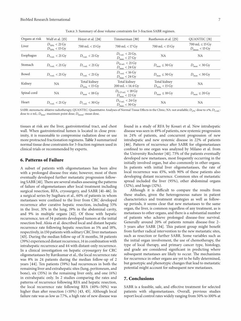
BioMed Research International 7
Table 3: Summary of dose volume constraints for 3-fraction SABR regimen.
Organs at risk Wulf et al. [15] Hoyer et al. [24] Timmerman [30] Rusthoven et al. [25] QUANTEC [31]
Liver 𝐷30% < 21 Gy𝐷50% < 15 Gy
700mL < 15Gy 700mL < 17Gy 700mL < 15 Gy 700mL ≤ 15Gy𝐷mean < 15 Gy
Esophagus 𝐷5mL < 21 Gy 𝐷1mL < 21 Gy
𝐷5mL < 21 Gy,𝐷max < 27Gy
NA NA
Stomach 𝐷5mL < 21 Gy 𝐷
1mL < 21Gy𝐷10mL < 21Gy𝐷max < 24Gy
𝐷max ≤ 30Gy 𝐷max < 30Gy
Bowel 𝐷5mL < 21 Gy 𝐷1mL < 21 Gy
𝐷5mL < 16Gy𝐷max < 24Gy
𝐷max ≤ 30Gy 𝐷max < 30Gy
Kidney NA Total kidney𝐷35% < 15Gy
Total kidney200mL < 14.4Gy
Total kidney𝐷35% < 15Gy
NA
Spinal cord NA 𝐷max < 18Gy𝐷0.25mL < 18Gy𝐷max < 22Gy
𝐷max ≤ 18Gy 𝐷max ≤ 20Gy
Heart 𝐷5mL < 21 Gy 𝐷
1mL < 30Gy𝐷15mL < 24Gy𝐷max < 30Gy
NA NA
SABR: stereotactic ablative radiotherapy; QUANTEC: Quantitative Analyses of Normal Tissue Effects in the Clinic; NA: not available;𝐷𝑥%: dose to 𝑥%;𝐷𝑥mL:dose to 𝑥mL;𝐷max: maximum point dose;𝐷mean: mean dose.
tissues at risk are the liver, gastrointestinal tract, and chestwall. When gastrointestinal lumen is located in close prox-imity, it is reasonable to compromise radiation dose or usemore protracted fractionation regimens. Table 3 summarizednormal tissue dose constraints for 3-fraction regimen used inclinical trials or recommended by experts.
6. Patterns of Failure
A subset of patients with oligometastases has been alivewith a prolonged disease-free state; however, most of themeventually developed further metastatic progression follow-ing SABR [41].There are several studies assessing the patternsof failure of oligometastases after local treatment includingsurgical resection, RFA, cryosurgery, and SABR [41–46]. Ina surgical series by Sugihara et al., 60% of patients in whommetastases were confined to the liver from CRC developedrecurrence after curative hepatic resection, including 53%in the liver, 31% in the lung, 19% in the abdominal cavity,and 9% in multiple organs [42]. Of those with hepaticrecurrence, ten of 34 patients developed tumors at the initialresection bed. Aloia et al. described local and distant hepaticrecurrence rate following hepatic resection as 5% and 18%,respectively, in 150 patientswith solitaryCRC livermetastases[43]. During the median follow-up of 31 months, 58 patients(39%) experienced distant recurrence, 14 in combinationwithintrahepatic recurrence and 44 with distant-only recurrence.In a clinical investigation on hepatic cryosurgery for CRColigometastases by Ravikumar et al., the local recurrence ratewas 8% in 24 patients during the median follow-up of 2years [44]. Ten patients (59%) had recurrences in both theremaining liver and extrahepatic sites (lung, peritoneum, andbone), six (35%) in the remaining liver only, and one (6%)in extrahepatic only. In 2 studies comparing the rates andpatterns of recurrence following RFA and hepatic resection,the local recurrence rate following RFA (40%–50%) washigher than after resection (<10%) [43, 45]. Although localfailure rate was as low as 7.7%, a high rate of new disease was
found in a study of RFA by Kosari et al. New intrahepaticdisease was seen in 49%of patients, new systemic progressionin 24% of patients, and concurrent progression of newintrahepatic and new systemic disease in 22% of patients[46]. Pattern of recurrence after SABR for oligometastasesconfined to one organ was analyzed by Milano et al. fromthe University Rochester [41]. 73% of the patients eventuallydeveloped new metastases, most frequently occurring in theinitially involved organ, but also commonly in other organs.In patients with initial liver oligometastases, the rate oflocal recurrence was 45%, with 90% of these patients alsodeveloping distant recurrence. Common sites of metastaticspread included the liver (95%), other abdominal organs(32%), and lungs (32%).
Although it is difficult to compare the results fromthese studies, given the heterogeneous nature in patientcharacteristics and treatment strategies as well as follow-up periods, it seems clear that new metastases to the sameorgan, the liver, is common regardless of any treatment, as ismetastases to other organs, and there is a substantial numberof patients who achieve prolonged disease-free survival.Generally around 20% of patients remain disease-free 2–5 years after SABR [34]. This patient group might benefitfrom further radical intervention to the new metastatic sites,such as resection or further SABR. Some variables such asthe initial organ involvement, the use of chemotherapy, thetype of local therapy, and primary cancer type, histology,and grade are considered significant in predicting wheresubsequent metastases are likely to occur. The mechanismsfor recurrence in other organs are yet to be fully determined,but genotypic and phenotypic changes that lead to metastaticpotential might account for subsequent new metastases.
7. Conclusions
SABR is a feasible, safe, and effective treatment for selectedpatients with oligometastases. Overall, previous studiesreport local control rates widely ranging from 50% to 100% at
8 BioMed Research International
one or two years. And actuarial survival at one and two yearshas been reported ranging from 72% to 94% and from 30%to 62%, respectively, without severe toxicity. The wide varietyof treatment techniques and dose fractionation scheduleshas been reported in the literatures and as yet, there is noconsensus on the standard approach to the SABR for liveroligometastases. To find out the best dose fractionationschedules and whether SABR really does improve survivalrate, randomized trials will be essential. Although no rando-mized studies between themajor nonsurgical ablative techni-ques have been completed, a randomized phase III study,comparing RFA and SBAR, is being addressed by a cur-rently ongoing trial in Europe (http://clinicaltrials.gov/show/NCT01233544).Theoptimal combination of systemic therapyand local therapy is yet to be determined and a few clinicaltrials integrating systemic therapy with SABR are ongoing.An ongoing phase I/II trial performed by the PrincessMargaret Hospital is investigating the use of SABR with sora-fenib for the treatment of unresectable liver metastases(http://clinicaltrials.gov/show/NCT00892424). Further inves-tigations are needed to determine whether SABR should bedone before, during, or after the systemic therapy session.
Conflict of Interests
The authors declare that they have no conflict of interests.
References
[1] R. Siegel, D.Naishadham, andA. Jemal, “Cancer statistics, 2012,”CA Cancer Journal for Clinicians, vol. 62, no. 1, pp. 10–29, 2012.
[2] V. P.Khatri,N. J. Petrelli, and J. Belghiti, “Extending the frontiersof surgical therapy for hepatic colorectal metastases: is there alimit?” Journal of Clinical Oncology, vol. 23, no. 33, pp. 8490–8499, 2005.
[3] S. R. Alberts, W. L. Horvath, W. C. Sternfeld et al., “Oxaliplatin,fluorouracil, and leucovorin for patientswith unresectable liver-onlymetastases from colorectal cancer: a North Central CancerTreatment Group phase II study,” Journal of Clinical Oncology,vol. 23, no. 36, pp. 9243–9249, 2005.
[4] J. A. Meyerhardt and R. J. Mayer, “Drug therapy: systemictherapy for colorectal cancer,” The New England Journal ofMedicine, vol. 352, no. 5, pp. 476–487, 2005.
[5] F. G. Fernandez, J. A. Drebin, D. C. Linehan et al., “Five-yearsurvival after resection of hepatic metastases from colorectalcancer in patients screened by positron emission tomographywith F-18 fluorodeoxyglucose (FDG-PET),” Annals of Surgery,vol. 240, no. 3, pp. 438–450, 2004.
[6] T. M. Pawlik, C. R. Scoggins, D. Zorzi et al., “Effect of surgicalmargin status on survival and site of recurrence after hepaticresection for colorectal metastases,” Annals of Surgery, vol. 241,no. 5, pp. 715–724, 2005.
[7] A. C. Wei, P. D. Greig, D. Grant, B. Taylor, B. Langer, and S.Gallinger, “Survival after hepatic resection for colorectal meta-stases: a 10-year experience,” Annals of Surgical Oncology, vol.13, no. 5, pp. 668–676, 2006.
[8] T. K. Sotsky and T. S. Ravikumar, “Cryotherapy in the treatmentof liver metastases from colorectal cancer,” Seminars in Oncol-ogy, vol. 29, no. 2, pp. 183–191, 2002.
[9] S. L. Wong, P. B. Mangu, M. A. Choti et al., “American Societyof Clinical Oncology 2009 clinical evidence review on radio-frequency ablation of hepatic metastases from colorectal can-cer,” Journal of Clinical Oncology, vol. 28, no. 3, pp. 493–508,2010.
[10] T. J. Vogl, S. Zangos, K. Eichler, D. Yakoub, and M. Nabil, “Col-orectal livermetastases: regional chemotherapy via transarterialchemoembolization (TACE) and hepatic chemoperfusion: anupdate,” European Radiology, vol. 17, no. 4, pp. 1025–1034, 2007.
[11] T. S. Lawrence, J. M. Robertson, M. S. Anscher, R. L. Jirtle, W.D. Ensminger, and L. F. Fajardo, “Hepatic toxicity resulting fromcancer treatment,” International Journal of Radiation Oncology,Biology, Physics, vol. 31, no. 5, pp. 1237–1248, 1995.
[12] S. Hellman and R. R.Weichselbaum, “Oligometastases,” Journalof Clinical Oncology, vol. 13, no. 1, pp. 8–10, 1995.
[13] D. M. MacDermed, R. R. Weichselbaum, and J. K. Salama,“A rationale for the targeted treatment of oligometastases withradiotherapy,” Journal of Surgical Oncology, vol. 98, no. 3, pp.202–206, 2008.
[14] H. Blomgren, I. Lax, I. Naslund, and R. Svanstrom, “Stereotactichigh dose fraction radiation therapy of extracranial tumorsusing an accelerator. Clinical experience of the first thirty-onepatients,” Acta Oncologica, vol. 34, no. 6, pp. 861–870, 1995.
[15] J. Wulf, M. Guckenberger, U. Haedinger et al., “Stereotacticradiotherapy of primary liver cancer and hepatic metastases,”Acta Oncologica, vol. 45, no. 7, pp. 838–847, 2006.
[16] A. W. Katz, M. Carey-Sampson, A. G. Muhs, M. T. Milano, M.C. Schell, and P. Okunieff, “Hypofractionated stereotactic bodyradiation therapy (SBRT) for limited hepatic metastases,” Inter-national Journal of Radiation Oncology, Biology, Physics, vol. 67,no. 3, pp. 793–798, 2007.
[17] A. E. M. van der Pool, A. Mendez Romero,W.Wunderink et al.,“Stereotactic body radiation therapy for colorectal liver meta-stases,”The British Journal of Surgery, vol. 97, no. 3, pp. 377–382,2010.
[18] D. T. Chang, A. Swaminath, M. Kozak et al., “Stereotactic bodyradiotherapy for colorectal liver metastases: a pooled analysis,”Cancer, vol. 117, no. 17, pp. 4060–4069, 2011.
[19] C. Vautravers-Dewas, S. Dewas, F. Bonodeau et al., “Image-guided robotic stereotactic body radiation therapy for livermetastases: is there a dose response relationship?” InternationalJournal of Radiation Oncology, Biology, Physics, vol. 81, no. 3, pp.e39–e47, 2011.
[20] R. Lanciano, J. Lamond, J. Yang et al., “Stereotactic body radia-tion therapy for patients with heavily pretreated livermetastasesand liver tumors,” Frontiers in Oncology, vol. 2, article 23, 2012.
[21] D. Habermehl, K. K. Herfarth, J. L. Bermejo et al., “Single-doseradiosurgical treatment for hepatic metastases—therapeuticoutcome of 138 treated lesions from a single institution,”Radiation Oncology, vol. 8, no. 1, article 175, 2013.
[22] K. K. Herfarth, J. Debus, F. Lohr et al., “Stereotactic single-doseradiation therapy of liver tumors: results of a phase I/II trial,”Journal of Clinical Oncology, vol. 19, no. 1, pp. 164–170, 2001.
[23] A. Mendez Romero, W. Wunderink, S. M. Hussain et al.,“Stereotactic body radiation therapy for primary andmetastaticliver tumors: a single institution phase i-ii study,” Acta Oncolog-ica, vol. 45, no. 7, pp. 831–837, 2006.
[24] M. Hoyer, H. Roed, A. T. Hansen et al., “Phase II study onstereotactic body radiotherapy of colorectal metastases,” ActaOncologica, vol. 45, no. 7, pp. 823–830, 2006.
BioMed Research International 9
[25] K. E. Rusthoven, B. D. Kavanagh, H. Cardenes et al., “Multi-institutional phase I/II trial of stereotactic body radiationtherapy for liver metastases,” Journal of Clinical Oncology, vol.27, no. 10, pp. 1572–1578, 2009.
[26] M. T. Lee, J. J. Kim, R. Dinniwell et al., “Phase i study of indi-vidualized stereotactic body radiotherapy of liver metastases,”Journal of Clinical Oncology, vol. 27, no. 10, pp. 1585–1591, 2009.
[27] G. Ambrosino, F. Polistina, G. Costantin et al., “Image-guidedrobotic stereotactic radiosurgery for unresectable liver metas-tases: preliminary results,” Anticancer Research, vol. 29, no. 8,pp. 3381–3384, 2009.
[28] W. Rule, R. Timmerman, L. Tong et al., “Phase i dose-escalationstudy of stereotactic body radiotherapy in patients with hepaticmetastases,”Annals of Surgical Oncology, vol. 18, no. 4, pp. 1081–1087, 2011.
[29] M. Scorsetti, S. Arcangeli, A. Tozzi et al., “Is stereotactic bodyradiation therapy an attractive option for unresectable livermetastases? A preliminary report from a phase 2 trial,” Interna-tional Journal of Radiation Oncology, Biology, Physics, vol. 86,no. 2, pp. 336–342, 2013.
[30] R. D. Timmerman, “An overview of hypofractionation andintroduction to this issue of seminars in radiation oncology,”Seminars in RadiationOncology, vol. 18, no. 4, pp. 215–222, 2008.
[31] C. C. Pan, B. D. Kavanagh, L. A. Dawson et al., “Radiation-asso-ciated liver injury,” International Journal of Radiation Oncology,Biology, Physics, vol. 76, no. 3, pp. S94–S100, 2010.
[32] L. Potters, B. Kavanagh, J.M.Galvin et al., “American Society forTherapeutic Radiology and Oncology (ASTRO) and AmericanCollege of Radiology (ACR) practice guideline for the perform-ance of stereotactic body radiation therapy,” International Jour-nal of Radiation Oncology, Biology, Physics, vol. 76, no. 2, pp.326–332, 2010.
[33] S. S. Lo, A. J. Fakiris, E. L. Chang et al., “Stereotactic body radia-tion therapy: a novel treatment modality,”Nature Reviews: Clin-ical Oncology, vol. 7, no. 1, pp. 44–54, 2010.
[34] A. C. Tree, V. S. Khoo, R. A. Eeles et al., “Stereotactic body radio-therapy for oligometastases,”The Lancet Oncology, vol. 14, no. 1,pp. e28–e37, 2013.
[35] M. Høyer, A. Swaminath, S. Bydder et al., “Radiotherapy forliver metastases: a review of evidence,” International Journal ofRadiation Oncology, Biology, Physics, vol. 82, no. 3, pp. 1047–1057, 2012.
[36] F. Alongi, S. Arcangeli, A. R. Filippi, U. Ricardi, andM. Scorsetti,“Review and uses of stereotactic body radiation therapy foroligometastases,” The Oncologist, vol. 17, no. 8, pp. 1100–1107,2012.
[37] H. Blomgren, I. Lax, H. Goranson et al., “Radiosurgery fortumors in the body: clinical experience using a new method,”Journal of Radiosurgery, vol. 1, no. 1, pp. 63–74, 1998.
[38] J. Wulf, U. Hadinger, U. Oppitz, W. Thiele, R. Ness-Dour-doumas, andM. Flentje, “Stereotactic radiotherapy of targets inthe lung and liver,” Strahlentherapie und Onkologie, vol. 177, no.12, pp. 645–655, 2001.
[39] S.-X. Liang, X.-D. Zhu, Z.-Y. Xu et al., “Radiation-induced liverdisease in three-dimensional conformal radiation therapy forprimary liver carcinoma: the risk factors and hepatic radiationtolerance,” International Journal of Radiation Oncology, Biology,Physics, vol. 65, no. 2, pp. 426–434, 2006.
[40] H. R. Cardenes, T. R. Price, S. M. Perkins et al., “Phase i feasi-bility trial of stereotactic body radiation therapy for primaryhepatocellular carcinoma,” Clinical and Translational Oncology,vol. 12, no. 3, supplement, pp. 218–225, 2010.
[41] M. T. Milano, A. W. Katz, and P. Okunieff, “Patterns of recur-rence after curative-intent radiation for oligometastases con-fined to one organ,”The American Journal of Clinical Oncology:Cancer Clinical Trials, vol. 33, no. 2, pp. 157–163, 2010.
[42] K. Sugihara, K. Hojo, Y. Moriya, S. Yamasaki, T. Kosuge, andT. Takayama, “Pattern of recurrence after hepatic resection forcolorectal metastases,”TheBritish Journal of Surgery, vol. 80, no.8, pp. 1032–1035, 1993.
[43] T. A. Aloia, J.-N. Vauthey, E. M. Loyer et al., “Solitary colorectalliver metastasis: resection determines outcome,” Archives ofSurgery, vol. 141, no. 5, pp. 460–467, 2006.
[44] T. S. Ravikumar, G. Steele Jr., R. Kane, and V. King, “Exper-imental and clinical observations on hepatic cryosurgery forcolorectal metastases,” Cancer Research, vol. 51, no. 23, part 1,pp. 6323–6327, 1991.
[45] R. R.White, I. Avital, C. T. Sofocleous et al., “Rates and patternsof recurrence for percutaneous radiofrequency ablation andopen wedge resection for solitary colorectal liver metastasis,”Journal of Gastrointestinal Surgery, vol. 11, no. 3, pp. 256–263,2007.
[46] K. Kosari, M. Gomes, D. Hunter, D. J. Hess, E. Greeno, and T.D. Sielaff, “Local, intrahepatic, and systemic recurrence patternsafter radiofrequency ablation of hepatic malignancies,” Journalof Gastrointestinal Surgery, vol. 6, no. 2, pp. 255–263, 2002.

Submit your manuscripts athttp://www.hindawi.com
Stem CellsInternational
Hindawi Publishing Corporationhttp://www.hindawi.com Volume 2014
Hindawi Publishing Corporationhttp://www.hindawi.com Volume 2014
MEDIATORSINFLAMMATION
of
Hindawi Publishing Corporationhttp://www.hindawi.com Volume 2014
Behavioural Neurology
EndocrinologyInternational Journal of
Hindawi Publishing Corporationhttp://www.hindawi.com Volume 2014
Hindawi Publishing Corporationhttp://www.hindawi.com Volume 2014
Disease Markers
Hindawi Publishing Corporationhttp://www.hindawi.com Volume 2014
BioMed Research International
OncologyJournal of
Hindawi Publishing Corporationhttp://www.hindawi.com Volume 2014
Hindawi Publishing Corporationhttp://www.hindawi.com Volume 2014
Oxidative Medicine and Cellular Longevity
Hindawi Publishing Corporationhttp://www.hindawi.com Volume 2014
PPAR Research
The Scientific World JournalHindawi Publishing Corporation http://www.hindawi.com Volume 2014
Immunology ResearchHindawi Publishing Corporationhttp://www.hindawi.com Volume 2014
Journal of
ObesityJournal of
Hindawi Publishing Corporationhttp://www.hindawi.com Volume 2014
Hindawi Publishing Corporationhttp://www.hindawi.com Volume 2014
Computational and Mathematical Methods in Medicine
OphthalmologyJournal of
Hindawi Publishing Corporationhttp://www.hindawi.com Volume 2014
Diabetes ResearchJournal of
Hindawi Publishing Corporationhttp://www.hindawi.com Volume 2014
Hindawi Publishing Corporationhttp://www.hindawi.com Volume 2014
Research and TreatmentAIDS
Hindawi Publishing Corporationhttp://www.hindawi.com Volume 2014
Gastroenterology Research and Practice
Hindawi Publishing Corporationhttp://www.hindawi.com Volume 2014
Parkinson’s Disease
Evidence-Based Complementary and Alternative Medicine
Volume 2014Hindawi Publishing Corporationhttp://www.hindawi.com


![Stereotactic Ablative Body Radiation Therapy (SABR): A Resource€¦ · the recommendations detailed in the NPSA report ‘Towards Safety in Radiotherapy’ [1]. In particular the](https://img.pdfslide.net/doc/110x75/5f1cdaf36d3823431859a3cb/stereotactic-ablative-body-radiation-therapy-sabr-a-resource-the-recommendations.jpg)


![Optimizing SABR delivery for synchronous multiple lung ... · have been treated radically using stereotactic ablative radio-therapy (SABR) [1–3]. SABR to multiple lung targets has](https://img.pdfslide.net/doc/110x75/602978eef386213e667256eb/optimizing-sabr-delivery-for-synchronous-multiple-lung-have-been-treated-radically.jpg)

![Radiotherapy Current Awareness Newsletter August 2016...2016] [Languages English] 26 Contents 26 of 26 results on Medline - (Liver SABR OR Stereotactic Ablative Body Radiotherapy).ti,ab](https://img.pdfslide.net/doc/110x75/5fed19ebdd0e6b0061328f97/radiotherapy-current-awareness-newsletter-august-2016-languages-english-26.jpg)