Embed Size (px)
Citation preview

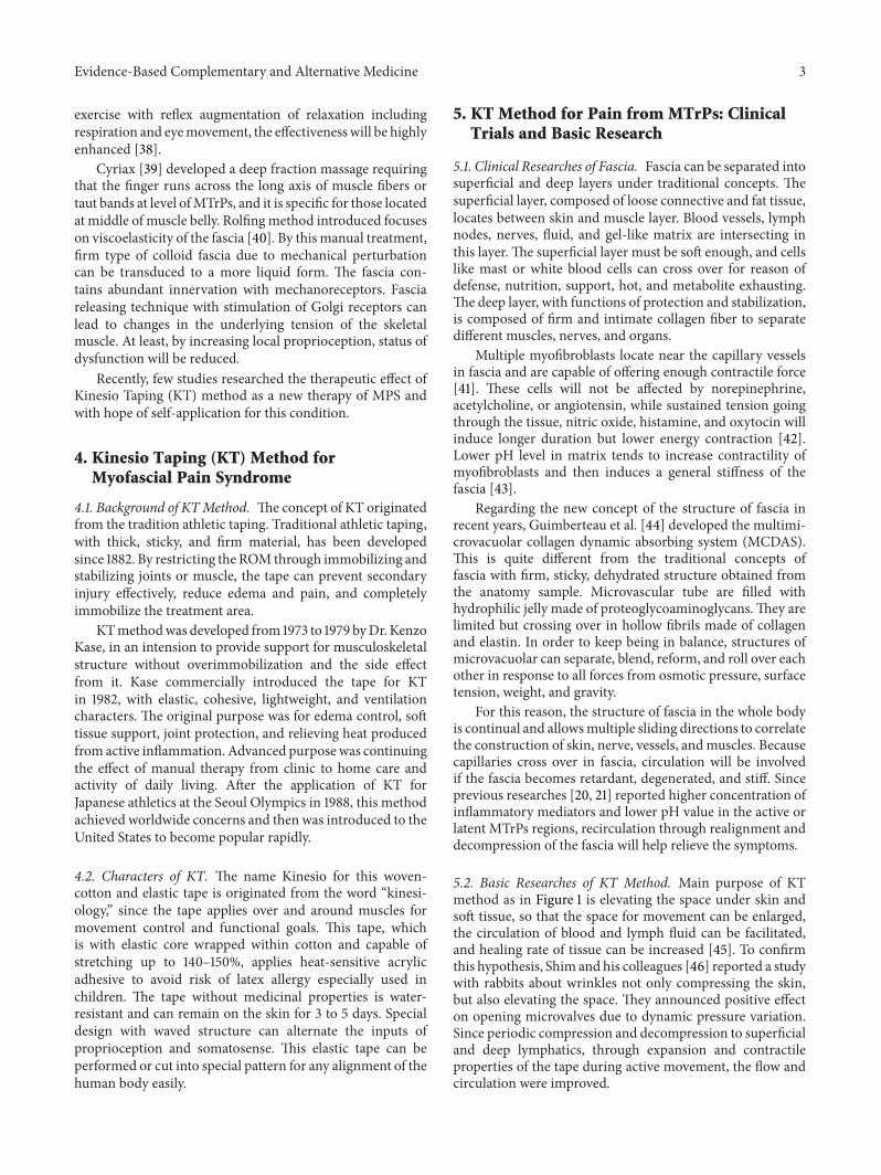
Review ArticleThe Kinesio Taping Method for Myofascial Pain Control
Wei-Ting Wu,1 Chang-Zern Hong,2 and Li-Wei Chou1,3,4
1Department of Physical Medicine and Rehabilitation, China Medical University Hospital, Taichung 40447, Taiwan2Department of Physical Therapy, Hungkuang University, Taichung 43302, Taiwan3School of Chinese Medicine, College of Chinese Medicine, China Medical University, Taichung 40402, Taiwan4Research Center for Chinese Medicine & Acupuncture, China Medical University, Taichung 40402, Taiwan
Correspondence should be addressed to Li-Wei Chou; [email protected]
Received 8 March 2015; Revised 2 April 2015; Accepted 8 April 2015
Academic Editor: Alan Needle
Copyright © 2015 Wei-Ting Wu et al. This is an open access article distributed under the Creative Commons Attribution License,which permits unrestricted use, distribution, and reproduction in any medium, provided the original work is properly cited.
Many people continue suffering from myofascial pain syndrome (MPS) defined as a regional pain syndrome characterized bymuscle pain caused by myofascial trigger points (MTrPs) clinically. Muscle spasm and block of blood circulation can be noticed inthe taut bands. In the MTrP region, nociceptors can be sensitized by the peripheral inflammatory factors and contracture of fasciacan also be induced. Traditional treatments of MPS include stretching therapy, thermal treatment, electrical stimulation, massage,manipulation, trigger points injection, acupuncture, and medicine. However, the pain syndrome may not be relieved even undermultiple therapies. Recently, the Kinesio Taping (KT)method is popularly used in sports injuries, postoperative complications, andvarious pain problems, but little research is focused on MPS with KT method. In this paper, we review the research studies on theapplication to KT in treating MPS and other related issues. It appears that the KT application can elevate the subcutaneous spaceand then increase the blood circulation and lymph fluid drainage to reduce the chemical factors around theMTrP region.Therefore,it is suggested that KT method can be used as a regular treatment or added to the previous treatment for myofascial pain.
1. Introduction
1.1. Myofascial Pain Syndrome (MPS). Myofascial pain syn-drome, defined as muscle pain due to myofascial triggerpoints (MTrPs) [1], has been considered to be related topoor postures, neuromusculoskeletal disorders, or systemicdiseases [2]. Besides, chronic repetitive minor muscle strain,bursitis, enthesopathy, arthritis, or disc lesion can also induceMPS [2]. Clinically, patients with myofascial pain complainabout local pain in the muscle, often with referred pain.If the associated pathologic reasons are not well treated,the pain often recurs later [2]. When performing physicalexamination, the MTrP in a taut band of skeletal musclecan be palpated and local twitch response can be elicited bysnapping of the MTrP [3]. Other symptoms of myofascialpain include range of motion (ROM) limitation, soonerexhausting, and referred spasm.
1.2. Myofascial Trigger Point (MTrP). For the diagnosis ofMTrP, “spot tenderness,” “taut band,” and “pain recognition”
are suggested as the three basic criteria, and “referred pain”and “local twitch responses” are the “signs” for it [4]. Inpatients suffering from MPS, both latent and active MTrPsmay be noted, with characters of spontaneous pain sensationor pain in response to the muscle movement in active MTrPsand tender without spontaneous pain sensation in latentMTrPs. Patient with MPS begins with one active MTrP,called primary MTrP, in the affected muscle due to reasonsmentioned above. When under inappropriate treatment,expanding of pain region and additional active MTrPs, calledsecondary or satellite MTrPs, will develop [1].
2. Hypothetical Mechanism ofMyofascial Pain Syndrome
2.1. Etiology of Myofascial Trigger Point. Acute muscle over-load can activate MTrPs. If the lesion is not well controlled,progressive scar tissue will be formed and become a chroniclesion. It may be the major cause of degeneration andactivation of MTrP in later life [5].
Hindawi Publishing CorporationEvidence-Based Complementary and Alternative MedicineVolume 2015, Article ID 950519, 9 pageshttp://dx.doi.org/10.1155/2015/950519

2 Evidence-Based Complementary and Alternative Medicine
2.2. Integrated Hypothesis of MTrP. In the hypothesis ofMTrP as energy crisis postulated by Simons and Travell[6], they have considered “excessive acetylcholine releasing,”“sarcomere shortening,” and “increasing of sensitizing sub-stances” as the three essential characters for the formationof MTrP [7]. An MTrP is composed of multiple contractionknots with sarcomeres overcontracture and increased diame-ter of that muscle [8, 9]. AnMTrP contains multiple sensitiveloci suspected as nociceptors and active loci in neuromuscu-lar junctions activated with excessive acetylcholine leakageeven under relaxation in the nonendplate zone [10]. In thiscondition, the sarcomeres in the endplate zone will contractcontinually and form the contraction knot in the endplatezone and the taut band in the whole muscle fiber, the painthreshold of nociceptors will be decreased, and the symptomsbecome severe. When energy crisis occurs, ischemic tissueslack adenosine triphosphate to promote calcium pump inthe sarcoplasmic reticulum. It will make the muscle contractcontinually with regional sarcomere shortening and thenblocks the supplements of nutrition and oxygen moreover[7, 11]. Local ischemia and hypoxia can induce secretion ofsensitizing substances to cause pain and release abnormalacetylcholine resulting in a vicious cycle [7].
2.3. Clinical Researches of MTrP. To investigate the pain,research of MacDonald [12] showed that muscles with activeMTrPs have restricted passive ROM. Since the tension ofinvolved muscle fibers has been increased even at rest,stretching the muscle beyond limitation can produce severepain. Painful contraction can be also noted when performingtest of fixed resistance [12]. After treating the MTrPs andreleasing the taut band, the ROM can be returned to originalstatus.
In surface electromyographic (EMG) studies performedby Headley [13, 14], it was found that muscles with activeMTrPs beginning fatigue, exhausting sooner, and recoveringlater than normal muscles. Another research using surfaceEMG for endurance test on myofascial pain demonstratedthat amplitudes of EMG activity increased and mean powerfrequency decreasedwith time. And on themore painful side,accelerated fatigability was noted with shorter duration ofendurance comparing to the normal side [15]. In a researchwith surface EMG, it was shown that the muscle containingactive MTrPs was under status of fatigue, and exhaustingthe energy earlier than the normal one [16]. Besides, afterinjection of 2% lidocaine solution on trigger points of uppertrapezius muscle, significant reduction in pain intensity (𝑃 <0.001) and EMG activity (𝑃 < 0.03) in ipsilateral massetermuscle was noted with confirmation of referred spasm due tomyofascial pain [17].
In consideration of image finding, Sikdar et al. applyultrasound on taut band in the upper trapezius [18]. Theyfound MTrPs as focal, hypoechoic regions and reducedvibration amplitude under vibration sonoelastography. Itindicated local changes in tissue echogenicity and appearedwith elliptic nodules of size about 0.16± 0.11 cm2. Chen et al.[19], through MRI study, noted that stiffness of the taut band
in patients with myofascial pain was about 9.0 kPa and 50%greater than surrounding tissue.
To confirm the theory of energy crisis, Shah and hiscolleagues used micropipettes to determine the pH valueand the electrolyte concentration in both active and latenttrigger points and also in control points [20, 21]. They foundsignificant higher concentration of inflammatory mediators(like bradykinin, substance P, tumor necrosis factor-alpha,interleukin-1 beta, serotonin, and norepinephrine) and lowerpH value in the active or latent MTrPs regions than those inthe normal points.These inflammatorymediators can induceperipheral sensitization of nociceptors in muscle or centralsensitization in central nervous system. When the messagewas transferred to spinal cord through nociceptors, it caninduce neural circuits of MTrP in central nervous systemand can form latent MTrPs in muscle [2, 21, 22]. Whenincreasing the stimulation to this neural circuit due to acuteor chronic injury, latent MTrPs can be activated into painfulactive MTrPs.
Researches conducted by Mense [23, 24] for centralsensitization reported that persistent stimulation of sensoryafference from muscles would lead to neuroplastic changesin the posterior horn of the spinal cord and allodyniaoften associated with active MTrPs. Releasing of substanceP, glutamate, and calcitonin gene-related peptide from theprimary afferent fibers can sensitize the nociceptors eitherat receptive or spinal ends. These neuropeptides will alsoenter into other synaptic associations with other posteriorhorn neurons with consequence of hyperalgesia. Besides,nociceptors near the site of pathology can transmit messagesto neural connections of associated MTrPs and then inducedthe latent MTrPs to active MTrPs.
3. Treatment of Myofascial Trigger Point
Due to multiple factors of MPS, single management ortherapy may not overcome the problem effectively. Treat-ments ofMTrP includemanual therapies [1], physical therapymodalities [25], needling therapy (including MTrP injection[26], dry needling [27–29], acupuncture [30–32], percuta-neous soft tissue release [33], and subcutaneous needling[34]), or oral medicine. Eliminating any perpetuating factorsand introducing adequate education and home programs topatients are also important [1, 35].
The earliest effective therapy suggested for treatment isspraying ethyl chloride on skin combined with stretch [1].Travell suggested applying two or three sweeps of spray beforeor concurrently while gently stretching the muscle to its fulllength [36]. But due to the side effects, such as respiratorytract injury, freezing, and environmental destruction, thespray was displaced with ice rubbing.
In exercise therapy, Lewit and Simons [37] introducedpostisometric relaxation (PIR) exercise as a treatment.Patients perform isometric contraction on thosemuscleswith10–25% of full strength. Then they make the patient relaxthe muscle three to ten seconds later, following mild stretchof the same muscle by clinician, and relax again. The circleshall be performed for several times. When combining PIR
Evidence-Based Complementary and Alternative Medicine 3
exercise with reflex augmentation of relaxation includingrespiration and eyemovement, the effectivenesswill be highlyenhanced [38].
Cyriax [39] developed a deep fraction massage requiringthat the finger runs across the long axis of muscle fibers ortaut bands at level ofMTrPs, and it is specific for those locatedat middle of muscle belly. Rolfingmethod introduced focuseson viscoelasticity of the fascia [40]. By this manual treatment,firm type of colloid fascia due to mechanical perturbationcan be transduced to a more liquid form. The fascia con-tains abundant innervation with mechanoreceptors. Fasciareleasing technique with stimulation of Golgi receptors canlead to changes in the underlying tension of the skeletalmuscle. At least, by increasing local proprioception, status ofdysfunction will be reduced.
Recently, few studies researched the therapeutic effect ofKinesio Taping (KT) method as a new therapy of MPS andwith hope of self-application for this condition.
4. Kinesio Taping (KT) Method forMyofascial Pain Syndrome
4.1. Background of KTMethod. The concept of KT originatedfrom the tradition athletic taping. Traditional athletic taping,with thick, sticky, and firm material, has been developedsince 1882. By restricting the ROM through immobilizing andstabilizing joints or muscle, the tape can prevent secondaryinjury effectively, reduce edema and pain, and completelyimmobilize the treatment area.
KTmethodwas developed from 1973 to 1979 byDr. KenzoKase, in an intension to provide support for musculoskeletalstructure without overimmobilization and the side effectfrom it. Kase commercially introduced the tape for KTin 1982, with elastic, cohesive, lightweight, and ventilationcharacters. The original purpose was for edema control, softtissue support, joint protection, and relieving heat producedfrom active inflammation. Advanced purposewas continuingthe effect of manual therapy from clinic to home care andactivity of daily living. After the application of KT forJapanese athletics at the Seoul Olympics in 1988, this methodachieved worldwide concerns and then was introduced to theUnited States to become popular rapidly.
4.2. Characters of KT. The name Kinesio for this woven-cotton and elastic tape is originated from the word “kinesi-ology,” since the tape applies over and around muscles formovement control and functional goals. This tape, whichis with elastic core wrapped within cotton and capable ofstretching up to 140–150%, applies heat-sensitive acrylicadhesive to avoid risk of latex allergy especially used inchildren. The tape without medicinal properties is water-resistant and can remain on the skin for 3 to 5 days. Specialdesign with waved structure can alternate the inputs ofproprioception and somatosense. This elastic tape can beperformed or cut into special pattern for any alignment of thehuman body easily.
5. KT Method for Pain from MTrPs: ClinicalTrials and Basic Research
5.1. Clinical Researches of Fascia. Fascia can be separated intosuperficial and deep layers under traditional concepts. Thesuperficial layer, composed of loose connective and fat tissue,locates between skin and muscle layer. Blood vessels, lymphnodes, nerves, fluid, and gel-like matrix are intersecting inthis layer. The superficial layer must be soft enough, and cellslike mast or white blood cells can cross over for reason ofdefense, nutrition, support, hot, and metabolite exhausting.The deep layer, with functions of protection and stabilization,is composed of firm and intimate collagen fiber to separatedifferent muscles, nerves, and organs.
Multiple myofibroblasts locate near the capillary vesselsin fascia and are capable of offering enough contractile force[41]. These cells will not be affected by norepinephrine,acetylcholine, or angiotensin, while sustained tension goingthrough the tissue, nitric oxide, histamine, and oxytocin willinduce longer duration but lower energy contraction [42].Lower pH level in matrix tends to increase contractility ofmyofibroblasts and then induces a general stiffness of thefascia [43].
Regarding the new concept of the structure of fascia inrecent years, Guimberteau et al. [44] developed the multimi-crovacuolar collagen dynamic absorbing system (MCDAS).This is quite different from the traditional concepts offascia with firm, sticky, dehydrated structure obtained fromthe anatomy sample. Microvascular tube are filled withhydrophilic jelly made of proteoglycoaminoglycans.They arelimited but crossing over in hollow fibrils made of collagenand elastin. In order to keep being in balance, structures ofmicrovacuolar can separate, blend, reform, and roll over eachother in response to all forces from osmotic pressure, surfacetension, weight, and gravity.
For this reason, the structure of fascia in the whole bodyis continual and allowsmultiple sliding directions to correlatethe construction of skin, nerve, vessels, andmuscles. Becausecapillaries cross over in fascia, circulation will be involvedif the fascia becomes retardant, degenerated, and stiff. Sinceprevious researches [20, 21] reported higher concentration ofinflammatory mediators and lower pH value in the active orlatent MTrPs regions, recirculation through realignment anddecompression of the fascia will help relieve the symptoms.
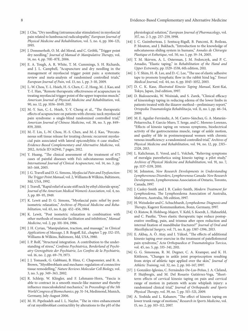
5.2. Basic Researches of KT Method. Main purpose of KTmethod as in Figure 1 is elevating the space under skin andsoft tissue, so that the space for movement can be enlarged,the circulation of blood and lymph fluid can be facilitated,and healing rate of tissue can be increased [45]. To confirmthis hypothesis, Shim and his colleagues [46] reported a studywith rabbits about wrinkles not only compressing the skin,but also elevating the space. They announced positive effecton opening microvalves due to dynamic pressure variation.Since periodic compression and decompression to superficialand deep lymphatics, through expansion and contractileproperties of the tape during active movement, the flow andcirculation were improved.

4 Evidence-Based Complementary and Alternative Medicine
Kinesio tape
Epidermis
Dermis
Blood and lymph vessels
Muscle
Lymph fluid
Bleeding, pressure, and pain
Before taping After taping
Figure 1: These two pictures showed the mechanism of KT application on soft tissue. Before taping, the lesion site, such as a taut band orbruise, may contain bleeding, pressure, and lymph fluid accumulation and then cause pain. After taping as shown with blue curve, the space-lifting mechanism will help the drainage of fluid. Then the inflammation factors and pressure can be reduced, and the movement of musclecan be improved.
To confirm this effect, Kase [47] once researched theinfluence of taping on blood circulation. The participantswere randomly tested through ultrasound under Dopplerview for radial, superficial temporal, and dorsalis pedis arterybefore and after taping. It was found that the flow rate wasincreased immediately after KT.
Bialoszewski and his colleagues [48] studied 24 patientstreated with Ilizarov method for lower limb lengthening andcomplicated with thigh edema. They were divided into twogroups. Both groups received 10 days of standard physio-therapy, and additional application of KT was performed inthe experimental group.They reported statistically significantdecrease in the circumference of thigh and leg in both groups(𝑃 = 0.02, 𝑃 = 0.03, resp.), with more significance inthe experimental group than the control group with onlystandard lymphatic massage. However, they did not providestatistical data for intergroup comparison research.
Aguilar-Ferrandiz and his colleagues [49] reported anarticle of treating patients with chronic venous insufficiencyfor 4 weeks. Participants were randomly assigned into anexperimental group for standardized KT application or acontrol group for sham KT treatment. Only experimentalgroup showed improvements compared to pretreatment val-ues in swelling (𝑃 < 0.002), muscle cramps (𝑃 < 0.001),and pain distribution (𝑃 < 0.001). Intragroup comparisonsfor the improvement of pain score between baseline andposttreatment were significantly greater in the experimentalgroup than the sham group. But placebo effect should alsobe taken into consideration because of mild reducing of painscore in control group.
Kalichman et al. [50] reported a case suffering frommer-algia paresthetica with symptoms of numbness, paresthesias,and pain in the anterolateral thigh. After using KT methodfor 4 weeks, symptoms and quality of life were significantlyimproved.
5.3. Mechanism of KT Method on Lymphatic System. Ininvestigation of lymphatic system, a one-way system belowthe surface of skin, it depends on negative-pressure pumpingto guide the fluid flow from superficial to deep layer. Thisnegative-pressure effect is facilitated by the alternativemusclecontraction and relaxation. If interstitial pressure aroundthe lymphatic system is much increasing due to edema,intercellular junction doors will be closed [51]. By musclecontraction, relaxation, and therapies such as massage orcompression garment, pressure in each segment can bechanged. The lymph and interstitial fluid can recirculate anddecrease the swelling and pain sensation [52, 53].
Besides, due to the drainage effect of lymph edema, thecirculation of lesions can be improved and then acceleratesthe healing of the tissue. Ristow et al. [54] once researched theapplication of Kinesio Tex Tape after surgery for mandibularfracture. They announced statistically significant decreasingthe incidence of swelling when applying KT during the first 2days after operation.
5.4. Other Probable Mechanisms of KT Method for Propri-oception and Pain. Many articles have reported differentconclusions of effects on taping, such as proprioception,placebo effect, warningmessage, or biomechanics. For exam-ple, treatment with additional KT method over exercise forpatellofemoral pain syndrome could provide significantlybetter hamstring flexibility than the control group (𝑃 < 0.05)[55]. On the other hand, a research of athletic tapes alsoshowed that increasing cutaneous sensory feedback wouldimprove ankle joint position perception (𝑃 < 0.05) undernon-weight bearing condition [56].
Besides, some researchers reported that KT methodwould improve the ROM of neck [57] and lower trunk[58]. For example, Osterhues [59] researched the effect oftaping on those who suffered from acute whiplash injury.

Evidence-Based Complementary and Alternative Medicine 5
The study group showed a significantly greater decrease inpain immediately (𝑃 < 0.001) and also significant increasein ROM in all directions. Osterhues [59] treated patientsof patella bony dislocation with KT based on the conceptof stimulation to mechanical receptors of skin from tapingand found that the balance and motor control were betterthan previous status. When performing the kneeling downor eccentric contraction of leg under-weight bearing, visualanalogue scale (VAS) was decreased.
Effect of pain control of KT method may associate withgate control theory [60]. A𝛽-fiber, afferent fiber from sensoryneurons of touch, is bigger in diameter and conductionvelocity than those for pain including A𝛿-fiber and C-fiber.By stimulating the afferent receptors with light touch inthe skin can activate glial cells in the spinal cord. Thentransmission of pain will be inhibited at the spinal cord levelfrom transmitting to the cortex. Until now, no research canconfirm that KT can provide effectiveness via this theory butcan depend on literatures of other physical modalities usedfor reduction of pain [61, 62]. Other issues such as balanceof agonist and antagonist, tendon and ligament protection,decreasing muscle activity of protective contracture, andsensing of movement still require further studies.
5.5. Clinical Taping for Facilitation and Inhibition. Inresearching of facilitation for muscle, Rood facilitationincluding fast brushing, light moving touch, and icing onfascia is reported as a useful method [63]. On the other hand,the inhibitory pattern is correlated with the Golgi tendonorgan (GTO) located at muscle-tendon junction in theinsertion end. These methods include lightly compressingthe joints, compressing the muscle-tendon junctions, andkeeping tendon in stretched condition [64]. GTO, sensitiveto changes in muscle tension, connects to muscle fibers andcommunicates the muscle spindle status. When in action,GTO inhibits its own muscle and excites the antagonist [65].
The input of stimulation for facilitation and inhibitionmentioned above can be achieved through taping. By chang-ing proprioception, biofeedback from correct movementpattern, and training or rehabilitation in high intensity, thetarget muscle groups and coordination can be improved.Then let the patient perform similar suitable movement afterremoving the tape.
Kaya et al. [66] applied KT combined with home exerciseprogram on patients who suffered from shoulder pain dueto impingement syndrome. The tapes were applied with“insertion to origin technique” over the supraspinatus, teresminor, and deltoid muscles. Significant improvement inpain and disability was noted in the taping group one totwo weeks later. In Simsek’s research [67], for outcomes insubacromial impingement syndrome with KT in addition toexercise therapy comparing to the sham taping, pain duringmovement in the therapeutic group was significantly lowerat the 5th day (𝑃 < 0.01) in intergroup comparisons. Andnight pain, pain with movement, shoulder external rotationmuscle strength, and pain-free shoulder abduction ROMin the therapeutic group at the 12th day were significantlyimproved (𝑃 < 0.05).
5.6. Probable Mechanism of KT Method for MTrPs. Theconcepts mentioned above can be compatible with thehypothesis proposed by Kase about the space, movement,and cooling effect of taping. Increasing the space of fasciato improve the circulation can remove the heat producedfrom inflammation. Pain sensation can be diminished dueto reduction of the pressure on nociceptors. The theory issimilar to that of treatment for MPS [2, 68, 69]; therefore wehypothesize that KT method can block the vicious circle ofenergy crisis. Figure 2 showed the possibleKTmechanism forthe relief of myofascial pain.
5.7. Researches of KT Method for Myofascial Pain. Wang andher colleagues [70] investigated the effect of KT methodfor the relief of MPS. Taping with “insertion to origintechnique” was performed on the upper trapezius muscleand statistically significant pain relief was found immediatelyafter the treatment. They considered the effects due totaut band stretching and stimulation of skin receptors. Noimprovement was reported by the patients in the controlgroup. The improvement of VAS in the experimental groupremained statistically significant 24 hours later.
Garcıa-Muro et al. [71] reported a patient with shoulderpain of myofascial origin under intervention of KT method.They found that this patient had significant improvementin the VAS, algometry, functional tests, and active ROM,resolving the problem in the following days. Therefore, theyannounced that KT methods were appropriate for treatingMTrPs.
6. Clinical Application of KT Method:Prescription and Contraindication
KT method combined with other therapeutic interventionsmay reduce pain, regulate (facilitate or inhibit) muscle func-tion, provide proprioceptive feedback, and stabilize joint.Before prescribing KT, the practitioner shall understandthe past history, posture at daily living, type of work orexercise, biomechanics and duration of injury, and previoustreatments and the effects. Detailed physical examinationincludes inspection, palpation, active and passive ROM,resistant test, and identification of the MTrPs and taut bands.It is also important to consider the “upper and lower crossedsyndrome” announced by Janda [72], such as tightness ofupper trapezius and pectoralis combined with weakness ofrhomboid and anterior serratus. After history taking, exami-nation, and locating the MTrPs, manual therapy or needling(dry needling or injection to MTrPs) can be performed asnecessary. For relieving the swelling and tenderness afterinjection, taping can be used following the guidance for thedesired direction of drainage.
The basic principle of prescription of KT for myofascialpain focuses on the patterns of facilitation and inhibition.When the tape applies on the muscle from its origin tothe insertion site, it can provide the effect of facilitationto the muscle contraction [47]. On the other hand, whentaping from insertion to origin, inhibition and relaxation ofmuscle spasm will be the effect [47] which is most useful formyofascial pain and muscle spasm.

6 Evidence-Based Complementary and Alternative Medicine
Placebo effectImprovement of motor control
Prevention of further injury or reinjury
BiofeedbackProprioceptive stimulation
(1) Removal of inflammatory substance
(2) Removal of inflammatoryheat
(3) Decrease of pressure on nociceptors
Immobilization
Somatosensory stimulation
Gate control
Increase of space in soft tissue
Facilitation of circulation
Golgi tendon organ stimulation
due to MTrPsRelief of pain
Figure 2: The possible mechanism of Kinesio Taping for the relief of myofascial pain.
The contraindication of KT method includes taping onsites of acute infection, open wounds, deep vein thrombosis,malignancy, and severe allergy. Djordjevic et al. [73] sug-gested checking skin for allergy to the tape by applying a small(1 × 1 cm2) patch of tape on the volar side of forearm. Thepositive finding of this allergy test was redness or other skinchanges noted in 15 minutes. If patient suffered from diabetesmellitus, taping shall be carefully used due to possible sensorydefect.
7. Limitation of Researches on KT Method
Researches for the effect of KT method often show differentresults. Hsu and his colleagues [74] applied taping on baseballplayers with shoulder impingement and then measured theEMG activities of the upper and lower trapezius. They foundsignificant increase of the lower trapezius muscle activity inthe arm-lowering phase (60–30∘, 𝑃 < 0.05) in comparisonto the placebo taping. Similar research with increasing bio-electrical activity of vastus medialis muscle was reported bySłupik et al. [75]. Other researchers such as Firth et al. [76]used the Hoffman reflex amplitudes to assess the effect oftaping on people with Achilles tendinopathy. They reportedthat the H reflex remained unchanged in the soleus andgastrocnemius in study group.
Therefore, more research studies are necessary to clarityof the effectiveness of KT method. Many limitations inresearches shall be overcome, and it is very difficult inbuilding random or double blind trials. Furthermore, thetaping techniques were variable in previous studies. Althoughall procedures of KT for a similar disorder were performedaccording to Kase’s original concept, different practitionersmight perform different technique based on their previousexperience and manner and could induce the bias [77].
Another limitation for research is the placebo effect. Someresearch announced that visual input of different color andthe sensation stuck on skinmaymake the positive expectancy[78] and may make the patients feel confidence, stability, andreassurance [79].
Besides, no suitable machine or image data can confirmthe effect of taping at anytime and anywhere. With thefunctional improvement of ultrasound, certain landmarkcould be tracked more easily. By using sonography, we canexactly define the depth, certain muscle, and surroundingtissue and note the twitch response during injection. We cannot only avoid injury from treatment but also increase thespecificity of the target tissue for taping. For example, incases with epicondylalgia after KT method application, Liuand his colleagues [80] reported improvement of epicondylarmuscles sliding in ultrasonic image when wrist was moving.Since the flexibility of tissue correlates with the pathologicstatus, sonoelastography can be used for identifying thelocation and may be considered for researching of outcomesafter taping.
8. Conclusion
In clinical practices, KT method was applied in sportsinjuries, postoperative complications, various pain problems,and many other conditions. The tape is simple to carry out,economic, and less traumatic. In treatment of patient withMPS who cannot be rehabilitated regularly, some researcherssuggested taping through self-application as a new therapy[70]. However, self-application of tape may be difficult insome aspects including the limited knowledge in anatomyor biomechanics, the inadequate knowledge in trigger pointexamination, the lack of experience of taping method,the requirement of using both hands, and the location of

Evidence-Based Complementary and Alternative Medicine 7
the MTrP (such as rhomboidmuscle).Therefore,most peoplecannot tape by themselves. In order to obtain a bettereffect, it is also necessary to combine therapeutic exercise,postural changing, and adjustment of daily living. Finally,we considered that KT method could be applied as anotherchoice of MTrP therapy but could need more researches toconfirm the effectiveness.
Conflict of Interests
Financial disclosure statements have been obtained, and noconflict of interests has been reported by the authors or byany individuals in control of the content of this paper.
Authors’ Contribution
Chang-ZernHong hadmade the same effort asWei-TingWu.
Acknowledgments
This study was supported in part by Taiwan Ministry ofHealth and Welfare Clinical Trial and Research Center ofExcellence (MOHW104-TDU-B-212-113002) and by ChinaMedical University (CMU) under the Aim for TopUniversityPlan of the Ministry of Education, Taiwan.
References
[1] J. G. Travell andD. G. Simons,Myofascial Pain andDysfunction:The Trigger PointManual, vol. 1, LippincottWilliams&Wilkins,Baltimore, Md, USA, 1983.
[2] C.-Z. Hong, “Myofascial pain therapy,” Journal of Musculoskele-tal Pain, vol. 12, no. 3-4, pp. 37–43, 2004.
[3] D. G. Simons, C.-Z. Hong, and L. S. Simons, “Endplate poten-tials are common to midfiber myofacial trigger points,” TheAmerican Journal of Physical Medicine and Rehabilitation, vol.81, no. 3, pp. 212–222, 2002.
[4] R. D. Gerwin, S. Shannon, C.-Z. Hong, D. Hubbard, andR. Gevirtz, “Interrater reliability in myofascial trigger pointexamination,” Pain, vol. 69, no. 1-2, pp. 65–73, 1997.
[5] R. Cailliet, Soft Tissue Pain and Disability, F.A. Davis Company,Philadelphia, Pa, USA, 1977.
[6] D. G. Simons and J. Travell, “Myofascial trigger points, apossible explanation,” Pain, vol. 10, no. 1, pp. 106–109, 1981.
[7] D. G. Simons, “New aspects of myofascial trigger points:etiological and clinical,” Journal of Musculoskeletal Pain, vol. 12,no. 3-4, pp. 15–21, 2004.
[8] D. G. Simons and W. C. Stolov, “Microscopic features andtransient contraction of palpable bands in canine muscle,”American Journal of Physical Medicine, vol. 55, no. 2, pp. 65–88,1976.
[9] C. Bron and J. D. Dommerholt, “Etiology of myofascial triggerpoints,” Current Pain and Headache Reports, vol. 16, no. 5, pp.439–444, 2012.
[10] C. Z. Hong, J. T. Chen, S. M. Chen, J. J. Yan, and Y. J. Su,“Histological findings of responsive loci in a myofascial triggerspot of rabbit skeletal muscle from where localized twitchresponses could be elicited,” Archives of Physical Medicine andRehabilitation, vol. 77, article 962, 1996.
[11] D. G. Simons, “Clinical and etiological update of myofascialpain from trigger points,” Journal of Musculoskeletal Pain, vol.4, no. 1-2, pp. 93–121, 1996.
[12] A. J. R. MacDonald, “Abnormally tender muscle regions andassociated painful movements,” Pain, vol. 8, no. 2, pp. 197–205,1980.
[13] B. J. Headley, “Evaluation and treatment of myofascial painsyndrome utilizing biofeedback,” in Clinical EMG for SurfaceRecordings, vol. 2, pp. 235–254, Clinical Resources, 1990.
[14] B. J. Headley, “Physiologic risk factors,” in Management ofCumulative Trauma Disorder, M. Sanders, Ed., pp. 107–127,Butterworth-Heinemann, Boston, Mass, USA, 1997.
[15] M. Hagberg and S. Kvarnstrom, “Muscular endurance and elec-tromyographic fatigue in myofascial shoulder pain,” Archives ofPhysical Medicine and Rehabilitation, vol. 65, no. 9, pp. 522–525,1984.
[16] S. Mense and D. G. Simons, Muscle Pain. UnderstandingIts Nature, Diagnosis, and Treatment, Lippincott Williams &Wilkins, Philadelphia, Pa, USA, 2001.
[17] C. R. Carlson, J. P. Okeson, D. A. Falace, A. J. Nitz, and J. E.Lindroth, “Reduction of pain and EMG activity in the masseterregion by trapezius trigger point injection,” Pain, vol. 55, no. 3,pp. 397–400, 1993.
[18] S. Sikdar, J. P. Shah, T. Gebreab et al., “Novel applications ofultrasound technology to visualize and characterize myofascialtrigger points and surrounding soft tissue,” Archives of PhysicalMedicine and Rehabilitation, vol. 90, no. 11, pp. 1829–1838, 2009.
[19] Q. Chen, S. Bensamoun, J. R. Basford, J. M. Thompson,and K.-N. An, “Identification and quantification of myofascialtaut bands with magnetic resonance elastography,” Archives ofPhysical Medicine and Rehabilitation, vol. 88, no. 12, pp. 1658–1661, 2007.
[20] J. P. Shah, T. M. Phillips, J. V. Danoff, and L. H. Gerber, “Anin vivo microanalytical technique for measuring the local bio-chemical milieu of human skeletal muscle,” Journal of AppliedPhysiology, vol. 99, no. 5, pp. 1977–1984, 2005.
[21] J. P. Shah, J. V. Danoff, M. J. Desai et al., “Biochemicalsassociated with pain and inflammation are elevated in sites nearto and remote from active myofascial trigger points,” Archivesof Physical Medicine and Rehabilitation, vol. 89, no. 1, pp. 16–23,2008.
[22] Y.-L. Hsieh, L.-W. Chou, Y.-S. Joe, and C.-Z. Hong, “Spinal cordmechanism involving the remote effects of dry needling on theirritability of myofascial trigger spots in rabbit skeletal muscle,”Archives of Physical Medicine and Rehabilitation, vol. 92, no. 7,pp. 1098–1105, 2011.
[23] S. Mense, “Nociception from skeletal muscle in relation toclinical muscle pain,” Pain, vol. 54, no. 3, pp. 241–289, 1993.
[24] S. Mense, “Neurobiological basis of muscle pain,” Schmerz, vol.13, no. 1, pp. 3–17, 1999.
[25] C.-Z.Hong, Y.-C. Chen, C.H. Pon, and J. Yu, “Immediate effectsof various physical medicine modalities on pain threshold ofmyofascialtrigger points,” Journal of Musculoskeletal Pain, vol.1, no. 2, pp. 37–52, 1993.
[26] C. Z. Hong, “Myofascial trigger point injection,” Critical Reviewof Physical and RehabilitationMedicine, vol. 5, no. 2, pp. 203–217,1993.
[27] P. Baldry, “Superficial dry needling at myofascial trigger pointsites,” Journal of Musculoskeletal Pain, vol. 3, no. 3, pp. 117–126,1995.
8 Evidence-Based Complementary and Alternative Medicine
[28] J. Chu, “Dry needling (intramuscular stimulation) inmyofascialpain related to lumbosacral radiculopathy,” European Journal ofPhysical Medicine and Rehabilitation, vol. 5, no. 4, pp. 106–121,1995.
[29] J. Dommerholt, O. M. del Moral, and C. Grobli, “Trigger pointdry needling,” Journal of Manual & Manipulative Therapy, vol.14, no. 4, pp. 70E–87E, 2006.
[30] E. A. Tough, A. R. White, T. M. Cummings, S. H. Richards,and J. L. Campbell, “Acupuncture and dry needling in themanagement of myofascial trigger point pain: a systematicreview and meta-analysis of randomised controlled trials,”European Journal of Pain, vol. 13, no. 1, pp. 3–10, 2009.
[31] L.-W. Chou, Y.-L. Hsieh, H.-S. Chen, C.-Z.Hong,M.-J. Kao, andT.-I. Han, “Remote therapeutic effectiveness of acupuncture intreatingmyofascial trigger point of the upper trapezius muscle,”American Journal of Physical Medicine and Rehabilitation, vol.90, no. 12, pp. 1036–1049, 2011.
[32] M.-Y. Sun, C.-L. Hsieh, Y.-Y. Cheng et al., “The therapeuticeffects of acupuncture on patients with chronic neckmyofascialpain syndrome: a single-blind randomized controlled trial,”American Journal of Chinese Medicine, vol. 38, no. 5, pp. 849–859, 2010.
[33] M.-T. Lin, L.-W. Chou, H.-S. Chen, and M.-J. Kao, “Percuta-neous soft tissue release for treating chronic recurrent myofas-cial pain associated with lateral epicondylitis: 6 case studies,”Evidence-Based Complementary and Alternative Medicine, vol.2012, Article ID 142941, 7 pages, 2012.
[34] Y. Huang, “The clinical assessment of the treatment of 675cases of painful diseases with Fu’s subcutaneous needling,”International Journal of Clinical Acupuncture, vol. 14, no. 3, pp.165–168, 2005.
[35] J. G. Travell andD. G. Simons,Myofascial Pain andDysfunction:TheTrigger PointManual, vol. 2,Williams&Wilkins, Baltimore,Md, USA, 1992.
[36] J. Travell, “Rapid relief of acute stiff neck by ethyl chloride spray,”Journal of the AmericanMedicalWomen’s Association, vol. 4, no.3, pp. 89–95, 1949.
[37] K. Lewit and D. G. Simons, “Myofascial pain: relief by post-isometric relaxation,” Archives of Physical Medicine and Reha-bilitation, vol. 65, no. 8, pp. 452–456, 1984.
[38] K. Lewit, “Post isometric relaxation in combination withother methods of muscular facilitation and inhibition,”ManualMedicine, vol. 2, pp. 101–104, 1986.
[39] J. H. Cyriax, “Manipulation, traction, and massage,” in ClinicalApplications of Massage, J. B. Rogoff, Ed., chapter 7, pp. 152–155,Williams &Wilkins, Baltimore, Md, USA, 1980.
[40] I. P. Rolf, “Structural integration. A contribution to the under-standing of stress,” Confinia Psychiatrica, Borderland of Psychi-atry Grenzgebiete der Psychiatrie, Les Confins de la Psychiatrie,vol. 16, no. 2, pp. 69–79, 1973.
[41] J. J. Tomasek, G. Gabbiani, B. Hinz, C. Chaponnier, and R. A.Brown, “Myofibroblasts and mechano: regulation of connectivetissue remodelling,” Nature Reviews Molecular Cell Biology, vol.3, no. 5, pp. 349–363, 2002.
[42] R. Schleip, W. Klingler, and F. Lehmann-Horn, “Fascia isable to contract in a smooth muscle-like manner and therebyinfluence musculoskeletal mechanics,” in Proceedings of the 5thWorldCongress of Biomechanics, pp. 51–54,Medimond,Munich,Germany, July-August 2006.
[43] M. H. Pipelzadeh and I. L. Naylor, “The in vitro enhancementof rat myofibroblast contractility by alterations to the pH of the
physiological solution,” European Journal of Pharmacology, vol.357, no. 2-3, pp. 257–259, 1998.
[44] J. C. Guimberteau, J. Sentucq-Rigall, B. Panconi, R. Boileau,P. Mouton, and J. Bakhach, “Introduction to the knowledge ofsubcutaneous sliding system in humans,” Annales de ChirurgiePlastique et Esthetique, vol. 50, no. 1, pp. 19–34, 2005.
[45] T. M. Skirven, A. L. Osterman, J. M. Fedorczyk, and P. C.Amadio, “Elastic taping,” in Rehabilitation of the Hand andUpper Extremity, pp. 1529–1538, 6th edition, 2011.
[46] J.-Y. Shim, H.-R. Lee, andD.-C. Lee, “The use of elastic adhesivetape to promote lymphatic flow in the rabbit hind leg,” YonseiMedical Journal, vol. 44, no. 6, pp. 1045–1052, 2003.
[47] D. C. K. Kase, Illustrated Kinesio Taping Manual, Kent-Kai,Tokyo, Japan, 2nd edition, 1997.
[48] D. Bialoszewski, W. Wozniak, and S. Zarek, “Clinical efficacyof kinesiology taping in reducing edema of the lower limbs inpatients treated with the ilizarov method—preliminary report,”Ortopedia Traumatologia Rehabilitacja, vol. 11, no. 1, pp. 46–54,2009.
[49] M. E. Aguilar-Ferrandiz, A.M. Castro-Sanchez, G. A.Mataran-Penarrocha, F. Garcıa-Muro, T. Serge, and C. Moreno-Lorenzo,“Effects of kinesio taping on venous symptoms, bioelectricalactivity of the gastrocnemius muscle, range of ankle motion,and quality of life in postmenopausal women with chronicvenous insufficiency: a randomized controlled trial,”Archives ofPhysical Medicine and Rehabilitation, vol. 94, no. 12, pp. 2315–2328, 2013.
[50] L. Kalichman, E. Vered, and L. Volchek, “Relieving symptomsof meralgia paresthetica using kinesio taping: a pilot study,”Archives of Physical Medicine and Rehabilitation, vol. 91, no. 7,pp. 1137–1139, 2010.
[51] M. Johnston, New Research Developments in UnderstandingLymphovenousDisorders, Lymphovenous Canada: NewResearchDevelopments, Lymphovenous Association of Ontario, Toronto,Canada, 1997.
[52] J. Casley-Smith and J. R. Casley-Smith, Modern Treatment forLymphoedema, The Lymphoedema Association of Australia,Malvern, Australia, 5th edition, 1997.
[53] H.Weissleder and C. Schuchhardt, Lymphedema: Diagnosis andTherapy, Kagerer Kommunikation, Bonn, Germany, 1997.
[54] O. Ristow, B. Hohlweg-Majert, V. Kehl, S. Koerdt, L. Hahnefeld,and C. Pautke, “Does elastic therapeutic tape reduce postop-erative swelling, pain, and trismus after open reduction andinternal fixation of mandibular fractures?” Journal of Oral andMaxillofacial Surgery, vol. 71, no. 8, pp. 1387–1396, 2013.
[55] E. Akbas, A. O. Atay, and I. Yuksel, “The effects of additionalkinesio taping over exercise in the treatment of patellofemoralpain syndrome,” Acta Orthopaedica et Traumatologica Turcica,vol. 45, no. 5, pp. 335–341, 2011.
[56] G. G. Simoneau, R. M. Degner, C. A. Kramper, and K. H.Kittleson, “Changes in ankle joint proprioception resultingfrom strips of athletic tape applied over the skin,” Journal ofAthletic Training, vol. 32, no. 2, pp. 141–147, 1997.
[57] J. Gonzalez-Iglesias, C. Fernandez-De-Las-Penas, J. A. Cleland,P. Huijbregts, and M. Del Rosario Gutierrez-Vega, “Short-term effects of cervical kinesio taping on pain and cervicalrange of motion in patients with acute whiplash injury: arandomized clinical trial,” Journal of Orthopaedic and SportsPhysical Therapy, vol. 39, no. 7, pp. 515–521, 2009.
[58] A. Yoshida and L. Kahanov, “The effect of kinesio taping onlower trunk range of motions,” Research in Sports Medicine, vol.15, no. 2, pp. 103–112, 2007.

Evidence-Based Complementary and Alternative Medicine 9
[59] D. J. Osterhues, “The use of Kinesio Taping in the managementof traumatic patella dislocation. A case study,” PhysiotherapyTheory and Practice, vol. 20, no. 4, pp. 267–270, 2004.
[60] R. Melzack and P. D. Wall, “Pain mechanisms: a new theory,”Science, vol. 150, no. 3699, pp. 971–979, 1965.
[61] M. Cameron, Physical Agents in Rehabilitation: From Researchto Practice, WB Saunders, Philadelphia, Pa, USA, 1999.
[62] M. Hardy and W. Woodall, “Therapeutic effects of heat, cold,and stretch on connective tissue,” Journal of HandTherapy, vol.11, no. 2, pp. 148–156, 1998.
[63] L. Pedretti, Occupational Therapy: Practice Skills for PhysicalDysfunction, Mosby, St. Louis, Mo, USA, 1996.
[64] K. MacGregor, S. Gerlach, R. Mellor, and P. W. Hodges,“Cutaneous stimulation from patella tape causes a differentialincrease in vasti muscle activity in people with patellofemoralpain,” Journal of Orthopaedic Research, vol. 23, no. 2, pp. 351–358, 2005.
[65] A. Shumway-Cook and M. Woollacott, Motor Control: Theoryand Practical Applications, Lippincott Williams & Wilkins,Philadelphia, Pa, USA, 2nd edition, 2001.
[66] E. Kaya, M. Zinnuroglu, and I. Tugcu, “Kinesio taping com-pared to physical therapy modalities for the treatment ofshoulder impingement syndrome,” Clinical Rheumatology, vol.30, no. 2, pp. 201–207, 2011.
[67] H. H. Simsek, S. Balki, S. S. Keklik, H. Ozturk, and H.Elden, “Does Kinesio taping in addition to exercise therapyimprove the outcomes in subacromial impingement syndrome?A randomized, double-blind, controlled clinical trial,” ActaOrthopaedica et Traumatologica Turcica, vol. 47, no. 2, pp. 104–110, 2013.
[68] L.-W. Chou, M.-J. Kao, and J.-G. Lin, “Probable mechanisms ofneedling therapies for myofascial pain control,” Evidence-BasedComplementary and Alternative Medicine, vol. 2012, Article ID705327, 11 pages, 2012.
[69] L.-W. Chou, Y.-L. Hsieh, T.-S. Kuan, and C.-Z. Hong, “Needlingtherapy for myofascial pain: recommended technique withmultiple rapid needle insertion,” BioMedicine, vol. 4, no. 2, 2014.
[70] Y.-H. Wang, S.-M. Chen, J.-T. Chen, W.-C. Yen, T.-S. Kuan,and C.-Z. Hong, “The effect of taping therapy on patients withMyofascial pain syndrome: a pilot study,” Taiwan Journal ofPhysical Medicine and Rehabilitation, vol. 36, no. 3, pp. 145–150,2008.
[71] F. Garcıa-Muro, A. L. Rodrıguez-Fernandez, and A. Herrero-de-Lucas, “Treatment of myofascial pain in the shoulder withKinesio Taping. A case report,” Manual Therapy, vol. 15, no. 3,pp. 292–295, 2010.
[72] V. Janda, “Evaluation of muscular imbalance,” in Rehabilitationof the Spine: A Practitioner's Manual, C. Liebenson, Ed., chapter6, pp. 97–112, Williams &Wilkins, Baltimore, Md, USA, 1996.
[73] O. C. Djordjevic, D. Vukicevic, L. Katunac, and S. Jovic,“Mobilization with movement and kinesiotaping comparedwith a supervised exercise program for painful shoulder: resultsof a clinical trial,” Journal of Manipulative and PhysiologicalTherapeutics, vol. 35, no. 6, pp. 454–463, 2012.
[74] Y.-H. Hsu, W.-Y. Chen, H.-C. Lin, W. T. J. Wang, and Y.-F.Shih, “The effects of taping on scapular kinematics and muscleperformance in baseball players with shoulder impingementsyndrome,” Journal of Electromyography and Kinesiology, vol. 19,no. 6, pp. 1092–1099, 2009.
[75] A. Słupik, M. Dwornik, D. Białoszewski, and E. Zych, “Effect ofKinesio Taping on bioelectrical activity of vastus medialis mus-cle. Preliminary report,”Ortopedia Traumatologia Rehabilitacja,vol. 9, no. 6, pp. 644–651, 2007.
[76] B. L. Firth, P. Dingley, E. R. Davies, J. S. Lewis, and C. M.Alexander, “The effect of kinesiotape on function, pain, andmotoneuronal excitability in healthy people and people withachilles tendinopathy,” Clinical Journal of Sport Medicine, vol.20, no. 6, pp. 416–421, 2010.
[77] A. Kalron and S. Bar-Sela, “A systematic review of the effective-ness of Kinesio Taping—fact or fashion?” European Journal ofPhysical and RehabilitationMedicine, vol. 49, no. 5, pp. 699–709,2013.
[78] M. E. Aguilar-Ferrandiz, A.M. Castro-Sanchez, G. A.Mataran-Penarrocha, R. Guisado-Barrilao, M. C. Garcıa-Rıos, and C.Moreno-Lorenzo, “A randomized controlled trial of a mixedKinesio taping-compression technique on venous symptoms,pain, peripheral venous flow, clinical severity and overallhealth status in postmenopausal women with chronic venousinsufficiency,” Clinical Rehabilitation, vol. 28, no. 1, pp. 69–81,2014.
[79] E. Delahunt, A.McGrath, N. Doran, andG. F. Coughlan, “Effectof taping on actual and perceived dynamic postural stabilityin persons with chronic ankle instability,” Archives of PhysicalMedicine and Rehabilitation, vol. 91, no. 9, pp. 1383–1389, 2010.
[80] Y. H. Liu, S. M. Chen, C. Y. Lin, C. I. Huang, and Y. N.Sun, “Motion tracking on elbow tissue from ultrasonic imagesequence for patients with lateral epicondylitis,” in Proceedingsof the Annual International Conference of the IEEE Engineeringin Medicine and Biology Society (EMBC ’07), vol. 2007, pp. 95–98, IEEE Service Center, August 2007.

Submit your manuscripts athttp://www.hindawi.com
Stem CellsInternational
Hindawi Publishing Corporationhttp://www.hindawi.com Volume 2014
Hindawi Publishing Corporationhttp://www.hindawi.com Volume 2014
MEDIATORSINFLAMMATION
of
Hindawi Publishing Corporationhttp://www.hindawi.com Volume 2014
Behavioural Neurology
EndocrinologyInternational Journal of
Hindawi Publishing Corporationhttp://www.hindawi.com Volume 2014
Hindawi Publishing Corporationhttp://www.hindawi.com Volume 2014
Disease Markers
Hindawi Publishing Corporationhttp://www.hindawi.com Volume 2014
BioMed Research International
OncologyJournal of
Hindawi Publishing Corporationhttp://www.hindawi.com Volume 2014
Hindawi Publishing Corporationhttp://www.hindawi.com Volume 2014
Oxidative Medicine and Cellular Longevity
Hindawi Publishing Corporationhttp://www.hindawi.com Volume 2014
PPAR Research
The Scientific World JournalHindawi Publishing Corporation http://www.hindawi.com Volume 2014
Immunology ResearchHindawi Publishing Corporationhttp://www.hindawi.com Volume 2014
Journal of
ObesityJournal of
Hindawi Publishing Corporationhttp://www.hindawi.com Volume 2014
Hindawi Publishing Corporationhttp://www.hindawi.com Volume 2014
Computational and Mathematical Methods in Medicine
OphthalmologyJournal of
Hindawi Publishing Corporationhttp://www.hindawi.com Volume 2014
Diabetes ResearchJournal of
Hindawi Publishing Corporationhttp://www.hindawi.com Volume 2014
Hindawi Publishing Corporationhttp://www.hindawi.com Volume 2014
Research and TreatmentAIDS
Hindawi Publishing Corporationhttp://www.hindawi.com Volume 2014
Gastroenterology Research and Practice
Hindawi Publishing Corporationhttp://www.hindawi.com Volume 2014
Parkinson’s Disease
Evidence-Based Complementary and Alternative Medicine
Volume 2014Hindawi Publishing Corporationhttp://www.hindawi.com