Embed Size (px)
Citation preview

EditorialsCan Cardiopulmonary Exercise Test Contribute to Train Soccer Players?
Is Cardiorespiratory Optimal Point Measured During the Maximal Cardiopulmonary Exercise Test a Relevant Indicator of Sports Performance?
Original ArticlesCardiorespiratory Optimal Point in Professional Soccer Players: A Novel Submaximal Variable During ExerciseComparison between Myocardial Ischemia Evaluation by Fractional Flow Reserve and Myocardial Perfusion ScintigraphyDisparities in Acute Myocardial Infarction Treatment Between Users of the Public and Private Healthcare System in SergipeCarotid Atherosclerosis in Pre- and Post-Menopausal Women with a History of Pregnancy-Induced Hypertension: Case-Control StudyEvaluation of Lipid Profile in AdolescentsWaiting for Cardiac Procedure in Congenital Heart Disease: Portrait of an Amazonian HospitalAdherence score for Users of Oral AnticoagulantsSuperior Cardiovascular Effect of the Periodized Model for Prescribed Exercises as Compared to the Conventional one in Coronary Diseases
Volu
me
31 -
Num
ber
4 | J
uly
/ A
ugus
t | IS
SN 2
359-
4802
| IS
SN o
nlin
e 2
359-
5647
Prevalence of Peripheral Artery Disease and Associated Risk Factors in a Brazilian Rural Population: The Baependi Heart StudyMethods of Screening for Depression in Outpatients with Heart Failure
Review ArticlesVitamin D Deficiency and Cardiovascular DiseasesFatigue: A Complex Symptom and its Impact on Cancer and Heart FailureTelerehabilitation for Cardiac Patients: Systematic Review
ViewpointCardiology and Films: An Important Teaching Tool
Case ReportsSirolimus-Eluting Balloon Treatment of Distal Internal Mammary Artery Anastomosis: Optical Coherence Tomography FindingsCardiac Amyloidosis with Heart Failure and Middle Range Ejection Fraction
News
See in The Next Edition
SUMARY
318
320
323
333
339
359
367
374
383
393
• Editorials
Can Cardiopulmonary Exercise Test Contribute to Train Soccer Players? ....................................................................... Miguel Mendes
Is Cardiorespiratory Optimal Point Measured During the Maximal Cardiopulmonary Exercise Test a Relevant Indicator of Sports Performance? ............................................................................................................................................
Jari A. Laukkanen
• Original Articles
Cardiorespiratory Optimal Point in Professional Soccer Players: A Novel Submaximal Variable During Exercise ......... Christina Grüne de Souza e Silva, Claudia Lucia Barros de Castro, João Felipe Franca, Altamiro Bottino, Jonathan Myers,
Claudio Gil Soares de Araújo
Comparison between Myocardial Ischemia Evaluation by Fractional Flow Reserve and Myocardial Perfusion Scintigraphy ................................................................................................................................................................................
Aurora Felice Castro Issa, Felipe Pittella, Sergio Martins Leandro, Patricia Paço, Judas Tadeu, Renata Felix
Disparities in Acute Myocardial Infarction Treatment Between Users of the Public and Private Healthcare System in Sergipe ......................................................................................................................................................................
Jussiely Cunha Oliveira, Laís Costa Souza Oliveira, Jeferson Cunha Oliveira, Ikaro Daniel de Carvalho Barreto, Marcos Antonio Almeida-Santos, Ticiane Clair Remacre Munareto Lima, Larissa Andreline Maia Arcelino, Luiz Flávio Andrade Prado, Fábio Serra Silveira, Thiago Augusto Nascimento, Eduardo José Pereira Ferreira, Rafael Vasconcelos Barreto, Enilson Vieira Moraes, José Teles de Mendonça, Antonio Carlos Sobral Sousa, José Augusto Barreto-Filho, em nome do grupo de pesquisadores do Registro VICTIM
Carotid Atherosclerosis in Pre- and Post-Menopausal Women with a History of Pregnancy-Induced Hypertension: Case-Control Study .........................................................................................................................................
Rafael Alessandro Ferreira Gomes, Isly Maria Lucena de Barros, Moacir de Novaes Lima Ferreira, Laura Olinda Bregieiro Fernandes Costa
Evaluation of Lipid Profile in Adolescents ............................................................................................................................ Eduardo del Bosco Brunetti Cunha, Rafael Pereira Fagundes, Edson Emílio Scalabrin, Roberto Hirochi Herai
Waiting for Cardiac Procedure in Congenital Heart Disease: Portrait of an Amazonian Hospital .............................. Valeria Santos de Jesus, Aline Marques Nascimento, Rogério dos Anjos Miranda, Joel Silva Lima, Milene de Andrade
Gouvea Tyll, Adriana de Oliveira Lameira Veríssimo
Adherence score for Users of Oral Anticoagulants .............................................................................................................. Sérgio Henrique Simonetti, Ana Cristina Mancussi e Faro, Estela Regina Ferraz Bianchi
Superior Cardiovascular Effect of the Periodized Model for Prescribed Exercises as Compared to the Conventional one in Coronary Diseases ................................................................................................................................
Rafael Michel de Macedo, Ana Carolina Brandt de Macedo, Jose R. Faria-Neto, Costantino R. Costantini, Costantino O. Costantini, Marcia Olandoski, Flavio Sebastião Neto, Rafael P. da Silveira, Katherine A. Teixeira de Carvalho, Luiz Cesar Guarita-Souza

Prevalence of Peripheral Artery Disease and Associated Risk Factors in a Brazilian Rural Population: The Baependi Heart Study ...............................................................................................................................................................
Rafael de Oliveira Alvim, Fernando Augusto Lavezzo Dias, Camila Maciel de Oliveira, Andréa Roseli Vançan Russo Horimoto, Anderson Zampier Ulbrich, José Eduardo Krieger, Alexandre da Costa Pereira
Methods of Screening for Depression in Outpatients with Heart Failure ....................................................................... Thaís de Rezende Bessa Guerra, Isabella Cristina Diniz Venancio, Daniel Mählmann de Moura Pinheiro, Mauro Vitor
Mendlowicz, Ana Carla Dantas Cavalcanti, Evandro Tinoco Mesquita
• Review Articles Vitamin D Deficiency and Cardiovascular Diseases ............................................................................................................ Antonio José Lagoeiro Jorge, Jamerson Reis Cordeiro, Maria Luiza Garcia Rosa, Diego Braga Campos Bianchi
Fatigue: A Complex Symptom and its Impact on Cancer and Heart Failure .................................................................... Jacqueline Aparecida Borges, Mônica Maria Pena Quintão, Sergio S. M.C. Chermont, Hugo Tannus Furtado de Mendonça
Filho, Evandro Tinoco Mesquita
Telerehabilitation for Cardiac Patients: Systematic Review ............................................................................................... Danieli de Cristo, Natan Pinto do Nascimento, Alexandre Simões Dias, Amanda Sachetti
• Viewpoint Cardiology and Films: An Important Teaching Tool ........................................................................................................... Ana Luisa Rocha Mallet, Fatima Geovanini, Luciana Andrade, David Kestenberg
• Case Reports Sirolimus-Eluting Balloon Treatment of Distal Internal Mammary Artery Anastomosis: Optical Coherence
Tomography Findings ............................................................................................................................................................... Marcos Garcia-Guimarães, Ramón Maruri-Sanchez, Javier Cuesta, Fernando Rivero, Teresa Bastante, Fernando Alfonso
Cardiac Amyloidosis with Heart Failure and Middle Range Ejection Fraction .............................................................. Antonio Jose Lagoeiro Jorge, Diane Xavier de Ávila, Enoï Guedes Vilar, Mario Luiz Ribeiro, Karima Elias Hallack Bruno,
Ana Carolina Pires
• News ...........................................................................................................................................................................................
• See in The Next Edition ......................................................................................................................................................
405
414
422
433
443
451
454
457
461
462

ISSN 2359-4802 / IJCS ONLINE: ISSN 2359-5647
EditorCláudio Tinoco Mesquita – Hospital Universitário Antônio Pedro (HUAP), Universidade Federal Fluminense (UFF), Niterói, Rio de Janeiro, RJ – Brazil
Associated EditorsClério Francisco Azevedo Filho (Cardiovascular Imaging Area) – Universidade do Estado do Rio de Janeiro (UERJ), Rio de Janeiro, RJ - Brazil
Gláucia Maria Moraes de Oliveira (Clinical Cardiology Area) – Departamento de Clínica Médica, Faculdade de Medicina (FM), Universidade Federal do Rio de Janeiro (UFRJ), Rio de Janeiro, RJ - Brazil
BrazilAndréia Biolo – Faculdade de Medicina, Universidade Federal do Rio Grande do Sul (UFRGS), Porto Alegre, RS – BrazilAngelo Amato Vincenzo de Paola – Escola Paulista de Medicina (EPM), Universidade Federal de São Paulo (UNIFESP), São Paulo, SP – BrazilAntonio Cláudio Lucas da Nóbrega – Centro de Ciências Médicas, Universidade Federal Fluminense (UFF), Niterói, Rio de Janeiro, RJ – BrazilAri Timerman – Unidades de Internação, Instituto Dante Pazzanese de Cardiologia (IDPC), São Paulo, SP - BrazilArmando da Rocha Nogueira – Departamento de Clínica Médica, Universidade Federal do Rio de Janeiro (UFRJ), Rio de Janeiro, RJ - BrazilCarísi Anne Polanczyk – Hospital de Clínicas de Porto Alegre, Universidade Federal do Rio Grande do Sul (UFRGS), Porto Alegre, RS – BrazilCarlos Eduardo Rochitte – Departamento de Cardiopneumologia, Hospital das Clínicas da Faculdade de Medicina da Universidade de São Paulo (HCFMUSP), São Paulo, SP – BrazilCarlos Vicente Serrano Júnior – Faculdade de Medicina da Universidade de São Paulo, Instituto do Coração (InCor), São Paulo, SP – BrazilCláudio Gil Soares de Araújo – Instituto do Coração Edson Saad, Universidade Federal do Rio de Janeiro (UFRJ), Rio de Janeiro, RJ - BrazilCláudio Pereira da Cunha – Departamento de Clínica Médica, Universidade Federal do Paraná (UFPR), Paraná, PR – BrazilCláudio Tinoco Mesquita – Hospital Universitário Antônio Pedro (HUAP), Universidade Federal Fluminense (UFF), Niterói, Rio de Janeiro, RJ – BrazilDenílson Campos de Albuquerque – Faculdade de Ciências Médicas, Universidade do Estado do Rio de Janeiro (UERJ), Rio de Janeiro, RJ – BrazilDenizar Vianna Araujo – Departamento de Clínica Médica, Universidade do Estado do Rio de Janeiro (UERJ), Rio de Janeiro, RJ – BrazilEsmeralci Ferreira – Hospital Universitário Pedro Ernesto (HUPE), Universidade do Estado do Rio de Janeiro (UERJ), Rio de Janeiro, RJ - BrazilEvandro Tinoco Mesquita – Hospital Universitário Antônio Pedro (HUAP), Universidade Federal Fluminense (UFF), Niterói, Rio de Janeiro, RJ – BrazilFernando Nobre – Faculdade de Medicina de Ribeirão Preto (FMRP), Universidade de São Paulo, São Paulo, SP – BrazilGabriel Blacher Grossman – Serviço de Medicina Nuclear, Hospital Moinhos de Vento, Porto Alegre, RS – BrazilHenrique César de Almeida Maia – Governo do Distrito Federal (GDF), Brasília, DF - BrazilHumberto Villacorta Júnior – Hospital Universitário Antônio Pedro (HUAP), Universidade Federal Fluminense (UFF), Niterói, Rio de Janeiro, RJ – BrazilIran Castro – Fundação Universitária de Cardiologia (FUC), Instituto de Cardiologia do Rio Grande do Sul (IC), Porto Alegre, RS – BrazilJoão Vicente Vitola – Quanta Diagnóstico e Terapia (QDT), Curitiba, PR – BrazilJosé Geraldo de Castro Amino – Sessão Clínica, Instituto Nacional de Cardiologia (INC), Rio de Janeiro, RJ – BrazilJosé Márcio Ribeiro – Clínica Médica (Ambulatório), União Educacional Vale do Aço (UNIVAÇO), Ipatinga, MG - Brazil Leonardo Silva Roever Borges – Departamento de Pesquisa Clínica, Universidade Federal de Uberlândia (UFU), MG – Brazil
Leopoldo Soares Piegas – Fundação Adib Jatene, Instituto Dante Pazzanese de Cardiologia (IDPC/FAJ), São Paulo, SP - Brazil
Luís Alberto Oliveira Dallan – Serviço Coronariopatias, Instituto do Coração (INCOR), São Paulo, SP - Brazil
Marcelo Iorio Garcia – Clínica de Insuficiência Cardíaca, Universidade Federal do Rio de Janeiro (UFRJ), Rio de Janeiro, RJ – Brazil
Marcelo Westerlund Montera – Centro de Insuficiência Cardíaca, Hospital Pró Cardíaco (PROCARDIACO), Rio de Janeiro, RJ – Brazil
Marcio Luiz Alves Fagundes – Divisão de Arritmia e Eletrofisiologia, Instituto Nacional de Cardiologia Laranjeiras (INCL), Rio de Janeiro, RJ – Brazil
Marco Antonio Mota Gomes - Fundação Universitária de Ciências da Saúde Governador Lamenha Filho (UNCISAL), Maceió, AL - Brazil
Marco Antonio Rodrigues Torres – Departamento de Medicina Interna, Hospital de Clínicas de Porto Alegre, Porto Alegre, RS – Brazil
Marcus Vinicius Bolivar Malachias – Instituto de Pesquisas e Pós-graduação (IPG), Faculdade de Ciências Médicas de Minas Gerais (FCMMG), Belo Horizonte, MG – Brazil
Maria Eliane Campos Magalhães – Departamento de Especialidades Médicas, Universidade do Estado do Rio de Janeiro (UERJ), Rio de Janeiro, RJ – Brazil
Mário de Seixas Rocha – Unidade Coronariana, Hospital Português, Salvador, BA – Brazil
Maurício Ibrahim Scanavacca – Unidade Clínica de Arritmia, Instituto do Coração do Hospital das Clínicas da FMUSP, São Paulo, SP – Brazil
Nadine Oliveira Clausell – Faculdade de Medicina, Universidade Federal do Rio Grande do Sul (UFRGS), Porto Alegre, RS – Brazil
Nazareth de Novaes Rocha – Centro de Ciências Médicas, Universidade Federal Fluminense, UFF - Rio de Janeiro, RJ – Brazil
Nelson Albuquerque de Souza e Silva – Departamento de Clínica Médica, Universidade Federal do Rio de Janeiro (UFRJ), Rio de Janeiro, RJ – Brazil
Paola Emanuela Poggio Smanio – Seção Médica de Medicina Nuclear, Instituto Dante Pazzanese de Cardiologia (IDPC) São Paulo, SP - Brazil
Paulo Cesar Brandão Veiga Jardim – Liga de Hipertensão Arterial, Universidade Federal de Goiás (UFGO), Goiânia, GO – Brazil
Ronaldo de Souza Leão Lima – Pós-Graduação em Cardiologia, Universidade Federal do Rio de Janeiro (UFRJ), Rio de Janeiro, RJ – Brazil
Salvador Manoel Serra – Setor de Pesquisa Clínica, Instituto Estadual de Cardiologia Aloysio de Castro (IECAC), Rio de Janeiro, RJ – Brazil
Sandra Cristina Pereira Costa Fuchs – Departamento de Medicina Social, Universidade Federal do Rio Grande do Sul (UFRGS), Porto Alegre, RS – Brazil
Tiago Augusto Magalhães – Ressonância Magnética e Tomografia Cardíaca, Hospital do Coração (HCor), São Paulo, SP – Brazil
Walter José Gomes – Departamento de Cirurgia, Universidade Federal de São Paulo (UFESP), São Paulo, SP – Brazil
Washington Andrade Maciel – Serviço de Arritmias Cardíacas, Instituto Estadual de Cardiologia Aloysio de Castro (IECAC), Rio de Janeiro, RJ – Brazil
Wolney de Andrade Martins – Centro de Ciências Médicas, Universidade Federal Fluminense (UFF), Niterói, Rio de Janeiro, RJ – Brazil
Guilherme Vianna e Silva (Interventionist Cardiology Area) – Texas Heart Institute, USAJoão Augusto Costa Lima (Integrative Imaging Area) – Johns Hopkins Hospital – Baltimore, USALauro Casqueiro Vianna (Multiprofessional Area) – Faculdade de Educação Física, Universidade de Brasília (UnB), Brasília, DF – BrazilMiguel Mendes (Ergometric and Cardiac Rehabilitation Area) – Sociedade Portuguesa de Cardiologia, PortugalRicardo Mourilhe-Rocha (Heart Failure and Myocardiopathy Area) – Hospital Universitário Pedro Ernesto, Universidade do Estado do Rio de Janeiro (UERJ), Rio de Janeiro, RJ - Brazil
EDITORIAL BOARD
ExteriorAmalia Peix - Instituto de Cardiología y Cirugía Cardiovascular, Havana – Cuba Amelia Jiménez-Heffernan - Hospital Juan Ramón Jiménez, Huelva – SpainAna Isabel Venâncio Oliveira Galrinho - Hospital Santa Marta, Lisboa – PortugalAna Maria Ferreira Neves Abreu - Hospital Santa Marta, Lisboa – PortugalAna Teresa Timóteo - Hospital Santa Marta, Lisboa – PortugalCharalampos Tsoumpas - University of Leeds, Leeds – EnglandChetal Patel - All India Institute of Medical Sciences, Delhi – IndianEdgardo Escobar - Universidad de Chile, Santiago – Chile Enrique Estrada-Lobato - International Atomic Energy Agency, Vienna – Austria Erick Alexanderson - Instituto Nacional de Cardiología - Ignacio Chávez, Ciudad de México – México Fausto Pinto - Universidade de Lisboa, Lisboa - Portugal Ganesan Karthikeyan - All India Institute of Medical Sciences, Delhi – IndianGuilherme Vianna e Silva - Texas Heart Institute, Texas – USA
Horacio José Faella - Hospital de Pediatría S.A.M.I.C. “Prof. Dr. Juan P. Garrahan”, Caba – ArgentinaJames A. Lang - Des Moines University, Des Moines – USA James P. Fisher - University of Birmingham, Birmingham – England João Augusto Costa Lima - Johns Hopkins Medicine, Baltimore – USA Jorge Ferreira - Hospital de Santa Cruz, Carnaxide, PortugalManuel de Jesus Antunes - Centro Hospitalar de Coimbra, Coimbra – Portugal Marco Alves da Costa - Centro Hospitalar de Coimbra, Coimbra – Portugal Maria João Soares Vidigal Teixeira Ferreira - Universidade de Coimbra, Coimbra – PortugalMassimo Francesco Piepoli - Ospedale “Guglielmo da Saliceto”, Piacenza – ItalyNuno Bettencourt - Universidade do Porto, Porto – PortugalRaffaele Giubbini - Università degli Studi di Brescia, Brescia – ItalyRavi Kashyap - International Atomic Energy Agency, Vienna – Austria Roberto José Palma dos Reis - Hospital Polido Valente, Lisboa – PortugalShekhar H. Deo - University of Missouri, Columbia – USA
BIENNIUM BOARD 2018/2019
SOCIEDADE BRASILEIRA DE CARDIOLOGIA/ BRAZILIAN SOCIETY OF CARDIOLOGY
President
Oscar Pereira Dutra
Vice-President
José Wanderley Neto
Scientific Director
Dalton Bertolim Précoma
Financial Director
Denilson Campos de Albuquerque
Administrative Director
Wolney de Andrade Martins
Government Liaison Director
José Carlos Quinaglia e Silva
Information Technology Director
Miguel Antônio Moretti
Communication Director
Romeu Sergio Meneghelo
Research Director
Fernando Bacal
Assistance Quality Director
Evandro Tinoco Mesquita
Specialized Departments Director
Audes Diógenes de Magalhães Feitosa
State and Regional Relations Director
Weimar Kunz Sebba Barroso de Souza
Cardiovascular Health Promotion Director - SBC/Funcor
Fernando Augusto Alves da Costa
Chief Editor of the Arquivos Brasileiros de Cardiologia
Carlos Eduardo Rochitte
Chief Editor of the International Journal of Cardiovascular Sciences
Claudio Tinoco Mesquita
PRESIDENTS OF STATE AND REGIONAL BRAZILIAN SOCIETIES OF CARDIOLOGY
SBC/AL – Edvaldo Ferreira Xavier Júnior
SBC/AM – João Marcos Bemfica Barbosa Ferreira
SBC/BA – Emerson Costa Porto
SBC/CE – Maria Tereza Sá Leitão Ramos Borges
SBC/DF – Ederaldo Brandão Leite
SBC/ES – Fatima Cristina Monteiro Pedroti
SBC/GO – Gilson Cassem Ramos
SBC/MA – Aldryn Nunes Castro
SBC/MG – Carlos Eduardo de Souza Miranda
SBC/MS – Christiano Henrique Souza Pereira
SBC/MT – Roberto Candia
SBC/NNE – Maria Alayde Mendonca da Silva
SBC/PA – Moacyr Magno Palmeira
SBC/PB – Fátima Elizabeth Fonseca de Oliveira Negri
SBC/PE – Audes Diógenes de Magalhães Feitosa
SBC/PI – Luiza Magna de Sá Cardoso Jung Batista
SBC/PR – João Vicente Vitola
SBC/RN – Sebastião Vieira de Freitas Filho
SBC/SC – Wálmore Pereira de Siqueira Junior
SBC/SE – Sheyla Cristina Tonheiro Ferro da Silva
SBC/TO – Wallace André Pedro da Silva
SOCERGS – Daniel Souto Silveira
SOCERJ – Andréa Araujo Brandão
SOCERON – Fernanda Dettmann
SOCESP – José Francisco Kerr Saraiva
PRESIDENTS OF DEPARTAMENTS AND STUDY GROUPS
SBC/DA – Maria Cristina de Oliveira Izar
SBC/DCC – João Luiz Fernandes Petriz
SBC/DCC/CP – Andressa Mussi Soares
SBC/DCM – Marildes Luiza de Castro
SBC/DECAGE – Elizabeth da Rosa Duarte
SBC/DEIC – Salvador Rassi
SBC/DERC – Tales de Carvalho
SBC/DFCVR – Antoinette Oliveira Blackman
SBC/DHA – Rui Manuel dos Santos Povoa
SBC/DIC – Marcelo Luiz Campos Vieira
SBCCV – Rui Manuel de Sousa S. Antunes de Almeida
SOBRAC – Jose Carlos Moura Jorge
SBHCI – Viviana de Mello Guzzo Lemke
DCC/GAPO – Pedro Silvio Farsky
DERC/GECESP – Antonio Carlos Avanza Jr
DERC/GECN – Rafael Willain Lopes
DERC/GERCPM – Mauricio Milani
DCC/GECETI – Luiz Bezerra Neto
DCC/GECO – Roberto Kalil Filho
DEIC/GEICPED – Estela Azeka
DCC/GEMCA – Roberto Esporcatte
DEIC/GEMIC – Fabio Fernandes
DCC/GERTC – Juliano de Lara Fernandes
DEIC/GETAC – Silvia Moreira Ayub Ferreira

INTERNATIONAL JOURNAL OF CARDIOVASCULAR SCIENCES
Volume 31, Nº 4, July/August 2018Indexing: Index Medicus Latino-Americano – LILACS and Scientific Electronic Library Online - SciELO
Commercial DepartmentTelephone Number: (11) 3411-5500 e-mail: [email protected]
Editorial Production SBC - Gerência Científica - Núcleo de Publicações
Desktop Publishing and Graphic DesignAlodê Produções Artísticas & Eventos
Former SOCERJ Magazine (ISSN 0104-0758) up to December 2009; Revista Brasileira de Cardiologia
(print ISSN 2177-6024 and online ISSN 2177-7772) from January 2010 up to December 2014.
International Journal of Cardiovascular Sciences (print ISSN 2359-4802 and online ISSN 2359-5647)
from January 2015.
ÓRGÃO OFICIAL DA SOCIEDADE BRASILEIRA DE CARDIOLOGIA - SBC
PUBLICAÇÃO BIMESTRAL / PUBLISHED BIMONTHLY INTERNATIONAL JOURNAL OF CARDIOVASCULAR SCIENCES
(INT J CARDIOVASC SCI)
This work is available per guidelines from the Creative Commons License. Attribution 4.0 International. Partial or total reproduction of this work is permitted upon citation.
O International Journal of Cardiovascular Sciences (ISSN 2359-4802) é editado bimestralmente pela SBC:
Av. Marechal Câmara, 160 - 3º andar - Sala 33020020-907 • Centro • Rio de Janeiro, RJ • Brasil
Tel.: (21) 3478-2700 e-mail: [email protected]
<www.onlineijcs.org>

DOI: 10.5935/2359-4802.20180042
318
EDITORIAL
International Journal of Cardiovascular Sciences. 2018;31(4)318-319
Mailing Address: Miguel MendesHospital de Santa Cruz - Av. Prof. Reynaldo dos Santos, s/n. 2790-134 - Carnaxide, Portugal.E-mail: [email protected]
Can Cardiopulmonary Exercise Test Contribute to Train Soccer Players?Miguel MendesHospital de Santa Cruz/Centro Hospitalar de Lisboa Ocidental, Lisbon - Portugal
Exercise; Football / trends; Spirometry / methods; Athletic Performance.
Keywords
Soccer, which draws crowds and moves huge sums of money, has not been dissociated from science, being frequently approached in studies conducted by the academy or upon request of technical teams, aimed at optimizing the sports outcomes.
A training plan is used to prepare for the soccer competition and comprises purely physical, psychological, technical (p. ex.: pass, dribble, feint, leaps) and tactical components. In the preseason and during the competitive season, coaches measure and monitor different variables of the training.
The players of a soccer team, although having the same baseline physical fitness, face different physical challenges depending on their field position in the game. From the goalkeeper, exceptional capacity of instantaneous reaction, impulsion, flexibility and motor coordination, particularly of the upper limbs, are required. Field soccer players, however, must have good baseline aerobic conditioning associated with the capacity to repeatedly sprint during 2 to 4 seconds, every 90 seconds, covering distances that can range from 5 m to 40 m, in the case of lateral defenders and attackers, or be shorter, in the case of central-defenders and midfielders. During the 90 minutes of the game, elite soccer players walk or run approximately 10 km at a mean intensity similar to that achieved at the anaerobic threshold, with multiple explosive efforts, namely sprints, corresponding to as much as 11% of the distance covered during the game.1
The intermittent nature of soccer games requires the use of three types of energy substrates. The aerobic pathway supports the periods of walking or slow running (90% of game duration), while phosphocreatine and the
anaerobic pathway are the sources of energy used in repeated explosive efforts, frequently carried out at a velocity superior to that achieved at the maximal effort of exercise testing and that the athlete will be able to repeat only after properly restoring the different energy substrates to the muscles.
The study by Souza e Silva et al.,2 published in this IJCS issue, assessed, for the first time in athletes, the cardiorespiratory optimal point (COP) determined in a maximal cardiopulmonary exercise test (CPX), performed on a treadmill according to the ramp protocol in 198 soccer players of a major team of a Carioca club, between January 2005 and December 2016. They concluded that COP values do not differ according to the soccer players’ field positions.
The COP is the minimum value of the ventilatory equivalent for oxygen (ratio between ventilation per minute and oxygen consumption: VE/VO2) during a CPX. It represents the effort with the lowest ventilation per liter of oxygen consumed, considered the best integration point between respiration and circulation.
The COP occurs at the initial phase of the CPX, at 30 - 50% of maximal oxygen consumption, correlated with neither maximal oxygen consumption nor anaerobic threshold. It is easily determined in incremental tests, independently of the observer or the athlete’s motivation, seeming useful for the assessment of healthy or ill individuals unable to achieve their maximum effort because of physical, psychological or other limitations.
This new parameter has shown an inverse relationship with all-cause mortality of healthy and ill individuals aged 40 to 85 years, as well as an ability to estimate mortality.3
In the discussion of their article, Souza e Silva et al.2 hypothesized that the low COP values of those elite soccer players could represent a physiological advantage for sports practice, which, although logical, lacks confirmation.

319Miguel Mendes
Cardiopulmonary exercise test to train soccer players?
Int J Cardiovasc Sci. 2018;31(4)318-319
Editorial
This is an open-access article distributed under the terms of the Creative Commons Attribution License
The consideration of COP, a parameter that occurs before the anaerobic threshold, to assess or guide the training of athletes, namely soccer players, raises doubts because the most intense and eventually discriminative efforts of soccer players occur at intensities close to the maximal effort.
Psotta et al.,4 studying young soccer players, have reported that the ability to perform sets of 10 repeated sprints can be predicted based on the mean velocity obtained in a 20-m sprint and in a 2-km race, suggesting the need for high-level anaerobic and aerobic abilities to properly respond to the demands of the game.
Edwards et al.5 have reported that training brings the values of oxygen consumption at the anaerobic threshold and at the ventilatory threshold close to the values of oxygen consumption at peak effort, but it does not change the latter, as if the maximal oxygen consumption had already been optimized. The values of oxygen consumption at the anaerobic threshold and at maximal effort specifically reflect the ability to perform aerobic efforts. The COP should be studied in the context of sports training to assess whether it identifies athletes with excellent aerobic capacity at submaximal level or
whether it can assess and monitor training during the competitive season.
Regarding the possibility of parameters provided by an incremental and maximal CPX being capable of identifying the ability to sustain and repeat sudden and intense efforts in anaerobiosis and to recover rapidly, it seems more useful to focus on the parameters present close to the end of the exercise test, after overcoming the 2nd ventilatory threshold, which precedes the phase of exhaustion and defines the intensity of the effort the individual will be able to maintain during a few minutes, being useful to consider the load at which it occurs (e. g., the treadmill velocity).
In addition, it might be useful to study how long an individual can sustain exercise at high lactatemia levels (e.g. > 6 - 8 mmol) or high respiratory quotient (> 1.10), considering the training of soccer players or other sports practitioners with similar physical requirements.
This is a very interesting and challenging field of work and study for the scientific community, which continues committed to produce knowledge that might contribute to enhance the performance of athletes with access to new technologies.
1. Stølen T, Chamari K, Castagna C, Wisløff U. Physiology of soccer an update. Sports Med. 2005;35(6):501-36.
2. Souza e Silva CG, Castro CL, France JF, Bottino A, Myers J, Araujo CG. Ponto ótimo cardiorrespiratório em futebolistas profissionais:uma nova variável submáxima de exercício. Int J Cardiovasc Sci. 2018;[online].ahead print.PP.0-0
3. Ramos PS, Araújo CG. Cardiorespiratory optimal point during exercise testing as a predictor of all-cause mortality. Rev Port Cardiol. 2017;36(4):261-9.
4. Psotta R, Bun V, Hendl J, Tenney D, Heller J. Is repeated-sprint ability of soccer players predictable from field-based or laboratory physiological tests? J Sports Med Phys Fitness. 2011;51(1):18-25.
5. Edwards AM, Clark N, Macfadyen AM. Lactate and ventilatory thresholds reflect the training status of professional soccer players where maximum aerobic power is unchanged. J Sports Sci Med. 2003;2:23-9.
References
DOI: 10.5935/2359-4802.20180041
320
EDITORIAL
International Journal of Cardiovascular Sciences. 2018;31(4)320-322
Mailing Address: Jari A. LaukkanenSeminaarinkatu 15. PO Box 35. FI-40014. University of Jyväskylä, Jyväskylä - FinlandE-mail: [email protected]
Is Cardiorespiratory Optimal Point Measured During the Maximal Cardiopulmonary Exercise Test a Relevant Indicator of Sports Performance?Jari A. LaukkanenInstitute of Public Health and Clinical Nutrition - University of Eastern Finland,1 Kuopio - FinlandFaculty of Sport and Health Sciences, University of Jyväskylä,2 Jyväskylä - FinlandCentral Finland Health Care District, Department of Medicine,3 Jyväskylä - Finland
Exercise; Respiratory Function Tests; Athletes; Cardiorespiratory Fitness; Athletic Performance.
Keywords
Cardiorespiratory fitness (CRF) is considered the gold standard for assessing aerobic performance among athletes and normal population and has recently been named as a clinical vital sign, being an essential indicator of cardiovascular and pulmonary function.1 Cardiorespiratory fitness is associated with lower risk of non-fatal and fatal cardiovascular disease (CVD) events, with studies demonstrating a consistent, inverse association between CRF and mortality even after adjustment for the traditional risk factor burden.2 Additionally, both maximal oxygen consumption (VO2max) and VO2 at ventilatory threshold (VT) have been associated with a reduced risk of adverse health outcomes.1,3-5 A literature-based meta-analysis of 33 observational cohort studies has better delineated the relationship of CRF with CVD and all-cause mortality outcomes.2 However, VO2max and VT are often used to evaluate athletes’ performance and to monitor their training responses. During the cardiopulmonary exercise test (CPX), many variables could be used to assess specific training responses to the cardiovascular, respiratory and musculoskeletal systems based on the analysis of submaximal and maximal responses to a progressively incremental exercise.
Modern CPX systems allow for the analysis of gas exchange at rest, during mild, moderate and maximal exercise levels, and during recovery and yield measures of VO2, carbon dioxide output (VCO2), and ventilation (VE).6 These advanced computerized systems provide both simple and complex analyses of these data that
are easy to retrieve and store, which makes CPX widely available. Oxygen uptake at VT, often referred to as the anaerobic threshold, is a variable assessed at submaximal level of CPX.6 For majority of healthy individuals, the anaerobic threshold lies at exercise intensities between 50% and 75% of VO2max, while in trained endurance athletes, it can reach intensities as high as 80% of VO2max.6
Observing the oxygen ventilatory equivalents (the ratio between VE in l/min and VO2 in l/min, VE/VO2) in a given minute during CPX, it is possible to identify a U-shaped pattern with a clear minimal value. Ramos et al.7 have named this minimal VE/VO2, a dimensionless variable, as cardiorespiratory optimal point (COP) with age- and sex-reference data and suggested that COP reflects circulation-respiration integration and the most economical use of ventilation to obtain oxygen for the active tissues during exercise.
In this context, it is worthwhile to comment that VO2max depends on performing a truly maximal exercise test. Although VT can be assessed at the submaximal level,5 it also requires a more intense exercise level compared to the assessment of COP, and VT measurement may be hindered by the existence of several distinct criteria for its identification and/or characterization, because it cannot be accurately defined in all cases, limiting its use in both clinical practice and sports performance.
Applicability of the COP for the assessment of the athletes’ exercise performance is potentially interesting.8 In addition to the fact that, as a submaximal variable of CPX, the use of COP is particularly interesting for people unable to achieve a maximal CPX because of functional limitations. In the sports scenario, where
321Laukkanen
Is cardiorespiratory optimal point a relevant indicator of sports performance?
Int J Cardiovasc Sci. 2018;31(4)320-322
Editorial
there are very limited opportunity or intention to have the athletes performing repeated maximal CPX during the competition season, COP could be a much easier and acceptable variable to be measured and followed along the season.8 As previously described by the same research group,9 the COP value increases with age and tends to be slightly higher in women, with associations being modest with other ventilation measures, suggesting an independent and potential contribution in the interpretation of the cardiorespiratory response at the CPX. Indeed, Ramos and Araujo,9 have also showed that COP provides valuable information on the risk of all-cause mortality in middle-aged and older men and women. In healthy subjects with COP < 22, there were no deaths during the six-year follow-up suggesting that the lowest level of COP is an indicator of good prognosis. Over the years, one can consider that there is a worsening in VE and a reduction in VO2max, i.e. variables directly involved in the calculation of the COP. However, it is possible that the decline in pulmonary ventilation is less significant or numerically important than the reduction in VO2, thereby explaining the higher COP values in older individuals.9
The study published in this issue of the Int J Cardiovasc Sci by de Souza e Silva et al.10 is the first one to describe the COP profile in athletes, as it was based on high-level soccer players undergoing CPX on a treadmill following the ramp protocol. They found that COP values did not significantly vary within the
athlete’s field position.10 The absence of association with VO2max and VT indicates that COP provides additional information on the top of conventional CPX parameters; however, it remains to be determined if this COP plays a significant role in terms of soccer performance and/or to the monitoring of the training responses along the competitive season. Notwithstanding, the information provided by this novel study10 is original and it should be confirmed by future studies including the interpretation of the various CPX variables in athletes, especially for those participanting in very long endurance sport events, such as marathon or triathlon, situations in which the athlete performs at an exercise intensity that is below VT and likely closer to COP.
In conclusion, COP, defined as the lowest VE/VO2 value in a given minute of CPX, has been associated with all-cause mortality in a population that is frequently seen for routine clinical exercise testing. COP is a reproducible and physiologically-based CPX variable. Additionally, the availability of age- and sex-reference data in a large sample of healthy subjects is an advantage compared to other CPX indices often obtained in a maximal CPX. The recent study by de Souza e Silva et al.10 moves COP one step ahead by suggesting its potential use among adult professional soccer players. Future longitudinal studies are needed to confirm COP relevance and if its measurement would become a possible substitute for some other relevant CPX ventilatory variables, such as VT or VO2max in athletes.
1. Ross R, Blair SN, Arena R, Church TS, Despres JP, Franklin BA, et al; American Heart Association Physical Activity Committee of the Council on Lifestyle and Cardiometabolic Health; Council on Clinical Cardiology; Council on Epidemiology and Prevention; Council on Cardiovascular and Stroke Nursing; Council on Functional Genomics and Translational Biology; Stroke Council. Importance of assessing cardiorespiratory fitness in clinical practice: a case for fitness as a clinical vital sign: a scientific statement From the American Heart Association. Circulation. 2016;134(24):e653-99.
2. Kodama S, Saito K, Tanaka S, Maki M, Yachi Y, Asumi M, et al. Cardiorespiratory fitness as a quantitative predictor of all-cause mortality and cardiovascular events in healthy men and women: a meta-analysis. JAMA. 2009;301(19):2024-35.
3. Laukkanen JA, Kurl S, Salonen R, Rauramaa R, Salonen JT. The predictive value of cardiorespiratory fitness for cardiovascular events in men with various risk profiles: a prospective population-based cohort study. Eur Heart J. 2004;25(16):1428-37.
4. Laukkanen JA, Zaccardi F, Khan H, Kurl S, Jae SY, Rauramaa R. Long-term change in cardiorespiratory fitness and all-cause mortality: a population-based follow-up study. Mayo Clin Proc. 2016;91(9):1183-8.
5. Kunutsor SK, Kurl S, Khan H, Zaccardi F, Laukkanen JA. Associations of cardiovascular and all-cause mortality events with oxygen uptake at ventilatory threshold. Int J Cardiol. 2017 Jun 1;236:444-50.
6. Balady GJ, Arena R, Sietsema K, Myers J, Coke L, Fletcher GF, et al; American Heart Association Exercise, Cardiac Rehabilitation, and Prevention Committee of the Council on Clinical Cardiology; Council on Epidemiology and Prevention; Council on Peripheral Vascular Disease; Interdisciplinary Council on Quality of Care and Outcomes Research. Clinician's guide to cardiopulmonary exercise testing in adults: a scientific statement from the American Heart Association. Circulation. 2010;122(2):191-225.
7. Ramos PS, Ricardo DR, Araujo CG. Cardiorespiratory optimal point: a submaximal variable of the cardiopulmonary exercise testing. Arq Bras Cardiol. 2012;99(5):988-96.
8. Ramos PS, Sardinha A, Nardi AE, de Araujo CG. Cardiorespiratory optimal point: a submaximal exercise variable to assess panic disorder patients. PLoS One. 2014;9(8):e104932.
9. Ramos PS, Araujo CG. Cardiorespiratory optimal point during exercise testing as a predictor of all-cause mortality. Rev Port Cardiol. 2017;36(4):261-9.
10. de Souza e Silva CG, Castro CL, Franca JF, Bottino A, Myers J, Araujo CG. Cardiorespiratory optimal point in professional soccer players: a novel submaximal variable during exercise. Int J Cardiovasc Sci. 2018. (in press).
References

322Laukkanen
Is cardiorespiratory optimal point a relevant indicator of sports performance?
Int J Cardiovasc Sci. 2018;31(4)320-322
Editorial
This is an open-access article distributed under the terms of the Creative Commons Attribution License
DOI: 10.5935/2359-4802.20180030
323
ORIGINAL ARTICLE
International Journal of Cardiovascular Sciences. 2018;31(4)323-332
Mailing Address: Christina Grüne de Souza e SilvaRua Professor Rodolpho Paulo Rocco, 255 - 8º andar - Cidade Universitária, Campus do Fundão. Postal Code: 21941-913, Rio de Janeiro, RJ - Brazil.E-mail: [email protected]
Cardiorespiratory Optimal Point in Professional Soccer Players: A Novel Submaximal Variable During ExerciseChristina Grüne de Souza e Silva,1,2 Claudia Lucia Barros de Castro,3 João Felipe Franca,3 Altamiro Bottino,4 Jonathan Myers,2 Claudio Gil Soares de Araújo3
Instituto do Coração Edson Saad, Universidade Federal do Rio de Janeiro,1 Rio de Janeiro, RJ - BrazilVeterans Affairs Palo Alto Health Care System/Stanford University,2 Palo Alto, California - USAClínica de Medicina do Exercício, CLINIMEX,3 Rio de Janeiro, RJ - Brazil São Paulo Futebol Clube,4 São Paulo, SP - Brazil
Manuscript received January 26, 2018; revised manuscript April 09, 2018; accepted April 22, 2018.
Abstract
Background: Maximal oxygen consumption (VO2max) and ventilatory threshold (VT) obtained during a cardiopulmonary exercise test (CPX) are used in the evaluation of athletes. However, the identification of these variables may sometimes be unreliable, which limits their use. In contrast, the cardiorespiratory optimal point (COP) is a submaximal variable derived from CPX with objective measurement and prognostic significance. However, its behavior in athletes is unknown.
Objective: To describe the behavior of COP in professional soccer players and its association with VO2max and VT.
Methods: VO2max, VT and COP were obtained retrospectively from 198 soccer players undergoing maximal treadmill CPX using ramp protocol. COP was defined as the lowest value of the ventilation/oxygen consumption ratio in a given minute of the CPX. The soccer players were stratified according to their field position: goalkeeper, center-defender, left/right-back, midfielder and forwarder. Continuous variables were compared using unpaired Student t test or ANOVA, or Mann-Whitney test or Kruskal-Wallis test depending on their distribution, and categorical variables were compared using chi-square test. Pearson correlation was used to test the association between COP and other ventilatory variables. A level of 5% was used for statistical significance.
Results: COP (mean ± SD) was 18.2 ± 2.1 and was achieved at a speed 4.3 ± 1.4 km.h-1 lower than that achieved at the VT. While VO2max (62.1 ± 6.2 mL.kg-1.min-1) tended to be lower in goalkeepers (p < 0.05), the COP did not vary according to field position (p = 0.41). No significant association was observed between COP and VO2max (r = 0.032, p = 0.65) or between COP and VT (r = -0.003, p = 0.96).
Conclusion: COP can be easily determined during submaximal exercise performed with incremental speed in soccer players and does not vary according to the athlete’s field position. The absence of association with VO2max and VT indicates that COP provides distinct and complementary information to these variables. Future studies are needed to determine the practical implications of COP in assessing athletes. (Int J Cardiovasc Sci. 2018;31(4)323-332)
Keywords: Exercise; Football / trends; Spirometry / methods; Bronchospirometry / methods; Athletic Performance.
Introduction
The cardiopulmonary exercise test (CPX) is a functional and noninvasive procedure used to assess the integration of the cardiovascular, respiratory and musculoskeletal systems based on the analysis of submaximal and
maximal responses to exercise.1 The information obtained from CPX is important to the prognostic assessment of healthy and unhealthy individuals,2,3 and the measures of maximal aerobic power, represented by maximal oxygen consumption (VO2max), and of ventilatory threshold (VT) are often used to assess and monitor athletes’
324de Souza e Silva et al.
Cardiorespiratory optimal point in soccer players
Int J Cardiovasc Sci. 2018;31(4)323-332
Original Article
training.4-6 For example, soccer players with higher VO2max are known to cover longer distances during a match,7 and their mean exercise intensity during the match is approximately 75% of their VO2max, similarly to the VT level of those players.7,8
However, limitations such as low reproducibility, different techniques and criteria for identification of both VO2max and VT9-13 hinder their routine use. In addition, mistakes in such measurements can jeopardize the planning of individualized trainings, impairing the athlete’s performance improvement.14
In 2012, Ramos et al.15 showed the minimum value of the ventilatory equivalent for oxygen (minimum VE/VO2) during a CPX – the cardiorespiratory optimal point (COP) – and described its behavior, which, theoretically, represents the point of the best association or integration between the respiratory and cardiovascular systems. Based on the assessment of more than 600 healthy and non-athletes individuals aged aged between 23 and 73 years, those authors showed that COP tends to be higher in women and increases with age. In addition, studies conducted by that same group have shown that COP measurement is easy, objective and stable in CPX performed in adults,16 supporting its potential use in physiological research and in clinical practice. Similarly to VO2max and VT, COP proved to be an excellent predictor of all-cause mortality in healthy and unhealthy individuals aged between 40 and 85 years.17
So far, the behavior of COP in athletes is unknown. Thus, our objectives are: a) to describe the behavior of COP in professional soccer players; and b) to assess its association with VO2max and VT.
Materials and Methods
Sample
This study analyzed retrospectively the data of 247 soccer players of the major team of a Rio de Janeiro club of the Brazilian Soccer Championship A series, who underwent a maximal CPX at a private Exercise and Sports Medicine clinic between January 2005 and December 2016. Of those, 198 players concomitantly meeting the following inclusion criteria were selected: a) to have undergone a treadmill CPX; b) to have completed a truly maximal CPX, which was not interrupted due to clinical reasons or lack of motivation; c) to have no history of cardiorespiratory diseases. Based on the information provided by the soccer players, they were categorized according to their
predominant field positions: goalkeeper, center-defender, left/right-back, midfielder and forwarder.
Assessment protocol
Clinical assessment
Included clinical history and physical examination, as well as anthropometric, spirometric and resting 12-lead electrocardiographic data.
Resting spirometry test
At least three maneuvers were carried out to determine the flow-volume curves using a pneumograph (SP-1 Spirometer, Schiller, Switzerland or KoKo, United States) periodically calibrated according to the protocol recommended by the North American and European guidelines.18
Maximal cardiopulmonary exercise test
The CPX were performed on a treadmill (ATL Master, Inbramed, Brazil) in a properly climatized room. All players underwent the same ramp protocol, at an initial velocity of 8.0 km.h-1, with progressive increase of 0.1 km.h-1 every 7.5 seconds, and without any inclination. All CPX were conducted by specialized physicians with large experience in assessing athletes, following a well-defined routine, mainly regarding the stimulus to achieve truly maximal exertion. CPX was considered maximal based on the physician’s subjective assessment and other objective variables, such as: occurrence of VT, U-pattern ventilatory equivalent, and a 10-score in the 0-10 Borg scale.19 During the CPX, the players were monitored continuously by use of a digital electrocardiograph (ErgoPC Elite versions 3.2.1.5 or 3.3.4.3 or 3.3.6.2, Micromed, Brazil), which measured heart rate (HR) on the electrocardiographic tracing in the CC5 or CM5 leads at the end of every minute.
Analysis of the expired gases
During the CPX, the expired gases were collected by use of a Prevent pneumograph (MedGraphics, United States) coupled to a mouth piece, with concomitant use of a nose clip. The expired gases were measured and analyzed with the VO2000 metabolic analyzer (MedGraphics, United States), which was calibrated with a 2L-serinje and with gases of known concentrations before the first assessment of the day, and this procedure was repeated when necessary. Pulmonary ventilation
325de Souza e Silva et al.
Cardiorespiratory optimal point in soccer players
Int J Cardiovasc Sci. 2018;31(4)323-332
Original Article
(VE) and oxygen and carbon dioxide partial fractions were expressed every 10 seconds, and their mean values for each minute of CPX were then calculated.
Determining maximal oxygen consumption and ventilatory threshold
The VO2max was considered the highest value at a given minute of CPX. The VT was visually determined as the point at which an interruption in VE's curve linearity and a sustained increment in VE/VO2 ratio occurred, being described as the percentage of VO2max at that velocity. In addition, the velocity and the VO2 at which the VT occurred were recorded.
Determining the cardiorespiratory optimal point
The COP was obtained by identifying the lowest VE/VO2 ratio at a given minute of CPX, being, thus, a non-dimensional value. In addition, the VO2 and the running velocity in the ramp protocol at that point were recorded.
Statistical analysis
Data distribution was assessed by use of the Shapiro-Wilk normality test. Continuous variables with parametric distribution were expressed as mean ± standard deviation (SD), being compared by use of the unpaired Student t test or ANOVA and post-hoc Bonferroni test, when appropriate. Continuous variables with non-parametric distribution were expressed as median (interquartile range) and compared by use of Mann-Whitney test or Kruskal-Wallis test, when appropriate. Categorical variables were expressed as percentage of the frequency and compared by use of the chi-square test. The coefficients of variation of the variables COP, VT and VO2max, obtained by the ratio between standard deviation and mean, were calculated. Pearson correlation was used to test the association between COP and other ventilatory variables. The statistical calculations were performed using the Stata14® software, adopting a significance level of 5%.
Ethical considerations
All soccer players underwent the assessment willingly, having read and signed the specific written informed consent before the CPX, and having authorized the use of their data for scientific research. The retrospective analysis of data was previously approved by the Ethics Committee on Research of the institution.
Results
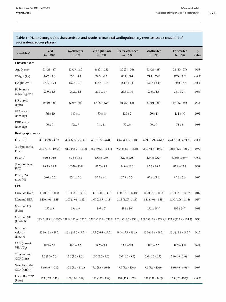
Table 1 describes the major demographic characteristics, and the resting spirometry and CPX results of the soccer players. Age, weight, height and body mass index (BMI) ranged from 16 to 36 years, from 57.5 to 102.0 kg, from 163.3 to 196.3 cm, and from 19.3 to 29.6 kg.m-2, respectively. COP, VT and VO2max showed a parametric distribution (p > 0.05), with values ranging from 13.1 to 25.3, from 61.8 to 92.7% of VO2max, and from 45.0 to 76.2 mL.kg-1.min-1, respectively. The coefficients of variation for COP, VT and VO2max were 16.1%, 10.7% and 10.0%, respectively. On average, COP, VT and VO2max occurred at the velocities of 10.0 ± 1.0, 14.3 ± 1.1, and 18.7 ± 0.9 km.h-1, respectively (p < 0.01).
When stratified by their field positions during the match (Table 1), the only characteristics that differed were weight and height, with goalkeepers showing the highest values for both variables (p < 0.01). The BMI, however, was similar among the soccer players of different field positions (p = 0.86). Regarding CPX, goalkeepers achieved the lowest VO2max values relative to their body weight (mL.kg-1.min-1) (p = 0.01) and reached the COP at a higher HR and percentage of VO2max than the players of other field positions (p < 0.01). However, the values of COP (p = 0.41) and VT (% of VO2max) (p = 0.42) did not differ according to the soccer players’ field positions.
The coefficients of correlation between COP and VO2max (mL.kg-1.min-1) and between COP and VT (% of VO2max) were 0.032 (p = 0.65) and -0.003 (p = 0.96), respectively, evidencing the low association between those variables. Figure 1 shows those data.
Discussion
During an exercise training with progressive intensity increase up to the voluntary maximum, the relationship between VE and VO2 is nonlinear,20 and the curve that illustrates that relation has a U shape, suggesting higher ventilatory efficiency (lower VE/VO2) at submaximal exercise levels when compared to rest and to the highest exertion intensities. Based on that, COP was described as the lowest VE/VO2 value at a given minute during an incremental exercise, representing the time point with the lowest amount of ventilation per liter of oxygen to be consumed, which is the best integration of the relationship between circulation and respiration.15 Recent studies have shown the clinical applicability of the COP for the diagnostic and prognostic assessment
326de Souza e Silva et al.
Cardiorespiratory optimal point in soccer players
Int J Cardiovasc Sci. 2018;31(4)323-332
Original Article
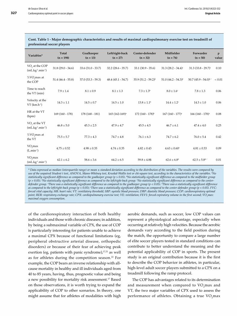
Table 1 - Major demographic characteristics and results of maximal cardiopulmonary exercise test on treadmill of professional soccer players
Variables*Total
(n = 198)
Goalkeeper
(n = 13)
Left/right-back
(n = 27)
Center-defender
(n = 32)
Midfielder
(n = 76)
Forwarder
(n = 50)
p
value
Characteristics
Age (years) 23 (21 - 27) 22 (19 - 24) 26 (21 - 28) 22 (21 - 26) 23 (21 - 28) 24 (10 - 27) 0.35
Weight (kg) 76.7 ± 7.6 85.1 ± 4.7 74.3 ± 6.2 80.7 ± 5.4 74.1 ± 7.6ß 77.3 ± 7.6ß < 0.01
Height (cm) 179.2 ± 6.4 187.5 ± 4.1 175.5 ± 4.2 184.3 ± 3.8 176.5 ± 6.0ß 180.0 ± 5.8 < 0.01
Body mass
index (kg.m-2)23.9 ± 1.8 24.2 ± 1.1 24.1 ± 1.7 23.8 ± 1.6 23.8 ± 1.8 23.9 ± 2.1 0.86
HR at rest
(bpm)59 (53 - 66) 62 (57 - 66) 57 (51 - 62)a 61 (53 - 65) 61 (54 - 66) 57 (52 - 66) 0.15
SBP at rest
(mm Hg)130 ± 10 130 ± 8 130 ± 14 129 ± 7 129 ± 11 131 ± 10 0.92
DBP at rest
(mm Hg)70 ± 9 72 ± 7 71 ± 11 70 ± 8 70 ± 9 71 ± 9 0.95
Resting spirometry
FEV1 (L) 4.31 (3.94 - 4.69) 4.74 (4.35 - 5.04) 4.16 (3.96 - 4.41) 4.44 (4.13 - 5.00)$ 4.24 (3.79 - 4.61)ß 4.41 (3.90 - 4.71)ß,#,€ < 0.01
% of predicted
FEV198.5 (90.8 - 105.6) 101.9 (93.9 - 105.3) 96.7 (93.5 - 104.8) 98.5 (88.6 - 105.8) 98.5 (91.6 - 105.0) 100.8 (87.3 - 107.0) 0.99
FVC (L) 5.05 ± 0.68 5.70 ± 0.68 4.83 ± 0.50 5.23 ± 0.66 4.94 ± 0.62ß 5.05 ± 0.75ß,€,# < 0.01
% of predicted
FVC96.2 ± 10.5 100.5 ± 10.8 95.7 ± 8.4 94.0 ± 10.3 97.0 ± 10.0 95.6 ± 12.1 0.38
FEV1/FVC
ratio (%)86.0 ± 5.3 83.1 ± 5.6 87.3 ± 4.1a 87.6 ± 5.3a 85.4 ± 5.1c 85.8 ± 5.9 0.05
CPX
Duration (min) 13.0 (13.0 - 14.0) 13.0 (13.0 - 14.0) 14.0 (13.0 - 14.0) 13.0 (13.0 - 14.0)b 14.0 (13.0 - 14.0) 13.0 (13.0 - 14.0)b 0.09
Maximal RER 1.10 (1.06 - 1.15) 1.09 (1.06 - 1.13) 1.09 (1.05 - 1.15) 1.13 (1.07 - 1.16) 1.11 (1.06 - 1.15) 1.10 (1.06 - 1.14) 0.59
Maximal HR
(bpm)192 ± 9 194 ± 8 187 ± 7 194 ± 10$ 192 ± 10$,€ 192 ± 8$,€,# 0.01
Maximal VE
(L.min-1)123.2 (113.1 - 133.2) 129.8 (122.6 - 135.2) 123.1 (112.8 - 133.7) 125.4 (113.7 - 136.0) 121.7 (111.6 - 129.9)a 122.9 (113.9 - 134.4) 0.30
Maximal
velocity
(km.h-1)
18.8 (18.4 - 19.2) 18.4 (18.0 - 19.2) 19.2 (18.4 - 19.5) 18.5 (17.9 - 19.2)b 18.8 (18.4 - 19.2) 18.6 (18.4 - 19.2)b 0.13
COP (lowest
VE/VO2)18.2 ± 2.1 19.1 ± 2.2 18.7 ± 2.1 17.9 ± 2.5 18.1 ± 2.2 18.2 ± 1.9ß 0.41
Time to reach
COP (min)2.0 (2.0 - 3.0) 3.0 (2.0 - 4.0) 2.0 (2.0 - 3.0) 2.0 (2.0 - 3.0) 2.0 (2.0 - 2.5)a 2.0 (2.0 - 2.0)a.c 0.07
Velocity at the
COP (km.h-1)9.6 (9.6 - 10.4) 10.4 (9.6 - 11.2) 9.6 (9.6 - 10.4) 9.6 (9.6 - 10.4) 9.6 (9.6 - 10.0)a 9.6 (9.6 - 9.6)a.c 0.07
HR at the COP
(bpm)132 (122 - 142) 142 (134 - 148) 131 (122 - 138) 139 (128 - 152)$ 131 (121 - 140)$ 128 (121-137)ß,# < 0.01
327de Souza e Silva et al.
Cardiorespiratory optimal point in soccer players
Int J Cardiovasc Sci. 2018;31(4)323-332
Original Article
Cont. Table 1 - Major demographic characteristics and results of maximal cardiopulmonary exercise test on treadmill of professional soccer players
Variables*Total
(n = 198)
Goalkeeper
(n = 13)
Left/right-back
(n = 27)
Center-defender
(n = 32)
Midfielder
(n = 76)
Forwarder
(n = 50)
p
value
VO2 at the COP
(mL.kg-1.min-1)31.8 (29.0 - 34.6) 33.6 (31.0 - 33.7) 32.2 (28.6 - 35.7) 33.1 (30.9 - 35.6) 31.3 (28.2 - 34.4)c 31.3 (33.8 - 29.7)c 0.10
%VO2max at
the COP51.4 (46.4 - 55.8) 57.0 (53.3 - 59.3) 48.4 (45.1 - 54.7) 55.9 (51.2 - 59.2)$ 51.0 (44.2 - 54.3)ß 50.7 (45.9 - 54.0)ß,# < 0.01
Time to reach
the VT (min)7.9 ± 1.4 8.1 ± 0.9 8.1 ± 1.3 7.3 ± 1.3b 8.0 ± 1.6c 7.8 ± 1.3 0.06
Velocity at the
VT (km.h-1)14.3 ± 1.1 14.5 ± 0.7 14.5 ± 1.0 13.8 ± 1.1b 14.4 ± 1.2c 14.3 ± 1.0 0.06
HR at the VT
(bpm)169 (160 - 178) 178 (168 - 181) 165 (162-169)a 172 (160 - 178)b 167 (160 - 177)a 166 (160 - 178)a 0.08
VO2 at the VT
(mL.kg-1.min-1)46.8 ± 5.0 45.3 ± 2.5 47.9 ± 4.7 45.5 ± 4.5 46.7 ± 6.1 47.4 ± 4.0 0.25
%VO2max at
the VT75.5 ± 5.7 77.3 ± 4.3 74.7 ± 4.8 76.1 ± 6.3 74.7 ± 6.2 76.0 ± 5.4 0.42
VO2max
(L.min-1)4.75 ± 0.52 4.98 ± 0.35 4.74 ± 0.35 4.82 ± 0.43 4.63 ± 0.60a 4.81 ± 0.53 0.09
VO2max
(mL.kg-1.min-1)62.1 ± 6.2 58.6 ± 3.6 64.2 ± 6.5 59.8 ± 4.8$ 62.6 ± 6.8ß 62.5 ± 5.8ß,# 0.01
* Data expressed as median (interquartile range) or mean ± standard deviation according to the distribution of the variables. The results were compared by use of the unpaired Student t test, ANOVA, Mann-Whitney test, Kruskal-Wallis test or chi-square test, according to the characteristics of the variables. $No statistically significant difference as compared to the goalkeeper group (p > 0.05). #No statistically significant difference as compared to the midfielder group (p > 0.05). ßNo statistically significant difference as compared to the left/right-back group. €No statistically significant difference as compared to the center-defender group. aThere was a statistically significant difference as compared to the goalkeeper group (p < 0.05). bThere was a statistically significant difference as compared to the left/right-back group (p < 0.05); cThere was a statistically significant difference as compared to the center-defender group (p < 0.05). FVC: forced vital capacity; HR: heart rate; VT: ventilatory threshold; SBP: systolic blood pressure; DBP: diastolic blood pressure; COP: cardiorespiratory optimal point; RER: respiratory exchange rate; CPX: cardiopulmonary exercise test; VE: ventilation; FEV1: forced expiratory volume in the first second; VO2max: maximal oxygen consumption.
of the cardiorespiratory interaction of both healthy individuals and those with chronic diseases; in addition, by being a submaximal variable of CPX, the use of COP is particularly interesting for patients unable to achieve a maximal CPX because of functional limitations (eg, peripheral obstructive arterial disease, orthopedic disorders) or because of their fear of achieving peak exertion (eg, patients with panic syndrome),17,21 as well as for athletes during the competition season.22 For example, the COP bears an inverse relationship with all-cause mortality in healthy and ill individuals aged from 40 to 85 years, having, thus, prognostic value and being a new possibility for mortality risk assessment.17 Based on those observations, it is worth trying to expand the applicability of COP to other scenarios. In theory, one might assume that for athletes of modalities with high
aerobic demands, such as soccer, low COP values can represent a physiological advantage, especially when occurring at relatively high velocities. Because the aerobic demands vary according to the field position during the match, the opportunity to compare a large number of elite soccer players tested in standard conditions can contribute to better understand the meaning and the potential applicability of COP in sports. The present study is an original contribution because it is the first to describe the COP behavior in athletes, in particular, high-level adult soccer players submitted to a CPX on a treadmill following the ramp protocol.
The COP has advantages related to its determination and measurement when compared to VO2max and VT, the two major variables of CPX used to assess the performance of athletes. Obtaining a true VO2max

328
Figure 1 - Diagram of dispersion between variables: a) cardiorespiratory optimal point (COP) and maximal oxygen consumption (VO2max); and b) COP and percentage of VO2max reached at the ventilatory threshold (VT).
COP vs. VO2max
COP vs. VT
Goalkeeper Left/right-back
Center-defender
Midfielder Forwarder
VO2m
ax (m
L.kg
-1.m
in-1)
% o
f VO
2max
reac
hed
at th
e VT
Cardiorespiratory optimal point
de Souza e Silva et al.
Cardiorespiratory optimal point in soccer players
Int J Cardiovasc Sci. 2018;31(4)323-332
Original Article

329de Souza e Silva et al.
Cardiorespiratory optimal point in soccer players
Int J Cardiovasc Sci. 2018;31(4)323-332
Original Article
suggests the existence of a plateau in the VO2 curve, which is not always possible, and it can vary according to the CPX protocol used and the gas sampling or collection interval.10,11 In addition, VO2max depends on performing a truly maximal exercise test, whose determination criteria vary in the literature, being subjective to a certain extent. On the other hand, although the VT does not require a maximal test, it requires a more intense exercise than COP does, and VT measurement is hindered by the existence of several distinct criteria for its identification and/or characterization, which, in a significant percentage of cases, cannot be obtained, limiting its use in clinical practice and sports.13 In addition, although both VO2max and VT can be detected automatically with commercial software, the methods available for that have been developed from varied definitions and algorithms, implying the need for its review by at least one experienced observer, making those measures subjective and widening the potential of high inter- and intraobserver variability.23,24 In contrast, COP is easily determined from the identification of the lowest value of the VE/VO2 ratio for each minute of CPX, not depending, thus, on the interpretation and experience of the observer, and relying on a relatively small effort, because it occurs at relatively low exercise intensities, before the VT.
Regarding the COP of the soccer players assessed, some findings are worth noting: 1- as expected, COP was obtained at lower percentage of VO2max and velocity than those at the VT; 2- similarly to VT, but opposite to VO2max, COP did not differ according to the different field positions of the soccer players; 3- no significant association was observed between COP and the variables VO2max and VT; and 4- the coefficient of variation of oxygen consumption at the time of the COP was greater than that observed at the VT and VO2max. It is interesting to point out that, on average, the COP values found for the soccer players were below the 50th percentile of the values found for healthy male non-athletes of the same age group in a previous study,15 and that only eight (4%) soccer players had COP over 22, considered the cutoff point for optimal clinical prognosis,17 suggesting that those soccer players have a privileged circulation-respiration interaction, probably more economic at the submaximal exercise. However, it is worth noting that the COP values described for non-athletes were obtained from a CPX performed on a lower limb cycle ergometer, with an individualized ramp protocol. Thus, the description of COP in different exercise modalities and protocols should be approached in future studies,
because there is evidence that the behavior of some variables obtained in CPX differ depending on the ergometer and protocol used.
The running velocity on the treadmill and the exercise intensity represented by the percentage of VO2max at which the soccer players assessed in this study reached the COP (10.0 ± 1.0 km.h-1 and 51.3 ± 8.7%, respectively) were lower than the values obtained at the VT by soccer players assessed in other studies, even when compared to those of players of lower athletic performance, who are expected to reach an earlier VT. For example, according to Ziogas et al.,25 soccer players of the first, second and third Greek division submitted to a CPX in the pre-season period reached the VT at a mean velocity of 13.2, 12.6 and 12.3 km.h-1, respectively. Boone et al.,26 however, assessing 289 soccer players of the first Belgian division, have reported mean running velocities on the treadmill at the VT ranging from 12.7 ± 1.4 in goalkeepers to 14.4 ± 0.7 km.h-1 in center-defenders. Regarding the exercise intensity, Impellizzeri et al.27 and Helgerud et al.28 have reported that junior soccer players reached the VT at a mean percentage of VO2max greater than 80%. Considering that the running velocity and the exercise intensity at which the VT is reached reflect the training status of the soccer players, future studies should assess whether COP is also useful to differentiate the physical performance of athletes.
When comparing the soccer players according to their field positions, goalkeepers, midfielders, left/right-backs, center-defenders and forwarders did not differ regarding the COP. Manari et al.,29 comparing the VT and the VO2max of 450 European elite soccer players of different field positions, have found no differences regarding the VT, similarly to our study’s findings regarding VT and COP. However, similarly to our study’s findings, VO2max was lower in goalkeepers. Tonessem et al.,30 assessing 1,545 male soccer athletes, have found small to moderate differences in VO2max according to the athlete’s field position, with greater values in the midfielders, followed, in decreasing order, by the defense athletes, forwarders and goalkeepers. Similarly, Balikian et al.,31 assessing 25 professional soccer players, have found lower mean VO2max values of goalkeepers (52.68 mL.kg-1.min-1) as compared to the mean values of soccer players of other field positions. However, in contrast to our study’s findings, the mean velocity at which the players reached the VT differed according to their field position, being lower for goalkeepers (12.66 km.h-1) and higher for left/right-
330de Souza e Silva et al.
Cardiorespiratory optimal point in soccer players
Int J Cardiovasc Sci. 2018;31(4)323-332
Original Article
backs (14.33 km.h-1) and midfielders (14.11 km.h-1). Nevertheless, it is worth noting that the heterogeneity of the methods used to measure VT hinders the comparison of the results between the studies.
Finally, the COP failed to show a linear association with the variables VT and VO2max. Ramos et al.15 have not only described a moderate association with VO2max (-0.47) and VT (-0.42), but have also observed that the combination of COP and VO2max adds more prognostic information to all-cause mortality than each variable in isolation.17 Such findings suggest a possible independence and complementarity of COP regarding VO2max and VT, which could contribute with additional information to the interpretation of the relationship between the cardiovascular and respiratory systems during a CPX. Thus, one can speculate that the submaximal variables – COP and VT – might better reflect the energetic demands of a soccer match in the current context, in which the differences in distance and in percentage of time spent in intense efforts are less evident in soccer players of different field positions.
The present study has some limitations in addition to those already mentioned. The CPX analyzed were limited to those performed in the pre-season period, not allowing us to assess the COP behavior in different training periods of the soccer players. In addition, this study only assessed male adult elite soccer players, which limits the extrapolation of the results to female soccer players, other age groups, different technical levels and other sport modalities.
Conclusion
The present study described the COP behavior and its absence of association with VO2max and VT of male adult
elite soccer players. Thus, future studies are required to assess whether COP can provide additional and relevant information to other sport contexts.
Author contributions
Conception and design of the research: de Souza e Silva CG, Castro CLB, Franca JF, Bottino A, Myers J, Araújo CGS; Acquisition of data: Castro CLB, Franca JF, Araújo CGS; Analysis and interpretation of the data, Statistical analysis and Writing of the manuscript: de Souza e Silva CG, Araújo CGS; Critical revision of the manuscript for intellectual content: Castro CLB, Franca JF, Bottino A, Myers J, Araújo CGS.
Potential Conflict of Interest
No potential conflict of interest relevant to this article was reported.
Sources of Funding
There were no external funding sources for this study.
Study Association
This study is not associated with any thesis or dissertation work.
Ethics approval and consent to participate
This study was approved by the Ethics Committee of the Suprema - Faculdade de Ciências Médicas e da Saúde de Juiz de Fora under the protocol number 0218/11. All the procedures in this study were in accordance with the 1975 Helsinki Declaration, updated in 2013. Informed consent was obtained from all participants included in the study.
1. Albouaini K, Egred M, Alahmar A, Wright DJ. Cardiopulmonary exercise testing and its application. Postgrad Med J. 2007;83(985):675-82.
2. Ross R, Blair SN, Arena R, Church TS, Després JP, Franklin BA, et al; American Heart Association Physical Activity Committee of the Council on Lifestyle and Cardiometabolic Health; Council on Clinical Cardiology; Council on Epidemiology and Prevention; Council on Cardiovascular and Stroke Nursing; Council on Functional Genomics and Translational Biology; Stroke Council. Importance of assessing cardiorespiratory fitness in clinical practice: a case for fitness as a clinical vital sign: a scientific statement from the American Heart Association. Circulation. 2016;134(24):e653-e99.
3. Gitt AK, Wasserman K, Kilkowski C, Kleemann T, Kilkowski A, Bangert M, et al. Exercise anaerobic threshold and ventilatory efficiency
identify heart failure patients for high risk of early death. Circulation. 2002;106(24):3079-84.
4. Edwards AM, Clark N, Macfadyen AM. Lactate and ventilatory thresholds reflect the training status of professional soccer players where maximum aerobic power is unchanged. J Sports Sci Med. 2003;2(1):23-9.
5. Hoff J. Training and testing physical capacities for elite soccer players. J Sports Sci. 2005;23(6):573-82.
6. Midgley AW, McNaughton LR, Jones AM. Training to enhance the physiological determinants of long-distance running performance: can valid recommendations be given to runners and coaches based on current scientific knowledge? Sports Med. 2007;37(10):857-80. Erratum in: Sports Med. 2007;37(11):1000.
References

331de Souza e Silva et al.
Cardiorespiratory optimal point in soccer players
Int J Cardiovasc Sci. 2018;31(4)323-332
Original Article
7. Stølen T, Chamari K, Castagna C, Wisløff U. Physiology of soccer: an update. Sports Med. 2005;35(6):501-36.
8. Reilly T. Physiological aspects of soccer. Biol Sport. 1994;11:3-20.
9. Taylor HL, Buskirk E, Henschel A. Maximal oxygen intake as an objective measure of cardiorespiratory performance. J Appl Physiol.1955;8(1):73-80.
10. Myers J, Walsh D, Buchanan N, Froelicher VF. Can maximal cardiopulmonary capacity be recognized by a plateau in oxygen uptake? Chest. 1989;96(6):1312-6.
11. Myers J, Walsh D, Sullivan M, Froelicher V. Effect of sampling on variability and plateau in oxygen uptake. J Appl Physiol (1985). 1990;68(1):404-10.
12. Doherty M, Nobbs L, Noakes TD. Low frequency of the "plateau phenomenon" during maximal exercise in elite British athletes. Eur J Appl Physiol. 2003;89(6):619-23.
13. Yeh MP, Gardner RM, Adams TD, Yanowitz FG, Crapo RO. Anaerobic threshold: problems of determination and validation. J Appl Physiol Respir Environ Exerc Physiol. 1983;55(4):1178-86.
14. Zinner C, Sperlich B, Wahl P, Mester J. Classification of selected cardiopulmonary variables of elite athletes of different age, gender, and disciplines during incremental exercise testing. Springerplus. 2015;4:544.
15. Ramos PS, Ricardo DR, Araújo CG. Cardiorespiratory optimal point: a submaximal variable of the cardiopulmonary exercise testing. Arq Bras Cardiol. 2012;99(5):988-96.
16. Ramos PS, Araújo CG. Análise da estabilidade de uma variável submáxima em teste cardiopulmonar de exercício: ponto ótimo cardiorrespiratório. Rev Bras Ativ Fis e Saúde (Pelotas-RS). 2013;18(5):585-93.
17. Ramos PS, Araújo CG. Cardiorespiratory optimal point during exercise testing as a predictor of all-cause mortality. Rev Port Cardiol. 2017;36(4):261-9.
18. Miller MR, Hankinson J, Brusasco V, Burgos F, Casaburi R, Coates A, et al; ATS/ERS Task Force. Standardisation of spirometry. Eur Respir J. 2005;26(2):319-38.
19. Borg GA. Psychophysical bases of perceived exertion. Med Sci Sports Exerc. 1982;14(5):377-81.
20. Hagan RD, Smith MG. Pulmonary ventilation in relation to oxygen uptake and carbon dioxide production during incremental load work. Int J Sports Med. 1984;5(4):193-7.
21. Ramos PS, Sardinha A, Nardi AE, de Araújo CG. Cardiorespiratory optimal point: a submaximal exercise variable to assess panic disorder patients. PLoS One. 2014;9(8):e104932.
22. Nikolaidis PT. Can maximal aerobic running speed be predicted from submaximal cycle ergometry in soccer players? The effects of age, anthropometry and positional roles. Adv Biomed Res. 2015;4:226.
23. Myers J, Goldsmith RL, Keteyian SJ, Brawner CA, Brazil DA, Aldred H, et al. The ventilatory anaerobic threshold in heart failure: a multicenter evaluation of reliability. J Card Fail. 2010;16(1):76-83.
24. Gaskill SE, Ruby BC, Walker AJ, Sanchez OA, Serfass RC, Leon AS. Validity and reliability of combining three methods to determine ventilatory threshold. Med Sci Sports Exerc. 2001;33(11):1841-8.
25. Ziogas GG, Patras KN, Stergiou N, Georgoulis AD. Velocity at lactate threshold and running economy must also be considered along with maximal oxygen uptake when testing elite soccer players during preseason. J Strength Cond Res. 2011;25(2):414-9.
26. Boone J, Vaeyens R, Steyaert A, Vanden Bossche L, Bourgois J. Physical fitness of elite Belgian soccer players by player position. J Strength Cond Res. 2012;26(8):2051-7.
27. Impellizzeri FM, Marcora SM, Castagna C, Reilly T, Sassi A, Iaia FM, et al. Physiological and performance effects of generic versus specific aerobic training in soccer players. Int J Sports Med. 2006;27(6):483-92.
28. Helgerud J, Engen LC, Wisloff U, Hoff J. Aerobic endurance training improves soccer performance. Med Sci Sports Exerc. 2001;33(11):1925-31.
29. Manari D, Manara M, Zurini A, Tortorella G, Vaccarezza M, Prandelli N, et al. VO2max and VO2AT: athletic performance and field role of elite soccer players Sport Sci Health. 2016;12(2):221-6.
30. Tønnessen E, Hem E, Leirstein S, Haugen T, Seiler S. Maximal aerobic power characteristics of male professional soccer players, 1989-2012. Int J Sports Physiol Perform. 2013;8(3):323-9.
31. Balikian P, Lourenção A, Ribeiro LF, Festuccia WTL, Neiva CM. Consumo máximo de oxigênio e limiar anaeróbio de jogadores de futebol: comparação entre as diferentes posições. Rev Bras Med Esporte. 2002;8(2):32-6.

332de Souza e Silva et al.
Cardiorespiratory optimal point in soccer players
Int J Cardiovasc Sci. 2018;31(4)323-332
Original Article
This is an open-access article distributed under the terms of the Creative Commons Attribution License

DOI: 10.5935/2359-4802.20180021
Abstract
Background: Moderate coronary artery lesions can be, or not, responsible for myocardial ischemia. The functional analysis of these lesions can be performed by invasive and noninvasive methods.
Objective: To compare the functional analysis of moderate coronary lesions by fractional flow reserve and myocardial perfusion scintigraphy.
Methods: 47 patients with stable coronary artery disease and at least one moderate coronary artery obstruction were prospectively studied. They were submitted to fractional flow reserve and myocardial perfusion scintigraphy with a median interval of 24.5 days between January 2013 and December 2015. There was no change in clinical status or revascularization procedure between the exams. The population variables were described as medians and interquartile range. Fractional flow reserve was performed in one left main coronary artery; 37 left descending coronary arteries; 12 circumflex arteries and 4 right coronary arteries. Fractional flow reserve < 0.8 was considered positive. The comparative analysis between the results of the tests was performed by two-tailed Fisher’s test and a p-value ≤ 0.05 was considered significant.
Results: Fractional flow reserve < 0.8 was found in the left main coronary artery (100%); 13 in the left descending coronary artery (35.14%); 6 in circumflex artery (50%) and 2 in the right coronary artery (50%). Among the patients with positive fractional flow reserve, 83% had myocardial ischemia demonstrated by the myocardial perfusion scintigraphy (p = 0.058). When analyzing specifically the left descending coronary artery, 83% of the patients with negative fractional flow reserve showed no ischemia at the myocardial perfusion scintigraphy, but 69% of the patients with positive fractional flow reserve showed no ischemia at the myocardial perfusion scintigraphy (p = 0.413).
Conclusion: Disagreements can occur between the results of the functional analysis of moderate coronary lesions by invasive and noninvasive tests. (Int J Cardiovasc Sci. 2018;31(4)333-338)
Keywords: Myocardial Ischemia; Praqctional Flow Reserve, Myocardial; Myocardial Perfusion / Diagnostic Imaging; Microvascular Angina.
Introduction
The presence of myocardial ischemia is one of the important prognostic factors in coronary artery disease (CAD) and in the decision-making on the best treatment to be implemented. The combination of coronary anatomy and information on the hemodynamic implication of the obstructive lesion is essential to define the treatment strategy to be carried out in patients with CAD.
The fractional flow reserve (FFR) measurement is a valuable tool to evaluate the functional severity of a coronary stenosis, identifying changes in coronary flow resistance. The FFR can be obtained in the hemodynamic laboratory and can be performed together with the angiography. The FFR is defined as the maximum blood flow to the myocardium in the presence of a certain stenosis, divided by this flow, if there was no such stenosis. The FFR can be determined by dividing the
333
ORIGINAL ARTICLE
International Journal of Cardiovascular Sciences. 2018;31(4)333-338
Mailing Address: Aurora Felice Castro IssaRua Timóteo da Costa, 623/110. Postal Code: 22450-130, Leblon, Rio de Janeiro, RJ - BrazilE-mail: [email protected]; [email protected]
Comparison between Myocardial Ischemia Evaluation by Fractional Flow Reserve and Myocardial Perfusion ScintigraphyAurora Felice Castro Issa, Felipe Pittella, Sergio Martins Leandro, Patricia Paço, Judas Tadeu, Renata FelixInstituto Nacional de Cardiologia, Rio de Janeiro, RJ - Brazil
Manuscript received November 27, 2016, revised manuscript October 18, 2017, accepted November 13, 2017.
334
mean pressure distal to the coronary lesion by the mean aortic pressure during maximal adenosine-induced vasodilation. The FFR has a normal value of 1, and values less than 0.8 indicate myocardial ischemia. Studies have shown that coronary vessels with FFR ≥ 0.8 can be clinically treated, with cardiovascular event rates similar to those of patients with normal noninvasive tests (< 1% per year). Patients with FFR ≤ 0.8 could benefit from percutaneous or surgical revascularization procedures.1-3
Although the FFR has its defined role in moderate lesions and is not very useful in angiographically severe lesions, it helps in the decision-making regarding when to revascularize patients with multivessel disease. In these patients, it helps to define the revascularization strategy, as well as to better evaluate its extent, according to the functional evaluation of stenosis in critical coronary sites.4
Myocardial perfusion scintigraphy (MPS) with tomographic images has been validated by several studies in the evaluation of diagnosis and prognosis for patients at risk of cardiovascular events. The functional repercussion of coronary lesions constitutes one of the main purposes of the method, which is based on the perfusion deficit assessment in myocardial segments irrigated by partially occluded arteries. Risk stratification is based on the ability to identify patients according to the test results. SPECT with normal or slightly altered perfusion has an excellent prognosis, with a low mortality risk (< 1%) per year. The risk associated with perfusion alterations varies according to the ischemia extent and severity. The greater the perfusion defects, the higher the likelihood of future events. In those with moderate perfusion defects, the incidence of events is 1 to 3% per year, being > 3% in patients with major perfusion defects.5
Most percutaneous coronary interventions are performed based on angiographic criteria alone, with no objective evidence of myocardial ischemia. Coronary angiography has limitations in establishing functional severity, because the stenosis degree of a lesion does not always correlate with functional impairment in the myocardium.6 Thus, it is important to complement anatomical data with functional tests capable of adequately guiding the therapeutic approach regarding a myocardial revascularization procedure. Several studies have been carried out to evaluate the agreement between the FFR with functional methods (MPS, dobutamine stress echocardiogram and exercise testing) to define the presence of myocardial ischemia, with the FFR having the advantage of being specific for each vessel and obstruction.7 In multi-vessel patients, MPS tends to
underestimate or overestimate the functional importance of coronary stenosis when compared to FFR.8
The functional tests are performed in a minority of patients referred to coronary angioplasty at Instituto Nacional de Cardiologia. In this sense, the FFR can be a useful tool in the hemodynamics room to aid in decision-making regarding whether or not to perform a percutaneous coronary intervention, saving time and costs to the health system. The objective of the present study was to compare the functional analysis between FFR and MPS in patients with moderate lesions at the coronary angiography.
Methods
This is a prospective, observational study of patients of both genders, aged 18 years or older, admitted to the Department of Coronary Disease unit or referred to the Hemodynamic Service of Instituto Nacional de Cardiologia, who had an FFR indication after the coronary angiography by the multidisciplinary “Heart team”. The sample size of 47 patients was selected by convenience.
Patients with no previous MPS were submitted to the examination. Coronary lesions were classified as moderate (between 50 and 70%) and severe (≥ 70%) according to visual estimation.
Patients with moderate lesions and those for whom there was doubt regarding the indication of myocardial revascularization were included in the study. Patients with chronic occlusion, ST-segment elevation acute myocardial infarction, unstable patients, those with severe valvular disease or cardiomyopathies from other causes, patients with contraindications to the use of adenosine and to scintigraphy (pregnant women, infants and women with suspected pregnancy) were excluded from the study.
The study was approved by the Ethics and Research Committee of Instituto Nacional de Cardiologia, and all the participants agreed to sign the Free and Informed Consent Form. The present study has no sources of funding.
Fractional flow reserve measurement
Coronary catheterization was performed with 6 and 7F guide catheters. Prior to the angiography, 10,000 u of intravenous heparin and intracoronary nitroglycerin at a dose of 0.25 to 0.5 mg were administered. Then, pressure measurements were performed in vessels with stenosis ≥ 50% by visual estimation using a guidewire with a
Issa et al.
Comparison between Fraction Flow Reserve and SPECT in Myocardial Ischemia
Int J Cardiovasc Sci. 2018;31(4)333-338
Original Article
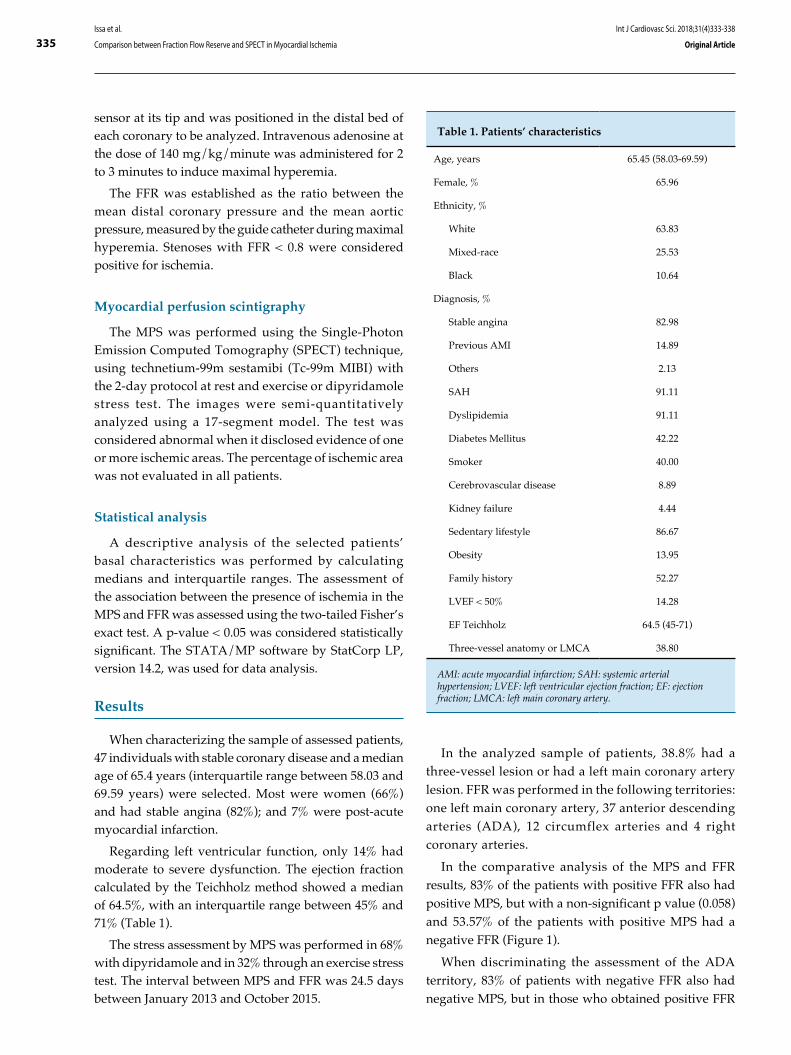
335
Table 1. Patients’ characteristics
Age, years 65.45 (58.03-69.59)
Female, % 65.96
Ethnicity, %
White 63.83
Mixed-race 25.53
Black 10.64
Diagnosis, %
Stable angina 82.98
Previous AMI 14.89
Others 2.13
SAH 91.11
Dyslipidemia 91.11
Diabetes Mellitus 42.22
Smoker 40.00
Cerebrovascular disease 8.89
Kidney failure 4.44
Sedentary lifestyle 86.67
Obesity 13.95
Family history 52.27
LVEF < 50% 14.28
EF Teichholz 64.5 (45-71)
Three-vessel anatomy or LMCA 38.80
AMI: acute myocardial infarction; SAH: systemic arterial hypertension; LVEF: left ventricular ejection fraction; EF: ejection fraction; LMCA: left main coronary artery.
sensor at its tip and was positioned in the distal bed of each coronary to be analyzed. Intravenous adenosine at the dose of 140 mg/kg/minute was administered for 2 to 3 minutes to induce maximal hyperemia.
The FFR was established as the ratio between the mean distal coronary pressure and the mean aortic pressure, measured by the guide catheter during maximal hyperemia. Stenoses with FFR < 0.8 were considered positive for ischemia.
Myocardial perfusion scintigraphy
The MPS was performed using the Single-Photon Emission Computed Tomography (SPECT) technique, using technetium-99m sestamibi (Tc-99m MIBI) with the 2-day protocol at rest and exercise or dipyridamole stress test. The images were semi-quantitatively analyzed using a 17-segment model. The test was considered abnormal when it disclosed evidence of one or more ischemic areas. The percentage of ischemic area was not evaluated in all patients.
Statistical analysis
A descriptive analysis of the selected patients’ basal characteristics was performed by calculating medians and interquartile ranges. The assessment of the association between the presence of ischemia in the MPS and FFR was assessed using the two-tailed Fisher’s exact test. A p-value < 0.05 was considered statistically significant. The STATA/MP software by StatCorp LP, version 14.2, was used for data analysis.
Results
When characterizing the sample of assessed patients, 47 individuals with stable coronary disease and a median age of 65.4 years (interquartile range between 58.03 and 69.59 years) were selected. Most were women (66%) and had stable angina (82%); and 7% were post-acute myocardial infarction.
Regarding left ventricular function, only 14% had moderate to severe dysfunction. The ejection fraction calculated by the Teichholz method showed a median of 64.5%, with an interquartile range between 45% and 71% (Table 1).
The stress assessment by MPS was performed in 68% with dipyridamole and in 32% through an exercise stress test. The interval between MPS and FFR was 24.5 days between January 2013 and October 2015.
In the analyzed sample of patients, 38.8% had a three-vessel lesion or had a left main coronary artery lesion. FFR was performed in the following territories: one left main coronary artery, 37 anterior descending arteries (ADA), 12 circumflex arteries and 4 right coronary arteries.
In the comparative analysis of the MPS and FFR results, 83% of the patients with positive FFR also had positive MPS, but with a non-significant p value (0.058) and 53.57% of the patients with positive MPS had a negative FFR (Figure 1).
When discriminating the assessment of the ADA territory, 83% of patients with negative FFR also had negative MPS, but in those who obtained positive FFR
Issa et al.
Comparison between Fraction Flow Reserve and SPECT in Myocardial Ischemia
Int J Cardiovasc Sci. 2018;31(4)333-338
Original Article
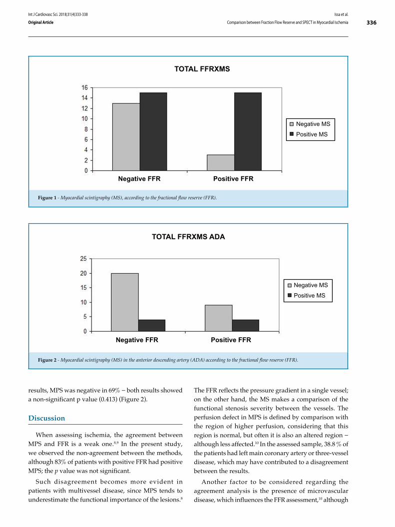
336
The FFR reflects the pressure gradient in a single vessel; on the other hand, the MS makes a comparison of the functional stenosis severity between the vessels. The perfusion defect in MPS is defined by comparison with the region of higher perfusion, considering that this region is normal, but often it is also an altered region − although less affected.10 In the assessed sample, 38.8 % of the patients had left main coronary artery or three-vessel disease, which may have contributed to a disagreement between the results.
Another factor to be considered regarding the agreement analysis is the presence of microvascular disease, which influences the FFR assessment,10 although
Figure 1 - Myocardial scintigraphy (MS), according to the fractional flow reserve (FFR).
Figure 2 - Myocardial scintigraphy (MS) in the anterior descending artery (ADA) according to the fractional flow reserve (FFR).
results, MPS was negative in 69% − both results showed a non-significant p value (0.413) (Figure 2).
Discussion
When assessing ischemia, the agreement between MPS and FFR is a weak one.8,9 In the present study, we observed the non-agreement between the methods, although 83% of patients with positive FFR had positive MPS; the p value was not significant.
Such disagreement becomes more evident in patients with multivessel disease, since MPS tends to underestimate the functional importance of the lesions.8
Negative MS
Positive MS
TOTAL FFRXMS
Negative FFR Positive FFR
Negative MS
Positive MS
TOTAL FFRXMS ADA
Negative FFR Positive FFR
Issa et al.
Comparison between Fraction Flow Reserve and SPECT in Myocardial Ischemia
Int J Cardiovasc Sci. 2018;31(4)333-338
Original Article
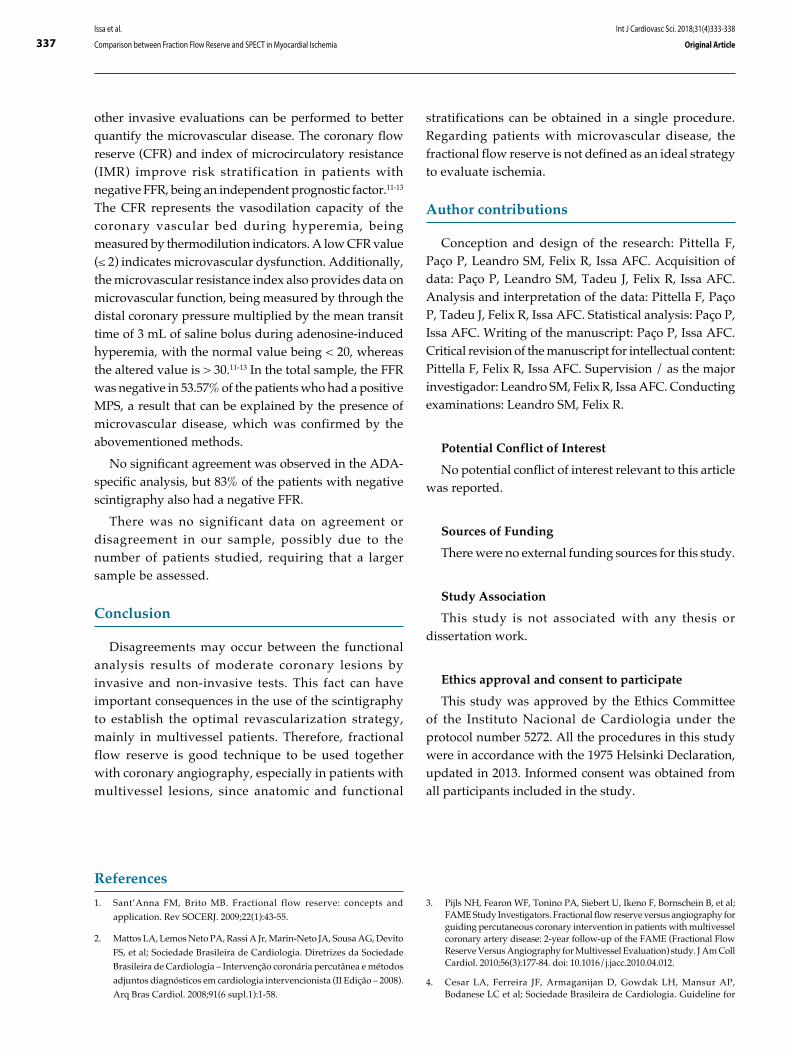
337
1. Sant’Anna FM, Brito MB. Fractional flow reserve: concepts and
application. Rev SOCERJ. 2009;22(1):43-55.
2. Mattos LA, Lemos Neto PA, Rassi A Jr, Marin-Neto JA, Sousa AG, Devito
FS, et al; Sociedade Brasileira de Cardiologia. Diretrizes da Sociedade
Brasileira de Cardiologia – Intervenção coronária percutânea e métodos
adjuntos diagnósticos em cardiologia intervencionista (II Edição – 2008).
Arq Bras Cardiol. 2008;91(6 supl.1):1-58.
3. Pijls NH, Fearon WF, Tonino PA, Siebert U, Ikeno F, Bornschein B, et al; FAME Study Investigators. Fractional flow reserve versus angiography for guiding percutaneous coronary intervention in patients with multivessel coronary artery disease: 2-year follow-up of the FAME (Fractional Flow Reserve Versus Angiography for Multivessel Evaluation) study. J Am Coll Cardiol. 2010;56(3):177-84. doi: 10.1016/j.jacc.2010.04.012.
4. Cesar LA, Ferreira JF, Armaganijan D, Gowdak LH, Mansur AP, Bodanese LC et al; Sociedade Brasileira de Cardiologia. Guideline for
References
other invasive evaluations can be performed to better quantify the microvascular disease. The coronary flow reserve (CFR) and index of microcirculatory resistance (IMR) improve risk stratification in patients with negative FFR, being an independent prognostic factor.11-13 The CFR represents the vasodilation capacity of the coronary vascular bed during hyperemia, being measured by thermodilution indicators. A low CFR value (≤ 2) indicates microvascular dysfunction. Additionally, the microvascular resistance index also provides data on microvascular function, being measured by through the distal coronary pressure multiplied by the mean transit time of 3 mL of saline bolus during adenosine-induced hyperemia, with the normal value being < 20, whereas the altered value is > 30.11-13 In the total sample, the FFR was negative in 53.57% of the patients who had a positive MPS, a result that can be explained by the presence of microvascular disease, which was confirmed by the abovementioned methods.
No significant agreement was observed in the ADA-specific analysis, but 83% of the patients with negative scintigraphy also had a negative FFR.
There was no significant data on agreement or disagreement in our sample, possibly due to the number of patients studied, requiring that a larger sample be assessed.
Conclusion
Disagreements may occur between the functional analysis results of moderate coronary lesions by invasive and non-invasive tests. This fact can have important consequences in the use of the scintigraphy to establish the optimal revascularization strategy, mainly in multivessel patients. Therefore, fractional flow reserve is good technique to be used together with coronary angiography, especially in patients with multivessel lesions, since anatomic and functional
stratifications can be obtained in a single procedure. Regarding patients with microvascular disease, the fractional flow reserve is not defined as an ideal strategy to evaluate ischemia.
Author contributions
Conception and design of the research: Pittella F, Paço P, Leandro SM, Felix R, Issa AFC. Acquisition of data: Paço P, Leandro SM, Tadeu J, Felix R, Issa AFC. Analysis and interpretation of the data: Pittella F, Paço P, Tadeu J, Felix R, Issa AFC. Statistical analysis: Paço P, Issa AFC. Writing of the manuscript: Paço P, Issa AFC. Critical revision of the manuscript for intellectual content: Pittella F, Felix R, Issa AFC. Supervision / as the major investigador: Leandro SM, Felix R, Issa AFC. Conducting examinations: Leandro SM, Felix R.
Potential Conflict of Interest
No potential conflict of interest relevant to this article was reported.
Sources of Funding
There were no external funding sources for this study.
Study Association
This study is not associated with any thesis or dissertation work.
Ethics approval and consent to participate
This study was approved by the Ethics Committee of the Instituto Nacional de Cardiologia under the protocol number 5272. All the procedures in this study were in accordance with the 1975 Helsinki Declaration, updated in 2013. Informed consent was obtained from all participants included in the study.
Issa et al.
Comparison between Fraction Flow Reserve and SPECT in Myocardial Ischemia
Int J Cardiovasc Sci. 2018;31(4)333-338
Original Article

338
stable coronary artery disease. Arq Bras Cardiol. 2014;103(2 Suppl 2):1-56. doi: http://dx.doi.org/10.5935/abc.2014S004.
5. Sociedade Brasileira de Cardiologia. I Diretriz da Sociedade Brasileira de Cardiologia sobre Cardiologia Nuclear. Arq Bras Cardiol. 2002;78(Suppl 3):1-42.
6. Sant’Anna FM, da Silva ER, Batista LA, Brito MB, Ventura FM, Ferraz HA. What is the angiography error when defining myocardial ischemia during percutaneous coronary interventions? Arq Bras Cardiol. 2008;91(3):162-7. doi: http://dx.doi.org/10.1590/S0066-782X2008001500007.
7. Pijls NH, De Bruyne B, Peels K, Van Der Voort PH, Bonnier HJ, Bartunek J Koolen JJ, et al. Measurement of fractional flow reserve to assess the functional severity of coronary artery stenoses. N Engl J Med. 1996;334(26):1703-8. doi: 10.1056/NEJM199606273342604.
8. Melikian N, De Bondt P, Tonino P, De Winter O, Wyffels E, Bartunek J, et al. Fractional flow reserve and myocardial perfusion imaging in patients with angiographic multivessel coronary artery disease. JACC Cardiovasc Interv. 2010;3(3):307-14. doi: 10.1016/j.jcin.2009.12.010.
9. Christou MA, Siontis GC, Katritsis DG, Ioannidis JP. Meta-analysis of fractional flow reserve versus quantitative coronary angiography and noninvasive imaging for evaluation of myocardial ischemia. Am J Cardiol. 2007;99(4):450-6. doi: 10.1016/j.amjcard.2006.09.092.
10. Crea F, Camici PG, Merz CN. Coronary microvascular dysfunction: an update. Eur Heart J. 2014;35(17):1101-11.
11. Schelbert HR. FFR and coronary flow reserve: friends or foes?. JACC Cardiovasc Imaging. 2012;5(2):203-6. doi: 10.1016/j.jcmg.2011.12.003.
12. Berry C. Fractional flow reserve, coronary flow reserve and the index of microvascular resistance in clinical practice. Radcliffe Cardiology. Feb. 2014:1-6.
13. Lee JM, Jung JH, Hwang D, Park J, Fan Y, Na SH, et al. Coronary flow reserve and microcirculatory resistance in patients with intermediate coronary stenosis. J Am Coll Cardiol. 2016;67(10):1158-69. doi: 10.1016/j.jacc.2015.12.053.
Issa et al.
Comparison between Fraction Flow Reserve and SPECT in Myocardial Ischemia
Int J Cardiovasc Sci. 2018;31(4)333-338
Original Article
This is an open-access article distributed under the terms of the Creative Commons Attribution License
DOI: 10.5935/2359-4802.20180043
339
ORIGINAL ARTICLE
International Journal of Cardiovascular Sciences. 2018;31(4)339-358
Mailing Address: José Augusto Barreto-FilhoAvenida Gonçalo Prado Rollemberg, 211, sala 202. Postal Code: 49010-410, Aracaju, SE - Brazil.E-mail: [email protected], [email protected]
Disparities in Acute Myocardial Infarction Treatment Between Users of the Public and Private Healthcare System in SergipeJussiely Cunha Oliveira1,2, Laís Costa Souza Oliveira1,3, Jeferson Cunha Oliveira3,4, Ikaro Daniel de Carvalho Barreto5, Marcos Antonio Almeida-Santos2,6, Ticiane Clair Remacre Munareto Lima1, Larissa Andreline Maia Arcelino1,7, Luiz Flávio Andrade Prado2,8, Fábio Serra Silveira8, Thiago Augusto Nascimento6,9, Eduardo José Pereira Ferreira6,8,10, Rafael Vasconcelos Barreto8, Enilson Vieira Moraes3, José Teles de Mendonça8,9,11, Antonio Carlos Sobral Sousa1,6,10,11, José Augusto Barreto-Filho1,6,10,11, em nome do grupo de pesquisadores do Registro VICTIMNúcleo de Pós-Graduação em Ciências da Saúde da Universidade Federal de Sergipe (UFS)1, São Cristóvão, SE - BrazilUniversidade Tiradentes (UNIT)2, Aracaju, SE - BrazilHospital Primavera3, Aracaju, SE - BrazilDepartamento de Farmácia da Universidade Federal de Sergipe (UFS)4, Lagarto, SE - BrazilDepartamento de Estatística da Universidade Federal de Sergipe (UFS)5, Aracaju, SE - BrazilCentro de Ensino e Pesquisa da Fundação São Lucas6, Aracaju, SE - BrazilFaculdade Estácio de Sá7, Aracaju, SE - BrazilFundação Beneficência Hospital de Cirurgia8, Aracaju, SE - BrazilHospital do Coração9, Aracaju, SE - BrazilDepartamento de Medicina da Universidade Federal de Sergipe (UFS)10, São Cristóvão, SE - BrazilDivisão de Cardiologia do Hospital Universitário da UFS11, São Cristóvão, SE - Brazil
Manuscript received July 05, 2017, revised manuscript October 03, 2017, accepted October 15, 2017.
Abstract
Background: The Brazilian Unified Health System (SUS) was created to ensure universal, integral and equitable access to quality healthcare to Brazilians. However, studies scrutinizing the quality of the healthcare provided by the SUS are scarce. This is especially critical for patients with ST-elevation myocardial infarction (STEMI), who depend on healthcare system responsiveness and timely reperfusion to achieve better outcomes.
Objective: To describe the methodology of the VICTIM Registry aimed at characterizing and comparing the access to effective therapies and the outcomes of patients with STEMI, who use the SUS and the private healthcare system at hospitals capable of performing angioplasty in Sergipe. In addition, that registry aimed at identifying and measuring possible disparities in the quality of the care provided.
Methods and Results: The VICTIM Registry is an observational study, launched in December 2014, being still in the data collection phase, to investigate: the epidemiology of STEMI in Sergipe, the temporal and geographic courses of the patients up to their admission to one of the hospitals capable of performing angioplasty, the reperfusion therapy rates, the quality of the healthcare provided during the event, and the 30-day mortality. It compares the results obtained in the SUS with those of the private healthcare system.
Conclusions: The VICTIM Registry is an interinstitutional effort to identify opportunities for healthcare improvement for SUS and private healthcare system patients with STEMI. It is expected to provide healthcare managers with information to support new, more efficient and equitable healthcare policies. (Int J Cardiovasc Sci. 2018;31(4)339-358)
Keywords: Myocardial Infarction; Healthcare Disparities; Unified Health System; Private Health Care Coverage.

340Oliveira et al
VICTIM Registry
Int J Cardiovasc Sci. 2018;31(4)339-358
Original Article
Introduction
The guarantee that health is a constitutional right and the subsequent creation of the Brazilian Unified Health System (SUS) are fundamental landmarks of the Brazilian public health.1,2 Based on that, every Brazilian would have universal, integral and equitable access to quality healthcare.2 Although the SUS is more than three decades old, the quality of the healthcare it provides has been insufficiently scrutinized by the Science of Results.3 This is particularly critical because 72.1% of the Brazilian population essentially depends on SUS, and only 27.9% of the Brazilians have some other type of healthcare coverage.4
Acute myocardial infarction (AMI) continues to be the major cause of cardiovascular morbidity and mortality in Brazil and worldwide.5-7 In ST-segment elevation myocardial infarction (STEMI), the immediate access to reperfusion therapies increases substantially the chance of survival.5-8 Although myocardial reperfusion for STEMI has been established since the 1980s,9 contemporary data from several countries and regions have shown the variability and underuse of that therapy and several other pharmacological or procedural practices, essential to the treatment of patients with STEMI.10-14 Developing countries, however, lack studies on the quality of the care provided to patients with AMI. In Brazil, studies investigating the quality of the healthcare provided by SUS are scarce.3
Therefore, generating representative and comprehensive knowledge on the healthcare quality provided by SUS is justified, in addition to assessing the existence of disparity as compared to the healthcare quality provided by the private system, which, if confirmed, should be quantified. However, assessing the healthcare provided to patients with STEMI in the huge territory of Brazil is a challenge. To fill that gap, limiting the research field to a circumscribed geography and developing pilot projects can be the most realistic strategy.11,12,15,16
Thus, Sergipe, by being the smallest state in Brazil, counting on only four referral hospitals specialized in cardiovascular diseases, can serve as a laboratory to measure the presumed disparity in the healthcare provided by the SUS and the private system to treat patients with STEMI.
Context of the VICTIM Registry
The VICTIM (Via Crucis para o Tratamento do Infarto do Miocárdio) Registry was designed to investigate and
compare patients with STEMI cared for in the public and private health systems considering the following major objectives: 1) celerity in the search for medical care; 2) temporal and geographic course of patients, from symptom onset to search for care and access to referral hospitals specialized in cardiovascular disease; 3) demographic and clinical characteristics of the patients with STEMI referred to the centers specialized in cardiovascular disease in the State of Sergipe; 4) access to the myocardial reperfusion therapies occurring during transportation to those centers and those occurring upon arrival there; 5) to assess whether the healthcare practices of public and private health services are aligned with the metric indicators that represent hospital care quality for the management of STEMI; 6) the rate of cardiovascular events occurring in-hospital and up to 30 days from the index event. In addition, the VICTIM Registry has the following general objectives: 7) to collaborate with the institutions participating in the process of improving the quality of the care provided to patients with STEMI; 8) to identify opportunities of improving the quality of the care provided to patients with STEMI in the entire State of Sergipe; 9) to disseminate knowledge at local and national levels; 10) to serve as a research platform for larger, multicenter and national studies; 11) to influence the public policies regarding the healthcare provided to patients with STEMI at state and national levels, in addition to other countries with similar socioeconomic characteristics.
The present study describes the methodology of the VICTIM Registry and discusses its potential implications.
Domains analyzed
For the outline of the VICTIM Registry, the following domains were considered (Figure 1):
A. Epidemiology of STEMI at referral hospitals
B. Pre-hospital healthcare quality
C. Healthcare quality at the referral center
D. Clinical outcomes
E. Post-discharge healthcare quality
History of the project
Pilot projects for the VICTIM Registry were conducted from May 2013 to November 2014 aimed at training the data collection team and at raising awareness in each referral center about the need for studies on the healthcare quality provided to patients with STEMI.
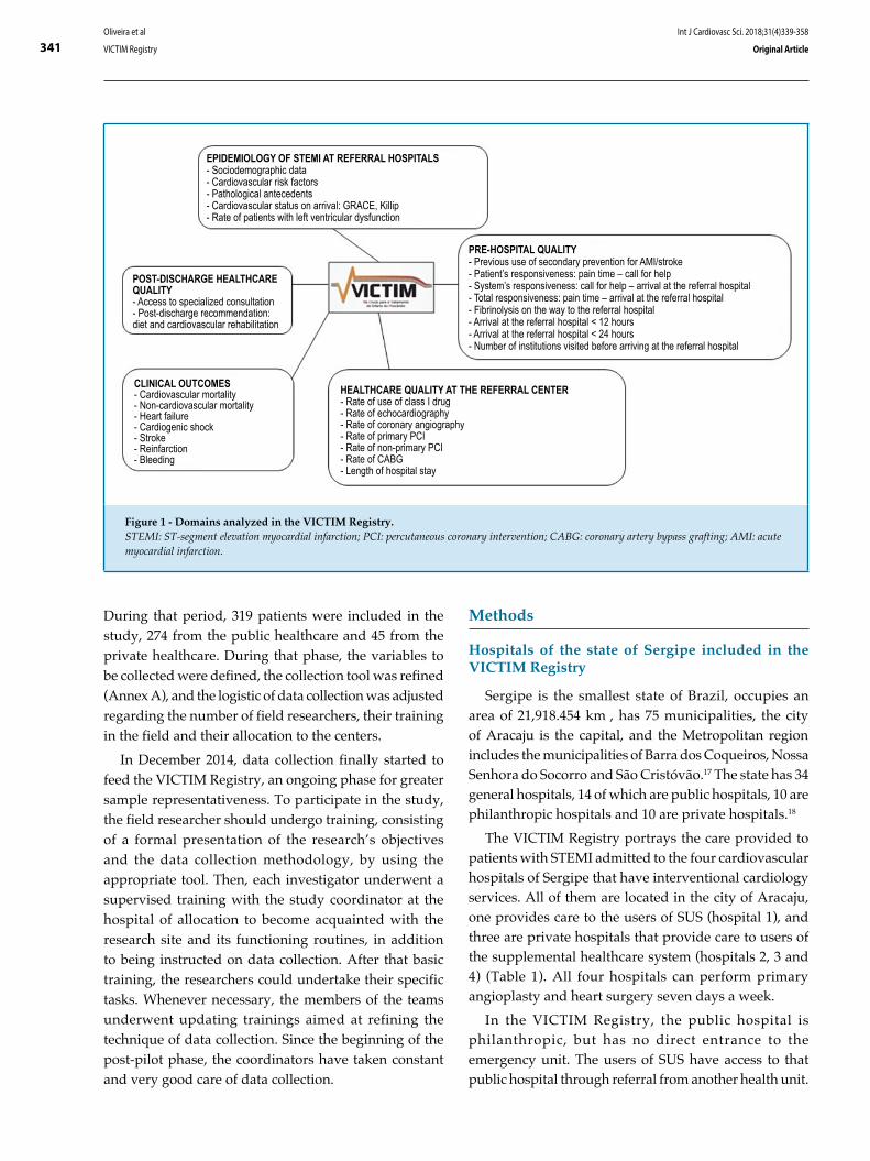
341
Figure 1 - Domains analyzed in the VICTIM Registry.STEMI: ST-segment elevation myocardial infarction; PCI: percutaneous coronary intervention; CABG: coronary artery bypass grafting; AMI: acute myocardial infarction.
POST-DISCHARGE HEALTHCARE QUALITY- Access to specialized consultation- Post-discharge recommendation: diet and cardiovascular rehabilitation
CLINICAL OUTCOMES- Cardiovascular mortality- Non-cardiovascular mortality- Heart failure- Cardiogenic shock- Stroke- Reinfarction- Bleeding
HEALTHCARE QUALITY AT THE REFERRAL CENTER- Rate of use of class I drug- Rate of echocardiography- Rate of coronary angiography- Rate of primary PCI- Rate of non-primary PCI- Rate of CABG- Length of hospital stay
EPIDEMIOLOGY OF STEMI AT REFERRAL HOSPITALS- Sociodemographic data- Cardiovascular risk factors- Pathological antecedents- Cardiovascular status on arrival: GRACE, Killip- Rate of patients with left ventricular dysfunction
PRE-HOSPITAL QUALITY- Previous use of secondary prevention for AMI/stroke- Patient’s responsiveness: pain time – call for help- System’s responsiveness: call for help – arrival at the referral hospital- Total responsiveness: pain time – arrival at the referral hospital- Fibrinolysis on the way to the referral hospital- Arrival at the referral hospital < 12 hours- Arrival at the referral hospital < 24 hours- Number of institutions visited before arriving at the referral hospital
Oliveira et al
VICTIM Registry
Int J Cardiovasc Sci. 2018;31(4)339-358
Original Article
During that period, 319 patients were included in the study, 274 from the public healthcare and 45 from the private healthcare. During that phase, the variables to be collected were defined, the collection tool was refined (Annex A), and the logistic of data collection was adjusted regarding the number of field researchers, their training in the field and their allocation to the centers.
In December 2014, data collection finally started to feed the VICTIM Registry, an ongoing phase for greater sample representativeness. To participate in the study, the field researcher should undergo training, consisting of a formal presentation of the research’s objectives and the data collection methodology, by using the appropriate tool. Then, each investigator underwent a supervised training with the study coordinator at the hospital of allocation to become acquainted with the research site and its functioning routines, in addition to being instructed on data collection. After that basic training, the researchers could undertake their specific tasks. Whenever necessary, the members of the teams underwent updating trainings aimed at refining the technique of data collection. Since the beginning of the post-pilot phase, the coordinators have taken constant and very good care of data collection.
Methods
Hospitals of the state of Sergipe included in the VICTIM Registry
Sergipe is the smallest state of Brazil, occupies an area of 21,918.454 km², has 75 municipalities, the city of Aracaju is the capital, and the Metropolitan region includes the municipalities of Barra dos Coqueiros, Nossa Senhora do Socorro and São Cristóvão.17 The state has 34 general hospitals, 14 of which are public hospitals, 10 are philanthropic hospitals and 10 are private hospitals.18
The VICTIM Registry portrays the care provided to patients with STEMI admitted to the four cardiovascular hospitals of Sergipe that have interventional cardiology services. All of them are located in the city of Aracaju, one provides care to the users of SUS (hospital 1), and three are private hospitals that provide care to users of the supplemental healthcare system (hospitals 2, 3 and 4) (Table 1). All four hospitals can perform primary angioplasty and heart surgery seven days a week.
In the VICTIM Registry, the public hospital is philanthropic, but has no direct entrance to the emergency unit. The users of SUS have access to that public hospital through referral from another health unit.

342Oliveira et al
VICTIM Registry
Int J Cardiovasc Sci. 2018;31(4)339-358
Original Article
Table 1 - Characteristics of the hospitals participating in the VICTIM Registry
Characteristics of the
hospitals
Public hospital
patients
N (370*)
Private hospital patients
N (82*)
Hospital 1
(370*)
Hospital 2
(35*)
Hospital 3
(17*)
Hospital 4
(30*)
Location Capital Capital Capital Capital
Type Non-profit-making Profit-making Profit-making Profit-making
Total number of beds 279 208 147 49
COU beds 10 10 0 8
General hospital YES YES YES NO
Access as user of SUS YES NO NO NO
Ability to perform PCI YES YES YES YES
Ability to perform heart surgery YES YES YES YES
Patients admitted via direct
access*6 (1.5%) 27 (77%) 15 (88%) 12 (40%)
Patients admitted via referral* 364 (98.5%) 8 (23%) 2 (12%) 18 (60%)
N: Number of patients; COU: Coronary Unit; SUS: Brazilian Unified Health System; PCI: Percutaneous Coronary Intervention; (*) Period: December 2014 to April 2016.
The private hospitals, however, provide care to a heterogeneous population, comprising patients with different health insurance plans and those who choose to pay for their own healthcare. Each of the three private hospitals has its specific set of health insurance plans, which makes their population heterogeneous. Such hospitals have direct entrance to their emergency units, thus, the patient can have direct access to those hospitals or can be referred from another health institution.
In the state of Sergipe, 80.7% of the population has no health insurance, relying, therefore, on the SUS, depending consequently on one single hospital as reference for the treatment of STEMI. The other 19.3% of the population has health insurance, counting on three hospitals with catheterization laboratory. Because of the lack of the necessary responsiveness in the SUS, some patients, even with neither health insurance nor a favorable economic condition, opt for the private service care.19
Except for those four hospitals, no other hospital of the Sergipe healthcare system has a team of cardiologists on call or a clinical team capable of identifying and treating patients with STEMI, especially regarding the
prescription of thrombolytic agents or the infrastructure to perform primary angioplasty.
The basic assumption is that the care provided to patients with STEMI in the four cardiovascular referral hospitals has the best quality in the state (Figure 2). Thus, to compare the quality of the care provided to users of the SUS with that provided at the three private hospitals will reflect the best public and private healthcare provided in the state of Sergipe.
Eligibility of the patients
Patients with the following characteristics are considered eligible for the VICTIM Registry: both sexes; older than 18 years; clinical findings compatible with acute coronary syndrome and electrocardiogram (ECG) showing persistent ST-segment elevation > 1 mm on two contiguous leads;7,8 and who provide written informed consent.
The diagnosis of AMI is confirmed later, based on the classical changes of the biomarkers CK-MB and/or troponin,7,8 taking into consideration the final opinion of the medical team.
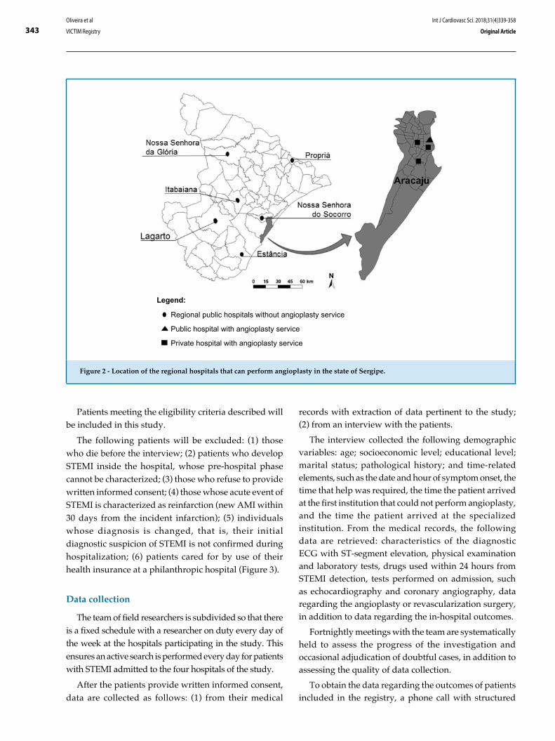
343
Figure 2 - Location of the regional hospitals that can perform angioplasty in the state of Sergipe.
Legend:
Regional public hospitals without angioplasty service
Public hospital with angioplasty service
Private hospital with angioplasty service
Oliveira et al
VICTIM Registry
Int J Cardiovasc Sci. 2018;31(4)339-358
Original Article
Patients meeting the eligibility criteria described will be included in this study.
The following patients will be excluded: (1) those who die before the interview; (2) patients who develop STEMI inside the hospital, whose pre-hospital phase cannot be characterized; (3) those who refuse to provide written informed consent; (4) those whose acute event of STEMI is characterized as reinfarction (new AMI within 30 days from the incident infarction); (5) individuals whose diagnosis is changed, that is, their initial diagnostic suspicion of STEMI is not confirmed during hospitalization; (6) patients cared for by use of their health insurance at a philanthropic hospital (Figure 3).
Data collection
The team of field researchers is subdivided so that there is a fixed schedule with a researcher on duty every day of the week at the hospitals participating in the study. This ensures an active search is performed every day for patients with STEMI admitted to the four hospitals of the study.
After the patients provide written informed consent, data are collected as follows: (1) from their medical
records with extraction of data pertinent to the study; (2) from an interview with the patients.
The interview collected the following demographic variables: age; socioeconomic level; educational level; marital status; pathological history; and time-related elements, such as the date and hour of symptom onset, the time that help was required, the time the patient arrived at the first institution that could not perform angioplasty, and the time the patient arrived at the specialized institution. From the medical records, the following data are retrieved: characteristics of the diagnostic ECG with ST-segment elevation, physical examination and laboratory tests, drugs used within 24 hours from STEMI detection, tests performed on admission, such as echocardiography and coronary angiography, data regarding the angioplasty or revascularization surgery, in addition to data regarding the in-hospital outcomes.
Fortnightly meetings with the team are systematically held to assess the progress of the investigation and occasional adjudication of doubtful cases, in addition to assessing the quality of data collection.
To obtain the data regarding the outcomes of patients included in the registry, a phone call with structured

344
Figure 3 - Flowchart representing the exclusion criteria for patients with ST-segment elevation myocardial infarction (STEMI).AMI: acute myocardial infarction.
PATIENTS WITH INITIAL DIAGNOSIS OF STEMI
PATIENTS AT A PUBLIC HOSPITAL THAT CAN
PERFORM ANGIOPLASTY
PATIENTS EXCLUDED FROM THE VICTIM REGISTRY
- Death before the interview- In-hospital AMI- Did not provide written informed consent- Reinfarction- Change in diagnosis- Patients with health insurance coverage at a philanthropic hospital
PACIENTES INCLUDED IN THE VICTIM REGISTRY
PATIENTS AT PRIVATE HOSPITALS THAT CAN
PERFORM ANGIOPLASTY
Oliveira et al
VICTIM Registry
Int J Cardiovasc Sci. 2018;31(4)339-358
Original Article
interview is performed 30 days after the detection of STEMI. On the occasion, the coordinator responsible for the calls gets information with the patients and/or their guardians on the occurrence of death, reinfarction, heart failure, cardiogenic shook, angina pectoris, stroke, hemorrhage, cardiac arrest and/or new hospitalization, in addition to assessing whether the patients attended a specialized consultation after discharge, and, if not, whether they have one scheduled.
When the patient cannot be reached via telephone, other resources are used, such as a relative’s or neighbor’s telephone contact, e-mail or post letter with the major researcher’s contacts, to minimize data loss.
If the patient remains hospitalized for 30 days, the final visit is performed during hospitalization, and after that the patient’s participation in the study ends.
Case report form and data bank
Case report form (CRF) is the collection tool (Annex A) adopted by the VICTIM Registry and comprises the following: (1) patient’s identification; (2) eligibility; (3) time line; (4) clinical presentation; (5) hospitalization; (6) outcomes. In 2015, the CRF passed from the print version to the electronic version, in which data storage is virtually fed, facilitating their maintenance and reducing the form filling out process errors. The data collected in
loco are stored in an electronic cloud, ensuring lower risk for data loss.
Data originating from the electronic CRF are transferred to a spreadsheet, facilitating their analysis and interpretation. The system is always fed by a researcher who underwent previous training and is the sole responsible for that activity. Aiming at minimizing errors of data bank input, the procedure is performed systematically right after patient’s assessment. Each CRF entered into the system receives an identification number, eliminating, thus, the need for contact with the names of the patients included in the study, and ensuring the right to anonymity.
Statistical analysis
Qualitative variables will be expressed as frequency (percentage), and quantitative variables will undergo Kolmogorov-Smirnov test to determine the distribution type; those meeting the normality assumption will be expressed as mean and standard deviation. The variables without a normal distribution will be described as median and interquartile range or maximum and minimum values. The qualitative variables will be compared by using Pearson’s chi-square test or Fisher exact test, when appropriate.20 Non-paired Student t test will be used to compare between the two major

345Oliveira et al
VICTIM Registry
Int J Cardiovasc Sci. 2018;31(4)339-358
Original Article
groups when the continuous or discrete variables have normal distribution. In case of asymmetric distribution, Wilcoxon-Mann-Whitney test will be used.21
To assess the effect of demography, clinical data, laboratory data and the time for reperfusion treatment to be performed, a model of multivariate logistic regression will be used with generalized equations that consider the clustering effect22 and stratified Cox regression.23
The Kaplan-Meier method24 and the log-rank test25 will be used to compare event-free survival curves in users of the SUS and of the private hospitals, with and without adjustment for the confounding variables. The SPSS Statistics program for Windows version 17 and R Core Team 201426 will be used for the statistical analysis. The significance level adopted in future analyses will be 5%.
Ethical considerations
Before entering the study, all volunteers or their guardians provide a written informed consent. Illiterate individuals who choose to participate in the study complete the informed consent process by signing with a fingerprint and two literate witnesses verify the process with a signature. This study was approved by the Ethics Committee in Research of the Federal University of Sergipe (nº 23392313.4.0000.5546).
Commitment of the VICTIM team
In addition to answering specific questions, the leaders of the VICTIM Registry are committed to continuously spreading the study results aiming at contributing to improve the healthcare quality for AMI. The present investigation is expected to provide constantly and systematically the health managers with technical information that can support new health policies or care strategies, contributing to the construction of a more efficient and equitable healthcare system. The central idea is to identify in the presently practiced line of care opportunities to improve the care provided regarding infrastructure, logistics of healthcare processes and especially the healthcare results.
In addition, the VICTIM Registry is expected to constitute a continuous field of training in several research areas, such as cardiovascular biomedicine, outcomes research and health services, for post-graduate and graduate students, to aid in the scientific qualification and formation of researchers in the health sciences area.
Author contributions
Conception and design of the research: Oliveira JC, Oliveira LCS, Oliveira JC, Barreto IDC, Arcelino LAM, Prado LFA, Silveira FS, Nascimento TA, Ferreira EJP, Barreto RV, Moraes EV, Mendonça JT, Sousa ACS, Barreto-Filho JA. Acquisition of data: Oliveira JC, Oliveira LCS, Oliveira JC, Lima TCRM, Arcelino LAM, Barreto-Filho JA. Analysis and interpretation of the data: Oliveira JC, Oliveira LCS, Oliveira JC, Barreto IDC, Almeida-Santos MA, Lima TCRM, Arcelino LAM, Sousa ACS, Barreto-Filho JA. Statistical analysis: Oliveira JC, Oliveira LCS, Barreto IDC, Almeida-Santos MA, Barreto-Filho JA. Obtaining financing: Oliveira JC, Oliveira LCS, Oliveira JC, Barreto-Filho JA. Writing of the manuscript: Oliveira JC, Oliveira LCS, Oliveira JC, Arcelino LAM, Barreto-Filho JA. Critical revision of the manuscript for intellectual content: Oliveira JC, Oliveira LCS, Oliveira JC, Barreto IDC, Almeida-Santos MA, Lima TCRM, Arcelino LAM, Prado LFA, Silveira FS, Nascimento TA, Ferreira EJP, Barreto RV, Moraes EV, Mendonça JT, Sousa ACS, Barreto-Filho JA. Supervision / as the major investigador: Barreto-Filho JA.
Potential Conflict of Interest
No potential conflict of interest relevant to this article was reported.
Sources of Funding
This study was partially funded by CNPq 14/2013 – Universal.
Study Association
This article is part of the thesis of Doctoral submitted by Jussiely Cunha Oliveira, from Universidade Federal de Sergipe.
Ethics approval and consent to participate
This study was approved by the Ethics Committee of the Universidade Federal de Sergipe under the protocol number 483.749. All the procedures in this study were in accordance with the 1975 Helsinki Declaration, updated in 2013. Informed consent was obtained from all participants included in the study.
346Oliveira et al
VICTIM Registry
Int J Cardiovasc Sci. 2018;31(4)339-358
Original Article
This is an open-access article distributed under the terms of the Creative Commons Attribution License
1. Brasil. Constituição 1988. Constituição da República Federativa do Brasil. Brasília, DF: Senado Federal; 1988. [Acesso em 2016 jan 19]. Disponível em: http://www.planalto.gov.br/ccivil_03/constituicao/ConstituicaoCompilado.htm
2. Brasil. Lei nº 8.080, de 19 de setembro de 1990. Dispõe sobre as condições para a promoção, proteção e recuperação da saúde, a organização e o funcionamento dos serviços correspondentes e dá outras providências. Brasília, DF; 1990. [Acesso em 2016 jan 19]. Disponível em: http://www.planalto.gov.br/ccivil_03/Leis/L8080.htm
3. Oliveira JC, Barreto-Filho, JA. Public health policy based on “Made-In-Brazil” science: a challenge for the Arquivos Brasileiros de Cardiologia. Arq Bras Cardiol. 2015;105(3):211-3. doi: http://dx.doi.org/10.5935/abc.20150120.
4. Instituto Brasileiro de Geografia e Estatísica. (IBGE). Pesquisa nacional de saúde 2013: acesso e utilização dos serviços de saúde, acidentes e violências: Brasil, grandes regiões e unidades da federação. Rio de Janeiro: IBGE; 2015. [Acesso em 2017 fev 12]. Disponível em http://biblioteca.ibge.gov.br/visualizacao/livros/liv94074.pdf
5. Andrade JP, Mattos LA, Carvalho AC, Machado AC, Oliveira GM. National physician qualification program in cardiovascular disease prevention and integral care. Arq Bras Cardiol. 2013;100(3):203-11. doi: http://dx.doi.org/10.5935/abc.20130061.
6. Steg PG, James SK, Atar D, Badano LP, Blömstrom-Lundqvist C, Borger MA, et al; ESC Guidelines for the management of acute myocardial infarction in patients presenting with ST-segment elevation. Task Force on the management of ST-segment elevation acute myocardial infarction of the European Society of Cardiology (ESC). Eur Heart J. 2012;33(20):2569-619. doi: 10.1093/eurheartj/ehs215.
7. Piegas LS, Timerman A, Feitosa GS, Nicolau JC, Mattos LA, Andrade MD, et al; Sociedade Brasileira de Cardiologia. V Diretriz da Sociedade Brasileira de Cardiologia sobre tratamento do infarto agudo do miocárdio com supradesnível do segmento ST. Arq Bras Cardiol. 2015;105(2):1-105. doi: http://dx.doi.org/10.5935/abc.20150107.
8. O'Gara PT, Kushner FG, Ascheim DD, Casey DE Jr, Chung MK, de Lemos JA, et al; American College of Cardiology Foundation/American Heart Association Task Force on Practice Guidelines. 2013 ACCF/AHA guideline for the management of ST-elevation myocardial infarction: a report of the American College of Cardiology Foundation/American Heart Association Task Force on Practice Guidelines. Circulation. 2013;127(4):e362-425. doi: 10.1161/CIR.0b013e3182742cf6.
9. Effectiveness of intravenous thrombolytic treatment in acute myocardial infarction. Gruppo Italiano per lo Studio della Streptochinasi nell'Infarto Miocardico (GISSI). Lancet. 1986;1(8478):397-402. PMID: 2868337.
10. Terkelsen CJ, Sørensen JT, Maeng M, Jensen LO, Tilsted HH, Trautner S, et al. System delay and mortality among patients with STEMI treated with primary percutaneous coronary intervention. JAMA. 2010;304(7):763-71. doi: 10.1001/jama.2010.1139.
11. Li J, Li X, Wang Q, Hu S, Wang Y, Masoudi FA, et al. ST-segment elevation myocardial infarction in China from 2001 to 2011 (the China PEACE Retrospective Acute Myocardial Infarction Study): a retrospective analysis of hospital data. Lancet. 2015;385(9966):441-51. doi: 10.1016/S0140-6736(14)60921-1.
12. Alexander T, Mullasari AS, Joseph G, Kannan K, Veerasekar G, Victor SM, et al. A system of care for patients with ST-segment elevation myocardial infarction in India. The Tamil Nadu–ST-segment elevation
myocardial infarction program. JAMA Cardiol. 2017;2(5):498-505. doi: 10.1001/jamacardio.2016.5977.
13. Ferreira GM, Correia LC, Reis H, Ferreira Filho CB, Freitas F, Ferreira GM, et al. Increased mortality and morbidity due to acute myocardial infarction in a public hospital, in Feira de Santana, Bahia. Arq Bras Cardiol. 2009;93(2):97-104. doi: http://dx.doi.org/10.1590/S0066-782X2009000800006.
14. Nicolau JC, Franken M, Lotufo PA, Carvalho AC, Marin Neto JA, Lima FG, et al. Use of demonstrably effective therapies in the treatment of acute coronary syndromes: comparison between different Brazilian Regions. Analysis of the Brazilian Registry on Acute Coronary Syndromes (BRACE). Arq Bras Cardiol. 2012;98(4):282-9. doi: http://dx.doi.org/10.1590/S0066-782X2012000400001.
15. Lichtman JH, Lorenze NP, D'Onofrio G, Spertus JA, Lindau ST, Morgan TM, et al. Variation in recovery: role of gender on outcomes of young AMI patients (VIRGO) study design. Circ Cardiovasc Qual Outcomes. 2010;3(6):684-93. doi: 10.1161/CIRCOUTCOMES.109.928713.
16. Dharmarajan K, Li J, Li X, Lin Z, Krumholz HM, Jiang L. The China Patient-Centered Evaluative Assessment of Cardiac Events (China PEACE) retrospective study of acute myocardial infarction: study design. Circ Cardiovasc Qual Outcomes. 2013;6(6):732-40. DOI: 10.1161/CIRCOUTCOMES.113.000441.
17. Instituto Brasileiro de Geografia e Estatísica. (IBGE). Estados. [publicação online]. IBGE; 2015. [Acesso em 2016 dez 1]. Disponível em http://www.ibge.gov.br/estadosat/perfil.php?lang=&sigla=se
18. Brasil. Ministério da Saúde. Cadastro Nacional de Estabelecimentos de Saúde: Situação da base de dados nacional. BRASIL. [publicação online]; 2010 [Acesso em 2017 mar 2]. Disponível em: http://tabnet.datasus.gov.br/cgi/tabcgi.exe?cnes/cnv/estabSE.def.
19. Instituto Brasileiro de Geografia e Estatística. (IBGE). Pesquisa Nacional de Saúde 2013: Acesso e Utilização dos Serviços de Saúde, Acidentes e Violências. Diretoria de Pesquisas, Coordenação de Trabalho e Rendimento, Pesquisa Nacional de Saúde. [publicação online]. Sergipe: IBGE; 2013. [Acesso em 2017 mai 2]. Disponível em: http://www.ibge.gov.br/estadosat/temas.php?sigla=se&tema=pns_2013_util_serv_saude.
20. Agresti A. Categorical data analysis. 2nd ed. New York: John Wiley & Sons; 2009.
21. Conover WJ. Practical nonparametric statistics. 3rd ed. New York: John Wiley & Sons; 1999.
22. Dobson AJ, Barnett AG. An introduction to generalized linear models. 3rd ed. Boca Raton; CRC Press; 2008.
23. Hosmer DW, Lemeshow S, May S. Applied survival analysis: regression modeling of time-to-event data. 2nd ed. New Jersey: Wiley-Interscience; 2008.
24. Kaplan EL, Meier P. Nonparametric estimation from incomplete observations. J Am Stat Assoc. 1958;53(282):457-81. doi: https://doi.org/10.1080/01621459.1958.70501452.
25. Mantel N. Evaluation of survival data and two new rank order statistics arising in its consideration. Cancer Chemother Rep.1966;50(3):163-70. PMID: 5910392.
26. R Core Team (2014). R: a language and environment for statistical computing. Vienna (Austria). R Foundation for Statistical Computing, Vienna, Austria. [Access in 2016 jan 19]. Available from: http://www.R-project.org/.
References

347Oliveira et al
VICTIM Registry
Int J Cardiovasc Sci. 2018;31(4)339-358
Original Article
Annex A
Data collection tool

348Oliveira et al
VICTIM Registry
Int J Cardiovasc Sci. 2018;31(4)339-358
Original Article
REGISTRATION
Patient’s registration number:ID: Brazilian Social Security number:
Socioeconomic level*
Self-employed
Widow Unmarried couple
Post-graduation
PrivateWhich?
Address:
*Socioeconomic level: A: income > 20 minimum wagesB: 10-20 minimum wagesC: 4-10 minimum wages
D: 2-4 minimum wagesE: <2 minimum wagesNI: not informed

349Oliveira et al
VICTIM Registry
Int J Cardiovasc Sci. 2018;31(4)339-358
Original Article
ELIGIBILITY
Clinical findings compatible with AMI Age ≥ 18 years
Provided written informed consent
Time of STEMI detection:
Persistent ST-segment elevation in two leads:
0.5mm:
T-WAVE INVERSION
350Oliveira et al
VICTIM Registry
Int J Cardiovasc Sci. 2018;31(4)339-358
Original Article
DECISION TO CALL TRANSPORTATION
Address at the time of symptom onset:
Address at the time transportation was requested:
Mobile emergency medical service
Own transportation
ARRIVAL AT THE 1st HOSPITAL WITHOUT PCI
Which?
ARRIVAL AT THE 2nd HOSPITAL WITHOUT PCI
Which?
ARRIVAL AT THE 3rd HOSPITAL WITHOUT PCI
Which?
ARRIVAL AT THE HOSPITAL WITH PCI
Which?
STEMI DETECTION:
DOOR-BALLOON Door-balloon:
Which?
Cardiopulmonary arrest during transfer?
Place:
*Hospitals:

351Oliveira et al
VICTIM Registry
Int J Cardiovasc Sci. 2018;31(4)339-358
Original Article
PRODROMAL SYMPTOMS FOR MORE THAN 24H FROM THE MAJOR FINDING
week
PRESENTATION SYMPTOMS
Typical anginal chest pain/epigastric pain
CLINICAL PRESENTATION
Atypical chest pain
Sweating
Nausea/vomiting
INFARCTION TRIGGERSStrenuous physical exertion 2h before symptom onsetSexual intercourse 2 hours before symptom onsetCocaine or other illicit drug use within the previous 24h ________Infection in the past 10 days: _____________________
Severe emotional stress within the previous 24h
Alcohol use within the previous 24h
PREVIOUS PATHOLOGICAL HISTORY AND CARDIOVASCULAR RISK FACTORS
Ex-smoker:
Family history of early CAD / male<55 and female<65:
Previous CAD (>50%):
Previous CABG:
352Oliveira et al
VICTIM Registry
Int J Cardiovasc Sci. 2018;31(4)339-358
Original Article
Previous stroke or TIA:
Chronic kidney disease:
PREVIOUS DRUG THERAPY
Beta-blocker
Calcium-channel blocker
If yes, which?
mm Hg HEIGHT:
In-hospital
High CK-MB:
High TpN +:

353Oliveira et al
VICTIM Registry
Int J Cardiovasc Sci. 2018;31(4)339-358
Original Article
DRUG THERAPY IN THE FIRST 24 HOURS
Eligible:
Eligible:
Eligible:
Eligible:
Eligible:
Eligible:
Eligible:
Eligible:
Eligible:
Eligible:
Eligible:
Eligible:
Beta-blocker:
Calcium-channel blocker:
Low-molecular-weight heparin:
Conventional heparin:
If yes, which?
IN-HOSPITAL TESTS
ECHOCARDIOGRAPHY
LA volume:
Segment deficit:
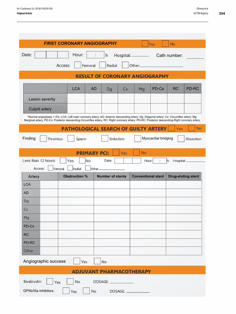
354Oliveira et al
VICTIM Registry
Int J Cardiovasc Sci. 2018;31(4)339-358
Original Article
FIRST CORONARY ANGIOGRAPHY
Cath number:
Lesion severity
Culprit artery
LCA AD PD-Cx RC PD-RC
*Normal angioplasty = 0%; LCA: Left main coronary artery; AD: Anterior descending artery; Dg: Diagonal artery; Cx: Circumflex artery; Mg: Marginal artery; PD-Cx: Posterior descending-Circumflex artery; RC: Right coronary artery; PD-RC: Posterior descending-Right coronary artery.
Finding Myocardial bridging
Obstruction % Number of stents Conventional stent Drug-eluting stent
LCA
AD
PD-Cx
RC
PD-RC
Angiographic success
GPIIb/IIIa inhibitors
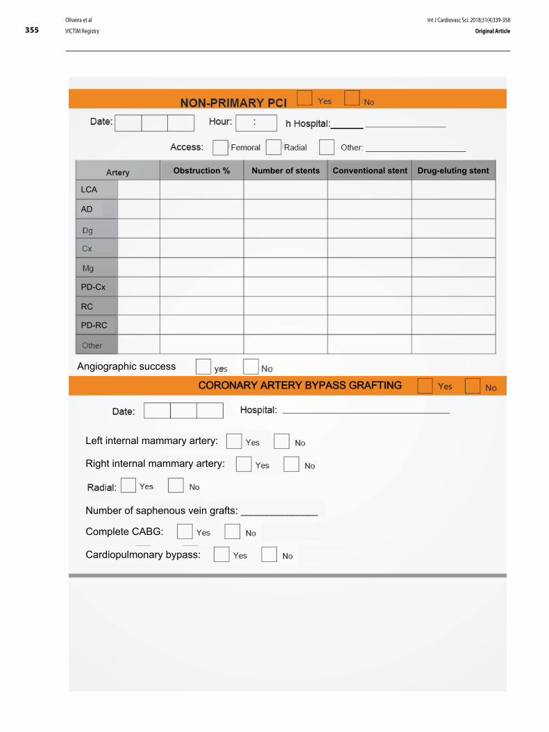
355Oliveira et al
VICTIM Registry
Int J Cardiovasc Sci. 2018;31(4)339-358
Original Article
Obstruction % Number of stents Conventional stent Drug-eluting stent
LCA
AD
PD-Cx
RC
PD-RC
Angiographic success
Left internal mammary artery:
Right internal mammary artery:
Number of saphenous vein grafts: _______________
Complete CABG:
Cardiopulmonary bypass:

356Oliveira et al
VICTIM Registry
Int J Cardiovasc Sci. 2018;31(4)339-358
Original Article
IN-HOSPITAL OUTCOMES
CARDIOVASCULAR DEATH:
NON-CARDIOVASCULAR DEATH:
POST-AMI ANGINA:
- Lowest hemoglobin recorded
- Lowest hematocrit recorded
- Puncture site-related hemorrhage
If yes, how many blood bags? ____________________
DIAGNOSTIC OUTCOME
:
If not, what was the final diagnosis?
Does the medical record have any post-infarction dietary/nutritional instruction?
If yes, who recorded the instruction?
Does the medical record have any instruction on post-infarction cardiac rehabilitation?
If yes, who recorded the instruction? Physical trainer

357Oliveira et al
VICTIM Registry
Int J Cardiovasc Sci. 2018;31(4)339-358
Original Article
DISCHARGE MEDICATION
Clopidogrel:
Beta-blocker:
Calcium-channel blocker:
If yes, which?
Eligible:
Eligible:
Eligible:
Eligible:
Eligible:
Eligible:
Eligible:
Eligible:
Eligible:
Contact after 30 days? CARDIOVASCULAR DEATH:NON-CARDIOVASCULAR DEATH:
POST-AMI ANGINA:
REHOSPITALIZATION:

358Oliveira et al
VICTIM Registry
Int J Cardiovasc Sci. 2018;31(4)339-358
Original Article
DOI: 10.5935/2359-4802.20180033
359
ORIGINAL ARTICLE
International Journal of Cardiovascular Sciences. 2018;31(4)359-366
Mailing Address: Rafael Alessandro Ferreira GomesRua dos Palmares, s/n, Santo Amaro. Postal Code: 50100-060, Recife, PE - Brazil.E-mail: [email protected]
Carotid Atherosclerosis in Pre- and Post-Menopausal Women with a History of Pregnancy-Induced Hypertension: Case-Control StudyRafael Alessandro Ferreira Gomes, Isly Maria Lucena de Barros, Moacir de Novaes Lima Ferreira, Laura Olinda Bregieiro Fernandes CostaUniversidade de Pernambuco (UPE), Recife, PE - Brazil
Manuscript received on July 10, 2017, revised manuscript received on November 18, 2017, accepted on December 20, 2017.
Abstract
Background: Cardiovascular disease mortality among women remains high. Observational studies are controversial about the participation of a history of gestational hypertensive disorder in cardiovascular risk.
Objective: To verify the association between carotid atherosclerosis in menopausal women who had pregnancy-induced hypertension.
Methods: Case-control study, with cases consisting of women with carotid atherosclerosis, defined as carotid intima-media thickness > 1 mm and/or presence of carotid plaques; the controls did not have these alterations. The significance level was set at 95%.
Results: A total of 504 women without previous cardiovascular disease were assessed, 126 cases and 378 controls. Of the total, 67% were hypertensive; 76% were dyslipidemic; and 16% were diabetic. Approximately 10% reported a history of hypertension during pregnancy. Women with carotid atherosclerosis had higher values of systolic blood pressure (134.18 mmHg vs. 128.59 mmHg, p = 0.008) and LDL-cholesterol (156.52 mg% vs. 139.97 mg%; p = 0.0005). No statistical difference was found regarding the presence of carotid atherosclerosis and history of hypertension during pregnancy (OR 1.672, 95% CI: 0.883-3.131).
Conclusion: The history of hypertension during pregnancy was not associated with subclinical carotid atherosclerosis in menopausal women. However, an association was observed between carotid atherosclerosis and classic risk factors, such as elevated systolic blood pressure and LDL-cholesterol levels. (Int J Cardiovasc Sci. 2018;31(4)359-366)
Keywords: Carotid Artery Diseases/physiopathology; Hypertension, Pregnancy-Induced; Women, Premenopause; Postmenopause; Case-Control Studies.
Introduction
Cardiovascular diseases (CVD) are the leading cause of death among women worldwide.1,2 In the United States, they account for almost a third of all causes of death in the female gender,3,4 and similar data are observed in Europe5 and in Brazil.6 Advances in CVD treatment in the last three decades have allowed a sustained decrease in mortality. However, socioeconomic and behavioral aspects have interrupted this process in recent years.7 In 2014, there were 340,284 CVD deaths among Brazilian women, representing an increase of almost 20% in relation
to those occurring 10 years earlier.6 The cardiovascular risk stratification in the female population has failed to detect and prevent the disease. The exploration of new risk factors thus becomes essential to reduce such indices.
Pregnancy is an important moment to evaluate women’s cardiovascular health, since the development of complications during this period may indicate an increase in future cardiovascular risk.8 Several observational studies have shown a higher prevalence of atherosclerosis in women with a history of gestation-induced hypertension,9-11 and some have observed an association between the number of cardiac events and
360Gomes et al.
Atherosclerosis and hypertension in pregnancy
Int J Cardiovasc Sci. 2018;31(4)359-366
Original Article
the number of complicated pregnancies,12 even after the normalization of blood pressure levels after childbirth.
Nonetheless, Romundstad et al.13 questioned whether such an association would be a factor of ambiguity, because pre-gestational characteristics – especially obesity, hypertension and dyslipidemia – would attenuate the effect that the gestational hypertensive disorder has on the late cardiovascular outcome. Unfortunately, most studies have insufficient evidence, such as limited sample size and clinical follow-up.
Considering that atherosclerosis is a gradual process that starts in childhood, the aim of this study was to verify the association between carotid atherosclerosis in menopausal women who had pregnancy-induced hypertension.
Methods
A case-control study was carried out, with a population of women aged between 45 and 65 years, who had had menstrual irregularities or interruption in the last year. Women receiving hormone replacement therapy, those with chronic inflammatory conditions or any previously diagnosed conditions with high cardiovascular risk or heart disease were excluded from the analysis.
The sample was calculated based on the systematic review performed by Brown et al.,14 using as reference a hypertension exposure during pregnancy of around 8% and Odds Ratio (OR) to increase the risk of atherosclerosis of 2.28. For a paired study with a one-tailed hypothesis test, we calculated at least 116 cases and 348 controls in order to obtain a 95% level of significance and 80% of test power with a ratio of one case for three controls. Controls were obtained from the same database, and were paired by age group.
All women underwent carotid ultrasound with the same examiner; the carotid intima-media thickness (CIMT) was quantified and the presence of carotid plaques was assessed. For image acquisition, a high-resolution device (EnVisor, Philips) was used with a 12.3 MHz linear transducer. The data were recorded for subsequent analysis using the QLAB-Intima Media Thickness (QLAB-IMT, Philips) software.
The presence of carotid atherosclerosis was defined when the CIMT was greater than 1 mm (mean values obtained in the analyzed segments of the right and left carotid arteries) and / or the presence of atheroma plaque. Atheroma plaque was defined as: (1) localized parietal structure with a thickness greater than 1.5 mm;
(2) protrusion into vessel lumen > 0.5 mm or; (3) thickness > 1.5-fold the adjacent CIMT, according to the Mannheim Carotid Intima-Media Thickness and Plaque Consensus.15
The cases consisted of women who had carotid atherosclerosis and the controls, of women who did not have this alteration at the ultrasonographic assessment.
The independent variable was pregnancy-induced hypertension, considered as the self-reported information of blood pressure increase during pregnancy. According to Diehl et al.,16 this information shows good accuracy (specificity of 96% and sensitivity of 79.6%) for the antecedents of pregnancy-induced hypertension, even 24.5 years after the pregnancy. Other variables were considered, namely: blood pressure, income, smoking, type 2 diabetes mellitus, family history of coronary artery disease (CAD), body mass index (BMI), number of pregnancies, preterm birth, low birth-weight offspring, fasting glycemia, total cholesterol (CT), high-density lipoprotein cholesterol (HDL-cholesterol), low-density lipoprotein cholesterol (LDL-cholesterol), triglycerides and ultrasensitive C-reactive protein (us-CRP).
The study was approved by the Research Ethics Committee of Complexo Hospitalar Hospital Universitário Oswaldo Cruz/ Pronto-Socorro Cardiológico de Pernambuco under CAAE number 55361416.0.0000.5192 and Opinion number 1,593,189 of June 16, 2016.
Statistical analysis
The results were expressed as percentages for categorical variables and as statistical measures such as means, standard deviation and medians, when indicated, for numerical variables. The association between the occurrence of carotid atherosclerosis and the categorical variables was performed using Pearson’s chi-square test, whereas the non-paired Student’s t test was used to compare carotid atherosclerosis in relation to numerical variables. Cox regression analysis was performed to evaluate the influence of covariates on carotid atherosclerosis development in the menopausal period. The strength of the association between the categorical variables was evaluated using the odds ratio (OR) with the respective confidence interval. The normality hypothesis verification was performed using the Kolmogorov-Smirnov test. The level of significance used in the statistical test decisions was 5% and the intervals had 95% of confidence. The Statistical Package for the Social Sciences (SPSS) version 21 was the statistical program used for the statistical calculations.
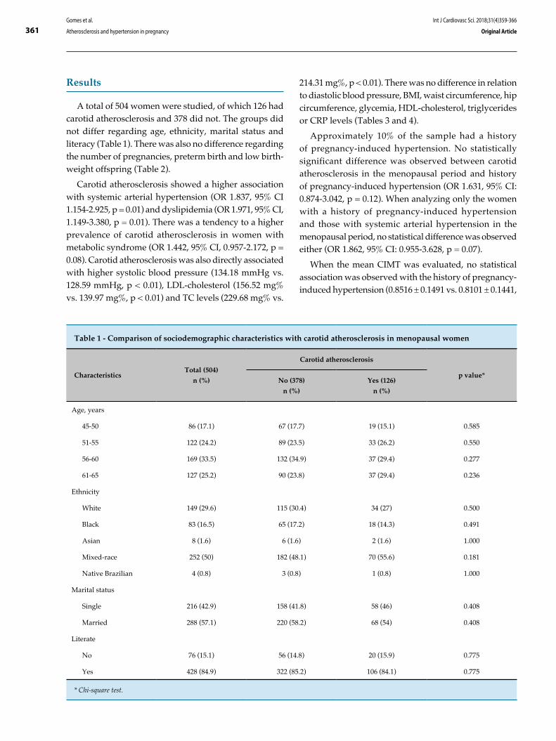
361Gomes et al.
Atherosclerosis and hypertension in pregnancy
Int J Cardiovasc Sci. 2018;31(4)359-366
Original Article
Results
A total of 504 women were studied, of which 126 had carotid atherosclerosis and 378 did not. The groups did not differ regarding age, ethnicity, marital status and literacy (Table 1). There was also no difference regarding the number of pregnancies, preterm birth and low birth-weight offspring (Table 2).
Carotid atherosclerosis showed a higher association with systemic arterial hypertension (OR 1.837, 95% CI 1.154-2.925, p = 0.01) and dyslipidemia (OR 1.971, 95% CI, 1.149-3.380, p = 0.01). There was a tendency to a higher prevalence of carotid atherosclerosis in women with metabolic syndrome (OR 1.442, 95% CI, 0.957-2.172, p = 0.08). Carotid atherosclerosis was also directly associated with higher systolic blood pressure (134.18 mmHg vs. 128.59 mmHg, p < 0.01), LDL-cholesterol (156.52 mg% vs. 139.97 mg%, p < 0.01) and TC levels (229.68 mg% vs.
214.31 mg%, p < 0.01). There was no difference in relation to diastolic blood pressure, BMI, waist circumference, hip circumference, glycemia, HDL-cholesterol, triglycerides or CRP levels (Tables 3 and 4).
Approximately 10% of the sample had a history of pregnancy-induced hypertension. No statistically significant difference was observed between carotid atherosclerosis in the menopausal period and history of pregnancy-induced hypertension (OR 1.631, 95% CI: 0.874-3.042, p = 0.12). When analyzing only the women with a history of pregnancy-induced hypertension and those with systemic arterial hypertension in the menopausal period, no statistical difference was observed either (OR 1.862, 95% CI: 0.955-3.628, p = 0.07).
When the mean CIMT was evaluated, no statistical association was observed with the history of pregnancy-induced hypertension (0.8516 ± 0.1491 vs. 0.8101 ± 0.1441,
Table 1 - Comparison of sociodemographic characteristics with carotid atherosclerosis in menopausal women
CharacteristicsTotal (504)
n (%)
Carotid atherosclerosis
p value*No (378)
n (%)
Yes (126)
n (%)
Age, years
45-50 86 (17.1) 67 (17.7) 19 (15.1) 0.585
51-55 122 (24.2) 89 (23.5) 33 (26.2) 0.550
56-60 169 (33.5) 132 (34.9) 37 (29.4) 0.277
61-65 127 (25.2) 90 (23.8) 37 (29.4) 0.236
Ethnicity
White 149 (29.6) 115 (30.4) 34 (27) 0.500
Black 83 (16.5) 65 (17.2) 18 (14.3) 0.491
Asian 8 (1.6) 6 (1.6) 2 (1.6) 1.000
Mixed-race 252 (50) 182 (48.1) 70 (55.6) 0.181
Native Brazilian 4 (0.8) 3 (0.8) 1 (0.8) 1.000
Marital status
Single 216 (42.9) 158 (41.8) 58 (46) 0.408
Married 288 (57.1) 220 (58.2) 68 (54) 0.408
Literate
No 76 (15.1) 56 (14.8) 20 (15.9) 0.775
Yes 428 (84.9) 322 (85.2) 106 (84.1) 0.775
* Chi-square test.
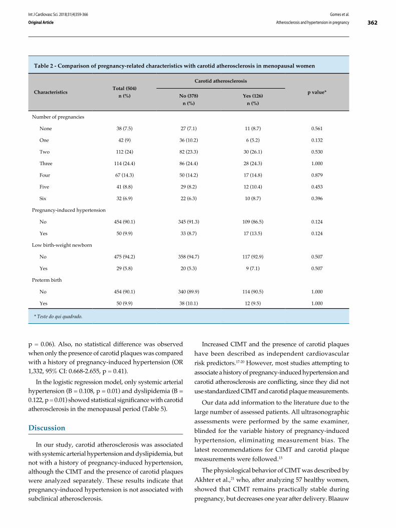
362Gomes et al.
Atherosclerosis and hypertension in pregnancy
Int J Cardiovasc Sci. 2018;31(4)359-366
Original Article
Table 2 - Comparison of pregnancy-related characteristics with carotid atherosclerosis in menopausal women
CharacteristicsTotal (504)
n (%)
Carotid atherosclerosis
p value*No (378)
n (%)
Yes (126)
n (%)
Number of pregnancies
None 38 (7.5) 27 (7.1) 11 (8.7) 0.561
One 42 (9) 36 (10.2) 6 (5.2) 0.132
Two 112 (24) 82 (23.3) 30 (26.1) 0.530
Three 114 (24.4) 86 (24.4) 28 (24.3) 1.000
Four 67 (14.3) 50 (14.2) 17 (14.8) 0.879
Five 41 (8.8) 29 (8.2) 12 (10.4) 0.453
Six 32 (6.9) 22 (6.3) 10 (8.7) 0.396
Pregnancy-induced hypertension
No 454 (90.1) 345 (91.3) 109 (86.5) 0.124
Yes 50 (9.9) 33 (8.7) 17 (13.5) 0.124
Low birth-weight newborn
No 475 (94.2) 358 (94.7) 117 (92.9) 0.507
Yes 29 (5.8) 20 (5.3) 9 (7.1) 0.507
Preterm birth
No 454 (90.1) 340 (89.9) 114 (90.5) 1.000
Yes 50 (9.9) 38 (10.1) 12 (9.5) 1.000
* Teste do qui quadrado.
p = 0.06). Also, no statistical difference was observed when only the presence of carotid plaques was compared with a history of pregnancy-induced hypertension (OR 1,332, 95% CI: 0.668-2.655, p = 0.41).
In the logistic regression model, only systemic arterial hypertension (B = 0.108, p = 0.01) and dyslipidemia (B = 0.122, p = 0.01) showed statistical significance with carotid atherosclerosis in the menopausal period (Table 5).
Discussion
In our study, carotid atherosclerosis was associated with systemic arterial hypertension and dyslipidemia, but not with a history of pregnancy-induced hypertension, although the CIMT and the presence of carotid plaques were analyzed separately. These results indicate that pregnancy-induced hypertension is not associated with subclinical atherosclerosis.
Increased CIMT and the presence of carotid plaques have been described as independent cardiovascular risk predictors.17-20 However, most studies attempting to associate a history of pregnancy-induced hypertension and carotid atherosclerosis are conflicting, since they did not use standardized CIMT and carotid plaque measurements.
Our data add information to the literature due to the large number of assessed patients. All ultrasonographic assessments were performed by the same examiner, blinded for the variable history of pregnancy-induced hypertension, eliminating measurement bias. The latest recommendations for CIMT and carotid plaque measurements were followed.15
The physiological behavior of CIMT was described by Akhter et al.,21 who, after analyzing 57 healthy women, showed that CIMT remains practically stable during pregnancy, but decreases one year after delivery. Blaauw
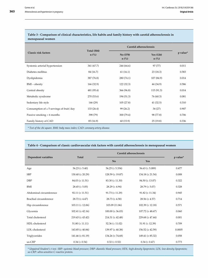
363Gomes et al.
Atherosclerosis and hypertension in pregnancy
Int J Cardiovasc Sci. 2018;31(4)359-366
Original Article
Table 3 - Comparison of clinical characteristics, life habits and family history with carotid atherosclerosis in menopausal women
Classic risk factorsTotal (504)
n (%)
Carotid atherosclerosis
p value*No (378)
n (%)
Yes (126)
n (%)
Systemic arterial hypertension 341 (67.7) 244 (64.6) 97 (77) 0.011
Diabetes mellitus 84 (16.7) 61 (16.1) 23 (18.3) 0.583
Dyslipidemia 387 (76.8) 280 (74.1) 107 (84.9) 0.014
BMI – obesity 166 (32.9) 122 (32.3) 44 (34.9) 0.586
Central obesity 481 (95.4) 366 (96.8) 115 (91.3) 0.014
Metabolic syndrome 270 (53.6) 194 (51.3) 76 (60.3) 0.081
Sedentary life style 146 (29) 105 (27.8) 41 (32.5) 0.310
Consumption of ≥ 5 servings of fruit/day 133 (26.4) 99 (26.2) 34 (27) 0.907
Passive smoking < 6 months 398 (79) 300 (79.4) 98 (77.8) 0.706
Family history of CAD 85 (16.9) 60 (15.9) 25 (19.8) 0.336
* Test of the chi square. BMI: body mass index; CAD: coronary artery disease.
Table 4 - Comparison of classic cardiovascular risk factors with carotid atherosclerosis in menopausal women
Dependent variables Total
Carotid atherosclerosis
p value*
No Yes
Age 56.23 (± 5.40) 56.25 (± 5.334) 56.63 (± 5.089) 0.477
SBP 130.40 (± 20.29) 128.59 (± 19.87) 134.18 (± 21.54) 0.008
DBP 84.03 (± 11.51) 83.30 (± 11.30) 84.50 (± 13.07) 0.322
BMI 28.45 (± 5.05) 28.29 (± 4.94) 28.79 (± 5.07) 0.328
Abdominal circumference 92.11 (± 11.51) 91.73 (± 11.29) 91.82 (± 11.34) 0.945
Brachial circumference 28.72 (± 4.47) 28.73 (± 4.58) 28.56 (± 4.57) 0.714
Hip circumference 103.11 (± 12.06) 103.09 (11.86) 102.39 (± 12.18) 0.571
Glycemia 102.41 (± 42.16) 100.00 (± 36.03) 107.72 (± 48.67) 0.060
Total cholesterol 219.43 (± 43.42) 214.31 (± 42.48) 229.68 (± 47.44) 0.001
HDL-cholesterol 51.80 (± 11.11) 52.36 (± 11.02) 51.91 (± 12.39) 0.709
LDL-cholesterol 143.85 (± 40.84) 139.97 (± 40.38) 156.52 (± 42.59) 0.0005
Triglycerides 141.46 (± 81.19) 134.26 (± 74.68) 149.41 (± 85.32) 0.058
us-CRP 0.34 (± 0.54) 0.32 (± 0.52) 0.34 (± 0.47) 0.773
* Unpaired Student’s t test. SBP: systemic blood pressure; DBP: diastolic blood pressure; HDL: high-density lipoprotein; LDL: low-density lipoprotein; us-CRP: ultra-sensitive C-reactive protein.
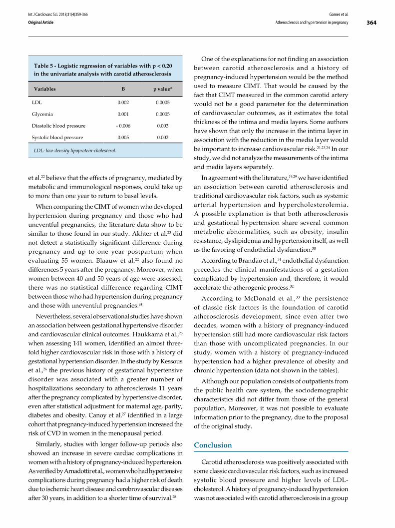
364Gomes et al.
Atherosclerosis and hypertension in pregnancy
Int J Cardiovasc Sci. 2018;31(4)359-366
Original Article
Table 5 - Logistic regression of variables with p < 0.20 in the univariate analysis with carotid atherosclerosis
Variables B p value*
LDL 0.002 0.0005
Glycemia 0.001 0.0005
Diastolic blood pressure - 0.006 0.003
Systolic blood pressure 0.005 0.002
LDL: low-density lipoprotein-cholesterol.
et al.22 believe that the effects of pregnancy, mediated by metabolic and immunological responses, could take up to more than one year to return to basal levels.
When comparing the CIMT of women who developed hypertension during pregnancy and those who had uneventful pregnancies, the literature data show to be similar to those found in our study. Akhter et al.23 did not detect a statistically significant difference during pregnancy and up to one year postpartum when evaluating 55 women. Blaauw et al.22 also found no differences 5 years after the pregnancy. Moreover, when women between 40 and 50 years of age were assessed, there was no statistical difference regarding CIMT between those who had hypertension during pregnancy and those with uneventful pregnancies.24
Nevertheless, several observational studies have shown an association between gestational hypertensive disorder and cardiovascular clinical outcomes. Haukkama et al.,25 when assessing 141 women, identified an almost three-fold higher cardiovascular risk in those with a history of gestational hypertension disorder. In the study by Kessous et al.,26 the previous history of gestational hypertensive disorder was associated with a greater number of hospitalizations secondary to atherosclerosis 11 years after the pregnancy complicated by hypertensive disorder, even after statistical adjustment for maternal age, parity, diabetes and obesity. Canoy et al.27 identified in a large cohort that pregnancy-induced hypertension increased the risk of CVD in women in the menopausal period.
Similarly, studies with longer follow-up periods also showed an increase in severe cardiac complications in women with a history of pregnancy-induced hypertension. As verified by Arnadottir et al., women who had hypertensive complications during pregnancy had a higher risk of death due to ischemic heart disease and cerebrovascular diseases after 30 years, in addition to a shorter time of survival.28
One of the explanations for not finding an association between carotid atherosclerosis and a history of pregnancy-induced hypertension would be the method used to measure CIMT. That would be caused by the fact that CIMT measured in the common carotid artery would not be a good parameter for the determination of cardiovascular outcomes, as it estimates the total thickness of the intima and media layers. Some authors have shown that only the increase in the intima layer in association with the reduction in the media layer would be important to increase cardiovascular risk.21,23,24 In our study, we did not analyze the measurements of the intima and media layers separately.
In agreement with the literature,19,29 we have identified an association between carotid atherosclerosis and traditional cardiovascular risk factors, such as systemic arterial hypertension and hypercholesterolemia. A possible explanation is that both atherosclerosis and gestational hypertension share several common metabolic abnormalities, such as obesity, insulin resistance, dyslipidemia and hypertension itself, as well as the favoring of endothelial dysfunction.30
According to Brandão et al.,31 endothelial dysfunction precedes the clinical manifestations of a gestation complicated by hypertension and, therefore, it would accelerate the atherogenic process.32
According to McDonald et al.,33 the persistence of classic risk factors is the foundation of carotid atherosclerosis development, since even after two decades, women with a history of pregnancy-induced hypertension still had more cardiovascular risk factors than those with uncomplicated pregnancies. In our study, women with a history of pregnancy-induced hypertension had a higher prevalence of obesity and chronic hypertension (data not shown in the tables).
Although our population consists of outpatients from the public health care system, the sociodemographic characteristics did not differ from those of the general population. Moreover, it was not possible to evaluate information prior to the pregnancy, due to the proposal of the original study.
Conclusion
Carotid atherosclerosis was positively associated with some classic cardiovascular risk factors, such as increased systolic blood pressure and higher levels of LDL-cholesterol. A history of pregnancy-induced hypertension was not associated with carotid atherosclerosis in a group

365Gomes et al.
Atherosclerosis and hypertension in pregnancy
Int J Cardiovasc Sci. 2018;31(4)359-366
Original Article
of menopausal and asymptomatic women, from the cardiovascular point of view. More studies are needed to understand the atherosclerosis process in women with a history of pregnancy-induced hypertension.
Author contributions
Conception and design of the research: Gomes RAF, Barros IML. Acquisition of data: Gomes RAF, Barros IML. Analysis and interpretation of the data: Gomes RAF, Barros IML. Statistical analysis: Gomes RAF, Barros IML. Writing of the manuscript: Gomes RAF. Critical revision of the manuscript for intellectual content: Barros IML, Ferreira MNL, Costa LOBF.
Potential Conflict of Interest
This manuscript is part of the master of the Graduate Program in Health Sciences of the University of Pernambuco by Rafael Alessandro Ferreira.
Sources of Funding
This study was funded by Fundação de Amparo à Ciência do estado de Pernambuco (FACEPE) (APQ-1386-4.00/08).
Study Association
This article is part of the thesis of master submitted by Rafael Alessandro Ferreira Gomes, from Universidade de Pernambuco.
Ethics approval and consent to participate
This study was approved by the Ethics Committee of the Complexo Hospitalar Hospital Universitário Oswaldo Cruz/Pronto-Socorro Cardiológico de Pernambuco under the protocol number 55361416.0.0000.5192 (CAAE) and number 1.593.189 of June 16, 2016. All the procedures in this study were in accordance with the 1975 Helsinki Declaration, updated in 2013. Informed consent was obtained from all participants included in the study.
1. Gawryszewski VP, Souza Mde F. Mortality due to cardiovascular diseases in the Americas by region, 2000-2009. Sao Paulo Med J. 2014;132(2):105-10.
2. Stramba-Badiale M, Fox KM, Priori SG, Collins P, Daly C, Graham I, et al. Cardiovascular diseases in women: a statement from the policy conference of the European Society of Cardiology. Eur Heart J. 2006;27(8):994-1005.
3. Ahmed R, Dunford J, Mehran R, Robson S, Kunadian V. Pre-eclampsia and future cardiovascular risk among women: a review. J Am Coll Cardiol. 2014;63(18):1815-22.
4. Mozaffarian D, Benjamin EJ, Go AS, Arnett DK, Blaha MJ, Cushman M, et al; American Heart Association Statistics Committee and Stroke Statistics Subcommittee. Heart disease and stroke statistics--2015 update: a report from the American Heart Association. Circulation. 2015;131(4):e29-322.
5. Vaccarino V, Badimon L, Corti R, de Wit C, Dorobantu M, Hall A, et al; Working Group on Coronary Pathophysiology and Microcirculation. Ischaemic heart disease in women: are there sex differences in pathophysiology and risk factors?: Position Paper from the Working Group on Coronary Pathophysiology and Microcirculation of the European Society of Cardiology. Cardiovasc Res. 2010;90(1):9-17.
6. Brasil. Ministério da Saude, Secretaria Executiva, DATASUS. Informações de saúde: informaçoes epidemiológicas e morbidade. [Citado em 2015 maio 1]. Disponível em http://www.datasus.gov.br.
7. Mansur Ade P, Favarato D. Mortality due to cardiovascular diseases in women and men in the five Brazilian Regions, 1980-2012. Arq Bras Cardiol. 2016;107(2):137-46.
8. Williams D. Pregnancy: a stress test for life. Curr Opin Obstet Gynecol. 2003;15(6):465-71.
9. Hashemi S, Ramezani Tehrani F, Mehrabi Y, Azizi F. Hypertensive pregnancy disorders as a risk factor for future cardiovascular and
metabolic disorders (Tehran Lipid and Glucose Study). J Obstet Gynaecol Res. 2013;39(5):891-7.
10. Smith GC, Pell JP, Walsh D. Pregnancy complications and maternal risk of ischaemic heart disease: a retrospective cohort study of 129,290 births. Lancet. 2001;357(9273):2002-6.
11. Ben-Ami S, Oron G, Ben-Haroush A, Blickstein D, Hod M, Bar J. Primary atherothrombotic occlusive vascular events in premenopausal women with history of adverse pregnancy outcome. Thromb Res. 2010;125(2):124-7.
12. Freibert SM, Mannino DM, Bush H, Ph D, Crofford LJ. The association of adverse pregnancy events and cardiovascular disease in women 50 years of age and older. J Womens Health (Larchmt). 2011;20(2):287-93.
13. Romundstad PR, Magnussen EB, Smith GD, Vatten LJ. Hypertension in pregnancy and later cardiovascular risk: Common antecedents? Circulation. 2010;122(6):579-84.
14. Brown MC, Best KE, Pearce MS, Waugh J, Robson SC, Bell R. Cardiovascular disease risk in women with pre-eclampsia: Systematic review and meta-analysis. Eur J Epidemiol. 2013;28(1):1-19.
15. Touboul P, Hennerici M, Meairs S, Adams H, Amarenco P, Bornstein N, et al. Mannheim carotid intima-media thickness and plaque consensus (2004-2006-2011). An update on behalf of the advisory board of the 3rd, 4th and 5th watching the risk symposia, at the 13th, 15th and 20th European Stroke Conferences, Mannheim, Germany, 2004, Brussels, Belgium, 2006, and Hamburg, Germany, 2011. Cerebrovasc Dis. 2012;34(4):290-6.
16. Diehl CL, Brost BC, Hogan MC, Elesber AA, Offord KP, Turner ST, et al. Preeclampsia as a risk factor for cardiovascular disease later in life : validation of a preeclampsia questionnaire. Am J Obstet Gynecol. 2008;198(5)e11-3.
17. Koskinen J, Kahonen M, Viikari JS, Taittonen L, Laitinen T, Ronnemaa T, et al. Conventional cardiovascular risk factors and metabolic syndrome
References

366Gomes et al.
Atherosclerosis and hypertension in pregnancy
Int J Cardiovasc Sci. 2018;31(4)359-366
Original Article
This is an open-access article distributed under the terms of the Creative Commons Attribution License
in predicting carotid intima-media thickness progression in young adults: the cardiovascular risk in young Finns study. Circulation. 2009;120(3):229-36.
18. Peters SA, den Ruijter HM, Bots ML, Moons KG. Improvements in risk stratification for the occurrence of cardiovascular disease by imaging subclinical atherosclerosis: a systematic review. Heart. 2012;98(3):177-84.
19. O’Leary DH, Polak JF, Kronmal RA, Manolio TA, Burke GL, Wolfson SK Jr. Carotid-artery intima and media thickness as a risk factor for myocardial infarction and stroke in older adults. Cardiovascular Health Study Collaborative Research Group. N Engl J Med. 1999;340(1):14-22.
20. Den Ruijter HM, Peters SA, Anderson TJ, Britton AR, Dekker JM, Eijkemans MJ, et al. Common carotid intima-media thickness measurements in cardiovascular risk prediction: a meta-analysis. JAMA. 2012;308(8):796-803.
21. Akhter T, Larsson A, Larsson M, Wikström A, Naessen T. Artery wall layer dimensions during normal pregnancy : a longitudinal study using noninvasive high-frequency ultrasound. Am J Physiol Heart Circ Physiol. 2013;304(2):H229-34.
22. Blaauw J, Souwer ET, Coffeng SM, Smit AJ, van Doormaal JJ, Faas MM, et al. Follow up of intima-media thickness after severe early-onset preeclampsia. Acta Obstet Gynecol Scand. 2014;93(12):1309-16.
23. Akhter T, Wikstrom AK, Larsson M, Naessen T. Individual common carotid artery wall layer dimensions, but not carotid intima-media thickness, indicate increased cardiovascular risk in women with preeclampsia: an investigation using noninvasive high-frequency ultrasound. Circ Cardiovasc Imaging. 2013;6(5):762-8.
24. Akhter T, Larsson M, Wikström AK, Naessen T. Thicknesses of individual layers of artery wall indicate increased cardiovascular risk in severe pre-eclampsia. Ultrasound Obstet Gynecol. 2014;43(6):675-80.
25. Haukkamaa L, Salminen M, Laivuori H, Leinonen H, Hiilesmaa V, Kaaja R. Risk for subsequent coronary artery disease. Am J Cardiol. 2004;93(6):805-8.
26. Kessous R, Shoham-Vardi I, Pariente G, Sergienko R, Sheiner E. Long-term maternal atherosclerotic morbidity in women with pre-eclampsia. Heart. 2015;101(6):442-6.
27. Canoy D, Cairns BJ, Balkwill A, Wright FL, Khalil A, Beral V, et al. Hypertension in pregnancy and risk of coronary heart disease and stroke: a prospective study in a large UK cohort. Int J Cardiol. 2016;222:1012-8.
28. Arnadottir GA, Geirsson RT, Arngrimsson R. Cardiovascular death in women who had hypertension in pregnancy: a case–control study. BJOG. 2005;112(3):286-92.
29. Mosca L, Benjamin EJ, Berra K, Bezanson JL, Dolor RJ, Lloyd-Jones DM, et al. Effectiveness-based guidelines for the prevention of cardiovascular disease in women-2011 update: a guideline from the American Heart Association. Circulation. 2011;123(11):1243-62. Erratum in: Circulation. 2011;123(22):e624. Circulation. 2011;124(16):e427.
30. Craici I, Wagner S, Garovic VD. Preeclampsia and future cardiovascular risk: formal risk factor or failed stress test? Ther Adv Cardiovasc Dis. 2008;2(4):249-59.
31. Brandão A, Cabral M, Leite H, Cabral A. Endothelial function, uterine perfusion and central flow in pregnancies complicated by Preeclampsia. Arq Bras Cardiol. 2012;99(4):931-5.
32. Agatisa PK, Ness RB, Roberts JM, Costantino JP, Kuller LH, McLaughlin MK. Impairment of endothelial function in women with a history of preeclampsia: an indicator of cardiovascular risk. Am J Physiol Heart Circ Physiol. 2004;286(4):H1389-93.
33. McDonald SD, Ray J, Teo K, Jung H, Salehian O, Yusuf S, et al. Measures of cardiovascular risk and subclinical atherosclerosis in a cohort of women with a remote history of preeclampsia. Atherosclerosis. 2013;229(1):234–9.
DOI: 10.5935/2359-4802.20180034
367
ORIGINAL ARTICLE
International Journal of Cardiovascular Sciences. 2018;31(4)367-373
Mailing Address: Eduardo del Bosco Brunetti Cunha Rua Costa Rica, 858. Postal Code: 82510-180, Bacacheri, Curitiba, PR - Brazil.E-mail: [email protected], [email protected]
Evaluation of Lipid Profile in AdolescentsEduardo del Bosco Brunetti Cunha,1,2 Rafael Pereira Fagundes,2 Edson Emílio Scalabrin1, Roberto Hirochi Herai1
Pontifícia Universidade Católica do Paraná (PUCPR),1 PR - BrazilFaculdade Educacional Araucária,2 PR - Brazil
Manuscript received October 10, 2017; revised manuscript January 17, 2018; accepted February 16, 2018.
Abstract
Background: Atherosclerosis is a chronic, multifactorial and insidious disease that can begin in childhood and adolescence, and whose major consequences appear during adulthood. Serum levels of lipoproteins, such as LDL-c, total cholesterol (TC), HDL-c, and non-HDL-c can be used as a screening method for disease diagnosis. In Brazil, few studies have correlated the serum levels of those lipoproteins with age.
Objective: To evaluate the serum concentrations of TC, LDL-c, HDL-c, VLDL-c, non-HDL-c and triglycerides (TG) of adolescents aged 10 to 19 years in the municipality of Araucária, Paraná state.
Methods: Cross-sectional retrospective study, collecting the following data from 600 adolescents: age, sex and serum levels of TC, LDL-c, HDL-c and TG from June to December 2016. Data were analyzed using the SPSS software 2.0, with Mann-Whitney U test and Spearman coefficient of correlation to identify statistical significance (p < 0.05).
Results: The female sex showed higher serum levels of TC, TG and LDL-c than the male sex. The HDL-c levels were identical in both sexes, with 48% of desirable values and 52% of low values. This study identified a strong correlation between the lipids and association with the age group of 10 to 14 years.
Conclusion: Non-HDL-c showed stronger correlation with the other lipids (TG, LDL-c and TC) as compared to LDL-c, suggesting that non-HDL-c can be used as an effective complementary diagnostic method to assess the risks for atherosclerosis in adolescents. (Int J Cardiovasc Sci. 2018;31(4)367-373)
Keywords: Dyslipidemias/epidemiology; Adolescent; Lipoproteins; Hypercholesterolemia/epidemiology.
Such lipoproteins can adhere to the intimal layer of
arteries, causing the formation of atheromatous plaques
that lead to atherosclerosis.6 This atherosclerotic process
begins in childhood, before clinical symptoms can be
perceived.7 In the aorta, fatty streaks begin at the age of
3 years, while in the coronary arteries, 5 to 10 years later.8
Over time, such fatty streaks form fatty plaques that can
rupture, leading to different ischemic processes, such as
acute myocardial infarction and stroke.9
The lipid profile is a panel of blood tests to assess
the serum concentrations of lipoproteins, such as total
cholesterol (TC), high-density lipoprotein cholesterol
(HDL-c), LDL-c, very-low-density lipoprotein cholesterol
(VLDL-c), non-HDL-c, and triglycerides (TG).10
Introduction
Cardiovascular diseases (CVD) are the major cause of death of men and women worldwide.1 In Brazil, according to the last 2013 Ministry of Health survey, of a total of 201,062,789 inhabitants, 678,556 of the deaths were related to the circulatory system.2
The risk factors for CVD are classified as modifiable and nonmodifiable. Some of the modifiable risk factors are sedentary lifestyle, smoking, obesity and dyslipidemia.2 Some nonmodifiable risk factors are family history of CVD, age, sex and ethnicity.3 Dyslipidemia has a great influence on the development of CVD, since an inadequate diet increases the concentration of low-density lipoprotein cholesterol (LDL-c) in blood vessels.4,5

368Cunha et al.
Lipid profile in adolescents
Int J Cardiovasc Sci. 2018;31(4)367-373
Original Article
Knowing that dyslipidemia is associated with CVD, its diagnosis in adolescence can reduce the chances of future complications, because a change in lifestyle to healthier habits can be the best prevention.11,12
This study was aimed at assessing the lipid profile of adolescents of the municipality of Araucária, Paraná state. It collected data of 600 adolescents aged 10 to 19 years and compared them to those of studies conducted in other regions of Brazil.
Methods
This study was approved by the Ethics Committee in Research of the Instituto Paranaense de Otorrinolaringologia and is registered under the number 65932917.0.0000.5529, according to the Resolution 466/12 of the National Board of Health of the Ministry of Health, which regulates research with human beings. The results of 600 lipid profiles were collected from the Araucária Municipal Laboratory, which had been approved by the local coordinator, so that the study could begin even before approval by the Ethics Committee in Research.
This is a cross-sectional, retrospective study with convenience sampling of the lipid profile of 600 adolescents aged 10 to 19 years, through systematic random sampling, from July to December 2016. Tests with TG levels greater than 400 mg/dL were excluded. The samples were collected 5-mL tubes containing serum separating gel and particles to activate clotting, and were tested by enzymatic photometric assay (Abbott Architect c8000), using the direct precipitation method for HDL-c quantitation (Ultra HDL). To calculate VLDL-c, the TG level was divided by 5, and to calculate LDL-c, the Friedewald formula was used. The collected data were organized in Excel 2007 sheets and stratified as TC, LDL-c, HDL-c, non-HDL-c (sum of the lipoproteins without HDL), VLDL-c and TG, in addition to age and sex of the adolescents, being identified by codes and organized in tables and graphs.
Statistical analysis
The descriptive statistical analysis included percentage and median with respective interquartile range (IQR). Continuous variables were expressed as median and IQR, because they had no normal distribution, while categorical variables were expressed as percentages. Normality was assessed by use of the Kolmogorov-Smirnov test. The statistical analysis comprised Mann-
Whitney U test and Spearman coefficient of correlation (S), using the SPSS software 2.0. The adopted significance level was 5% of probability and 95% confidence interval, and all tests were two-tailed.
Results
The lipid profiles of 600 adolescents aged 10 to 19 years from the Araucária Municipal Laboratory were assessed. Of the 600 adolescents, 322 (54%) were of the female sex and 278 (46%) were of the male sex. Table 1 shows the analyzed data, which, in 1.83% (n = 11) of the adolescents, it is suggested familial hypercholesterolemia.10 Tables 2, 3 and 4 show the correlations between the lipids, where 1 means perfect positive correlation, that is, when one variable increases, the other increases at the same intensity, and -1 means perfect negative correlation, that is, when one variable increases, the other decreases at the same intensity.
When comparing between sexes, the female sex had a higher TC than that of the male sex (Figure 1A). Regarding non-HDL-c, the female sex had a median of 109 and IQR of 40.25, while the male sex had a median of 101 and IQR of 32.25, with no significant difference between them. In addition, in the non-stratified sample, positive correlations of non-HDL-c were found with TG, TC and VLDL-c as compared to LDL-c, and fewer negative correlations with HDL-c (Table 2).
In adolescents aged 10 to 14 years, more positive correlations of non-HDL-c with TG, TC and VLDL-c were found as compared to LDL-c, and some negative correlations of LDL-c, TC and non-HDL-c with age (Table 3). When comparing between sexes, only TG showed a difference (Figure 1B). Regarding sexes, in adolescents aged 15 to 19 years, the correlations showed the same trend of the other age group (Table 4), and the comparisons between sexes achieving significance for the LDL-c (Figure 1C) and TC (Figure 1D), while the other lipids showed no statistically significant variation.
Discussion
The study by Silva et al.11 has reported desirable values of TC of 50%, similar to those found in the present study (49%), but different from the 37% reported by Araki et al.13 This difference was observed in a study conducted in the city of Aracaju, Sergipe state, which has found a higher TC value in the female sex as compared to that in the male sex, a result that corroborates that found
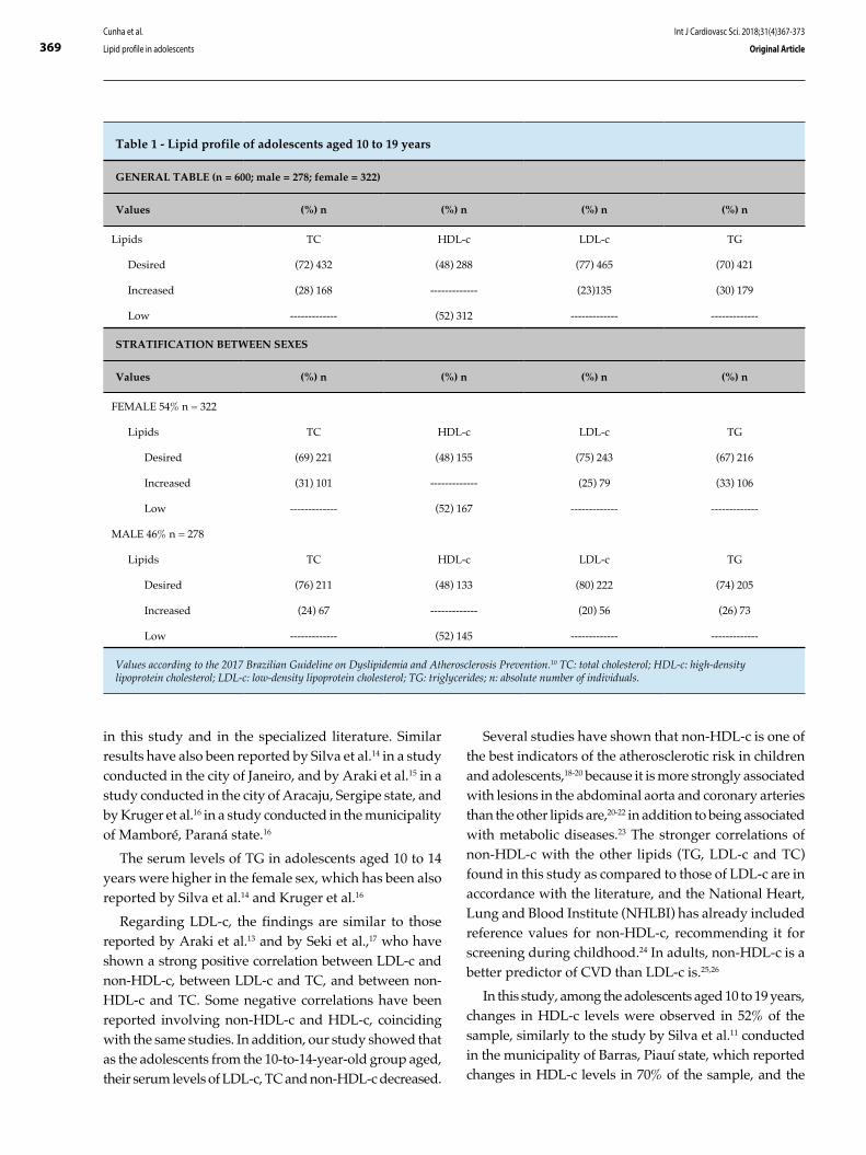
369
Table 1 - Lipid profile of adolescents aged 10 to 19 years
GENERAL TABLE (n = 600; male = 278; female = 322)
Values (%) n (%) n (%) n (%) n
Lipids TC HDL-c LDL-c TG
Desired (72) 432 (48) 288 (77) 465 (70) 421
Increased (28) 168 ------------- (23)135 (30) 179
Low ------------- (52) 312 ------------- -------------
STRATIFICATION BETWEEN SEXES
Values (%) n (%) n (%) n (%) n
FEMALE 54% n = 322
Lipids TC HDL-c LDL-c TG
Desired (69) 221 (48) 155 (75) 243 (67) 216
Increased (31) 101 ------------- (25) 79 (33) 106
Low ------------- (52) 167 ------------- -------------
MALE 46% n = 278
Lipids TC HDL-c LDL-c TG
Desired (76) 211 (48) 133 (80) 222 (74) 205
Increased (24) 67 ------------- (20) 56 (26) 73
Low ------------- (52) 145 ------------- -------------
Values according to the 2017 Brazilian Guideline on Dyslipidemia and Atherosclerosis Prevention.10 TC: total cholesterol; HDL-c: high-density lipoprotein cholesterol; LDL-c: low-density lipoprotein cholesterol; TG: triglycerides; n: absolute number of individuals.
Cunha et al.
Lipid profile in adolescents
Int J Cardiovasc Sci. 2018;31(4)367-373
Original Article
in this study and in the specialized literature. Similar results have also been reported by Silva et al.14 in a study conducted in the city of Janeiro, and by Araki et al.15 in a study conducted in the city of Aracaju, Sergipe state, and by Kruger et al.16 in a study conducted in the municipality of Mamboré, Paraná state.16
The serum levels of TG in adolescents aged 10 to 14 years were higher in the female sex, which has been also reported by Silva et al.14 and Kruger et al.16
Regarding LDL-c, the findings are similar to those reported by Araki et al.13 and by Seki et al.,17 who have shown a strong positive correlation between LDL-c and non-HDL-c, between LDL-c and TC, and between non-HDL-c and TC. Some negative correlations have been reported involving non-HDL-c and HDL-c, coinciding with the same studies. In addition, our study showed that as the adolescents from the 10-to-14-year-old group aged, their serum levels of LDL-c, TC and non-HDL-c decreased.
Several studies have shown that non-HDL-c is one of the best indicators of the atherosclerotic risk in children and adolescents,18-20 because it is more strongly associated with lesions in the abdominal aorta and coronary arteries than the other lipids are,20-22 in addition to being associated with metabolic diseases.23 The stronger correlations of non-HDL-c with the other lipids (TG, LDL-c and TC) found in this study as compared to those of LDL-c are in accordance with the literature, and the National Heart, Lung and Blood Institute (NHLBI) has already included reference values for non-HDL-c, recommending it for screening during childhood.24 In adults, non-HDL-c is a better predictor of CVD than LDL-c is.25,26
In this study, among the adolescents aged 10 to 19 years, changes in HDL-c levels were observed in 52% of the sample, similarly to the study by Silva et al.11 conducted in the municipality of Barras, Piauí state, which reported changes in HDL-c levels in 70% of the sample, and the

370
Table 2 - Correlations of the lipid variables in adolescents aged 10 to 19 years (n = 600)
Age TG LDL-c HDL-c TC VLDL-c Non-HDL-c
Age S 1 0.004 -0.079 -0.010 -0.071 0.004 -0.080
TG S 0.004 1 0.292** -0.232** 0.396** 0.999** 0.493**
LDL-c S -0.079 0.292** 1 0.024 0.896** 0.289** 0.935**
HDL-c S -0.010 -0.232** 0.024 1 0.282** -0.230** -0.032
TC S -0.071 0.396** 0.896** 0.282** 1 0.394** 0.934**
VLDL-c S 0.004 0.999** 0.289** -0.230** 0.394** 1 0.490**
Non-HDL-c S -0.080 0.493** 0.935** -0.032 0.934** 0.490** 1
TC: total cholesterol; HDL-c: high-density lipoprotein cholesterol; LDL-c: low-density lipoprotein cholesterol; TG: triglycerides; Non-HDL-c: sum of the lipoproteins without HDL-c; S: Spearman correlation. * p < 0.05; ** p < 0.001.
Table 3 - Correlations of the lipid variables in adolescents aged 10 to 14 years (n = 339)
Age TG LDL-c HDL-c TC VLDL-c Non-HDL-c
Age S 1 -0.056 -0.140** -0.079 -0.138* -0.056 -0.136*
TG S -0.056 1 0.262** -0.300** 0.337** 0.999** 0.457**
LDL-c S -0.140** 0.262** 1 0.039 0.912** 0.257** 0.949**
HDL-c S -0.079 -0.300** 0.039 1 0.282** -0.298** -0.053
TC S -0.138* 0.337** 0.912** 0.282** 1 0.333** 0.926**
VLDL-c S -0.056 0.999** 0.257** -0.298** 0.333** 1 0.452**
Non-HDL-c S -0.136* 0.457** 0.949** -0.053 0.926** 0.452** 1
TC: total cholesterol; HDL-c: high-density lipoprotein cholesterol; LDL-c: low-density lipoprotein cholesterol; TG: triglycerides; Non-HDL-c: sum of the lipoproteins without HDL-c; S: Spearman correlation. * p < 0.05; ** p < 0.001.
Table 4 - Correlations of the lipid variables in adolescents aged 15 to 19 years (n = 261)
Age TG LDL-c HDL-c TC VLDL-c Non-HDL-c
Age S 1 0.063 0.064 0.071 0.086 0.065 0.054
TG S 0.063 1 0.328** -0.143* 0.468** 0.999** 0.537**
LDL-c S 0.064 0.328** 1 0.006 0.878** 0.329** 0.919**
HDL-c S 0.071 -0.143* 0.006 1 0.281** -0.142* -0.007
TC S 0.086 0.468** 0.878** 0.281** 1 0.468** 0.944**
VLDL-c S 0.065 0.999** 0.329** -0.142* 0.468** 1 0.538**
Non-HDL-c S 0.054 0.537** 0.919** -0.007 0.944** 0.538** 1
TC: total cholesterol; HDL-c: high-density lipoprotein cholesterol; LDL-c: low-density lipoprotein cholesterol; TG: triglycerides; Non-HDL-c: sum of the lipoproteins without HDL-c; S: Spearman correlation. * p < 0.05; ** p < 0.001.
Cunha et al.
Lipid profile in adolescents
Int J Cardiovasc Sci. 2018;31(4)367-373
Original Article
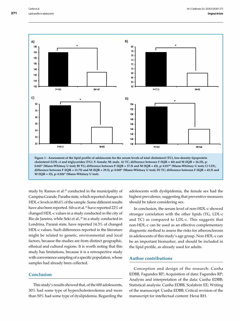
371
Figure 1 - Assessment of the lipid profile of adolescents for the serum levels of total cholesterol (TC), low-density lipoprotein cholesterol (LDL-c) and triglycerides (TG). F: female; M: male. A) TC; difference between F (IQR = 44) and M (IQR = 36.25), p: 0.043* (Mann-Whitney U test); B) TG; difference between F (IQR = 37.5) and M (IQR = 42), p: 0.017* (Mann-Whitney U test); C) LDL; difference between F (IQR = 31.75) and M (IQR = 29.5), p: 0.049* (Mann-Whitney U test); D) TC; difference between F (IQR = 43.5) and M (IQR = 32), p: 0.026* (Mann-Whitney U test).
Cunha et al.
Lipid profile in adolescents
Int J Cardiovasc Sci. 2018;31(4)367-373
Original Article
study by Ramos et al.25 conducted in the municipality of Campina Grande, Paraíba state, which reported changes in HDL-c levels in 80.6% of the sample. Some different results have also been reported. Silva et al.14 have reported 22% of changed HDL-c values in a study conducted in the city of Rio de Janeiro, while Seki et al.,28 in a study conducted in Londrina, Paraná state, have reported 14.3% of changed HDL-c values. Such differences reported in the literature might be related to genetic, environmental and local factors, because the studies are from distinct geographic, ethnical and cultural regions. It is worth noting that this study has limitations, because it is a retrospective study with convenience sampling of a specific population, whose samples had already been collected.
Conclusion
This study’s results showed that, of the 600 adolescents, 30% had some type of hypercholesterolemia and more than 50% had some type of dyslipidemia. Regarding the
adolescents with dyslipidemia, the female sex had the highest prevalence, suggesting that preventive measures should be taken considering sex.
In conclusion, the serum level of non-HDL-c showed stronger correlation with the other lipids (TG, LDL-c and TC) as compared to LDL-c. This suggests that non-HDL-c can be used as an effective complementary diagnostic method to assess the risks for atherosclerosis in adolescents of this study’s age group. Non-HDL-c can be an important biomarker, and should be included in the lipid profile, as already used for adults.
Author contributions
Conception and design of the research: Cunha EDBB, Fagundes RP; Acquisition of data: Fagundes RP; Analysis and interpretation of the data: Cunha EDBB; Statistical analysis: Cunha EDBB, Scalabrin EE; Writing of the manuscript: Cunha EDBB; Critical revision of the manuscript for intellectual content: Herai RH.
372Cunha et al.
Lipid profile in adolescents
Int J Cardiovasc Sci. 2018;31(4)367-373
Original Article
1. Schmidt MI, Duncan BB, Silva GA, Menezes AM, Monteiro CA, Barreto SM, et al. Doenças crônicas não transmissíveis no Brasil: carga e desafios atuais. In: Victora CG, Leal MC, Barreto ML, Schimidt MI, Monteiro CA. Saúde no Brasil: a série The Lancet. Rio de Janeiro: Fiocruz; 2011. p. 61-74.
2. Brasil.Ministerio da Saúde. DATASUS. Indicadores de mortalidade [Internet]. [access in 2017 May 16] Available in: http://tabnet2.datasus.gov.br/cgi/tabcgi.exe?idb2013/c08.def
3. Magalhães FJ, Mendonça LB, Rebouças CB, Lima FE, Custódio IL, de Oliveira SC. [Risk factors for cardiovascular diseases among nursing professionals: strategies for health promotion]. Rev Bras Enferm. 2014;67(3):394-400.
4. Boni A, Pugliese C, Cláudio CC, Patim RV, Olveira FL. Antioxidant vitamins and prevention of atherosclerosis in childhood. Rev Paul Pediatr. 2010;28(4):373-80.
5. Cromwell WC, Otvos JD, Keyes MJ, Pencina MJ, Sullivan L, Vasan RS, et al. LDL particle number and risk of future cardiovascular disease in the Framingham Offspring Study—Implications for LDL management. J Clin Lipidol. 2007;1(6):583-92.
6. Xavier HT, Izar MC, Faria-Neto JR, Assad MH, Rocha VZ, Sposito AC, et al; Sociedade Brasileira de Cardiologia. [V Brazilian Guidelines on Dyslipidemias and Prevention of Atherosclerosis]. Arq Bras Cardiol. 2013;101(4 Suppl 1):1-20.
7. Santos MG, Pengoraro M, Sandrini F, Macuco EC. Risk factors for the development of atherosclerosis in childhood and adolescence. Arq Bras Cardiol. 2008;90(4):301-8.
8. McGill HC Jr. Fatty streaks in the coronary arteries and aorta. Lab Invest. 1968;18(5):560-4.
9. Mackman N. Triggers, targets and treatments for thrombosis. Nature. 2008;451(7181):914-8.
10. Faludi AA, Izar MC, Saraiva JF, Chacra AP, Bianco HT, Afiune Neto A, et al; Sociedade Brasileira de Cardiologia. Atualização da Diretriz Brasileira de Dislipidemias e Prevenção da Aterosclerose – 2017. Arq Bras Cardiol. 2017;109(2 supl 1):1-76.
11. Silva IP, Lima HM. Perfil lipídico de adolescentes em uma escola de Barras-PI. Revista Interdisciplinar UNINOVAFAPI. 2015;8(1):157-66.
12. Medeiros YM, Pinheiro LB, Rodrigues PM. Analysis on the lipid profile of adolescents from a municipality of sertao paraibano. Temas em Saúde. 2016;16(2):45-8.
13. Araki MV, Martins IC, Barros C, Santos EG. Não-HDL colesterol em escolares e adolescentes. Scientia Plena. 2013;9(2):1-8.
14. Silva RA, Kanaan S, Silva LE, Peralta RH. Lipid profiles associated with dyslipidemic risk in children and teenagers from the pediatric clinic of the University Hospital Antônio Pedro. J Bras Med Lab. 2007;43(2):95-100.
15. Araki MV, Martins IC, Barros C, Santos EG. Análise de perfil lipídico de crianças e adolescentes do estado de Sergipe. Scientia Plena. 2010;6(12):45-58.
16. Kruger GR, Ribas-Silva RC. Perfil lipídico e índice de massa corporal de escolares de um colégio estadual da região centro-ocidental do Paraná. Adolesc Saude. 2014;11(4): 54-60.
17. Seki MO, Matsuo T, Seki M. [Non-HDL cholesterol levels in students aged 7 to 17 years in a Brazilian town]. Rev Panam Salud Publica. 2007;21(5):307-12.
18. Srinivasan SR, Frontini MG, Xu J, Berenson GS. Utility of childhood non-high-density lipoprotein cholesterol levels in predicting adult dyslipidemia and other cardiovascular risks: the Bogalusa Heart Study. Pediatrics. 2006;118(1):201-6.
19. Rainwater DL, McMahan CA, Malcom GT, Scheer WD, Roheim PS, McGill HC Jr, et al. Lipid and apolipoprotein predictors of atherosclerosis in youth: apolipoprotein concentrations do not materially improve prediction of arterial lesions in PDAY subjects. The PDAY Research Group. Arterioscler Thromb Vasc Biol. 1999;19(3):753-61.
20. Frontini MG, Srinivasan SR, Xu JH, Tang R, Bond MG, Berenson G. Utility of non-high-density lipoprotein cholesterol versus other lipoprotein measures in detecting subclinical atherosclerosis in young adults (The Bogalusa Heart Study). Am J Cardiol. 2007;100(1):64-8.
21. Virani SS. Non-HDL Cholesterol as a metric of good quality of care: opportunities and challenges. Tex Heart Inst J. 2011;38(2):160-2.
22. Ma H, Lin H, Hu Y, Li X, He W, Jin X, et al. Relationship between non–high-density lipoprotein cholesterol and carotid atherosclerosis in normotensive and euglycemic Chinese middle-aged and elderly adults. Lipids Health Dis. 2017;16(1):55.
23. Li C, Ford ES, McBride PE, Kwiterovich PO, McCrindle BW, Gidding SS. Non-high-density lipoprotein cholesterol concentration is associated with the metabolic syndrome among US youth aged 12-19 years. J Pediatr. 2011;158(2):201-7.
24. Expert Panel on Integrated Guidelines for Cardiovascular Health and Risk Reduction: National Heart, Lung and Blood Institute. Expert panel on integrated guidelines for cardiovascular health and risk reduction in children and adolescents: summary report. Pediatrics. 2011;128 Suppl 5:S213-56.
25. Rana JS, Boekholdt SM, Kastelein JJ, Shah PK. The role of non- HDL cholesterol in risk stratification for coronary artery disease. Curr Atheroscler Rep. 2012;14(2):130-4.
26. Boekholdt SM, Arsenault BJ, Mora S, Pedersen TR, LaRosa JC, Nestel PJ, et al. Association of LDL cholesterol, non-HDL cholesterol, and apolipoprotein B levels with risk of cardiovascular events among patients treated with statins: a meta-analysis. JAMA. 2012;307(12):1302-9. Erratum in: JAMA. 2012;307(18):1915. JAMA. 2012;307(16):1694.
References
Potential Conflict of Interest
No potential conflict of interest relevant to this article was reported.
Sources of Funding
There were no external funding sources for this study.
Study Association
This article is part of the thesis of Graduation by Rafael Pereira Fagundes and Doctoral submitted by Eduardo
del Bosco Brunetti Cunha, from Pontifícia Universidade Católica do Paraná and Faculdade Educacional Araucária.
Ethics approval and consent to participate
This study was approved by the Ethics Committee of the Instituto Paranaense de Otorrinolaringologia under the protocol number 65932917.0.0000.5529. All the procedures in this study were in accordance with the 1975 Helsinki Declaration, updated in 2013. Informed consent was obtained from all participants included in the study.

373Cunha et al.
Lipid profile in adolescents
Int J Cardiovasc Sci. 2018;31(4)367-373
Original Article
This is an open-access article distributed under the terms of the Creative Commons Attribution License
27. Ramos AT, Carvalho D, Gonzaga NC, Cardoso AS, Noronha JA, Cardoso MA. Perfil lipídico em crianças e adolescentes em excesso de peso. Rev Bras Crescimento Desenvolvimento Hum. 2011;21(3):780-8.
28. Seki M, Kseki MO, Lima AD, Onishi MH, Seki MO, Oliveira LA. Estudo de perfil lipídico de crianças e jovens até 19 anos de idade. J Bras Patol. 2001; 37(4):247-51.
DOI: 10.5935/2359-4802.20180035
374
ORIGINAL ARTICLE
International Journal of Cardiovascular Sciences. 2018;31(4)374-382
Mailing Address: Joel Silva LimaPassagem João Marcos, n: 9, Icuí-Guajará. Postal Code: 67125-099, Ananindeua, PA - Brazil.E-mail: [email protected]
Waiting for Cardiac Procedure in Congenital Heart Disease: Portrait of an a Hospital in the Amazonian RegionValeria Santos de Jesus, Aline Marques Nascimento, Rogério dos Anjos Miranda, Joel Silva Lima, Milene de Andrade Gouvea Tyll, Adriana de Oliveira Lameira VeríssimoFundação Hospital de Clínicas Gaspar Vianna, Belém, PA - Brazil
Manuscript received on February 5, 2017, revised on December 18, 2017, accepted on February 16, 2018.
Abstract
Background: Congenital heart disease is an important cause of morbidity and mortality in childhood, and in 50% of cases, surgery is required in the first year of life. A high deficit of surgical procedures is estimated in Northern Brazil.
Objective: To analyze the waiting time for elective surgical treatment and/ or intervention in children with congenital heart disease in a Cardiology referral center, and to make considerations about heart diseases and forms of treatment in that institution.
Methods: A cross-sectional study of all patients aged less than 14 years, with a diagnosis of congenital heart disease that were waiting for elective surgical or percutaneous cardiac treatment.
Results: Among the 407 children with congenital heart defects, the most prevalent age group was > 2 to 6 years (34.0%). The average waiting time was 23.1 ± 18.3 months, with a median of 19. The most frequent heart disease was ventricular septal defect (28.98%), patent ductus arteriosus (18.42%) and atrial septal defect (11.05%). Most children (63.4%) were not from the metropolitan area. The percutaneous interventions represented only 27.84% of the catheterization procedures and 14,85% of all heart treatments. Approximately 60% of the pediatric surgeries occurred in children who were not previously registered due to urgency cases.
Conclusion: Most of the children waiting for a cardiac procedure were not from the metropolitan area and had malformations potentially treatable by catheterization. It is necessary to increase the capacity of the single referral center in the state of Pará, as well as decentralize the high-complexity cardiological care in the metropolitan region. (Int J Cardiovasc Sci. 2018;31(4)374-382)
Keywords: Heart Defects, Congenital / therapy; Waiting Lists; Heart Defects, Congenital / surgery; Epidemiology.
Introduction
Congenital heart defects, defined as structural abnormalities of the heart or the intrathoracic vessels, in different anatomical forms, are one of the most frequent congenital anomalies identified at birth.1-3 These malformations are the ones with the greatest impact on children’s morbidity and mortality and on the cost of health services4 and they represent the main cause of death among all congenital malformations.5
The prevalence of congenital heart diseases is between
four and nine per thousand live births, with an estimated
1.5 million new cases per year worldwide.2,6,7
Hoffman estimated that between 1940 and 2002, 1.5
million people were born in the United States affected by
heart disease.2 In Brazil, 28,846 new cases of congenital
heart disease are estimated per year. Spontaneous cure
occurs in approximately 20% of the cases, related to less
complex defects with a mild hemodynamic effect.8
375Jesus et al.
The wait for surgical treatment of heart disease
Int J Cardiovasc Sci. 2018;31(4)374-382
Original Article
The estimated need for surgical procedures to repair congenital heart defects is 7.2 per thousand births, with reports of more significant deficits for treatment in the North and Northeast Regions, with rates close to 90%, and less significant in the Southern and Midwest regions, with rates of 46.4% and 57.4%, respectively.8,9
Congenital heart diseases are important causes of hospital admissions in the pediatric population, and the earlier the diagnosis and the therapeutic intervention of these conditions, the lower the mortality and hospital readmission rates, and the better the quality of life of these children.10
Considering the severe or potentially severe nature of these cardiopathies, which may have significant effects for morbidity and mortality, it is essential to know the reality of heart diseases in the only public referral hospital for these conditions in the State of Pará, Brazil.
The aim of this study was to analyze the waiting time for elective surgical and/or interventional treatment in children with congenital heart defects in a cardiology referral center, as well as to evaluate the patients’ origin and make considerations about heart diseases and their types of treatment in that institution.
Methods
Cross-sectional study of patients aged 14 years or less, diagnosed with congenital cardiac malformations, who were waiting for surgical or percutaneous cardiac treatments, including reoperation cases.
The data were obtained from the medical and statistical archive service (SAME) of Fundação Hospital de Clínicas Gaspar Vianna. The study variables were: gender, age, place of residence, diagnosis and time waiting for the procedure. Additionally, data from patients submitted to surgical and/or catheterization treatment were collected from January 2012 to October 2014.
The descriptive analysis of data was performed using the BioStat program, and the variables were shown as measures of central tendency and dispersion or frequencies.
The present study was submitted to and approved by the Research Ethics Committee Involving Human Beings of Fundação Hospital de Clínicas Gaspar Vianna, under CAAE number 39903014.2.0000.0016.
Results
Of the 417 children waiting for cardiac surgery or hemodynamic procedure, 407 had a diagnosis of
congenital heart disease; of these, 55.1% were females, and the most prevalent age groups were preschoolers (> 2 to 6 years), with 34.0%, and schoolchildren (> 6 to 12 years), with 33.3% (Table 1). The mean age was 5.7 (± 3.9), with a median of 5.0 years, ranging from 1 month to 14 years. There were no neonates waiting for treatment.
The mean waiting time, in months, was 23.1 ± 18.3, with a median of 19, a minimum of 1 month and a maximum of 94 months. The two patients who had been waiting for 91 and 94 months (longer waiting periods) were incommunicable by the social service of the institution, which may justify the delay.
Regarding their origin, 36.6% came from the municipality of Belém Metropolitan Mesoregion, followed by 27.2% from the Northeast of Pará, 17.6% from the Southeast of Pará and the remaining 18.1% from the Lower Amazon region, Southwest of Pará and Marajó together; one patient was from Amapá state (Figure 1).
The most commonly diagnosed type of congenital heart disease was ventricular septal defect (VSD), isolated or associated with other cardiac malformations, totaling 28.98%, followed by persistent ductus arteriosus (PDA) with 18.42%, atrial septal defect (ASD) with 11.05%, with or without associations, and Tetralogy of Fallot, with 8.59% (Table 2).
Regarding the performed surgeries, in 2012, 172 children underwent 201 pediatric cardiac surgeries; in 2013, 176 patients underwent 207 surgeries; and in 2014, until October 146 children underwent 158 cardiac surgeries. In 85.3% of the cases, it was possible to determine whether the child was previously enrolled for elective treatment or not: in 2012, 62.2% were not enrolled, being submitted to emergency procedures, and only 37.7% belonged to the elective enrollment group. In 2013, the same thing occurred, with 59.0% and 40.9% of cases, respectively (Table 3).
In 2012, an average of 16.7 pediatric heart surgeries were performed per month. In 2013, this average was 17.2 and in 2014, until the end of October, of 15.8. Among pediatric hemodynamic procedures, there was a greater increase: in 2012, the average number of monthly procedures was 9.5, in 2013, 9.8 and until October 2014, 13.6 (Table 4).
Regarding the type of hemodynamic procedure, the rate of cardiac diagnostic catheterization was 73.9%, while the rate of therapeutic interventions corresponded to 26.1% of the total procedures performed since 2012 (Table 5, Figure 2).
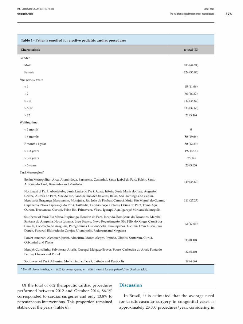
376Jesus et al.
The wait for surgical treatment of heart disease
Int J Cardiovasc Sci. 2018;31(4)374-382
Original Article
Table 1 - Patients enrolled for elective pediatric cardiac procedures
Characteristic n total (%)
Gender
Male 183 (44.94)
Female 224 (55.06)
Age group, years
< 1 45 (11.06)
1-2 66 (16.22)
> 2-6 142 (34.89)
> 6-12 133 (32.68)
> 12 21 (5.16)
Waiting time
< 1 month 0
1-6 months 80 (19.66)
7 months-1 year 50 (12.29)
> 1-3 years 197 (48.4)
> 3-5 years 57 (14)
> 5 years 23 (5.65)
Pará Mesoregion*
Belém Metropolitan Area: Ananindeua, Barcarena, Castanhal, Santa Izabel do Pará, Belém, Santo
Antonio do Tauá, Benevides and Marituba149 (36.60)
Northeast of Pará: Abaetetuba, Santa Luzia do Pará, Acará, Irituia, Santa Maria do Pará, Augusto
Corrêa, Aurora do Pará, Mãe do Rio, São Caetano de Odivelas, Baião, São Domingos do Capim,
Maracanã, Bragança, Marapanim, Mocajuba, São João de Pirabas, Cametá, Moju, São Miguel do Guamá,
Capanema, Nova Esperança do Piriá, Tailândia, Capitão Poço, Colares, Oeiras do Pará, Tomé-Açu,
Ourém, Tracuateua, Curuçá, Peixe-Boi, Primavera, Viseu, Igarapé-Açu, Igarapé-Miri and Salinópolis
111 (27.27)
Southeast of Pará: Rio Maria, Itupiranga, Rondon do Pará, Jacundá, Bom Jesus do Tocantins, Marabá,
Santana do Araguaia, Nova Ipixuna, Breu Branco, Novo Repartimento, São Félix do Xingu, Canaã dos
Carajás, Conceição do Araguaia, Paragominas, Curionópolis, Parauapebas, Tucumã, Dom Eliseu, Pau
D'arco, Tucuruí, Eldorado do Carajás, Ulianópolis, Redenção and Xinguara
72 (17.69)
Lower Amazon: Alenquer, Juruti, Almeirim, Monte Alegre, Prainha, Óbidos, Santarém, Curuá,
Oriximiná and Placas33 (8.10)
Marajó: Curralinho, Salvaterra, Anajás, Gurupá, Melgaço Breves, Soure, Cachoeira do Arari, Ponta de
Pedras, Chaves and Portel22 (5.40)
Southwest of Pará: Altamira, Medicilândia, Pacajá, Itaituba and Rurópolis 19 (4.66)
* For all characteristics, n = 407, for mesoregions, n = 406; † except for one patient from Santana (AP).
Of the total of 662 therapeutic cardiac procedures performed between 2012 and October 2014, 86.1% corresponded to cardiac surgeries and only 13.8% to percutaneous interventions. This proportion remained stable over the years (Table 6).
Discussion
In Brazil, it is estimated that the average need for cardiovascular surgery in congenital cases is approximately 23,000 procedures/year, considering in

377
Figure 1 - Distribution of patients waiting for elective pediatric cardiac procedures according to the place of residence in mesoregions.
Lower Amazon
Southwest of Pará
Belém Metropolitan Area
Northeast of Pará
Southeast of Pará
Marajó
Jesus et al.
The wait for surgical treatment of heart disease
Int J Cardiovasc Sci. 2018;31(4)374-382
Original Article
this estimate, in addition to new births with congenital heart disease, the reintervention cases. In 2002, a total of 8,092 patients underwent surgery, which shows a 65% gap – with higher rates in the Northern Region (93.5%).9
In the present study, it was observed that of the 407 children diagnosed with congenital heart disease, the most prevalent age groups were preschoolers (> 2 to 6 years) and schoolchildren (> 6 to 12 years), with no neonates waiting for treatment. These results differ from those observed in the analysis of the prevalence of congenital heart diseases at the time of the first consultation in a pediatric hospital in the city of Curitiba, state of Paraná, where there was a predominance of children with congenital heart disease in the infancy period, followed by the neonatal period, with 52.1%, and 19.4%, respectively.11 Considering that the sample of the present study refers to the patients waiting for elective procedures, this may reflect the differences regarding the time of referral for these patients and the delay during the waiting period.
Regarding the type of congenital heart disease, the most frequent one was VSD, followed by PDA and ASD. These results are consistent with those found in the study by Aragão et al.,12 who demonstrated the
following frequencies: VSD (21%), PDA (18%), Tetralogy of Fallot (14%) and ASD (7.7%). As for Huber et al.,13 they were as follows: VSD with or without associations (13.9%), Tetralogy of Fallot (12.9%), obstructive lesions of the right ventricular outflow tract (9.8%), and isolated ASD (9.6%). It can be said that the assessed institution had similar characteristics to those observed in other regions of Brazil.
The most frequent origin of the children who comprised the waiting list for cardiologic procedures was the Belém Metropolitan Mesoregion, a result consistent with those of a referral hospital in the Northeast region of Brazil, where most of the children came from the metropolitan region of the state.12
However, 63.4% of the children did not live in the Metropolitan Region of Belém; thus, a point to be discussed is the need to qualify new high cardiovascular complexity referral units in the State of Pará. For the geographical distribution of the High Complexity Care Services in Pediatric Cardiovascular Surgery, according to Ordinance 210,14 which is based on the proportion of 1:800 thousand inhabitants, the State of Pará needs nine centers capable of performing pediatric cardiovascular surgery, but the regionalization of services has not yet occurred, generating
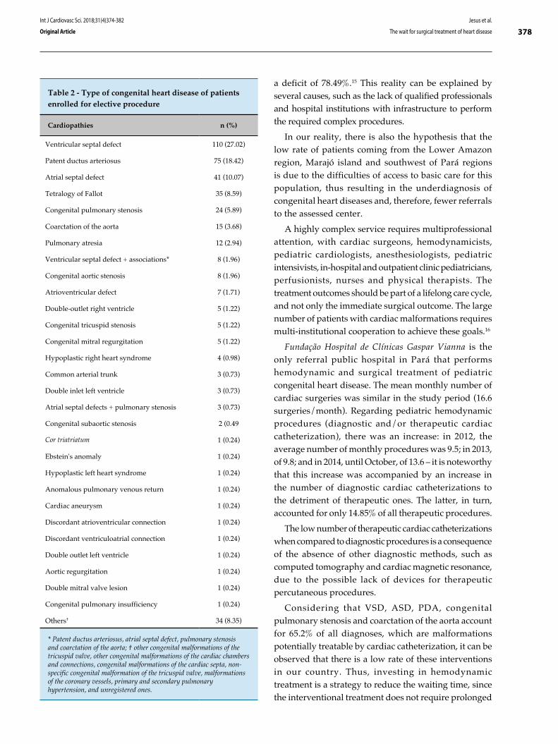
378Jesus et al.
The wait for surgical treatment of heart disease
Int J Cardiovasc Sci. 2018;31(4)374-382
Original Article
Table 2 - Type of congenital heart disease of patients enrolled for elective procedure
Cardiopathies n (%)
Ventricular septal defect 110 (27.02)
Patent ductus arteriosus 75 (18.42)
Atrial septal defect 41 (10.07)
Tetralogy of Fallot 35 (8.59)
Congenital pulmonary stenosis 24 (5.89)
Coarctation of the aorta 15 (3.68)
Pulmonary atresia 12 (2.94)
Ventricular septal defect + associations* 8 (1.96)
Congenital aortic stenosis 8 (1.96)
Atrioventricular defect 7 (1.71)
Double-outlet right ventricle 5 (1.22)
Congenital tricuspid stenosis 5 (1.22)
Congenital mitral regurgitation 5 (1.22)
Hypoplastic right heart syndrome 4 (0.98)
Common arterial trunk 3 (0.73)
Double inlet left ventricle 3 (0.73)
Atrial septal defects + pulmonary stenosis 3 (0.73)
Congenital subaortic stenosis 2 (0.49
Cor triatriatum 1 (0.24)
Ebstein's anomaly 1 (0.24)
Hypoplastic left heart syndrome 1 (0.24)
Anomalous pulmonary venous return 1 (0.24)
Cardiac aneurysm 1 (0.24)
Discordant atrioventricular connection 1 (0.24)
Discordant ventriculoatrial connection 1 (0.24)
Double outlet left ventricle 1 (0.24)
Aortic regurgitation 1 (0.24)
Double mitral valve lesion 1 (0.24)
Congenital pulmonary insufficiency 1 (0.24)
Others† 34 (8.35)
* Patent ductus arteriosus, atrial septal defect, pulmonary stenosis and coarctation of the aorta; † other congenital malformations of the tricuspid valve, other congenital malformations of the cardiac chambers and connections, congenital malformations of the cardiac septa, non-specific congenital malformation of the tricuspid valve, malformations of the coronary vessels, primary and secondary pulmonary hypertension, and unregistered ones.
a deficit of 78.49%.15 This reality can be explained by several causes, such as the lack of qualified professionals and hospital institutions with infrastructure to perform the required complex procedures.
In our reality, there is also the hypothesis that the low rate of patients coming from the Lower Amazon region, Marajó island and southwest of Pará regions is due to the difficulties of access to basic care for this population, thus resulting in the underdiagnosis of congenital heart diseases and, therefore, fewer referrals to the assessed center.
A highly complex service requires multiprofessional attention, with cardiac surgeons, hemodynamicists, pediatric cardiologists, anesthesiologists, pediatric intensivists, in-hospital and outpatient clinic pediatricians, perfusionists, nurses and physical therapists. The treatment outcomes should be part of a lifelong care cycle, and not only the immediate surgical outcome. The large number of patients with cardiac malformations requires multi-institutional cooperation to achieve these goals.16
Fundação Hospital de Clínicas Gaspar Vianna is the only referral public hospital in Pará that performs hemodynamic and surgical treatment of pediatric congenital heart disease. The mean monthly number of cardiac surgeries was similar in the study period (16.6 surgeries/month). Regarding pediatric hemodynamic procedures (diagnostic and/or therapeutic cardiac catheterization), there was an increase: in 2012, the average number of monthly procedures was 9.5; in 2013, of 9.8; and in 2014, until October, of 13.6 – it is noteworthy that this increase was accompanied by an increase in the number of diagnostic cardiac catheterizations to the detriment of therapeutic ones. The latter, in turn, accounted for only 14.85% of all therapeutic procedures.
The low number of therapeutic cardiac catheterizations when compared to diagnostic procedures is a consequence of the absence of other diagnostic methods, such as computed tomography and cardiac magnetic resonance, due to the possible lack of devices for therapeutic percutaneous procedures.
Considering that VSD, ASD, PDA, congenital pulmonary stenosis and coarctation of the aorta account for 65.2% of all diagnoses, which are malformations potentially treatable by cardiac catheterization, it can be observed that there is a low rate of these interventions in our country. Thus, investing in hemodynamic treatment is a strategy to reduce the waiting time, since the interventional treatment does not require prolonged
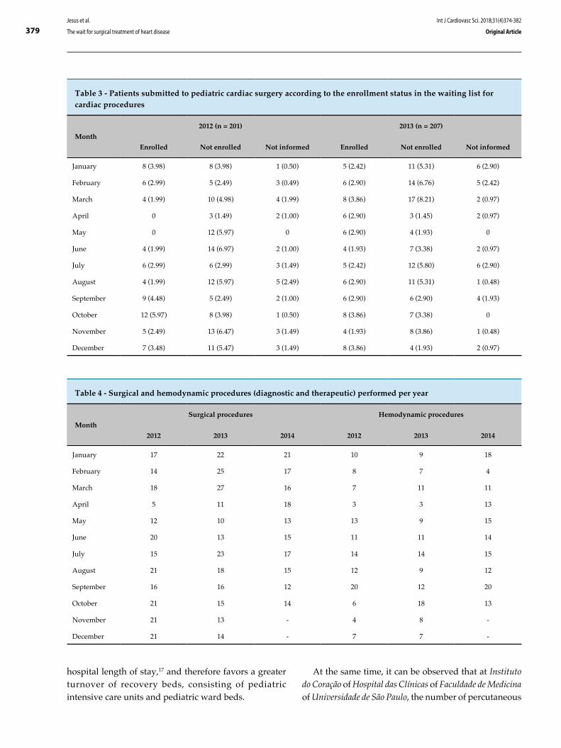
379Jesus et al.
The wait for surgical treatment of heart disease
Int J Cardiovasc Sci. 2018;31(4)374-382
Original Article
Table 3 - Patients submitted to pediatric cardiac surgery according to the enrollment status in the waiting list for cardiac procedures
Month
2012 (n = 201) 2013 (n = 207)
Enrolled Not enrolled Not informed Enrolled Not enrolled Not informed
January 8 (3.98) 8 (3.98) 1 (0.50) 5 (2.42) 11 (5.31) 6 (2.90)
February 6 (2.99) 5 (2.49) 3 (0.49) 6 (2.90) 14 (6.76) 5 (2.42)
March 4 (1.99) 10 (4.98) 4 (1.99) 8 (3.86) 17 (8.21) 2 (0.97)
April 0 3 (1.49) 2 (1.00) 6 (2.90) 3 (1.45) 2 (0.97)
May 0 12 (5.97) 0 6 (2.90) 4 (1.93) 0
June 4 (1.99) 14 (6.97) 2 (1.00) 4 (1.93) 7 (3.38) 2 (0.97)
July 6 (2.99) 6 (2.99) 3 (1.49) 5 (2.42) 12 (5.80) 6 (2.90)
August 4 (1.99) 12 (5.97) 5 (2.49) 6 (2.90) 11 (5.31) 1 (0.48)
September 9 (4.48) 5 (2.49) 2 (1.00) 6 (2.90) 6 (2.90) 4 (1.93)
October 12 (5.97) 8 (3.98) 1 (0.50) 8 (3.86) 7 (3.38) 0
November 5 (2.49) 13 (6.47) 3 (1.49) 4 (1.93) 8 (3.86) 1 (0.48)
December 7 (3.48) 11 (5.47) 3 (1.49) 8 (3.86) 4 (1.93) 2 (0.97)
Table 4 - Surgical and hemodynamic procedures (diagnostic and therapeutic) performed per year
Month
Surgical procedures Hemodynamic procedures
2012 2013 2014 2012 2013 2014
January 17 22 21 10 9 18
February 14 25 17 8 7 4
March 18 27 16 7 11 11
April 5 11 18 3 3 13
May 12 10 13 13 9 15
June 20 13 15 11 11 14
July 15 23 17 14 14 15
August 21 18 15 12 9 12
September 16 16 12 20 12 20
October 21 15 14 6 18 13
November 21 13 - 4 8 -
December 21 14 - 7 7 -
hospital length of stay,17 and therefore favors a greater turnover of recovery beds, consisting of pediatric intensive care units and pediatric ward beds.
At the same time, it can be observed that at Instituto do Coração of Hospital das Clínicas of Faculdade de Medicina of Universidade de São Paulo, the number of percutaneous
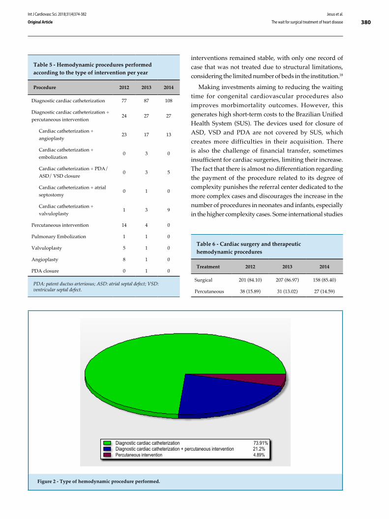
380
Figure 2 - Type of hemodynamic procedure performed.
Diagnostic cardiac catheterization 73.91%Diagnostic cardiac catheterization + percutaneous intervention 21.2%Percutaneous intervention 4.89%
Jesus et al.
The wait for surgical treatment of heart disease
Int J Cardiovasc Sci. 2018;31(4)374-382
Original Article
Table 5 - Hemodynamic procedures performed according to the type of intervention per year
Procedure 2012 2013 2014
Diagnostic cardiac catheterization 77 87 108
Diagnostic cardiac catheterization +
percutaneous intervention24 27 27
Cardiac catheterization +
angioplasty23 17 13
Cardiac catheterization +
embolization0 3 0
Cardiac catheterization + PDA/
ASD/ VSD closure0 3 5
Cardiac catheterization + atrial
septostomy0 1 0
Cardiac catheterization +
valvuloplasty1 3 9
Percutaneous intervention 14 4 0
Pulmonary Embolization 1 1 0
Valvuloplasty 5 1 0
Angioplasty 8 1 0
PDA closure 0 1 0
PDA: patent ductus arteriosus; ASD: atrial septal defect; VSD: ventricular septal defect.
Table 6 - Cardiac surgery and therapeutic hemodynamic procedures
Treatment 2012 2013 2014
Surgical 201 (84.10) 207 (86.97) 158 (85.40)
Percutaneous 38 (15.89) 31 (13.02) 27 (14.59)
interventions remained stable, with only one record of case that was not treated due to structural limitations, considering the limited number of beds in the institution.18
Making investments aiming to reducing the waiting time for congenital cardiovascular procedures also improves morbimortality outcomes. However, this generates high short-term costs to the Brazilian Unified Health System (SUS). The devices used for closure of ASD, VSD and PDA are not covered by SUS, which creates more difficulties in their acquisition. There is also the challenge of financial transfer, sometimes insufficient for cardiac surgeries, limiting their increase. The fact that there is almost no differentiation regarding the payment of the procedure related to its degree of complexity punishes the referral center dedicated to the more complex cases and discourages the increase in the number of procedures in neonates and infants, especially in the higher complexity cases. Some international studies

381Jesus et al.
The wait for surgical treatment of heart disease
Int J Cardiovasc Sci. 2018;31(4)374-382
Original Article
have shown that there is a linear association between the complexity and cost of the procedure.19
Another great difficulty that the service faces in reducing the waiting list for elective cardiac surgery is the high demand for urgent surgeries in patients without previous enrollment, as these cases are prioritized to the detriment of elective ones. A strategy to mitigate the problem in the short term would be the performance of congenital heart surgeries in patients already enrolled for it, aiming to reduce the repressed demand of SUS users.19
In this sense, it is proposed: the creation of outpatient care and specialized centers for the diagnosis and early treatment of the population, reducing underdiagnosing and improving pre- and postoperative clinical follow-up, with a possible reduction of costs for out-of-home treatment and, consequently, less social impact for the affected families; investment in diagnosis performed through computed tomography and cardiac magnetic resonance imaging, which would reduce diagnostic cardiac catheterizations and increase the availability of hospital support for therapeutic procedures; the promotion of hemodynamic procedures, including a funding policy for Orthoses, Devices and Special Materials (Órteses, Próteses e Materiais Especiais – OPME) not covered by SUS, due to the proven effectiveness and shorter hospital length of stay, with a consequent decrease in hospital expenses and an increase in the volume of treated cases per unit of time; increased functional capacity at the referral hospital; decentralization of surgical and cardiac hemodynamic care, with the internalization of this type of service in medium-sized municipalities, such as Marabá and Santarém, both in the state of Pará; and reliable, detailed and updated data registry regarding the surgical and hemodynamic procedures, for permanent control and evaluation of the outcomes. The promotion of improvements in the care for children with heart disease is a priority and involves the participation of all – public services, professionals and several sectors of society.
Conclusion
Most of the children awaiting cardiac procedures come from outside the metropolitan area and have malformations potentially treatable through cardiac catheterization. However, even with changes in the treatment profile, with
the increase in percutaneous procedures in the last years, it still requires further increase.
The limitations of the public hospital system in meeting the great demand of the region for elective therapeutic cardiovascular procedures generate an important care deficit, with the need to increase the functional capacity of the only public referral center for these diseases in the region, as well as decentralization of cardiological, clinical, surgical and hemodynamic care, to better treat the population that depends on SUS.
Author contributions
Conception and design of the research: Jesus VS, Nascimento AM, Miranda RA, Veríssimo AOL. Acquisition of data: Jesus VS, Nascimento AM, Lima JS. Analysis and interpretation of the data: Jesus VS, Nascimento AM, Veríssimo AOL. Statistical analysis: Jesus VS, Nascimento AM. Writing of the manuscript: Jesus VS, Nascimento AM, Lima JS, Tyll MAG. Critical revision of the manuscript for intellectual content: Miranda RA, Tyll MAG, Veríssimo AOL.
Potential Conflict of Interest
No potential conflict of interest relevant to this article was reported.
Sources of Funding
O presente estudo não teve fontes de financiamento externas.
Study Association
This study is not associated with any thesis or dissertation work.
Ethics approval and consent to participate
This study was approved by the Ethics Committee of the Fundação Pública Estadual Hospital das Clínicas Gaspar Vianna under the protocol number 39903014.2.0000.0016. All the procedures in this study were in accordance with the 1975 Helsinki Declaration, updated in 2013. Informed consent was obtained from all participants included in the study.

382Jesus et al.
The wait for surgical treatment of heart disease
Int J Cardiovasc Sci. 2018;31(4)374-382
Original Article
This is an open-access article distributed under the terms of the Creative Commons Attribution License
1. Petersen S, Peto V, Rayner M. Congenital heart diseases statistics. [Internet]. British Heart Foundation Statistics Database. [Cited in 2014 July 21]. Available from: http://www.heartstats.org.
2. Hoffman JI, Kaplan S, Liberthson RR. Prevalence of congenital heart disease. Am Heart J. 2004;147(3):425-39.
3. Hoffman JI, Kaplan S. The incidence of congenital heart disease. J Am Coll Cardiol. 2002;39(12):1890-900.
4. Bosi G, Garani G, Scorrano M, Calzolari E; IMER Working Party. Temporal variability in birth prevalence of congenital heart defects as recorded by a general birth defects registry. J Pediatr. 2003;142(6):690-8. Erratum in: J Pediatr. 2003;143(4):531.
5. Jenkins KJ, Correa A, Feinstein JA, Botto L, Britt AE, Daniels SR, et al; American Heart Association Council on Cardiovascular Disease in the Young. Noninherited risk factors and congenital heart defects: current knowledge: a scientific statement from the American Heart Association Council on Cardiovascular Disease in the Young. Circulation. 2007;115(23):2995-3014.
6. Pradat P, Francannet C, Harris JA, Robert E. The epidemiology of cardiovascular defects, part I: a study based on data from three large registries of congenital malformations. Pediatr Cardiol. 2003;24(3):195-221.
7. Amorim LF, Pires CA, Lana AM, Campos AS, Aguiar RA, Tibúrcio JD, et al. Presentation of congenital heart disease diagnosed at birth: analysis of 29,770 newborn infants. J Pediatr (Rio J). 2008;84(1):83-90.
8. Pinto Júnior VC, Daher CV, Sallum FS, Jatene MB, Croti UA. The situation of congenital heart surgeries in Brazil. Rev Bras Cir Cardiovasc. 2004;19(2):III-VI.
9. Pinto Júnior VC, Rodrigues, LC, Muniz CR. Reflexões sobre a formulação de política de atenção cardiovascular pediátrica no Brasil. Rev Bras Cir Cardiovasc. 2009;24(1):73-80.
10. Fernandes AM, Mansur AJ, Canêo LF, Lourenço DD, Piccioni MA, Franchi SM, et al. The reduction in hospital stay and costs in the care
of patients with congenital heart diseases undergoing fast-track cardiac surgery. Arq Bras Cardiol. 2004;83(1):27-34, 18-26.
11. Miyague NI, Cardoso SM, Meyer F, Ultramari FT, Araujo FH, Rozkowisk I, et al. Epidemiological study of congenital heart defects in children and adolescents. Analysis of 4,538 cases. Arq Bras Cardiol. 2003;80(3):269-73.
12. Aragão JA, Mendonça MP, Silva MS, Moreira AN, Sant´Anna ME, Reis FP. O perfil epidemiológico dos pacientes com cardiopatias congênitas submetidos à cirurgia no Hospital do Coração. R bras Ci Saúde 2013:17(3):263-9.
13. Huber J, Peres VC, Santos TJ, Beltrão Lda F, Baumont AC, Cañedo AD, et al. Congenital heart diseases in a reference service: clinical evolution and associated illnesses. Arq Bras Cardiol. 2010;94(3):333-8.
14. Brasil. Ministério da Saúde. Portaria nº 210 de 15 de junho de 2004. Institui Unidades de assistência de alta complexidade cardiovascular e os centros de referência em alta complexidade cardiovascular - Serviços de cirurgia cardiovascular pediátrica. Diário Oficial da União. 2004;117(1):43. Seção 1.
15. Pinto Júnior VC, Fraga MN, Freitas SM, Croti UA. Regionalization of Brazilian pediatric cardiovascular surgery. Rev Bras Cir Cardiovasc. 2013;28(2):256-62.
16. Haddad N, Bittar OJ, Pereira AA, da Silva MB, Amato VL, Farsky PS, et al. Consequences of the prolonged waiting time for patient candidates for heart surgery. Arq Bras Cardiol. 2002;78(5):452-65.
17. Costa Fde A, Kajita LJ, Martinez Filho EE. [Percutaneous interventions in congenital heart disease]. Arq Bras Cardiol. 2002;78(6):608-17.
18. Lisboa LA, Moreira LF, Mejia OV, Dallan LA, Pomerantzeff PM, Costa R, et al. [Evolution of cardiovascular surgery at the Instituto do Coração: analysis of 71,305 surgeries]. Arq Bras Cardiol. 2010;94(2):162-8, 174-81, 164-71.
19. Caneo LF, Jatene MB, Yatsuda N, Gomes WJ. A reflection on the performance of pediatric cardiac surgery in the State of São Paulo. Rev Bras Cir Cardiovasc. 2012;27(3):457-62.
References

DOI: 10.5935/2359-4802.20180028
383
ORIGINAL ARTICLE
International Journal of Cardiovascular Sciences. 2018;31(4)383-392
Mailing Address: Sérgio Henrique SimonettiRua das Tâmaras, 210. Postal Code: 04361-130, Vila Paulista, São Paulo, SP - Brazil.E-mail: [email protected], [email protected]
Adherence Score for Users of Oral AnticoagulantsSérgio Henrique Simonetti,1 Ana Cristina Mancussi e Faro,2 Estela Regina Ferraz Bianchi2
Instituto Dante Pazzanese de Cardiologia,1 São Paulo - BrazilEscola de Enfermagem da Universidade de São Paulo,2 São Paulo - Brazil
Manuscript received June 05, 2017, revised manuscript December 21, 2017, accepted January 15, 2018.
Abstract
Background: The score is an effective instrument for the assessment of treatment adherence in users of oral anticoagulants and maintenance of INR (international normalized ratio) within the therapeutic range.
Objective: To develop an adherence score for oral anticoagulant users for the maintenance of INR within the therapeutic range.
Methods: This was an analytical, observational, cross-sectional study, with a quantitative approach, conducted in a public cardiology hospital. A total of 607 patients who met the inclusion criteria were evaluated. Variables with a significance level of 5% in the exploratory analysis and considered clinically relevant were included and subjected to a multiple logistic regression model. Predictive accuracy of the model was determined using the C statistic.
Results: The variables with a significance level of 5% in the multivariate analysis were: education, family income, inadequate use of medications, drug-drug interactions, invasive procedures, food-drug interactions, physical activity, medical conditions, other factors, and complications related to oral anticoagulant therapy. The following prognostic variables were identified: family income, inadequate use of medications, invasive procedures, drug-drug interactions, dietary habits, medical conditions, other factors (stress, thinness, weight loss). C-statistic for adherence score was 0.94.
Conclusion: The Simonetti adherence score proved to be easily applicable and feasible, with high predictive value for influencing factors, promoting the improvement of adherence and maintenance of INR in the ideal range. (Int J Cardiovasc Sci. 2018;31(4)383-392)
Keywords: Cardiovascular Diseases; Anticoagulants/therapy; Probability; Medication Adherence; Health Education.
Introduction
Health promotion is a key strategy for prevention of thromboembolic events in patients on oral anticoagulant therapy (OAC-T) in addition to other aspects related to the assistance of this population. In this context, health professionals, nurses and physicians play the role of educators, particularly in providing clear guidance on risk factors and possible signs and symptoms and related complications associated with a OAC-T.1
Stability of OAC-T is associated with treatment adherence, in addition to other factors including age, concomitant use of other drugs, comorbidities, irregular use of vitamin K, inadequate use of the drug,
and hereditary polymorphism.2 Educational programs for outpatient management of patients on OAC-T have been crucial for the approach of drug and dietary interactions with coumarin derivatives, which may affect treatment adherence.2,3
A study4 pointed out important considerations about adherence to OAC-T. First, knowledge about adherence to OAC-T is still a challenge among health professionals in national and international contexts. Second, the study provides data on instruments proposed by health professionals aimed at improving patients’ adherence and knowledge about the treatment, and on patients’ sociodemographic status. The authors also reported the scarcity of studies on factors that may influence therapy
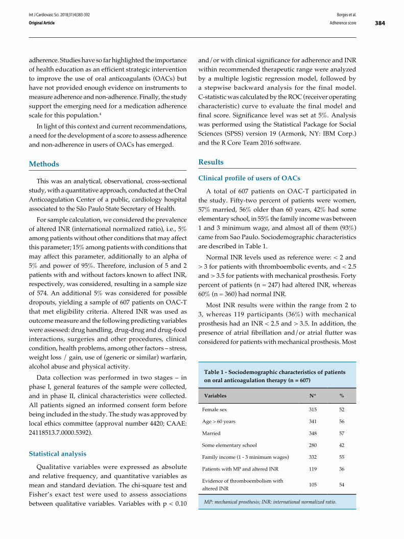
384Borges et al.
Adherence score
Int J Cardiovasc Sci. 2018;31(4)383-392
Original Article
adherence. Studies have so far highlighted the importance of health education as an efficient strategic intervention to improve the use of oral anticoagulants (OACs) but have not provided enough evidence on instruments to measure adherence and non-adherence. Finally, the study support the emerging need for a medication adherence scale for this population.4
In light of this context and current recommendations, a need for the development of a score to assess adherence and non-adherence in users of OACs has emerged.
Methods
This was an analytical, observational, cross-sectional study, with a quantitative approach, conducted at the Oral Anticoagulation Center of a public, cardiology hospital associated to the São Paulo State Secretary of Health.
For sample calculation, we considered the prevalence of altered INR (international normalized ratio), i.e., 5% among patients without other conditions that may affect this parameter; 15% among patients with conditions that may affect this parameter, additionally to an alpha of 5% and power of 95%. Therefore, inclusion of 5 and 2 patients with and without factors known to affect INR, respectively, was considered, resulting in a sample size of 574. An additional 5% was considered for possible dropouts, yielding a sample of 607 patients on OAC-T that met eligibility criteria. Altered INR was used as outcome measure and the following predicting variables were assessed: drug handling, drug-drug and drug-food interactions, surgeries and other procedures, clinical condition, health problems, among other factors – stress, weight loss / gain, use of (generic or similar) warfarin, alcohol abuse and physical activity.
Data collection was performed in two stages – in phase I, general features of the sample were collected, and in phase II, clinical characteristics were collected. All patients signed an informed consent form before being included in the study. The study was approved by local ethics committee (approval number 4420; CAAE: 24118513.7.0000.5392).
Statistical analysis
Qualitative variables were expressed as absolute and relative frequency, and quantitative variables as mean and standard deviation. The chi-square test and Fisher’s exact test were used to assess associations between qualitative variables. Variables with p < 0.10
and/or with clinical significance for adherence and INR within recommended therapeutic range were analyzed by a multiple logistic regression model, followed by a stepwise backward analysis for the final model. C-statistic was calculated by the ROC (receiver operating characteristic) curve to evaluate the final model and final score. Significance level was set at 5%. Analysis was performed using the Statistical Package for Social Sciences (SPSS) version 19 (Armonk, NY: IBM Corp.) and the R Core Team 2016 software.
Results
Clinical profile of users of OACs
A total of 607 patients on OAC-T participated in the study. Fifty-two percent of patients were women, 57% married, 56% older than 60 years, 42% had some elementary school, in 55% the family income was between 1 and 3 minimum wage, and almost all of them (93%) came from Sao Paulo. Sociodemographic characteristics are described in Table 1.
Normal INR levels used as reference were: < 2 and > 3 for patients with thromboembolic events, and < 2.5 and > 3.5 for patients with mechanical prosthesis. Forty percent of patients (n = 247) had altered INR, whereas 60% (n = 360) had normal INR.
Most INR results were within the range from 2 to 3, whereas 119 participants (36%) with mechanical prosthesis had an INR < 2.5 and > 3.5. In addition, the presence of atrial fibrillation and/or atrial flutter was considered for patients with mechanical prosthesis. Most
Table 1 - Sociodemographic characteristics of patients on oral anticoagulation therapy (n = 607)
Variables Nº %
Female sex 315 52
Age > 60 years 341 56
Married 348 57
Some elementary school 280 42
Family income (1 - 3 minimum wages) 332 55
Patients with MP and altered INR 119 36
Evidence of thromboembolism with
altered INR 105 54
MP: mechanical prosthesis; INR: international normalized ratio.
385Borges et al.
Adherence score
Int J Cardiovasc Sci. 2018;31(4)383-392
Original Article
patients on OAC-T with INR < 2.0 were those with atrial fibrillation (n = 105, 54%) (Table 1).
For patients with INR > 2 (conditions with risk for thromboembolic events) and/or > 2.5 (mechanical prosthesis), atrial fibrillation was the most frequent disease and mechanical prosthesis was the main valvular prosthesis – mitral valve (n = 58; 26%) and aortic valve (n = 41; 18%).
With respect to participants’ diseases and indication for OAC, 30 types of heart diseases were identified, 51% (783) clinical conditions and 24% (360) of surgical type, as well as related comorbidities (364; 25%).
Multiple logistic regression model
The following variables were selected for the multiple logistic regression model: family income, educational attainment, inadequate doses, invasive procedures, drug interactions, eating habits, physical activity, health problems, OAC-related complications, among other factors (Table 2).
Since all predictive variables showed a significative response, seven logistic regression models were considered for analysis at first. However, the model adopted in the study included the variables described in Table 2, considering also the interactions between them.
Simonetti medication adherence score
The variables used in the scores are shown in Table 3. First, the percentage of adherence was determined by Simonneti SH by multiplying the number of positive variables for adherence by the total number of predictive variables. A score was also developed to determine whether INR was within the normal range (Figure 1). The score was calculated by odds ratio.
Then, each condition known to affect INR was categorized into high (≤ 10 points), intermediate (11 – 30 points) and high (≥ 31 points) for normal INR (Figure 2).
The area under the ROC curve was determined based on the results of the variables proposed in the present study (Figure 3). For the logistic model, C-statistic was 0.940 (95% CI = 0.920 – 0.960; p < 0.001), indicating a satisfactory performance in detecting the occurrence of an event.
Discussion
The optimal dose of OAC is variable between individuals and should be adjusted to ensure that INR
is maintained within the therapeutic range. In addition, it is known that the patient may reduce, discontinue (e.g. bleeding) or increase (e.g. double dose to make up for a missed dose) the dose of OAC.5
OAC-T-related complications may also be associated with the use of Marevan®, due to underdosing (risk of thrombus formation) or overdosing (bleeding), and the seek for medical care due to gum bleeding, hematuria, and other complications.6
Therefore, the lack of appropriate instructions and recommendations for patients on OAC-T in the perioperative period of any clinical or surgical procedure may cause variability in routine practice and affect the maintenance of INR within therapeutic range. Nevertheless, evidence shows that changes in OAC-T are not required prior to tooth extraction for example, providing that INR is maintained between 2 and 4, and bleeding control measures are implemented in the perioperative period to prevent embolic events.7
Studies have suggested that patients on OAC-T may require parenteral anticoagulation in the perioperative period. The decision to discontinue anticoagulation and start an antithrombotic therapy is determined by the risk of bleeding, surgical treatment to which patients were submitted and the risk for thrombosis due to underlying diseases.8 However, for patients at low risk of bleeding (skin biopsies, cataract or dental procedures), the use of OAC may be continued, providing that INR is maintained at lower values and control of local bleeding is successfully achieved.8
Due to the risk of bleeding, when management of patients on OAC-T includes major surgery, it is recommended8 that these individuals are classified into patients at minimal risk (atrial fibrillation without history of venous thromboembolism), intermediate risk or high risk of thromboembolism.
In addition, one of the main factors that affect INR and treatment adherence is drug-drug interactions. Antonio et al.7 described the main drugs that interact with and potentiate the effect of OACs – allopurinol, amiodarone, cimetidine, cisapride, clofibrate and other fibrates, chloramphenicol, cotrimoxazole, erythromycin, fluconazole, isoniazid, metronidazole, miconazole, omeprazole, phenylbutazone, piroxicam, propafenone, propranolol, salicylate, phenylbutazone. These drugs may increase INR and cause bleeding. However, some drugs may inhibit the effects of OAC, including barbiturates, carbamazepine, chlordiazepoxide, cholestyramine,
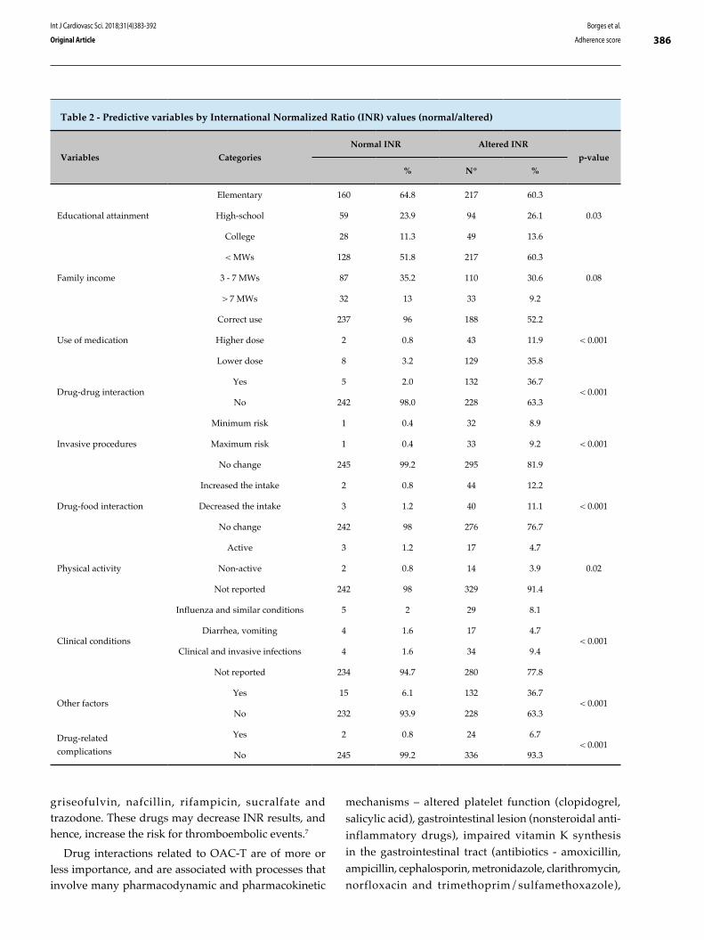
386Borges et al.
Adherence score
Int J Cardiovasc Sci. 2018;31(4)383-392
Original Article
Table 2 - Predictive variables by International Normalized Ratio (INR) values (normal/altered)
Variables Categories
Normal INR Altered INR
p-value
% Nº %
Educational attainment
Elementary 160 64.8 217 60.3
0.03High-school 59 23.9 94 26.1
College 28 11.3 49 13.6
Family income
< MWs 128 51.8 217 60.3
0.083 - 7 MWs 87 35.2 110 30.6
> 7 MWs 32 13 33 9.2
Use of medication
Correct use 237 96 188 52.2
< 0.001Higher dose 2 0.8 43 11.9
Lower dose 8 3.2 129 35.8
Drug-drug interactionYes 5 2.0 132 36.7
< 0.001No 242 98.0 228 63.3
Invasive procedures
Minimum risk 1 0.4 32 8.9
< 0.001Maximum risk 1 0.4 33 9.2
No change 245 99.2 295 81.9
Drug-food interaction
Increased the intake 2 0.8 44 12.2
< 0.001Decreased the intake 3 1.2 40 11.1
No change 242 98 276 76.7
Physical activity
Active 3 1.2 17 4.7
0.02Non-active 2 0.8 14 3.9
Not reported 242 98 329 91.4
Clinical conditions
Influenza and similar conditions 5 2 29 8.1
< 0.001Diarrhea, vomiting 4 1.6 17 4.7
Clinical and invasive infections 4 1.6 34 9.4
Not reported 234 94.7 280 77.8
Other factorsYes 15 6.1 132 36.7
< 0.001No 232 93.9 228 63.3
Drug-related
complications
Yes 2 0.8 24 6.7< 0.001
No 245 99.2 336 93.3
griseofulvin, nafcillin, rifampicin, sucralfate and trazodone. These drugs may decrease INR results, and hence, increase the risk for thromboembolic events.7
Drug interactions related to OAC-T are of more or less importance, and are associated with processes that involve many pharmacodynamic and pharmacokinetic
mechanisms – altered platelet function (clopidogrel, salicylic acid), gastrointestinal lesion (nonsteroidal anti-inflammatory drugs), impaired vitamin K synthesis in the gastrointestinal tract (antibiotics - amoxicillin, ampicillin, cephalosporin, metronidazole, clarithromycin, norfloxacin and trimethoprim/sulfamethoxazole),
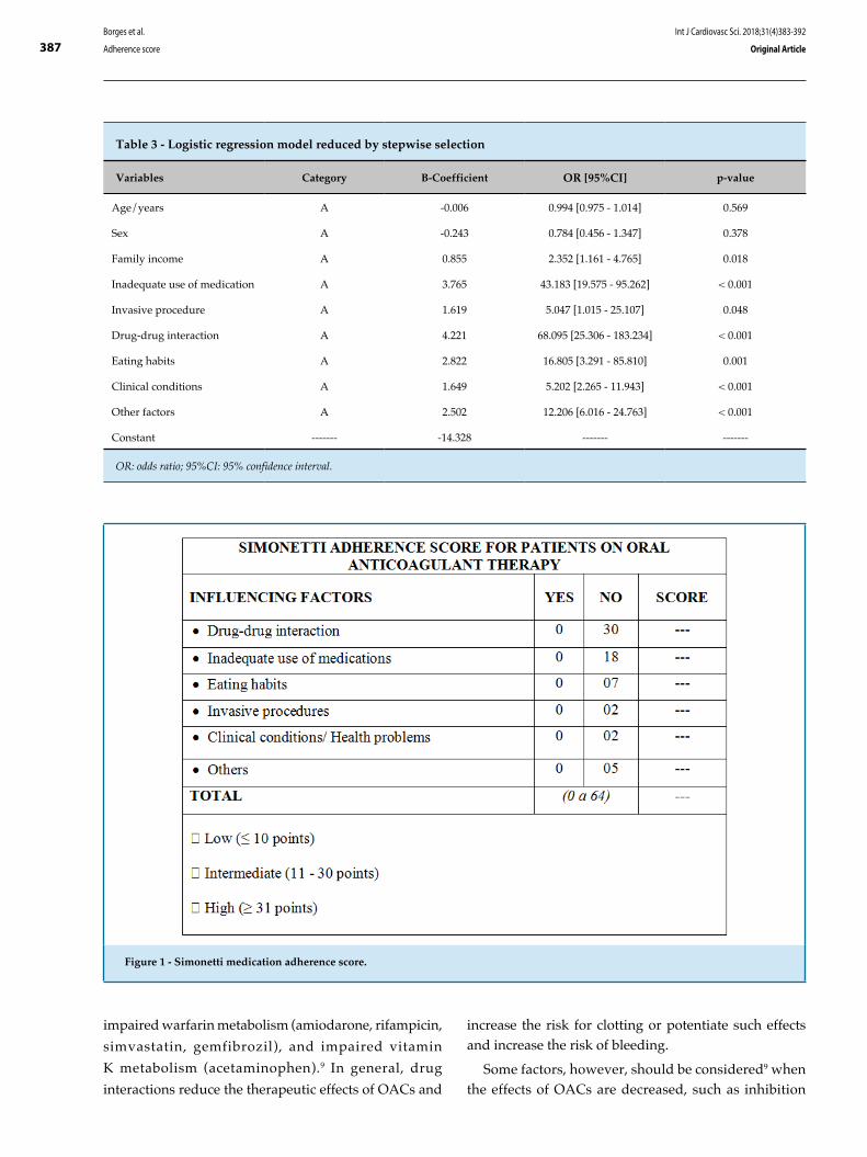
387Borges et al.
Adherence score
Int J Cardiovasc Sci. 2018;31(4)383-392
Original Article
Table 3 - Logistic regression model reduced by stepwise selection
Variables Category B-Coefficient OR [95%CI] p-value
Age/years A -0.006 0.994 [0.975 - 1.014] 0.569
Sex A -0.243 0.784 [0.456 - 1.347] 0.378
Family income A 0.855 2.352 [1.161 - 4.765] 0.018
Inadequate use of medication A 3.765 43.183 [19.575 - 95.262] < 0.001
Invasive procedure A 1.619 5.047 [1.015 - 25.107] 0.048
Drug-drug interaction A 4.221 68.095 [25.306 - 183.234] < 0.001
Eating habits A 2.822 16.805 [3.291 - 85.810] 0.001
Clinical conditions A 1.649 5.202 [2.265 - 11.943] < 0.001
Other factors A 2.502 12.206 [6.016 - 24.763] < 0.001
Constant ------- -14.328 ------- -------
OR: odds ratio; 95%CI: 95% confidence interval.
Figure 1 - Simonetti medication adherence score.
impaired warfarin metabolism (amiodarone, rifampicin,
simvastatin, gemfibrozil), and impaired vitamin
K metabolism (acetaminophen).9 In general, drug
interactions reduce the therapeutic effects of OACs and
increase the risk for clotting or potentiate such effects and increase the risk of bleeding.
Some factors, however, should be considered9 when the effects of OACs are decreased, such as inhibition

388Borges et al.
Adherence score
Int J Cardiovasc Sci. 2018;31(4)383-392
Original Article
of warfarin absorption by cholestyramine or by foods in the gastrointestinal tract; increased distribution volume and half-life reduction due to hypoproteinemia; increased metabolism caused by stimulation of CYP2C9 by other drugs (barbiturates); excess intake of vitamin K-rich foods. On the other hand, CYP2C9 inhibition by other drugs (fluoxetine, amiodarone, clopidogrel), protein binding displacement caused by diuretics and sodium valproate, as well as vitamin K deficiency may reduce vitamin K metabolism, and consequently increase its effect.10
Amiodarone and simvastatin were the main drugs that significantly affected the required dose of OACs. Amiodarone is an antiarrhythmic that inhibits warfarin, due to its role as a potent cytochrome P450 inhibitor including CYP2C9, which is involved in S-warfarin metabolism.10 Amiodarone inhibits warfarin metabolism and increases its effect, and the concomitant use of both drugs also affects prothrombin time. Simvastatin potentiates the effect of warfarin by binding to plasma
proteins, reduces platelet aggregation and promotes thrombus formation.11
Besides, it is essential that patients on OAC-T are aware of the consumption of vitamin K-rich foods. Variations in the amount or in the frequency of consumption of these foods may change INR to values outside the therapeutic range.10
In case of acute alcohol consumption, dehydrogenase pathway plays a key role in ethanol metabolism in the liver. Nevertheless, in case of chronic consumption, the microsomal ethanol oxidizing system, which requires the CYP2E1 enzyme, is used and increases ethanol metabolism by ten-fold.10 Therefore, in the pharmacokinetics of warfarin, there is an interaction between ethanol and the drug – in case of acute ethanol consumption, warfarin metabolism is decreased by enzymatic inhibition, resulting in increased OAC effect and increased risk of bleeding.10 In chronic alcohol consumption, enzymatic activity decreases, with consequent increase of warfarin metabolism, reduction
Figure 2 - Internal validation of Simonetti adherence score.
Score
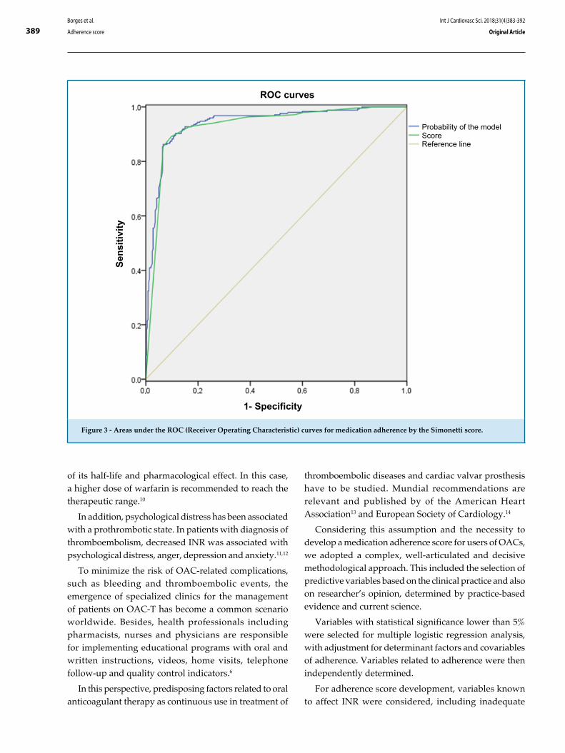
389Borges et al.
Adherence score
Int J Cardiovasc Sci. 2018;31(4)383-392
Original Article
of its half-life and pharmacological effect. In this case, a higher dose of warfarin is recommended to reach the therapeutic range.10
In addition, psychological distress has been associated with a prothrombotic state. In patients with diagnosis of thromboembolism, decreased INR was associated with psychological distress, anger, depression and anxiety.11,12
To minimize the risk of OAC-related complications, such as bleeding and thromboembolic events, the emergence of specialized clinics for the management of patients on OAC-T has become a common scenario worldwide. Besides, health professionals including pharmacists, nurses and physicians are responsible for implementing educational programs with oral and written instructions, videos, home visits, telephone follow-up and quality control indicators.6
In this perspective, predisposing factors related to oral anticoagulant therapy as continuous use in treatment of
thromboembolic diseases and cardiac valvar prosthesis have to be studied. Mundial recommendations are relevant and published by of the American Heart Association13 and European Society of Cardiology.14
Considering this assumption and the necessity to develop a medication adherence score for users of OACs, we adopted a complex, well-articulated and decisive methodological approach. This included the selection of predictive variables based on the clinical practice and also on researcher’s opinion, determined by practice-based evidence and current science.
Variables with statistical significance lower than 5% were selected for multiple logistic regression analysis, with adjustment for determinant factors and covariables of adherence. Variables related to adherence were then independently determined.
For adherence score development, variables known to affect INR were considered, including inadequate
Figure 3 - Areas under the ROC (Receiver Operating Characteristic) curves for medication adherence by the Simonetti score.
Sens
itivi
ty
1- Specificity
Probability of the modelScoreReference line
ROC curves
390Borges et al.
Adherence score
Int J Cardiovasc Sci. 2018;31(4)383-392
Original Article
use of medication (higher or lower doses, missing doses), history of invasive or surgical procedures, drug-drug interaction, nutrient-drug interaction (particularly vitamin K), health problems or clinical conditions, other related factors such as considerable psychological distress, weight gain or loss and OAC-related complications. Thus, the score was developed using the variables of several models, classified and compared based on descriptive analysis of the independent variables, as well as interaction tests.
Then, a score model for a Brazilian population of patients in OAC-T was developed (Table 3); this was a simple instrument, easily applicable in the clinical setting of OAC-T. Results of the score were presented as odds ratio and respective 95% confidence interval and descriptive analysis.
The tests revealed the independent nature of the variables and the impartiality of the event occurrence. Therefore, a score that attributed the same weight for the variables was developed.
The proportion of the occurrence of an event will then be calculated by the number of positive variables for adherence and the number of predictive variables established in the model.
Simonetti adherence score had a c-statistic of 0.94, indicating satisfactory performance and applicability. Therefore, the score proposed in the present study will contribute to a more accurate evaluation and decision-making process. However, a long-term evaluation of the instrument should be performed in the clinical setting, like any stratification model, in order to evaluate the occurrence of new variables that may affect adherence.
Some limitations of the study should be considered. First, since the study was conducted in only one center, the model may not be reproducible in other settings. Second, since the score was not applied in an independent population for external validation, the performance of the instrument may not be satisfactory. It is worth pointing out, however, that our next step is to validate the instrument in another group of patients on OAC-T.
Despite these limitations, we successfully developed a medication adherence score using a model with good statistical performance. We believe that the score is of public interest and can contribute to the improvement of oral anticoagulation centers.
This study aimed to ensure and improve the provision of nursing services to patients on OAC-T in a
specialized center, using a specific protocol. Although this characterized a limitation of the study, this would encourage different centers to revise their own protocols and to be informed of how treatment is being performed in other centers.
The proposed model of medication adherence score is in conformity with the significant and independent variables identified in the study. This ensures its accessibility and applicability. Using adequate statistical methods, we developed the Simonetti adherence score, and confirmed the hypothesis of this study.
Conclusion
The medication adherence score here proposed for patients on OAC-T was shown to be an easy-to-use instrument, with high predictive value and good performance. We successfully developed an instrument aimed at measuring and improving adherence of patients to OAC-T by means of the proposed methods and results obtained, confirming the hypothesis of the study.
This instrument will help professionals in accurately dealing with and hence improving adherence of patients to OAC-T and consequently improve the quality of life of this population.
Author contributions
Conception and design of the research: Simonetti SH, Bianchi ERF, Faro ACM. Acquisition of data: Simonetti SH. Analysis and interpretation of the data: Simonetti SH, Bianchi ERF, Faro ACM. Statistical analysis: Simonetti SH. Writing of the manuscript: Simonetti SH, Bianchi ERF, Faro ACM. Critical revision of the manuscript for intellectual content: Simonetti SH, Faro ACM. Supervision / as the major investigador: Simonetti SH.
Potential Conflict of Interest
No potential conflict of interest relevant to this article was reported.
Sources of Funding
There were no external funding sources for this study.
Study Association
This article is part of the thesis of Doctoral submitted by Sérgio Henrique Simonetti, from USP Nursing School.
391Borges et al.
Adherence score
Int J Cardiovasc Sci. 2018;31(4)383-392
Original Article
1. Freitas D, Ribeiro K, Oliveira JL, Matos FG, Carvalho AR, Ross C, et al. Diagnósticos de enfermagem entre usuários de anticoagulante oral acompanhados em ambulatório. Rev baiana enferm.2017;31(3):e20356
2. Ávila CW, Aliti GB, Feijó MKF, Rabelo ER. Adesão Farmacológica ao anticoagulante oral e os fatores que influenciam na estabilidade do índice de normatização internacional. Rev Latino-Am. Enfermagem.2011; 19(1): 1-8.
3. Lustosa MA, Alcaires J, Costa JC. Adesão do paciente ao tratamento no hospital geral. Rev SBPH. 2011;14(2):27-49.
4. Simonetti SH, Mancussi e Faro AC, Bianchi ERF. Adesão à terapia com anticoagulantes orais: revisão integrativa. Rev enferm UFPE. 2014;8(8): 2854-63.
5. Crowther M, Crowther MA. Intensity of Warfarin Coagulation in the Antiphoslipid Syndrome. Curr Rheumatol Rep. 2010;12(1):64-9.
6. Pelegrino FM, Bolela F, Corbi ISA, Carvalho ARS, Dantas RAS. Protocolo educativo para pacientes em uso de anticoagulante oral: construção e validação. Texto Contexto Enferm. 2014;23(3):799-806.
7. Antonio N, Castro G, Ramos D, Machado A, Gonçalves L, Macedo T,et al. Controvérsias na anticoagulação oral: continuar ou interromper os anticoagulantes orais durante a Intervenção estomatológica? Rev Port Cardiol. 2008;27(4):531-44.
8. Fernandes EO, Guerra EE, Pitrez FAB, Fernandes FM, Rosito GBA, Gonzáles HE, et al. Avaliação pré-operatório e cuidado
em cirurgia eletiva: recomendações baseadas em evidências. Rev AMRIGS.2010;54(2):240-58.
9. Silva PM. Velhos e novos anticoagulantes orais. Perspectiva farmacológica. Rev Port Cardiol. 2012;31(Supl 1):6-16.
10. Rohrbacher I, Brum EP. Knowledge of warfarin users about their own treatment. Rev AMRIGS. 2013;57:285-9.
11. Krummenacher R, Lukas PS, Demarmels Biasiutti F, Begré S, Znoj H, Von Känel R. Relationship between psychological distress and endogenous anticoagulants in patients with a previous venous thromboembolic event. Clin Appl Thromb Hemost. 2011;17(2):171-80.
12. Oldgren J, Wallentin L, Alexander JH, James S, Jönelid B, Steg G, et al. New oral anticoagulants in addition to single or dual antiplatelet therapy after an acute coronary syndrome: a systematic review and meta-analysis. Eur Heart J.2013; 34(22):1670-80.
13. January CT, Wann S, Alpert JS, Calkins H, Cigarroa JE, Cleveland JC, et al. 2014 AHA/ACC/HRS Guideline for the management of patients with atrial fibrillation: a report of the American College of Cardiology/American Heart Assosciation Task Force on Practice Guidelines and the Heart Rhythm Society. J Am Coll Cardiol. 2014;64(21):e1-76.
14. Kirchhof P, Benussi S, Kotecha D, Ahlsson A, Atar D, Casadei B, et al. 2016 ESC Guidelines for the management of atrial fibrillation developed in collaboration with EACTS. Eur Heart J (London).2016;37(38):2893-962.
References
Ethics approval and consent to participate
This study was approved by the Ethics Committee of the Instituto Dante Pazzanese de Cardiologia do Estado de São Paulo under the protocol number 4420 e CAAE:
24118513.7.0000.5392. All the procedures in this study were in accordance with the 1975 Helsinki Declaration, updated in 2013. Informed consent was obtained from all participants included in the study.

392Borges et al.
Adherence score
Int J Cardiovasc Sci. 2018;31(4)383-392
Original Article
This is an open-access article distributed under the terms of the Creative Commons Attribution License
DOI: 10.5935/2359-4802.20180036
393
ORIGINAL ARTICLE
International Journal of Cardiovascular Sciences. 2018;31(4)393-404
Mailing Address: Rafael Michel de MacedoRua Pedro Collere, 890. Postal Code: 80320-320, Vila Izabel, Curitiba, PR - Brazil.E-mail: [email protected], [email protected]
Superior Cardiovascular Effect of the Periodized Model for Prescribed Exercises as Compared to the Conventional one in Coronary DiseasesRafael Michel de Macedo,1,2 Ana Carolina Brandt de Macedo,3 Jose R. Faria-Neto,2 Costantino R. Costantini,1 Costantino O. Costantini,1 Marcia Olandoski,2 Flavio Sebastião Neto,1 Rafael P. da Silveira,1 Katherine A. Teixeira de Carvalho4, Luiz Cesar Guarita-Souza2
Hospital Cardiológico Costantini,1 Curitiba, PR - BrazilPontifícia Universidade Católica do Paraná,2 Curitiba, PR - BrazilUniversidade Federal do Paraná,3 Curitiba, PR - BrazilHospital Pequeno Príncipe,4 Curitiba, PR - Brazil
Manuscript received July 25, 2017, reviewed November 27, 2017, accepted December 19, 2017.
Abstract
Background: Physical exercise improves the survival and quality of life of coronary patients, but the ideal way of prescribing these exercises is still controversial.
Objective: To create a new periodized model for the prescription of exercises for coronary patients and compare it with a conventional model.
Methods: 62 coronary patients under pharmacological treatment were randomized into two groups: conventional (NPG, n = 33) and periodized (PG, n = 29) training. The two groups were submitted to the same exercises during the 36 sessions making up the program, but prescribed in different ways. All patients underwent an evaluation consisting of: medical admission consultancy, cardiopulmonary endurance testing, 1 maximum repetition test (1MR) and body composition evaluation.
Results: The VO2 peak improved in both groups, although more effectively in the PG (4% against 1.7%, p < 0.001). In addition, the functional capacity of this group improved by 13%, and there was a significant reduction in the percent body fat (2.1%, p < 0.005) and body weight (1.9 kg, p < 0.005). The muscle strength of both groups improved as diagnosed by the 1RM test for six different muscle groups (quadriceps, hamstrings, brachial biceps, brachial triceps, pectoral and large dorsal), and showed no significant difference between the groups, evidencing that the two models had the same efficiency.
Conclusions: The present study showed that periodization of the training of cardiac patients can improve their cardiorespiratory capacity and reduce the percent body fat more effectively than the conventional one. (Int J Cardiovasc Sci. 2018;31(4)393-404)
Keywords: Coronary Artery Disease / physiopathology; Exercise; Exercise Therapy; Exercise Movement Techniques; Percutaneous Coronary Intervention.
Introduction
According to the World Health Organization, cardiovascular disease is responsible for 33% of all deaths occurring in the world per year.1 In Brazil, more than 900,000 deaths of individuals over the age of 30 years were registered in 2011.2 Despite this, the number of patients over the age of 60 years who survive
a cardiovascular event and require secondary care is increasing every year.2 Therefore, the regular practice of physical exercise and/or of cardiac rehabilitation has become fundamental for the reduction in mortality and comorbidities associated with cardiovascular disease.3,4 Exercise training in coronary artery disease (CAD) patients include improvements in cardiovascular and skeletal muscle functions, endurance, inflammation,
394Macedo et al.
Periodized model for prescribed exercises
Int J Cardiovasc Sci. 2018;31(4)393-404
Original Article
quality of life, and cognitive functions, and relieved clinical symptoms (dyspnea, sleep disorders, stress and depressive symptoms).5,6
Guidelines which involve physical exercise as a form of treatment for CAD respect a relationship of equilibrium between safety and effect of training,7,8 and recommend that resistance training (RT) be performed in combination with aerobic exercise training (AT).5,6 For RT, they provide recommendations concerning the maximum load limits during training, such as 50% of intensity in the one repetition maximum (1RM) test.7-9 For AT, the ventilatory threshold measured during maximum cardiopulmonary exercise test (CPT) is often used in CAD patients. For beginners with low physical function/greater cardiac risk, the guidelines recommend 40% to 50% of maximum oxygen consumption (VO2 peak), and for CAD patients with higher fitness level or less cardiac risk, 50% to 75% of VO2 peak.5,6 However, none of those documents describe the way in which the prescription of the exercises should be organized by time. The maximum load limits for training allow for the elaboration of an exercise session but not for a progressive training program. Such organization, which should involve the type of stimulus according to the training phase (continuous and/or with intervals), the form of load progression (volume and/or intensity),10 the frequency (session/week) and the evaluation and reevaluation dates, is known as periodization.11
Periodization has been used in sport training since the 1990s,12 and its inclusion in rehabilitation has been recently debated.13-15 The training can be described in more detail using periodization, emphasizing its basic principles as: specificity, overload and reversibility. Periodization is the process of manipulating training variables to prevent overtraining, maximize training adaptations, and attain overcompensation or a training effect.9 The classical approach to periodization is linear periodized training which appears in exercise guidelines for cardiac patients.8 This type consists of initial high-volume and low-intensity. For this reason, the clinical and physical results obtained from periodized physical training in cardiopulmonary and metabolic rehabilitation programs could be improved, improving the quality of life of the patients involved.
Therefore, the objective of this study was to create a periodization model for the prescription of exercises aimed at patients with CAD in phase II of the cardiac rehabilitation program, and compare the results with those of patients submitted to a non-periodized program.
Method
Subjects
After approval of the project by the Ethics in Research of the Parana Pontific Catholic University (434/2010), 534 patients referred to the rehabilitation service of the Hospital Cardiológico Costantini (HCC) were evaluated.
The inclusion criterion was: men undergoing a percutaneous coronary intervention (angioplasty) or post-acute myocardial infarction with a left ventricular ejection fraction ≥ 50% (evaluated by transthoracic echocardiography) and stratified as of low or moderate risk for the practice of exercise according to the American Association of Cardiopulmonary Rehabilitation and Prevention.16 The exclusion criteria were: musculoskeletal injuries induced by exercise, failure to complete the 36 sessions and/or cardiovascular complications that lead to stop the exercise program. Patients stratified as at low or moderated risk according to the American College of Sports Medicine (ACSM)10 were submitted to a medical admission consultancy (MAC).
After evaluation, 62 patients who met the inclusion criterion were selected.
Outcomes of the measures
Cardiopulmonary exercise test
Cardiopulmonary exercise test was carried out by a doctor from the HCC using a gas analyzer (Cortex, model Metalyzer3B), an electric treadmill (Inbramed, model Inbrasport Super ATL) and a computer program (Ergo PC Elite). The CPT chosen was an individualized ramp protocol for each patient, measuring blood pressure every 3 minutes with an analogical sphygmomanometer (Missouri) and a stethoscope (BD). In addition, the electrocardiographic tracing was monitored using electrodes (3M) throughout the entire endurance phase and recovery period. The volumes and gases (O2 and CO2) were calibrated before the tests. The V-slope method was used to determine the first ventilatory threshold (VT1). The second ventilatory threshold (VT2) was determined by respiratory point compensation, that is, transition between aerobic and anaerobic system in CPT. At this moment, the production of CO2 loses linearity, exponentially increases and exceeds oxygen consumption (VO2). This point was considered the VT2. Maximum oxygen consumption was established from the mean measured during the last 30 seconds of exercise.

395Macedo et al.
Periodized model for prescribed exercises
Int J Cardiovasc Sci. 2018;31(4)393-404
Original Article
One repetition maximum test
The 1RM test was carried out by one of the instructors from the HCC rehabilitation service. It was defined as the heaviest weight that can be moved in an exercise with no more than one repetition. Before starting the test, all subjects performed a 5-minute general warm-up of cycling and, after that, they carried out 10 repetitions with no additional load to adjust the speed and angle of movement. First, the instructor explained how to carry out each movement. The 1RM test was done encompassing the large muscle groups (quadriceps, hamstrings, pectoral, biceps, triceps and large dorsal), and the weight was increased by 5 kg at every repetition, with 3-5 minutes of rest between lifts after three to four subsequent attempts. The test was interrupted when the patient was unable to complete the one repetition with the proposed load, and, in this case, the previous load was considered the ideal one. The MEGAMOVEMENT station was used for the test in the following positions: extensor chair, leg curl, hip adduction and abduction, bench press, biceps and triceps curl, and high pulley rear.
Body composition evaluation
The body composition (Bc) was evaluated by a rehabilitation instructor. The Faulkner protocol was composed of six circumference measures (calf, thigh, arm, forearm, hip, and abdomen) and four skinfold measures (abdomen, suprailiac, subscapular and triceps).15
A tape measure (Wiso model R88) was used combined with an adipometer (Cescorf). Fat percentages, ideal body mass, lean and fat masses were calculated using the Faulkner equation.17
The volunteers were reevaluated after 36 sessions (MAC + CPT + 1MR + Bc).
Experimental design
This study was a randomized controlled trial, in which 62 male patients were included and randomly assigned to two groups: a non-periodized training group (NPG, n = 33) and a periodized exercise training group (PG, n = 29). Blinded scaled envelopes were prepared with papers named PG and NPG and kept secure by an independent person (Figure 1).
Figure 1 - Study design.PG: periodized exercise training group; NPG: non-periodized exercise training group; LEVF: left ventricular ejection fraction; RM: maximum resistance.

396Macedo et al.
Periodized model for prescribed exercises
Int J Cardiovasc Sci. 2018;31(4)393-404
Original Article
Training protocols
All subjects of both groups carried out AT and RT for 12 weeks, 3 sessions per week (36 sessions) on non-consecutive days.
The AT was carried out on a treadmill (Movement models RT250, LX160 and LX150), while, in the RT, ankle weights, dumbbells, and a muscle toning machine (MEGAMOVEMENT II station) were used.
Resistance protocol
The RT was made in upper and lower limbs, being two sessions for lower limbs and one session for upper limbs. Hence, 24 sessions of AT were carried out on a treadmill and with lower resistance exercise (LRE), whereas, in the other 12 sessions, the treadmill and upper resistance exercise (URE) were used. Thus, every two consecutive sessions of treadmill + LRE were followed by one of treadmill + URE.
The exercise selection for RT was similar in the two groups and included: leg extension, leg curl, hip flexion, knee flexion, hip abduction and adduction, ankles planti-flexion and hip flexion associated with knee flexion, elbow flexion and extension, shoulder abduction, scapular adduction, shoulders anterior flexion, pendulum exercise for the decoaptation of the shoulder joint, bench press, lat pulldown, biceps and triceps curl and pulley. The two groups carried out three sets of 15 repetitions of each exercise and the intensity of the RT varied from 30% to 50% of the loads obtained in the 1RM test. The difference between the two groups was that, in the PG, the intensity was increased progressively in each microcycle (four weeks) and, in the NPG, the intensity was increased according to patient’s resilience (Table 1). According to the ACSM,10 the rest intervals between sets were of 1 to 2 minutes.
Aerobic protocol
The intensity of the AT on the electric treadmill for the two groups was defined from the result obtained in the CPT. The heart rate (HR) corresponding to the VT1 was defined as the lower limit training (HRVT1), whereas the HR corresponding to the VT2 was defined as the upper limit training (HRVT2). The interval between HRVT1 and HRVT2 corresponded to the ideal training intensity for each patient, known as the target zone (TZ).3
The two groups began the AT program with 25 minutes of activity divided into 5 minutes of warm-up,
Table 1 - Resistance and aerobic training programs for NPG and PG
Training periods Sets RepetitionsLoad
(%1RM)
Resistance training
NPG
Weeks 1 - 12 3 15 30-50%
GP
Weeks 1 - 4 3 15 30%
Weeks 5 - 8 3 15 40%
Weeks 9 - 12 3 15 50%
Endurance training Intensity
NPG
Weeks 1 - 12 HRVT1 to HRVT2
GP
Weeks 1 - 6 HRVT1
Weeks 7 - 12Interval training
(2 min HVT1+AHR, 1 min HRVT2)
PG: periodized exercise training group; NPG: non-periodized exercise training group; HRVT1: heart rate ventilatory threshold 1; HRVT2: heart rate ventilatory threshold 2; AHR: average heart rate.
15 minutes of training in the TZ and the 5 final minutes of cool down. After every three sessions, 5 extra minutes of training within the TZ were added. From the 10th to the 36th session, the total work time was of 40 minutes, 30 of which were within the TZ. The 5 minutes of warm up and cool down each were maintained throughout the 36 sessions.
The NPG trained along the 36 sessions within the TZ range proposed prescribed by HR (corresponding to the VT1 and VT2 of the CPT) without a predict load progression. The patient chose the training intensity, provided it was within the TZ (Figure 2A).
The AT of PG was divided in two microcycles of 18 sessions. First the average of HR (AHR) was determined between HRVT1 and HRVT2, obtained from the formula: AHR = (HRVT2-HRVT1)/2. The training intensity until the 18th session was determined by HRVT1 + AHR. This was designated as target zone 1 (TZ1). The second target zone (TZ2) was determined by the interval between HRVT1 + AHR and HRVT2. For instance, if the patient displayed HR in VT1 of 100 bpm and 130 bpm in VT2,
397Macedo et al.
Periodized model for prescribed exercises
Int J Cardiovasc Sci. 2018;31(4)393-404
Original Article
the TZ1 was the interval between 100 and 115 bpm, and the TZ2 between 115 to 130 bmp. After the 18th session, the interval training commenced, corresponding to 2 minutes of intensity in AHR and 1 minute in HRVT2.
Thus, the difference between the models of the AT proposed was based on the progression of load, that is, pre-determined in PG (18th session), regulated by the increase of HR of training and change within the TZ (TZ1 for TZ2), whereas in NPG, the intensity was regulated only by patient, always between TZ1 and TZ2 (Table 1). The patients of NPG and PG trained with a conventional HR monitor (Oregon model HR102). Additionally, the instructors check regularly the HR with finger oximeters (Nonin). It is important to emphasize that coronary patients at low risk for the practice of exercises were reminded to train between the ventilatory thresholds, following the recommendation of the Brazilian Society of Cardiology.3
Throughout the 36 training sessions of the NPG, the safety criteria for training and the intensity limits were respected, the loads for RT varied from 30% to 50% of the 1RM test, and the TZ limits for AT were also respected. Moreover, the volume of training was maintained, carrying out three sets of 15 repetitions for each localized exercise and a maximum time of 40 minutes of AT after the 10th session. These limits were presented to the patients, who defined their ideal training loads themselves according to their comfort zone and received orientation from the instructor regarding the implementation of the movements.
In the PG the prescription of their exercises was periodized. This group performed the same volume of training with the same intensity intervals prescribed for the NPG, but with the prescription organized by time. Thus, three training macrocycles were created, the first known as adaptation (MAD), the second as fundamental (MFU) and the third as specific (MSP). Each macrocycle, which presented a different objective, was composed of 12 microcycles and each microcycle was defined as a group of three classes or training sessions. The objective of MAD was to improve neuromuscular coordination and cardiopulmonary adaptation. The objective of MFU was to improve the ventilatory threshold and muscle fiber recruitment. And the objective of MSP was to improve VO2 peak (Figure 2) and resistance strength.
Data analysis
The results obtained in this study were expressed as means, medians, minimum and maximum and standard
deviations (quantitative variables) or frequencies and percentages (qualitative variables). The data were tested through normal distribution using the Kolmogorov-Smirnov test. The groups were compared regarding the quantitative variables using Student t test for dependent samples or Mann-Whitney nonparametric test. Regarding the qualitative variables, the comparisons were made considering Fisher exact test or chi-square test. Student t test was used to compare the moments of evaluation in the case of paired samples or nonparametric Wilcoxon test. In order to compare the groups and the evaluation moments (initial x final), a variance analysis model with a repeated measurements factor (split-plot) was considered. All variables which presented significant interaction between group and evaluation moment were analyzed by comparing the groups at each moment, and the evaluation moments within each group, where values for p < 0.05 indicated statistical significance. The data were analyzed using the Statistica V 8.0 program.
Results
Baseline characteristics
One NPG patient and another from the PG did not complete the 36 exercise sessions. As a consequence, a total of 60 patients (NPG n = 32 and PG n = 28) were reevaluated.
Table 2 provides the baseline characteristics of the 60 patients who met the inclusion criteria. All variables evaluated had a normal distribution (Kolmogorov-Smirnov test, p > 0.05) (Table 2).
Adverse events during treatment period
No significant adverse events were registered during the training period.
Body composition parameters
No significant differences were observed between groups. However, there was a significant difference within the groups in all variables in PG and only in %fat above ideal in NPG (Table 3).
Cardiopulmonary testing
There was no significant difference between baseline values for all cardiopulmonary variables between the two groups. However, significant post-training changes were observed in functional capacity (FCR) reached, VO2 peak and VO2 for the VT1 and VT2, with superior training
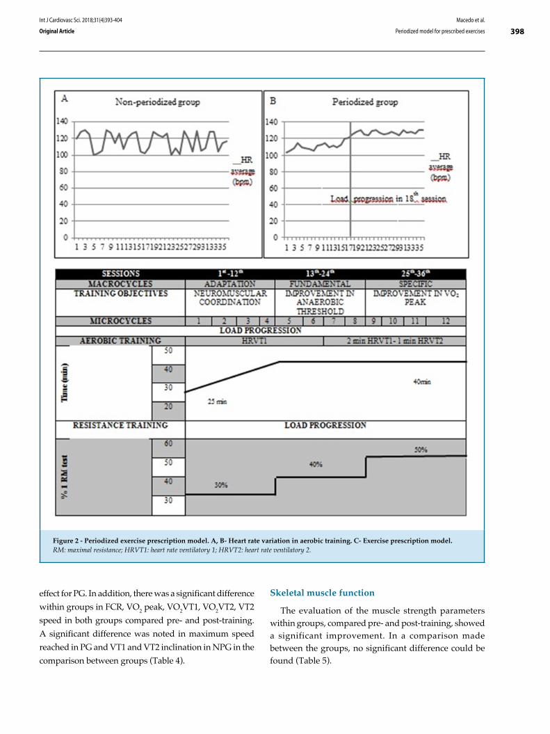
398Macedo et al.
Periodized model for prescribed exercises
Int J Cardiovasc Sci. 2018;31(4)393-404
Original Article
Figure 2 - Periodized exercise prescription model. A, B- Heart rate variation in aerobic training. C- Exercise prescription model.RM: maximal resistance; HRVT1: heart rate ventilatory 1; HRVT2: heart rate ventilatory 2.
effect for PG. In addition, there was a significant difference
within groups in FCR, VO2 peak, VO2VT1, VO2VT2, VT2
speed in both groups compared pre- and post-training.
A significant difference was noted in maximum speed
reached in PG and VT1 and VT2 inclination in NPG in the
comparison between groups (Table 4).
Skeletal muscle function
The evaluation of the muscle strength parameters within groups, compared pre- and post-training, showed a significant improvement. In a comparison made between the groups, no significant difference could be found (Table 5).
399Macedo et al.
Periodized model for prescribed exercises
Int J Cardiovasc Sci. 2018;31(4)393-404
Original Article
Table 2 - Baseline characteristics of the study population
Sample characteristic PG NPG
n (men) 28 (100%) 32 (100%)
Age ± SD, years 55.89 ± 8.2 62.4 ± 11.8
Left ventricular ejection
fraction, ** %65.57 ± 5.5 66.09 ± 5.7
Body mass index 28.2 ± 3.5 28.9 ± 4.4
Abdominal circumference 100.7 ± 9.0 101.0 ± 10.6
Positive family history /
cardiovascular disease20 (71.5%) 24 (80%)
Dyslipidemia 27 (96.7%) 32 (100%)
Obesity 12 (42.8%) 22 (68.75%)
Sedentary life style 22 (78.5%) 25 (78.1%)
Stress 25 (89.2.8%) 22 (68.75%)
Smoking habit 4 (14.2%) 8 (25%)
Diabetes mellitus 5 (17.8%) 3 (9.3%)
High blood pressure 12 (42.8%) 15 (46.8%)
Stratification of risk for exercises
Low risk 21 (75%) 25 (78.1%)
Moderate risk 8 (21.6%) 3 (8.1%)
Anatomic location of injuries
Right coronary 5 (14.2%)
Posterior descending 0
Left main coronary 2 (7.1%)
Anterior descending 18 (64.2%)
Diagonal 5 (17.8%)
Circumflex 6 (21.4%)
Marginal 0
Arteries with stent implants
Right coronary
1 stent 4 (14.2%)
2 stents 1 (3.5%)
3 stents 0
Posterior descending
1 stent 1 (3.5%)
Left main coronary
1 stent 2 (7.1%)
Anterior descending
1 stent 16 (57.1%)
Diagonal
1 stent 1 (2.7%)
Circumflex
1 stent 6 (21.4%)
2 stents 6 (21.4%)
Marginal
1 stent 0
Incomplete revascularization of
myocardium 1 (3.5%)
Prior surgery for
revascularization of
myocardium
2 (7.1%)
Prior angioplasties 5 (14.2%)
Medications,† %
Antiplatelet agents 28 (100%)
Anticoagulant 28 (100%)
Antihypertensive 13 (46.4%)
Beta-blockers 26 (92.8%)
PG: periodized exercise training group. NPG: non-periodized exercise training group; ** Obtained by transthoracic echocardiography; † Standard dose of medication.
Discussion
This study ascertained the following outcomes: superior improvement of body fat, fat above ideal and body mass, VO2 peak and VO2 at VT1-2 in the PG; muscle strength improvement in both groups. Periodization training is suggested in most recent guidelines.5-8 However, the superiority of periodized training (RT and AT) has been poorly studied in CAD patients.
The main finding of this study was that the periodized exercise prescription program was superior to the conventional one with respect to the increase in VO2 peak for coronary patients taking part in a rehabilitation program. VO2 peak is closely associated with morbidity and mortality in cardiac patients.18 This information is very important since periodization is still not in the rehabilitation programs of CAD patients.19 In addition, VO2 peak is recognized as being the best indicator of survival for this population.20-22 Therefore, the inclusion
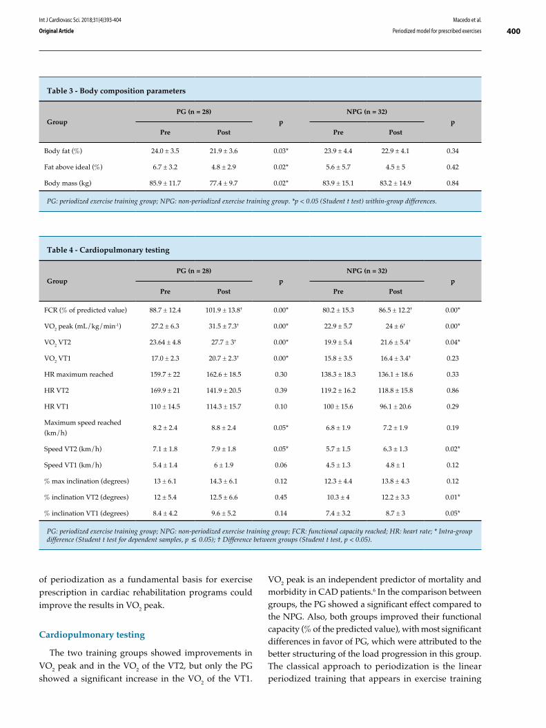
400Macedo et al.
Periodized model for prescribed exercises
Int J Cardiovasc Sci. 2018;31(4)393-404
Original Article
Table 3 - Body composition parameters
Group
PG (n = 28)
p
NPG (n = 32)
p
Pre Post Pre Post
Body fat (%) 24.0 ± 3.5 21.9 ± 3.6 0.03* 23.9 ± 4.4 22.9 ± 4.1 0.34
Fat above ideal (%) 6.7 ± 3.2 4.8 ± 2.9 0.02* 5.6 ± 5.7 4.5 ± 5 0.42
Body mass (kg) 85.9 ± 11.7 77.4 ± 9.7 0.02* 83.9 ± 15.1 83.2 ± 14.9 0.84
PG: periodized exercise training group; NPG: non-periodized exercise training group. *p < 0.05 (Student t test) within-group differences.
Table 4 - Cardiopulmonary testing
Group
PG (n = 28)
p
NPG (n = 32)
p
Pre Post Pre Post
FCR (% of predicted value) 88.7 ± 12.4 101.9 ± 13.8† 0.00* 80.2 ± 15.3 86.5 ± 12.2† 0.00*
VO2 peak (mL/kg/min-1) 27.2 ± 6.3 31.5 ± 7.3† 0.00* 22.9 ± 5.7 24 ± 6† 0.00*
VO2 VT2 23.64 ± 4.8 27.7 ± 3† 0.00* 19.9 ± 5.4 21.6 ± 5.4† 0.04*
VO2 VT1 17.0 ± 2.3 20.7 ± 2.3† 0.00* 15.8 ± 3.5 16.4 ± 3.4† 0.23
HR maximum reached 159.7 ± 22 162.6 ± 18.5 0.30 138.3 ± 18.3 136.1 ± 18.6 0.33
HR VT2 169.9 ± 21 141.9 ± 20.5 0.39 119.2 ± 16.2 118.8 ± 15.8 0.86
HR VT1 110 ± 14.5 114.3 ± 15.7 0.10 100 ± 15.6 96.1 ± 20.6 0.29
Maximum speed reached
(km/h)8.2 ± 2.4 8.8 ± 2.4 0.05* 6.8 ± 1.9 7.2 ± 1.9 0.19
Speed VT2 (km/h) 7.1 ± 1.8 7.9 ± 1.8 0.05* 5.7 ± 1.5 6.3 ± 1.3 0.02*
Speed VT1 (km/h) 5.4 ± 1.4 6 ± 1.9 0.06 4.5 ± 1.3 4.8 ± 1 0.12
% max inclination (degrees) 13 ± 6.1 14.3 ± 6.1 0.12 12.3 ± 4.4 13.8 ± 4.3 0.12
% inclination VT2 (degrees) 12 ± 5.4 12.5 ± 6.6 0.45 10.3 ± 4 12.2 ± 3.3 0.01*
% inclination VT1 (degrees) 8.4 ± 4.2 9.6 ± 5.2 0.14 7.4 ± 3.2 8.7 ± 3 0.05*
PG: periodized exercise training group; NPG: non-periodized exercise training group; FCR: functional capacity reached; HR: heart rate; * Intra-group difference (Student t test for dependent samples, p ≤ 0.05); † Difference between groups (Student t test, p < 0.05).
of periodization as a fundamental basis for exercise prescription in cardiac rehabilitation programs could improve the results in VO2 peak.
Cardiopulmonary testing
The two training groups showed improvements in VO2 peak and in the VO2 of the VT2, but only the PG showed a significant increase in the VO2 of the VT1.
VO2 peak is an independent predictor of mortality and morbidity in CAD patients.6 In the comparison between groups, the PG showed a significant effect compared to the NPG. Also, both groups improved their functional capacity (% of the predicted value), with most significant differences in favor of PG, which were attributed to the better structuring of the load progression in this group. The classical approach to periodization is the linear periodized training that appears in exercise training
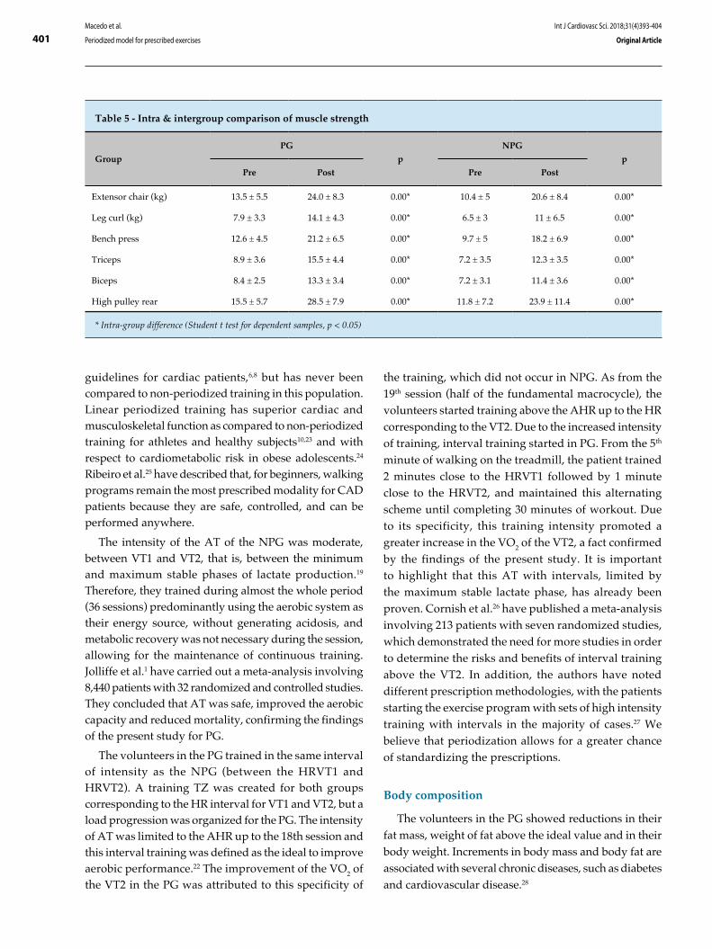
401Macedo et al.
Periodized model for prescribed exercises
Int J Cardiovasc Sci. 2018;31(4)393-404
Original Article
Table 5 - Intra & intergroup comparison of muscle strength
Group
PG
p
NPG
p
Pre Post Pre Post
Extensor chair (kg) 13.5 ± 5.5 24.0 ± 8.3 0.00* 10.4 ± 5 20.6 ± 8.4 0.00*
Leg curl (kg) 7.9 ± 3.3 14.1 ± 4.3 0.00* 6.5 ± 3 11 ± 6.5 0.00*
Bench press 12.6 ± 4.5 21.2 ± 6.5 0.00* 9.7 ± 5 18.2 ± 6.9 0.00*
Triceps 8.9 ± 3.6 15.5 ± 4.4 0.00* 7.2 ± 3.5 12.3 ± 3.5 0.00*
Biceps 8.4 ± 2.5 13.3 ± 3.4 0.00* 7.2 ± 3.1 11.4 ± 3.6 0.00*
High pulley rear 15.5 ± 5.7 28.5 ± 7.9 0.00* 11.8 ± 7.2 23.9 ± 11.4 0.00*
* Intra-group difference (Student t test for dependent samples, p < 0.05)
guidelines for cardiac patients,6,8 but has never been compared to non-periodized training in this population. Linear periodized training has superior cardiac and musculoskeletal function as compared to non-periodized training for athletes and healthy subjects10,23 and with respect to cardiometabolic risk in obese adolescents.24 Ribeiro et al.25 have described that, for beginners, walking programs remain the most prescribed modality for CAD patients because they are safe, controlled, and can be performed anywhere.
The intensity of the AT of the NPG was moderate, between VT1 and VT2, that is, between the minimum and maximum stable phases of lactate production.19 Therefore, they trained during almost the whole period (36 sessions) predominantly using the aerobic system as their energy source, without generating acidosis, and metabolic recovery was not necessary during the session, allowing for the maintenance of continuous training. Jolliffe et al.1 have carried out a meta-analysis involving 8,440 patients with 32 randomized and controlled studies. They concluded that AT was safe, improved the aerobic capacity and reduced mortality, confirming the findings of the present study for PG.
The volunteers in the PG trained in the same interval of intensity as the NPG (between the HRVT1 and HRVT2). A training TZ was created for both groups corresponding to the HR interval for VT1 and VT2, but a load progression was organized for the PG. The intensity of AT was limited to the AHR up to the 18th session and this interval training was defined as the ideal to improve aerobic performance.22 The improvement of the VO2 of the VT2 in the PG was attributed to this specificity of
the training, which did not occur in NPG. As from the 19th session (half of the fundamental macrocycle), the volunteers started training above the AHR up to the HR corresponding to the VT2. Due to the increased intensity of training, interval training started in PG. From the 5th minute of walking on the treadmill, the patient trained 2 minutes close to the HRVT1 followed by 1 minute close to the HRVT2, and maintained this alternating scheme until completing 30 minutes of workout. Due to its specificity, this training intensity promoted a greater increase in the VO2 of the VT2, a fact confirmed by the findings of the present study. It is important to highlight that this AT with intervals, limited by the maximum stable lactate phase, has already been proven. Cornish et al.26 have published a meta-analysis involving 213 patients with seven randomized studies, which demonstrated the need for more studies in order to determine the risks and benefits of interval training above the VT2. In addition, the authors have noted different prescription methodologies, with the patients starting the exercise program with sets of high intensity training with intervals in the majority of cases.27 We believe that periodization allows for a greater chance of standardizing the prescriptions.
Body composition
The volunteers in the PG showed reductions in their fat mass, weight of fat above the ideal value and in their body weight. Increments in body mass and body fat are associated with several chronic diseases, such as diabetes and cardiovascular disease.28

402Macedo et al.
Periodized model for prescribed exercises
Int J Cardiovasc Sci. 2018;31(4)393-404
Original Article
Studies have shown that moderate AT promotes an improvement in body composition.29,30 This was important because obesity is considered to be an important modifiable cardiovascular risk factor.31 A simple improvement in food habits is not sufficient for a rapid and appropriate decrease in fat mass. Therefore, the physical exercise association was fundamental for body weight decrease and long-term maintenance of these changes.31 Studies28,32,33 have recognized aerobic exercise as the most suitable form of training by providing positive effects on glucose and lipids and decrease on body fat and the strength exercises. Inoue et al.24 have shown that the association of strength and AT was more effective than only AT to improve lipid profile and insulin resistance sensitivity on obese adolescents.
The improvement in aerobic capacity or exercise tolerance results in a greater consumption of calories to maintain the activity and, consequently, burning more fat.24 Lira et al.34 have studied the effects of intensity and type of exercise on lipoprotein profiles and highlighted the higher energy expenditure achieved by associating volume and intensity. This fact justifies the finding that the PG, with its greater cardiopulmonary evolution and tolerance to exercise, presented greater body fat decrease. This is because improving the aerobic capacity increases the caloric expenditure per session, since the patient is walking more within a same time interval.
Skeletal muscle function
Both training groups presented a significant improvement in strength after the training period. In this case, the PG showed no advantage.
During MAD the patients worked with loads equivalent to 30% of the maximum determined in the 1RM test, in the MFU, with 40%, and in the MSP, with 50%. This organized progression of the load was not more effective than the random progression used for the NPG. This could be attributed to the fact that these low training loads did not recruit different energy sources and/or types of muscle fibers. In addition, in the first 12 weeks of training, the increase in strength occurs due to neural adaptation and not to hypertrophy, which is independent of the load.8,11 The increase in strength noted in both groups could have contributed to the improvement in the VO2 peak, in the walking speed and in the inclination reached during the treadmill test.27
Therefore, the training study is extremely important to both athletes, to reach high performance, and patients,
such as those with heart disease, to reduce the risk of mortality, which has great social relevance.
Conclusion
The present study showed that, within the cardiac rehabilitation programs for coronary disease patients, periodization of the training can improve the results as compared to the conventional model, when considering the following variables: VO2 peak, VO2 for the VT2, VO2 for the VT1, %fat and body weight. These findings are very important for future studies involving physical training and cardiac rehabilitation.
We believe that, at the present moment, before evolving into comparative studies between continuous exercises of moderate intensity versus high intensity exercises with intervals, periodization should be included as a prescription tool aimed at improving the results of the intervention or treatment of those with coronary disease with physical exercise.
Study limitations
Some of the study limitations were the small size of the sample and not using the Faulkner protocol to evaluate body composition. In addition, inflammatory biomarkers, oxidative stress analysis and drug reduction for hypertension were not performed.
Author contributions
Conception and design of the research and analysis and interpretation of the data: Macedo RM; Acquisition of data: Macedo RM, Sebastião Neto F; Statistical analysis: Macedo RM, Macedo ACB, Olandoski M; Obtaining financing:Macedo RM, Guarita-Souza LC, Silveira RP; Writing of the manuscript: Macedo RM, Macedo ACB, Faria-Neto JR, Costantini CR, Costantini CO, Guarita-Souza LC, Carvalho KAT; Critical revision of the manuscript for intellectual content: Macedo RM, Macedo ACB, Faria-Neto JR, Guarita-Souza LC.
Potential Conflict of Interest
No potential conflict of interest relevant to this article was reported.
Sources of Funding
There were no external funding sources for this study.

403Macedo et al.
Periodized model for prescribed exercises
Int J Cardiovasc Sci. 2018;31(4)393-404
Original Article
1. Joliffe JA, Rees K, Taylor RS, Thompson D, Oldridge N, Ebrahim S. Exercise-based rehabilitation for coronary artery disease. Cochrane Database Syst Rev. 2001;(1):CD001800.
2. Mansur Ade P, Favarato D. Mortality due to cardiovascular diseases in Brazil and in the metropolitan region of São Paulo: a 2011 update. Arq Bras Cardiol. 2012, 99(2):755-61.
3. Sociedade Brasileira de Cardiologia. [Guidelines for cardiopulmonary and metabolic rehabilitation: practical aspects]. Arq Bras Cardiol. 2006;86(1):74-82.
4. Anderson L, Oldridge N, Thompson DR, Zwisler AD, Rees K, Martin N, et al. Exercise-based cardiac rehabilitation for coronary artery disease: Cochrane systematic review and meta-analysis. J Am Coll Cardiol. 2016;67(1):1-12.
5. Balady GJ, Williams MA, Ades PA, Bittner V, Comoss P, Foody JM, et al; merican Heart Association Exercise, Cardiac Rehabilitation, and Prevention Committee, the Council on Clinical Cardiology; American Heart Association Council on Cardiovascular Nursing; American Heart Association Council on Epidemiology and Prevention; American Heart Association Council on Nutrition, Physical Activity, and Metabolism; American Association of Cardiovascular and Pulmonary Rehabilitation. Core components of cardiac rehabilitation/secondary prevention programs: 2007 update a scientific statement from the American Artery Association exercise, cardiac rehabilitation, and prevention committee, the council on clinical cardiology; the councils on cardiovascular nursing, epidemiology and prevention, and nutrition, physical activity, and metabolism; and the American Association of Cardiovascular and Pulmonary Rehabilitation. Circulation. 2007;115(20):2675-82.
6. Vanhees L, Geladas N, Hansen D, Kouidi E, Niebauer J, Reiner Z, et al. Importance of characteristics and modalities of physical activity and exercise in the management of cardiovascular health in individuals with cardiovascular risk factors: recommendations from the EACPR (Part II). Eur J Prev Cardiol. 2012;19(5):1005-33.
7. Fletcher GF, Ades PA, Kligfield P, Arena R, Balady GJ, Bittner VA, et al; American Heart Association Exercise, Cardiac Rehabilitation, and Prevention Committee of the Council on Clinical Cardiology, Council on Nutrition, Physical Activity and Metabolism, Council on Cardiovascular and Stroke Nursing, and Council on Epidemiology and Prevention. Exercise standards for testing and training: a statement for healthcare professionals from the American Artery Association. Circulation. 2013;128(8):873-934.
8. Piepoli MF, Conraads V, Corra U, Dickstein K, Francis DP, Jaarsma T, et al. Exercise training in artery failure: from theory to practice. A consensus document of the Artery Failure Association and the European Association for Cardiovascular Prevention and Rehabilitation. Eur J Heart Fail. 2011;13(4):347-57.
9. Piepoli MF, Corra U, Benzer W, Bjarnason-Wehrens B, Dendale P, Gaita D, et al; European Association of Cardiovascular Prevention and Rehabilitation Committee for Science Guidelines. Secondary prevention through cardiac rehabilitation: physical activity counseling and exercise training: key components of the position paper from the Cardiac Rehabilitation Section of the European Association of Cardiovascular Prevention and Rehabilitation. Eur Heart J. 2010;31(16):1967-74.
10. American College of Sports Medicine. Resource manual for guidelines for exercise testing and prescription. 9th. Baltimore: Williams and Wilkins; 2013. p. 34-6.
11. Bompa T. Periodization in sports training. São Paulo: Manole; 2002.
12. Gomes AC. Sports training, structuring and periodization. 2a ed. Porto Alegre: Artmed; 2009.
13. Macedo RM. Cardiorespiratory physiotherapy. a new concept for the treatment in the hospital phase. Curitiba (PR): Juruá; 2012.
14. Macedo RM, Faria-Neto JR, Costantini CO, Casali D, Muller AP, Costantini CR, et al. Phase I of cardiac rehabilitation: a new challenge for evidence based physiotherapy. World J Cardiol. 2011;3(7):248-55.
15. de Macedo RM, Faria-Neto JR, Costantini CO, Olandoski M, Casali D, de Macedo AC, et al. A periodized model for exercise improves the intra-hospital evolution of patients after myocardial revascularization: a pilot randomized controlled trial Clin Rehabil. 2012:26(11):982-9.
16. Guidelines for Cardiac Rehabilitation and Secondary Prevention Programs. 5th ed. Champaign (Ill): Human Kinetics; 2013.
17. Faulkner JA. Physiology of swimming and diving. Exercise Physiology. Baltimore: Academic Press; 1968.
18. Cahalin LP, Chase P, Arena R, Myers J, Bensimhon D, Peberdy MA, et al. A meta-analysis of the prognostic significance of cardiopulmonary exercise testing in patients with artery failure. Heart Fail Rev. 2013;18(1):79-94.
19. Sociedade Brasileira de Cardiologia. [Guidelines of cardiac rehabilitation]. Arq Bras Cardiol. 2005;84(5):431-40.
20. Williams MA, Haskell WL, Ades PA, Amsterdam EA, Bittner V, Franklin BA, et al; American Heart Association Council on Clinical Cardiology; American Heart Association Council on Nutrition, Physical Activity, and Metabolism. Resistance exercise in individuals with and without cardiovascular disease: 2007 update: a scientific statement from the American Heart Association Council on Clinical Cardiology and Council on Nutrition, Physical Activity, and Metabolism. Circulation. 2007;116(5):572-84.
21. Wisloff U, Stoylen A, Loennechen JP, Bruvold M, Rognmo O, Haram P, et al. Superior cardiovascular effect of aerobic interval training versus moderate continuous training in artery failure patients: a randomized study. Circulation. 2007;115(24):3086-94.
22. Myers J, Prakash M, Froelicher V, Do D, Partington S, Atwood JE. Exercise capacity and mortality among men referred for exercise testing. N Eng J Med. 2002;346(11):793-801.
23. Buchheit M, Laursen PB. High-intensity interval training, solutions to the programming puzzle: Part I: cardiopulmonary emphasis. Sports Med. 2013;43(5):313-38.
24. Inoue DS, De Mello MT, Foschini D, Lira FS, De Piano Ganen A, Da Silveira Campos RM, et al. Linear and undulating periodized strengh plus aerobic training promote similar benefits and lead to improvement of insulin resistance on obese adolescents. J Diabetes Complications. 2015;29(2):258-64.
References
Study Association
This article is part of the thesis of Post Doctoral submitted by Rafael Michel de Macedo, from Pontifícia Universidade Católica do Paraná.
Ethics approval and consent to participate
This study was approved by the Ethics Committee of the Pontifícia Universidade Católica do Paraná under the protocol number 434/2010. All the procedures in this study were in accordance with the 1975 Helsinki Declaration, updated in 2013. Informed consent was obtained from all participants included in the study.

404Macedo et al.
Periodized model for prescribed exercises
Int J Cardiovasc Sci. 2018;31(4)393-404
Original Article
This is an open-access article distributed under the terms of the Creative Commons Attribution License
25. Ribeiro PA, Boidin M, Juneau M, Nigam A, Gayda M. High-intensity interval traning in patients with coronary artery disease: prescription models and perspectives. Rehab. Ann Phys Rehabil Med. 2017;60(1):50-7.
26. Cornish AK, Broadbent S, Cheema BS. Interval training for patients with coronary artery disease: a systematic review. Eur J Appl Physiol. 2011,111(4):579-89.
27. Marchionni N, Fattirolli F, Fumagalli S, Oldridge N, Del Lungo F, Morosi L, et al. Patients after myocardial infarction: results of a randomized, controlled trial improved exercise tolerance and quality of life with cardiac rehabilitation of older. Circulation. 2003,107(17):2201-6.
28. Ahmadizad S, Ghorbani S, Ghasemikaram M, Bahmanzadeh M. Effects of short-term nonperiodized, linear periodized and daily undulating periodized resistance training on plasma adiponectin, leptin and insulin resistance. Clin Biochem. 2014;47(6):417-22.
29. Sanches PL, de Mello MT, Elias N, Fonseca FA, Campos RM. Hyperleptinemia; implications on the inflammatory state and vascular protection in obese adolescents submitted to an interdisciplinary therapy. Inflammation. 2014:37(1):35-43.
30. Willund KR, Feeney LA, Tomayko EJ, Weiss EP, Hagberg JM. Effects of endurance exercise training on markers of cholesterol absorption and synthesis. Physiol Res. 2009;58(4):545-52.
31. Thompson PD, Buchner D, Piña IL, Balady GJ, Williams MA, Marcua BH, et al; American Heart Association Council on Clinical Cardiology Subcommittee on Exercise, Rehabilitation, and Prevention; American Heart Association Council on Nutrition, Physical Activity, and Metabolism Subcommittee on Physical Activity.) Exercise and physical activity in the prevention and treatment of atherosclerotic cardiovascular disease: a statement from the Council on Clinical Cardiology (Subcommittee on Exercise, Rehabilitation, and Prevention) and the Council on Nutrition, Physical Activity, and Metabolism (Subcommittee on Physical Activity). Circulation. 2003;107(24):3109-16.
32. Marzolini S, Oh PI, Thomas SG, Goodmann JM. Aerobic and resistance training in coronary disease: single versus multiple sets. Med Sci Sports Exerc. 2008;40(9):1557-64.
33. Cauza E, Hanusch-Enserer U, Strasser B, Ludvik B, Metz-Schimmerl S, Pacini G, et al. The relative benefits of endurance and strength training on the metabolic factors and muscle function of people with type 2 diabetes mellitus. Arch Phys Med Rehabil. 2005;86(9):1527-33.
34. Lira FS, Carnevali LC Jr, Zanchi NE, Santos RV, Lavoie JM, Seelaender M. Exercise intensity modulation of hepatic lipid metabolism. J Nutr Metab. 2012;2012:809576.

DOI: 10.5935/2359-4802.20180031
405
ORIGINAL ARTICLE
International Journal of Cardiovascular Sciences. 2018;31(4)405-413
Mailing Address: Rafael de Oliveira AlvimRua Alaor de Queiroz Araújo, 135. Postal Code: 29050-245, Enseada do Suá, Vitória (ES) - Brazil.E-mail: [email protected]
Prevalence of Peripheral Artery Disease and Associated Risk Factors in a Brazilian Rural Population: The Baependi Heart StudyRafael de Oliveira Alvim,1 Fernando Augusto Lavezzo Dias,2 Camila Maciel de Oliveira,2 Andréa Roseli Vançan Russo Horimoto,3 Anderson Zampier Ulbrich,2 José Eduardo Krieger,3 Alexandre da Costa Pereira3
Universidade Federal do Espírito Santo (UFES),1 Vitória, ES - BrazilUniversidade Federal do Paraná (UFPR),2 Curitiba, PR - BrazilInstituto do Coração (InCor) - Faculdade de Medicina da Universidade de São Paulo,3 São Paulo, SP - Brazil
Manuscript received July 26, 2016, revised manuscript September 28, 2017, accepted October 15, 2017.
Abstract
Background: The identification of peripheral artery disease (PAD) can help prevent further progression of the disease and additional complications, considering that this condition is a risk factor for all-cause mortality and cardiovascular death.
Objective: To assess the prevalence of PAD in the Baependi Heart Study and investigate associated risk factors in different age groups.
Methods: A total of 1,627 individuals (of both genders and aged 18 - 102 years) residing in the municipality of Baependi (Minas Gerais, Brazil) were selected for this study. Anthropometric and biochemical parameters were evaluated by standard techniques. Physical activity level was determined by the International Physical Activity Questionnaire - Short Form (IPAQ-SF). The screening of PAD was performed by determination of the ankle-brachial index (ABI). The level of statistical significance was set at 5%.
Results: In the overall sample, the prevalence of PAD was 1.05%, and reached 5.2% after the age of 70 years. The frequency and intensity of smoking were higher in individuals with PAD. A prior history of myocardial infarction and a higher prevalence of hypertension, diabetes, obesity, and sedentary lifestyle were also associated with PAD. In addition, PAD was more frequent in blacks than whites. In multivariable analysis, age, diabetes, smoking, and physical inactivity remained independently associated with PAD.
Conclusion: The prevalence of PAD was low and increased clearly with age in our sample from a Brazilian rural population. Furthermore, the main risk factors for PAD in the investigated sample were smoking, sedentary lifestyle, diabetes mellitus, and age. (Int J Cardiovasc Sci. 2018;31(4)405-413)
Keywords: Peripheral Arterial Disease / prevalence; Risk Factors; Rural Population; Tobacco Use Disorder; Ankle-Brachial Index.
Introduction
Peripheral artery disease (PAD) is classically defined as a condition affecting noncardiac, nonintracranial arteries, majorly due to atherosclerosis, that leads to partial obstruction of the peripheral arteries, reducing perfusion to the tissues irrigated by these arteries.1,2 Even though PAD is asymptomatic in most patients, the disease may progress with clinical symptoms (like
claudication) and eventually lead to tissue necrosis. The identification of PAD can help prevent further progression of the disease itself and additional complications, considering that this condition is a risk factor for all-cause mortality3-8 and cardiovascular death,3,4,7,9 including coronary artery disease3,7 and stroke.5,7 PAD is also a major cause of quality of life impairment, which in turn, worsen even more with the progression of the disease.10,11 Therefore, information
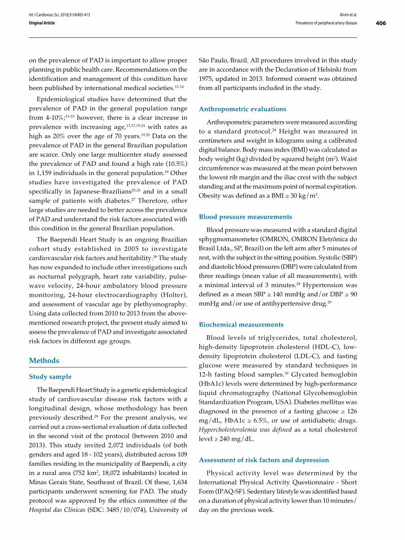
406Alvim et al.
Prevalence of peripheral artery disease
Int J Cardiovasc Sci. 2018;31(4)405-413
Original Article
on the prevalence of PAD is important to allow proper planning in public health care. Recommendations on the identification and management of this condition have been published by international medical societies.12-14
Epidemiological studies have determined that the prevalence of PAD in the general population range from 4-10%;15-23 however, there is a clear increase in prevalence with increasing age,13,17,19,24 with rates as high as 20% over the age of 70 years.19,20 Data on the prevalence of PAD in the general Brazilian population are scarce. Only one large multicenter study assessed the prevalence of PAD and found a high rate (10.5%) in 1,159 individuals in the general population.18 Other studies have investigated the prevalence of PAD specifically in Japanese-Brazilians25,26 and in a small sample of patients with diabetes.27 Therefore, other large studies are needed to better access the prevalence of PAD and understand the risk factors associated with this condition in the general Brazilian population.
The Baependi Heart Study is an ongoing Brazilian cohort study established in 2005 to investigate cardiovascular risk factors and heritability.28 The study has now expanded to include other investigations such as nocturnal polygraph, heart rate variability, pulse-wave velocity, 24-hour ambulatory blood pressure monitoring, 24-hour electrocardiography (Holter), and assessment of vascular age by plethysmography. Using data collected from 2010 to 2013 from the above-mentioned research project, the present study aimed to assess the prevalence of PAD and investigate associated risk factors in different age groups.
Methods
Study sample
The Baependi Heart Study is a genetic epidemiological study of cardiovascular disease risk factors with a longitudinal design, whose methodology has been previously described.28 For the present analysis, we carried out a cross-sectional evaluation of data collected in the second visit of the protocol (between 2010 and 2013). This study invited 2,072 individuals (of both genders and aged 18 - 102 years), distributed across 109 families residing in the municipality of Baependi, a city in a rural area (752 km2, 18,072 inhabitants) located in Minas Gerais State, Southeast of Brazil. Of these, 1,634 participants underwent screening for PAD. The study protocol was approved by the ethics committee of the Hospital das Clínicas (SDC: 3485/10/074), University of
São Paulo, Brazil. All procedures involved in this study are in accordance with the Declaration of Helsinki from 1975, updated in 2013. Informed consent was obtained from all participants included in the study.
Anthropometric evaluations
Anthropometric parameters were measured according to a standard protocol.28 Height was measured in centimeters and weight in kilograms using a calibrated digital balance. Body mass index (BMI) was calculated as body weight (kg) divided by squared height (m2). Waist circumference was measured at the mean point between the lowest rib margin and the iliac crest with the subject standing and at the maximum point of normal expiration. Obesity was defined as a BMI ≥ 30 kg/m2.
Blood pressure measurements
Blood pressure was measured with a standard digital sphygmomanometer (OMRON, OMRON Eletrônica do Brasil Ltda., SP, Brazil) on the left arm after 5 minutes of rest, with the subject in the sitting position. Systolic (SBP) and diastolic blood pressures (DBP) were calculated from three readings (mean value of all measurements), with a minimal interval of 3 minutes.28 Hypertension was defined as a mean SBP ≥ 140 mmHg and/or DBP ≥ 90 mmHg and/or use of antihypertensive drug.29
Biochemical measurements
Blood levels of triglycerides, total cholesterol, high-density lipoprotein cholesterol (HDL-C), low-density lipoprotein cholesterol (LDL-C), and fasting glucose were measured by standard techniques in 12-h fasting blood samples.30 Glycated hemoglobin (HbA1c) levels were determined by high-performance liquid chromatography (National Glycohemoglobin Standardization Program, USA). Diabetes mellitus was diagnosed in the presence of a fasting glucose ≥ 126 mg/dL, HbA1c ≥ 6.5%, or use of antidiabetic drugs. Hypercholesterolemia was defined as a total cholesterol level ≥ 240 mg/dL.
Assessment of risk factors and depression
Physical activity level was determined by the International Physical Activity Questionnaire - Short Form (IPAQ-SF). Sedentary lifestyle was identified based on a duration of physical activity lower than 10 minutes/day on the previous week.

407Alvim et al.
Prevalence of peripheral artery disease
Int J Cardiovasc Sci. 2018;31(4)405-413
Original Article
Information regarding medical history (angina pectoris, myocardial infarction, stroke, kidney failure, and depression) and environmental risk factors such as smoking and alcohol use were evaluated through a questionnaire completed by each participant. The questionnaire was based on the World Health Organization’s Multinational Monitoring Trends and Determinants in Cardiovascular Disease (MONICA)31 project epidemiological instrument, and was applied and filled out by research assistants specifically trained for this task.
Screening of peripheral artery disease
The screening of PAD was performed with the ankle-brachial index (ABI), which was measured by a single trained examiner using a sphygmomanometer (Heidji, Brazil) with a cuff suitable for the circumference of the limbs and a 10 MHz portable Doppler device (DV 610B, MEDMEGA, SP, Brazil).
The ABI was determined for each leg by the ratio between the highest SBP obtained at the ankle (posterior tibial and dorsalis pedis arteries) and highest SBP obtained in the arms (brachial artery).12-14 The methodology of the test and the ABI classification were based on recommendations of the American College of Cardiology / American Heart Association (ACC/AHA).12,13 ABI values between 0.91 and 1.39 were considered normal. Values ≤ 0.90 were considered compatible with PAD and values ≥ 1.40 were considered inconclusive for PAD and were excluded from the analysis.
Statistical analysis
Categorical variables were compared using the chi-square test and are presented as percentage, while continuous variables are presented as mean ± standard deviation. The normality of the data was confirmed with the Kolmogorov-Smirnov test. Unpaired Student t test was performed to analyze demographic, hemodynamic, and biochemical data according to PAD status. Since the cutoff value for PAD diagnosis based on ABI is well-established in the literature, we carried out univariate and multivariate logistic regression analyses to determine the association between PAD (ABI < 0.9) as the dependent variable and age, hypertension, diabetes, myocardial infarction, smoking, and sedentary lifestyle as predictor variables. Statistical analyses were carried out using SPSS
(version 19) software (Chicago, IL, USA), with the level of significance set at 5%.
Results
A total of 1,634 individuals were screened for PAD. Seven individuals presented an ABI above 1.4 and were excluded from the analysis. Therefore, 1,627 volunteers were included in the study. The age of the participants ranged from 18 to 102 years (mean 44.9 ± 16.4 years). Table 1 shows the demographic, anthropometric, biochemical, and hemodynamic characteristics of the individuals with and without PAD, defined as an ABI equal to or lower than 0.9. Age, BMI, HbA1c, and SBP were higher in volunteers with PAD. The presence of PAD was also more frequent in elderly compared with younger individuals, and in blacks compared with whites.
Figure 1 presents the data related to the prevalence of PAD using the ABI in different age groups. Overall, the prevalence was very low (1.05%). Only one case of PAD was observed below the age of 30 years, and the prevalence of PAD increased after the fifth decade, peaking at the age of 70 years, when it reached 5.2%. The frequency of PAD by decade is presented in the Table 2.
Table 3 presents the data related to the lifestyle characteristics of the volunteers. The frequency and amount of smoking were higher in individuals with PAD. There was also a higher frequency of physically inactive volunteers in the PAD group.
Table 4 presents a comparison of clinical characteristics in individuals with and without PAD. A higher prevalence of hypertension, diabetes, and obesity was observed in individuals with a diagnosis of PAD. The presence of hypercholesterolemia was not different between groups. Also, a prior history of myocardial infarction was more frequent in the PAD group.
Table 5 presents univariate and multivariate logistic regression models for PAD. In multivariate analysis, age, diabetes, smoking, and physical inactivity were significantly and independently associated with PAD.
Discussion
In terms of the number of individuals included, the present study is the largest investigation of the prevalence of PAD in a Brazilian population. The Baependi Heart Study is a Brazilian cohort study investigating cardiovascular risk factors and heritability in residents of
408Alvim et al.
Prevalence of peripheral artery disease
Int J Cardiovasc Sci. 2018;31(4)405-413
Original Article
Table 1 - Demographic, anthropometric, biochemical, and hemodynamic characteristics of individuals with and without peripheral artery disease
Variables Total PAD present PAD absent P value
n 1,627 17 1,610 ***
Age (years) 44.9 ± 16.4 66 ± 15 45 ± 16 < 0.001
Gender
Men (%) 41.5 41.2 41.50.59
Women (%) 58.5 58.8 58.5
Ethnicity
White (%) 76.7 70.6 76.8
0.001Black (%) 5.5 29.4 5.2
Mulatto (%) 17.5 0 17.7
Others (%) 0.3 0 0.4
ABI (ratio) 1.11 ± 0.1 0.78 ± 0.11 1.12 ± 0.7 < 0.001
BMI (kg/m2) 25.8 ± 5.1 28.7 ± 5.3 25.7 ± 5.1 0.02
WC (cm) 91 ±12 100 ± 10 91 ± 12 0.003
TC (mg/dL) 197.8 ± 40.7 195.6 ± 59.9 197.9 ± 40.5 0.83
LDL-c (mg/dL) 124.5 ± 35.4 122.2 ± 56.3 124.6 ± 35.2 0.79
HDL-c (mg/dL) 47.2 ± 11.7 45.2 ± 13.1 47.2 ± 11.8 0.49
Triglycerides (mg/dL) 130.3 ± 68.1 141.1 ± 60.0 130.2 ± 68.2 0.52
Fasting glucose (mg/dL) 92.6 ± 19.0 114.6 ± 43.2 92.3 ± 18.5 0.06
HbA1c (%) 5.7 ± 0.7 6.5 ± 1.2 5.7 ± 0.8 0.02
SBP (mmHg) 125.4 ± 16.3 133.5 ± 19.2 125.4 ± 16.2 0.04
DBP (mmHg) 76.2 ± 10.3 75.3 ± 9.2 76.2 ± 10.4 0.72
Categorical variables were compared using the chi-square test, and continuous variables were compared using Student t test. PAD: peripheral artery disease; n: sample size; ABI: ankle-brachial index; BMI: body mass index; WC: waist circumference; TC: total cholesterol; HDL-C: high-density lipoprotein; LDL-C: low-density lipoprotein; HbA1c: glycated hemoglobin; SBP: systolic blood pressure; DBP: diastolic blood pressure.
the rural city of Baependi, located in Southeastern Brazil. In the study sample, which included 1,627 volunteers, the overall prevalence of PAD was 1.05%. The prevalence clearly increased with age, peaking at 5.2% above the age of 70 years. Individuals with PAD were older, heavier, more often smokers, and less physically active. Identified risk factors for PAD in the study sample were age (odds ratio [OR] = 1.08), smoking (OR = 4.01), sedentary lifestyle (OR = 3.75), and diabetes mellitus (OR = 3.07).
The occurrence of PAD in the general population has been assessed in different countries.15-17,19,20,22,23,32 In Brazil, Makdisse et al.18 reported in 2008 a high prevalence of PAD (10.5%) in a population with a mean age of 43.8 ±
14.7 years. The study involved volunteers in large cities (over 100 thousand inhabitants) from different Brazilian regions. Another study specifically assessed the Japanese-Brazilian population and determined a prevalence of PAD of 21.1%;26 however, the mean age of the participants in this study was higher (about 56.5 years). The present study assessed volunteers from a city with an estimated population of 18,072 inhabitants at the time of the study. The prevalence found (around 1%) is considerably low.
Most studies assessing the prevalence of PAD have included samples of individuals older than 40 years (sometimes above the ages of 55 or 65 years). If we consider only the prevalence of PAD in individuals

409
Figure 1 - Prevalence of peripheral artery disease (PAD) in the overall population and stratified by age group.
Prevalence of PAD%
(PA
D)
18 - 30 years
31 - 40 years
41 - 50 years
51 - 60 years
61 - 70 years
Total Above 70 years
7.0
6.0
5.0
4.0
3.0
2.0
1.0
0.0
Alvim et al.
Prevalence of peripheral artery disease
Int J Cardiovasc Sci. 2018;31(4)405-413
Original Article
older than 40 years in the present study (1.7%), the data are lower but in agreement to those of other studies such as the ones performed in Sri Lanka by Weragoda et al.32 in 2015 (3.6%, n = 2,912) and in the
US by Razzouk et al.22 in 2015 (the Life Line Screening®, 4.1%, n = 3.67 million), and Eraso et al.16 in 2015 (4.6%, n = 7,058), which identified prevalence rates below 5% in the general population. In a study published in 2016, Alzamora et al.15 described a prevalence of new PDA of 4.3% (n = 2,256) in a study with a follow-up of 5 years conducted in a Spanish population aged more than 55 years and described as having low-risk factors for cardiovascular disease.
In the present study, the prevalence of PAD above the age of 50 years was 2.6%. As expected, age was independently associated with a diagnosis of PAD, as identified by the present and previous studies. Therefore, the low prevalence of PAD in the present study is partially due to the low mean age of the participants, the inclusion of volunteers with age starting at 18 years, and the fact that most volunteers (62.1%) were below the age of 50 years. Additionally, our sample had only about 7% (n = 116) of the volunteers above the age of 70 years, a group in which a higher prevalence of PAD is expected.15-17,19,20,32
Volunteers with PAD in the present study were older and had higher BMI, HbA1c, and SBP levels compared
Table 2 - PAD prevalence by decade in men and women
Age (n)
PAD overall
prevalence
n cases (%)
PAD
prevalence
men
n cases (%)
PAD
prevalence
women
n cases (%)
Below 30
(397)1 (0.3) 1 (0.6) 0 (0)
31 - 40 (279) 0 (0) 0 (0) 0 (0)
41 - 50 (335) 0 (0) 0 (0) 0 (0)
51 - 60 (316) 3 (0.9) 1 (0.8) 2 (1.1)
61 - 70 (184) 7 (3.8) 2 (2.5) 5 (4.8)
Above 70
(116)6 (5.2) 3 (5.4) 3 (5.0)
PAD: peripheral artery disease.

410Alvim et al.
Prevalence of peripheral artery disease
Int J Cardiovasc Sci. 2018;31(4)405-413
Original Article
Table 3 - Lifestyle characteristics of individuals with and without peripheral artery disease
Lifestyle characteristicsTotal
(1,627)
PAD present
(n = 17)
PAD absent
(n = 1,610)p value
Smoking
Have you ever smoked cigarettes?
Yes, and still smoke 11.0% 5.9% 11.1%
0.02Yes, in the past 23.3% 58.8% 22.9%
No 65.7% 35.3% 66.0%
Nº of cigarrettes/day 13.3 ± 12.3 23.2 ± 14.3 13.1 ± 12.2 0.007
Alcohol consumption
Daily 0.9% 0% 0.9%
0.70
1-3 days/week 10.0% 0% 10.1%
4-6 days/week 0.9% 0% 0.9%
1-3 days/month 11.0% 5.9% 11.1%
Less than 1 day/month 6.0% 11.8% 6.0%
Gets drunk at least once
a month0.7% 0% 0.7%
None 70.4% 82.3% 70.3%
Physical activity
Very active 3.4% 0% 3.5%
0.05Active 30.1% 5.9% 30.4%
Irregularly active 25.2 % 17.6% 25.3%
Sedentary 41.2% 76.5% 40.8%
Alcohol consumption (mean frequency of alcohol intake in the last 12 months); physical activity (activities carried out in the last week - International Physical Activity Questionnaire - Short Form [IPAQ-SF]). All variables are categorical and were compared using chi-square test. PAD: peripheral artery disease.
with volunteers with normal ABI. Furthermore, a higher prevalence of physical inactivity, obesity and smoking frequency and intensity (cigarettes/day) was found in volunteers with PAD. These variables are frequently associated with PAD in other studies.15-17,19,20,22,23,32 The present study also identified a higher frequency of PAD in blacks compared with whites. This finding is aligned with recent data published by Eraso et al.,16 who assessed data from the National Health and Nutrition Examination Survey (NHANES), including 7,058 subjects above the age of 40 years. Another potential explanation for the low prevalence of PAD in the assessed population is the fact that the study was conducted in a small town where overall habits (e.g., nutritional)
and exposure to risk factor (e.g., mental stress), other than the investigated here, are different from those observed in large cities. Comparing our population with the one included in the study by Makdisse et al.18 (conducted in large Brazilian cities), our results showed a lower percentage of smokers (34.3% versus 46.7%) and physically inactive individuals (41.18% versus 64.8%). Also, the study by Makdisse et al.18 had a larger number of individuals with chronic kidney disease (2.46% versus 6.12%), which is a well-known risk factor for PAD.16,33
Upon applying a multivariable logistic regression model, we determined the independent predictors associated with PAD. Only age, diabetes mellitus, smoking, and sedentary lifestyle emerged as significant
411Alvim et al.
Prevalence of peripheral artery disease
Int J Cardiovasc Sci. 2018;31(4)405-413
Original Article
Table 4 - Comparison of clinical characteristics among individuals with and without peripheral artery disease
Clinical
characteristics
Total
(1,627)
PAD
present
(n = 17)
PAD
absent
(n = 1,610)
p value
Hypertension (%)
Yes 40.0 82.4 39.6< 0.001
No 60.0 17.6 60.4
Diabetes (%)
Yes 7.7 41.2 7.4< 0.001
No 92.3 58.8 92.6
Obesity (%)
Yes 19.1 41.2 18.90.03
No 80.9 58.8 81.1
Hypercholesterolemia (%)
Yes 15.9 12.5 15.90.52
No 84.1 87.5 84.1
Angina pectoris (%)
Yes 3.0 5.9 2.90.40
No 97.0 94.1 97.1
Myocardial infarction (%)
Yes 2.2 11.8 2.10.05
No 97.8 88.2 97.9
Stroke (%)
Yes 0.6 5.9 0.50.09
No 99.4 94.1 99.5
Kidney failure (%)
Yes 2.5 5.9 2.40.35
No 97.5 94.1 97.6
Depression (%)
Yes 19.2 29.4 19.10.21
No 80.8 70.6 80.9
All variables are categorical and were compared using the chi-square test. PAD: peripheral artery disease.
Table 5 - Univariate and multivariate logistic regression analysis of peripheral artery disease (defined as an ankle-brachial index < 0.90) in a Brazilian population
Variables
PAD
OR (95%CI), p value
Univariate Multivariate
Age1.08 (1.05 to 1.13),
< 0.001
1.08 (1.03 to 1.13),
0.001
Hypertension7.12 (2.04 to 24.90),
< 0.001
1.53 (0.39 to 5.98),
0.54
Diabetes8.77 (3.28 to 23.46),
< 0.001
3.07 (1.07 to 8.85),
0.04
Obesity3.01 (1.14 to 7.96),
0.03
2.89 (0.97 to 8.66),
0.06
Myocardial
infarction
6.18 (1.36 to 28.07),
0.02
2.19 (0.44 to 11.02),
0.34
Smoking3.56 (1.31 to 9.68),
0.01
4.01 (1.34 to 11.97),
0.01
Sedentary lifestyle4.70 (1.53 to 14.48),
0.007
3.75 (1.16 to 12.03),
0.03
Multivariate model: age, hypertension, diabetes, obesity, myocardial infarction, smoking, and sedentary lifestyle. PAD: peripheral artery disease; OR: odds ratio; 95%CI: 95% confidence interval.
factor. Other studies have also found a weak association between PAD and hypercholesterolemia when comparing risk factors16 or no association whatsoever.35
Our study has some limitations. First, as a cross-sectional analysis, a causal relationship between several cardiovascular risk factors and PAD could not be established. Second, the diagnosis of PAD in the present study was established only by ABI. Despite being a simple and inexpensive method, studies have shown that ABI has a high sensitivity (90 - 97%) and specificity (98 - 100%) for detection of arterial stenosis greater than 50%.36
Conclusion
In summary, in a sample from the Brazilian population, aged 18 years and above and residing in a small rural town, PAD had a low prevalence, clearly increased with age, and was more frequent in blacks than whites. Additionally, risk factors for PAD in the investigated population were smoking, sedentary lifestyle, diabetes mellitus, and age,
risk factors in the study sample. This finding is aligned with that of several other studies.16,17,19,32,34 Even though PAD is majorly caused by atherosclerotic disease, hypercholesterolemia was not found as a significant risk
412Alvim et al.
Prevalence of peripheral artery disease
Int J Cardiovasc Sci. 2018;31(4)405-413
Original Article
1. Kullo IJ, Rooke TW. Clinical Practice. Peripheral artery disease. N Engl J Med. 2016;374(9):861-71.
2. Olin JW, White CJ, Armstrong EJ, Kadian-Dodov D, Hiatt WR. Peripheral artery disease: evolving role of exercise, medical therapy, and endovascular options. J Am Coll Cardiol. 2016;67(11):1338-57.
3. Criqui MH, Langer RD, Fronek A, Feigelson HS, Klauber MR, McCann TJ, et al. Mortality over a period of 10 years in patients with peripheral arterial disease. N Engl J Med. 1992;326(6):381-6.
4. Criqui MH, Ninomiya JK, Wingard DL, Ji M, Fronek A. Progression of peripheral arterial disease predicts cardiovascular disease morbidity and mortality. J Am Coll Cardiol. 2008;52(21):1736-42.
5. Dormandy JA, Betteridge DJ, Schernthaner G, Pirags V, Norgren L; PROactive investigators. Impact of peripheral arterial disease in patients with diabetes--results from PROactive (PROactive 11). Atherosclerosis. 2009;202(1):272-81.
6. Jager A, Kostense PJ, Ruhe HG, Heine RJ, Nijpels G, Dekker JM, et al. Microalbuminuria and peripheral arterial disease are independent predictors of cardiovascular and all-cause mortality, especially among hypertensive subjects: five-year follow-up of the Hoorn Study. Arterioscler Thromb Vasc Biol. 1999;19(3):617-24.
7. Kornitzer M, Dramaix M, Sobolski J, Degre S, De Backer G. Ankle/arm pressure index in asymptomatic middle-aged males: an independent predictor of ten-year coronary heart disease mortality. Angiology. 1995;46(3):211-9.
8. Resnick HE, Lindsay RS, McDermott MM, Devereux RB, Jones KL, Fabsitz RR, et al. Relationship of high and low ankle brachial index to all-cause and cardiovascular disease mortality: the Strong Heart Study. Circulation. 2004;109(6):733-9.
9. Criqui MH, Langer RD, Fronek A, Feigelson HS. Coronary disease and stroke in patients with large-vessel peripheral arterial disease. Drugs. 1991;42 Suppl 5:16-21.
10. Breek JC, Hamming JF, De Vries J, van Berge Henegouwen DP, van Heck GL. The impact of walking impairment, cardiovascular risk factors, and comorbidity on quality of life in patients with intermittent claudication. J Vasc Surg. 2002;36(1):94-9.
11. Long J, Modrall JG, Parker BJ, Swann A, Welborn MB 3rd, Anthony T. Correlation between ankle-brachial index, symptoms, and health-related quality of life in patients with peripheral vascular disease. J Vasc Surg. 2004;39(4):723-7.
12. Hirsch AT, Haskal ZJ, Hertzer NR, Bakal CW, Creager MA, Halperin JL, et al; American Association for Vascular Surgery; Society for Vascular Surgery; Society for Cardiovascular Angiography and Interventions; Society for Vascular Medicine and Biology; Society of Interventional Radiology; ACC/AHA Task Force on Practice Guidelines; American Association of Cardiovascular and Pulmonary Rehabilitation; National Heart, Lung, and Blood Institute; Society for Vascular Nursing; TransAtlantic Inter-Society Consensus; Vascular Disease Foundation. ACC/AHA 2005 guidelines for the management of patients with peripheral arterial disease (lower extremity, renal, mesenteric, and abdominal aortic): executive summary a collaborative report from the American Association for Vascular Surgery/Society for Vascular Surgery, Society for Cardiovascular Angiography and Interventions, Society for Vascular Medicine and Biology, Society of Interventional Radiology, and the ACC/AHA Task Force on Practice Guidelines (Writing Committee to Develop Guidelines for the Management of Patients With Peripheral Arterial Disease) endorsed by the American Association of Cardiovascular and Pulmonary Rehabilitation; National Heart, Lung, and Blood Institute; Society for Vascular Nursing; TransAtlantic Inter-Society Consensus; and Vascular Disease Foundation. J Am Coll Cardiol. 2006;47(6):1239-312.
13. Norgren L, Hiatt WR, Dormandy JA, Nehler MR, Harris KA, Fowkes FG, et al; TASC II Working Group. Inter-society consensus for the management of peripheral arterial disease (TASC II). J Vasc Surg. 2007;45 Suppl S:S5-67.
14. Rooke TW, Hirsch AT, Misra S, Sidawy AN, Beckman JA, Findeiss LK, et al; American College of Cardiology Foundation/American Heart Association Task Force on Practice Guidelines; Society for Cardiovascular Angiography and Interventions; Society of Interventional Radiology; Society for Vascular Medicine; Society for Vascular Surgery. 2011 ACCF/AHA focused update of the guideline for the management of patients with peripheral artery disease (updating the 2005 guideline): a report of the American College of Cardiology Foundation/American Heart Association Task Force on Practice Guidelines: developed in collaboration with the Society for Cardiovascular Angiography and Interventions, Society of
References
which are similar to those in other epidemiological studies including different ethnic groups.
Author contributions
Conception and design of the research and writing of the manuscript: Alvim RO, Dias FAL, Krieger JE, Pereira AC; Acquisition of data abd analysis and interpretation of the data: Alvim RO, Dias FAL, Oliveira CM, Horimoto ARVR, Ulbrich AZ; Statistical analysis: Alvim RO; Obtaining financing: Krieger JE; Critical revision of the manuscript for intellectual content: Oliveira CM, Horimoto ARVR, Ulbrich AZ, Pereira AC.
Potential Conflict of Interest
No potential conflict of interest relevant to this article was reported.
Sources of Funding
This study was funded by FAPESP, Hospital Samaritano and SUS.
Study Association
This study is not associated with any thesis or dissertation work.
Ethics approval and consent to participate
This study was approved by the Ethics Committee of the Hospital das Clínicas da Faculdade de Medicina da Universidade de São Paulo under the protocol number SDC: 3485/10/074. All the procedures in this study were in accordance with the 1975 Helsinki Declaration, updated in 2013. Informed consent was obtained from all participants included in the study.

413Alvim et al.
Prevalence of peripheral artery disease
Int J Cardiovasc Sci. 2018;31(4)405-413
Original Article
This is an open-access article distributed under the terms of the Creative Commons Attribution License
Interventional Radiology, Society for Vascular Medicine, and Society for Vascular Surgery. J Vasc Surg. 2011;54(5):e32-58.
15. Alzamora MT, Fores R, Pera G, Baena-Diez JM, Heras A, Sorribes M, et al. Incidence of peripheral arterial disease in the ARTPER population cohort after 5 years of follow-up. BMC Cardiovasc Disord. 2016;16:8.
16. Eraso LH, Fukaya E, Mohler ER 3rd, Xie D, Sha D, Berger JS. Peripheral arterial disease, prevalence and cumulative risk factor profile analysis. Eur J Prev Cardiol. 2014;21(6):704-11.
17. Fowkes FG, Housley E, Cawood EH, Macintyre CC, Ruckley CV, Prescott RJ. Edinburgh Artery Study: prevalence of asymptomatic and symptomatic peripheral arterial disease in the general population. Int J Epidemiol. 1991;20(2):384-92.
18. Makdisse M, Pereira Ada C, Brasil Dde P, Borges JL, Machado-Coelho GL, Krieger JE, et al; Hearts of Brazil Study and Peripheral Arterial Disease Committee of the Brazilian Society of Cardiology/Funcor. Prevalence and risk factors associated with peripheral arterial disease in the Hearts of Brazil Project. Arq Bras Cardiol. 2008;91(6):370-82.
19. Meijer WT, Hoes AW, Rutgers D, Bots ML, Hofman A, Grobbee DE. Peripheral arterial disease in the elderly: The Rotterdam Study. Arterioscler Thromb Vasc Biol. 1998;18(2):185-92.
20. Newman AB, Siscovick DS, Manolio TA, Polak J, Fried LP, Borhani NO, et al. Ankle-arm index as a marker of atherosclerosis in the Cardiovascular Health Study. Cardiovascular Heart Study (CHS) Collaborative Research Group. Circulation. 1993;88(3):837-45.
21. Norgren L, Hiatt WR, Dormandy JA, Nehler MR, Harris KA, Fowkes FG, et al. Inter-Society Consensus for the Management of Peripheral Arterial Disease (TASC II). Eur J Vasc Endovasc Surg. 2007;33 Suppl 1:S1-75.
22. Razzouk L, Rockman CB, Patel MR, Guo Y, Adelman MA, Riles TS, et al. Co-existence of vascular disease in different arterial beds: Peripheral artery disease and carotid artery stenosis--Data from Life Line Screening(®). Atherosclerosis. 2015;241(2):687-91.
23. Velescu A, Clara A, Penafiel J, Grau M, Degano IR, Marti R, et al. Peripheral Arterial Disease Incidence and Associated Risk Factors in a Mediterranean Population-based Cohort. The REGICOR Study. Eur J Vasc Endovasc Surg. 2016;51(5):696-705.
24. Hirsch AT, Criqui MH, Treat-Jacobson D, Regensteiner JG, Creager MA, Olin JW, et al. Peripheral arterial disease detection, awareness, and treatment in primary care. JAMA. 2001;286(11):1317-24.
25. Garofolo L, Barros N Jr, Miranda F Jr, D'Almeida V, Cardien LC, Ferreira SR. Association of increased levels of homocysteine and peripheral arterial disease in a Japanese-Brazilian population. Eur J Vasc Endovasc Surg. 2007;34(1):23-8.
26. Garofolo L, Ferreira SR, Miranda Junior F. Study of risk factors associated with peripheral arteriopathy in Japanese-Brazilians from Bauru (SP). Arq Bras Cardiol. 2014;102(2):143-50.
27. Sales AT, Fregonezi GA, Silva AG, Ribeiro CT, Dourado-Junior ME, Sousa AG, et al. Identification of peripheral arterial disease in diabetic patients and its association with quality of life, physical activity and body composition. J Vasc Bras. 2015;14(1):46-54.
28. Egan KJ, von Schantz M, Negrao AB, Santos HC, Horimoto AR, Duarte NE, et al. Cohort profile: the Baependi Heart Study-a family-based, highly admixed cohort study in a rural Brazilian town. BMJ Open. 2016;6(10):e011598.
29. Whitworth JA; World Health Organization, International Society of Hypertension Writing Group. 2003 World Health Organization (WHO)/International Society of Hypertension (ISH) statement on management of hypertension. J Hypertens. 2003;21(11):1983-92.
30. Pereira AC, Sposito AC, Mota GF, Cunha RS, Herkenhoff FL, Mill JG, et al. Endothelial nitric oxide synthase gene variant modulates the relationship between serum cholesterol levels and blood pressure in the general population: new evidence for a direct effect of lipids in arterial blood pressure. Atherosclerosis. 2006;184(1):193-200.
31. Tunstall-Pedoe H, Kuulasmaa K, Amouyel P, Arveiler D, Rajakangas AM, Pajak A. Myocardial infarction and coronary deaths in the World Health Organization MONICA Project. Registration procedures, event rates, and case-fatality rates in 38 populations from 21 countries in four continents. Circulation. 1994;90(1):583-612.
32. Weragoda J, Seneviratne R, Weerasinghe MC, Wijeyaratne M, Samaranayaka A. A cross-sectional study on peripheral arterial disease in a district of Sri Lanka: prevalence and associated factors. BMC Public Health. 2015 Aug 28;15:829.
33. Wattanakit K, Folsom AR, Selvin E, Coresh J, Hirsch AT, Weatherley BD. Kidney function and risk of peripheral arterial disease: results from the Atherosclerosis Risk in Communities (ARIC) Study. J Am Soc Nephrol. 2007;18(2):629-36.
34. Bennett PC, Silverman S, Gill PS, Lip GY. Ethnicity and peripheral artery disease. QJM. 2009;102(1):3-16.
35. Diehm C, Lange S, Trampisch HJ, Haberl R, Darius H, von Stritzky B, et al; GetABI Study Group. Relationship between lipid parameters and the presence of peripheral arterial disease in elderly patients. Curr Med Res Opin. 2004;20(12):1873-5.
36. Lamina C, Meisinger C, Heid IM, Rantner B, Döring A, Löwel H, et al. Ankle-brachial index and peripheral arterial disease. Gesundheitswesen. 2005;67 Suppl 1:S57-61.
DOI: 10.5935/2359-4802.20180037
414
ORIGINAL ARTICLE
International Journal of Cardiovascular Sciences. 2018;31(4)414-421
Mailing Address: Thais de Rezende Bessa GuerraRua dos Cristais, 22, quadra 92. Postal Code: 24342-685, Itaipu, Niterói, RJ - Brazil.E-mail: [email protected], [email protected]
Methods of Screening for Depression in Outpatients with Heart FailureThaís de Rezende Bessa Guerra,1 Isabella Cristina Diniz Venancio,1 Daniel Mählmann de Moura Pinheiro,2 Mauro Vitor Mendlowicz,1 Ana Carla Dantas Cavalcanti,2 Evandro Tinoco Mesquita1
Programa de Pós Graduação em Ciências Cardiovasculares, Universidade Federal Fluminense;1 Niterói, RJ - BrazilEscola de Enfermagem Aurora de Afonso Costa, Universidade Federal Fluminense;2 Niterói, RJ - Brazil
Manuscript received August 03, 2017, revised manuscript November 07, 2017, accepted December 19, 2017.
Abstract
Background: Depression is a syndromic clinical condition underdiagnosed in patients with heart failure. Several instruments are currently applied to screen for depression.
Objective: To determine the prevalence of depression and the agreement among screening methods for depression in patients with heart failure.
Methods: Cross-sectional study conducted between March 2015 and January 2017 including 76 outpatients following up at a clinic specialized in heart failure. Depression was screened with the Hamilton Depression Rating Scale (HAM-D), Beck Depression Inventory-II (BDI-II), and Patient Health Questionnaire-9 (PHQ-9). The agreement among the three instruments was analyzed with Fleiss’ kappa coefficient (kF), Krippendorff’s alpha coefficient (Ck) and Cronbach’s alpha coefficient. The accuracy, sensitivity, and specificity, as well as false-positive and false-negative results of the HAM-D and PHQ-9 were calculated considering the BDI-II as the gold-standard instrument in the diagnosis of depression.
Results: The prevalence rates of depression were 72.4% (n = 55) with the HAM-D, 67.1% (n = 51) with the BDI-II, and 40.8% (n = 31) with the PHQ-9 scales. The prevalence of depression simultaneously identified by all three instruments was 28.9% (n = 22) and the diagnostic agreement (presence or absence of depression) was 47.4% (n = 36). The analysis revealed a superficial agreement (kF = Ck = 0.27) and moderate consistency ((↓C = 0.602, significantly not null, p = 0.000). Sociodemographic and clinical variables were not risk factors for depression in the evaluated sample.
Conclusion: The screening methods analyzed showed agreement and were useful in detecting depression among outpatients with heart failure. (Int J Cardiovasc Sci. 2018;31(4)414-421)
Keywords: Heart Failure; Depression / diagnosis; Depression / prevalence; Medical Records; Surveys and Questionnaires; Cross-Sectional Studies.
Introduction
Depression is a disorder of multifactorial nature.1 When associated with heart failure (HF), depression compromises the functional capacity, quality of life, and survival of the patient.2-7 It is necessary to explore the screening methods used to diagnose depression because despite the variety of applied instruments, no studies have been conducted to evaluate the agreement of these methods in patients with HF.1,8
The diagnosis of depression is established through the patient’s clinical history and duration of signs and symptoms, as well as the application of specific scales.1,8-9 There are approximately 49 scales used in the multidimensional assessment of depression,10 including the Hamilton Depression Rating Scale (HAM-D),11 Beck Depression Inventory-II,12 and Patient Health Questionnaire-9 (PHQ-9),13 among others.
A study conducted by Matias et al.1 comparing the screening of depression with the PHQ-9 and the
415Guerra et al.
Screening: depression in heart failure
Int J Cardiovasc Sci. 2018;31(4)414-421
Original Article
Geriatric Depression Scale of Yesavage concluded that both instruments are useful in identifying depression in elderly individuals, with a rho correlation of 0.387 (p < 0.000), kappa reliability 0.41 (p < 0.001), sensitivity 80%, specificity 44%, and moderate agreement.
A randomized study conducted by Freedland et al.14 evaluated the efficacy of an integrative cognitive behavior therapy intervention for depression (measured by the scales BDI-II [46%] and HAM-D [51%]) and self-care in outpatients with HF (n = 158); the results of the study showed that the intervention was effective for depression, but not for self-care.
Another study,15 conducted to determine the best sensitivity and specificity values of the BDI-II and HAM-D in patients (n = 73) seen at a reference center in neuropsychiatric showed that the BDI-II had higher sensitivity and specificity (94.4% and 90.6%, respectively) than the HAM-D (95% and 75.5%, respectively).
The BDI-II has been described as a gold-standard instrument to screen for depression in HF.3,16 However, as a self-rating scale, the BDI-II has limited application in patients with cognitive impairment or low education level. In these cases, interviewer-rated scales may be applied by professionals experienced in interviewing depressed patients.15,17
The objective of this study was to determine the prevalence of depression and the agreement among methods of screening for depression in patients with HF.
Methods
Type of study
Cross-sectional study with a consecutive sample, carried out between March 2015 and January 2017 in a clinic specialized in HF in Universidade Federal Fluminense (UFF), Niterói, RJ, Brazil.
Participants
The participants were enrolled in a multidisciplinary program of a clinic specialized in HF (UFF).
The participants included in the study were patients enrolled in the clinic’s program, diagnosed with HF according to the criteria by McMurray et al.18, of both genders, and with the ability to answer the questionnaires. We excluded patients with cognitive impairment, as identified in their medical records, difficulty or inability of understanding the
instruments, and with a prior history of cognitive therapy or use of antidepressants.
In all, 76 patients comprised the final study sample and were assessed with three questionnaires for the screening of depressive symptoms: HAM-D, BDI-II, and PHQ-9.
The interview was conducted by a single interviewer, who followed the guiding protocol of the interviewer-rated questionnaire (HAM-D).17
The questionnaires BDI-II and PHQ-9 were applied as recommended in the literature.
The questionnaires were applied during the same interview, conducted by a single examiner. In the case of the self-rating questionnaires (BDI-II and PHQ-9), the patients were informed that these questionnaires were meant to evaluate their mental health status. They were then instructed to read the questionnaires attentively and mark the answers according to the intensity of their symptoms, with all following the same direction (i.e., the greater the severity of the symptom, the higher the score to be checked). There was no time constraint for the participants to complete the questionnaires and the examiner did not interfere with the reading of the questions to avoid a biased interpretation by the patient.
In regards to the HAM-D questionnaire, whose interview follows a protocol, the examiner conducted structured interviews and assigned a score to each response according to the intensity of the patient’s signs and symptoms.
Instruments
Hamilton Depression Rating Scale
The HAM-D was the first interviewer-rated scale, i.e., applied by an interviewer. This scale was designed and developed by Hamilton at the end of the 1950s decade.19 In 1994, the HAM-D was adapted to the Brazilian population as a valid instrument for an early diagnosis of a depressive episode. The HAM-D scale initially had 21 items but was subsequently reduced to a 17-item version after some items were removed (paranoid symptoms, obsessional symptoms, derealization, and diurnal variation), due to the low incidence or reliability of these items in relation to the measure of depression.11,19
In 1988, a structured manual was prepared for the HAM-D scale interview20 to standardize the questions by the interviewer. Hamilton did not establish a cutoff value to discriminate normality from morbidity. Currently, it is accepted that scores with more than 25 points characterize

416Guerra et al.
Screening: depression in heart failure
Int J Cardiovasc Sci. 2018;31(4)414-421
Original Article
severely depressed patients, scores between 18 and 24 points characterize patients moderately depressed, and scores between 7 and 17 points characterize patients with mild depression.17,19
The items of the HAM-D scale focus on somatic symptoms (28%), cognitive symptoms (28%), motor symptoms (12%), anxiety (16%), mood (8%), and social symptoms (8%).19
This scale has been validated by several studies comparing the scores in groups of patients with diseases of different severity, including HF.10
Beck Depression Inventory-II
The BDI-II12 has been validated in psychiatric inpatients and outpatients by comparison with the HAM-D, in which the BDI-II has been shown to be more effective.12 The BDI-II is a self-rating instrument that screens for the presence of depressive symptoms using a scoring scale comprising 21 questions with four answer choices ranging from zero to three, ordered according to severity. To fill out the inventory, the individual selects the option that best fits the way he feels at the moment, with the scores ranging from zero (absence of symptoms) to three (highest intensity of the symptom).21
The overall BDI-II assessment is done by adding the numbers marked beside each question. A sum of 0-9 is considered normal, while 10-15 indicates mild depression, 16-23 indicates moderate depression, and 24 or more points indicate severe depression.
The screening properties of the BDI-II have been validated in Brazil. This scale is considered the gold-standard method for depression screening and has demonstrated good psychometric and operational characteristics.3,10,12,16,21
Patient Health Questionnaire-9 (PHQ-9)
The PHQ-9 evaluates the presence of depressive symptoms using a Likert-like scale comprising nine questions categorized into four response options ranging from “not at all” (zero points) to “nearly every day” (3 points) and a total score ranging from zero to 27 points. Thus, the higher the score, the worse the severity of the depressive signs. The PHQ-9 is a quick self-rating instrument that screens individuals at higher risk of presenting a major depressive episode.
The screening properties of this questionnaire were validated in the general Brazilian population in
2013,22 and the questionnaire has demonstrated good psychometric and operational characteristics, with a sensitivity of 77% to 98% and specificity of 75% to 80%, having also been validated for the population of adults and elderly individuals.1,23,24
Procedures for data analysis
The descriptive analysis characterized the studied population according to frequencies and proportions. For quantitative variables, we used the statistics of minimum, maximum, mean, median, values, standard deviation, and coefficient of variation (CV). The variability of the distribution of a quantitative variable was considered low if CV < 0.20, moderate if 0.20 ≤ CV < 0.40, and high if CV ≥ 0.40. In order to investigate the significance of the association between two qualitative variables, we used the chi-square test, and when this proved inconclusive, we applied Fisher’s exact test for 2 x 2 tables.25
In the inferential analysis, complementary proportions were compared using the binomial test. The hypothesis of normality of a quantitative variable distribution was verified by the Kolmogorov-Smirnov (KS) and Shapiro-Wilk (SW) tests. The distribution was considered normal if both tests did not reject the null hypothesis of normality. In order to compare two independent groups when the variable had a normal distribution, we used unpaired Student’s t test; otherwise, the comparison between the groups was performed with the Mann-Whitney non-parametric test.25
The agreement of the three instruments in diagnosing depression in the HF population was analyzed by Fleiss’ kappa (kF) and Krippendorff’s alpha coefficients (Ck). The consistency of the three instruments in diagnosing depression in the HF population was assessed by Cronbach’s alpha coefficient (Cc). Agreement and consistent were evaluated according to the Landis & Koch classification (1977).
We calculated the accuracy, sensitivity, specificity, and rates of false-positive and false-negative results of the HAM-D and PHQ-9 instruments considering the BDI-II as the gold-standard instrument in the diagnosis of depression (the definition of these concepts can be found in Medronho et al., 2009).
The statistical analysis was performed using the program Statistical Package for the Social Sciences (SPSS), version 20.0, and all analyses were carried out considering a maximum significance level of 5.0%.
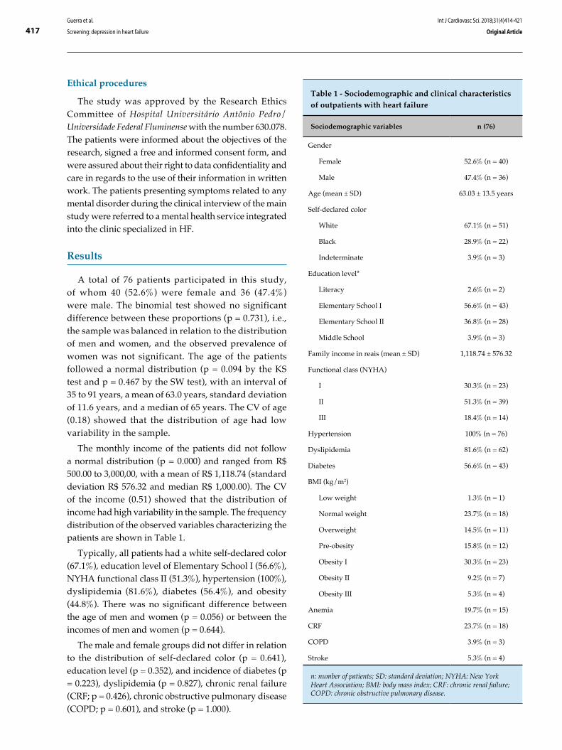
417Guerra et al.
Screening: depression in heart failure
Int J Cardiovasc Sci. 2018;31(4)414-421
Original Article
Ethical procedures
The study was approved by the Research Ethics Committee of Hospital Universitário Antônio Pedro/Universidade Federal Fluminense with the number 630.078. The patients were informed about the objectives of the research, signed a free and informed consent form, and were assured about their right to data confidentiality and care in regards to the use of their information in written work. The patients presenting symptoms related to any mental disorder during the clinical interview of the main study were referred to a mental health service integrated into the clinic specialized in HF.
Results
A total of 76 patients participated in this study, of whom 40 (52.6%) were female and 36 (47.4%) were male. The binomial test showed no significant difference between these proportions (p = 0.731), i.e., the sample was balanced in relation to the distribution of men and women, and the observed prevalence of women was not significant. The age of the patients followed a normal distribution (p = 0.094 by the KS test and p = 0.467 by the SW test), with an interval of 35 to 91 years, a mean of 63.0 years, standard deviation of 11.6 years, and a median of 65 years. The CV of age (0.18) showed that the distribution of age had low variability in the sample.
The monthly income of the patients did not follow a normal distribution (p = 0.000) and ranged from R$ 500.00 to 3,000,00, with a mean of R$ 1,118.74 (standard deviation R$ 576.32 and median R$ 1,000.00). The CV of the income (0.51) showed that the distribution of income had high variability in the sample. The frequency distribution of the observed variables characterizing the patients are shown in Table 1.
Typically, all patients had a white self-declared color (67.1%), education level of Elementary School I (56.6%), NYHA functional class II (51.3%), hypertension (100%), dyslipidemia (81.6%), diabetes (56.4%), and obesity (44.8%). There was no significant difference between the age of men and women (p = 0.056) or between the incomes of men and women (p = 0.644).
The male and female groups did not differ in relation to the distribution of self-declared color (p = 0.641), education level (p = 0.352), and incidence of diabetes (p = 0.223), dyslipidemia (p = 0.827), chronic renal failure (CRF; p = 0.426), chronic obstructive pulmonary disease (COPD; p = 0.601), and stroke (p = 1.000).
Table 1 - Sociodemographic and clinical characteristics of outpatients with heart failure
Sociodemographic variables n (76)
Gender
Female 52.6% (n = 40)
Male 47.4% (n = 36)
Age (mean ± SD) 63.03 ± 13.5 years
Self-declared color
White 67.1% (n = 51)
Black 28.9% (n = 22)
Indeterminate 3.9% (n = 3)
Education level*
Literacy 2.6% (n = 2)
Elementary School I 56.6% (n = 43)
Elementary School II 36.8% (n = 28)
Middle School 3.9% (n = 3)
Family income in reais (mean ± SD) 1,118.74 ± 576.32
Functional class (NYHA)
I 30.3% (n = 23)
II 51.3% (n = 39)
III 18.4% (n = 14)
Hypertension 100% (n = 76)
Dyslipidemia 81.6% (n = 62)
Diabetes 56.6% (n = 43)
BMI (kg/m2)
Low weight 1.3% (n = 1)
Normal weight 23.7% (n = 18)
Overweight 14.5% (n = 11)
Pre-obesity 15.8% (n = 12)
Obesity I 30.3% (n = 23)
Obesity II 9.2% (n = 7)
Obesity III 5.3% (n = 4)
Anemia 19.7% (n = 15)
CRF 23.7% (n = 18)
COPD 3.9% (n = 3)
Stroke 5.3% (n = 4)
n: number of patients; SD: standard deviation; NYHA: New York Heart Association; BMI: body mass index; CRF: chronic renal failure; COPD: chronic obstructive pulmonary disease.

418Guerra et al.
Screening: depression in heart failure
Int J Cardiovasc Sci. 2018;31(4)414-421
Original Article
The prevalence rates of depression diagnosed by the instruments were: 72.4% (n = 55) by the HAM-D, 67.1% (n = 51) by the BDI-II, and 40.8% (n = 31) by the PHQ-9, as shown in Table 2. Considering a simultaneous diagnosis by all three instruments, the prevalence of depression was 28.9% (n = 22). The three instruments showed a diagnostic agreement (presence or absence of depression) in only 47.4% of the sample (n = 36).
Comparing the three instruments in terms of diagnosing depression in the HF population, there was superficial agreement (kF = Ck = 0,27, as evaluated by Fleiss’ kappa (kF) and Krippendorff’s alpha coefficients (Ck), and moderate consistency (significantly not null, p = 0.000), as assessed by Cronbach’s alpha coefficient (CC).
Table 3 shows the quality measures of the HAM-D and PHQ-9 instruments as diagnostic tests for depression in outpatients with HF, using the BDI-II as the gold-standard instrument. The HAM-D scale proved to be the best instrument to diagnose depression, as it showed higher accuracy and sensitivity and a lower percentage of false-negative results. The PHQ-9 instrument was
Table 2 - Prevalence (P) of depression in outpatients with heart failure
Instrument Results
HAM-D 72.4% (n = 55)
BDI-II 67.1% (n = 51)
PHQ-9 40.8% (n = 31)
Agreement among all three
instrumentsk↓F = C↓k = 0.27
Consistency of the three instruments (↓C 0.602; p < 0.000
n: number of patients; HAM-D: Hamilton Depression Rating Scale; BDI-II: Beck Depression Inventory-II; PHQ-9: Patient Health Questionnaire-9.
Table 3 - Quality measures of the HAM-D and PHQ-9 instruments as diagnostic tests for depression in outpatients with heart failure
Instrument Accuracy Sensitivity Specificity% of false-
positives
% of false-
negatives
HAM-D 76.3 86.3 56.0 14.5 9.2
PHQ-9 55.3 47.1 72.0 9.2 35.5
HAM-D: Hamilton Depression Rating Scale; PHQ-9: Patient Health Questionnaire-9.
conservative in diagnosing depression, with a high percentage of false-negative results and a low sensitivity in identifying patients who in fact had depression.
Considering the BDI-II as the gold-standard instrument in diagnosing depression, we investigated the association of depression with the patients’ characteristics. There was no significant association between depression and the following factors: gender (p = 0.291), self-declared color (p = 0.976), education level (p = 0.918), obesity (p = 0.324), diabetes (p = 0.316), dyslipidemia (p = 0.056), COPD (p = 0.250), stroke (p = 0.296), and CRF (p = 0.536).
The age and income of the patients with and without depression were also not associated with depression (p = 0.862 [unpaired Student’s t test] and p = 0.776 [Mann-Whitney test], respectively).
Therefore, none of the variables included in this study was associated with depression or was a risk factor for depression in outpatients with HF.
Discussion
This is the first study comparing screening methods for depression in outpatients in a multidisciplinary clinic specialized in HF. Depression has not been systematically analyzed in patients with HF, but when specifically researched, has been observed to be frequent in this population.28-36 This condition affects between 14.0% and 26.0% of the patients without HF, but the incidence increases to 24.0% to 85.0% in patients with HF.33,34
The main findings of this study indicate a relevant prevalence of depression in HF patients when screened by the HAM-D, BDI-II, and PHQ-9 instruments. BDI-II has been considered the gold-standard instrument for depression screening in patients with HF, but in individuals with cognitive impairment or illiterate, this instrument is not recommended.10,12,21 Therefore, the results of our investigation in relation to the internal
419Guerra et al.
Screening: depression in heart failure
Int J Cardiovasc Sci. 2018;31(4)414-421
Original Article
consistency of the scales show that the BDI-II, HAM-D, and PHQ-9 proved to be useful tools for application in patients with HF.
We observed in this study a possible agreement advantage of the HAM-D with the BDI-II, the gold-standard instrument. This is probably due to the number of items in both questionnaires. The significant agreement among the scales indicates an evaluation of the intensity of the symptoms in the same direction, i.e., the greater the score, the greater the severity.37,40
A possible explanation for the difference in prevalence identified in the PHQ-9 scale may be due to its self-rating features since they portray an individual’s subjective response (as how he perceives his health and symptom). Although the instrument has already been tested in various levels of health care attention and different cultural contexts,1 still limited research has been carried out in Brazil using the PHQ-9 to screen for depression in outpatients with HF. A study carried out in Minnesota applied the PHQ-9 scale to evaluate the occurrence of depression in a sample of 425 outpatients with HF and identified a prevalence of 42.1% (n = 179),38 which is in line with our findings.
Self- and interviewer-rating scales should take into account several aspects, including the individual’s educational level and time availability for the assessment, as well as the objective of the evaluation.
The BDI-II and HAM-D scales are instruments used in more than 50% of the studies;10 the sensitivity and specificity of these instruments is approximately 0.84 and 0.72, respectively,37,39 which is also in line with our results.
A review9 has assessed the scales HAM-D, BDI-II, Zung Self-Rating Depression Scale, Geriatric Depression Scale of Yesavage, and Montgomery-Åsberg Depression Rating Scale (MADRS). The results showed relevance in the identification of signs and symptoms of depressive disorders, directing the attention to mental health interventions in the elderly.
We observed that the patients in the present study had important clinical comorbidities (diabetes, dyslipidemia, obesity, CRF, and hypertension), which were not associated with depression. In a study conducted by Aguiar et al. (2010) in hospitalized patients with HF (n = 43), patients with depression (55.8%, n = 24) according to the HAM-D scale did not differ from non-depressed ones in regards to gender, age, anemia, and renal function, factors that are known to influence the occurrence of clinical manifestations.
Depression is an important risk factor associated with HF,2 a syndromic clinical condition. When depression is not specified, it is mistaken and underdiagnosed in these individuals,35 probably due to the superposition of symptoms of HF (dyspnea, weight change, sleep, fatigue) and the neurovegetative symptoms of depression (insomnia, psychomotor slowing, and decreased energy, concentration, and appetite).36
A study conducted by Freedland et al.32 in a sample of 682 patients with HF showed that 245 (36%) patients had clinically significant depression (according to the Diagnostic and Statistical Manual of Mental Disorders [DSM-IV] criteria), while 436 (64%) were classified as non-depressed. Patients with depression also were not significantly affected by the presence of other important comorbidities, such as diabetes or kidney disease, or by the presence of only one of these conditions. These results are aligned with those of our study and differ in some aspects from the observations described in the literature. However, considering that depression emphasizes the clinical manifestations and worsens the progression of patients with HF, more attention should be dedicated to this condition, including to its screening. The importance of this initiative has been increasing in clinics specialized in HF, since studies have documented that the treatment of depression promotes improvement of the symptoms and quality of life of the patients.2
Limitations
This study presents limitations due to its experimental, observational, and cross-sectional design, which did not allow us to establish the variables predicting depression in HF. However, through the data presented, it becomes evident the importance of this discussion, because different depression scales are applied in this population.
This research demonstrates a need for further work on the screening of depression in outpatients in practices specialized on HF, due to the relevant prevalence of and damage from this association. It is important to highlight the need to deepen the knowledge of the association of depression with the sociodemographic and clinical characteristics present in this population for the development of preventive work in outpatients with HF.
Conclusions
Based on the results of this study on the prevalence of depression associated with HF, we found the following:
420Guerra et al.
Screening: depression in heart failure
Int J Cardiovasc Sci. 2018;31(4)414-421
Original Article
1) depression has a relevant prevalence in outpatients with HF; 2) the diagnosis and detection of depression are obtained through the use of questionnaires in outpatients with HF; 3) the three questionnaires evaluated have a superficial agreement and moderate consistency in the diagnosis of depression in the population with HF; 4) the HAM-D scale proved to be the best instrument in diagnosing depression, since it showed greater accuracy and sensitivity, and a lower percentage of false-negative results; 5) the PHQ-9 instrument was conservative in diagnosing depression, with a high percentage of false-negative results and low sensitivity to identify patients who are in fact depressed.
In the present study, the HAM-D questionnaire showed greater accuracy in the diagnosis of depression, due to its convenience in grading this condition and for demonstrating (in several studies) the ability to evaluate the severity of the depression.
These conclusions identify areas with gaps requiring further research, in addition to new questions in the screening of depression, and adds more information on the prevalence of depression in HF, guiding researchers and clinicians in terms of screening for depression and issues relevant to the association of these two pathologies, depression and HF.
Author contributions
Conception and design of the research: Guerra TRB, Mendlowicz MV, Mesquita ET, Cavalcanti ACD;
Acquisition of data: Guerra TRB, Venancio ICD, Pinheiro DMM; Analysis and interpretation of the data: Guerra TRB, Mesquita ET, Cavalcanti ACD; Statistical analysis: Guerra TRB; Obtaining financing: Mesquita ET, Cavalcanti ACD; Writing of the manuscript: Guerra TRB, Pinheiro DMM; Critical revision of the manuscript for intellectual content: Mendlowicz MV, Mesquita ET, Cavalcanti ACD.
Potential Conflict of Interest
No potential conflict of interest relevant to this article was reported.
Sources of Funding
This study was funded by CAPES and CNPq.
Study Association
This article is part of the thesis of Doctoral submitted by Thais de Rezende Bessa Guerra, from Universidade Federal Fluminense.
Ethics approval and consent to participate
This study was approved by the Ethics Committee of the Hospital Universitário Antônio Pedro under the protocol number 630.078. All the procedures in this study were in accordance with the 1975 Helsinki Declaration, updated in 2013. Informed consent was obtained from all participants included in the study.
1. Matias AGC, Fonsêca MA, Gomes MLF, Matos MAA. Indicadores de depressão em idosos e os diferentes métodos de rastreamento. Einstein. 2016 Jan/Mar;14(1):6-11.
2. Bocchi EA, Marcondes-Braga FG, Bacal F, Ferraz AS, Albuquerque D, Rodrigues D, et al. Sociedade Brasileira de Cardiologia. Atualização da Diretriz Brasileira de Insuficiência Cardíaca Crônica. Arq Bras Cardiol. 2012 Jan;98(1 supl. 1):1-33.
3. Aguiar VB, Ochiai ME, Cardoso JN, Carlo CHD, Morgado PC, Munhoz RT et al. Relação entre depressão, nível de BNP e comprometimento ventricular na insuficiência cardíaca. Arq Bras Cardiol. 2010 Dez;96(6):732-7.
4. Altay H, Zorlu A, Kocum HT, Demircan S, Yilmaz N, Yilmaz MB. Relationship between parathyroid hormone and depression in heart failure. Arq Bras Cardiol. 2012 Oct;99(4):915-23.
5. Faller H, Störk S, Gelbrich G, Schowalter M, Ertl G, Angermann CE. Depressive symptoms in heart failure: Independent prognostic factor or marker of functional status? J Psychosom Res. 2015 Jun;78(6):569-72.
6. Sokoreli I, de Vries JJ, Riistama JM, Pauws SC, Steyerberg EW, Tesanovic A et al. Depression as an independent prognostic factor for all-cause mortality after a hospital admission for worsening heart failure. Int J Cardiol. 2016 Oct;220:202-7.
7. Gottlieb SS, Khatta M, Friedmann E, Einbinder L, Katzen S, Baker B, et al. The influence of age, gender, and race on the prevalence of depression in heart failure patients. J Am Coll Cardiol. 2004 May;43(9):1542-9.
8. Lossnitzer N, Herzog W, Störk S, Wild B, Müller-Tasch T, Lehmkuhl E, et al. Incidence rates and predictors of major and minor depression in patients with heart failure. Int J Cardiol. 2013 Jul;167(2):502-7.
9. Tier CG, Santos SSC, Pelzer MT, Bulhosa MS. (2007). Escalas de avaliação da depressão em idosos. Rev Baiana de Enferm. 2007 Mai/Dez;21(2-3):27-36.
10. Aros MS, Yoshida EMP. Estudos da depressão: Instrumentos de avaliação e gênero. Bol Psicol. 2009 Jun;59(130)61-76.
11. Hamilton M. A rating scale for depression. J Neurol Neurosurg Psychiatry. 1960 Feb;23:56-62.
References

421Guerra et al.
Screening: depression in heart failure
Int J Cardiovasc Sci. 2018;31(4)414-421
Original Article
This is an open-access article distributed under the terms of the Creative Commons Attribution License
12. Beck AT, Ward CH, Mendelson M, Mock J, Erbaugh J. An inventory for measuring depression. Arch Gen Psychiatry. 1961 Jun;4:561-71.
13. Staab JP, Datto CJ, Weinrieb RM, Gariti P, Rynn M, Evans DL. Detection and diagnosis of psychiatric disorders in primary medical care settings. Med Clin North Am. 2001 May;85(3):579-96.
14. Freedland KE, Carney RM, Rich MW, Steinmeyer BC, Rubin EH. Cognitive behavior therapy for depression and self-care in heart failure patients: a randomized clinical trial. JAMA Intern Med. 2015 Nov;175(11):1773-82.
15. Oliveira GNM, Araújo Filho GM, Kummer A, Salgado JV, Portela EJ, Sousa-Pereira SRS et al. Inventário de Depressão de Beck (BDI) e Escala de Avaliação de Depressão de Hamilton (HAM-D) em pacientes com epilepsia. J Bras Psiquiatr. 2011;60(2):131-134.
16. Pena FM, Amorim A, Fassbender C, Oliveira RFJ, Faria CAC. Insuficiência cardíaca e depressão: uma associação com desfechos negativos. Insuf card. 2011 Nov;6(4):170-8.
17. Moreno RA, Moreno DH. Escalas de depressão de Montgomery & Asberg (MADRS) e de Hamilton (HAM-D). Rev Psiquiatr Clín. 1998;25(5):262-72.
18. McMurray JJ, Adamopoulos S, Anker SD, Auricchio A, Böhm M, Dickstein K et al. ESC Guidelines for the diagnosis and treatment of acute and chronic heart failure 2012: The Task Force for the Diagnosis and Treatment of Acute and Chronic Heart Failure 2012 of the European Society of Cardiology. Developed in collaboration with the Heart Failure Association (HFA) of the ESC.. Eur Heart J. 2012 Jul;33(14):1787-847.
19. Neto JG, Campos MS Jr, Hübner CK. Escala de Depressão de Hamilton (HAM-D): Revisão dos 40 anos de sua utilização. Rev Fac Ciênc Méd Sorocaba. 2001;3(1):10-4.
20. Williams JB. A structured interview guide for the Hamilton Depression Rating Scale. Arch Gen Psychiatry. 1988 Aug;45(8):742-7.
21. Argimon IIL, Terroso LB, Barbosa AS, Lopes RMF. Intensidade de sintomas depressivos em adolescentes através da escala de depressão de Beck (BDI-II). Acad Paul Psicol. 2013 Dez;33(85):354-72.
22. Santos IS, Tavares BF, Munhoz TN, Almeida LSP, Silva NTB, Tams BD, et al. Sensibilidade e especificidade do Patient Health Questionnaire-9 (PHQ-9) entre adultos da população geral. Cad Saúde Pública. 2013 Ago;29(8):1533-43.
23. Bächle C, Lange K, Stahl-Pehe A, Castillo K, Scheuing N, Holl RW et al. Symptoms of eating disorders and depression in emerging adults with early-onset, long-duration type 1 diabetes and their association with metabolic control. PLoS One. 2015 Jun;10(6):e0131027.
24. Sung SC, Low CC, Fung DS, Chan YH. Screening for major and minor depression in a multiethnic sample of Asian primary care patients: a comparison of the nine-item Patient Health Questionnaire (PHQ-9) and the 16-item Quick Inventory of Depressive Symptomatology - Self-Report (QIDS-SR16). Asia Pac Psychiatry. 2013 Dec;5(4):249-58.
25. Pagano M, Gauvreau K. Princípios de Bioestatística. 1a ed. São Paulo: Thomson Pioneira; 2004.
26. Landis JR, Koch GG. The measurement of observer agreement for categorical data. Biometrics. 1977 Mar;33(1):159-74.
27. Medronho RA, Bloch KV, Luiz RR, Werneck GL. Epidemiologia. 2a ed. São Paulo: Editora Atheneu; 2008.
28. Havranek EP, Spertus JA, Masoudi FA, Jones PG, Rumsfeld JS. Predictors of the onset of depressive symptoms in patients with heart failure. J Am Coll Cardiol. 2004 Dec;44(12):2333-8.
29. Konstam V, Moser DK, De Jong MJ. Depression and anxiety in heart failure. J Card Fail. 2005 Aug;11(6):455-63.
30. Joynt KE, Whellan DJ, O'Connor CM. Why is depression bad for the failing heart? A review of the mechanistic relationship between depression and heart failure. J Card Fail. 2004 Jun;10(3):258-71.
31. Laprérie AL, Hossler V, Stora O, Mariaux C, Lacaze G, Lambert H, et al. Education and multidisciplinary management in chronic heart failure. Presse Med. 2007 Jun;36(6 pt 2):985-9.
32. Freedland KE, Carney RM, Rich MW, Steinmeyer BC, Skala JA, Dávila-Román VG. Depression and multiple rehospitalizations in patients with heart failure. Clin Cardiol. 2016;39(5):257-262.
33. Sherwood A, Blumenthal JA, Trivedi R, Johnson KS, O'Connor CM, Adams KF Jr, et al. Relationship of depression to death or hospitalization in patients with heart failure. Arch Intern Med. 2007 Feb;167(4):367-73.
34. Pena FM, Carreira MAMQ, Faria CAC, Modenesi RF, Barcelos AF, Piraciaba MCT, et al. Sintomas depressivos e hospitalizações por insuficiência cardíaca: prevalência, preditores e mortalidade. Insuf Card. 2010 Out/Dez;5(4):178-84.
35. Artinian NT, Artinian CG, Saunders MM. Identifying and treating depression in patients with heart failure. J Cardiovasc Nurs. 2004 Nov/Dec;19(6 suppl):S47-56.
36. Bichara VM, Santillán J, Rosa R, Estofan L. Depression en insuficiencia cardíaca crónica: causa o consecuencia. Insuf Card. 2016; 11(4):173-200.
37. Parcias S, Rosario BP, Sakae T, Monte F, Guimarães ACA, Xavier AJ. Validação da versão em português do Inventário de Depressão Maior. J Bras Psiquiatr. 2011;60(3):164-70.
38. Jani BD, Mair FS, Roger VL, Weston SA, Jiang R, Chamberlain AM. Comorbid depression and heart failure: a community cohort study. PLoS One. 2016 Jun;11(6):1-11.
39. Menezes PR. Validade e confiabilidade das escalas de avaliação em psiquiatria. Rev Psiquiatr Clín. 1998;25(5):214-6.
40. Pinho MX, Custódio O, Makdisse M, Carvalho ACC. Confiabilidade e validade da escala de depressão geriátrica em idosos com doença arterial coronariana. Arq Bras Cardiol. 2010 Mai;94(5)570-9.
DOI: 10.5935/2359-4802.20180025
422
REVIEW ARTICLE
International Journal of Cardiovascular Sciences. 2018;31(4)422-432
Mailing Address: Antonio José Lagoeiro JorgeRua Marques de Paraná, 303 - 6º andar; Postal Code: 24033-900, Centro, Niterói, RJ - Brazil.E-mail: [email protected], [email protected]
Vitamin D Deficiency and Cardiovascular DiseasesAntonio José Lagoeiro Jorge, Jamerson Reis Cordeiro, Maria Luiza Garcia Rosa, Diego Braga Campos BianchiUniversidade Federal Fluminense, Niterói, RJ - Brazil
Manuscript received May 27, 2017; revised manuscript August 21, 2017; accepted September 25, 2017.
Vitamin D Deficiency/physiopathology; Cardiovascular Diseases; Solar Radiation; Calcium; Phosphorus.
Keywords
Abstract
Vitamin D is considered a steroid hormone with a broad spectrum of action in the human body. Its action arises from the binding of its active metabolite (1α,25-dihydroxyvitamin D) to its receptor (VDR), which is present throughout the body, including vascular smooth muscle cells and cardiomyocytes. Initially, vitamin D deficiency was related only to changes in the musculoskeletal system, but in recent years, researchers have demonstrated its relationship with several pathologies related to other systems, such as cardiovascular diseases. The objective of this study is to review vitamin D’s pathophysiology, describe its relationship with cardiovascular diseases based on the most recent publications, and highlight the results of vitamin supplementation in the prevention of such pathologies.
Introduction
Vitamin D, the fourth vitamin to be described, was initially characterized as a factor capable of curing rickets, a disease characterized by bone demineralization and skeletal deformities.1
Currently, vitamin D comprises a group of secosteroid molecules derived from 7-dehydrocholesterol (7-DHC) that includes the active metabolite (1α,25-dihydroxyvitamin D or calcitriol), its precursors (cholecalciferol or vitamin D3, ergocalciferol or vitamin D2 , and 25-hydroxyvitamin D or calcidiol), as well as its degradation products.2 These molecules, along with their carrier proteins and receptors, comprise an important metabolic axis: the endocrine vitamin D system.3
The active vitamin D has a fundamental role in regulating bone and mineral physiology, in particular, calcium and phosphorus metabolism. It is also involved in the homeostasis of several other cellular processes, such as the modulation of autoimmunity and synthesis of inflammatory interleukins,4 blood pressure control,5 and participation in the process of cell multiplication and differentiation.6 The spectrum of action of vitamin D is so broad that microarray studies show that 1α,25-dihydroxyvitamin has more than 900 potential gene targets, corresponding to approximately 3% of the human genome.7
Epidemiological studies have found that a significant portion of the world population, regardless of age, ethnicity, and geographical location, has low serum levels of vitamin D,8 as illustrated in Figure 1. Some countries even present rates of vitamin D deficiency above 50%, as observed in Brazil, Denmark, and Germany.
Recent studies have associated inadequate serum vitamin D levels with several diseases unrelated to the musculoskeletal system, such as cancer (colon, prostate, and breast), autoimmune and inflammatory diseases (multiple sclerosis, Crohn’s disease), depression, and cardiovascular diseases (CVDs) such as hypertension, coronary artery disease (CAD), and heart failure (HF).9
The objective of this study was to review vitamin D’s pathophysiology, describe its relationship with CVD based on the most recent publications, and highlight the results of vitamin supplementation in the prevention of such diseases.
Physiology and mechanism of action
In humans, only 10 to 20% of the vitamin D derives from the diet, and the remaining 80% is synthesized endogenously.10 Few foods have significant amounts of this vitamin, of which the main ones are listed in Table 1.
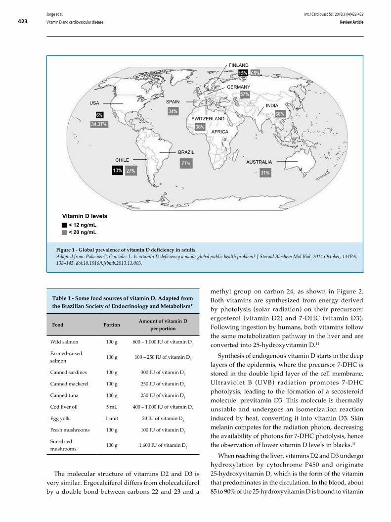
423Jorge et al.
Vitamin D and cardiovascular disease
Int J Cardiovasc Sci. 2018;31(4)422-432
Review Article
Table 1 - Some food sources of vitamin D. Adapted from the Brazilian Society of Endocrinology and Metabolism11
Food PortionAmount of vitamin D
per portion
Wild salmon 100 g 600 – 1,000 IU of vitamin D3
Farmed-raised
salmon100 g 100 – 250 IU of vitamin D3
Canned sardines 100 g 300 IU of vitamin D3
Canned mackerel 100 g 250 IU of vitamin D3
Canned tuna 100 g 230 IU of vitamin D3
Cod liver oil 5 mL 400 – 1,000 IU of vitamin D3
Egg yolk 1 unit 20 IU of vitamin D3
Fresh mushrooms 100 g 100 IU of vitamin D2
Sun-dried
mushrooms100 g 1,600 IU of vitamin D2
methyl group on carbon 24, as shown in Figure 2. Both vitamins are synthesized from energy derived by photolysis (solar radiation) on their precursors: ergosterol (vitamin D2) and 7-DHC (vitamin D3). Following ingestion by humans, both vitamins follow the same metabolization pathway in the liver and are converted into 25-hydroxyvitamin D.11
Synthesis of endogenous vitamin D starts in the deep layers of the epidermis, where the precursor 7-DHC is stored in the double lipid layer of the cell membrane. Ultraviolet B (UVB) radiation promotes 7-DHC photolysis, leading to the formation of a secosteroid molecule: previtamin D3. This molecule is thermally unstable and undergoes an isomerization reaction induced by heat, converting it into vitamin D3. Skin melanin competes for the radiation photon, decreasing the availability of photons for 7-DHC photolysis, hence the observation of lower vitamin D levels in blacks.11
When reaching the liver, vitamins D2 and D3 undergo hydroxylation by cytochrome P450 and originate 25-hydroxyvitamin D, which is the form of the vitamin that predominates in the circulation. In the blood, about 85 to 90% of the 25-hydroxyvitamin D is bound to vitamin
The molecular structure of vitamins D2 and D3 is very similar. Ergocalciferol differs from cholecalciferol by a double bond between carbons 22 and 23 and a
Figure 1 - Global prevalence of vitamin D deficiency in adults.Adapted from: Palacios C, Gonzalez L. Is vitamin D deficiency a major global public health problem? J Steroid Biochem Mol Biol. 2014 October; 144PA: 138–145. doi:10.1016/j.jsbmb.2013.11.003.
USA
CHILE
SPAIN
SWITZERLAND
FINLAND
GERMANY
AFRICA
AUSTRALIA
INDIA
Vitamin D levels< 12 ng/mL< 20 ng/mL
BRAZIL

424Jorge et al.
Vitamin D and cardiovascular disease
Int J Cardiovasc Sci. 2018;31(4)422-432
Review Article
D-binding protein (VDBP), 10 to 15% to albumin, and the remaining (less than 1%) circulate in a free form. There are few data in the literature on the bioavailability of vitamin D bound to albumin; therefore, the expression “vitamin D bioavailability” is used for the 25- hydroxyvitamin D form not bound to VDBP.12
When reaching target tissues, 25-hydroxyvitamin D is converted by the enzyme 1α-hydroxylase into 1α,25-dihydroxyvitamin D, which is the metabolically active form of the vitamin.
The effects of 1α,25-dihydroxyvitamin D are mediated by its receptor, VDR, which belongs to the family of nuclear receptors 1. Both the enzyme 1α-hydroxylase and the VDR receptor are found in almost all human cells, including cardiomyocytes13 and vascular smooth muscle cells.14 Experimental models with absence of VDR allow the understanding of the tissue-specific activity of the receptor. As an example, the absence of VDR results in increased ventricular mass, increased brain natriuretic peptide (BNP) levels and deregulation of cardiac metalloproteinases and fibroblasts, promoting a fibrotic extracellular matrix and leading to ventricular dilation and electromechanical uncoupling.15 After the binding of vitamin D to the VDR, the complex promotes gene activation or suppression, with the
help of coregulator proteins. On the other hand, the VDR also presents prompt nongenomic responses by inducing voltage-dependent calcium channels, leading to increased cell inflow of calcium and activation of other messengers, such as cyclic AMP, protein kinase A, and phospholipase C.16
Vitamin D deficiency
The American guideline for evaluation, prevention, and treatment of vitamin D deficiency17 establishes that the body pool of the vitamin should be determined by measurement of serum 25-hydroxyvitamin D with the following cutoff points: (i) deficiency, when below or equal to 20 ng/mL, (ii) insufficiency, when between 20 and 30 ng/mL, and (iii) sufficiency when greater than 30 ng/mL.
Some risk factors for vitamin D deficiency have been observed and relate to sun exposure, dietary habits, and intestinal absorption. These include an indoor lifestyle (sun deprivation), use of sunscreens, advanced age, distance from the Equator, black skin, air pollution, smoking, poor food absorption (malabsorption syndromes), drugs (anticonvulsants, glucocorticoids), and kidney and liver disease.10,17
Figure 2 - Vitamin D2 and D3 molecules, showing the difference in methyl group (CH3), which is present in the former and absent in the later (arrows).Extracted from: Peixoto PV et al. Hypervitaminosis D in animals [Hipervitaminose D em animais]. Pesq. Vet. Bras. 32(7):573-594, julho 2012.
vitamin D2 vitamin D3
425Jorge et al.
Vitamin D and cardiovascular disease
Int J Cardiovasc Sci. 2018;31(4)422-432
Review Article
Despite the increased prevalence of vitamin D deficiency in the adult population and the growing evidence of its association with CVD, both American and Brazilian guidelines recommend that serum 25-hydroxyvitamin D levels should not be measured routinely in the general population and should only be measured in patients of populations considered at risk for deficiency of this vitamin.10,17
Association of vitamin d deficiency and cardiovascular diseases
Although numerous studies have confirmed an association between vitamin D and CVD, a cause-effect relationship between both remains unclear.
In this review, we discuss the association of vitamin D deficiency with the main cardiovascular pathologies and, subsequently, we analyze some outcomes with vitamin replacement.
Hypertension
The association of vitamin D deficiency and hypertension has its basis on the renin-angiotensin-aldosterone system (RAAS). Renin is synthesized by renal juxtaglomerular cells and stimulates the production of angiotensin II (from angiotensin I) and aldosterone, which increase blood pressure (BP) directly by vasoconstriction and indirectly by fluid and salt retention.18 Inappropriately increased RAAS activation has been reported in studies with VDR and 1α-hydroxylase knockout mice.19 Vitamin D acts by inhibiting renin gene expression, decreasing the synthesis of renin and, thus, preventing hyperstimulation by this system.20
The Third National Health and Nutrition Examination Survey (NHANES III),21 a large population study that analyzed a sample of 12,644 North-Americans, has shown that systolic BP and pulse pressure correlate inversely with levels of 25-hydroxyvitamin D. These results have been confirmed by subgroup analyses in which increases in BP associated with age were significantly lower in vitamin D sufficient individuals.22 The prevalence of hypertension was also associated with vitamin D deficiency in other large studies such as the German National Interview and Examination Survey23 and the British Birth Cohort.24 A study carried out in Brazil with 91 hypertensive elderly patients has shown that the serum concentrations of 25-hydroxyvitamin D
is inversely associated with BP and positively associated with the weekly frequency of fish consumption.25
Few prospective studies have evaluated the association between vitamin D and changes in BP or emergence of hypertension. In 2015, van Ballegooijen et al.26 followed up 5,066 individuals without hypertension at the Dutch city of Groningen; the individuals had their serum vitamin D level measured and were followed up for 6.4 years. At the end of follow-up, 1,036 (20.5%) developed hypertension and, as expected, low levels of vitamin D were associated with a greater risk of development of the disease.26
Diabetes mellitus
Type 1 diabetes occurs due to an autoimmune destruction of pancreatic beta cells leading to complete deficiency of insulin production. As for the development of type 2 diabetes, the major mechanisms involved are beta cell dysfunction, peripheral insulin resistance, and systemic inflammation. According to evidence, vitamin D deficiency is associated with all these processes.27
Vitamin D may exert effects on beta cell function through a direct connection to VDR receptors and by local expression of the enzyme 1α-hydroxylase. Vitamin D may also increase insulin sensitivity by stimulating VDR expression in peripheral tissues and activating peroxisome proliferator-activated receptor-gamma (PPAR) receptors, a factor that is involved in regulating the metabolism of fatty acids in skeletal muscles and adipose tissue. On the other hand, vitamin D may also act through indirect pathways in insulin secretion and sensitivity by regulating calcium concentration and flux in beta cell membranes and peripheral tissues.27
Observational studies have shown that the incidence and prevalence of type 1 diabetes are higher in countries with higher latitude and that the disease is most often diagnosed in the winter months.28 Some studies have related vitamin D deficiency in pregnant women with the incidence of type 1 diabetes in children after birth.29 Other studies have evaluated the protective role of vitamin D supplementation in early childhood against the development of type 1 diabetes, showing a lower incidence of the disease in children who received vitamin supplementation.30
With respect to insulin resistance and type 2 diabetes, the results have been conflicting. Some studies have associated low concentrations of 25-hydroxyvitamin D with insulin resistance and dysfunction of pancreatic

426Jorge et al.
Vitamin D and cardiovascular disease
Int J Cardiovasc Sci. 2018;31(4)422-432
Review Article
beta cells in western populations.31 While studying 1,807 healthy Korean individuals, Ock et al.32 have recently reported that vitamin D has an inverse association with insulin resistance.32 While analyzing the relationship between vitamin D deficiency, diabetes, and CAD, Nardin et al.33 evaluated 1,859 patients undergoing elective angiography for evaluation of CAD and concluded that diabetes is not an independent predictor of vitamin D deficiency, but diabetic patients with vitamin D deficiency presented increase CAD prevalence and severity.33 In a recent study, Schafer et al.34 followed up more than 5,000 elderly women for 8.6 ± 4.4 years to investigate a possible relationship between vitamin D levels and the emergence of type 2 diabetes; the authors concluded that the serum levels of vitamin D were not independent predictors of the incidence of type 2 diabetes in this population.34
Obesity
Recent evidence suggests that vitamin D deficiency is associated with obesity and other components of the metabolic syndrome.35
Low levels of 25-hydroxyvitamin D are common in obese individuals, and many studies have demonstrated an inverse relationship between serum vitamin D levels and body mass index (BMI).36 Vitamin D has also been associated with regional fat distribution, and high levels of the vitamin have been associated with a lower amount of visceral and subcutaneous fat.37 Some of the explanations proposed for this association are: differences in dietary intake between obese and nonobese individuals, decreased sun exposure among obese individuals, lower vitamin D bioavailability in obesity, and altered vitamin D metabolism in obese individuals.38
Wortsman et al.39 proposed the hypothesis of sequestration of vitamin D by fat tissue to explain the prevalence of low levels of this vitamin in obese individuals.39 They demonstrated that obese individuals presented a lower increase in serum 25-hydroxyvitamin D when compared with nonobese individuals under the same conditions of exposure to sunlight and vitamin intake. Since vitamin D is liposoluble, they proposed that the vitamin must accumulate in fatty tissue and not be readily available in the circulation, which would lead to low serum levels of this vitamin.
On the other hand, Drincic et al.40 suggested that the difference in serum levels of vitamin D between obese and nonobese individuals is related to the distribution volume of this vitamin, which is greater in obese
individuals and would justify its lower serum levels in these individuals.40
Smoking and lifestyle habits
Smoking is a risk factor for CVD and systemic inflammation, and vitamin D has been associated with both these conditions. Lee et al.41 studied 560 Korean individuals aged 60 years or older to investigate the association between vitamin D and inflammatory markers and evaluate whether this association would change with the smoking profile of the patients.41 The authors observed a significant association between vitamin D deficiency and high-sensitivity C-reactive protein (hsCRP) and a modifying effect of smoking on this association, in which smokers show a stronger association between vitamin D deficiency and hsCRP than nonsmokers.41
With the aim of relating lifestyle characteristics with vitamin D deficiency, Skaaby et al.42 conducted a longitudinal study with 4,185 individuals with a follow-up time of 5 years. In this study, multivariate analyses of repeated serum measurements 25-hydroxyvitamin D were used to evaluate the association of this vitamin deficiency with BMI, practice of physical activity, type of diet (more healthy versus less healthy), alcohol consumption, and smoking. As a result, lower serum levels of vitamin D were associated with higher BMI, lower levels of physical activity, consumption of a less healthy diet, increased alcohol consumption, and smoking.42
Coronary artery disease
The occurrence of CAD has been associated with vitamin D deficiency, but the pathophysiological mechanisms of this association have not been well understood yet. The main evidence to suggest such an association is the VDR presence in both the myocardium and vascular cells, and the demonstration by epidemiological studies that the incidence of both CAD and vitamin D deficiency increase in winter months and in countries furthest from the Equator.43
Vitamin D deficiency appears to be common in acute myocardial infarction (AMI), and preliminary studies indicate a possible association of vitamin deficiency with AMI prognosis in the short and long term.43 Moreover, vitamin D deficiency seems to predispose to recurrent adverse cardiac events, due to its association with the number of affected coronary arteries, AMI complications, and cardiac remodeling.44
427Jorge et al.
Vitamin D and cardiovascular disease
Int J Cardiovasc Sci. 2018;31(4)422-432
Review Article
The Health Professionals Follow-up Study followed up 18,225 men during 10 years and observed an association between low vitamin D levels and increased AMI risk, even after adjustment for other risk factors.45 Prospective studies have also found a high prevalence of vitamin D deficiency in patients hospitalized with AMI. A multicenter study carried out with 239 patients with acute coronary syndrome (ACS) showed that 96% of the individuals had low vitamin D levels at hospital admission.46
Some studies show a potential independent association between severe vitamin D deficiency and intrahospital mortality in patients with ACS. Correia et al.47 studied 206 patients with ACS and found that individuals with serum vitamin D levels lower than 10 ng/mL had a 24% rate of intrahospital cardiovascular mortality, which was significantly higher than that observed in the remaining patients (4.9%).47
Heart failure
HF has been associated with vitamin D deficiency. Shane et al. demonstrated a high prevalence of vitamin D deficiency in patients with HF, as well as an inverse correlation between serum levels of vitamin D with left ventricular function and disease severity.48
Vitamin D deficiency has been associated with severe adverse events, such as hospitalization due to HF and mortality. Liu et al.49 reported in a study with 548 patients that low levels of 25-hydroxyvitamin D were associated with higher BNP levels, as well as a higher rate of hospitalization due to HF and increased mortality rate from all causes.49 In the LURIC study, a prospective cohort study with 3,299 patients undergoing coronary angiography, the levels of N-terminal (NT)-proBNP related inversely to the levels of vitamin D.50
In regards to HF with normal ejection fraction (HFNEF), studies have shown conflicting results in terms of its association with vitamin D deficiency. In 2013, Lagoeiro et al. studied 85 outpatients with suspected HFNEF, of whom 32 had confirmed HFNEF, and observed a negative correlation between vitamin D deficiency and E/E’ ratio.51 On the other hand, Pandit et al.52, in 2014, conducted a retrospective study with 1,011 patients and found no significant association between vitamin D levels and left ventricular diastolic performance.52
Despite evidence demonstrating an association between vitamin D and HF, the exact mechanism by which this vitamin’s deficiency leads to worse clinical
outcomes in patients with HF has not been clearly established yet. A potential mechanism could be through the occurrence of cardiorenal syndrome or worsening of renal function.53 It is well known that the cardiovascular and renal systems are interrelated and that a decline in one of them could influence the other. Progression of cardiorenal syndrome involves hyperactivation of RAAS and sympathetic nervous system, as well as systemic inflammation, which may lead to electrolyte disturbances and disorders in fluid regulation, causing endothelial dysfunction, potentially leading to left ventricular remodeling and myocardial fibrosis. These changes generate a vicious cycle in which a decline in a system’s function contributes to its further deterioration.54
Evidence supports vitamin D as an important regulator in the progression of cardiorenal syndrome. Deregulations in vitamin D metabolism due to reduced activity of the enzyme 1α-hydroxylase and depletion of VDBPs due to proteinuria are responsible for vitamin D deficiency in chronic renal patients; given the high prevalence of chronic renal insufficiency in patients with HF, such changes can be prevalent in these patients.55
Other evidence supporting the role of vitamin D deficiency in the pathogenesis of cardiorenal syndrome relates to the involvement of RAAS and inflammatory cytokines. Vitamin D deficiency leads to RAAS hyperactivation, contributing to left ventricular remodeling and emergence or worsening of HF.56 Vitamin D deficiency can lead to increased production and release of inflammatory cytokines, which may have a direct or indirect negative effect in the myocardium, contributing to cell apoptosis, hypertrophy, fibrosis, ventricular remodeling, and negative inotropic effects, in addition to increased renal fibrosis and renal insufficiency.57
Vitamin D deficiency and myalgia induced by statins
Statins are very effective agents in primary and secondary cardiovascular prevention in high-risk patients.58 However, the side effects most frequently observed in the musculoskeletal system, such as myalgia, have been commonly observed in patients treated with statins, and these effects directly affect the adherence to treatment using these medications.
Observational studies show that myalgia may occur in approximately 15 to 20% of the individuals treated with statins.59 However, evidence from daily clinical practice shows that this prevalence is even greater. VDRs are present in muscle cells, and low vitamin D levels are
428Jorge et al.
Vitamin D and cardiovascular disease
Int J Cardiovasc Sci. 2018;31(4)422-432
Review Article
associated with hypotonia, proximal muscle weakness, and nonspecific musculoskeletal pain.60 Recent studies have reported that vitamin D deficiency is associated with a higher prevalence of myalgia induced by statins.61
In 2014, Shantha et al.62 performed a retrospective study with 5,526 patients followed by a prospective analysis in which the patients were followed up for 7 years. The patients with measured serum vitamin D levels who started treatment with statins were considered as the exposure group. The aim was to analyze the association between statin-induced myalgia and vitamin D levels, as well as to establish a cutoff level for vitamin D that would demonstrate a high accuracy for the emergence of myalgia. The authors concluded that low levels of vitamin D were associated with myalgia and that a cutoff level of 15 ng/mL for vitamin D showed a high accuracy in predicting the emergence of myalgia induced by statins.62
In 2015, Morioka et al.63 performed a study with 5,907 patients to analyze if the level of vitamin D would modify the association between the use of statin and the emergence of musculoskeletal pain. The authors concluded that the group with vitamin D level below 15 ng/mL and using statins presented an approximately two-fold greater chance of developing musculoskeletal pain than patients who also had vitamin D levels below 15 ng/mL but were not treated with statins.63
Prospective and randomized studies are needed to confirm the actual association between vitamin D deficiency and the emergence of myalgia induced by statins. In addition, the pathophysiological mechanism that could explain this association still needs to be elucidated.
Genetic factors of vitamin D and its implications in cardiovascular disease
The increased worldwide prevalence of vitamin D deficiency, or at least of its measurable circulating form, 25-hydroxyvitamin D, can be explained in part by genetic determinants. In 2010, an important multicenter study carried out by Wang et al. pointed out that serum vitamin D levels may be influenced by genetic variations involving its synthesis (7-DHC), hydroxylation (CYP2R1, CYP24A1), and transport protein (VDBP).64
Most studies analyzing the association between vitamin D deficiency and CVD are epidemiological, which prevents discrimination between association and causality. In this context, Mendelian randomization (MR) is an alternative approach to estimate the causal relationship between modifiable biological exposures and clinical outcomes
of interest using genetic variants (single nucleotide polymorphisms, SNPs) as instrumental variables. Thus, MR using summarized data allows the combination of results already published in previous studies, becoming a relevant alternative to investigating causality.65
Some studies have used MR to investigate a possible causal relationship between vitamin D deficiency and CVD. With respect to hypertension, Vimaleswaran et al.66 found that increased vitamin D levels could reduce the risk of development of CVD, showing a causal relationship.66 On the other hand, the results of MR studies have not demonstrated a causal relationship for diabetes mellitus67 and CAD,68 in which vitamin D deficiency appears to be a confounding factor.
Vitamin D replacement
Based on growing evidence of an association between vitamin D deficiency and CVD, many authors have investigated the role of vitamin D supplementation in the prevention and treatment of these pathologies.
A randomized study conducted by Hsia et al.69 with 36,282 postmenopausal women evaluated the supplementation of vitamin D 200 IU plus calcium carbonate twice a day or placebo during a follow-up of 7 years and found that supplementation of vitamin D was unable to reduce cardiovascular risk.69 This was one of the few randomized studies that evaluated the impact of vitamin D in reducing hard outcomes, namely, incidence of AMI, stroke, and CAD-related death.
With regard to hypertension, studies with vitamin D replacement have shown conflicting results. An important systematic review and meta-analysis published by Wu et al.70 comprising 36,806 patients showed no significant effect of calcium plus vitamin D supplementation in variations in systolic and diastolic BP when compared with lack of supplementation of both.70
In diabetes mellitus, studies involving vitamin D supplementation have proved disappointing. A study involving 70 children with type 1 diabetes of recent onset, supplementation with calcitriol had a modest effect on the residual function of pancreatic beta cells, but the reduction of glycated hemoglobin after 1 year of treatment was not statistically significant.71 With respect to type 2 diabetes, study results are conflicting, perhaps due to lack of standardization of the supplemented vitamin D dose or the use of small samples in these studies. A meta-analysis involving 35 controlled studies evaluated the impact of vitamin D supplementation
429Jorge et al.
Vitamin D and cardiovascular disease
Int J Cardiovasc Sci. 2018;31(4)422-432
Review Article
in healthy patients and individuals with vitamin D deficiency, obesity, prediabetes, and diabetes. Compared with placebo, vitamin D had no effect on insulin resistance, insulin secretion, or glycated hemoglobin.72
In regards to obesity, several studies have evaluated the effect of vitamin D supplementation with and without the addition of calcium on weight and body composition. Most of these studies showed no significant effect of vitamin D on BMI or body composition.73
Additionally, CAD does not seem to be significantly influenced by vitamin D supplementation. An important study named RECORD74 involving 5,292 individuals compared the effects of vitamin D, calcium, vitamin D plus calcium, or placebo on cardiovascular events. The results showed that although vitamin D can exert a protective role on HF, it does not seem to protect against AMI and stroke. A meta-analysis of 51 controlled studies found that supplementation of vitamin D has no significant impact on AMI.75
Vitamin D supplementation seems to have some benefits on HF, although the mechanisms of action have not been well established. Recent studies have reported that in individuals with established HF and vitamin D deficiency, vitamin supplementation is associated with improved survival.76
Conclusions
CVDs remain the main cause of mortality in several countries worldwide. An understanding of the pathophysiological mechanisms involved, as well as their risk factors, is essential for planning of prevention and treatment strategies.
In recent years, many studies have shown a relationship between vitamin D deficiency and CVDs, with a direct influence on prognosis. Based on the understanding of this association, the focus of researchers has been in the correction of vitamin deficiency with the aim of preventing diseases and improving the prognosis of established diseases. However, there are still no consistent data to recommend vitamin D replacement in the context of cardiac diseases.
One point that deserves attention is the broad worldwide variation in the prevalence of vitamin D deficiency. Since endogenous vitamin D synthesis is dependent on solar exposure, which in turn varies according to latitude, perhaps the reference level for serum vitamin D also differs among countries depending on sunlight exposure.
It is unclear whether the disappointment of the results of studies with vitamin D supplementation is due to an inability of the vitamin in exerting effects on established disease, or use of inappropriate supplementation doses. It is important to understand the doses required to maintain the serum levels of vitamin D above the desired level, as well as serial measurements of 25-hydroxyvitamin D with the aim of maintaining adequate levels of this vitamin during the entire follow-up duration.
Author contributions
Conception and design of the research: Jorge AJL, Cordeiro JR. Acquisition of data: Cordeiro JR. Writing of the manuscript: Jorge AJL, Cordeiro JR, Bianchi DBC. Critical revision of the manuscript for intellectual content: Jorge AJL, Rosa MLG.Supervision / as the major investigador: Jorge AJL.
Potential Conflict of Interest
No potential conflict of interest relevant to this article was reported.
Sources of Funding
There were no external funding sources for this study.
Study Association
This article is part of the thesis of master submitted by Jamerson Reis Cordeiro, from Universidade Federal Fluminense.
Ethics approval and consent to participate
This article does not contain any studies with human participants or animals performed by any of the authors.
1. Holick MF. McCollum Award Lecture, 1994: vitamin D – new horizonts for the 21st century. Am J Clin Nutr. 1994;60(4):619-30. PMID:8092101
2. Castro LCG. O sistema endocrinológico vitamina D. Arq Bras Endocrinol Metab. 2011;55(8):566-75. PMID:22218438
References

430Jorge et al.
Vitamin D and cardiovascular disease
Int J Cardiovasc Sci. 2018;31(4)422-432
Review Article
3. Normam AW. From vitamin D to hormone D: fundamentals of the vitamin D endocrine system essential for good health. Am J ClinNutr. 2008;88(Suppl 2):491S-9S. PMID:1868389
4. Cantorna MT, Mahon BD. D-hormone and the immune system. J Rheumatol Suppl. 2005;76:11-20. PMID: 18689389
5. Li YC, Qiao G, Uskokovic M, Xiang W, Zheng W, Kong J. Vitamin D: a negative endocrine regulator of the rennin-angiotensin system and blood pressure. J Steroid Biochem Mol Biol. 2004;89-90(1-5):387-92. Doi: 10.1016/j.jsbmb.2004.03.004
6. Deeb KK, Trump DL, Johnson CS. Vitamin D signaling pathways in cancer: potential for anticancer therapeutics. Nat Rev Cancer. 2007; 7(9):684-700. Doi: 10.1038/nrc2196
7. Wang TT, Tavera-Mendoza LE, Laperriere D, Libby E, MacLeod NB, Nagai Y, et al. Large-scale in silico and microarray-based identification of direct 1,25-dihydroxyvitamin D3 target genes. Mol Endocrinol. 2005;19(11):2685-95. Doi:10.1210/me.2005-0106
8. Palacios C, Gonzalez L. Is vitamin D deficiency a major global public health problem? J Steroid Biochem Mol Biol. 2014;144(pPta):138-45. Doi:10.1016/jsbmb2013.11.003.
9. Holick MF. Vitamin D deficiency. N Eng J Med. 2007;357(3):266-81. PMID:17634462
10. Maeda SS, Borba VZ, Camargo MB, Silva DM, Borges JL, Bandeira F, et al. et al. Recomendações da Sociedade Brasileira de Endocrinologia e Metabologia (SBEM) para o diagnóstico e tratamento da hipovitaminose. Arq Bras Endocrinol Metabol.2014; 58(5):411-33. PMID:25166032
11. Holick MF. Sunlight and vitamin D for bone health and prevention of autoimmune diseases, cancers, and cardiovascular disease. Am J Clin Nutr. 2004;80(6 Suppl):1678S-88S. PMID: 15585788
12. Bikle D, Bouillon R, Thadhani R, Schoenmakers I. Vitamin D metabolites in captivity? Should we measure free or total 25(OH)D to assess vitamin D status? J Steroid Biochem Mol Biol. 2017173:105-16. Doi:10.1016/i.isbmb.2017.01.007
13. Somjen D, Wisman Y, Kohen F, Gayer B, Limor R, Sharon O, 25-hydroxyvitamin D3 – 1alpha-hydroxylase is expressed in human vascular smooth muscle cells and is up regulated by parathyroid hormone and estrogenic compounds. Circulation. 2005;111(13):1666-71. Doi:10.1161/01.CIR.0000160353.27927.70
14. Bouillon R, Carmeliet G, Verlinden L, van Elton E, Verstuyf A, Luderer HF, et al. Vitamin D and human health: lessons from vitamin D receptor null mice. Endocr Rev. 2008;9(6):726-76. Doi: 10.1210/er.2008-0004
15. Al Mheid I, Quyyumi AA. Vitamin D and cardiovascular disease. J Am Coll Cardiol. 2017;70(1):89-100. Doi:10.1016/j.jacc.2017.05.031
16. Holick MF, Binkley NC, Bischoff-Ferrari HA, Gordon CM, Hnley DA, Hearney RP, et al. Evaluation, treatment, and prevention of vitamin D deficiency: an endocrine society clinical practice guideline. J Clin Endocrinol Metab. 2011;96(7)1911-30. Doi:10.1210/jc.2011-0385
17. Lee JH, O’Keefe JH, Bell D, Vitamin D deficiency. An important, common, and easily treatable cardiovascular risk factor? J Am Coll Cardiol. 2008;52(24):1949-56. Doi:10.016/j.jacc.2008.08.050
18. Connell JM, Mackenzie SM, Freel EM, Fraser R, Davies E. et al. A lifetime of aldosterone excess: long-term consequences of altered regulation of aldosterone production for cardiovascular function. Endocr. Rev. 2008; 29(2):133-54. Doi:10.1210/er.2007-0030
19. Li YC, Kong J, Wei M, Chen ZF, Liu SQ,Cao LP. 1,25-dihydroxyvitamin D(3) is a negative endocrine regulator of the rennin-angiotensin system. J Clin Invest.2002;110():229-38. Doi:10.1172/JCI15219
20. Yuan W , Pan W, Kong J, Zheng W, Szeto FL, Wong KE, et al. 1,25-dihydroxyvitamin D3 suppresses renin gene transcription by blocking the activity of the cyclic AMP response element in the renin gene promoter. J Biol Chem. 2007;282(41):29821-30. Doi: 10.1074/jbc.m705495200
21. Scragg R, Sowers M, Bell C. Serum 25-hydroxyvitamin D, ethnicity, and blood pressure in the third National Health and Nutrition
Examination Survey. Am J Hypertens. 2007;20(7):713-9. Doi: 10.1016/j.amjhyper2007.01.017
22. Judd SE, Nanes MS, Ziegler TR, Wilson PW,Tangpricha V .Optimal vitamin D status attenuates the age-associated increase in systolic blood pressure in white Americans: results from the third National Health and Nutrition Examination Survey. Am. J. Clin Nutr. 2008;87(1):136-41. PMID:18175747
23. Hintzpeter B, Mensink GB, Thierfelder W, Muller MJ, Scheidt-Nave C. Vitamin D status and health correlates among German adults. Eur J Clin Nutr. 2008;62(9):1079-89. Doi: 10.1007/s10620-007-9833-1
24. Hyppönen E, Boucher BJ, Berry DJ, Power C. 25-hydroxyvitamin D, IGF-1, and metabolic syndrome at 45 years of age: a cross-sectional study in the 1958 British Birth Cohort. Diabetes. 2008;57(2):298-305. Doi: 10.2337/db07-1122
25. Neves JPR , Silva AS, Morais LC, Diniz AS, Costa MJ, et al. Concentrações de 25-hidroxivitamina D e níveis pressóricos em idosos hipertensos. Arq Bras Endocrinol Metabol. 2012; 56(7):415-22. PMID:23108745
26. Van Ballegooijen AJ, Kestenbaum B, Sachs MC, de Boer IH, Siscovick DS, Hoofenagle AN, et al. Association of 25-hydroxyvitamin D and parathyroid hormone with incident hypertension: MESA (Multi-Ethnic Study of Atherosclerosis). J Am Coll Cardiol. 2014; 63(12):1214-22. Doi:10.1016/j.jacc.2014.01.012
27. Pittas AG, Dawson-Hugues B. Vitamin D and diabetes. J Steroid Biochem Mol Biol. 2010;121(1-2):425-9. Doi:10.1016/j.jsbmb.2010.03.042
28. Mohr SB, Garland CF, Gorham ED, Garland FC. The association between ultraviolet B irradiance, vitamin D status and incidence rates of type 1 diabetes in 51 regions worldwide. Diabetologia. 2008;51(8):1391–8. Doi:10.1007/s00125-008-1061-5
29. Zipitis CS, Akobeng AK. Vitamin D supplementation in early childhood and risk of type 1 diabetes: a systematic review and meta-analysis. Arch Dis Child. 2008;93(6):512-7. Doi:10.1136/adc.2007.128579
30. Tenconi MT, Devoti G, Comelli M. Major childhood infectious diseases and other determinants associated with type 1 diabetes: a casecontrol study. Acta Diabetol.2007;44(1):14–9. Doi:10.1007/s00592-007-0235-9
31. Liu E, Meigs JB, Pittas AG, MvKeown NM, Economos CD, Booth SL, et al.Plasma 25-hydroxyvitamin D is associated with markers of the insulin resistant phenotype in non diabetic adults. J Nutr. 2009;139:329-34. Doi: 10.3945/jn.108.093831
32. Ock SY, Ha KH, Kim BK, Kim HC, Shim JS, Lee MH, et al. Serum 25-hydroxyvitamin Dconcentration is independently inversely associated with insulin resistance in the healthy, non-obese Korean population. Diabetes Metab J. 2016;40(5):367-75. Doi: 10.4093/dmj.2016.40.5.367
33. Nardin M, VerDoia M, Schahffer A, Barbieri L, Marino P, De Luca G, et al. Vitamin D status, diabetes mellitus and coronary artery disease in patients undergoing coronary angiography. Atherosclerosis. 2016;250:114-21. Doi: 10.1016/j.atherosclerosis.2016.05.019
34. Schafer AL, Napoli N, Lui L, Schwartz AV, Black DM, Study of Osteoporotic Fractures. Serum 25-hydroxyvitamin D concentration does not independently predict incident diabetes in older women. Diabet Med. 2014;31(5):564-9. Doi: 10.1111/dme.12368
35. McGill AT, Stewart JM, Lithander FE, Stri KCM, Poppitt SD Relationships of low serum vitamin D3 with anthropometry and markers of the metabolic syndrome and diabetes in overweight and obesity. Nutr J. 2008;7:4. Doi:10.1186/1475-2891-7-4
36. Vilarrasa N, Maravall J, Estepa A. Low 25-hydroxyvitamin D concentrations in obese women: their clinical significance and relationship with anthropometric and body composition variables. J Endocrinol Invest. 2007;30(8):653–8. Doi:10.1007/BFO3347445
37. Caron-Jobin M, Morisset AS, Tremblay A, Hot C, Légaré D, Tcheernof A. Elevated serum 25(OH)D concentrations, vitamin D, and calcium intakes are associated with reduced adipocyte size in women. Obesity(Silver Spring).2011;19(7):1335–41. Doi:10.1036/oby.2011.90
38. Wamberg L, Pedersen SB, Rejnmark L, Richelsen B. Causes of vitamin D deficiency and effect of vitamin D supplementation on metabolic

431Jorge et al.
Vitamin D and cardiovascular disease
Int J Cardiovasc Sci. 2018;31(4)422-432
Review Article
complications in obesity: a review. Curr Obes Rep. 2015;4(4):429-40. Doi:10.1007/s13679-015-0176-5
39. Wortsman J, Matsuoka LY, Chen TC, Luz Z, Holick MF, et al. Decreased bioavailability of vitamin D in obesity. Am J Clin Nutr. 2000;72(3):690–3. PMID:10966885
40. Drincic AT, Armas LAG, Van Diest EE. Volumetric dilution, rather than sequestration best explains the low vitamin D status of obesity. Obesity. 2012;20(7):1444–8. Doi:10.1038/oby.2011.404
41. Lee H, Kim KN, Lim YH, Hong Yc. Interaction of vitamin D and smoking on inflammatory markers in the urban elderly. J Prev Med Public Health. 2015;48(5):249-56. Doi: 10.3961/jpmph.15.042
42. Skaaby T, Husemoen LLN, Thuesen BH, Pisinges C, Hannemann A, Jorgensen T, et al. Longitudinal associations between lifestyle and vitamin D: a general population study with repeated vitamin D measurements. Endocrine. 2016;51:342-50. Doi: 10.1007/s12020-015-0641-7
43. Milazzo V, De Metrio M, Cosentino N, Marenzi G, Tremoli E, Vitamin D and acute myocardial infarction. Word J Cardiol. 2017;;9(1):14-20. Doi: 10.4330/wje.v9.i1.14
44. De Metrio M, Milazzo V, Rubino M, Cabiesti A Moltrasio M, Marana I, et al. Vitamin D plasma levels and in-hospital and 1-year outcomes in acute coronary syndromes: a prospective study. Medicine(Baltimore). 2015;94(19):e857. Doi: 10.1097/MD.00000000000 857
45. Giovannucci E, Liu Y, Hollis BW, Rimm EB. 25-hydroxyvitamin D and risk of myocardial infarction in men: a prospective study. Arch Intern Med 2008;168(11):1174-80. Doi:10.1001/archinte.168.11.1174
46. Lee JH, Gadi R, Spertus JA, Tang F,O’Keefe JH. Prevalence of vitamin D deficiency in patients with acute myocardial infarction. Am J Cardiol. 2011;107(11):1636-8. Doi:10.1016/j.amjcard.2011.01.048
47. Correia LC, Sodré F, Garcia G, Sabino M, Brito M, Kalil F. Relation of severe deficiency of vitamin D to cardiovascular mortality during acute coronary syndromes. Am J Cardiol. 2013;111(3):324-7. Doi:10.1016/j.amjcard.2012.10.006
48. Shane E, Mancini D, Aaronson K, Silverberg SI, Seibel MJ, Adessa VD. Bone mass, vitamin D deficiency, and hyperparathyroidism in congestive heart failure. Am J Med .1997;103(3):197–207. PMID: 9316552
49. Liu LC, Voors AA, van Veldhuisen DJ, Belonje AM, Szymanski MK, Lillie HH, et al. Vitamin D status and outcomes in heart failure patients. Eur J Heart Fail. 2011;13(6):619–25. Doi: 10.1093/Eurhf/hfr032
50. Murr C, Pilz S, Grammer TB, Kleber ME, Meinitzer A, Boehm BO, et al. Vitamin D deficiency parallels inflammation and immune activaction, the Ludwigshafen Risk and Cardiovascular Health (LURIC) study. Clin Chem Lab Med. 2012;50(12):2205-12. PMID: 23093269
51. Lagoeiro AJ, Rosa ML, Freire MC, Correia DM, Fernandes LC, Ribeiro ML, et al. Deficiência de vitamina D em pacientes com suspeita de insuficiência cardíaca e fração de ejeção normal. RevBras Cardiol. 2013;26(4):253-8.
52. Pandit A, Mookadam F, Boddu S, Aryal Pandit A, Tandar A, Chaliki H, et al. Vitamin D levels and left ventricular diastolic function. 2014. Open Heart. 2014;1(1):e000011 Doi: 10.1136/openhrt-2013-000011.eCollection 2014
53. Ronco C, Ronco F. Cardio-renal syndromes: a systematic approach for consensus definition and classification. Heart Fail Rev. 2012;17(2):151–60. Doi: 10.1007/s10741-010-9224-0
54. Sun Y, Zhang J, Zhang JQ, ramires PJ. Local angiotensin II and transforming growth factor-beta1 in renal fibrosis of rats. Hypertension. 2000;35(5):1078–84. Doi:https//Doi.org/10.116101.HYP35.5.1078
55. Levin A, Bakris GL, Molitch M, SmuldersM, Tian J, Williams LA, et al. Prevalence of abnormal serum vitamin D, PTH, calcium, and phosphorus in patients with chronic kidney disease: results of the study to evaluate early kidney disease. Kidney Int. 2007;71(1):31–8. Doi: 10.1038/ki.5002009
56. McMurray JJ. Clinical practice. Systolic heart failure. N Engl J Med. 2010;362(3):228–38. Doi: 10.1056/NEJMc0909392
57. Gullestad L, Ueland T, Vinge LE, Finsen A, Yndestad A, Aukrust P. Inflammatory cytokines in heart failure: mediators and markers. Cardiology. 2012;122(1):23–35. Doi:10.1159/000338.166
58. Taylor F, Huffman MD, Macedo AF, Moore TH, BurkeM, Davey Smith G, et al. Statins for the primary prevention of cardiovascular disease. Cochrane Database Syst Rev. 2013 ; (1):CD004816. Doi:10.1002/14651858CD0048
59. Joy TR, Hegele RA. Narrative review: statin-related myopathy. Ann Intern Med. 2009;150(12):858-68. Doi: 10.7326/0003-4819-150-12-200090616000009
60. Harari M, Dramsdahl E, Shany S, et al. Increased vitamin D serum levels correlate to clinical improvement of rheumatic disease after Dead Sea climatotherapy. Isr Med Assoc J. 2011Apr13;4:212-5.
61. Michalska-Kasiczak M, Sahebkar A, Mikhailidis DP, Rysz J,Muntner P, Toth PP,,et al. Analysis of vitamin D levels in patients with and without statin-associated myalgia – a systematic review and meta-analysis of 7 studies with 2420 patients. Int J Cardiol. 2015;178:111-6. Doi:10.1016/j.ijcard.2014.10.118
62. PalamanerSubashShantha G, Ramos J, Thomas-Hemak L, Pancholy SB. Association of vitamin D and incident statin induced myalgia—a retrospective cohort study. PLoS One. 2014;9(2):e88877. Doi:10.1371/journal.pone.0088877.eCollection2014
63. Morioka TY, Lee AJ, Bertisch S, Buetner A. Vitamin D status modifies the association between statin use and musculoskeletal pain: a population based study. Atherosclerosis. 2015;238(1):77-82. Doi:10.1016/j.atherosclerosis.2014.11.012
64. Wang TJ, Zhang F, Richards JB, Kestenbaum B, van Meurs JB, Berry D, et al. Common genetic determinants of vitamin D insufficiency: a genome-wide association study. Lancet. 2010; 376(9736): 180–8. Doi:10.1016/S0140-6736(10)60588-0
65. Bracco PA. Randomização mendeliana: um método para estimação de efeitos causais utilizando variantes genéticas como variáveis instrumentais.[Citado em 2016 jan15] Disponível em http://hdl.handle.net/10183/158115.
66. Vimaleswaran KS, Cavadino A, Berry DJ, Lifelines Cohort Study Investigators, Jorde R, Lu C, Alves AC, et al. Association of vitamin D status with arterial blood pressure and hypertension risk: a mendelian randomization study. Lancet Diabetes Endocrinol. 2014;2(9):719-29. Doi: 10.1016/S2213-8587(14)70113-5
67. Ye Z, Sharp SJ, Burgess S, Scott RA, Imamura F, InterAct Consortium, et al. Association between circulating 25-hydroxyvitamin D and incident type 2 diabetes: a mendelian randomization study. Lancet Diabetes Endocrinol. 2015;3(1):35-42. Doi: 10.1016/S2213-8587(14)70184-6
68. Manousaki D, Mokry LE, Ross S, Gotzman D, Richards JB. Mendelian Randomization Studies do not Support a Role for Vitamin D in Coronary Artery Disease. Cir Cardiovasc Genet. 2016;9(4):349-56. Doi:10.1161/CIRCGENETICS.116.001396
69. Hsia J, Heiss G, Ren H, Allison M, Dolan NC, Greenland P, et al. Calcium/vitamin D supplementation and cardiovascular events. Circulation. 2007;115(7):846–54. Doi:10.1161/CIRCULATIONAHA.106.673491
70. Wu L, Sun D. Effects of calcium plus vitamin D supplementation on blood pressure: a systematic review and meta-analysis of randomized controlled trials. J Human Hypertens. 2017;1(9):547-54.
71. Pitocco D, Crino A, Di Stasio E, Manfrini S, Guglielmi C, Spera S, et al.The effects of calcitriol and nicotinamide on residual pancreatic beta-cell function in patients with recent-onset Type 1 diabetes (IMDIAB XI). Diabet Med 2006;23(8):920–3. Doi: 10.1111/j.1484-5491.2006.01 921.X
72. Seida J, Mitri J, Colmers IN, et al. Effect of vitamin D3 supplementation on improving glucose homeostasis and preventing diabetes: a systematic review and meta-analysis. J ClinEndocrinolMetab. 2014;99:3551-60.
73. Salehpour A, Shidfar F, Hosseinpanah F, vafa M, Razaghi M, Amiri F. Does vitamin D3 supplementation improve glucose homeostasis in overweight or obese women? A double-blind, randomized, placebo-controlled clinical trial. Diabet Med. 2013;30(12):1477–81. Doi: 10.1111/dme,12273

432Jorge et al.
Vitamin D and cardiovascular disease
Int J Cardiovasc Sci. 2018;31(4)422-432
Review Article
This is an open-access article distributed under the terms of the Creative Commons Attribution License
74. Ford JA, MacLennan GS, Avenell A, Bolland M, Grey A, Witham M, RECORD Trial Group. Cardiovascular disease and vitamin D supplementation: trial analysis, systematic review, and meta-analysis. Am J Clin Nutr. 2014;100(3):746-55. Doi:10.3945/ajcn.113.082 602
75. Elamin MB, Abu Elnour NO, Elamin KB, Fatourechi MM, Alkatib AA, et al. Vitamin D and cardiovascular outcomes: a systematic review and
metaanalysis. J Clin Endocrinol Metab. 2011;96(7)1931–42. Doi:10.1210/jc.2011-0398
76. Gotsman I, Shauer A, Zwas DR, Hellman Y, Keren A, Lotan C, et al. Vitamin D deficiency is a predictor of reduced survival in patients with heart failure; vitamin D supplementation improves outcome. Eur J Heart Fail 2012;14(4):357- 66. Doi:10.1093/eurjhf/hfr175
DOI: 10.5935/2359-4802.20180027
433
REVIEW ARTICLE
International Journal of Cardiovascular Sciences. 2018;31(4)433-442
Mailing Address: Jacqueline Aparecida BorgesRua Martins Ferreira, 81/704. Postal Code: 22271-010, Botafogo, Rio de Janeiro, RJ - Brazil.E-mail: [email protected]; [email protected]
Fatigue: A Complex Symptom and its Impact on Cancer and Heart FailureJacqueline Aparecida Borges, Mônica Maria Pena Quintão, Sergio S. M.C. Chermont, Hugo Tannus Furtado de Mendonça Filho, Evandro Tinoco MesquitaUniversidade Federal Fluminense (UFF), Niterói, RJ - Brazil
Manuscript received April 11, 2017, revised manuscript December 12, 2017, accepted December 19, 2017.
Heart Failure / physiopathology; Neoplasms / physiopathology; Cardiotoxicity / complications; Ventricular Dysfunction, Left; Neoplasias / drug therapy.
Keywords
Abstract
In chronic diseases like cancer and heart failure (HF), fatigue is a common and complex symptom from an etiological and pathophysiological point of view, thus, a relevant issue in the recent area of oncocardiology. Fatigue is prevalent in 80-90% of the oncological patients treated with chemotherapy and/or radiotherapy and affects approximately 50-96% of the individuals with IC. The toxicity attributed to chemotherapeutic agents can determine the patients’ degree of fatigue and may even predict their survival. In recent decades, the advancement of antineoplastic therapies has substantially impacted the survival of patients with cancer, and the risks of harmful effects from these therapies to the cardiovascular system have been increasingly described. Therefore, the cooperation between oncologists and cardiologists has led to the emergence of oncocardiology and the new concept of cardiac surveillance. Cardiotoxicity is one of the clinical complications in the treatment of cancer, and its typical manifestation is left ventricular systolic dysfunction. New diagnostic and therapeutic strategies have been employed in the cardiac surveillance of patients with cancer. Fatigue in these patients has been carefully studied with a multidisciplinary approach and with the development of visual scales to quantify and correlate better its real impact on these individuals’ quality of life and survival. The Fatigue Pictogram and Piper Fatigue Scale are tools increasingly used in research and clinical practice. The mechanisms involved in fatigue, from a conceptual point of view, may be of central (central
nervous system) or peripheral (muscular skeletal) origin, both of which may be present in patients with cancer. The present review aims to discuss the new concepts in the assessment of fatigue in oncological patients. These concepts are fundamental to professionals who work in the emerging area of oncocardiology.
Introduction
The survival rate of patients with cancer has improved substantially in recent decades with the emergence of new chemotherapeutic agents and advancement of radiotherapy. However, oncological patients are more susceptible to cardiotoxic effects developed during treatment, which can increase the morbidity and mortality of this population.1 Within this new scenario, oncocardiology emerged as a new area of specialization based on a multidisciplinary integrative approach. Oncocardiology seeks to improve the quality of cardiologic care offered to patients with cancer, and to study the different dimensions of cardiotoxicity.
Among cardiovascular symptoms, fatigue is a common and very prevalent clinical manifestation in patients with cancer, and its characterization and mechanisms still defy healthcare professionals. Fatigue associated with cancer is a subjective experience characterized by fatigue not relieved by sleep or rest, and is considered a predictor of decreased personal satisfaction and quality of life.2 The symptom fatigue varies in duration and intensity, reduces in varying degrees the patient’s ability to develop daily activities, and decreases the functional capacity of patients with cancer.3 Fatigue can affect 80-99% of the patients with cancer treated with chemotherapy and/or radiotherapy4 and may persist for months to years. Cella et al.5 reported that one-third of the patients cured of cancer showed fatigue for 5 years after the end of chemotherapy.
434Borges et al.
Oncocardiology and the symptom fatigue
Int J Cardiovasc Sci. 2018;31(4)433-442
Review Article
The multifactorial nature of fatigue associated with cancer is a crucial point to be considered by professionals dealing with oncological patients. The main causes of fatigue are associated with the cancer effects and treatment on the central nervous system. Other causes include depression and anxiety, anemia, endocrine abnormalities (such as hypothyroidism and diabetes), activation of the immune system, inflammatory mediators, emotional stress, electrolytic disorders, myopathies, pulmonary fibrosis, and heart failure (HF).6
HF is a complex and multisystemic syndrome found in elderly patients presenting with the clinical triad fatigue, dyspnea, and edema. The mechanisms associated with fatigue in HF are triggered by inadequate blood perfusion affecting the respiratory and peripheral muscles, leading to decreased oxidative capacity.7,8 Fatigue affects 50-96% of the patients with HF and is associated with a reduction in quality of life, restriction of physical activity, and worse prognosis.8
A successful HF treatment depends on a comprehensive assessment of the symptoms and knowledge of the available approaches to alleviating not only the physical aspects of the patient but also his emotional and spiritual suffering. The “person-centered care” strategy, including a partnership between healthcare professionals and patients with HF, decreases the duration of hospitalization.9 The prescriptions are specific to those patients with HF who present with dyspnea at the end of life, with the objective of symptom relief, in addition to full support of the team specialized in palliative care.10,11
In clinical practice, healthcare professionals dedicated to oncology and cardiovascular diseases encounter patients, especially elderly ones, presenting with both conditions. Therefore, the identification and evaluation of fatigue by these professionals is fundamental and must include scientifically validated instruments, as well as a clinical assessment and complementary tests to perform an adequate therapeutic plan.
The objective of this review is to discuss the new aspects of fatigue present in oncological patients and emphasize the importance of early detection of HF and monitoring of cardiac function for more appropriate management of patients undergoing chemotherapy and radiotherapy.
Defining fatigue in clinical practice
There is no consensus in regards to the concept of fatigue. Describing fatigue is a difficult task due to a
large number of synonyms associated with this term. Healthcare professionals attribute different terms to fatigue, such as asthenia, lethargy, exhaustion, feeling of weakness, extreme tiredness, and lack of motivation. Patients with cancer refer to fatigue using different terms, such as weakness, exhaustion, fatigue, depletion, slowness, or weight.11
The scientific literature, in turn, defines fatigue as “a subjective feeling of physical tiredness or exhaustion disproportionate to the level of activity.” Additionally, “fatigue may manifest as difficulty or inability in initiating an activity (perception of general weakness); reduced capacity of maintaining an activity (easy tiredness); and difficult concentration, memory problems, and emotional stability (mental fatigue).”12
Muscle fatigue, in turn, is regarded by many authors as “an inability to maintain a level of potency or strength during repeated muscle contractions,”13 “decreased strength in sustained maximal contraction,”14 and “reduced availability of energy substrate for the skeletal muscle during exercise.”15
Mechanisms of fatigue
Fatigue originates in the cerebral cortex and may extend up to the cross-bridges of the muscle, induced by a reduction in the number of functional motor units involved in the activity or in the frequency of triggering. The mechanisms responsible for fatigue may be central or peripheral and are investigated through kinesthetic sensations (effort and strength) and by electromyography.15
Electromyographic signs allow the identification of the manifestation of fatigue of a given muscle through a reduction in the amplitude of the electrical impulse registered, indicative of loss of recruitment or synergic activation of multiple muscles. Another method of study of the physiology of fatigue is the addition of a force by supramaximal electrical stimulation during a maximal voluntary contraction, which translates into impaired muscle activation (at a level proximal to the neuromuscular junction).16
The central mechanism of fatigue occurs due to changes in the neural input arriving at the muscles, i.e., the recruitment of motor units remains below the ideal one for generation of adequate muscle strength during exercise.16
Peripheral fatigue stems from homeostatic changes in the skeletal muscle itself and from decreased contractile force. One of the mechanisms inducing muscle fatigue
435Borges et al.
Oncocardiology and the symptom fatigue
Int J Cardiovasc Sci. 2018;31(4)433-442
Review Article
during exercises influencing the production of force is the depletion of energy substrate required for ATP synthesis and the variation in intracellular concentration of Ca++, H+, lactate, phosphate, and ADP. A failure of the muscle in maintaining homeostasis (depending on variations in Ca++ and H+ levels, for example) compromises force production at the cross-bridge level, resulting in the development of fatigue. Another mechanism that contributes to muscle fatigue is the production of free radicals. Current evidence suggests that free radicals can damage the contractile proteins myosin and troponin and decrease the number of cross-bridges, compromising muscle strength. The increased production of free radicals can also compromise the function of the sodium/potassium pump in the skeletal muscle and cause muscle fatigue.17
Skeletal muscle contraction is a complex process that involves a certain number of cellular proteins and the energy production system, with the interaction of the contractile proteins actin and myosin in the presence of intracellular ATP and Ca++. The process of muscle contraction begins with the arrival of a nerve impulse in the neuromuscular junction. The action potential of the motor neuron causes the release of acetylcholine in the synaptic cleft, which in turn leads to depolarization of the muscle cell. When it reaches the sarcoplasmic reticulum, the action potential promotes the release of Ca++, which binds to troponin and causes a change in the position of tropomyosin. The active sites in actin are then exposed, allowing an “energized” myosin cross-bridge to bind to the actin molecule. When the neural activity ceases at the level of the neuromuscular junction, Ca++ is removed from the sarcoplasm and actively pumped into the sarcoplasmic reticulum by the Ca++ pump, breaking the cycle of muscle contraction. The term “excitation-contraction coupling” is defined as the sequence of events in which the nerve impulse reaches the muscle membrane and causes shortening of the muscle via cross-bridge activity.18
Fatigue in chronic diseases
Clinical fatigue is often found in chronic diseases like HF and cancer. Several metabolic, neurological, and myofibrillar adaptations are involved in these conditions and implicated in the onset of fatigue.19 Ewans & Lambert4 have pointed out that cachexia and deconditioning are probably involved in the persistence of fatigue at the end of treatment and after resolution of the disease.
Fatigue in heart failure
Fatigue and dyspnea are cardinal symptoms of HF. Fatigue is triggered by inadequate blood perfusion affecting the respiratory and peripheral muscles and leads to reduced oxidative capacity. Dyspnea, in turn, is caused by an excessive ventilatory demand or a ventilatory disorder arising from sensory systems involved in breathing. The symptom fatigue can be caused by cardiac cachexia and malnutrition that accompany the severe metabolic stage of the disease.8 Patients with advanced HF may develop sarcopenia associated with aging and physical inactivity, resulting in worsening of fatigue. The symptom fatigue connected to HF is also related to anemia, sleep apnea, electrolyte disturbance, use of beta-blockers and diuretics, in addition to depression.20
The exercise intolerance present in HF may be associated with central (chronotropic response and reduced ejection fraction) or peripheral (endothelial dysfunction with decreased release of nitric oxide, increased total peripheral resistance and lower vasodilatory response) limitations. The ventilatory muscle weakness present in HF, in turn, is also a limitation that may reflect a greater increase in the work of the diaphragm, triggering a sensation of dyspnea.21
Another adaptation found in HF that can contribute to aggravate fatigue is the decrease in contractile function. The myopathy observed in HF clearly reflects the reduction in oxidative phosphorylation with increased type IIb fibers and decreased type I fibers, which are regarded as determinants in the reduction of functional capacity. The administration of drugs used in HF, such as losartan and enalapril, improves exercise tolerance with normalization of the composition of the muscle fibers (i.e., reduction in glycolytic fibers [type IIb] and increase in aerobic fibers [type 1]), in addition to improving the maximum energy expenditure (VO2).22 Similar results have been obtained with exercise training in patients with HF, which resulted in improved endurance, physical activity, and oxidative phosphorylation of the skeletal muscle.23
The muscle weakness eventually observed in patients with HF can be attributed to changes in function and amount of proteins in the myofilaments and not only to muscle atrophy. These changes are probably secondary and apparent in relation to the deconditioning and/or disuse resulting from the disease and allow the definition of the muscle phenotype in patients with HF.24,25

436Borges et al.
Oncocardiology and the symptom fatigue
Int J Cardiovasc Sci. 2018;31(4)433-442
Review Article
Assessment of fatigue in heart failure
Several validated scales are available to measure symptoms during care of patients with HF, which allows for individualized treatment to each patient based on his scores. For a better understanding of the symptoms, numerical scales are used with the purpose of evaluating physical, emotional, and cognitive aspects of the patient in relation to the aspects observed by other professionals, for example, the Edmonton Symptom Assessment System (ESAS). The information collected with ESAS helps to measure HF symptoms not traditionally evaluated.26
ESAS is a simple instrument, of easy application, which may be filled out by the patient himself (self-assessment) or by a family member or professional. This scale comprises 10 common symptoms found in patients with cancer receiving palliative care, including lack of appetite, fatigue, nausea, depression, sleepiness, anxiety, pain, dyspnea, malaise, and other symptoms. The scale is graded from 0 to 10, where 0 represents the absence of the evaluated symptom and 10 represents the presence of the evaluated symptom in its strongest intensity.26
A prospective study conducted in Canada assessed the applicability of different questionnaires of palliative care in patients with HF. The study correlated the New York Heart Association (NYHA) functional class and the Kansas City Cardiomyopathy Questionnaire (KCCQ) with the palliative care scales ESAS and Palliative Performance Scale (PPS). The study found a positive correlation of NYHA with PPS and ESAS (R2 = 0.57, p = 0.001); however, the KCCQ questionnaire correlated negatively with ESAS (R2 = - 0.72, p = 0.001). Depending on the difficulty of the identification of patients with HF eligible for palliative care, these tools may be useful in clinical practice.27
Fatigue related to cancer
The symptom fatigue is directly associated with cancer itself and the side effects of its treatment, including toxicity from chemotherapy. Patients with cancer who present severe fatigue during treatment remain fatigued after the end of the therapy or the resolution of the disease. The chronicity of fatigue is implicated in possible metabolic and physiological adaptations, such as deconditioning and cachexia. Increased physical activity is a strategy adopted to reduce the loss of skeletal muscle during chemotherapy.28
Cachexia in cancer is characterized by a continuous loss of skeletal muscle mass and may cause generalized
weakness and fatigue. Roberts et al.29 investigated the weakness of the diaphragm muscle due to cachexia associated with cancer in animal models and observed that muscle weakness was attributed to muscle atrophy and contractile dysfunction.
Lee et al.30 evaluated the difference in physical performance between women and men with and without lymphoma, with the application of the 6-minute walk test (6MWT) and the Brief Fatigue Inventory. The results obtained showed a higher fatigue score in patients who presented worse functional physical capacity.
The multifactorial nature of fatigue associated with cancer hinders the identification of underlying mechanisms involved in the disease. Bower et al.31 have confirmed the relationship between increased inflammatory cytokines with worse fatigue in patients with breast and prostate cancer during treatment. Dower et al.32 demonstrated that women with breast cancer and fatigue had reduced levels of cortisol in the morning, suggesting possible abnormalities in the hypothalamic-pituitary-adrenal axis. Fink et al.33 found that low levels of hemoglobin, depression, and physical limitation may be considered predisposing factors of fatigue.
The diagnosis of fatigue related to cancer is established with the exclusion of reversible causes that can be treated and investigated. Among cited reversible causes are the types of fatigue, hypothyroidism, anemia, sleep disorder, pain, emotional stress, menopause, electrolyte abnormalities, adverse effects of medications, cardiac dysfunction, renal and hepatic failure, myopathy, and pulmonary fibrosis.34 The diagnosis of fatigue can be complemented with information from the patient’s clinical history, physical exam, and laboratory tests, with the application by a multidisciplinary team of instruments for the assessment of fatigue.
The classification of cancer-related fatigue follows the Common Terminology Criteria for Adverse Events (CTCAE, version 4.0) of the National Cancer Institute of the United States and is widely used by Brazilian oncologists (figure 1).35
Dimeo et al.36 concluded that exercises are the only factors with strong evidence in the control of fatigue during and after treatment of tumors of the breast, prostate, and several other solid tumors. Schwartz et al.37 pointed out the effectiveness of therapeutic exercises in improving the patients’ fatigue and quality of life, with a reduction of the adverse effects of therapies against cancer. Aerobic training performed during 4 months by women with hypertension, cardiovascular disease,

437Borges et al.
Oncocardiology and the symptom fatigue
Int J Cardiovasc Sci. 2018;31(4)433-442
Review Article
and breast cancer undergoing treatment resulted in a reduction of systolic and diastolic blood pressure and resting heart rate. A systematic review involving 4,826 participants with cancer, in turn, showed improved quality of life and functional capacity during and after a training program with exercises (tables 1 and 2).38
Risks of cardiotoxicity
The signs and symptoms of HF may be similar to those observed in oncological patients. Fatigue is a common clinical manifestation in both pathologies. In cancer, it can deteriorate due to oncological treatment, which increases the risk of injury to the myocardium, which in turn triggers cardiovascular complications.39
The substantial decline in cardiopulmonary capacity is a consequence of immobility, chronic fatigue, loss
Table 1 - Potential benefits of exercises before and after cancer treatment38
Before treatment After treatment
Improved cardiorespiratory
and cardiovascular function
Improved self-esteem, mood,
and self-image
Effects on body composition
(preservation or increase in
muscle mass, loss of fat mass)
Reduced duration of
hospitalization
Improved muscle strength and
flexibility
Reduced stress, depression,
and anxiety
Improved immune system
Reduced of serious adverse
effectsk including nausea,
fatigue and pain
Source: Adapted from Mishra SI et al, 201238
Figure 1 - Conceptual map of fatigue related to cancer
FATIGUE ASSOCIATED CANCER
Chemotherapy
Cardiotoxicity
Cardiac failure
Cardio-oncology
Quality of life
Survival of patients with cancer
Subjective feeling of fatigue
Subjective scales
Piper Scale: multidimensional
Fatigue pictogram
Impact on daily activities Intensity
of fatigue
Dyspnea
Fatigue
EdemaDiastolic dysfunction
Systolic dysfunction
Acute
Subacute
Chronic
Reduction in hemoglobin levels
Inflammation
Adverse effects
Peripheral myopathy
TroponinsBNP
Strain echocardiography
Hypothyroidism
Depression
Inactivity
Menopause
Sleep disorders
Cardiac and hepatic dysfunction
Pulmonary disease
Physical conditioning
Respiratory and peripheral
muscle strength Functional capacity
Improve patient careCardiac
surveillance
affects
defined as
due to
associated with
evaluated with
may be
like
reflectscauses may be
may lead to
diagnosed by
may cause
studied by
seeks
may reduce
leads to

438Borges et al.
Oncocardiology and the symptom fatigue
Int J Cardiovasc Sci. 2018;31(4)433-442
Review Article
Table 2 - Differences between oncologic fatigue and fatigue related to heart failure
Fatigue associated with cancerFatigue related to heart
failure
Generalized muscle weaknessWeakness of the peripheral
and respiratory muscles
No improvement with rest or
sleepImproves with rest and sleep
Worsens with chemotherapy and
radiotherapy
Worsens with corticosteroids
and anti-inflammatory drugs
There is no association direct
with dyspnea
Associated with dyspnea on
exertion
Dysfunction of the central and
peripheral nervous system
Dysfunction of the peripheral
nervous system
Disuse of muscle fibers and
contractile alteration
Atrophy of Type I aerobic
muscle fibers
Triggered by low levels of
hemoglobin, cortisol, TSH, and
free T4
Triggered by an increase in
inflammatory mediators
Related with worsening
nutritional status
Associated with cardiac
cachexia with disease
progression
Associated with mild, moderate,
or severe pain
Source: Author, 2017
of muscle mass, anemia, increased inflammatory activity, changes in coagulation, and adverse events from chemotherapy and/or radiotherapy. All these changes consequently lead to worse quality of life among cancer patients.39
Cardiotoxicity induced by chemotherapy has been a major concern among oncologists and cardiologists in search of early identification of cardiac dysfunction and monitoring of cardiovascular function during treatment. Cardiac toxicity is one of the most important complications of cancer therapy and is responsible for considerable morbidity and mortality.40
Several drugs used in cancer treatment have been associated with left ventricular dysfunction, in particular, drugs in the anthracyclines group, like doxorubicin. Anthracycline-induced cardiotoxicity manifests early (< 3 months after treatment) or late (3 to 12 months after treatment), but can also occur 1 year after treatment. According to Suter & Ewer,41
medications can be classified according to the injury that they cause to the myocardium as leading to reversible (type 1) and irreversible (type 2) injury. One of the effects of cardiac toxicity by anthracyclines involves oxidative stress and lipid peroxidation of the cardiomyocytes. Swain et al.42 identified 149 cardiac events and reduced left ventricular ejection fraction (LVEF) in 50% of 630 cancer patients treated with doxorubicin.43 A noninvasive hemodynamic evaluation of patients with HF showed an increase of the following variables: cardiac output, stroke volume, heart rate, and blood pressure.43
The harmful events of chemotherapeutic agents/drugs on the cardiovascular system include HF, hypertension, thromboembolic disease, and myocardial diseases (table 3). The main risk factors for cardiotoxicity associated with chemotherapeutic agents are hypertension, age above 60 years, prior left ventricular dysfunction, and prior thoracic irradiation. According to its clinical presentation, cardiotoxicity may have an acute, subacute, or late presentation.1
The diagnosis of cardiotoxicity is established using biomarkers (including brain natriuretic peptide [BNP] and troponins) and echocardiographic resources. Approximately one-third of the patients have elevated levels of troponins, which are sensitive and specific markers of myocardial injury with the ability to signal the development of ventricular dysfunction in patients receiving elevated doses of chemotherapeutics.44
The European Society of Cardiology currently proposes a discussion about the relevance of the biomarkers and serial evaluations of LVEF in clinical practice and research. The simultaneous use of blood samples for measurement of biomarkers levels and characterization of genetic and epigenetic factors may be useful in identifying patients with cancer susceptible or resistant to cardiotoxicity. With this approach, it is possible to compare the clinically relevant results before and during cancer treatment, allowing the planning of strategies based on evidence through oncocardiology.44
Evaluation of oncological fatigue
Standardized questionnaires have been incorporated into the assessment of fatigue. Several instruments for assessment of fatigue are available, seven of which have been validated in Brazil for the evaluation of the impact of fatigue on quality of life of oncological patients.52
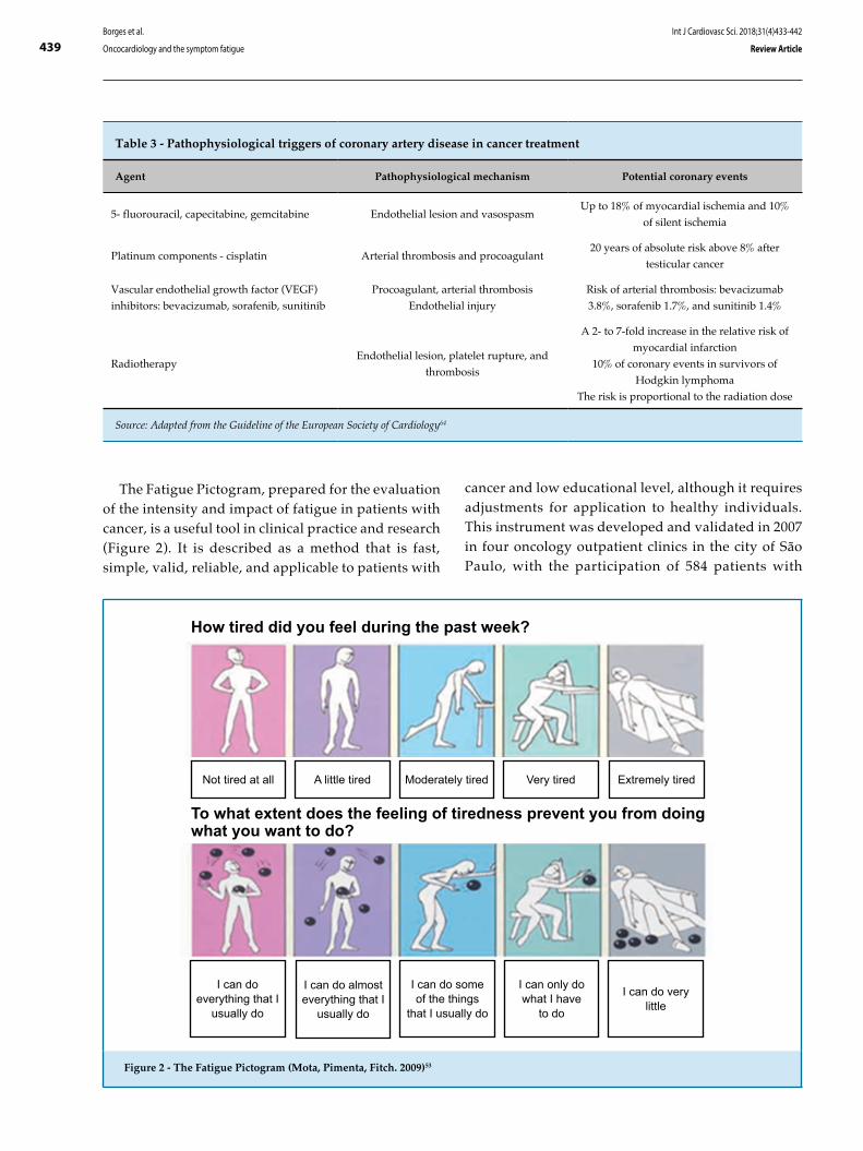
439
Not tired at all
How tired did you feel during the past week?
To what extent does the feeling of tiredness prevent you from doing what you want to do?
A little tired Moderately tired Very tired Extremely tired
I can do everything that I
usually do
I can do almost everything that I
usually do
I can do some of the things
that I usually do
I can only do what I have
to do
I can do very little
Borges et al.
Oncocardiology and the symptom fatigue
Int J Cardiovasc Sci. 2018;31(4)433-442
Review Article
Figure 2 - The Fatigue Pictogram (Mota, Pimenta, Fitch. 2009)53
Table 3 - Pathophysiological triggers of coronary artery disease in cancer treatment
Agent Pathophysiological mechanism Potential coronary events
5- fluorouracil, capecitabine, gemcitabine Endothelial lesion and vasospasmUp to 18% of myocardial ischemia and 10%
of silent ischemia
Platinum components - cisplatin Arterial thrombosis and procoagulant20 years of absolute risk above 8% after
testicular cancer
Vascular endothelial growth factor (VEGF)
inhibitors: bevacizumab, sorafenib, sunitinib
Procoagulant, arterial thrombosis
Endothelial injury
Risk of arterial thrombosis: bevacizumab
3.8%, sorafenib 1.7%, and sunitinib 1.4%
RadiotherapyEndothelial lesion, platelet rupture, and
thrombosis
A 2- to 7-fold increase in the relative risk of
myocardial infarction
10% of coronary events in survivors of
Hodgkin lymphoma
The risk is proportional to the radiation dose
Source: Adapted from the Guideline of the European Society of Cardiology64
The Fatigue Pictogram, prepared for the evaluation of the intensity and impact of fatigue in patients with cancer, is a useful tool in clinical practice and research (Figure 2). It is described as a method that is fast, simple, valid, reliable, and applicable to patients with
cancer and low educational level, although it requires adjustments for application to healthy individuals. This instrument was developed and validated in 2007 in four oncology outpatient clinics in the city of São Paulo, with the participation of 584 patients with
440Borges et al.
Oncocardiology and the symptom fatigue
Int J Cardiovasc Sci. 2018;31(4)433-442
Review Article
different types and stages of cancer receiving or not treatment with radiotherapy.53
Fatigue may also be assessed with the Piper Fatigue Scale. Revised and validated in Brazil in 2009, this scale covers all dimensions of fatigue and can be applied to cancer patients at all stages of the disease. This scale establishes a cutoff point from which the individual should be regarded as fatigued.54 Another widely used questionnaire is the Functional Assessment of Cancer Therapy-Fatigue (FACT-F), which was validated and applied in a study carried out at the Instituto Nacional de Câncer José Alencar Gomes da Silva (INCA) in 2008 and showed a negative impact of fatigue on the quality of life of patients with breast cancer undergoing chemotherapy.55
Conclusion
Although fatigue is a common symptom in patients with cancer, it receives little attention in daily clinical practice. In recent decades, fatigue has been progressively recognized by its impact on the patients’ quality of life and survival. Fatigue is also one of the cardinal symptoms of HF. Cardiac surveillance and oncocardiology are concepts that are being incorporated by multidisciplinary teams caring for patients with cancer. Therefore, the identification of fatigue and its pathophysiological mechanisms, as well as its correct stratification and therapeutic approach, are fundamental steps to be met by healthcare professionals involved in the care of patients with cancer.
Author contributions
Conception and design of the research: Borges JA, Quintão MMP, Chermont SSMC, Mendonça Filho HTF, Mesquita ET. Acquisition of data: Borges JA, Quintão MMP, Chermont SSMC, Mendonça Filho HTF, Mesquita ET. Analysis and interpretation of the data: : Borges JA, Quintão MMP, Chermont SSMC, Mendonça Filho HTF, Mesquita ET. Statistical analysis: Borges JA. Writing of the manuscript: Borges JA, Quintão MMP, Chermont SSMC, Mendonça Filho HTF, Mesquita ET. Critical revision of the manuscript for intellectual content: Borges JA, Quintão MMP, Chermont SSMC, Mendonça Filho HTF, Mesquita ET.
Potential Conflict of Interest
No potential conflict of interest relevant to this article was reported.
Sources of Funding
There were no external funding sources for this study.
Study Association
This article is part of the thesis of master submitted by Jacqueline Aparecida Borges, from Universidade Federal Fluminense and INCA - RJ.
Ethics approval and consent to participate
This article does not contain any studies with human participants or animals performed by any of the authors.
1. Kalil Filho R, Hajjar LA, Bacal F, Hoff PM, Diz Mdel P, Galas FR, et al; Grupo de Estudos em Insuficiência Cardíaca da Sociedade Brasileira de Cardiologia (GEIC/SBC); Sociedade Brasileira de Oncologia Clínica; Instituto do Coração – Faculdade de Medicina da Universidade de São Paulo; Instituto do Câncer do Estado de São Paulo – Faculdade de Medicina da Universidade de São Paulo. [I Brazilian guideline for cardio-oncology from Sociedade Brasileira de Cardiologia]. Arq Bras Cardiol. 2011;96(2 Suppl.1):1-52.
2. Lis CG, Rodeghier M, Grutsch JF, Gupta D. Distribution and determinants of patient satisfaction in oncology with a focus on heath related quality of life. BMC Heath Serv Res. 2009 Oct 21;9:190.
3. Mota DD, Cruz DA, Pimenta CA. Fadiga: uma análise de conceito. Acta Paul Enferm. 2005;18(3):285-93.
4. Evans WJ, Lambert CP. Physiological basis of fatigue. Am J Phys Med Rehabil. 2007;86(1 Suppl):S29-46.
5. Cella D, Davis K, Breitbart W, Curt G; Fatigue Coalition. Cancer-related fatigue: prevalence of proposed diagnostic criteria in a United States sample of cancer survivors. J Clin Oncol. 2001;19(14):3385-91.
6. Gutstein HB. The biologic basis of fatigue. Cancer. 2000; 92(6 Suppl):1678-83.
7. Mettauer B, Zoll J, Sandchez H, Lampert E, Ribera F, Veksler V, et al. Oxidative capacity of skeletal muscle in heart failure patients versus sedentary or active control subjects J Am Coll Cardiol. 2001;38(4):947-54.
8. Alpert CM, Smith MA, Hummel SL, Hummel EK. Symptom burden in heart failure: assement, impact on outcomes, and management. Heart Fail Rev. 2017;22(1):25-39.
9. Brannstrom S, Boman K. Effects of person-central and integrated chronic heart failure and palliative home care. PREFER: a randomized controlled study. Eur J Heart Fail. 2014;16(10):1142-51.
10. Markowitz AJ, Rabow MW. Palliative management of fatigue at the close of life: "it feels like my body is just worn out". JAMA 2007;298(2):217.
11. Barsevick AM, Whitmer K, Walker L. In their own words: using the common sense model to analyze patient descriptions of cancer-related fatigue. Oncol Nurs. 2001;28(9):1363-9.
References

441Borges et al.
Oncocardiology and the symptom fatigue
Int J Cardiovasc Sci. 2018;31(4)433-442
Review Article
12. Zwarts MJ, Bleijenberg G, van Engelen BG. Clinical neurophysiology of fatigue. Clin Neurophysiol. 2008;119(1):2-10.
13. Barry BK, Enoka RM. The neurobiology of muscle fatigue: 15 years later. Integr Comp Biol. 2007;47(4):465-73.
14. Saibene F, Minetti AE. Biomechanical and physiological aspects of legged locomotion in humans. Eur J Appl Physiol. 2003;88(4-5):297-316.
15. Powers SK, Howley ET. Fisiologia do exercício. In: Músculo esquelético; estrutura e função. São Paulo: Manole; 2014.
16. Davis MP, Walsh D. Mechanisms of fatigue. J Support Oncol. 2010;8(4):164-74.
17. Ascensão A, Magalhães J, Oliveira J, Duarte J, Soares J. Fisiologia da fadiga muscular. Delimitação conceptual, modelos de estudo e mecanismos de fadiga de origem central e periférica. Revista Portuguesa de Ciências do Desporto. 2003;3(1):108-23.
18. Mckenna MJ, Medved I, Goodman CA, Brown MJ, Bjorksten AR, Murphy KT, et al. N-acetycysteine attenuates the decline in muscle Na+, K+- pump activity and and delays fatigue during prolonged exercise in humans. J Physiol. 2006;576(Pt 1):279-88.
19. Maughan D, Toth M. Discerning primary and secundary factors responsible for clinical fatigue in multisystem diseases. Biology (Basel). 2014;3(3):606-22.
20. Zambroski CH, Moser DK, Bhat G, Ziegler C. Impact of symptom prevalence and symptom burden on quality of life in patients with heart failure. Eur J Cardiovasc Nurs. 2005;4(3):198-206.
21. Doehmer W, Frenneaux M, Anker SD. Metabolic impairment in heart failure: the myocardic and systemic perspective. J Am Coll Cardiol. 2014;64(13):1388-400.
22. Vescovo G, Dalla Libera L, Serafin F, Leprotti C, Facchin L, Volterrani M, et al. Improved exercise tolerance after Losartan and Enalapril in heart failure: correlation with changes in skeletal muscle myosin heavy chain composition. Circulation. 1998;98(17):1742-9.
23. Dall’ Ago P, Stein R, Ribeiro JP. Exercício em pacientes com Insuficiência Cardíaca: do dogma às evidências. Revista da Sociedade de Cardiologia do Rio Grande do Sul. 2005;13(4):1-5.
24. Miller MS, VanBuren P, LeWinter MM, Braddock JM, Ades PA, Maughan DW, et al. Chronic heart failure decreases cross-bridge kinetics in single skeletal muscle fibres from humans. J Physiol. 2010;588(Pt 20):4039-53.
25. Rehn TA, Munkvik, M, Lunde PK, Sjaastad L, Sejersted OM. Intrinsic skeletal muscle alterations in chronic heart failure patients: a disease-specific myopathy or a result of deconditioning? Heart Fail Rev. 2012;17(3):421-36.
26. Bruera E, Kuehn N, Miller MJ, Selmser P, Macmillan K. The Edmonton Symptom Assssment System (ESAS): a simple method for the assessment of palliative care patients. J Palliat Care.1991;7(2):6-9.
27. Ezekowitz JA, Thai V, Hodnefield TS, Sanderson L, Cujec B. The correlation of standard heart failure assessment and palliative care questionnaries in a multidisciplinary heart failure clinic. J Pain Symptom Manage. 2011;42(3):379-87.
28. Servaes P, Verhagen S, Schreuder HW, Veth RP, Bleijenberg G. Fatigue after treatment for malignant and benign bone and soft tissue tumors. J Pain Symptom Manage. 2003;26(6):1113-22.
29. Roberts BM, Ahn B, Smuder AJ, Al-Rajhi M, Gill LC, Beharry AW, et al. Diaphragm and ventilatory dysfunction during cancer cachexia. FASEB J. 2013;27(7):2600-10.
30. Lee JQ, Simmonds MJ, Wang XS, Novy DM. Difference in physical performance between men and women with and without lymphoma. Arch Phys Med Rehabil 2003;84(12):1747-52.
31. Bower JE, Ganz PA, Tao ML, Hu W, Belin TR, Sepah S, et al. Inflammation biomarkers and fatigue during radiation therapy for breast cancer and prostate cancer. Clin Cancer Res. 2009;15(17):5534-40.
32. Doehner W, von Haehling S, Anker SD, Lainscak M. Neurohormonal activation and inflammation in chronic cardiopulmonary disease: a brief systematic review. Wien Klin Wochenschr. 2009;121(9-10):293-6.
33. Fink MA, Sullivan SL, Zerwic JJ, Piano MR. Fatigue with systolic heart failure. J Cardiovasc Nurs. 2009;24(5):410-7.
34. Escalante CP, Kallen MA, Valdres RU, Morrow PK, Manzullo EF. Outcomes of a cancer-related fatigue clinic in a comprehensive cancer centre. J Pain Simptom Manage. 2010;39(4):691-701.
35. Common Terminology Criteria for Adverse Events (CTCAE) Version 4.0. May 28, 2009. (4.03: June 14, 2010). Bethesda: U.S. Department of Health and Human Services; National Institutes of Health; National Cancer Institute.
36. Dimeo F, Schwartz S, Wesel N, Voigt A, Thiel E. Effects of an endurance and resistance exercice program on persistent cancer-fatigue after treatment. Ann Oncol. 2008;19(8):1495-9.
37. Schwartz AL, Mori M, Gao R, Nail LM, King ME. Exercise reduces daily fatigue in women with breast cancer receiving chemotherapy. Med Sci Sports Exerc. 2001;33(5):718-23.
38. Mishra SI, Scherer RW, Snyder C, Geigle PM, Berlanstein DR, Topaloglu O. Exercise interventions on health-related quality of life for people with cancer during active treatment. Cochrane Database Syst Rev. 2012 Aug 15;(8):CD008465.
39. Yeh ET, Bickford CL. Cardiovascular complications of cancer therapy: incidence, pathogenesis, diagnosis, and management. J Am Coll Cardiol. 2009;53(24):2231-47.
40. Adão R, de Keulenaer G, Leite-Moreira A, Brás-Silva C. Cardiotoxicity associated with cancer therapy: pathophysiology and prevention strategies. Rev Port Cardiol. 2013;32(5):395-409.
41. Suter TM, Ewer MS. Cancer drugs and the heart: importance and management. Eur Heart J. 2013;34(15):1102-11.
42. Swain SM, Whaley FS, Ewer MS. Congestive heart failure in patients treated with doxorubicin: a retrospective analysis of three trials. Cancer. 2003;97(11):2869-79.
43. von Haehling S, Lainscak M, Kung T, Cramer L, Fülster S, Pelzer U, et al. Non-invasive assessment of cardiac hemodynamics in patients with advanced cancer and with chronic heart failure: a pilot feasibility study. Arch Med Sci. 2013;9(2):261-7.
44. Cardinale D, Sandri MT. Role of biomarkers in chemotherapy-induced cardiotoxicity. Prog Cardiovasc Dis. 2010;53(2):121-9.
45. Frickhofen N, Beck FJ, Jung B, Fuhr HG, Andrasch H, Sigmund M. Capecitabine can induce acute coronary syndrome similar to 5-fluorouracil. Ann Oncol. 2002;13(5):797-801.
46. Kosmas C, Kallistratos MS, Kopterides P, Syrios J, Skopelitis H, Mylonaki N, et al. Cardiotoxicity of fluoropyrimidines in different schedulesof administration: a prospective study. J Cancer Res Clin Oncol. 2008;134(1):75-82.
47. Moore RA, Adel N, Riedel E, Bhutani M, Feldman DR, Tabbara NE, et al. High incidence of thromboembolic events in patients treated with cisplatin-based chemotherapy: a large retrospective analysis. J Clin Oncol. 2011;29(25):3466-73.
48. Haugnes HS, Wethal T, Aass N, Dahl O, Klepp O, Langberg CW, et al. Cardiovascular risk factors and morbidity in long-term survivors of testicular cancer: a 20-year follow-up study. J Clin Oncol. 2010;28(30):4649-57.
49. Huddart RA, Norman A, Shahidi M, Horwich A, Coward D, Nicholls J, et al. Cardiovascular disease as a long-term complication of treatment for testicular cancer. J Clin Oncol. 2003;21(8):1513-23.
50. Scappaticci FA, Skillings JR, Holden SN, Gerber HP, Miller K, Kabbinavar F, et al. Arterial thromboembolic events in patients with metastatic carcinoma treated with chemotherapy and bevacizumab. J Natl Cancer Inst. 2007;99(16):1232-9. Erratum in: J Natl Cancer Inst. 2008;100(2):156, J Natl Cancer Inst. 2008;100(9):685.

442Borges et al.
Oncocardiology and the symptom fatigue
Int J Cardiovasc Sci. 2018;31(4)433-442
Review Article
This is an open-access article distributed under the terms of the Creative Commons Attribution License
51. Choueiri TK, Schutz FA, Je Y, Rosenberg JE, Bellmunt J. Risk of arterial thromboembolic events with sunitinib and sorafenib: a systematic review and meta-analysis of clinical trials. J Clin Oncol. 2010;28(13):2280-5.
52. Caponero R. Consenso Brasileiro de fadiga. Revista Brasileira de Cuidados Paliativos. 2010;3(2 supl 1):3-31.
53. Mota DD, Pimenta CA, Fitch MI. Fatigue Pictogram: an option for assessing fatigue severity and impact. Rev Esc Enferm USP. 2009;43(Spe):1079-86.
54. Mota DD, Pimenta CA, Piper BF. Fatigue in Brazilian cancer patients, caregivers, and nursing students: a psychometric validation study of the Piper Fatigue Scale-Revised. Support Care Cancer. 2009;17(6):645-52
55. Ishikawa NM, Thuler LC, Giglio AG, Baldotto CS, de Andrade CJ, Derchain SF, et al. Validation of the Portuguese version of Functional Assessment of Cancer Therapy-Fatigue (FACT-F) in Brazilian cancer patients. Support Care Cancer. 2010;18(4):481-90.

DOI: 10.5935/2359-4802.20180032
443
REVIEW ARTICLE
International Journal of Cardiovascular Sciences. 2018;31(4)443-450
Mailing Address: Danieli de CristoRua Ludovico Della Mea, 685. Postal Code: 99040-170, Vera Cruz, Passo Fundo, RS - Brazil.E-mail: [email protected], [email protected]
Telerehabilitation for Cardiac Patients: Systematic ReviewDanieli de Cristo,1 Natan Pinto do Nascimento,1 Alexandre Simões Dias,2 Amanda Sachetti1
Universidade de Passo Fundo (UPF),1 Rio Grande do Sul, RS - BrazilHospital de Clínicas de Porto Alegre, Universidade Federal do Rio Grande do Sul,2 Rio Grande do Sul, RS - Brazil
Manuscript received 01/04/2018, reviewed 01/16/2018, accepted 02/16/2018.
Cardiovascular Diseases; Cardiac Rehabilitation; Telerehabilitation; Physical Therapy Specialty; Review.
Keywords
Abstract
C a r d i o v a s c u l a r r e h a b i l i t a t i o n i s o n e nonpharmacological intervention used to treat cardiovascular diseases. Despite the proven benefits of cardiovascular rehabilitation, the adherence of patients with heart disease is low. Thus, the alternative of telerehabilitation has gained importance, and many studies are being carried out to verify its efficacy.
To review the literature and assess the efficacy of telerehabilitation for the cardiac population.
This is a systematic review of the literature. The search was conducted in the electronic databases MEDLINE/PubMed (Medical Literature Analysis and Retrieval System Online), PubMed Central® (PMC), Cochrane Library, and Physiotherapy Evidence Database (PEDro), using the combination of descriptors, including terms of the Medical Subject Headings (MeSH) and its entry terms. The MeSH terms used in combination were: “telerehabilitation” AND “cardiac rehabilitation” (Table 1). Then, a manual search by use of the articles selected, as well as a search in the gray literature, was conducted.
The search strategy collected 154 studies, of which 109 were excluded because of duplication in the databases and 29 for not being clinical studies. Sixteen clinical studies were included for full analysis, of which 2 were excluded for being prospective, 2 for being duplicate and 5 for not including any outcome. Thus, 7 studies were included.
Cardiac rehabilitation using telerehabilitation is a feasible and safe alternative to conventional rehabilitation, and has high adherence of patients with heart disease. It can be added to conventional cardiovascular rehabilitation programs or used in isolation.
Introduction
Cardiovascular diseases (CVD) are increasingly frequent. Their epidemiology has been compared to that of the great epidemics of the past centuries.1 According to the World Health Organization (WHO), in recent decades, approximately 30% of a total of 50 million deaths were caused by CVD, 17 million people worldwide.2,3
Similarly, Brazil has equally alarming indices, with CVD as the major cause of death, representing 30% of all causes of death recorded, and being the third major cause of hospitalization in the country.2,3 In addition, WHO states that those diseases are a threat to the socioeconomic development, mainly due to the large number of premature deaths that could be prevented by reducing the risk factors.4
Some nonpharmacological interventions are used to treat CVD, such as cardiovascular rehabilitation (CVR), consisting in the set of interventions aimed at improving the patients’ physical, psychological and social conditions.5 Over the past 40 years, the role of the CVR services in the secondary prevention of cardiovascular events has been recognized and accepted by health organizations, and the interventions used in the care provided to patients with CVD have proved fundamental to treat those individuals.6
Despite the confirmed CVR benefits, low adherence of the patients with CVD has been observed. Some studies have attributed it to the lack of transportation for the patients, lack of time, return to work or
444Cristo et al.
Telerehabilitation for cardiac patients
Int J Cardiovasc Sci. 2018;31(4)443-450
Review Article
financial problems, those being the major hindrances to participation in CVR programs. Some authors have reported that only 27% of the patients adhere to CVR.7
Therefore, different strategies to encourage physical exercise and changes in behavior and lifestyle are necessary and should be implemented to modify the patients’ risk factors, preventing new cardiovascular events and enabling the patients’ return to their usual daily activities.7
Considering all that and the recent technological advance, an alternative to conventional CVR has been the use of the technology of telemedicine,8 which proposes the delivery of healthcare services by use of information and communication technologies in situations where a health professional and a patient (or two health professionals), each in a different place, can communicate in real time, or even enables data storage for further analysis, consultation and opinion. In addition, it provides the safe transmission of medical data via texts, sounds and images required for prevention, diagnosis, treatment and patients’ follow-up.9
Rehabilitation using telemedicine resources is known as telerehabilitation and has gained importance. Several ongoing studies are assessing its efficacy, but they are heterogeneous and use different tools to conduct telerehabilitation. Thus, this study was aimed at reviewing the literature and assessing the efficacy of telerehabilitation for cardiac patients.
Methodology
Study design and search strategy
This is a systematic review of the literature, which does not require Ethics Committee in Research approval, but is being analyzed by the International Prospect ive Regis ter of Systemat ic Reviews (PROSPERO). In addition, this systematic review has met the recommendations of the Preferred Reporting Items for Systematic Reviews and Meta-Analyses (PRISMA). The search was conducted in the electronic databases MEDLINE/PubMed (Medical Literature Analysis and Retrieval System Online), PubMed Central® (PMC), Cochrane Library, and Physiotherapy Evidence Database (PEDro), using the combination of descriptors, including terms of the Medical Subject Headings (MeSH) and its entry terms. The MeSH terms used in combination were: “telerehabilitation” AND “cardiac rehabilitation” (Table 1). Then, a manual search by use of the articles selected, as well as a search in the gray literature, was conducted.
Inclusion and exclusion criteria
This study included all randomized or nonrandomized clinical trials found in the databases, published in Portuguese, English or Spanish, with the full text available and no date restriction, conducted in human beings aged at least 18 years, in which patients with CVD participated in CVR programs, using telerehabilitation or telemedicine resources.
Studies with the following characteristics were excluded: duplicate studies; not performed in human beings; not published in full text; whose population had been studied in more than one study and whose outcomes were similar, situations in which the first study was considered for inclusion in this review.
Two reviewers evaluated independently the abstracts. The studies selected had their full text assessed for inclusion according to the criteria established.
Identification and selection of studies
Two reviewers independently read the titles and abstracts of each pre-selected study, identifying
Table 1 - Search strategy used in PubMed
#1
(“Telerehabilitation”[mesh] OR “Telerehabilitations”
OR “Tele-rehabilitation” OR “Tele rehabilitation” OR
“Tele-rehabilitations” OR “Remote Rehabilitation” OR
“Rehabilitation, Remote” OR “Rehabilitations, Remote” OR
“Remote Rehabilitations” OR “Virtual Rehabilitation” OR
“Rehabilitation, Virtual” OR “Rehabilitations, Virtual” OR
“Virtual Rehabilitations”)
#2
(“Cardiac rehabilitation” [mesh] OR “Cardiac
Rehabilitations” OR “Rehabilitation, Cardiac” OR
“Rehabilitations, Cardiac” OR “Cardiovascular
Rehabilitation” OR “Cardiovascular Rehabilitations” OR
“Rehabilitation, Cardiovascular” OR “Rehabilitations,
Cardiovascular”)
#3
(randomized controlled trial[pt] OR controlled clinical
trial[pt] OR randomized controlled trials[mh] OR random
allocation[mh] OR double-blind method[mh] OR single-
blind method[mh] OR clinical trial[pt] OR clinical trials[mh]
OR (“clinical trial”[tw]) OR ((singl*[tw] OR doubl*[tw] OR
trebl*[tw] OR tripl*[tw]) AND (mask*[tw] OR blind*[tw]))
OR (“latin square”[tw]) OR placebos[mh] OR placebo*[tw]
OR random*[tw] OR research design[mh:noexp] OR follow-
up studies[mh] OR prospective studies[mh] OR cross-
over studies[mh] OR control*[tw] OR prospectiv*[tw] OR
volunteer*[tw]) NOT (animal[mh] NOT human[mh])
#4 (#1 AND #2 AND #3)
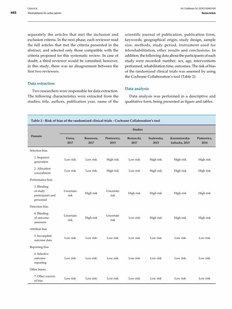
445Cristo et al.
Telerehabilitation for cardiac patients
Int J Cardiovasc Sci. 2018;31(4)443-450
Review Article
Table 2 - Risk of bias of the randomized clinical trials - Cochrane Collaboration’s tool
Domain
Studies
Vieira,
2017
Brouwers,
2017
Piotrowicz,
2015
Bernocchi,
2017
Szalewska,
2015
Korzeniowska-
kubacka, 2015
Piotrowicz,
2014
Selection bias
1. Sequence
generationLow risk Low risk High risk Low risk High risk High risk High risk
2. Allocation
concealmentLow risk Low risk High risk Low risk High risk High risk High risk
Performance bias
3. Blinding
of study
participants and
personnel
Uncertain
riskHigh risk
Uncertain
riskHigh risk High risk High risk High risk
Detection bias
4. Blinding
of outcome
assessors
Uncertain
riskHigh risk
Uncertain
riskLow risk High risk High risk High risk
Attrition bias
5. Incomplete
outcome dataLow risk Low risk Low risk Low risk Low risk Low risk Low risk
Reporting bias
6. Selective
outcome
reporting
Low risk Low risk Low risk Low risk Low risk Low risk Low risk
Other biases
7. Other sources
of biasLow risk Low risk Low risk Low risk Low risk Low risk Low risk
separately the articles that met the inclusion and exclusion criteria. In the next phase, each reviewer read the full articles that met the criteria presented in the abstract, and selected only those compatible with the criteria proposed for this systematic review. In case of doubt, a third reviewer would be consulted; however, in this study, there was no disagreement between the first two reviewers.
Data extraction
Two researchers were responsible for data extraction. The following characteristics were extracted from the studies: title, authors, publication year, name of the
scientific journal of publication, publication form, keywords, geographical origin, study design, sample size, methods, study period, instrument used for telerehabilitation, other results and conclusions. In addition, the following data about the participants of each study were recorded: number, sex, age, interventions performed, rehabilitation time, outcomes. The risk of bias of the randomized clinical trials was assessed by using the Cochrane Collaboration’s tool (Table 2).
Data analysis
Data analysis was performed in a descriptive and qualitative form, being presented as figure and tables.
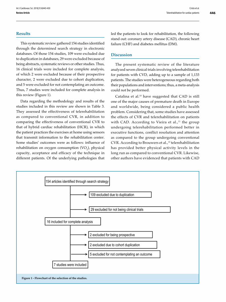
446Cristo et al.
Telerehabilitation for cardiac patients
Int J Cardiovasc Sci. 2018;31(4)443-450
Review Article
Figure 1 - Flowchart of the selection of the studies.
29 excluded for not being clinical trials
154 articles identified through search strategy
109 excluded due to duplication
16 included for complete analysis
2 excluded for being prospective
2 excluded due to cohort duplication
5 excluded for not contemplating an outcome
7 studies were included
Results
This systematic review gathered 154 studies identified through the determined search strategy in electronic databases. Of those 154 studies, 109 were excluded due to duplication in databases, 29 were excluded because of being abstracts, systematic reviews or other studies. Thus, 16 clinical trials were included for complete analysis, of which 2 were excluded because of their prospective character, 2 were excluded due to cohort duplication, and 5 were excluded for not contemplating an outcome. Thus, 7 studies were included for complete analysis in this review (Figure 1).
Data regarding the methodology and results of the studies included in this review are shown in Table 3. They assessed the effectiveness of telerehabilitation as compared to conventional CVR, in addition to comparing the effectiveness of conventional CVR to that of hybrid cardiac rehabilitation (HCR), in which the patient practices the exercises at home using sensors that transmit information to the rehabilitation center. Some studies’ outcomes were as follows: influence of rehabilitation on oxygen consumption (VO2), physical capacity, acceptance and efficacy of the technique in different patients. Of the underlying pathologies that
led the patients to look for rehabilitation, the following stand out: coronary artery disease (CAD), chronic heart failure (CHF) and diabetes mellitus (DM).
Discussion
The present systematic review of the literature analyzed seven clinical trials involving telerehabilitation for patients with CVD, adding up to a sample of 1,133 patients. The studies were heterogeneous regarding both their populations and interventions; thus, a meta-analysis could not be performed.
Catalina et al.10 have suggested that CAD is still one of the major causes of premature death in Europe and worldwide, being considered a public health problem. Considering that, some studies have assessed the effects of CVR and telerehabilitation on patients with CAD. According to Vieira et al.,11 the group undergoing telerehabilitation performed better in executive functions, conflict resolution and attention as compared to the group undergoing conventional CVR. According to Brouwers et al.,12 telerehabilitation has provided better physical activity levels in the long run as compared to conventional CVR. Likewise, other authors have evidenced that patients with CAD
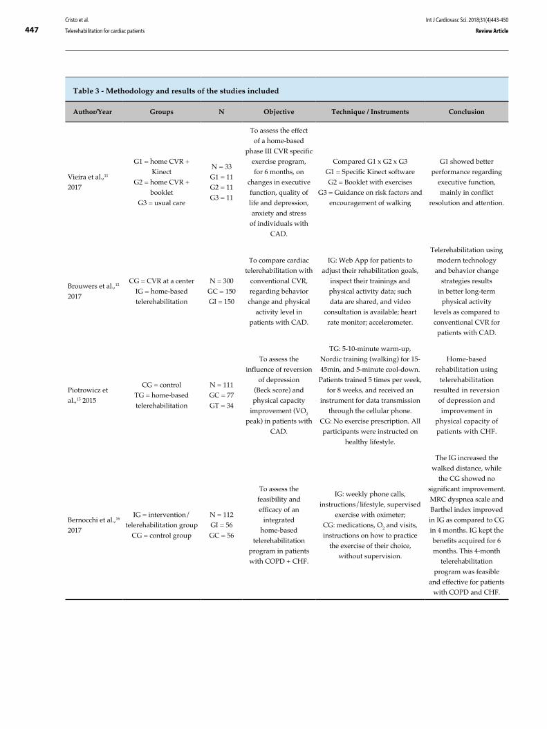
447
Table 3 - Methodology and results of the studies included
Author/Year Groups N Objective Technique / Instruments Conclusion
Vieira et al.,11
2017
G1 = home CVR +
Kinect
G2 = home CVR +
booklet
G3 = usual care
N = 33
G1 = 11
G2 = 11
G3 = 11
To assess the effect
of a home-based
phase III CVR specific
exercise program,
for 6 months, on
changes in executive
function, quality of
life and depression,
anxiety and stress
of individuals with
CAD.
Compared G1 x G2 x G3
G1 = Specific Kinect software
G2 = Booklet with exercises
G3 = Guidance on risk factors and
encouragement of walking
G1 showed better
performance regarding
executive function,
mainly in conflict
resolution and attention.
Brouwers et al.,12
2017
CG = CVR at a center
IG = home-based
telerehabilitation
N = 300
GC = 150
GI = 150
To compare cardiac
telerehabilitation with
conventional CVR,
regarding behavior
change and physical
activity level in
patients with CAD.
IG: Web App for patients to
adjust their rehabilitation goals,
inspect their trainings and
physical activity data; such
data are shared, and video
consultation is available; heart
rate monitor; accelerometer.
Telerehabilitation using
modern technology
and behavior change
strategies results
in better long-term
physical activity
levels as compared to
conventional CVR for
patients with CAD.
Piotrowicz et
al.,15 2015
CG = control
TG = home-based
telerehabilitation
N = 111
GC = 77
GT = 34
To assess the
influence of reversion
of depression
(Beck score) and
physical capacity
improvement (VO2
peak) in patients with
CAD.
TG: 5-10-minute warm-up,
Nordic training (walking) for 15-
45min, and 5-minute cool-down.
Patients trained 5 times per week,
for 8 weeks, and received an
instrument for data transmission
through the cellular phone.
CG: No exercise prescription. All
participants were instructed on
healthy lifestyle.
Home-based
rehabilitation using
telerehabilitation
resulted in reversion
of depression and
improvement in
physical capacity of
patients with CHF.
Bernocchi et al.,16
2017
IG = intervention/
telerehabilitation group
CG = control group
N = 112
GI = 56
GC = 56
To assess the
feasibility and
efficacy of an
integrated
home-based
telerehabilitation
program in patients
with COPD + CHF.
IG: weekly phone calls,
instructions/lifestyle, supervised
exercise with oximeter;
CG: medications, O2 and visits,
instructions on how to practice
the exercise of their choice,
without supervision.
The IG increased the
walked distance, while
the CG showed no
significant improvement.
MRC dyspnea scale and
Barthel index improved
in IG as compared to CG
in 4 months. IG kept the
benefits acquired for 6
months. This 4-month
telerehabilitation
program was feasible
and effective for patients
with COPD and CHF.
Cristo et al.
Telerehabilitation for cardiac patients
Int J Cardiovasc Sci. 2018;31(4)443-450
Review Article
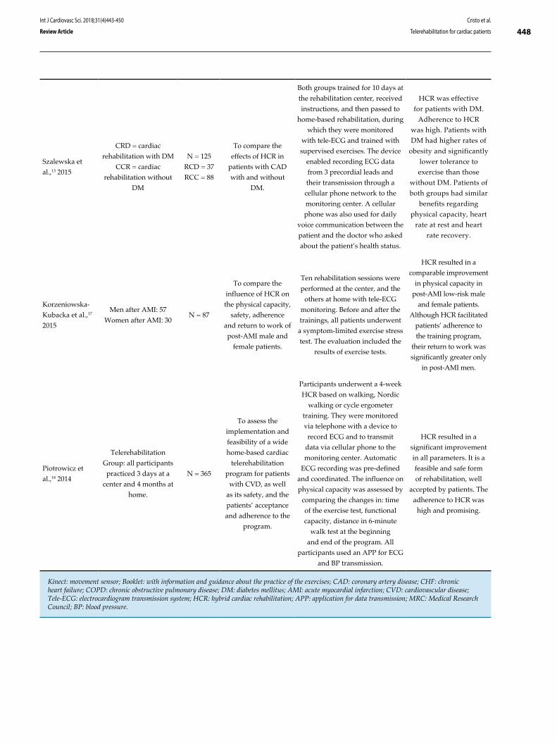
448
Szalewska et
al.,13 2015
CRD = cardiac
rehabilitation with DM
CCR = cardiac
rehabilitation without
DM
N = 125
RCD = 37
RCC = 88
To compare the
effects of HCR in
patients with CAD
with and without
DM.
Both groups trained for 10 days at
the rehabilitation center, received
instructions, and then passed to
home-based rehabilitation, during
which they were monitored
with tele-ECG and trained with
supervised exercises. The device
enabled recording ECG data
from 3 precordial leads and
their transmission through a
cellular phone network to the
monitoring center. A cellular
phone was also used for daily
voice communication between the
patient and the doctor who asked
about the patient’s health status.
HCR was effective
for patients with DM.
Adherence to HCR
was high. Patients with
DM had higher rates of
obesity and significantly
lower tolerance to
exercise than those
without DM. Patients of
both groups had similar
benefits regarding
physical capacity, heart
rate at rest and heart
rate recovery.
Korzeniowska-
Kubacka et al.,17
2015
Men after AMI: 57
Women after AMI: 30N = 87
To compare the
influence of HCR on
the physical capacity,
safety, adherence
and return to work of
post-AMI male and
female patients.
Ten rehabilitation sessions were
performed at the center, and the
others at home with tele-ECG
monitoring. Before and after the
trainings, all patients underwent
a symptom-limited exercise stress
test. The evaluation included the
results of exercise tests.
HCR resulted in a
comparable improvement
in physical capacity in
post-AMI low-risk male
and female patients.
Although HCR facilitated
patients’ adherence to
the training program,
their return to work was
significantly greater only
in post-AMI men.
Piotrowicz et
al.,18 2014
Telerehabilitation
Group: all participants
practiced 3 days at a
center and 4 months at
home.
N = 365
To assess the
implementation and
feasibility of a wide
home-based cardiac
telerehabilitation
program for patients
with CVD, as well
as its safety, and the
patients’ acceptance
and adherence to the
program.
Participants underwent a 4-week
HCR based on walking, Nordic
walking or cycle ergometer
training. They were monitored
via telephone with a device to
record ECG and to transmit
data via cellular phone to the
monitoring center. Automatic
ECG recording was pre-defined
and coordinated. The influence on
physical capacity was assessed by
comparing the changes in: time
of the exercise test, functional
capacity, distance in 6-minute
walk test at the beginning
and end of the program. All
participants used an APP for ECG
and BP transmission.
HCR resulted in a
significant improvement
in all parameters. It is a
feasible and safe form
of rehabilitation, well
accepted by patients. The
adherence to HCR was
high and promising.
Kinect: movement sensor; Booklet: with information and guidance about the practice of the exercises; CAD: coronary artery disease; CHF: chronic heart failure; COPD: chronic obstructive pulmonary disease; DM: diabetes mellitus; AMI: acute myocardial infarction; CVD: cardiovascular disease; Tele-ECG: electrocardiogram transmission system; HCR: hybrid cardiac rehabilitation; APP: application for data transmission; MRC: Medical Research Council; BP: blood pressure.
Cristo et al.
Telerehabilitation for cardiac patients
Int J Cardiovasc Sci. 2018;31(4)443-450
Review Article

449Cristo et al.
Telerehabilitation for cardiac patients
Int J Cardiovasc Sci. 2018;31(4)443-450
Review Article
1. Simão AF, Précoma DB, Andrade JP, Correa Filho H, Saraiva JF, Oliveira GM; Brazilian Society of Cardiology. I cardiovascular prevention guideline of the Brazilian Society of Cardiology - executive summary. Arq Bras Cardiol. 2014;102(5):420-31.
2. Braig S, Peter R, Nagel G, Hermann S, Rohrmann S, Linseisen J. The impact of social status inconsistency on cardiovascular risk factors, myocardial infarction and stroke in the EPIC-Heidelberg cohort. BMC Public Health 2011 Feb 16;11:104.
References
submitted to telerehabilitation showed a significant increase in their daily activity level and VO2 peak after 6 weeks.
Hybrid cardiac rehabilitation has been used for patients with CAD. Szalewska et al.13 have compared the use of that technique in patients with CAD and DM and patients with DAC but without DM. Those authors have reported that adherence to HCR was high, and that HCR was effective in patients with and without DM.
According to Bocchi et al.,14 heart failure (HF) is the common end of most heart diseases, being classified as an epidemic and representing one of the most important current clinical challenges in health care. Telemedicine has been increasingly used for that population. In a clinical study with 111 patients with HF, Piotrowicz et al.15 have shown that home-based rehabilitation using telerehabilitation caused reversion of depression and improved physical capacity in those patients.
Corroborating the results demonstrated, Bernocchi et al.16 have suggested, in a study with 112 patients diagnosed with HF and chronic obstructive pulmonary disease (COPD), that a home-based telerehabilitation program increased the walked distance, reduced the dyspnea and improved the functionality of those individuals as compared to those of the group undergoing conventional CVR, confirming the feasibility and effectiveness of telerehabilitation programs for patients with HF and COPD.
Hybrid cardiac rehabilitation has also been used for post-acute myocardial infarction (AMI) patients. In a study with 87 post-AMI patients, the authors have evidenced that HCR facilitated patients’ adherence to the training program, but the return-to-work indices were higher in men than in women, although the physical capacity improvement was similar for both sexes.17
Similarly to the studies assessed, Piotrowicz et al.18 have confirmed in a sample of 365 patients that HCR using telerehabilitation resulted in a significant improvement in functional capacity, being a feasible,
safe and well accepted rehabilitation form, with a high index of patients’ adherence.
Conclusion
After analyzing the studies, we concluded that HCR and home-based rehabilitation using telerehabilitation are feasible and safe alternatives, with high adherence by patients with CVD. They can be added to conventional CVR programs or be used in isolation. In addition, they help to improve depression, functional capacity and the physical activity level.
Author contributions
Conception and design of the research: Cristo D, Dias AS, Sachetti A. Acquisition of data: Cristo D, Nascimento NP, Sachetti A. Analysis and interpretation of the data: Cristo D, Nascimento NP, Sachetti A. Statistical analysis: Cristo D. Writing of the manuscript: Cristo D, Sachetti A. Critical revision of the manuscript for intellectual content: Cristo D, Dias AS, Sachetti A.
Potential Conflict of Interest
No potential conflict of interest relevant to this article was reported.
Sources of Funding
There were no external funding sources for this study.
Study Association
This study is not associated with any thesis or dissertation work.
Ethics approval and consent to participate
This article does not contain any studies with human participants or animals performed by any of the authors.

450Cristo et al.
Telerehabilitation for cardiac patients
Int J Cardiovasc Sci. 2018;31(4)443-450
Review Article
This is an open-access article distributed under the terms of the Creative Commons Attribution License
3. Tuan TS, Venâncio TS, Nascimento LF. Effects of air pollutant exposure on acute myocardial infarction, according to gender. Arq Bras Cardiol. 2016;107(3):216-22.
4. Duncan BB, ChorII D, Aquino EML, Bensenor IM, Mil JG, SchmidtI MI, et al. Chronic non-communicable diseases in Brazil: priorities for disease management and research. Rev Saúde Pública. 2012;46(Suppl):126-34.
5. Trevisan MD. Reabilitação cardiopulmonar e metabólica fase i no pós-operatório de cirurgia de revascularização do miocárdio utilizando cicloergômetro: um ensaio clínico randomizado. [Dissertação]. Porto Alegre: Pontifícia Universidade Católica do Rio Grande do Sul - Programa de Pós-graduação em Gerontologia Biomédica; 2015.
6. Herdy AH, López-Jimenez F, Terzic CP, Milani M, Stein R, Carvalho T; Sociedade Brasileira de Cardiologia. South American guidelines for cardiovascular disease prevention and rehabilitation. Arq Bras Cardiol. 2014;103(2 Suppl 1):1-31.
7. Frederix I, Vanhees L, Dendale P, Goetschalckx K. A review of telerehabilitation for cardiac patients. J Telemed Telecare. 2015;21(1):45-53.
8. Piotrowicz E. How to do: telerehabilitation in heart failure patients. Cardiol J. 2012;19(3):243-8.
9. Oliveira Jr MT, Canesin MF, Marcolino MS, Ribeiro AL, Carvalho AC, Reddy S, et al; Sociedade Brasileira de Cardiologia. [Telemedicine guideline in patient care with acute coronary syndrome and other heart diseases]. Arq Bras Cardiol. 2015;104(5 Supp. 1):1-26.
10. Catalina CO, Adina B, Smarandita BE, Angela D, Dan G, Silvia M. Cardiovascular lipid risk factors and rate of cardiovascular events after myocardial revascularization. Int J Cardiovasc Sci. 2017;30(1):4-10.
11. Vieira A, Melo C, Machado J, Joaquim Gabriel. Virtual reality exercise on a home-based phase III cardiac rehabilitation program, effect on executive
function, quality of life and depression, anxiety and stress: a randomized controlled trial. Disabil Rehabil Assist Technol. 2018;13(2):112-23.
12. Brouwers RW, Kraal JJ, Traa SC, Spee RF, Oostveen LM, Kemps HM. Effects of cardiac telerehabilitation in patients with coronary artery disease using a personalised patient-centred web application: protocol for the SmartCare-CAD randomised controlled trial. BMC Cardiovasc Disord. 2017;17(1):46.
13. Szalewska D, Zieliński P, Tomaszewski J, Kusiak-Kaczmarek M, Łepska L, Gierat-Haponiuk K, et al. Effects of outpatient followed by home-based telemonitored cardiac rehabilitation in patients with coronary artery disease. Kardiol Pol. 2015;73(11):1101-7.
14. Bocchi EA, Marcondes-Braga FG, Bacal F, Ferraz AS, Albuquerque D, Rodrigues Dde A, et al. [Updating of the Brazilian guideline for chronic heart failure - 2012]. Arq Bras Cardiol. 2012;98(1 Suppl 1):1-33.
15. Piotrowicz E, Piotrowski W, Piotrowicz R. Positive effects of the reversion of depression on the sympathovagal balance after telerehabilitation in heart failure patients. Ann Noninvasive Electrocardiol. 2016;21(4):358-68.
16. Bernocchi P, Vitacca M, La Rovere MT, Volterrani M, Galli T, Baratt D, et al. Home-based telerehabilitation in older patients with chronic obstructive pulmonary disease and heart failure: a randomised controlled trial. Age Ageing. 2018;47(1):82-88.
17. Korzeniowska-Kubacka I, Bilińska M, Dobraszkiewicz-Wasilewska B, Piotrowicz R. Hybrid model of cardiac rehabilitation in men and women after myocardial infarction. Cardiol J. 2015;22(2):212-8.
18. Piotrowicz E, Korzeniowska-Kubacka I, Chrapowicka A, Wolszakiewicz J, Dobraszkiewicz-Wasilewska B, Batogowski M, et al. Feasibility of home-based cardiac telerehabilitation: results of TeleInterMed study. Cardiol J. 2014;21(5):539-46.
DOI: 10.5935/2359-4802.20180029
451
VIEWPOINT
International Journal of Cardiovascular Sciences. 2018;31(4)451-453
Mailing Address: Ana Luisa Rocha MalletRua Almirante Alexandrino, 1808/SS-302. Postal Code: 20241-263, Santa Teresa, RJ - Brazil.E-mail: [email protected]
Cardiology and Films: An Important Teaching ToolAna Luisa Rocha Mallet,1,2 Fatima Geovanini,2 Luciana Andrade,2 David Kestenberg2
Hospital Clementino Fraga Filho, Universidade Federal do Rio de Janeiro (UFRJ);1 Rio de Janeiro, RJ - BrazilUniversidade Estácio de Sá,2 Rio de Janeiro, RJ - Brazil
Manuscript received 11/12/2017, reviewed 12/18/2017, accepted 02/16/2018.
Communication; Humans; Cardiovascular Diseases; Neoplasms; Physician Patient Relations; Prognosis; Truth Disclosure.
Keywords
The use of films and literary texts as didactic material in medical school has increased based on the perception that it might benefit the discussion of certain technical aspects of clinical conditions and enable students to experience the illness process.
An article on literature and cardiology1 has shown that the description of the death process in Tolstoy’s novel “The death of Ivan Ilyich” could be more powerful to provide medical students and physicians with the perception of the finitude process than reading theoretical texts about the end of life. Similarly, Marguerite Yourcenar’s “Memoirs of Hadrian” could help to understand the suffering of a patient with heart failure.
Similarly to literature, films have been used for medical education to discuss questions related mainly to ethics and medical practice. Some films, such as “Patch Adams”, “Cry Danger”, “The Sea Inside”, “Black Swan”, “As Good as it Gets”, “Wit”, are classically presented to medical students for medical education.
If on the one hand, by telling a story and showing an image, films can reduce our ability to imagine how the story’s characters and places would be, on the other, they provide us with a different aesthetic experience, usually shorter than book reading, by offering images and sounds that affect our sensitivity in a different way than book reading does. Both literature and films allow us access to a level of internalization difficult to achieve when reading a medical book or text about a certain disease. Medical books provide the description of cells, organs and diseases, how to establish a diagnosis, in
addition to the drugs and other treatments to be used, while literature and films provide us with the experience of patients with their illnesses.
Regarding cardiology, the year 2016 was particularly interesting, because two awarded films, “Manchester by the Sea” and “I, Daniel Blake”, approached under different aspects one of the most prevalent heart conditions worldwide: heart failure.
Kenneth Lonergan’s “Manchester by the Sea” was awarded with the 2017 Oscar for best actor in a leading role (Casey Affleck) and best original screenplay. Its central plot is the return of a man to his hometown after the death of his brother to take care of his 16-year-old nephew, re-opening an unspeakable tragedy. The film scene that interests us, due to its potential to foster a cardiology class discussion, lasts less than three minutes and approaches the disclosure of his brother’s heart failure diagnosis to the patient and his family (wife, brother and father) by a female doctor during one of his hospitalizations. The doctor talks with the patient about his diagnosis and prognosis, explaining that the data presented were statistics and not definitive numbers about a particular patient. The scene raises ethical and technical questions, such as possible heart failure etiologies, its treatment, its mechanisms of death, its prognosis. In addition, that scene provides us with the different ways to react to such talk, always difficult, especially the patient’s and his wife’s reactions.
Talks involving the disclosure of the diagnosis and prognosis of life-threatening diseases, also known as bad news, are one of the most difficult attributions of a doctor. Bad news in the healthcare field are any information that will drastically affect the future perspective of patients and families. Difficulties in communicating bad news usually postpone discussions with the patients and families about terminal diseases,

452Mallet et al.
Cardiology and films
Int J Cardiovasc Sci. 2018;31(4)451-453
Viewpoint
proximity of death and indication for exclusive palliative care.2-4
But we might have to face even tougher situations when managing patients with heart failure, such as answering the following questions: “How many years do I still have?”, “Will I live to attend my child’s graduation?”, “Will I live to see my grandchildren grow up?”, and other questions that might cross their mind. Perhaps one of the defenses we put up is to restrict all room for that sort of questioning. In addition to not being the proper solution, this attitude somehow postpones coping with those issues, which ends up avoiding that discussion, because, not rarely, the outcome soon becomes clear to both patients and families. However, that attitude hinders the communication with patients and families, delaying their understanding of the situation.
In addition, the discussion about that scene can go beyond medicine as we realize that the diagnosis is established by a female doctor of Asian origin. This can lead us to debate our reality in the Brazilian medicine, in whose graduation programs female students predominate. How will this affect medical practice? Will this change the doctor-patient relationship? Moreover, the Asian origin of the doctor can open the discussion about “the other”, “the foreigner” in a country that seems to move away from its cosmopolitan tradition. Perhaps we could draw a parallel to the issue that “the other”, through the quota system, affirmative actions, ProUni and FIES, has become an increasing presence at medical schools in Brazil, one of the most elitist courses in the country.
The other film from 2016, Ken Loach’s “I, Daniel Blake”, was awarded with the Palm d’Or in the Cannes Film Festival and also approaches the heart failure issue, but from a social perspective. In that film, set in England, Daniel Blake, interpreted by Dave Johns, after having a heart attack, is advised by his doctor not to return to his work at a carpentry, and, thus, must fight the bureaucratic forces of the system to receive Employment and Support Allowance. He faces the bureaucratic system, the coldness of the institutions, which supposedly exist to make the patients’ lives easier, the difficulties enlarged by his digital illiteracy, and a whole process that reminds us of the Franz Kafka’s novel “The Trial”, which tells about the distressing, meaningless and cruel bureaucratic trial to which citizens are submitted.
Watching this film can bring to the classroom discussions about situations rarely approached in lectures on myocardial infarction and heart failure, but which are extremely frequent
when managing patients with such conditions. Who has not been asked: “Doctor, when can I go back to work?”, “Doctor, am I entitled to retirement benefits?”, “How do I receive my support allowance?”, “Doctor, they have cut my benefits. What should I do?”. These situations are so frequent for those following patients up after myocardial infarction and with heart failure that we have to know something about the legislation to instruct our patients in a responsible way regarding their rights, providing them with reports to allow them to have their rights ensured. Usually such reports need to be systematically renewed, which not rarely makes the patient feel shy, fearing to be bothering the doctor.
An additional theme for discussion in that film is the question of “the other”. In one of his several visits to the government offices, Daniel Blake befriends a white woman who had just moved to the city and also seeks for social support. She has two children, one of them is a black girl, who also suffers with the social security insensitivity. Although our patients might face much bigger difficulties than English citizens do, this film shows that bureaucracy and insensitivity are not exclusive characteristics of our society.
Conclusion
The use of films or film scenes is an important tool to discuss heart conditions, not only regarding the disease’s technical aspects, but also the social, ethical and existential aspects involved in the diagnosis of a severe heart disease. To quote Arthur Kleinman, the laureate films “Manchester by the Sea” and “I, Daniel Blake” are examples that can help us practice medicine in a more sensitive and reflexive, alert and morally responsible way in face of the emotional and experiential challenges presented by the profession.5
Author contributions
Conception and design of the research: Mallet ALR, Geovanini F, Andrade L, Kestenberg D. Analysis and interpretation of the data: Mallet ALR. Writing of the manuscript: Mallet ALR, Geovanini F, Andrade L, Kestenberg D. Critical revision of the manuscript for intellectual content: Mallet ALR, Geovanini F, Andrade L, Kestenberg D.
Potential Conflict of Interest
No potential conflict of interest relevant to this article was reported.

453Mallet et al.
Cardiology and films
Int J Cardiovasc Sci. 2018;31(4)451-453
Viewpoint
This is an open-access article distributed under the terms of the Creative Commons Attribution License
1. Mallet AL, Andrade L, Dias MC. Literature and cardiology. Int J Cardiovasc Sci. 2015;28(4):335-7.
2. Baile WF, Buckman R, Lenzi R, Glober G, Beale EA, Kudelka AP. SPIKES-A six-step protocol for delivering bad news: application to the patient with cancer. Oncologist. 2000;5(4):302-11.
3. Geovanini F. Apoderando-se do câncer “dos outros”: contribuições da ética das virtudes ao estudo da comunicação de prognósticos
de câncer a pacientes e familiares. [Tese]. Rio de Janeiro: Fundação Oswaldo Cruz; 2015.
4. Ragan SL, Wittenberg E, Hall HT. The communication of palliative care for the elderly cancer patients. Health Commun. 2003;15(2):219-26.
5. Kleinman A. The illness narratives: suffering, healing & the human condition. United States of America: Basic Books; 1989.
References
Sources of Funding
This study was funded by "Research Productivity" study grant of the medical course of Universidade Estácio de Sá.
Study Association
This study is not associated with any thesis or dissertation work.
DOI: 10.5935/2359-4802.20180040
454
CASE REPORT
International Journal of Cardiovascular Sciences. 2018;31(4)454-456
Mailing Address: Marcos Garcia-GuimaraesCalle Tutor 68 Bajo interior Izquierda. Postal Code: 28008, Madrid - Spain.E-mail: [email protected], [email protected]
Sirolimus-Eluting Balloon Treatment of Distal Internal Mammary Artery Anastomosis: Optical Coherence Tomography FindingsMarcos Garcia-Guimarães, Ramón Maruri-Sanchez, Javier Cuesta, Fernando Rivero, Teresa Bastante, Fernando AlfonsoHospital Universitario de La Princesa, Madrid - Spain
Manuscript received March 25, 2017, revised manuscript October 23, 2017, accepted February 22, 2018.
Angioplasty, Balloon, Coronary / methods; Coronary Artery Disease / diagnosis; Drug-Eluting Stents; Treatment Outcome; Sirolimus.
Keywords
Drug-eluting balloons (DEB) represent an interesting alternative to current generation drug-eluting stents (DES) in selected anatomic scenarios, such as in-stent restenosis, small-vessel disease and bifurcation lesions, where the systematic implantation of DES may result less appealing.1,2 In particular, lesions located at the distal anastomosis of a bypass graft (an artificially created bifurcation) constitute a unique anatomic setting, where DEB may overcome some inherent limitations of DES resulting from the implantation of a metallic platform throughout the bypass graft into and across the native distal vessel.
Herein we present, to the best of our knowledge, the first report of the treatment of a lesion located at the distal anastomosis of a left internal mammary artery (LIMA) to the left anterior descending coronary artery (LAD) with a novel sirolimus-DEB. Interestingly, optical coherence tomography (OCT) findings proved to be of major value to guide the procedure.
An 83-year-old man with previous history of hypertension, hyperlipidemia and ex-smoker was admitted with acute coronary syndrome. Coronary angiography revealed 3-vessel coronary artery disease, with a total occlusion at the mid-segment of the LAD and additional significant lesions at the obtuse marginal branch (OM) and mid-segment of the right coronary artery (RCA). After discussion in a Heart-Team meeting, a decision for a hybrid revascularization strategy was made. Off-pump coronary artery bypass grafting (CABG) was performed with a LIMA anastomosed to the distal
LAD and a saphenous vein graft from LIMA to OM. The postoperative period was uneventful. A week later, the scheduled percutaneous coronary intervention was successfully performed with a DES implanted at the mid RCA. However, during the procedure a revision of the bypass grafts revealed the presence of a significant lesion at the distal anastomosis of LIMA to LAD (Figure 1A). Due to a suspicious of pseudo-stenosis secondary to edema or spasm related to the recent surgery, it was decided to reevaluate this lesion later on. The scheduled control angiogram at 1 month revealed the persistence of the same image of a severe lesion at the distal anastomosis of the LIMA. The lesion appearance remained unaltered after repeated boluses of nitroglycerin. To gain further diagnostic insights, OCT imaging was performed. This imaging technique confirmed the presence of a critical stenosis with a severely reduced lumen (minimal lumen area 1.0 mm2) (Figure 2A). After lesion predilation with a 2.0 mm semi-compliant balloon, a 2.5/15 mm sirolimus-DEB (MagicTouch, Concept Medical Inc, Surat, India) was used to treat the distal anastomosis (Figure 1B). A good immediate result was confirmed both by angiography (Figure 1C) and OCT (Figures 2B and 2C). The patient was discharged the day after and remains completely asymptomatic at the 9-month follow-up.
The diagnosis of lesions located in the distal LIMA anastomosis may be very challenging. When these lesions are detected in the early postoperative period technical problems should be differentiated from reversible causes, such as edema or spasm. Notably, however, the value of OCT to get further diagnostic insights on these elusive lesions has not been previously reported. Our findings underscore the usefulness of this technique to disclose the underlying substrate and to assess lesion severity before intervention and to guide and optimize the results of interventions. Lesions
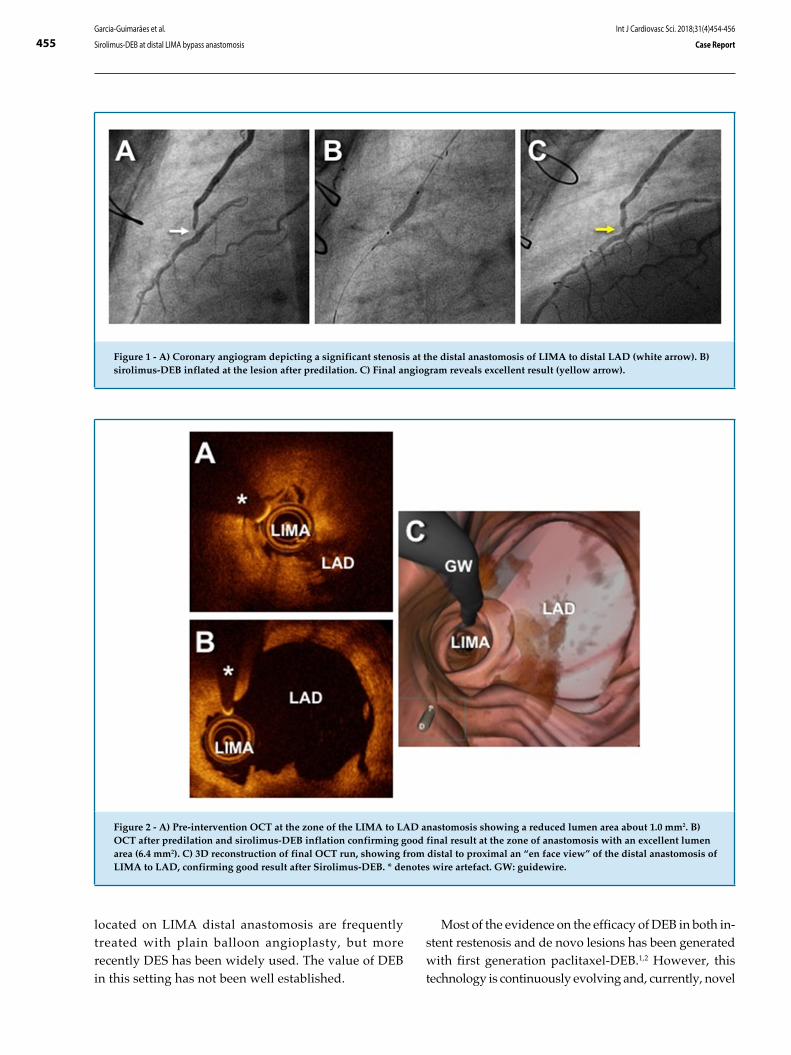
455
Figure 1 - A) Coronary angiogram depicting a significant stenosis at the distal anastomosis of LIMA to distal LAD (white arrow). B) sirolimus-DEB inflated at the lesion after predilation. C) Final angiogram reveals excellent result (yellow arrow).
Figure 2 - A) Pre-intervention OCT at the zone of the LIMA to LAD anastomosis showing a reduced lumen area about 1.0 mm2. B) OCT after predilation and sirolimus-DEB inflation confirming good final result at the zone of anastomosis with an excellent lumen area (6.4 mm2). C) 3D reconstruction of final OCT run, showing from distal to proximal an “en face view” of the distal anastomosis of LIMA to LAD, confirming good result after Sirolimus-DEB. * denotes wire artefact. GW: guidewire.
Garcia-Guimarães et al.
Sirolimus-DEB at distal LIMA bypass anastomosis
Int J Cardiovasc Sci. 2018;31(4)454-456
Case Report
located on LIMA distal anastomosis are frequently treated with plain balloon angioplasty, but more recently DES has been widely used. The value of DEB in this setting has not been well established.
Most of the evidence on the efficacy of DEB in both in-stent restenosis and de novo lesions has been generated with first generation paclitaxel-DEB.1,2 However, this technology is continuously evolving and, currently, novel
456Garcia-Guimarães et al.
Sirolimus-DEB at distal LIMA bypass anastomosis
Int J Cardiovasc Sci. 2018;31(4)454-456
Case Report
This is an open-access article distributed under the terms of the Creative Commons Attribution License
devices with limus-type drugs and more modern carrying systems, are available.3 We selected a novel sirolimus-DEB with a carrying system based on nanoparticles that enable an efficient drug transfer. Preclinical studies have shown that these nanocarriers have better bio-availability and in-tissue uptake, therefore, allowing a reduction in the drug dosage, therefore, potentially diminishing vessel toxicity.3 Preliminary experience with these novel devices appears highly promising.4 Our findings suggest the value of sirolimus-DEB in the treatment of de novo lesions at the distal anastomosis of LIMA bypass grafts. This therapy may represent a safe and valid alternative to current-generation DES in this setting. However, further studies with long-term clinical and angiographic follow-up are required to fully elucidate the relative value of this novel strategy in this unique anatomic scenario.
Novel sirolimus-DEB technology seems to be an alternative to current DES at curious scenarios such as distal LIMA bypass anastomosis. OCT may be helpful to assess the results after DEB treatment.
Author contributions
Conception and design of the research: Garcia-Guimarães M. Acquisition of data: Garcia-Guimarães M. Writing of the manuscript: Garcia-Guimarães M, Alfonso F. Critical revision of the manuscript for intellectual content: Maruri-Sanchez R, Cuesta J, Rivero F, Bastante T, Alfonso F.
Potential Conflict of Interest
No potential conflict of interest relevant to this article was reported.
Sources of Funding
There were no external funding sources for this study.
Study Association
This study is not associated with any thesis or dissertation work.
1. Byrne RA, Joner M, Alfonso F, Kastrati A. Drug-coated balloon therapy in coronary and peripheral artery disease. Nat Rev Cardiol. 2014;11(1):13-23.
2. Cortese B, Piraino D, Buccheri D, Alfonso F. Treatment of bifurcation lesions with drug-coated balloons: a review of currently available scientific data. Int J Cardiol. 2016;220:589-94.
3. Granada JF, Tellez A, Baumbach WR, Bingham B, Keng YF, Wessler J, et al. In vivo delivery and long-term tissue retention of nano-encapsulated sirolimus using a novel porous balloon angioplasty system. EuroIntervention. 2016;12(6):740-7.
4. Clever YP, Peters D, Calisse J, Bettink S, Berg MC, Sperling C, et al. Novel sirolimus–coated balloon catheter in vivo evaluation in a porcine coronary model. Circ Cardiovasc Interv. 2016;9(4):e003543.
References
DOI: 10.5935/2359-4802.20180039
457
CASE REPORT
International Journal of Cardiovascular Sciences. 2018;31(4)457-460
Mailing Address: Antonio Jose Lagoeiro JorgeAvenida Marques do Paraná, 303. Postal Code: 24033-900, Centro, Niterói, RJ - Brazil.E-mail: [email protected]
Cardiac Amyloidosis with Heart Failure and Middle Range Ejection FractionAntonio Jose Lagoeiro Jorge, Diane Xavier de Ávila, Enoï Guedes Vilar, Mario Luiz Ribeiro, Karima Elias Hallack Bruno, Ana Carolina PiresUniversidade Federal Fluminense, Niterói, RJ - Brazil
Manuscript received October 30, 2017; revised manuscript February 23, 2018; accepted March 3, 2018.
Heart Failure / physiopathology; Amyloidosis; Stroke Volume; Hypertrophy, Left Ventricular; Aged.
Keywords
Introduction
Cardiac amyloidosis (CA) is a disease with a difficult diagnosis, limited management and a reserved prognosis.1,2 A high level of suspicion is necessary for its identification. There are some clinical clues, such as elderly individuals with unexplained left ventricular hypertrophy (LVH), heart failure with preserved ejection fraction (HFpEF) and restrictive pattern, dissociation between LVH on echocardiography and low voltage on the electrocardiogram, among others.1,2
CA can occur with several hemodynamic forms and patterns of remodeling, according to the disease evolution stage. It may occur as the restrictive form, with left ventricular ejection fraction > 50%; and dilated form, with reduced ejection fraction.1,2 Recently, the European Society of Cardiology has established a new classification with the creation of “Heart Failure with Mid-Range Ejection Fraction” (HFmrEF).3 We report a case of CA with HFmrEF.
Case report
A female patient, aged 80 years old, was treated at the emergency unit at the first evaluation showing fatigue with medium exertion, orthopnea, nocturnal paroxysmal dyspnea and lower limb edema. She also had frequent complaints of muscle weakness and asthenia. She was admitted with a diagnosis of acute heart failure (HF). She reported macrocytic anemia 3 years before, with no defined etiological diagnosis. She was submitted to
an echocardiogram, which showed LVH and ejection fraction of 64%. She was discharged without an etiological definition of HF.
Fifteen days after discharge, she came to the outpatient clinic with pallor (2+/4+) and jugular swelling at 45°; ictus cordis in the sixth intercostal space in the anterior axillary line; presence of third heart sound; pulmonary component of the second heart sound greater than the aortic component, without murmurs. Pulmonary auscultation showed vesicular murmur abolished at both bases; crepitant rales up to the middle third of both hemithoraces. The liver was palpable at 2 cm from the right costal border. She had symmetrical lower-limb swelling, with pitting edema up to the thigh root, cold and painless. The patient was hospitalized for HF compensation and etiological investigation.
Laboratory tests showed macrocytic and hypochromic anemia; vitamin B12 deficiency; erythrocyte sedimentation rate of 134 mm; electrophoresis; and proteins with monoclonal lambda chain peak.
The electrocardiogram showed junctional rhythm, with low voltage. Chest x-ray showed signs of pulmonary congestion and moderate bilateral pleural effusion. The transthoracic echocardiogram showed dilation of the left and right atria, left ventricular ejection fraction (LVEF) of 42% and alterations suggestive of infiltrative heart disease. Global strain with apical sparing pattern (Figure 1A to 1D) was observed. Myocardial resonance (MRI) was suggestive of the presence of subendocardial amyloid deposits and late enhancement of 35%. LVEF was 45% (Figures 1E to 1H).
The bone marrow aspirate showed a predominance of plasma cells in > 90% of the slide. Immunohistochemistry confirmed the diagnosis of multiple myeloma. Biopsy of facial lesion and abdominal fat with histopathological analysis showed amyloid deposits (Figure 2).

458Jorge et al
Cardiac amyloidosis
Int J Cardiovasc Sci. 2018;31(4)457-460
Case Report
Figure 1 - (A) Apical four-chamber view showing biatrial enlargement, thickened interatrial septum, left ventricular wall hypertrophy, with hyperechoic texture of the myocardium. (B) Parasternal short axis view. Concentric left ventricular hypertrophy. (C) Bullseyes. Global longitudinal systolic strain (mean value of (-)12%), with prominent basal and medium impairment, and preserved apical mechanical function. (D) Apical four-chamber strain showing that the ratio between the apical inferior septal strain and the lower basal septal strain is greater than 3. (E) Cine-mode of the stationary state free precession showing diffuse left ventricular myocardial thickening. (F) Long axis post-gadolinium image showing diffuse late improvements (arrows) in the left atrium, as well as subendocardial enlargement of the left ventricle, more prominent in the anterior wall. (G) Short axis post-gadolinium image showing diffuse subendocardial enhancement (arrows) in the left ventricle. (H) Four-chamber post-gadolinium image showing late enhancement (arrows) not only in the left chambers, but also of the right atrium.
The diagnosis was systemic amyloidosis and multiple myeloma associated with cardiac involvement. Pulse therapy with prednisone was initiated. The HF became refractory to treatment, and the patient died 3 months after the disease onset.
Discussion
Amyloidosis is a rare and multisystemic disease. Patients with amyloidosis usually have few specific symptoms, which makes the diagnosis difficult in the initial phase, as the case presented herein. Cardiac impairment due to amyloidosis can lead to HF, as well as conduction system involvement, with low voltage at the ECG, which increases clinical suspicion.4 In addition to myocardial infiltration, amyloid infiltrates can be found
in the conduction system, valve tissues, coronary arteries, large vessels and autonomic or peripheral nerves, leading to many clinical manifestations.2
More than 25 proteins have been described as possible amyloid-forming agents; however, two of them predominate in cardiovascular impairment: transthyretin (TTR) and immunoglobulin light chains – amyloid light chains or AL.2 The TTR protein is synthesized and secreted by the liver and choroid plexus, and functions as a carrier of thyroxine and retinol binding protein. This protein is typically found in soluble tetramers in their native form. TTR has become the most prevalent form of CA found in clinical practice, with greater identification being made by noninvasive imaging tools.5 Cardiac involvement by TTR occurs most commonly in the sixth and seventh decades of life as HFpEF,6 with the wild-type or senile
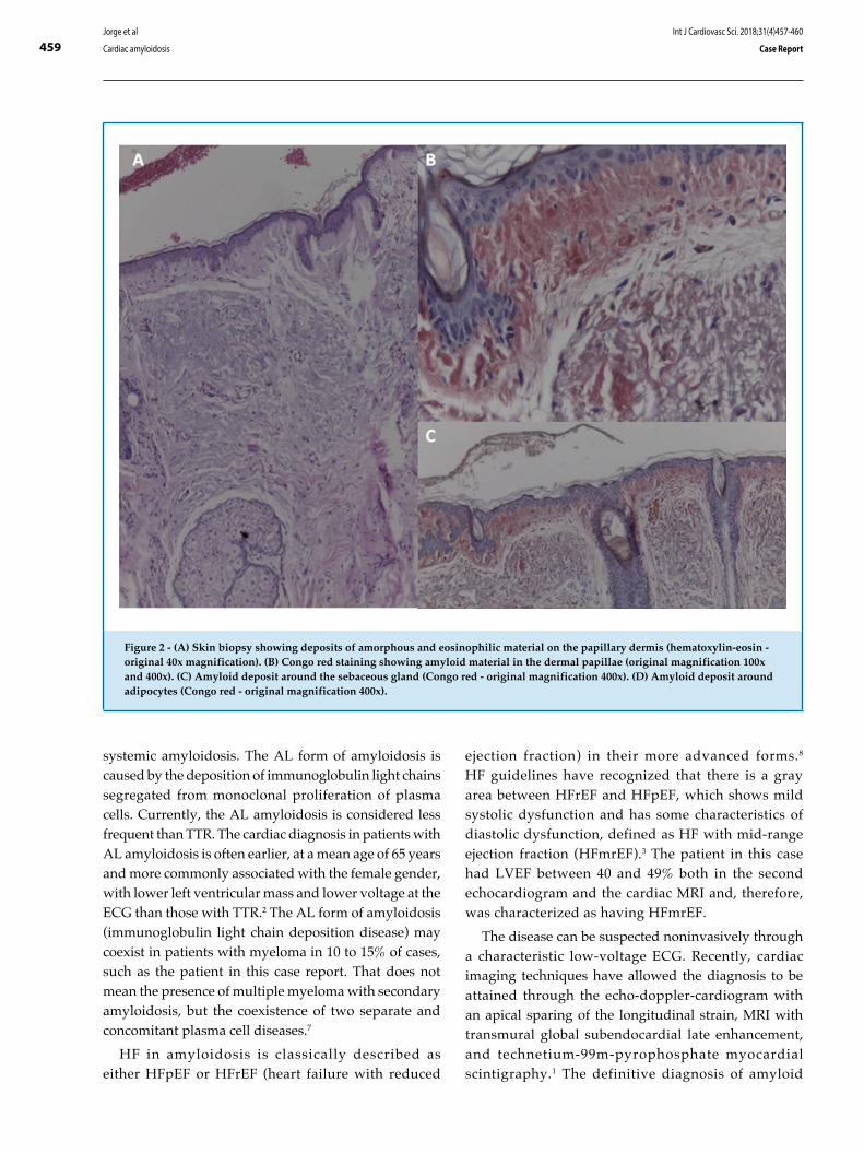
459
Figure 2 - (A) Skin biopsy showing deposits of amorphous and eosinophilic material on the papillary dermis (hematoxylin-eosin - original 40x magnification). (B) Congo red staining showing amyloid material in the dermal papillae (original magnification 100x and 400x). (C) Amyloid deposit around the sebaceous gland (Congo red - original magnification 400x). (D) Amyloid deposit around adipocytes (Congo red - original magnification 400x).
Jorge et al
Cardiac amyloidosis
Int J Cardiovasc Sci. 2018;31(4)457-460
Case Report
systemic amyloidosis. The AL form of amyloidosis is caused by the deposition of immunoglobulin light chains segregated from monoclonal proliferation of plasma cells. Currently, the AL amyloidosis is considered less frequent than TTR. The cardiac diagnosis in patients with AL amyloidosis is often earlier, at a mean age of 65 years and more commonly associated with the female gender, with lower left ventricular mass and lower voltage at the ECG than those with TTR.2 The AL form of amyloidosis (immunoglobulin light chain deposition disease) may coexist in patients with myeloma in 10 to 15% of cases, such as the patient in this case report. That does not mean the presence of multiple myeloma with secondary amyloidosis, but the coexistence of two separate and concomitant plasma cell diseases.7
HF in amyloidosis is classically described as either HFpEF or HFrEF (heart failure with reduced
ejection fraction) in their more advanced forms.8 HF guidelines have recognized that there is a gray area between HFrEF and HFpEF, which shows mild systolic dysfunction and has some characteristics of diastolic dysfunction, defined as HF with mid-range ejection fraction (HFmrEF).3 The patient in this case had LVEF between 40 and 49% both in the second echocardiogram and the cardiac MRI and, therefore, was characterized as having HFmrEF.
The disease can be suspected noninvasively through a characteristic low-voltage ECG. Recently, cardiac imaging techniques have allowed the diagnosis to be attained through the echo-doppler-cardiogram with an apical sparing of the longitudinal strain, MRI with transmural global subendocardial late enhancement, and technetium-99m-pyrophosphate myocardial scintigraphy.1 The definitive diagnosis of amyloid

460Jorge et al
Cardiac amyloidosis
Int J Cardiovasc Sci. 2018;31(4)457-460
Case Report
cardiomyopathy is obtained from an endomyocardial biopsy using Congo red or thioflavin staining2,9 technique and identifying the type of amyloid infiltrate by molecular genetic techniques.
The gold standard for the diagnosis of amyloidosis is the myocardial biopsy. The guidelines of the American Heart Association/American College of Cardiology Foundation show a II-A recommendation for endomyocardial biopsy in the presence of HF associated with unexplained restrictive cardiomyopathy.10 The abdominal fat biopsy can confirm the diagnosis in 70% of cases and, in this reported case, amyloidosis was confirmed by the abdominal biopsy.
The prognosis of patients with amyloidosis is reserved. The mean untreated survival is 13 months and may be extended to 17 months with melphalan and prednisone, which in this case were not used due to the patient’s clinical worsening. The cardiac impairment makes the prognosis even worse, with a life expectancy of approximately 6 months.
Author contributions
Conception and design of the research: Jorge AJL, Avila D, Ribeiro ML, Bruno KEH, Pires C; Acquisition of data: Jorge AJL, Avila D, Vilar EG, Ribeiro ML, Bruno KEH, Pires C; Analysis and interpretation of the data: Jorge AJL, Avila D, Bruno KEH; Writing of the manuscript: Jorge AJL, Avila D, Pires C; Critical revision of the manuscript for intellectual content: Jorge AJL.
Potential Conflict of Interest
No potential conflict of interest relevant to this article was reported.
Sources of Funding
There were no external funding sources for this study.
Study Association
This study is not associated with any thesis or dissertation work.
1. Maurer MS, Elliott P, Comenzo R, Semigran M, Rapezzi C. Addressing common questions encountered in the diagnosis and management of cardiac amyloidosis. Circulation. 2017;135(14):1357-77.
2. Mesquita ET, Jorge AJ, Souza CV Junior, Andrade TR. Cardiac amyloidosis and its new clinical phenotype: heart failure with preserved ejection fraction. Arq Bras Cardiol. 2017;109(1):71-80.
3. Ponikowski P, Voors AA, Anker SD, Bueno H, Cleland JG, Coats AJ, et al. 2016 ESC Guidelines for the diagnosis and treatment of acute and chronic heart failure: the Task Force for the diagnosis and treatment of acute and chronic heart failure of the European Society of Cardiology (ESC). Developed with the special contribution of the Heart Failure Association (HFA) of the ESC. Eur J Heart Fail. 2016;18(8):891-975.
4. Murtagh B, Hammill SC, Gertz MA, Kyle RA, Tajik AJ, Grogan M. Electrocardiographic findings in primary systemic amyloidosis and biopsy-proven cardiac involvement. Am J Cardiol. 2005;95(4):535-7.
5. Gillmore JD, Maurer MS, Falk RH, Merlini G, Damy T, Dispenzieri A, et al. Nonbiopsy diagnosis of cardiac transthyretin amyloidosis. Circulation. 2016;133(24):2404-12.
6. Sperry BW, Vranian MN, Hachamovitch R, Joshi H, Ikram A, Phelan D, et al. Subtype-specific interactions and prognosis in cardiac amyloidosis. J Am Heart Assoc. 2016;24(3):e002877.
7. Dubrey SW, Hawkins PN, Falk RH. Amyloid diseases of the heart: assessment, diagnosis, and referral. Heart. 2011;97(1):75-84.
8. Liu D, Niemann M, Hu K, Herrmann S, Störk S, Knop S, et al. Echocardiographic evaluation of systolic and diastolic function in patients with cardiac amyloidosis. Am J Cardiol. 2011;108(4):591-8.
9. Phelan D, Collier P, Thavendiranathan P, Popović ZB, Hanna M, Plana JC, et al. Relative apical sparing of longitudinal strain using two-dimensional speckle-tracking echocardiography is both sensitive and specific for the diagnosis of cardiac amyloidosis. Heart. 2012;98(19):1442-8.
10. Cooper LT, Baughman KL, Feldman AM, Frustaci A, Jessup M, Kuhl U, et al; American Heart Association; American College of Cardiology; European Society of Cardiology. The role of endomyocardial biopsy in the management of cardiovascular disease: a scientific statement from the American Heart Association, the American College of Cardiology, and the European Society of Cardiology. Circulation. 2007;116(19):2216-33.
References
This is an open-access article distributed under the terms of the Creative Commons Attribution License

461
XXXVIII Congresso Norte-Nordeste de Cardiologia / XXIII Congresso Paraibano de Cardiologia
De 2 a 4 de Agosto de 2018
Centro de Convenções do Hotel Tambaú (PB)
http://sociedades.cardiol.br/nn/congresso.html
8º Congresso Brasileiro de Imagem Cardiovascular
De 9 a 11 de Agosto de 2018
Centro De Convenções Centro Sul (SC)
http://www.congressodic.com.br/
XXX Congresso da SBC/ES
De 16 a 18 de Agosto de 2018
http://sociedades.cardiol.br/es/
XIX Congresso Cardiologia da SBC-MT / IX Simpósio de Hipertensão Arterial da SBC-MT
De 27 a 29 de Setembro de 2018
Hotel Gran Odara
http://www.sbcmt.com.br/
73º Congresso Brasileiro de Cardiologia
De 14 a 16 de setembro de 2018.
CICB - Centro Internacional de Convenções do Brasil (DF)
http://cardio2018.com.br/
XV Congresso Brasileiro de Cardiogeriatria - DECAGE 2018
De 12 a 13 de outubro de 2018
Florianópolis (SC)
http://departamentos.cardiol.br/decage2014/
XXV Congresso Nacional do SBC/DERC
De 25 a 27 de Outubro de 2018
Costão do Santinho Resort
http://departamentos.cardiol.br/sbc-derc/2016/
XXV Congresso Brasileiro de Cardiologia e Cirurgia Cardiovascular Pediátrica
De 31 de Outubro a 3 de Novembro de 2018
Maceió (AL)
http://departamentos.cardiol.br/sbc-dcp/2010/default.asp
XV Congresso do Departamento de Hipertensão Arterial da SBC
De 01 a 02 de novembro de 2018
Salvador (BA)
http://departamentos.cardiol.br/sbc-dha/
XXXV Congresso Brasileiro de Arritmias Cardíacas
De 22 a 24 de Novembro de 2018
Centro de Convenções, Goiânia, GO
http://sobrac.org/sobrac2018/
Calendar
NEWS

462
Neutrophil-Lymphocyte Ratio in the Risk Assessment of Cardiovascular Disease
Heitor Oliveira Santos and Luiz Fernando Moreira Izidoro
Incidence and Characteristics Angiographic of Patients with Acute Myocardial
Cynthia Kallás Bachur, José Alexandre Bachur, Juliana Pereira Machado, Eugenia Velludo Veiga, Sarah da Silva Candido, Ricardo Barbosa, Julia Granado Carraro, Danielle de Freitas Gonçalves, Maria Georgina Marques Tonello
SAMe-TT2R2 Score: A Useful Tool in Oral Anticoagulation Decision-Making for Venous Thromboembolism Patients?
Fernando Pivatto Júnior, Rafaela Fenalti Salla, Lísia Cunha Cé, Andréia Biolo, André Luís Ferreira Azeredo da Silva, Bruno Führ, Luís Carlos Amon, Marina Bergamini Blaya, Rafael Selbach Scheffel
Mortality and Survival in Surgery Aortic Arch with Preservation of Supra-aortic Vessels: Thirteen Years of Experience
Paula Ferraz de Oliveira, Gustavo Luiz Gouvêa de Almeida Junior, Fabrício Braga da Silva, Mauro Paes Leme de Sá, Valdo José Carreira, Bruno Soares da Silva Rangel, Sicilia Pacheco e Silva
Vol. 31, Nº 5, September and October 2018
SEE IN THE NEXT EDITION