Embed Size (px)
Citation preview

REVIEW
Indeterminate colitisM Guindi, R H Riddell. . . . . . . . . . . . . . . . . . . . . . . . . . . . . . . . . . . . . . . . . . . . . . . . . . . . . . . . . . . . . . . . . . . . . . . . . . . . . . . . . . . . . . . . . . . . . . . . . . . . . . . . . . . . . . . . . . . . . . . . . . . . . . .
J Clin Pathol 2004;57:1233–1244. doi: 10.1136/jcp.2003.015214
Indeterminate colitis (IC) originally referred to those 10–15% of cases of inflammatory bowel disease (IBD) in whichthere was difficulty distinguishing between ulcerative colitis(UC) and Crohn’s disease (CD) in the colectomy specimen.However, IC is increasingly used when a definitivediagnosis of UC or CD cannot be made at colonoscopy, incolonic biopsies or at colectomy. The diagnostic difficultiesmay explain the variably reported prevalence of IC.Clinically, most patients with IC evolve to a definitediagnosis of UC or CD on follow up. The role of ancillarytests in the distinction of UC from CD is reviewed. The lowsensitivity of serological markers limits their usefulness.Other tests include upper endoscopy and magneticresonance imaging. The definition of IC may not be apurely histological one derived from resected specimens,but rather a clinicopathological one. This review offerssome personal observations and viewpoints, and proposesan approach to some of the relatively more esotericcombinations of findings.. . . . . . . . . . . . . . . . . . . . . . . . . . . . . . . . . . . . . . . . . . . . . . . . . . . . . . . . . . . . . . . . . . . . . . . . . . .
See end of article forauthors’ affiliations. . . . . . . . . . . . . . . . . . . . . . .
Correspondence to:Dr M Guindi, Departmentof Pathology, TorontoGeneral Hospital, 200Elizabeth Street, 4th floor,Room EC4-305, Toronto,Ontario, Canada, M5G2C4; [email protected]
Accepted for publication1 June 2004. . . . . . . . . . . . . . . . . . . . . . .
The distinction between ulcerative colitis(UC) and Crohn’s disease (CD) has majorimplications, including the choice of medical
treatment, timing of surgery, prognosis, anddisease course. In addition, there is a need forsurveillance in UC, and possibly also in Crohn’scolitis, because it may have a similar risk to UC.The distinction between UC and CD usuallydetermines whether an ileal pouch anal anasto-mosis (IPAA) is offered to the patient, andcorrelates with the occurrence of morbidity andcomplications from pouch and pouch failure.Because there can be overlapping features of UCand CD in the colons of some patients, the termindeterminate colitis (IC) was coined in anattempt to classify these entities more effectively.Patients with indeterminate colitis appear tohave a higher rate of pouch failure and longtermcomplications than those with UC.
‘‘The distinction between ulcerative colitis andCrohn’s disease has major implications,including the choice of medical treatment,timing of surgery, prognosis, and diseasecourse’’
This review of IC will deal with its originaldefinition and features, how it evolved over time,and the controversies associated with this evolu-tion. The incidence, prevalence, and clinical
evolution of IC will be followed by a detaileddiscussion of the settings that give rise to thediagnosis of IC, and the problems and pitfallsassociated with them, including variations ofinflammatory bowel disease (IBD), both UC andcolitis of CD, which can be mistaken for IC. Theseinclude the more common features such as IBDin the fulminant or refractory phase, but also IBDin the chronic phase, and the effects of treatmenton the histology of IBD, especially UC, which canresult in pronounced focality, relative rectalsparing, and focal healing. The issues of inter-observer variability in the diagnosis and classi-fication of IBD and colitis as a result of causesother than UC or CD are discussed. Recentdevelopments, and the usefulness of ancillarytests that are potentially helpful, in the furtherclassification of IC are summarised, with specialreference to serological markers and the role ofupper endoscopy. The outcome of ileoanal pouchconstruction in IC is also discussed.
DEFINITIONS AND FEATURESUC characteristically affects the rectum, involvesthe colon contiguously and symmetrically, andthe disease is more severe distally. Crohn’s colitisis not continuous or symmetrical, and need notinvolve the rectum.1–5 In 1978, Price introducedthe term IC to refer to a subgroup of approxi-mately 10–15% of IBD cases in which therewas difficulty in distinguishing between UCand CD in the excised colectomy specimenbecause the features of typical severe UC werereplaced by deep ulcers—often with knife-likefissures—relative rectal sparing, and transmuralinflammation.6
Table 1 summarises the histological featuresand incidence of discriminating attributes inPrice’s UC, CD, and IC groups.Idiopathic granulomas were only present in
CD. Typical CD fissuring-type ulcerations (mostlysingle and narrow) were present in 13% of casesof IC, but the other histological features of theseIC cases were reportedly so atypical that adiagnosis of CD could not be justified. A secondform of fissuring ulceration, V shaped clefts,usually multiple, was present in 60% of cases ofIC, and appeared to be a feature of any form offulminant colitis. Transmural lymphoid aggre-gates, a feature accepted as very important in thediagnosis of CD, was present in 6% of cases of IC,
Abbreviations: ASCA, anti-Saccharomyces cervisiaeantibodies; ASLC, acute self limited colitis; CD, Crohn’sdisease; DDAC, diverticular disease associated colitis;IBD, inflammatory bowel disease; IC, indeterminatecolitis; IPAA, ileal pouch anal anastomosis; NSAID, non-steroidal anti-inflammatory drug; P-ANCA, perinuclearantineutrophil cytoplasmic antibodies; UC, ulcerativecolitis
1233
www.jclinpath.com
on October 22, 2021 by guest. P
rotected by copyright.http://jcp.bm
j.com/
J Clin P
athol: first published as 10.1136/jcp.2003.015214 on 24 Novem
ber 2004. Dow
nloaded from

but were poorly developed. Transmural inflammation waspresent in most cases of IC, but was only related to areas ofsevere ulceration. The mucosal glandular pattern/architectureand goblet cell population were obscured or modified byextensive ulceration in the IC cases. The islands of survivingmucosa were only mildly inflamed, with a regular glandularpattern and preserved goblet cells—features that favouredCD, or disease in mucosa that was previously unaffected bythe disease process, conceivably including fulminant infec-tions by organisms that were not identified on routine stoolculture. However, these features were present in cases wherethe cumulative evidence was unclear, and two thirds of theseproved to be UC on subsequent follow up in Price’s series.7
‘‘The presence of scattered mononuclear inflammatorycells in the muscularis propria adjacent to ulceration wasregarded as a non-specific response, and not a discrimi-nating feature between ulcerative colitis and Crohn’sdisease’’
Approximately a half of the cases of IC described by Price6
had uneven disease that fell into two patterns, both of whichwould normally bias towards CD (table 2).In the subgroup with relative rectal sparing, the description
of the rectal mucosa was not very detailed. The rectal mucosawas described as being ‘‘mildly abnormal’’ on histologicalexamination in all seven cases. In the three judged to beprobably UC, glandular irregularity was noted. In theremainder of the IC cases with relative rectal sparing, theinflammation was described as mild, without specificreference to the presence or absence of basal plasmacytosisor architectural distortion. The second pattern was ofintermittent ulceration and superficially resembled the skiplesions of CD.
Lee et al found IC in 16% of colectomy specimens.8 Some ofthe features of IC they described are similar to those of Priceet al, such as normal or minimally altered colonic mucosaadjacent to or between ulcers and the absence of granulomas.They described deep slit-like fissures in the IC cases, but didnot specifically describe the V shaped cleft-like fissures seenin IC by Price, although these may just be synonyms. Theyalso emphasised that the term ‘‘transmural inflammation’’, afeature of CD, is loosely used and that accurate definition ofthis criterion removes much of the difficulty from thedifferential diagnosis of IBD. They defined transmuralinflammation as lymphocytes in an aggregated pattern inall layers of the colon, including the serosa. The presence ofscattered mononuclear inflammatory cells in the muscularispropria adjacent to ulceration was regarded as a non-specificresponse, and not a discriminating feature between UC andCD. Their IC cases contained scattered non-aggregatedinflammatory cells involving the full thickness of the bowelwall, but only in those areas deep to ulceration. Unlike Price’sseries, all their patients had an acute onset and requiredurgent colectomy after a short period of unsuccessful medicalmanagement.8
THE EVOLUTION OF ICThe term IC is increasingly being used clinically andhistologically in patients with some form of IBD in whom adefinitive diagnosis of either UC or CD has not been made,either on colonoscopy or colonic biopsy before colectomy. ICmay not be a purely histological entity, but rather a
Table 1 Incidence of discriminating histological features in UC, CD, and IC*
IC
CD UC
Urgent surgery Elective surgery Urgent surgery Elective surgery
Granulomas 0 2 23 0 0FissuresTypical CD 4 2 12 0 0V shaped clefts 18 Not given Not given Not given Not given
Transmural inflammation 28 2 30 20 0Normal goblet cells, regular gland pattern, and focal mucosalinflammation
20 2 27 5 2
Goblet cell depletion, glandular irregularity, ¡ diffuse mucosalinflammation
10 0 3 25 28
Feature suggestive of acute disease ¡ dilatationMyocytolysis and capillary congestion 21 0 0 22 0V shaped clefts 18 0 0 6 0
*Modified from Price.6
CD, Crohn’s disease; ID, indeterminate colitis; UC, ulcerative colitis.
Table 2 Findings in uneven disease pattern inindeterminate colitis from Price6
Uneven pattern (n = 14)
Relative rectal sparinggroup
Intermittent ulcerationgroup
Number of cases 7 cases 7 casesUC more likely 3 cases 3 casesCD more likely 2 cases 2 casesRectal mucosa Mildly abnormal Not specifically mentionedMucosa betweenulcers
Not specificallymentioned
Mild inflammation in allcases
CD, Crohn’s disease; UC, ulcerative colitis.
Figure 1 Relative distal sparing. Colectomy specimen showing lesscongested and less diseased appearing mucosa at the distal end of thecolon, compared with the erythematous ulcerated colonic mucosa seenmore proximally. In the right colon, the transition from normal todiseased mucosa is irregular and patchy.
1234 Guindi, Riddell
www.jclinpath.com
on October 22, 2021 by guest. P
rotected by copyright.http://jcp.bm
j.com/
J Clin P
athol: first published as 10.1136/jcp.2003.015214 on 24 Novem
ber 2004. Dow
nloaded from
clinicopathological one, whereby the label is applied toindeterminate disease in the colectomy specimen (not inthe preceding biopsies), and may remain as such, or befurther refined into UC or CD as clinical events warrant. Aftercorrelating the pathology, preoperative clinical information,and radiological findings of 46 patients with a diagnosis ofIC, Wells et al reclassified 46 patients from the original Priceseries with IC as follows: 19 patients classified as probableCD, 11 classified as probable UC, whereas 16 retained thediagnosis of IC (40% were reclassified as CD). Over a medianfollow up of 10 years, the patients in the last group continuedto be classified as IC, and evidence of CD was not founddespite many clinical investigations.7
Even if the definition were standardised, in practice, thepatients carrying a diagnosis of IC may be a heterogeneousgroup. Some may have colitis that cannot be subclassifiedinto UC or CD (therefore labelled IC) despite investigations atreferral centres by dedicated multidisciplinary teams ofgastroenterologists, pathologists, and surgeons. Others, alsodesignated as IC in less specialised centres or practices, maywell have UC or CD if investigated by a more expert team.
FACTORS LEADING TO A DIAGNOSIS OF ICIt can be difficult to distinguish UC from CD in cases offulminant or refractory IBD, the chronic phase of IBD, whenunusual patterns are seen in otherwise typical UC, in treatedcases of IBD, and in the earliest stages of IBD. When thedistinction between UC and CD becomes difficult, a diagnosisof IC is rendered. In addition, some patients may not have
IBD at all, and other possible aetiologies may need to beconsidered.The difficulty in distinguishing UC from CD, thereby
leading to a diagnosis of IC, is related to several confoundingfactors, as detailed below.
IBD in the fulminant or refractory phaseSome of the gross and microscopic features that are useful indistinguishing the two diseases in the chronic state arecommon to both in the fulminant or refractory phase. Forexample, relative rectal sparing (fig 1), intermittent ulcera-tion, a regular glandular pattern, and lack of mucin depletion(fig 2)—features that in classic teaching would sway theevidence in favour of CD—are seen in some indeterminatecases in Price’s series6 that subsequently proved to be UC, orthat lacked other evidence of CD. It has been surmised thatthe diagnosis of IC is more likely to follow the examination ofa severely diseased colectomy specimen.9 In one series,fulminant disease was more common in patients with ICcompared with either UC or CD, despite similar rates ofimmunosuppression.10 However, it should be noted that theinclusion of specimens from colectomy performed forfulminant colitis in a series selects for IC, as described byPrice6—the histological distinction of UC from CD is moredifficult in the fulminant setting because the histologicalfeatures overlap. It has been argued that intensive steroidtreatment for fulminant disease may lead to the histologicalappearance of IC.10 Histopathological evaluation alone haslimitations in the accurate classification of fulminant IBD.Granulomas and transmural lymphoid hyperplasia/aggre-gates, especially when not in areas of ulceration, appear to bethe two most specific indicators of CD in colectomy speci-mens from patients with fulminant colitis.11 Poorly formedmicrogranulomas may be good indicators of CD, althoughwhen an aggregate of histiocytes becomes a granuloma ishighly subjective. Some have used five histiocytes to definemicrogranulomas—that at least provides some objectivity.12
Greater emphasis should perhaps be placed on theiridentification.11 13 Isolated giant cells and well definedepithelioid granulomas distant from crypts do not, as a rule,occur in UC, and hence their presence in a colonoscopicbiopsy showing features of chronic IBD is a strong pointertowards the diagnosis of CD. Crypt associated giant cells andgranulomas can occur in UC (fig 3), and in themselves areunreliable features for the discrimination between CD andUC.12 A pathological review of the original histologicaldiagnoses in Swan’s series suggests that the reluctance tocommit to a diagnosis of UC by some was based on thepresence of features that are considered to be characteristic of
Figure 2 Residual mucosa in a colectomy specimen from fulminantinflammatory bowel disease. (A) Mucosal changes are minimal, withoutmucin depletion, and with only slight architectural distortion. (B) Mildinflammation only with basal lymphoplasmacytosis (haematoxylin andeosin stain).
Figure 3 Crypt associated giant cells and granulomas. Note theassociated ruptured crypt. The lesion forms as a reaction to extravasatedmucin (haematoxylin and eosin stain).
Indeterminate colitis 1235
www.jclinpath.com
on October 22, 2021 by guest. P
rotected by copyright.http://jcp.bm
j.com/
J Clin P
athol: first published as 10.1136/jcp.2003.015214 on 24 Novem
ber 2004. Dow
nloaded from

the fulminant nature of IBD, such as transmural inflamma-tion and fissuring ulceration, and which may superficially bereminiscent of CD.11 Macroscopic examination appears tohave rather limited value in differentiating UC from CD inthe fulminant state.1 11 In the fulminant colitis specimen,linear ulceration (fig 4) and fissures are relatively common,regardless of the precise cause, and are not specific indicatorsof CD.
‘‘The histological distinction of ulcerative colitis fromCrohn’s disease is more difficult in the fulminant settingbecause the histological features overlap’’
IBD in the chronic phaseEven in the chronic phase, the classic endoscopic andhistological features of UC do not always conform to thetraditional rules, which may lead to confusion with CD,change the diagnosis to Crohn’s colitis, or lead to a diagnosisof IC. In practice, this may occur at the time of examinationof endoscopic biopsies in the non-fulminant setting, and notjust upon examination of colectomy specimens resected forfulminant disease.14–18 There are sequential biopsy changes inthe mucosa of UC that occur with time, and pertain to diseasedistribution and extent. In one study of patients with UC,who had a median follow up of seven years,16 these changesincluded: development of non-diffuse distribution, finding ofnormal rectal mucosa both endoscopically and histologically,either at the beginning or later on in the course of the disease,variation in extent of involvement over time, and lack of anendoscopic–histological correlation. The presence of some ofthese features in biopsy specimens may not fit with the restof the cumulative evidence for UC in a given case, or mayrender the cumulative evidence equivocal, such that adiagnosis of IC is made. One caveat to this study is thatbecause several biopsy sets from the same patient had beentaken over time, some of the variability in histological andendoscopic appearances may have been related to the effectsof treatment and were not necessarily a ‘‘natural’’ evolutionof extent and distribution. Particularly in quiescent disease,some pathologists may be less inclined to offer a specificdiagnosis other than IBD when the pattern and distributionof activity that would favour one or other diagnosis cannot beassessed. A diagnosis of quiescent IBD based primarily onarchitectural distortion and its distribution is not the samething as IC. When IBD is inactive, only minimal histologicalchanges are found, making a histological differentialdiagnosis of CD and UC difficult,19–21 and also making itdifficult to distinguish from infection, especially retrospec-tively. This raises the issue of whether one can or should lend
more credence to the features at first presentation or thoseseen after treatment, when the acute phase has passed.
Unusual patterns in UCSome patients with either subtotal or left sided colitis mayshow patchy, mild, caecum (caecal patch), and/or peri-appendiceal orifice and/or ascending colon chronic activeinflammation, which is separated from the distal disease bynormal intervening colonic mucosa, giving the false impres-sion of a skip area, and therefore CD.22–24 In one series of 20patients with established left sided UC, six showed a sharpdemarcation between affected and unaffected portions ofcolon, whereas 14 showed a more gradual transition.22 Thearea of transition may appear somewhat patchy (fig 1) andgive the false impression of skip lesions. Furthermore, threequarters of this last group of patients showed an area ofinflammation in the caecum, primarily in the periappendicealmucosa, which was separate from the distal inflamedsegment. The natural history of UC in patients with a caecalpatch suggests that patchy right sided inflammation inpatients with left sided colitis has little clinical relevance, butshould be recognised by pathologists to prevent a falsediagnosis of CD in this setting.25 Discontinuous involvementof the appendix in 13 of 62 cases of UC was first described byDavison and Dixon.26 Others subsequently reported thisobservation. In the series of Groisman et al there were twocases with limited left sided involvement combined withappendiceal involvement.27 The appendiceal involvement ismucosal but can be quite severe; however, it appears not toproduce clinical symptoms of appendicitis (or these aremasked by those of the underlying colitis).
‘‘Definite diagnostic criteria for backwash ileitis have notbeen determined, especially those to delineate the extremeend of its spectrum’’
Backwash ileitis represents a mild (but occasionally severeand extensive) degree of active inflammation in the distalfew centimetres of terminal ileum in some patients withsevere pancolitis. It is presumed to be secondary to the refluxof colonic contents.28 When present and exaggerated, it mayraise the differential diagnosis of CD. However, CD moretypically involves longer segments of the distal ileum, andmay show other features of CD, such as granulomas. Definitediagnostic criteria for backwash ileitis have not beendetermined, especially those to delineate the extreme endof its spectrum, but conventionally diffuse mucosal disease incontinuity with severe colonic disease is accepted, as long asno other features of CD or other diseases are present. Despiteearlier reports that backwash ileitis was not an important riskfactor for the development of pouchitis,29 one subsequentstudy suggested that patients who are preoperatively assessedto have ileal disease appear to be at greater risk for thedevelopment of pouchitis.30
Effects of treatment on histology of IBDSome of the changes described above may be related to theeffects of treatment.15 16 31 A study that prospectively eval-uated rectal histology in patients enrolled in a 5-aminos-alicylic acid enema treatment trial found that seven of 11patients with UC treated with 5-aminosalicylic acid had oneor more biopsies that showed histologically normal rectalmucosa.32 In two of the three patients treated with placebo,one or more biopsies were normal. Focality of healing,including rectal healing, occurs in all forms of treatment.Using sequential endoscopic biopsies, Kim et al have reportedendoscopic and histological patchiness of inflammation andrectal sparing in 59% of well documented patients with UCover time.31 The Leuven group (Geboes and Dalle) performed
Figure 4 Colectomy specimen from a patient with refractoryinflammatory bowel disease. Longitudinal ulcers are present and are notspecific for Crohn’s disease. Note the almost normal or minimallyinvolved mucosa.
1236 Guindi, Riddell
www.jclinpath.com
on October 22, 2021 by guest. P
rotected by copyright.http://jcp.bm
j.com/
J Clin P
athol: first published as 10.1136/jcp.2003.015214 on 24 Novem
ber 2004. Dow
nloaded from

a literature review of clinical drug trials in IBD and the effectsof various drugs on the microscopic features of CD.33
Diagnostic microscopic features and the features character-istic of disease activity vary with time and treatment. Themore recently developed drugs (especially the immuno-modulators azathiaprine and infliximab22 34) used for CDcan induce mucosal healing. Medical treatment can haveprofound effects on mucosal histology, but the effect is highlyvariable. It depends on time, the type of treatment (whethertopical or systemic), the intensity of the inflammation, theseverity of the lesion, and probably several other factors thatare unknown at the present time. In all drug studiesinvolving CD it must be remembered that there is about a30% response rate for any form of treatment (a placeboresponse), so that genuine responses must result in con-siderably greater degrees of therapeutic responsiveness.Iatrogenic inflammatory changes that may be confused
with IBD may represent the effects or bowel preparationcompounds administered before lower endoscopy. Sodiumphosphate, a commonly used oral cathartic agent, causesaphthoid ulcers or focal active colitis in the colon and rectumthat might lead to an endoscopic diagnosis of CD, especiallyin quiescent IBD.35
Early stage IBDIt has been shown that the microscopic features used for thediagnosis of IBD are often not present in the very early stageof disease,36 especially in children.37 38 For example, childrenmay show relative, or complete, rectal sparing or evenpatchiness of disease at initial presentation before treat-ment.36 37 39 40 In a study of rectal and sigmoid colon biopsiesfrom untreated paediatric patients with UC evaluated shortlyafter the onset of symptoms, approximately 50% of patientshad completely normal biopsy results and two had anendoscopically normal rectum and sigmoid.39 Compared withadults, the paediatric group showed significantly fewerpatients with chronic active disease, and more patients withmicroscopic skip areas and relative rectal sparing.41 Atypicalfeatures of UC in children at initial presentation such as theabsence of features of chronicity, mild active disease, andmicroscopic skip areas creates several dilemmas: they do notexclude the possibility of UC, may lead to a diagnosis of CD inerror, and make it difficult to discern IBD from infection. Adiagnostic re-evaluation of patients in the southeasternNorway study has shown that in 33% the presence ofIBD was either excluded or strongly questioned duringfollow up.42
Colitis other than UC and CD, including fulminantcolitis of any aetiologyWhen there is no previous clinical diagnosis, or in a firstattack that is fulminant, colitis may have features that arefound commonly, irrespective of the aetiology. These includedeep ulcers with associated non-aggregated polymorphoustransmural inflammation (fig 5), residual mucosa with mildinflammation, and no features of chronicity from pre-existing IBD such as architectural distortion, minimal ulcersat bases of innominate grooves (fig 6), and pronouncedcapillary vasodilatation and haemorrhage.1 In this situation,some cases presenting as fulminant colitis may not easily fitinto either UC or CD because the underlying disease hasanother cause. If the diagnosis of IC is made by default, itwould inadvertently or erroneously imply that the underlyingdisease is IBD (UC or CD), or an intermediate form betweenthem.It has been proposed that the use of the term ‘‘fulminant
colitis of uncertain aetiology’’ rather than IC should be usedwhen there is no clear evidence of either previous UC orfeatures that allow a diagnosis of CD to remove one area ofambiguity.1
‘‘In acute infectious colitis, a pathogen cannot be grown inup to 50% of patients, and a similar proportion of culturenegative infections may also occur in fulminant colitis’’
The differential diagnosis in such cases should include IBD,in addition to forms of colitis that can occur as fulminantdisease, such as colitis caused by infection (for example,Clostridium difficile, salmonellosis, shigellosis, and Escherichiacoli) and drugs, such as non-steroidal anti-inflammatorydrugs (NSAIDs).1 In acute infectious colitis, a pathogencannot be grown in up to 50% of patients, and a similarproportion of culture negative infections may also occur infulminant colitis. Even in a patient with known UC or CD,fulminant colitis may be infection mediated.NSAIDs have been linked to flares of IBD and to triggering
a first episode of IBD.43 NSAIDs can cause non-specific colitisor exacerbate pre-existing colonic disease.44
There is no clear agreement on the diagnostic criteria forexcluding IBD, especially in patients with colonic diseaseonly.45 Histological features can sometimes differentiate
Figure 5 Deep wide ulcers that can be seen in fulminant colitis of anyaetiology and can be associated with surrounding, sometimestransmural, inflammation in their vicinity but without lymphoidaggregates (Haematoxylin and eosin).
Figure 6 Aphthoid-like ulcers at the bases of innominate grooves maybe seen in fulminant colitis of any aetiology (haematoxylin and eosinstain).
Indeterminate colitis 1237
www.jclinpath.com
on October 22, 2021 by guest. P
rotected by copyright.http://jcp.bm
j.com/
J Clin P
athol: first published as 10.1136/jcp.2003.015214 on 24 Novem
ber 2004. Dow
nloaded from

between acute self limited colitis (ASLC) and UC,46 47 and alsobetween IBD and other types of colitis, including ischaemiaand infection.48 Basal plasmacytosis in the lamina propriadistal to the ileocaecal valve region (where it is normal) andmucosal distortion differentiate first and recurrent attacks ofUC from cases of ASLC,46 47 with the caveat that focal cryptitisduring the resolving phase of ASLC could be confused withsimilar lesions in CD. However, as discussed above, thehistological features used for the diagnosis of IBD are oftenabsent in the very early stages of the disease.Diverticular disease can mimic IBD in a variety of ways.
Diverticular disease can be associated with chronic inflam-matory changes of the luminal mucosa of the colon in thesegments involved by diverticula, usually the sigmoid colon,known as diverticular disease associated colitis (DDAC). Theinflammatory pattern ranges from mild non-specific inflam-mation, with or without mild crypt distortion, to apronounced chronic active colitis picture that may mimicCD or UC, with cryptitis and crypt abscesses present.49 50 TheCD-like variant of DDAC can be seen in patients who have noevidence of CD elsewhere in the gastrointestinal tract, andappears to represent a local chronic inflammatory reaction tocomplicated (perforated and resolving) diverticulitis. ThisCD-like variant exhibits all the histological features of CD,including transmural lymphoid aggregates.51–53 Inflammatorymasses related to diverticular disease may be associated withIBD-like changes of the overlying luminal mucosa.54
Diverticular disease may be associated with prolapsingmucosal polyps of the colon, especially the sigmoid colon,which may be confused endoscopically with inflammatorypolyps of IBD.55 Any combination of the above features maybe present in biopsies or resections from a patient withdiverticular disease. If the pathologist is unaware of thepresence of diverticula when interpreting biopsies in thissetting, or if the pathologist does not recognise the potentialreaction patterns associated with diverticular disease, anerroneous diagnosis of IBD can be made. Classification of the‘‘IBD’’ into UC or CD in this setting is difficult and may resultin an inadvertent diagnosis of IC. The finding that in somepatients diagnosed with DDAC the disease subsequentlyevolved into distal UC, despite initial normal rectal biopsies,49
further confounds the situation.In ischaemic colitis, the endoscopic and clinical findings
can overlap with UC or CD, making the distinctiondifficult,56 57 especially in the elderly. It may be difficult, ifnot impossible, to distinguish ischaemia from IBD histologi-cally.1 Segmental distribution may occur in ischaemia, similarto CD. Submucosal oedema and haemorrhage occur inischaemia and in CD, leading to cobblestoning and thumb-printing, which may be seen in both. Low flow ischaemiamay be impossible to distinguish from UC if diffuse and withrectal involvement, or from CD if focal. Mucosal fibrosis andtelangiectasia of capillaries, if present, favour ischaemia, buthaemosiderin laden macrophages may be seen in ischaemiccolitis and in IBD. Ischaemic proctosigmoiditis has beenreported without proximal colonic involvement.58 Biopsiesmay show a non-specific ulcer base similar to the non-specific ulcer base seen in IBD related ulcers and erosions.Finally, mass lesions of diverse aetiologies, such as primary
and metastatic malignancy, endometriosis, and pneumatosis,may cause overlying mucosal changes that closely mimic IBDin mucosal biopsies54; however, the clinical context in suchcases would not be consistent with IBD.
INTEROBSERVER VARIABILITY (OBSERVER BIAS)There is interobserver disagreement in the diagnosis andclassification of colonic IBD, which can result in a change ofdiagnosis that can include either more or less patients in thatcategory. Some pathologists are more sensitive to features
that might be found in CD, and are also more prepared toaccept a larger range of changes in UC, thereby diminishingthe number of patients with IC or CD.59–61 Theodossi et alfound that the range of agreement among 10 observers with aspecial interest in gastrointestinal pathology was 65–76%.60
The best agreement was when discriminating betweennormal slides and those of confirmed IBD cases. Using kstatistics, Farmer et al compared the histological diagnosis ofcolonic IBD made by board certified, university affiliatedpathologists versus that made by a specialist gastrointestinalpathologist, and found significant interobserver variation.61
The gastrointestinal pathologist’s diagnoses differed from theinitial diagnoses in 45% of surgical specimens and 54% ofbiopsy specimens. Forty three per cent of cases initiallydiagnosed as UC were changed to CD or IC, whereas 17%initially diagnosed as CD were changed to UC or IC. Thisstudy raises several interesting observations. Whereas nodiagnosis of IC was made by the general pathologists, thegastrointestinal pathologist reclassified the IBD as IC inapproximately 24% of cases. It would be tempting to assumethat in the hands of a gastrointestinal pathologist fewer casesof IBD would fall into the IC category. However, this does notappear to be the case. One explanation may be the more indepth knowledge and/or experience of the gastrointestinalpathologist with regard to the overlapping features betweenCD and UC, and familiarity with the features of fulminantdisease and with treatment effects that result in focality orsome degree of rectal sparing. The number of paraffin waxblocks for each surgical colectomy specimen varied greatly,between two and 14. Sampling of colectomy specimens didnot appear to be uniform, and therefore may have affectedthe ability of the pathologist to classify IBD. The adequacy ofsampling in the IBD biopsy sets in Farmer’s study was notclear. Inadequate sampling can potentially increase thefrequency of the diagnosis of IC, both in colectomy resectionspecimens and in IBD biopsies.
‘‘Some pathologists are more sensitive to features thatmight be found in Crohn’s disease (CD), and are alsomore prepared to accept a larger range of changes inulcerative colitis, thereby diminishing the number ofpatients with indeterminate colitis or CD’’
The contributions of the extent of sampling (multiple andsingle biopsies), expert status, brief exposure to guidelines,and the use of particular evidence based diagnostic criteria tothe accuracy and reproducibility of the diagnosis of IBD werestudied at an international workshop.62 Expert and non-expert international diagnostic histopathologists attended aworkshop. Sixty cases with full follow up were reviewed,blinded, in two rounds. Diagnoses were made on rectalbiopsies and then full colonoscopic series. Experts correctlyidentified only 24% of CD cases (non-experts, 12%) from therectal biopsies (many were normal). This improved to 64%(non-experts, 60%) with the full series. The accuracy of thediagnosis of UC also improved slightly with the full series—from 64% to 74% overall. Experts had a similar (moderate)level of agreement and accuracy compared with non-experts.A full colonoscopic series gave more accurate diagnosis thana rectal biopsy.Tanaka et al found that interobserver agreement for
histological features that distinguish IBD from other formsof colitis, and UC from CD (crypt atrophy, crypt distortion,basal plasmacytosis, grade of mononuclear infiltration,Paneth cell metaplasia, grade of mucin depletion, mucinpreservation, focal or diffuse mononuclear infiltration, andepithelioid granulomas) was fair to good when assessed by kstatistics.2 Agreement was maintained for all the examined
1238 Guindi, Riddell
www.jclinpath.com
on October 22, 2021 by guest. P
rotected by copyright.http://jcp.bm
j.com/
J Clin P
athol: first published as 10.1136/jcp.2003.015214 on 24 Novem
ber 2004. Dow
nloaded from

features except Paneth cell metaplasia. It would appear thatinterobserver agreement regarding the identification ofindividual histological features is sufficiently reproducible,whereas interobserver disagreement regarding the synthesisof the overall diagnosis may be significant. There aredifferences in the identification and interpretation oflymphoid aggregates and equivocal granulomas amongpathologists, which may lead to shifting the diagnosis fromCD to IC, or vice versa.11 12 Systematised protocols andchecklists for endoscopic biopsies and colectomy specimensmay help to improve interobserver variability.
INCIDENCE/PREVALENCE AND CLINICALEVOLUTION OF ICFactors that may contribute to variation in the prevalence ofIC include interobserver variation in histological interpreta-tion, making the diagnosis of IC based on the evaluation ofendoscopic biopsy sets not colectomy specimens, determiningprevalence based on colectomy specimen diagnosis with orwithout interval follow up during which CD may declareitself, and variable sampling of colectomy specimens. Thestudy by Swan et al exemplifies several of these factors.11 Of67 patients with IBD who underwent colectomy, 40 werediagnosed as UC, 16 as CD, and 11 as IC immediately aftersurgery by several pathologists. The prevalence of IC changedto nine of 67 upon review of the cases by the studypathologist, and to six of 67 after a mean follow up periodof 43 months. There appears to be reluctance among generalpathologists to make a diagnosis of Crohn’s colitis in theabsence of granulomas.61 This may impact upon theprevalence of IC in study series of colectomy or biopsy sets,and adds to interobserver variation.The prevalence of IC diagnosed preoperatively may increase
or decrease after surgery and evaluation of the colectomyspecimen; it fell from 13% to 4% in one study.63 This can beexplained by the small and superficial nature of theendoscopic biopsies compared with the full thickness surgicalcolectomy specimens, and the practice of prolonged medicaltreatment. Rudolph et al found that the prevalence of IC in aseries of patients diagnosed preoperatively with UC rose from0% to 29% when a single specialist gastrointestinal pathol-ogist reviewed all colectomy specimens postoperatively.10
In recent epidemiological studies,64–67 and in one retro-spective study,68 IC accounted for 5–6% of all initial diagnosesof IBD. However, rates as high as 23% have been reported inchildren.69
Given the difficulties in determining the presence ofulcerative or Crohn’s colitis, it would be surprising if therewere not interobserver variability in IC, and this issue isclearly a major concern.68 Given the lack of a universallyaccepted definition of IC, criteria for the diagnosis of ICinevitably differ between studies.64–68 The data suggest thatduring follow up, most patients with IC acquire a definitediagnosis of UC or CD.7 68 69
ANCILLARY TESTS STUDIED FOR DISTINCTION OFUC FROM CD, AND ICSerological markersThe measurement of perinuclear antineutrophil cytoplasmicantibodies (P-ANCA) and anti-Saccharomyces cervisiae (ASCA)antibodies has been suggested as a method for differentiatingUC from CD.70 71 The presence of ASCA has a specificity ofapproximately 90% for CD and a positive predictive value of88%, but this test seems to be most positive in patients inwhom there is no real diagnostic dilemma—for example,those with small bowel disease. In UC the presence ofP-ANCA has a specificity approaching 90%, but may be abetter marker of large bowel disease than any specificsubtype. The sensitivity of these tests is only 40–60%, limiting
their usefulness in IC.72 The clinical usefulness of thesemarkers is controversial.73–75 A large prospective study byJoossens et al showed that ASCA and ANCA might be helpfulto some degree in categorising IC.76 Patients with an initialdiagnosis of IC who had positive serology were given adefinitive diagnosis of CD or UC, respectively, more oftenthan patients with negative serology. Thus, the combinationof ASCA+/P-ANCA2 was predictive of CD in 80%, and thecombination of ASCA2/P-ANCA+ was predictive of UC orUC-like CD in all patients. Patients with an initial diagnosisof IC and negative serology were more likely to retain thediagnosis of IC. These patients may represent an as yetundefined clinicoserological subgroup of IBD and mayexpress not yet specified antibodies.
Upper endoscopyTraditionally, inflammation in the upper gastrointestinaltract in children, in whom Helicobacter pylori infection is fairlyuncommon, was tantamount to a diagnosis of CD. However,several recent case reports and series suggest that gastro-duodenal involvement, which used to be diagnostic of CD,may also occur in UC, especially in children precolectomy.77–82
Although the presence of granulomas can support a diagnosisof CD, severe inflammation and other abnormalities occur inthe proximal gastrointestinal tract in CD and in UV.One study in a paediatric population suggests that routine
baseline upper endoscopy with gastric antral biopsies mayhelp to distinguish between UC and CD. The finding ofgastric antral granulomas may facilitate a diagnosis of CD inchildren whose colonoscopic findings are indeterminate orsuggestive of UC. Diffuse antral H pylori negative inflamma-tion is of no value in differentiating CD from UC, and appearsto be common in both. Focal antral gastritis is suggestive of,although not exclusive to, CD.77 Several other investigatorsconfirm the occurrence of focal antral H pylori negativegastritis in CD.83–87
A large retrospective study of children and adolescentsconfirms the high prevalence of focal gastritis amongchildren with IBD relative to children without IBD or thosewith H pylori infection, but also shows that focal gastritis canbe found in patients with UC.88 The specificity and positivepredictive value of focal gastritis for CD range from 79% to84% and 71% to 81%, respectively, in different studies.81 84 87 89
Diffuse acute duodenitis is especially suggestive of UC.82
Magnetic resonance imagingA study of magnetic resonance imaging in differentiating UCfrom CD in a paediatric population showed poor interobser-ver reliability. Magnetic resonance imaging interpretation ofIBD did not adequately recognise CD in children.90 1Hmagnetic resonance spectroscopy performed on colonicmucosal biopsies was used to differentiate UC from CDdisease, and showed some promise in the classification ofIC.91
Table 3 Examples of variable rates of pouch failure inindeterminate colitis (IC)
StudyPouch failure in ICpatients
Delaney et al (Cleveland Clinic Foundation,USA)
3.4%
McIntyre et al (Mayo Clinic, USA) 19%Rudolph et al (Louisville, Kentucky, USA) 0%Yu et al (Mayo Clinic, USA) 28%Marcello et al (Labey Hitchcock MedicalCenter, Burlington, Massachusetts, USA)
12%
Indeterminate colitis 1239
www.jclinpath.com
on October 22, 2021 by guest. P
rotected by copyright.http://jcp.bm
j.com/
J Clin P
athol: first published as 10.1136/jcp.2003.015214 on 24 Novem
ber 2004. Dow
nloaded from
IC AND IPAAWhether IPAA is the operation of choice for IC is con-troversial.92 93 Studies from the Mayo Clinic, USA, show thesuccess rate in patients with IC who underwent IPAA to be73–85%, as compared with a success rate of 89% in definiteUC.94–96 The frequency of severe pouch complications overallappears to be 20%—intermediate between UC (8–10%) andCD (30–40%). Although this intermediate frequency of pouchcomplications may superficially reflect on IC as the ‘‘inter-mediate’’ form of chronic idiopathic IBD, the interpretationof these finding is not straightforward. In one series ofpatients with IBD who had undergone IPAA, Yu andcolleagues found that 2% of patients with UC and 15% withIC ultimately had the diagnosis changed to CD.96 When theoutcomes of the patients with CD were considered separately,the complication rate of the remaining patients with IC wasidentical to that of those with UC. McIntyre et al comparedfunctional outcomes, frequency of bowel movements, incon-tinence, or prevalence of pouchitis, in addition to pouch failurerate, in typical UC and in patients with IC.95 The failure rate in ICwas higher than in UC (19% v 8%). However, most patients withIC had longterm functional results similar to those of patientswith UC. Such findings raise the suspicion that most patientswith IC probably have UC, or perhaps more accurately may benon-CD. Pouch failure rates were higher as a result of perinealdisease in the IC group in McIntyre’s series (presumably relatedto CD).95 This supports the argument that the overall higher rateof severe pouch complications in patients with IC comparedwith those with UC who had undergone IPAA results from theheterogeneity of the patients included in the IC group in thedifferent series, such that they frequently represent anadmixture of patients with true UC and CD. The functionaloutcomes of patients with UC and IC after IPAA were found tobe similar in other studies.10 94 97
‘‘In general, we do not regard indeterminate colitis acontraindication for a pouch’’
Patients with IC and IPAA manifest more late complica-tions, such as more episodes of pelvic sepsis, pouch fistula,and pouch failure compared with patients with definiteUC.10 96 98
The incidence of pouch failure in patients with IC isvariable among different series (table 3).No patient with IC required permanent ileostomy in the
series by Rudolph et al.10 This contrasts with the Labeyand the Mayo experiences; these centres reported pouchfailure in 28%, 16–19%, and 12% of patients with IC,respectively.95 96 98 99
The most common problem is pouchitis, which increases inincidence as the duration of follow up increases.94 95 96 100 Thecaveat in interpretation of data regarding functional outcomeand complications of IPAA in patients with IC is that the levelof diligence exercised by surgeons, gastroenterologists, andpathologists in attempting to classify the colitis before IPAAmay vary among different series, and may contribute to someof the conflicting results seen. Gramlich et al proposed thatpathological stratification of the IC colectomy specimenfindings may predict those more likely to develop CD orother complications, but not pouch failure and that, on thisbasis, patients with IC should not be precluded from havingIPAA surgery.101 In their series, postoperative colectomydiagnoses were divided into UC if there was diffuse mucosaldisease involving the rectum, CD if there were granulomas ortransmural lymphoid aggregates not associated with deepulcers, and IC in the remaining specimens with atypicalfeatures. The IC group was subcategorised as IC favouring CD(segmental disease but without granulomas or transmural
lymphoid aggregates not associated with deep ulcers), ‘‘true’’IC, and IC favouring UC, if in addition to features of UC therewas one or a combination of (1) deep ulcers, (2) transmurallymphoid aggregates, (3) skip lesions, (4) terminal ilealinflammation, and/or (5) a caecal patch. In the patients withIC favouring UC, only those with evidence of deep ulcerationhad a significant increase in the incidence of CD. However, itwas not clear whether any or all of the patients with ICfavouring UC with deep ulcers had a fulminant presentation,such that the deep ulcers may be related to the fulminantstate alone. Furthermore, the incidence of CD in thissubgroup of patients was only 4.3%. The remainder of theircomplications (such as perianal abscess, perianal fistula, non-functioning pouch) may be related to factors other than deepulcers that have not been taken into consideration, such assurgical technique and the impact of hot colitis.In general, we do not regard IC as a contraindication for a
pouch. As the level of suspicion for CD rises, our concerns areraised usually by stating—for example: ‘‘In view of thepresence of … (for example, rare subserosal lymphoidaggregates) the possibility that the underlying disease maybe CD needs to be considered. Suggest consider delaying therestorative procedure for 6–12 months at which time otherevidence of CD should be sought if this will preclude theIPAA.’’ This can include endoscopy through the stoma,enteroclysis, or capsule studies and upper endoscopy to lookfor other features of CD.
SELECTED DIFFICULT SCENARIOS IN COLECTOMIESFOR IBDTable 4 summarises the histological features encountered incolectomy specimens and their importance. The criteria for
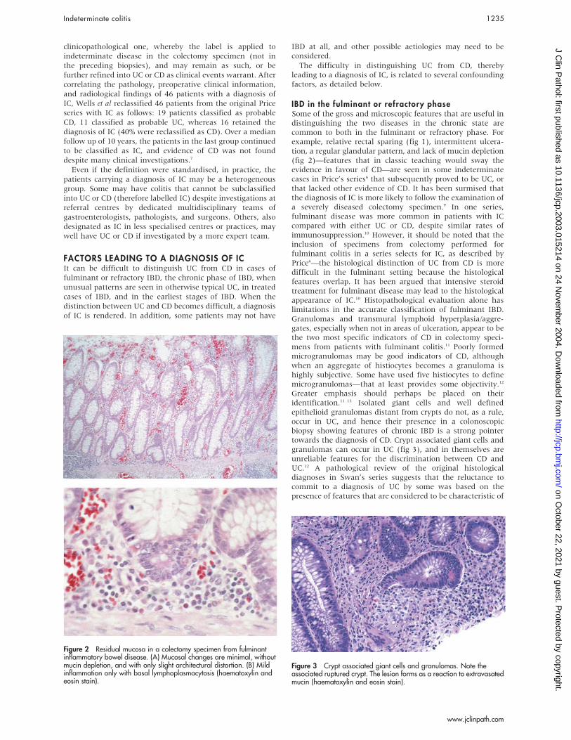
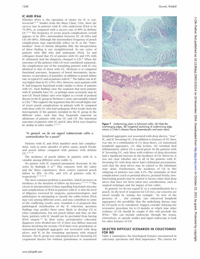
Figure 7 Undermining ulcers in fulminant colitis. (A) Note theoverhanging edges. (B) Tangential sectioning of undermining edgemimics a Crohn’s disease fissure (haematoxylin and eosin stain).
1240 Guindi, Riddell
www.jclinpath.com
on October 22, 2021 by guest. P
rotected by copyright.http://jcp.bm
j.com/
J Clin P
athol: first published as 10.1136/jcp.2003.015214 on 24 Novem
ber 2004. Dow
nloaded from

subclassification of IBD, especially in the fulminant setting,are not absolute and overlap. The final conclusion withregard to CD versus UC rests with the weight of evidencederived from the pattern of inflammation and a constellationof findings in a given caseIn this section, we would like to offer personal observations
and viewpoints, and propose an approach to some of therelatively more esoteric combinations of findings. It shouldbe emphasised that these are opinions and personalobservations that are not necessarily supported by existingstudies, and as such we acknowledge that some may disagreewith them.
Scenario 1: ulceration in backwash ileitisThere is no evidence in the literature that ulceration is part ofthe spectrum of backwash ileitis. The problem arises whenthe colon shows features compatible with UC, but theterminal ileum is involved by what appears to be backwashileitis, except that it is accompanied by ulceration, pyloricmetaplasia, or architectural distortion. These features,although not diagnostic of CD, are sufficient to raise thepossibility of CD, despite the lack of other evidence in thecolon. Pyloric metaplasia (ulcer associated lineage) is seenfrequently in ileal resections from patients with CD.1 Itrepresents a non-specific reparative reaction in intestinalulcers,102 and is not specific for CD. In a study of adults withdefinite evidence of CD, pyloric metaplasia was identified in22.2% of the terminal ileal biopsies. A previous study withsimilar data showed an incidence of only 2.27%.34 Thedifference could reflect the duration of the disease, theextent of pre-existing terminal ileal ulceration, the opportu-nity for mucosal repair before biopsy, or the thoroughness ofthe endoscopic sampling and/or pathological examination.Pyloric metaplasia lacks specificity for CD, but may be asensitive indicator of persistent ulceration with inflamma-tion. When found in terminal ileal biopsies, it could supportthe diagnosis of CD in the appropriate clinical setting.We believe that when this situation is encountered, a
diagnosis of ‘‘IBD, CD not excluded’’ should be given, with arecommendation to delay IPAA and follow up for a reason-able period of time, such as one year, to allow for otherevidence of CD to declare itself.
Scenario 2: aphthoid-type ulcers in UCOccasionally, the transition from normal to abnormal mucosaat the proximal end of an involved segment of UC showsaphthoid-type ulceration, endoscopically or grossly. In UCwith rectal sparing, the transition from spared rectum to thedistal end of the involved segment above may have a similarappearance. The gross/endoscopic appearances in theseinstances may be somewhat misleading and unnecessarilybias towards CD. If there is no other evidence to suggest CD,the diagnosis should still be UC or consistent with UC,especially if severely active/fulminant disease has beenpresent.
Scenario 3: proximal skip lesions in UCInfrequently, one encounters in a colectomy specimen distaldisease with features of UC, whereby the upper margin of thediseased distal mucosa may be separated from additionalmore proximal diseased mucosa by a grossly skipped area.The importance of the disease distribution in this scenario isnot clear. One approach would be to ‘‘ignore’’ this atypical UCdistribution if there is no other compelling evidence of CD,but at least not hasten into a firm diagnosis of CD.When an area of caecal inflammation is present, the
differential diagnosis of caecal involvement separated fromdisease of the distal colon or rectum by normal mucosa is UCwith caecal patch versus CD with a skip area. It is importantto ascertain that the intervening mucosa is uninvolved, and ifso not to assume that the disease is CD. Sampling of theterminal ileum is helpful, because it is usually uninvolved inUC with a caecal patch. If biopsies of the terminal ileum andintervening colonic mucosa are available and unremarkable,a diagnosis of IBD consistent with UC is made. However, ifterminal ileal and intervening colonic mucosal biopsies arenot available, a diagnosis of IBD is made but importantlywithout favouring CD. The clinician is advised to obtain suchbiopsies in the future and to confirm or exclude the presenceof other features of CD, such as small bowel involvement andperianal disease.
Scenario 4: scant deep lymphoid aggregates and‘‘early’’ (small) granulomasAlthough granulomas and deeply situated lymphoid folliclesappear to be the most reliable features for the diagnosis of CD
Table 4 Histological features in colectomy specimens and their importance
Histological feature Definition Importance
Focal LP inflammation Patchy increase of mononuclear cells in LP separatedby areas of normal LP cellularity
In UC can be seen after treatment, may be seen in infection; characteristicof CD especially if intense; may be seen in infection but tends to affect midand upper crypts and surface epithelium; not helpful in fulminant colitis
Diffuse cryptitis Evenly distributed inflammation of crypts, involvingmost crypts
Characteristic of UC, seen in pretreatment material and flares of disease.Avidity of neutrophils for crypts, unlike CD in biopsies and fulminantcolectomy specimens
Idiopathic granulomas Discrete aggregate of macrophages, more than 5histiocytes
Against a diagnosis of UC unless crypt related; characteristic of CD, butseen in some infections (TB, yersinia, others); in fulminant colitis against adiagnosis of UC, favours CD
Microgranulomas (smallaggregates)
A granuloma is defined as 5 histiocytes or more andeverything else is not a granuloma, so microgranulomaprobably does not exist
Some believe carries weight in favour of CD
Crypt related granulomasor giant cells/mucingranulomas
Giant cells or granulomatous reaction adjacent to aruptured or injured crypt
No special importance
Muscularis mucosaduplication/hyperplasia
Splaying, thickening, and disorganisation of smoothmuscle, sometimes blending with submucosa
Indicates previous ulceration; usually seen in CD; may be seen in UC afterimmunosuppressive treatment for temporisation of severe disease, nothelpful in fulminant colitis
Narrow fissures Cleft-like fissure extending into submucosa Characteristic of CD, but are tracts—usually histiocytes—at peripheryand neutrophils centrally (different from fissures seen in IC); may occur inIC and occasionally in UC
Wide fissures, deepundermining ulcers (fig 7A)
V shaped clefts that may extend into submucosa ormuscularis propria with overhanging edges
Typical for any form of fulminant colitis; typical of all severe IBD and someinfections. When undermining ulcers are tangentially cut, they may givethe false impression of linear fissures seen in CD (fig 7B)
CD, Crohn’s disease; LP, lamina propria; IBD, inflammatory bowel disease; IC, indeterminate colitis; UC, ulcerative colitis.
Indeterminate colitis 1241
www.jclinpath.com
on October 22, 2021 by guest. P
rotected by copyright.http://jcp.bm
j.com/
J Clin P
athol: first published as 10.1136/jcp.2003.015214 on 24 Novem
ber 2004. Dow
nloaded from

in the fulminant colectomy specimen, they are not alwayspresent to the same extent. Occasionally there are only one ortwo lymphoid aggregates within the distal colonic wall in IBDthat otherwise has features of UC. The dilemma is whether toattach any weight to such a minimal finding. Our approach isto surmise that given the minimal nature of the evidence, CDis unlikely, and a diagnosis of IBD but favouring UC isrendered. Lymphoid aggregates in the subserosa can bepolymorphous. Our personal view is to ignore inflammatoryaggregates in the subserosa if they are polymorphous—especially ones with plasma cells—but not if purelylymphoid.Where a small histiocytic aggregate ends and a granuloma
begins is a subjective matter. Although there is a precedent inthe literature that aggregates of five or more histiocytesrepresent granulomas, the cut off is nevertheless arbitrary. Itis not clear whether the presence of small histiocyticaggregates signifies CD at all times. If this is the onlyevidence suggesting CD, a diagnosis of IC favouring UC ismade, but one should refrain from making a definitediagnosis of CD, and suggest delaying IPAA, with a periodof follow up as outlined previously.
‘‘Although there is a precedent in the literature thataggregates of five or more histiocytes represent granulo-mas, the cut off is nevertheless arbitrary’’
With regard to ancillary tests, we personally do not useANCA/ASCA to influence the pathological diagnosis whenfaced with the above scenarios or with other difficulties. Thisapproach has the potential of combining the shortcomings ofhistological parameters with uncertainties of ANCA/ASCAthemselves. However, if enteroclysis in a given patient shows—for example, jejunal CD—that would stop us from makinga diagnosis of UC in colonic biopsies of that patient.We do not use the term IC when faced with difficulties in
classifying IBD into CD or UC on biopsy, but prefer to reserveit for colectomy specimens. We regard colectomy specimensas the court of final appeal for the histological classificationof colonic IBD. We accept that the designation IC in acolectomy specimen in a given patient may serve as theworking clinical diagnosis indefinitely, or the workingclinical diagnosis may change to UC or CD if and whenadditional evidence for one or the other becomes availablewith follow up—the ‘‘pending tray’’ concept.We prefer to use the term ‘‘IBD, not yet classified’’ when
faced with difficulty in classifying IBD into CD or UC onbiopsy. Although some may regard this as semantic nuance,we feel such a designation leaves the door open for biopsyclassification of the IBD in repeat or future biopsies. Thesemay provide more comprehensive sampling, or capture morecharacteristic features as the IBD evolves. The difference isthat, after colectomy, there is no further opportunity forexamining additional material from the colon, so if the IBD isnot classifiable pathologically at that point it is thenindeterminate as far as the pathological evidence in thecolon goes.
SUMMARYIt appears that the jury is still out as to whether IC is aseparate entity or a ‘‘third’’ type of IBD, or whether all arebasically a form of either UC or CD. If one accepts the notionthat the colon has a limited repertoire of response to injury,then IC may simply represent the end stage of any colitis—UC, CD, and other diseases including pseudomembranouscolitis, or other infections, because only 50% of acute selflimited colitis ever has an organism identified. However, theclinical evolution of IC indicates that based on follow up
investigations and clinical events it is possible to reclassify aproportion of cases into UC or CD. Price7 was able to reclassifyhalf of his 30 cases of IC after reviewing additional materialfrom precolectomy biopsies or from material that subse-quently became available. If reclassification of IC then restswith cumulative evidence derived from evolving clinicalsymptoms and physical findings, imaging, endoscopy, colec-tomy specimens, and the pathological examination ofendoscopic precolectomy and postcolectomy biopsies, ICmay not be a separate entity but a provisional diagnosis.Price103 had proposed that the term should be used as apending tray diagnosis, representing diagnostic inadequacy,and not as a specific nosological entity. Evidence emergingfrom the studies of serological markers (ASCA and P-ANCA)suggests that perhaps a subgroup of patients initiallydiagnosed as IC may be identified as a separate group basedon their distinct profile of ASCA and ANCA. Approximately50% of patients with IC in the series of Joossens72 did notshow ASCA or P-ANCA. Most of these patients retained thediagnosis of IC during their further clinical course, perhapsreflecting a distinct clinicoserological entity. ASCA andP-ANCA estimation has been suggested as a method fordifferentiating UC from CD, but sensitivity is only 40–60%,limiting its clinical usefulness.Some hold the view that distinguishing IC as a separate
entity has clinical value because of the higher incidence ofpouch complications and the greater risk of pouch failure.Regardless of whether one accepts IC as a separate entity,there are instances when there is a real need to classify theIBD—for example when deciding on specific medical treat-ments/trials, type of surgery (partial versus total colectomy),and the formation of pouches. These are really nodes in themanagement algorithm where one may need to make a firmdecision, but what is most important is not whether the IC isreally UC or CD, but whether there are any features of CDthat would preclude a specific type of treatment—CD versusIC/UC—given that IC seems to behave more like UC than CDfor most of these nodes.
‘‘We regard colectomy specimens as the court of finalappeal for the histological classification of colonicinflammatory bowel disease’’
Pathologists need to understand the different clinicalpathological scenarios that lead to a diagnosis of IC, andperhaps agree to apply the term only as Price described itoriginally—in colectomy specimens not biopsies. This isbecause biopsies are fraught with pitfalls, such as the effectsof treatment, and biopsies taken from large areas of the colonmay be labelled as coming from one site, resulting in ahistological pattern that is not typical of either UC or CD, andmaking the diagnosis of IC tempting. The histologicaldiagnostic criteria for classification of IBD seem to havedulled in recent years when applied to post treatmentmaterial. Pathologists should also familiarise themselveswith the features of fulminant disease, so that they are noterroneously swayed to a diagnosis of CD or IC in colectomyspecimen evaluation.There are numerous other confounding issues including
the effects of treatment and the presence of caecal (andappendiceal) inflammation, which may cause the observer toconsider CD in a patient who otherwise has a typical leftsided colitis, and which may result in doubt about thediagnosis of UC and lead to a diagnosis of IC or even CD.The terminology used in the diagnosis of biopsies and
colectomy specimens should reflect the degree of certainty, orlack thereof, as to whether the underlying disease is IBD inthe first place, and whether it is CD or UC. When the type of
1242 Guindi, Riddell
www.jclinpath.com
on October 22, 2021 by guest. P
rotected by copyright.http://jcp.bm
j.com/
J Clin P
athol: first published as 10.1136/jcp.2003.015214 on 24 Novem
ber 2004. Dow
nloaded from
IBD in biopsy specimens is uncertain, it is better to call it‘‘IBD of uncertain subtype’’ or ‘‘IBD not yet classified’’, and ifuncertain of IBD, a descriptive diagnosis may be appropriate.
Authors’ affiliations. . . . . . . . . . . . . . . . . . . . .
M Guindi, Department of Laboratory Medicine and Pathobiology,University of Toronto, and Department of Pathology, University HealthNetwork, Toronto, Ontario, Canada, M5G 2C4R H Riddell, Department of Pathology, Mount Sinai Hospital, Toronto,Ontario, Canada
REFERENCES1 Lewin KJ, Riddell RH, Weinstein WM. Gastrointestinal pathology and its
clinical implications. New York: Igaku-Shoin, 1992.2 Tanaka M, Riddell RH, Saito H, et al. Morphologic criteria applicable to
biopsy specimens for effective distinction of inflammatory bowel disease fromother forms of colitis and of Crohn’s disease from ulcerative colitis.Scand J Gastroenterol 1999;34:55–67.
3 Riddell RH. Histopathology of ulcerative colitis. In: Allan RN, Rhodes JM,Hanauer SB, et al, eds. Inflammatory bowel disease, 3rd ed. New York:Churchill-Livingstone, 1997:291–309.
4 Goldblum JK, Petras RE. Histopathology of Crohn’s disease. In: Allan RN,Rhodes JM, Hanauer SB, et al, eds. Histopathology of Crohn’s disease, 3rded. New York: Churchill-Livingstone, 1997:311–15.
5 Schachter H, Kirsner JB. Definitions of inflammatory bowel disease ofunknown etiology. Gastroenterology 1975;68:591–600.
6 Price AB. Overlap in the spectrum of non-specific inflammatory boweldisease: colitis indeterminate. J Clin Pathol 1978;31:567–77.
7 Wells AD, McMillan I, Price AB, et al. Natural history of indeterminate colitis.Br J Surg 1991;78:179–81.
8 Lee KS, Medline A, Shockey S. Indeterminate colitis in the spectrum ofinflammatory bowel disease. Arch Pathol Lab Med 1979;103:173–6.
9 Nicholls RJ, Wells AD. Indeterminate colitis. Baillieres Clin Gastroenterol1992;6:105–12.
10 Rudolph WG, Uthoff SM, McAuliffe TL, et al. Indeterminate colitis: the realstory. Dis Colon Rectum 2002;45:1528–34.
11 Swan NC, Geoghegan JG, O’Donoghue DP, et al. Fulminant colitis ininflammatory bowel disease: detailed pathologic and clinical analysis. DisColon Rectum 1998;41:1511–15.
12 Mahadeva U, Martin JP, Patel NK, et al. Granulomatous ulcerative colitis: are-appraisal of the mucosal granuloma in the distinction of Crohn’s diseasefrom ulcerative colitis. Histopathology 2002;41:50–5.
13 Lee FD, Maguire C, Obeidat W, et al. Importance of cryptolytic lesions andpericryptal granulomas in inflammatory bowel disease. J Clin Pathol1997;50:148–52.
14 Bernstein CN, Shanahan F, Anton PA, et al. Patchiness of mucosalinflammation in treated ulcerative colitis: a prospective study. GastrointestEndosc 1995;42:232–7.
15 Bernstein CN, Shanahan F, Weinstein WM. Histological patchiness andsparing of the rectum in ulcerative colitis: refuting the dogma. J Clin Pathol1997;50:354–5.
16 Kleer CG, Appelman HD. Ulcerative colitis: patterns of involvement withcolorectal biopsies and changes with time. Am J Surg Pathol1998;22:983–9.
17 Levine TS, Tzardi M, Mitchell S, et al. Diagnostic difficulty arising from rectalrecovery in ulcerative colitis. J Clin Pathol 1996;49:319–23.
18 Spiliadis CA, Lennard-Jones JE. Ulcerative colitis with relative sparing of therectum. Clinical features, histology, and prognosis. Dis Colon Rectum1987;30:334–6.
19 Hamilton SR. Diagnosis comparison of ulcerative colitis and Crohn’s diseaseinvolving the colon. In: Norris HT, ed. Pathology of the colon, small intestineand anus. New York: Churchill Livingstone, 1983:1–19.
20 Hamilton SR. The differential diagnosis of idiopathic inflammatory diseaseby colorectal biopsy. Int J Colorectal Dis 1987;2:113–17.
21 Greenson J, Odze R. Inflammatory bowel disease of the large intestine. In:Odze RD, Goldblum JR, Crawford JM, eds. Surgical pathology of the GItract, liver, biliary tract, and pancreas. Philadelphia, Pennsylvania:Saunders, 2004:216–17.
22 D’Haens G, Geboes K, Peeters M, et al. Patchy cecal inflammationassociated with distal ulcerative colitis: a prospective endoscopic study.Am J Gastroenterol 1997;92:1275–9.
23 Okawa K, Aoki T, Sano K, et al. Ulcerative colitis with skip lesions at themouth of the appendix. Am J Gastroenterol 1998;93:2405–10.
24 Yang SK, Jung HY, Kang GH, et al. Appendiceal orifice inflammation as askip lesion in ulcerative colitis: an analysis in relation to medical therapy anddisease extent. Gastrointest Endosc 1999;49:743–77.
25 Mutinga M, Farraye F, Wang H, et al. Clinical significance of right colonicinflammation in patients with left sided chronic ulcerative colitis [abstract].Gastroenterology 2001;120:A450.
26 Davison AM, Dixon MF. The appendix as a ‘‘skip lesion’’ in ulcerative colitis.Histopathology 1990;16:93–5.
27 Groisman GM, George J, Harpaz N. Ulcerative appendicitis in universal andnonuniversal ulcerative colitis. Mod Pathol 1994;7:322–35.
28 Saltzstein SL, Rosenberg BF. Ulcerative colitis of the ileum, and regionalenteritis of the colon; a comparative histological study. Am J Clin Pathol1963;40:610–23.
29 Gustavsson S, Weiland LH, Kelly KA. Relationship of backwash ileitis to ilealpouchitis after ileal pouch–anal anastomosis. Dis Colon Rectum1987;30:25–8.
30 Schmidt CM, Lazenby AJ, Hendrickson RJ, et al. Preoperative terminal ilealand colonic resection histopathology predicts risk of pouchitis in patients afterileoanal pull-through procedure. Ann Surg 1998;227:654–62.
31 Kim B, Barnett JL, Kleer CG, et al. Endoscopic and histological patchiness intreated ulcerative colitis. Am J Gastroenterol 1999;94:3258–62.
32 Odze R, Antonioli D, Peppercorn M, et al. Effects of topical 5-aminosalicylicacid (5-ASA) therapy on rectal mucosal biopsy morphology in chroniculcerative colitis. Am J Surg Pathol 1993;17:869–75.
33 Geboes K, Dalle I. Influence of treatment on morphological features ofmucosal inflammation. Gut 2002;50(suppl 3):II37–42.
34 Geboes K, Ectors N, D’Haens G, et al. Is ileoscopy with biopsy worthwhile inpatients presenting with symptoms of inflammatory bowel disease?Am J Gastroenterol 1998;93:201–6.
35 Driman DK, Preiksaitis HG. Colorectal inflammation and increased cellproliferation associated with oral sodium phosphate bowel preparationsolution. Hum Pathol 1998;29:972–8.
36 Schumacher G, Kollberg B, Sandstedt B. A prospective study of first attacks ofinflammatory bowel disease and infectious colitis. Histologic course duringthe 1st year after presentation. Scand J Gastroenterol 1994;29:318–32.
37 Washington K, Greenson JK, Montgomery E, et al. Histopathology ofulcerative colitis in initial rectal biopsy in children. Am J Surg Pathol2002;26:1441–9.
38 Tang LH, Reyes-Mugica M, Hao L, et al. Histologic differences betweenchildren and adults presenting with ulcerative colitis [abstract]. Mod Pathol2000;14:96A.
Take home messages
N The settings that give rise to the diagnosis ofindeterminate colitis (IC) include confounding histolo-gical features of inflammatory bowel disease (IBD) inthe fulminant or refractory phase, IBD in the chronicphase, the effects of treatment on the histology of IBD,unusual patterns of ulcerative colitis (UC), observerbias, colitis not caused by UC or Crohn’s disease (CD),and non-specific fulminant colitis
N The distinction between CD and UC rests with theweight of evidence derived from a pattern of inflam-mation and a constellation of findings in a given case
N It is preferable to reserve the term IC for colectomyspecimens
N When faced with difficulties in classifying IBD into CDor UC in biopsies, the term IC should not be used. Weprefer to use the term ‘‘IBD, not yet classified’’ or‘‘colitis of uncertain aetiology’’. This designation leavesthe door open for biopsy classification of IBD in repeator future biopsies or resections, or from other clinicaldata
N The diagnosis of IC in a colectomy specimen in a givenpatient may stand as the clinical diagnosis indefinitely,or may change to UC or CD if and when additionalevidence for one or the other becomes available withfollow up
N Serological markers, anti-Saccharomyces cervisiaeand perinuclear antineutrophil cytoplasmic antibodies,have been suggested as a method for differentiatingUC from CD, but their sensitivity is only 40–60%,limiting their clinical usefulness. They tend to be non-contributory when most needed
N IC should not be regarded as a contraindication forileal pouch anal anastomosis (IPAA). The overallhigher rate of severe pouch complications in patientswith IC compared with those with UC and IPAA mayresult from the heterogeneity of patients designated ashaving IC in different series, such that they frequentlyrepresent an admixture of patients with UC and CD
Indeterminate colitis 1243
www.jclinpath.com
on October 22, 2021 by guest. P
rotected by copyright.http://jcp.bm
j.com/
J Clin P
athol: first published as 10.1136/jcp.2003.015214 on 24 Novem
ber 2004. Dow
nloaded from

39 Markowitz J, Kahn E, Grancher K, et al. Atypical rectosigmoid histology inchildren with newly diagnosed ulcerative colitis. Am J Gastroenterol1993;88:2034–7.
40 Glickman J, Bousvaros A, Farraye F. Relative rectal sparing and skip lesionsare not uncommon at initial presentation in pediatric patients with chroniculcerative colitis [abstract]. Mod Pathol 2002;15:127A.
41 Glickman J, Bousvaros A, Farraye F. Relative rectal sparing and skip lesionsare not uncommon at initial presentation in pediatric patients with chroniculcerative colitis [abstract]. Mod Pathol 2002;15:127A.
42 Moum B, Ekbom A, Vatn MH, et al. Inflammatory bowel disease: re-evaluation of the diagnosis in a prospective population based study in southeastern Norway. Gut 1997;40:328–32.
43 Evans JM, McMahon AD, Murray FE, et al. Non-steroidal anti-inflammatorydrugs are associated with emergency admission to hospital for colitis due toinflammatory bowel disease. Gut 1997;40:619–22.
44 Faucheron JL, Parc R. Non-steroidal anti-inflammatory drug-induced colitis.Int J Colorectal Dis 1996;11:99–101.
45 Tanaka M, Riddell RH. The pathological diagnosis and differential diagnosisof Crohn’s disease. Hepatogastroenterology 1990;37:18–31.
46 Kumar NB, Nostrant TT, Appelman HD. The histopathologic spectrum ofacute self-limited colitis (acute infectious-type colitis). Am J Surg Pathol1982;6:523–9.
47 Surawicz CM, Belic L. Rectal biopsy helps to distinguish acute self-limitedcolitis from idiopathic inflammatory bowel disease. Gastroenterology1984;86:104–13.
48 Le Berre N, Heresbach D, Kerbaol M, et al. Histological discrimination ofidiopathic inflammatory bowel disease from other types of colitis. J ClinPathol 1995;48:749–53.
49 Makapugay LM, Dean PJ. Diverticular disease-associated chronic colitis.Am J Surg Pathol 1996;20:94–102.
50 Shepherd NA. Diverticular disease and chronic idiopathic inflammatorybowel disease: associations and masquerades. Gut 1996;38:801–2.
51 Goldstein NS, Leon-Armin C, Mani A. Crohn’s colitis-like changes in sigmoiddiverticulitis specimens is usually an idiosyncratic inflammatory response tothe diverticulosis rather than Crohn’s colitis. Am J Surg Pathol2000;24:668–75.
52 Gledhill A, Dixon MF. Crohn’s-like reaction in diverticular disease. Gut1998;42:392–5.
53 Burroughs SH, Bowrey DJ, Morris-Stiff GJ, et al. Granulomatousinflammation in sigmoid diverticulitis: two diseases or one? Histopathology1998;33:349–53.
54 Gupta J, Shepherd NA. Colorectal mass lesions masquerading as chronicinflammatory bowel disease on mucosal biopsy. Histopathology2003;42:476–81.
55 Tendler DA, Aboudola S, Zacks JF, et al. Prolapsing mucosal polyps: anunderrecognized form of colonic polyp—a clinicopathological study of 15cases. Am J Gastroenterol 2002;97:370–6.
56 Gurudu S, Fiocchi C, Katz JA. Inflammatory bowel disease. Best Pract ResClin Gastroenterol 2002;16:77–90.
57 Brandt L, Boley S, Goldberg L, et al. Colitis in the elderly. A reappraisal.Am J Gastroenterol 1981;76:239–45.
58 Bharucha AE, Tremaine WJ, Johnson CD, et al. Ischemic proctosigmoiditis.Am J Gastroenterol 1996;91:2305–9.
59 Tsang P, Rotterdam H. Biopsy diagnosis of colitis: possibilities and pitfalls.Am J Surg Pathol 1999;23:423–30.
60 Theodossi A, Spiegelhalter DJ, Jass J, et al. Observer variation anddiscriminatory value of biopsy features in inflammatory bowel disease. Gut1994;35:961–8.
61 Farmer M, Petras RE, Hunt LE, et al. The importance of diagnostic accuracy incolonic inflammatory bowel disease. Am J Gastroenterol 2000;95:3184–8.
62 Bentley E, Jenkins D, Campbell F, et al. How could pathologists improve theinitial diagnosis of colitis? Evidence from an international workshop. J ClinPathol 2002;55:955–60.
63 Kangas E, Matikainen M, Mattila J. Is ‘‘indeterminate colitis’’ Crohn’sdisease in the long-term follow-up? Int Surg 1994;79:120–3.
64 Shivananda S, Lennard-Jones J, Logan R, et al. Incidence of inflammatorybowel disease across Europe: is there a difference between north and south?Results of the European collaborative study on inflammatory bowel disease(EC-IBD). Gut 1996;39:690–7.
65 Moum B, Vatn MH, Ekbom A, et al. Incidence of ulcerative colitis andindeterminate colitis in four counties of southeastern Norway, 1990–93—aprospective population-based study. Scand J Gastroenterol 1996;31:362–6.
66 Martinez-Salmeron JF, Rodrigo M, de Teresa J, et al. Epidemiology ofinflammatory bowel disease in the province of Granada, Spain: aretrospective study from 1979 to 1988. Gut 1993;34:1207–9.
67 Russel MGVM, Dorant E, Volovics A, et al. High incidence of inflammatorybowel disease in the Netherlands—results of a prospective study. Dis ColonRectum 1998;41:33–40.
68 Meucci G, Bortoli A, Riccioli FA, et al. Frequency and clinical evolution ofindeterminate colitis: a retrospective multi-centre study in northern Italy.GSMII (Gruppo di Studio per le Malattie Infiammatorie Intestinali).Eur J Gastroenterol Hepatol 1999;11:909–13.
69 Hildebrand H, Fredrikzon B, Holmquist L, et al. Chronic inflammatory boweldisease in children and adolescents in Sweden. J Pediatr Gastroenterol Nutr1991;13:293–7.
70 Quinton JF, Sendid B, Reumaux D, et al. Anti-Saccharomyces cerevisiaemannan antibodies combined with antineutrophil cytoplasmic autoantibodies
in inflammatory bowel disease: prevalence and diagnostic role. Gut1998;42:788–91.
71 Zachos M, Reumaux D, Critch J, et al. Clinical utility of ASCA and ANCA inpediatric colitis. J Pediatr Gastroenterol Nutr 2001;33:378–81.
72 Legnani PE, Kornbluth A. Difficult differential diagnoses in IBD: ileitis andindeterminate colitis. Semin Gastrointest Dis 2001;12:211–22.
73 Targan SR. The utility of ANCA and ASCA in inflammatory bowel disease.Inflamm Bowel Dis 1999;5:61–3.
74 MacDermott RP. Lack of current clinical value of serological testing in theevaluation of patients with IBD. Inflamm Bowel Dis 1999;5:64–5.
75 Present DH, Banks PA. The role of pANCA and ASCA in differentiatingulcerative colitis, Crohn’s disease, and indeterminate colitis. Inflamm BowelDis 1999;5:66–7.
76 Joossens S, Reinisch W, Vermeire S, et al. The value of serologic markers inindeterminate colitis: a prospective follow-up study. Gastroenterology2002;122:1242–7.
77 Tobin JM, Sinha B, Ramani P, et al. Upper gastrointestinal mucosal disease inpediatric Crohn’s disease and ulcerative colitis: a blinded, controlled study.J Pediatr Gastroenterol Nutr 2001;32:443–8.
78 Kaufman SS, Vanderhoof JA, Young R, et al. Gastroenteric inflammation inchildren with ulcerative colitis. Am J Gastroenterol 1997;92:1209–12.
79 Sasaki M, Okada K, Koyama S, et al. Ulcerative colitis complicated bygastroduodenal lesions. J Gastroenterol 1996;31:585–9.
80 Honma J, Mitomi H, Murakami K, et al. Nodular duodenitis involving CD8+cell infiltration in patients with ulcerative colitis. Hepatogastroenterology2001;48:1604–10.
81 Kundhal PS, Stormon MO, Zachos M, et al. Gastral antral biopsy in thedifferentiation of pediatric colitides. Am J Gastroenterol 2003;98:557–61.
82 Valdez R, Appelman HD, Bronner MP, et al. Diffuse duodenitis associatedwith ulcerative colitis. Am J Surg Pathol 2000;24:1407–13.
83 Schmitz-Moormann P, Malchow H, Pittner PM. Endoscopic and bioptic studyof the upper gastrointestinal tract in Crohn’s disease patients. Pathol ResPract 1985;179:377–87.
84 Parente F, Cucino C, Bollani S, et al. Focal gastric inflammatory infiltrates ininflammatory bowel diseases: prevalence, immunohistochemicalcharacteristics, and diagnostic role. Am J Gastroenterol 2000;95:705–11.
85 Wright CL, Riddell RH. Histology of the stomach and duodenum in Crohn’sdisease. Am J Surg Pathol 1998;22:383–90.
86 Meining A, Bayerdorffer E, Bastlein E, et al. Focal inflammatory infiltrationsin gastric biopsy specimens are suggestive of Crohn’s disease.Scand J Gastroenterol 1997;32:813–18.
87 Oberhuber G, Puspok A, Oesterreicher C, et al. Focally enhanced gastritis: afrequent type of gastritis in patients with Crohn’s disease. Gastroenterology1997;112:698–706.
88 Sharif F, McDermott M, Dillon M, et al. Focally enhanced gastritis in childrenwith Crohn’s disease and ulcerative colitis. Am J Gastroenterol2002;97:1415–20.
89 Oberhuber G, Hirsch M, Stolte M. High incidence of upper gastrointestinaltract involvement in Crohn’s disease. Virchows Arch 1998;432:49–52.
90 Durno CA, Sherman P, Williams T, et al. Magnetic resonance imaging todistinguish the type and severity of pediatric inflammatory bowel diseases.J Pediatr Gastroenterol Nutr 2000;30:170–14.
91 Bezabeh T, Somorjai RL, Smith IC, et al. The use of 1H magnetic resonancespectroscopy in inflammatory bowel diseases: distinguishing ulcerative colitisfrom Crohn’s disease. Am J Gastroenterol 2001;96:442–8.
92 Wolff BG. Is ileoanal the proper operation for indeterminate colitis: the casefor. Inflamm Bowel Dis 2002;8:362–5.
93 Schoetz DJJ. Is ileoanal the proper operation for indeterminate colitis: thecase against. Inflamm Bowel Dis 2002;8:366–7.
94 Pezim ME, Pemberton JH, Beart RW, et al. Outcome of ‘‘indeterminant’’colitis following ileal pouch–anal anastomosis. Dis Colon Rectum1989;32:653–8.
95 McIntyre PB, Pemberton JH, Wolff BG, et al. Indeterminate colitis. Long-termoutcome in patients after ileal pouch–anal anastomosis. Dis Colon Rectum1995;38:51–4.
96 Yu CS, Pemberton JH, Larson D. Ileal pouch–anal anastomosis in patientswith indeterminate colitis: long-term results. Dis Colon Rectum2000;43:1487–96.
97 Delaney CP, Remzi FH, Gramlich T, et al. Equivalent function, quality of lifeand pouch survival rates after ileal pouch–anal anastomosis forindeterminate and ulcerative colitis. Ann Surg 2002;236:43–8.
98 Koltun WA, Schoetz DJJ, Roberts PL, et al. Indeterminate colitis predisposesto perineal complications after ileal pouch–anal anastomosis. Dis ColonRectum 1991;34:857–60.
99 Marcello PW, Schoetz DJ Jr, Roberts PL, et al. Evolutionary changes in thepathologic diagnosis after the ileoanal pouch procedure. Dis Colon Rectum1997;40:263–9.
100 Rauh SM, Schoetz DJJ, Roberts PL, et al. Pouchitis—is it a wastebasketdiagnosis? Dis Colon Rectum 1991;34:685–9.
101 Gramlich T, Delaney CP, Lynch AC, et al. Pathological subgroups maypredict complications but not late failure after ileal pouch–anal anastomosisfor indeterminate colitis. Colorectal Dis 2003;5:315–19.
102 Hanby AM, Wright NA. The ulcer-associated cell lineage: thegastrointestinal repair kit? J Pathol 1993;171:3–4.
103 Price AB. Indeterminate colitis—broadening the perspective. Curr DiagnPathol 1999;3:35–44.
1244 Guindi, Riddell
www.jclinpath.com
on October 22, 2021 by guest. P
rotected by copyright.http://jcp.bm
j.com/
J Clin P
athol: first published as 10.1136/jcp.2003.015214 on 24 Novem
ber 2004. Dow
nloaded from