Embed Size (px)
Citation preview
Hindawi Publishing CorporationISRN OceanographyVolume 2013, Article ID 604045, 16 pageshttp://dx.doi.org/10.5402/2013/604045
Review ArticleLipids in Marine Ecosystems
Christopher C. Parrish
Department of Ocean Sciences, Memorial University of Newfoundland, St. John’s, NL, Canada A1C 5S7
Correspondence should be addressed to Christopher C. Parrish; [email protected]
Received 10 January 2013; Accepted 2 February 2013
Academic Editors: M. Elskens and J. L. Zhou
Copyright © 2013 Christopher C. Parrish. This is an open access article distributed under the Creative Commons AttributionLicense, which permits unrestricted use, distribution, and reproduction in any medium, provided the original work is properlycited.
Lipids provide the densest form of energy in marine ecosystems. They are also a solvent and absorption carrier for organiccontaminants and thus can be drivers of pollutant bioaccumulation. Among the lipids, certain essential fatty acids and sterolsare considered to be important determinants of ecosystem health and stability. Fatty acids and sterols are also susceptible tooxidative damage leading to cytotoxicity and a decrease in membrane fluidity.The physical characteristics of biological membranescan be defended from the influence of changing temperature, pressure, or lipid peroxidation by altering the fatty acid and sterolcomposition of the lipid bilayer. Marine lipids are also a valuable tool to measure inputs, cycling, and loss of materials. Theirheterogeneous nature makes them versatile biomarkers that are widely used in marine trophic studies, often with the help ofmultivariate statistics, to delineate carbon cycling and transfer of materials. Principal components analysis has a strong followingas it permits data reduction and an objective interpretation of results, but several more sophisticated multivariate analyses whichare more quantitative are emerging too. Integrating stable isotope and lipid data can facilitate the interpretation of both data setsand can provide a quantitative estimate of transfer across trophic levels.
1. Introduction
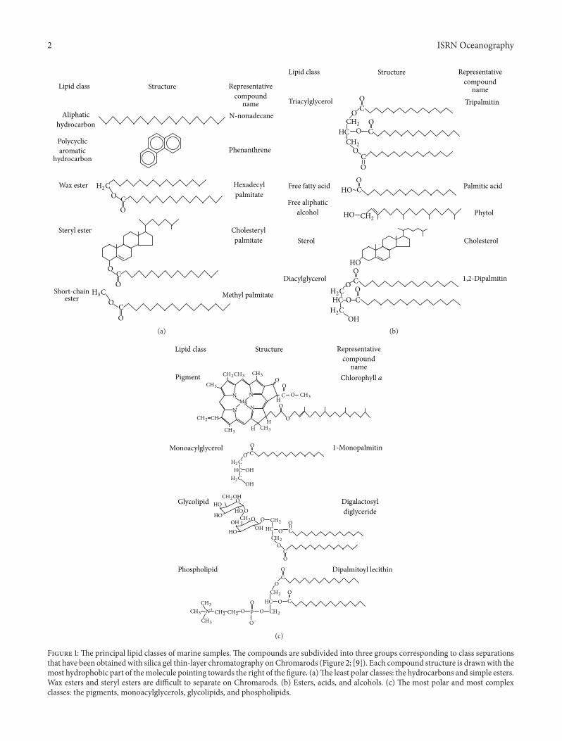
This paper concerns compounds that can be operationallydefined as lipids. The basis of this definition is theirextractability in nonpolar organic solvents which provides aconvenientmeans of separating them from other compoundsin an aqueous matrix. These extracts may contain multi-ple subclasses of both biogenic and anthropogenic origin(Figure 1). The heterogeneous nature of lipids means thatthey are widely used in ecological and biogeochemical studiesassessing the health of ecosystems and the degree to whichthey have been influenced by terrestrial and anthropogenicinputs.They can be used to determine production of biogenicmaterial of dietary value to marine organisms as well asto indicate water quality. While the emphasis here will beon biochemical and ecological aspects of marine lipids, themore stable compounds have also been extensively usedin paleoceanographic studies. Lipid marker determinationin sediment cores can show the sensitivity of sediments tochanges in land use patterns near land margins. The rela-tionship between aquatic and terrestrially derived products incores can be used to indicate the degree to which human land
use has impacted the pattern of aquatic biogenic productivityin the area.
2. Lipid Analyses
The analysis of lipids starts with sampling design and opti-mization, if possible, using chemometric procedures [1]. Ifseawater is being sampled, the next step normally involvesthe separation into operationally defined dissolved and par-ticulate factions. Kepkay [2] gives the particulate fractionas that retained by filters with a nominal pore size of 0.5–0.7 𝜇m and then gives the colloidal fraction as ranging from1 nm to 3 nm equivalent spherical diameter at the lower endto 0.2–1.0 𝜇m at the upper end. Samples are then collectedand lipids are isolated from the sample matrix usually withchloroform in the case of colloids and truly dissolvedmaterial[3], and with mixtures of chloroform and methanol in thecase of solids [4, 5]. At this point total lipids, lipid classes, andindividual compounds may be measured. Total lipids can bedetermined gravimetrically, colorimetrically, or by summingindividually determined lipid classes which have invariablybeen separated by chromatography [4].
2 ISRN Oceanography
Structure
N-nonadecane
Phenanthrene
Hexadecylpalmitate
Cholesterylpalmitate
Lipid class
Aliphatichydrocarbon
Polycyclicaromatic
hydrocarbon
Wax ester
Steryl ester
H2CO C
O
OCO
H3CO C
O
Representativecompound
name
Methyl palmitateShort-chainester
(a)
Tripalmitin
Palmitic acid
Phytol
Cholesterol
Triacylglycerol
Free fatty acid
Free aliphaticalcohol
Sterol
Diacylglycerol
OCO
CH2 OCOHC
CH2O
CO
OCHO
HO CH2
HOOC
HCH2C
CO
H2COH
O
O
StructureLipid class Representativecompound
name
1,2-Dipalmitin
(b)
O−
StructureLipid class Representativecompound
name
1-Monopalmitin
Digalactosyldiglyceride
Dipalmitoyl lecithin
Pigment
Monoacylglycerol
Glycolipid
Phospholipid
OCH
ON
NMg
N
NCH
O
C
O
HC OH
OCOHC
OOOHHO
HO
OC
OOCOHC
OO P
CH3
CH3
CH3
H2C
OCH2CH3
CH2 HCH3HCH3
O
O
H2COH
OC
OCH2
CH2OH
CH2OHO
OOHCH2
HO
CH2
CH2CH2CH2CH3
CH3
CH3 O
Chlorophyll 𝑎
N+
(c)
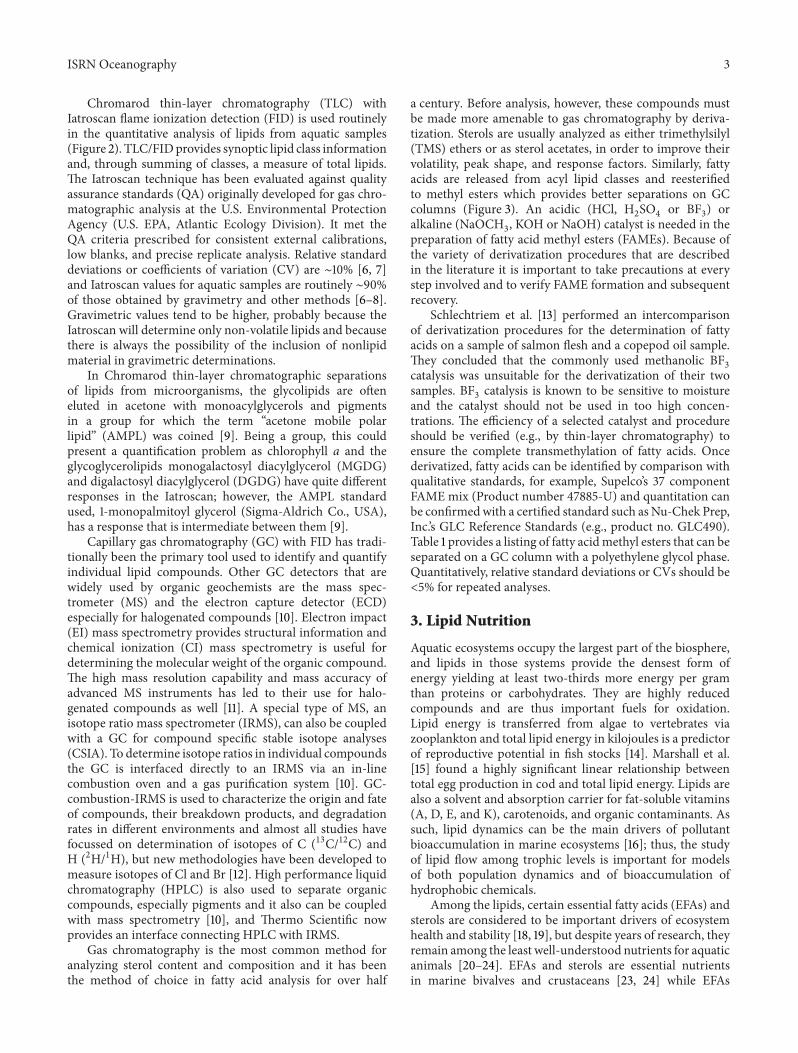
Figure 1: The principal lipid classes of marine samples. The compounds are subdivided into three groups corresponding to class separationsthat have been obtained with silica gel thin-layer chromatography onChromarods (Figure 2; [9]). Each compound structure is drawnwith themost hydrophobic part of themolecule pointing towards the right of the figure. (a)The least polar classes: the hydrocarbons and simple esters.Wax esters and steryl esters are difficult to separate on Chromarods. (b) Esters, acids, and alcohols. (c) The most polar and most complexclasses: the pigments, monoacylglycerols, glycolipids, and phospholipids.
ISRN Oceanography 3
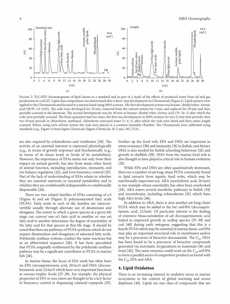
Chromarod thin-layer chromatography (TLC) withIatroscan flame ionization detection (FID) is used routinelyin the quantitative analysis of lipids from aquatic samples(Figure 2). TLC/FIDprovides synoptic lipid class informationand, through summing of classes, a measure of total lipids.The Iatroscan technique has been evaluated against qualityassurance standards (QA) originally developed for gas chro-matographic analysis at the U.S. Environmental ProtectionAgency (U.S. EPA, Atlantic Ecology Division). It met theQA criteria prescribed for consistent external calibrations,low blanks, and precise replicate analysis. Relative standarddeviations or coefficients of variation (CV) are ∼10% [6, 7]and Iatroscan values for aquatic samples are routinely ∼90%of those obtained by gravimetry and other methods [6–8].Gravimetric values tend to be higher, probably because theIatroscan will determine only non-volatile lipids and becausethere is always the possibility of the inclusion of nonlipidmaterial in gravimetric determinations.
In Chromarod thin-layer chromatographic separationsof lipids from microorganisms, the glycolipids are ofteneluted in acetone with monoacylglycerols and pigmentsin a group for which the term “acetone mobile polarlipid” (AMPL) was coined [9]. Being a group, this couldpresent a quantification problem as chlorophyll a and theglycoglycerolipids monogalactosyl diacylglycerol (MGDG)and digalactosyl diacylglycerol (DGDG) have quite differentresponses in the Iatroscan; however, the AMPL standardused, 1-monopalmitoyl glycerol (Sigma-Aldrich Co., USA),has a response that is intermediate between them [9].
Capillary gas chromatography (GC) with FID has tradi-tionally been the primary tool used to identify and quantifyindividual lipid compounds. Other GC detectors that arewidely used by organic geochemists are the mass spec-trometer (MS) and the electron capture detector (ECD)especially for halogenated compounds [10]. Electron impact(EI) mass spectrometry provides structural information andchemical ionization (CI) mass spectrometry is useful fordetermining the molecular weight of the organic compound.The high mass resolution capability and mass accuracy ofadvanced MS instruments has led to their use for halo-genated compounds as well [11]. A special type of MS, anisotope ratio mass spectrometer (IRMS), can also be coupledwith a GC for compound specific stable isotope analyses(CSIA). To determine isotope ratios in individual compoundsthe GC is interfaced directly to an IRMS via an in-linecombustion oven and a gas purification system [10]. GC-combustion-IRMS is used to characterize the origin and fateof compounds, their breakdown products, and degradationrates in different environments and almost all studies havefocussed on determination of isotopes of C (13C/12C) andH (2H/1H), but new methodologies have been developed tomeasure isotopes of Cl and Br [12]. High performance liquidchromatography (HPLC) is also used to separate organiccompounds, especially pigments and it also can be coupledwith mass spectrometry [10], and Thermo Scientific nowprovides an interface connecting HPLC with IRMS.
Gas chromatography is the most common method foranalyzing sterol content and composition and it has beenthe method of choice in fatty acid analysis for over half
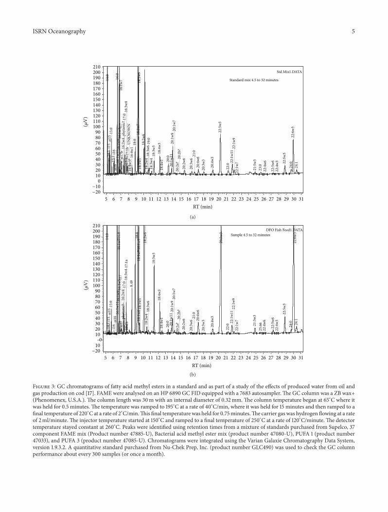
a century. Before analysis, however, these compounds mustbe made more amenable to gas chromatography by deriva-tization. Sterols are usually analyzed as either trimethylsilyl(TMS) ethers or as sterol acetates, in order to improve theirvolatility, peak shape, and response factors. Similarly, fattyacids are released from acyl lipid classes and reesterifiedto methyl esters which provides better separations on GCcolumns (Figure 3). An acidic (HCl, H
2SO4or BF
3) or
alkaline (NaOCH3, KOH or NaOH) catalyst is needed in the
preparation of fatty acid methyl esters (FAMEs). Because ofthe variety of derivatization procedures that are describedin the literature it is important to take precautions at everystep involved and to verify FAME formation and subsequentrecovery.
Schlechtriem et al. [13] performed an intercomparisonof derivatization procedures for the determination of fattyacids on a sample of salmon flesh and a copepod oil sample.They concluded that the commonly used methanolic BF
3
catalysis was unsuitable for the derivatization of their twosamples. BF
3catalysis is known to be sensitive to moisture
and the catalyst should not be used in too high concen-trations. The efficiency of a selected catalyst and procedureshould be verified (e.g., by thin-layer chromatography) toensure the complete transmethylation of fatty acids. Oncederivatized, fatty acids can be identified by comparison withqualitative standards, for example, Supelco’s 37 componentFAME mix (Product number 47885-U) and quantitation canbe confirmedwith a certified standard such asNu-Chek Prep,Inc.’s GLC Reference Standards (e.g., product no. GLC490).Table 1 provides a listing of fatty acidmethyl esters that can beseparated on a GC column with a polyethylene glycol phase.Quantitatively, relative standard deviations or CVs should be<5% for repeated analyses.
3. Lipid Nutrition
Aquatic ecosystems occupy the largest part of the biosphere,and lipids in those systems provide the densest form ofenergy yielding at least two-thirds more energy per gramthan proteins or carbohydrates. They are highly reducedcompounds and are thus important fuels for oxidation.Lipid energy is transferred from algae to vertebrates viazooplankton and total lipid energy in kilojoules is a predictorof reproductive potential in fish stocks [14]. Marshall et al.[15] found a highly significant linear relationship betweentotal egg production in cod and total lipid energy. Lipids arealso a solvent and absorption carrier for fat-soluble vitamins(A, D, E, and K), carotenoids, and organic contaminants. Assuch, lipid dynamics can be the main drivers of pollutantbioaccumulation in marine ecosystems [16]; thus, the studyof lipid flow among trophic levels is important for modelsof both population dynamics and of bioaccumulation ofhydrophobic chemicals.
Among the lipids, certain essential fatty acids (EFAs) andsterols are considered to be important drivers of ecosystemhealth and stability [18, 19], but despite years of research, theyremain among the least well-understood nutrients for aquaticanimals [20–24]. EFAs and sterols are essential nutrientsin marine bivalves and crustaceans [23, 24] while EFAs
4 ISRN Oceanography
Standard mix
Hyd
roca
rbon
sSt
eryl
/wax
este
rsKe
tone
s
Tria
cylg
lyce
rols
Free
fatty
acid
s
Alco
hols
Ster
ols
Acet
one m
obile
pola
rlip
ids
Phos
phol
ipid
s
30.6(mV)
(cm)0 2 4 6 8 10 12 14 16 18 20 22 24 26 28 30
100
(%)
80
60
40
20
0
(a)
Hyd
roca
rbon
sSt
eryl
/wax
este
rs
Keto
nes
Tria
cylg
lyce
rols
Free
fatty
acid
s
Ster
ols
Acet
one m
obile
pola
rlip
ids
Phos
phol
ipid
s
(cm)0 2 4 6 8 10 12 14 16 18 20 22 24 26 28 30
Sample
Met
hyl e
sters
Dia
cylg
lyce
rols
59.9(mV)
100
80
60
40
20
0
(%)
(b)
Figure 2: TLC/FID chromatograms of lipid classes in a standard and as part of a study of the effects of produced water from oil and gasproduction on cod [17]. Lipid class compositionwas determined after a three-step development onChromarods (Figure 1). Lipid extracts wereapplied to the Chromarods and focused to a narrow band using 100% acetone.The first development systemwas hexane : diethyl ether : formicacid (98.95 : 1.0 : 0.05). The rods were developed for 25min, removed from the solvent system for 5min, and replaced for 20min and thenpartially scanned in the Iatroscan. The second development was for 40min in hexane : diethyl ether : formic acid (79 : 20 : 1) after which therods were partially scanned. The final separation had two steps, the first was development in 100% acetone for two 15min time periods, thentwo 10min periods in chloroform :methanol : chloroform-extracted water (5 : 4 : 1), after which the rods were dried and their entire lengthscanned. Before using each solvent system the rods were placed in a constant humidity chamber. The Chromarods were calibrated usingstandards (e.g., Figure 1) from Sigma Chemicals (Sigma Chemicals, St. Louis, MI, USA).
are also required by echinoderms and vertebrates [24]. Theactivity of an essential nutrient is expressed physiologically(e.g., in terms of growth response) and biochemically (e.g.,in terms of its tissue levels or levels of its metabolites).However, the importance of EFAs stems not only from theirimpact on animal growth, but also from many other facetsof animal function, including reproduction, immunity, andion balance regulation [22], and even buoyancy control [25].Part of the lack of understanding of EFAs relates to whetherthey are essential nutrients or essential metabolites and towhether they are conditionally indispensable or conditionallydispensable [24].
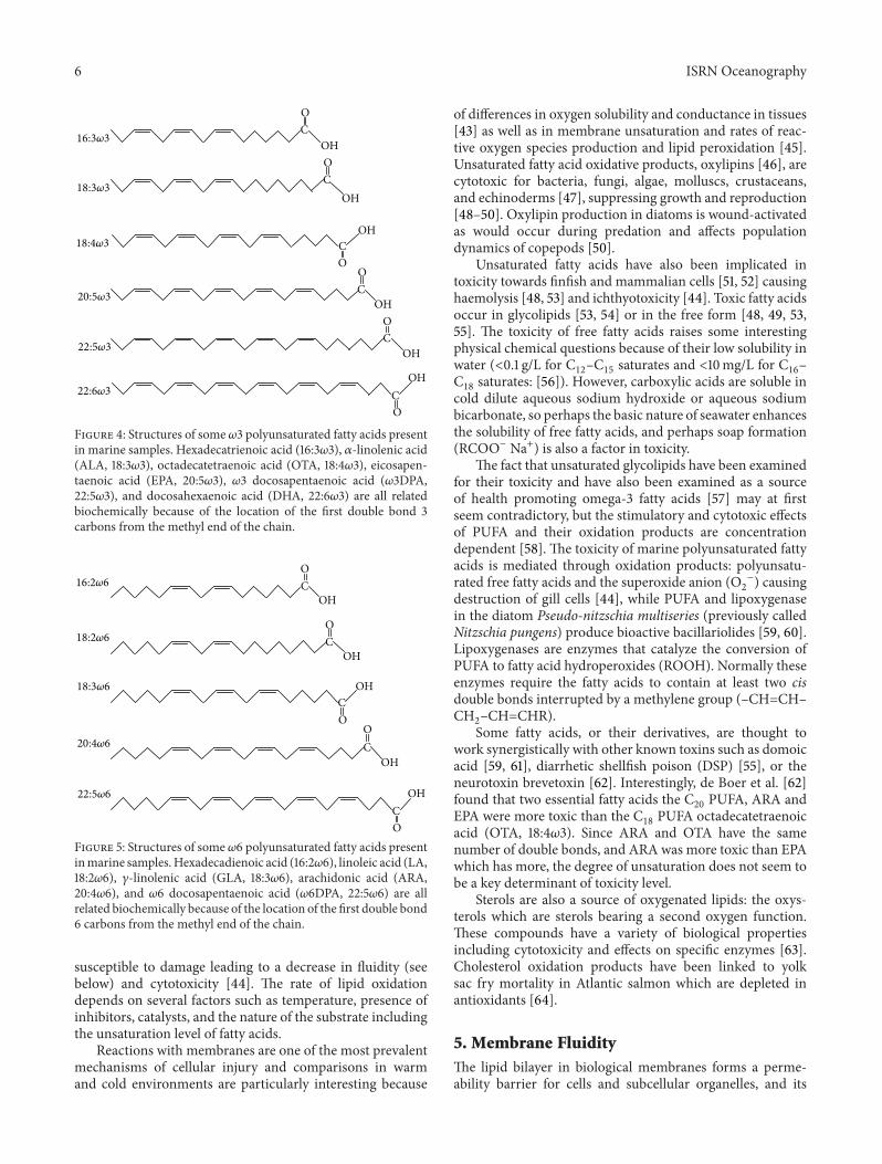
There are two related families of EFAs consisting of 𝜔3(Figure 4) and 𝜔6 (Figure 5) polyunsaturated fatty acids(PUFA). Fatty acids in each of the families are intercon-vertible usually through alternate use of desaturases andelongases. The extent to which a given species at a given lifestage can convert one 𝜔3 fatty acid to another or one 𝜔6fatty acid to another determines the degree of essentiality ofthe fatty acid for that species at that life stage. It should benoted that there are pathways of PUFA synthesis which do notrequire desaturation and elongation of saturated fatty acids.Polyketide synthase systems conduct the same reactions butin an abbreviated sequence [26]. It has been speculatedthat PUFA originally synthesized by the polyketide synthasepathway may be a significant contributor to PUFA in marinefish [26].
In marine fauna, the focus of EFA work has often beenon EPA (eicosapentaenoic acid, 20:5𝜔3) and DHA (docosa-hexaenoic acid, 22:6𝜔3) which have very important functionsat various trophic levels [27–29]. For example, the physicalproperties of EPA in wax esters are thought to be a key factorin buoyancy control in diapausing calanoid copepods [25].
Further up the food web, EPA and DHA are important instress resistance [30] and immunity [31] in finfish, and dietaryDHA is also needed for finfish schooling behaviour [32] andgrowth in shellfish [29]. DHA from the marine food web isalso thought to have played a critical role in human evolution[33].
While EPA and DHA are often quantitatively dominant,there are a number of 𝜔6 long-chain PUFA commonly foundin lipid extracts from aquatic food webs, which may benutritionally important too. ARA (arachidonic acid, 20:4𝜔6)is one example whose essentiality has often been overlooked[34]. ARA enters several metabolic pathways in finfish [34]and invertebrates, including echinoderms [35] which havehigh ARA levels [36].
In addition to ARA, there is now another 𝜔6 long-chainPUFA which may be added to the list: 𝜔6DPA (docosapen-taenoic acid, 22:5𝜔6). Of particular interest is the findingof extensive bioaccumulation of 𝜔6 docosapentaenoic acidlinked to improved growth in scallop species [37–39] andcod [40] during early ontogeny, suggesting that this is afourth PUFAwhichmay be essential inmarine fauna.𝜔6DPAmay play an important structural role in membranes and/ormay be a precursor of bioactive docosanoids. The C
22DHA
has been found to be a precursor of bioactive compoundsgenerated via enzymatic oxygenations in mammals [41] andtrout [42]. The same enzymes could work on the C
22𝜔6DPA
to form a parallel series of competitive products as foundwiththe C20EPA and ARA.
4. Lipid OxidationThere is an increasing interest in oxidative stress in marineecosystems in the context of global warming and ozonedepletion [43]. Lipids are one class of compounds that are
ISRN Oceanography 5
14:0
14:1
i15
ai15
15:0
15:1
i16
ai16
16:0
16:1
w11
16:1
w9
16:1
w7
16:1
w5 i1
7ai
17a
ai17
b16
:2w
4ph
ytan
ic? 1
7:0
16:3
w4
17:1
a 17:
1bU
NKN
OW
N16
:4w
3?16
:4w
118
:018
:1w
918
:1w
718
:1w
518
:2w
618
:2w
418
:3w
619
:018
:3w
418
:3w
318
:4w
318
:4w
1 20:0
20:1
w11
20:1
w9
20:1
w7
20:2
a?20
:2b?
20:2
w6
20:3
w6
21:0
20:4
w6
20:3
w3
20:4
w3
22:5
w3
22:0
22:1
w11
22:1
w9
22:1
w7
21:5
w3
23:0
22:4
w6
22:5
w6
22:4
w3 22
:5w
3 24
:022
:6w
324
:1
Std Mix1.DATA
Standard mix 4.5 to 32 minutes
RT (min)5 7 9 11 13 15 17 19 21 23 25 27 29 31
210
190
170
150
130
110
90
70
50
30
10
−10
(𝜇V
)
6 8 10 12 14 16 18 20 22 24 26 28 30
200
180
160
140
120
100
80
60
40
20
0
−20
(a)
14:0
14:1
i15
ai15
15:0
i16
ai16
16:0
16:1
w11
16:1
w9
16:1
w7
16:1
w5 i1
7ai
1716
:2w
4ph
ytan
ic?
17:0
16:3
w4
17:1
a17
:1b
8.49
18:0
18:1
w9
18:1
w7
18:1
w6 1
8:1w
518
:2w
618
:2w
418
:3w
618
:3w
318
:4w
318
:4w
120
:0 20:1
w11
20:1
w9
20:1
w7
20:2
a?20
:2b?
20:2
w6
20:3
w6
21:0
20:4
w6
20:3
w3
20:4
w3
20:5
w3
22:0
22:1
w11
22:1
w9
22:1
w7
21:5
w3
25.6
622
:4w
6
22:5
w6
22:4
w3
22:5
w3
24:0
22:6
w3
24:1
DFO Fish Feed1.DATASample 4.5 to 32 minutes
RT (min)5 7 9 11 13 15 17 19 21 23 25 27 29 31
210
190
170
150
130
110
90
70
50
30
10−10
(𝜇V
)
6 8 10 12 14 16 18 20 22 24 26 28 30
200
180
160
140
120
100
80
60
40
20
0
−20
(b)
Figure 3: GC chromatograms of fatty acid methyl esters in a standard and as part of a study of the effects of produced water from oil andgas production on cod [17]. FAME were analysed on an HP 6890 GC FID equipped with a 7683 autosampler. The GC column was a ZB wax+(Phenomenex, U.S.A.). The column length was 30m with an internal diameter of 0.32mm. The column temperature began at 65∘C where itwas held for 0.5 minutes. The temperature was ramped to 195∘C at a rate of 40∘C/min, where it was held for 15 minutes and then ramped to afinal temperature of 220∘C at a rate of 2∘C/min.This final temperature was held for 0.75minutes.The carrier gas was hydrogen flowing at a rateof 2ml/minute. The injector temperature started at 150∘C and ramped to a final temperature of 250∘C at a rate of 120∘C/minute. The detectortemperature stayed constant at 260∘C. Peaks were identified using retention times from a mixture of standards purchased from Supelco, 37component FAME mix (Product number 47885-U), Bacterial acid methyl ester mix (product number 47080-U), PUFA 1 (product number47033), and PUFA 3 (product number 47085-U). Chromatograms were integrated using the Varian Galaxie Chromatography Data System,version 1.9.3.2. A quantitative standard purchased from Nu-Chek Prep, Inc. (product number GLC490) was used to check the GC columnperformance about every 300 samples (or once a month).
6 ISRN Oceanography
16:3𝜔3
18:3𝜔3
18:4𝜔3
20:5𝜔3
22:5𝜔3
22:6𝜔3
OC
OHOC
OH
OC
OH
OC
OHOC
OH
OC
OH
Figure 4: Structures of some 𝜔3 polyunsaturated fatty acids presentin marine samples. Hexadecatrienoic acid (16:3𝜔3), 𝛼-linolenic acid(ALA, 18:3𝜔3), octadecatetraenoic acid (OTA, 18:4𝜔3), eicosapen-taenoic acid (EPA, 20:5𝜔3), 𝜔3 docosapentaenoic acid (𝜔3DPA,22:5𝜔3), and docosahexaenoic acid (DHA, 22:6𝜔3) are all relatedbiochemically because of the location of the first double bond 3carbons from the methyl end of the chain.
16:2𝜔6
18:2𝜔6
18:3𝜔6
20:4𝜔6
22:5𝜔6
OC
OH
OC
OH
OC
OH
O
COH
O
COH
Figure 5: Structures of some 𝜔6 polyunsaturated fatty acids presentinmarine samples.Hexadecadienoic acid (16:2𝜔6), linoleic acid (LA,18:2𝜔6), 𝛾-linolenic acid (GLA, 18:3𝜔6), arachidonic acid (ARA,20:4𝜔6), and 𝜔6 docosapentaenoic acid (𝜔6DPA, 22:5𝜔6) are allrelated biochemically because of the location of the first double bond6 carbons from the methyl end of the chain.
susceptible to damage leading to a decrease in fluidity (seebelow) and cytotoxicity [44]. The rate of lipid oxidationdepends on several factors such as temperature, presence ofinhibitors, catalysts, and the nature of the substrate includingthe unsaturation level of fatty acids.
Reactions with membranes are one of the most prevalentmechanisms of cellular injury and comparisons in warmand cold environments are particularly interesting because
of differences in oxygen solubility and conductance in tissues[43] as well as in membrane unsaturation and rates of reac-tive oxygen species production and lipid peroxidation [45].Unsaturated fatty acid oxidative products, oxylipins [46], arecytotoxic for bacteria, fungi, algae, molluscs, crustaceans,and echinoderms [47], suppressing growth and reproduction[48–50]. Oxylipin production in diatoms is wound-activatedas would occur during predation and affects populationdynamics of copepods [50].
Unsaturated fatty acids have also been implicated intoxicity towards finfish and mammalian cells [51, 52] causinghaemolysis [48, 53] and ichthyotoxicity [44]. Toxic fatty acidsoccur in glycolipids [53, 54] or in the free form [48, 49, 53,55]. The toxicity of free fatty acids raises some interestingphysical chemical questions because of their low solubility inwater (<0.1 g/L for C
12–C15saturates and <10mg/L for C
16–
C18saturates: [56]). However, carboxylic acids are soluble in
cold dilute aqueous sodium hydroxide or aqueous sodiumbicarbonate, so perhaps the basic nature of seawater enhancesthe solubility of free fatty acids, and perhaps soap formation(RCOO− Na+) is also a factor in toxicity.
The fact that unsaturated glycolipids have been examinedfor their toxicity and have also been examined as a sourceof health promoting omega-3 fatty acids [57] may at firstseem contradictory, but the stimulatory and cytotoxic effectsof PUFA and their oxidation products are concentrationdependent [58]. The toxicity of marine polyunsaturated fattyacids is mediated through oxidation products: polyunsatu-rated free fatty acids and the superoxide anion (O
2
−) causingdestruction of gill cells [44], while PUFA and lipoxygenasein the diatom Pseudo-nitzschia multiseries (previously calledNitzschia pungens) produce bioactive bacillariolides [59, 60].Lipoxygenases are enzymes that catalyze the conversion ofPUFA to fatty acid hydroperoxides (ROOH). Normally theseenzymes require the fatty acids to contain at least two cisdouble bonds interrupted by a methylene group (–CH=CH–CH2–CH=CHR).Some fatty acids, or their derivatives, are thought to
work synergistically with other known toxins such as domoicacid [59, 61], diarrhetic shellfish poison (DSP) [55], or theneurotoxin brevetoxin [62]. Interestingly, de Boer et al. [62]found that two essential fatty acids the C
20PUFA, ARA and
EPA were more toxic than the C18PUFA octadecatetraenoic
acid (OTA, 18:4𝜔3). Since ARA and OTA have the samenumber of double bonds, and ARA was more toxic than EPAwhich has more, the degree of unsaturation does not seem tobe a key determinant of toxicity level.
Sterols are also a source of oxygenated lipids: the oxys-terols which are sterols bearing a second oxygen function.These compounds have a variety of biological propertiesincluding cytotoxicity and effects on specific enzymes [63].Cholesterol oxidation products have been linked to yolksac fry mortality in Atlantic salmon which are depleted inantioxidants [64].
5. Membrane FluidityThe lipid bilayer in biological membranes forms a perme-ability barrier for cells and subcellular organelles, and its
ISRN Oceanography 7
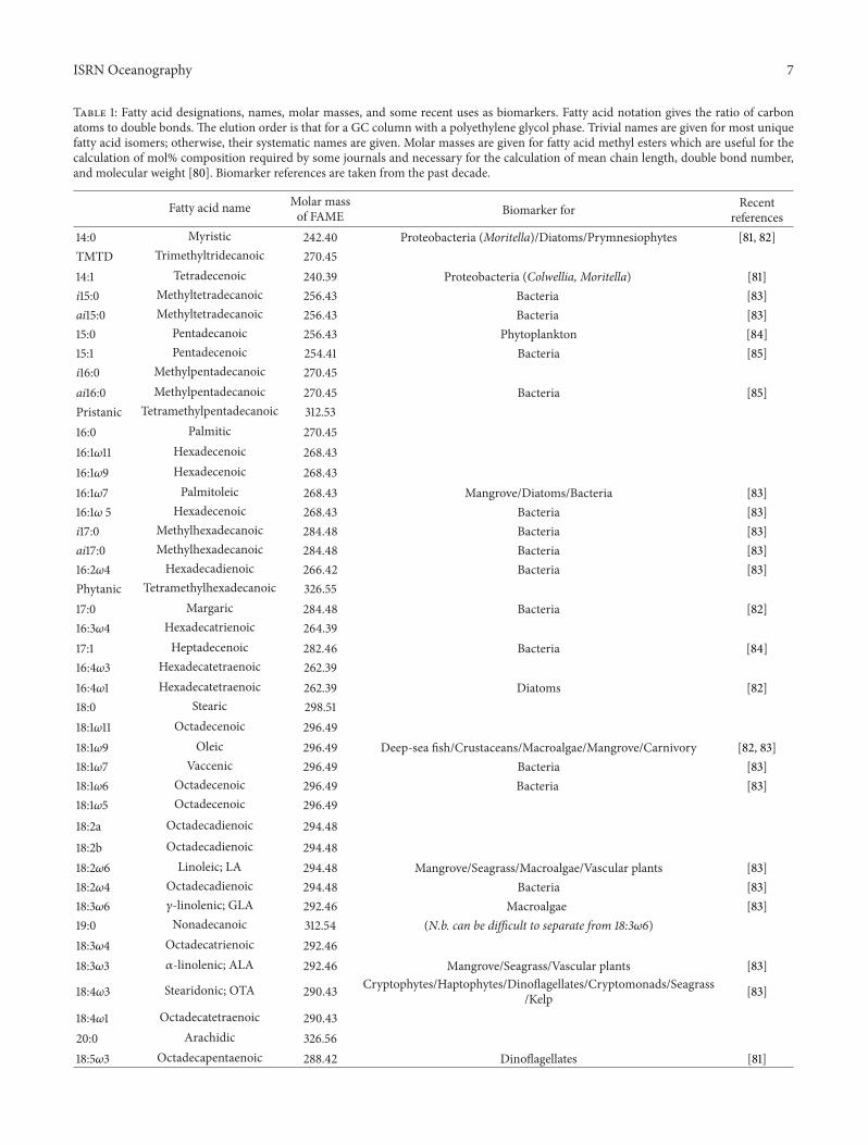
Table 1: Fatty acid designations, names, molar masses, and some recent uses as biomarkers. Fatty acid notation gives the ratio of carbonatoms to double bonds. The elution order is that for a GC column with a polyethylene glycol phase. Trivial names are given for most uniquefatty acid isomers; otherwise, their systematic names are given. Molar masses are given for fatty acid methyl esters which are useful for thecalculation of mol% composition required by some journals and necessary for the calculation of mean chain length, double bond number,and molecular weight [80]. Biomarker references are taken from the past decade.
Fatty acid name Molar massof FAME Biomarker for Recent
references14:0 Myristic 242.40 Proteobacteria (Moritella)/Diatoms/Prymnesiophytes [81, 82]TMTD Trimethyltridecanoic 270.4514:1 Tetradecenoic 240.39 Proteobacteria (Colwellia, Moritella) [81]i15:0 Methyltetradecanoic 256.43 Bacteria [83]ai15:0 Methyltetradecanoic 256.43 Bacteria [83]15:0 Pentadecanoic 256.43 Phytoplankton [84]15:1 Pentadecenoic 254.41 Bacteria [85]i16:0 Methylpentadecanoic 270.45ai16:0 Methylpentadecanoic 270.45 Bacteria [85]Pristanic Tetramethylpentadecanoic 312.5316:0 Palmitic 270.4516:1𝜔11 Hexadecenoic 268.4316:1𝜔9 Hexadecenoic 268.4316:1𝜔7 Palmitoleic 268.43 Mangrove/Diatoms/Bacteria [83]16:1𝜔 5 Hexadecenoic 268.43 Bacteria [83]i17:0 Methylhexadecanoic 284.48 Bacteria [83]ai17:0 Methylhexadecanoic 284.48 Bacteria [83]16:2𝜔4 Hexadecadienoic 266.42 Bacteria [83]Phytanic Tetramethylhexadecanoic 326.5517:0 Margaric 284.48 Bacteria [82]16:3𝜔4 Hexadecatrienoic 264.3917:1 Heptadecenoic 282.46 Bacteria [84]16:4𝜔3 Hexadecatetraenoic 262.3916:4𝜔1 Hexadecatetraenoic 262.39 Diatoms [82]18:0 Stearic 298.5118:1𝜔11 Octadecenoic 296.4918:1𝜔9 Oleic 296.49 Deep-sea fish/Crustaceans/Macroalgae/Mangrove/Carnivory [82, 83]18:1𝜔7 Vaccenic 296.49 Bacteria [83]18:1𝜔6 Octadecenoic 296.49 Bacteria [83]18:1𝜔5 Octadecenoic 296.4918:2a Octadecadienoic 294.4818:2b Octadecadienoic 294.4818:2𝜔6 Linoleic; LA 294.48 Mangrove/Seagrass/Macroalgae/Vascular plants [83]18:2𝜔4 Octadecadienoic 294.48 Bacteria [83]18:3𝜔6 𝛾-linolenic; GLA 292.46 Macroalgae [83]19:0 Nonadecanoic 312.54 (N.b. can be difficult to separate from 18:3𝜔6)18:3𝜔4 Octadecatrienoic 292.4618:3𝜔3 𝛼-linolenic; ALA 292.46 Mangrove/Seagrass/Vascular plants [83]
18:4𝜔3 Stearidonic; OTA 290.43 Cryptophytes/Haptophytes/Dinoflagellates/Cryptomonads/Seagrass/Kelp [83]
18:4𝜔1 Octadecatetraenoic 290.4320:0 Arachidic 326.5618:5𝜔3 Octadecapentaenoic 288.42 Dinoflagellates [81]
8 ISRN Oceanography
Table 1: Continued.
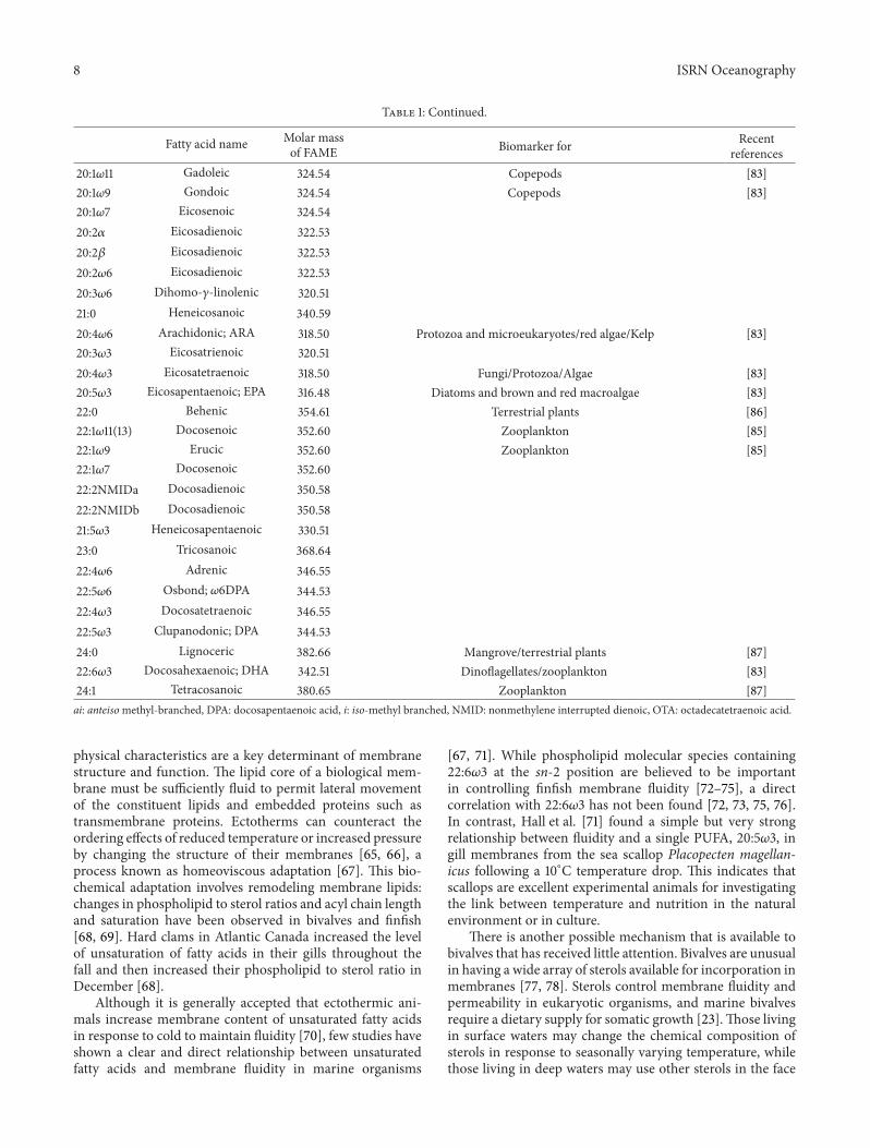
Fatty acid name Molar massof FAME Biomarker for Recent
references20:1𝜔11 Gadoleic 324.54 Copepods [83]20:1𝜔9 Gondoic 324.54 Copepods [83]20:1𝜔7 Eicosenoic 324.5420:2𝛼 Eicosadienoic 322.5320:2𝛽 Eicosadienoic 322.5320:2𝜔6 Eicosadienoic 322.5320:3𝜔6 Dihomo-𝛾-linolenic 320.5121:0 Heneicosanoic 340.5920:4𝜔6 Arachidonic; ARA 318.50 Protozoa and microeukaryotes/red algae/Kelp [83]20:3𝜔3 Eicosatrienoic 320.5120:4𝜔3 Eicosatetraenoic 318.50 Fungi/Protozoa/Algae [83]20:5𝜔3 Eicosapentaenoic; EPA 316.48 Diatoms and brown and red macroalgae [83]22:0 Behenic 354.61 Terrestrial plants [86]22:1𝜔11(13) Docosenoic 352.60 Zooplankton [85]22:1𝜔9 Erucic 352.60 Zooplankton [85]22:1𝜔7 Docosenoic 352.6022:2NMIDa Docosadienoic 350.5822:2NMIDb Docosadienoic 350.5821:5𝜔3 Heneicosapentaenoic 330.5123:0 Tricosanoic 368.6422:4𝜔6 Adrenic 346.5522:5𝜔6 Osbond; 𝜔6DPA 344.5322:4𝜔3 Docosatetraenoic 346.5522:5𝜔3 Clupanodonic; DPA 344.5324:0 Lignoceric 382.66 Mangrove/terrestrial plants [87]22:6𝜔3 Docosahexaenoic; DHA 342.51 Dinoflagellates/zooplankton [83]24:1 Tetracosanoic 380.65 Zooplankton [87]ai: anteisomethyl-branched, DPA: docosapentaenoic acid, i: iso-methyl branched, NMID: nonmethylene interrupted dienoic, OTA: octadecatetraenoic acid.
physical characteristics are a key determinant of membranestructure and function. The lipid core of a biological mem-brane must be sufficiently fluid to permit lateral movementof the constituent lipids and embedded proteins such astransmembrane proteins. Ectotherms can counteract theordering effects of reduced temperature or increased pressureby changing the structure of their membranes [65, 66], aprocess known as homeoviscous adaptation [67]. This bio-chemical adaptation involves remodeling membrane lipids:changes in phospholipid to sterol ratios and acyl chain lengthand saturation have been observed in bivalves and finfish[68, 69]. Hard clams in Atlantic Canada increased the levelof unsaturation of fatty acids in their gills throughout thefall and then increased their phospholipid to sterol ratio inDecember [68].
Although it is generally accepted that ectothermic ani-mals increase membrane content of unsaturated fatty acidsin response to cold to maintain fluidity [70], few studies haveshown a clear and direct relationship between unsaturatedfatty acids and membrane fluidity in marine organisms
[67, 71]. While phospholipid molecular species containing22:6𝜔3 at the sn-2 position are believed to be importantin controlling finfish membrane fluidity [72–75], a directcorrelation with 22:6𝜔3 has not been found [72, 73, 75, 76].In contrast, Hall et al. [71] found a simple but very strongrelationship between fluidity and a single PUFA, 20:5𝜔3, ingill membranes from the sea scallop Placopecten magellan-icus following a 10∘C temperature drop. This indicates thatscallops are excellent experimental animals for investigatingthe link between temperature and nutrition in the naturalenvironment or in culture.
There is another possible mechanism that is available tobivalves that has received little attention. Bivalves are unusualin having a wide array of sterols available for incorporation inmembranes [77, 78]. Sterols control membrane fluidity andpermeability in eukaryotic organisms, and marine bivalvesrequire a dietary supply for somatic growth [23].Those livingin surface waters may change the chemical composition ofsterols in response to seasonally varying temperature, whilethose living in deep waters may use other sterols in the face
ISRN Oceanography 9
of constantly very cold conditions and higher pressure. Theordering capacity provided by cholesterol is different to thatprovided by other sterols [23]. Sea scallops living at 10m and31m depths in Newfoundland waters have different sterolsin their adductor muscle, especially 24-methylenecholesterol[79], which is a major sterol in cold water bivalves [77].Whether this has any effect on membrane fluidity, or thereis a seasonal pattern, or there is any seasonal correlation withsource sterols have yet to be determined.
6. Molecular Biomarkers
Biological markers are receiving widespread attention inecological, paleoecological, and biogeochemical studies inaquatic environments [10]. Chemicalmarkers are compoundsor groups of compounds that can be used as indicatorsor signatures of individual organisms or of groupings oforganisms, or of certain environmental processes. In orderto measure inputs, cycling, and loss of materials in marineecosystems, there are now a wide range of compounds andinstrumental tools that are available. Anthropogenic com-pounds can be used as wastewater markers to locate sourcesand pathways of transport as well as to determine pollutantloadings. Molecular biomarkers can be DNA fragments orsmaller molecules that are easily determined using standardchromatographic separation techniques. Lipids are one suchgroup that are receiving continuing attention in ecological(e.g., [88]) and biogeochemical [89] studies.
The heterogeneous nature of lipids means that muchinformation can be gained by determining individual classes.Individual classes or groups of certain classes may be used toindicate the presence of certain types of organisms as well astheir physiological state and activity [90]. Lipid classes canalso be used to indicate sources of organic matter [91] includ-ing dissolved organic matter and hydrophobic contaminants[89, 92]. Iatroscan-determined free fatty acids, alcohols,diacylglycerols, and monoacylglycerols in filtrates indicatedseasonal, day/night, and depth differences in degradationin the North West Mediterranean Sea, while hydrocarbonsshowed the level of contamination [89, 92].
Lipid class information is particularly valuable whenused in conjunction with determination of individual fattyacids [93] or sterols [94] or both [95]. Another combination,fatty acids, n-alkanes and glycerol dialkyl glycerol tetraethers(GDGTs) has been used to assess differences in planktoncomposition and food quality in the Humboldt CurrentSystem [86]. GDGTs are found in prokaryotemembranes andthey contain ether linkages to glycerol (R–O–C–) rather thanester linkages as found in glycerophospholipids (RCOOC–).HPLC-MS analyses of intact GDGTs in sediments also showgreat promise for identifying the delivery of soil-derivedorganic matter to coastal regions as well as for paleother-mometry [10].
Fatty acids and sterols are versatile biomarkers that arewidely employed in oceanographic studies because of thelarge number of unique structures that are synthesized. Fattyacids have often been broadly used as biomarkers in trophictransfer studies in aquatic food webs (e.g., [96–98]) and nowthey are being increasingly used for analysing terrestrial food
webs as well [99]. Information provided by these markersmay be used to delineate carbon cycling and transfer ofmaterial through food webs especially when analysed withmultivariate statistics (see below).
Using fatty acids, bacteria, phytoplankton classes, andzooplankton orders may be distinguished in marine samples[81, 88, 93]. Table 1 gives a listing of recently used fatty acidsand it can be seen that many compounds are not uniquebiomarkers. When this occurs, fatty acid sums and ratios canbe added to help in the interpretation. For example, Pepinet al. [100], employing a variety of markers of diatoms (16:1𝜔7+ 16:4𝜔1 + 20:5𝜔3; 16:1𝜔7/16:0; ΣC
16/ΣC18), prymnesio-
phytes (18:1𝜔9 + 18:4𝜔3), and dinoflagellates (22:6𝜔3/20:5𝜔3)showed diapausing copepods in slope waters had fed mainlyon diatomswhile active ones in shelf waters had fedmainly ondinoflagellates and prymnesiophytes. Another ratio has beenused in benthic organisms, the 18:1 isomer ratio, to indicatediatom feeding and trophic level [101]. The rationale behindthe use of these 𝜔9/𝜔7 ratios is elongation of the diatom 16:1𝜔7 to 18:1𝜔7, but they could be biased if there was a significantintake of bacterial 18:1𝜔7. Low and seasonally highly variableratios have been observed in echinoderm gonads [102, 103].
Zooplankton grazing is an important link between lowerand higher trophic levels and fatty acid biomarkers may alsobe employed to determine the importance of zooplanktonsources [81, 88, 104]. For example, Kreibich et al. [105] used20:1𝜔9 and 22:1𝜔11 to show calanoid copepods were a majorfood source for two pelagic crustaceans in the Greenland Sea.
Sterols and hydrocarbons are also excellent biomarkersdue to their stability in addition to the diversity of their struc-tures. They have been extensively used in sedimentary geo-chemical studies (e.g., [106–108]) and aquatic biogeochemicalstudies [10]. For example, Hudson et al. [109] grouped sterolsin order to apportion inputs to a cold ocean ecosystem andshowed the dominance of marine origin C
27and C
28sterols
in plankton and settling particles contrasted with higherplant C
29sterols dominating in the underlying sediments. As
part of a trophic ecology study, Drazen et al. [95] measuredsterols to help assess feeding habits of abyssal echinodermsand used C
28and C
29sterols, especially 24-methylcholesta-
5,22E-dien-3𝛽-ol (brassicasterol) and 24-ethylcholest-5-en-3𝛽-ol (sitosterol), to show the importance of phytodetritus.The use of sterols as trophic markers in the lower food webshould be investigated further. Indeed, further sterol studiesinmodern environments will greatly aid source identificationin sediments (e.g., [108]).
7. Multivariate Data Analysis
Application of chemometric methods for experiment designand analysis of multivariate data is becoming increasinglycommon as it permits data reduction and an objectiveinterpretation of the results. In the marine biomarker field,principal components analysis (PCA) has gained popularityas a powerful data reduction procedure. This multivariatetechnique handles a large amount of variables simultaneously,and has been used by organic geochemists to determineorganic matter sources and degradation of suspended andsinking particles in the oceans [110, 111]. We have used PCA
10 ISRN Oceanography
to determine marine, terrestrial, and anthropogenic organicinputs, and trophic relationships (e.g., [100, 102, 104, 112–114]).Fatty acid PCA continues to have a strong following [115–117], but more sophisticated multivariate statistical analysesare emerging too (see below).
There are several refinements to PCA that have beenapplied, such as expansion beyond the first two principalcomponents. PC1 accounts for most of the variability inthe data set, PC2 the next largest, and so on. When PC1and PC2 account for most of the overall variation in thedata set between them, the data can easily be representeddiagrammatically in only two dimensions. For example, PC1and PC2 in Pepin et al. [100] explained 92% of the variation incopepod Calanus finmarchicus fatty acid summary data andthere was a clear separation of samples from shelf and slopelocations. Inmost studies, it requiresmore than two principalcomponents to account for that much of the variation. Xue etal. [111] used three principal components to construct organicmatter degradation trajectories as against a one-dimensionaldegradation index [110]. Xue et al. [111] used cluster analysisto help determine the number of principal components toconsider. Even if more than half of the variation in the dataset is accounted for by PC1 and PC2, indicating the sign (+or −) of the coefficients and scores on PC3 can be useful. Ina copepod feeding study the first two principal componentsaccounted for 54.2% of the variation and addition of the thirdprincipal component raised this value to 73.8% [114]. The co-location of Calanus spp. and its diet in three PC dimensionsshowed that there was little modification or sequestrationof dietary polyunsaturated fatty acids. This study also usedcluster analysis but this time the scores were grouped bysingle linkage cluster analysis in order to determine whichshould be encircled in the graphic representation. Allan et al.[117] used one-way analysis of variance (ANOVA) for thesame purpose and showed spatial changes in mussel diets.
An additional refinement is the further analysis of PCAdata. PC1 scores have been used to indicate how sourcecomposition of suspended and sinking particles varies withdepth [110], and how the accumulation of reserves in oystersvaries with season [118]. They have also been correlated withlengths of Calanus finmarchicus [100] and lengths of belugawhales [115] to reveal size-related dietary differences. Furtherprincipal components could be included in the analyses usingmultiple linear regressions [1].
8. Quantification of Trophic Relationships
Despite several refinements that can be applied, principalcomponents analysis remains essentially a means to providea simple graphic representation of an entire data set. Moresophisticated multivariate statistical analyses can provide amore quantitative analysis of patterns, and such techniquesare emerging for usewith fatty acids, especially from foodwebsamples [119, 120]. These authors used PRIMER (PlymouthRoutines in Multivariate Ecological Research) to investigatealgae feeding amphipods and sea urchins. This is a softwarepackage for analyzing ecological data marketed by PRIMEREnterprises (PRIMER-E) Ltd. They used analysis of similar-ities (ANOSIM), a nonparametric approximate analogue of
univariate ANOVA tests, to provide a measure of similaritybetween groups, and similarity of percentages analysis (SIM-PER) to examine the contribution of each fatty acid to averageresemblances between sample groups. SIMPER identifies thatwhich is primarily providing the discrimination betweenobserved sample clusters.
ANOSIM and SIMPER provide a more quantitativeanalysis of patterns in the lower food web, but they do notprovide quantitative estimates of predator diets as can be doneat higher trophic levels [96, 98]. Prey consumption of seals,seabirds, and polar bears has been estimated by statisticallycomparing predator fatty acid signatures with those of theirpotential prey.
The quantitative fatty acid signature analysis (QFASA)model [96] is based on minimizing the statistical distancebetween predator signatures and mean prey signatures.Predator signatures must also be corrected for modificationthat may occur due to predator fatty acid metabolism usingcalibration coefficients. Calibration coefficients are developedusing long-term controlled diet studies to estimate differ-ences between patterns of fatty acid intake and depositionin predators. The coefficients correspond to the ratio of preycomposition to predator tissue composition for each fattyacid, after assuming it has consumed the diet long enoughto have completely turned over all fatty acids. Thus for fattyacids to be quantitative determinants of diet, an experimentalapproach is required to determine selective retention ofspecific fatty acids, to detect de novo synthesis, elongationand/or desaturation, and tomeasure extents of tissue-specificdeposition. It is interesting that this is the same questionbeing asked in the field of aquaculture nutrition in differentontogenetic stages of multiple species from several phyla.For example, dietary and tissue fatty acids and sterols havebeen measured to determine incorporation efficiencies orenrichments and depletions in yellowtail flounder [121] andsea scallops [37, 78]. Such data may help in the application ofQFASA to fish and primary consumers; however, the breadthof species and differences in lipid metabolism in the lowerfood web may make quantitative estimates of fatty acid flowthrough the lower food web difficult.
The determination of isotopes of C (13C/12C) and N(15N/14N) may also help estimate fatty acid flow throughthe lower food web. Quantitative estimates of ice algaecontributions to higher trophic levels have been madeusing compound-specific stable isotope analyses of fattyacid 13C/12C ratios (𝛿13C in ‰: [122]). Fatty acid isotopicsignatures were also used in a three-step food chain feedingexperiment to show that polyunsaturated fatty acids origi-nally synthesized by the polyketide synthase pathway couldbe a significant contributor to PUFA in marine fish [40]. Inaddition there are implications for food web 𝛿13C as fewersynthetic steps will cause less kinetic fractionation of the iso-tope at the original point of synthesis.This stable isotopemassspectrometermethodology should be investigated further forfatty acid and sterol work.
Bulk 15N/14N ratios (𝛿15N in ‰) have traditionally beenused to determine trophic levels of consumers. Connelly et al.[84] correlated bulk stable isotope ratios (‰) and fatty acid
ISRN Oceanography 11
proportions (% of total fatty acids) in 26 taxa of zooplanktoncollected from the benthic boundary layer on the BeaufortSea shelf. For those fatty acids that had a significant corre-lation with 𝛿15N, a trophic multiplication factor (TMF) wascalculated from the slope of the linear regression on 𝛿15Ndetermined trophic level. Docosahexaenoic acid had by farthe greatest average increase per trophic level among thePUFA, suggesting that this essential fatty acid was highlyconserved through the food web and was not used as anenergy source.
Carreon-Palau et al. [123] calculated trophic retentionfactors (TRFs) for fatty acids in a coral reef food web withriver influence in the Gulf of Mexico. These factors werecalculated in a manner similar to that used for trophicmagnification factors (TMFs) of contaminants. Concentra-tions of the essential fatty acids 20:5𝜔3, 20:4𝜔6, and 22:6𝜔3showed positive slopes across trophic levels with the slopesincreasing in the order shown, suggesting an order of essen-tiality. Possible mechanisms for significantly positive TRFsinclude preferential assimilation of essential fatty acids as hasbeen documented in finfish [124] and preferential oxidationof nonessential fatty acids as again documented in finfish[125]. This regulation by consumers provides an importantdistinction from trophic magnification of highly lipophilicnonmetabolized contaminants.
A further refinement of the TRF approach is to estimatefatty acid TRFs according to source. Carreon-Palau et al. [123]used bulk 𝛿13C and 𝛿15N to apportion primary producersources of organic carbon in a coral reef food web. The mostprobable solution was calculated using an isotopic mixingmodel: Stable Isotope Analysis in R (SIAR) which is availableas an open source R package. R is a free software environmentfor statistical computing and graphics. Using these data, itwas possible to estimate the contribution from each primaryproducer to each fatty acid in consumers. In turn, these datawere used to calculate the trophic retention factors for eachfatty acid from each primary producer source.
9. Conclusions
In marine ecosystems, lipids provide the densest form ofenergy which is transferred from algae to vertebrates viazooplankton. As well, they contain essential fatty acids andsterols which are considered to be important drivers ofecosystem health and stability. Fatty acids and sterols are alsosusceptible to oxidative damage leading to cytotoxicity anda decrease in membrane fluidity. The physical characteristicsof biological membranes can be defended from the influenceof changing temperature, pressure, or lipid peroxidation byaltering the fatty acid and sterol composition of the lipidbilayer. The influence of essential lipids, lipid oxidation, andmembrane composition on food web structure and functionwill become increasingly important in the context of globalwarming and ozone depletion.
Lipids have been used as biomarkers in studies rangingfrom bacteria to bears. Lipid classes and their components,especially fatty acids, have been the main tools used, butthe determination of individual intact lipids or molecularspecies is gaining momentum. The measurement of intact
polar lipids such as glycerol dialkyl glycerol tetraethers ratherthan their subcomponents, such as fatty acids derived fromglycerophospholipids, would strengthen the identification ofmicroorganisms. The importance of different fatty acids andphospholipids with different head groups in regulation ofcellular processes, together with the fact that fluidity, may becontrolled by just a few compounds suggests that molecularspecies analysis would also help describemechanisms behindthe ecological effects of essential fatty acids.
Data sets of lipids from marine ecosystems are ofteninterpreted using chemometrics as they permit data reduc-tion and an objective interpretation of the results; however,a key step is to use multivariate analyses of biomarkers toobjectively define and then quantify trophic relationships inmarine ecosystems. This represents our current challenge inecosystem studies: simply identifying sources is no longerenough. To quantify trophic relationships, multiple markersmay be necessary. In this regard, the use of sterols as trophicmarkers in the lower food web should be investigated further.Sterols are excellent biomarkers due to their stability inaddition to the diversity of their structures.
The determination of isotopes of C (13C/12C) and N(15N/14N) may also help estimate lipid flow through thefood web. Integrating bulk stable isotope data with fatty aciddata can facilitate the interpretation of both data sets andcan provide a quantitative estimate of transfer across trophiclevels. Compound-specific stable isotope analyses can alsodifferentiate inorganic sources and biosynthetic pathways inprimary producers and provide estimates of contributions tohigher trophic levels. Application of bulk and compound-specific stable isotope mass spectrometer methodology andits modeling should be investigated further for both fatty acidand sterol work.
Abbreviations and Definitions𝐴
𝑍X: Atoms are symbolized by 𝐴
𝑍X where A is the
mass number in amu, Z is the atomic numberand X is the element symbol. Isotopes of anelement have the same Z but a different A
A:B𝜔X: Fatty acids are designated with theconvenient shorthand notation A:B𝜔X,where A is the number of carbon atoms, B isthe number of double bonds and X is theposition of the double bond closest to theterminal methyl group (Table 1). With thissystem, all double bonds are assumed to bemethylene-interrupted and cis inconfiguration
AMPL: Acetone-mobile polar lipidANOSIM: Analysis of similaritiesARA: Arachidonic acid (20:4𝜔6)CSIA: Compound specific stable isotope analyses𝛿13CFA: Natural carbon isotope compositions for
fatty acid methyl esters are reported in permille (‰) using the conventional deltanotation (𝛿13C), relative to the Vienna PDBstandard: 𝛿13 C
𝐹𝐴(‰)= [( 13C / 12CFA /
13C /12CPDB) − 1] ⋅ 1000
12 ISRN Oceanography
DGDG: Digalactosyl diacylglycerolDHA: Docosahexaenoic acid (22:6𝜔3)𝜔6DPA: Docosapentaenoic acid (22:5𝜔6)CI: Chemical ionizationECD: Electron capture detectorEFA: Essential fatty acid(s)EI: Electron impactEPA: Eicosapentaenoic acid (20:5𝜔3)FID: Flame ionization detectionGC: Gas chromatographyGDGTs: Glycerol dialkyl glycerol tetraethersHPLC: High performance liquid chromatographyIRMS: Isotope ratio mass spectrometerMGDG: Monogalactosyl diacylglycerolMS: Mass spectrometerOTA: Octadecatetraenoic acid (18:4𝜔3)PC: Principal component(s)PRIMER: Plymouth Routines inMultivariate Ecolog-
ical ResearchPUFA: Polyunsaturated fatty acid(s)QA: Quality-assuranceQFASA: Quantitative fatty acid signature analysisR: R, derived from radical, represents an alkyl
group: C𝑛H2𝑛+1
SIMPER: Similarity of percentagessn-2: Stereospecific numbering to indicate the
middle position on the glycerolTLC: Thin-layer chromatographyTMF: Trophic magnification/multiplication fac-
torTRF: Trophic retention factor.
Acknowledgments
C. C. Parrish thanks all members of Memorial University’sMarine Lipid Lab for reading this paper during preparationand the Natural Science and Engineering Research Councilof Canada for funding our research.The paper was completedwhile the author was an Ernest Frohlich Fellow at CSIROMarine and Atmospheric Research, Hobart, Australia.
References
[1] J. M. Miller and J. C. Miller, Statistics and Chemometrics forAnalytical Chemistry, Prentice Hall, 6th edition, 2010.
[2] P. E. Kepkay, “Colloids and the ocean carbon cycle,” in MarineChemistry, P. J.Wangersky, Ed., pp. 35–56, Springer, Heidelberg,Germany, 2000.
[3] Q. Liu, C. C. Parrish, and R. Helleur, “Lipid class and carbohy-drate concentrations inmarine colloids,”Marine Chemistry, vol.60, no. 3-4, pp. 177–188, 1998.
[4] C. C. Parrish, “Determination of total lipid, lipid classes, andfatty acids in aquatic samples,” in Lipids in Freshwater Ecosys-tems, M. T. Arts and B. C. Wainman, Eds., pp. 4–20, Springer,New York, NY, USA, 1999.
[5] S.M. Budge, S. J. Iverson, andH.N. Koopman, “Studying troph-ic ecology in marine ecosystems using fatty acids: a primer onanalysis and interpretation,” Marine Mammal Science, vol. 22,no. 4, pp. 759–801, 2006.
[6] B. J. Bergen, J. G. Quinn, and C. C. Parrish, “Quality-assurancestudy of marine lipid-class determination using Chromarod/Iatroscan thin-layer chromatography-flame ionization detec-tor,” Environmental Toxicology and Chemistry, vol. 19, no. 9, pp.2189–2197, 2000.
[7] Y. Lu, S. A. Ludsin, D. L. Fanslow, and S. A. Pothoven,“Comparison of three microquantity techniques for measuringtotal lipids in fish,” Canadian Journal of Fisheries and AquaticSciences, vol. 65, no. 10, pp. 2233–2241, 2008.
[8] C. C. Parrish, T. A. Abrajano, S. M. Budge et al., “Lipidand phenolic biomarkers in marine ecosystems: analysis andapplications,” inMarine Chemistry, P. J.Wangersky, Ed., pp. 193–223, Springer, Heidelberg, Germany, 2000.
[9] C. C. Parrish, G. Bodennec, and P. Gentien, “Determinationof glycoglycerolipids byChromarod thin-layer chromatographywith Iatroscan flame ionization detection,” Journal of Chro-matography A, vol. 741, no. 1, pp. 91–97, 1996.
[10] T. S. Bianchi and E. A. Canuel, Chemical Biomarkers in AquaticEcosystems, Princeton University Press, 2011.
[11] K. Fischer, E. Fries, W. Korner, C. Schmalz, and C. Zwiener,“New developments in the trace analysis of organic waterpollutants,”AppliedMicrobiology and Biotechnology, vol. 94, pp.11–28, 2012.
[12] A. Cincinelli, F. Pieri, Y. Zhang, M. Seed, and K. C. Jones,“Compound specific isotope analysis (CSIA) for chlorine andbromine: a review of techniques and applications to elucidateenvironmental sources and processes,”Environmental Pollution,vol. 169, pp. 112–127, 2012.
[13] C. Schlechtriem, R. James Henderson, and D. R. Tocher, “Acritical assessment of different transmethylation procedurescommonly employed in the fatty acid analysis of aquaticorganisms,” Limnology and Oceanography, vol. 6, pp. 523–531,2008.
[14] C. T. Marshall, N. A. Yaragina, B. Adlandsvik, and A. V. Dolgov,“Reconstructing the stock-recruit relationship for NortheastArctic cod using a bioenergetic index of reproductive potential,”Canadian Journal of Fisheries and Aquatic Sciences, vol. 57, no.12, pp. 2433–2442, 2000.
[15] C. T. Marshall, N. A. Yaragina, Y. Lambert, and O. S. Kjesbu,“Total lipid energy as a proxy for total egg production by fishstocks,” Nature, vol. 402, no. 6759, pp. 288–290, 1999.
[16] F. De Laender, D. Van Oevelen, S. Frantzen, J. J. Middelburg,and K. Soetaert, “Seasonal PCB bioaccumulation in an Arcticmarine ecosystem: amodel analysis incorporating lipid dynam-ics, food-web productivity and migration,” Environmental Sci-ence and Technology, vol. 44, no. 1, pp. 356–361, 2010.
[17] D. Hamoutene, H. Volkoff, C. Parrish et al., “Effect of producedwater on innate immunity, feeding and antioxidant metabolismin Atlantic cod (Gadusmorhua),” in ProducedWater, K. Lee andJ. Neff, Eds., chapter 16, pp. 311–328, Springer, New York, NY,USA, 2011.
[18] M. T. Arts, R. G. Ackman, and B. J. Holub, ““Essential fattyacids” in aquatic ecosystems: a crucial link between diet andhuman health and evolution,”Canadian Journal of Fisheries andAquatic Sciences, vol. 58, no. 1, pp. 122–137, 2001.
[19] M. T. Arts, M. T. Brett, and M. J. Kainz, Eds., Lipids in AquaticEcosystems, Springer, Dordrecht, The Netherlands, 2009.
[20] G. Ahlgren, T. Vrede, and W. Goedkoop, “Fatty acid ratiosin freshwater fish, zooplankton and zoobenthos—are therespecific optima?” in Lipids in Aquatic Ecosystems, M. T. Arts, M.T. Brett, and M. J. Kainz, Eds., chapter 7, pp. 147–178, Springer,Dordrecht, The Netherlands, 2009.
ISRN Oceanography 13
[21] M. T. Brett, D. C. Muller-Navarra, and J. Persson, “Crustaceanzooplankton fatty acid composition,” in Lipids in AquaticEcosystems,M. T. Arts,M. T. Brett, andM. J. Kainz, Eds., chapter6, pp. 115–146, Springer, Dordrecht, The Netherlands, 2009.
[22] B. D. Glencross, “Exploring the nutritional demand for essentialfatty acids by aquaculture species,” Reviews in Aquaculture, vol.1, pp. 71–124, 2009.
[23] D.Martin-Creuzburg and E. von Elert, in Ecological Significanceof Sterols in Aquatic FoodWebs Lipids in Aquatic Ecosystems, M.T. Arts, M. T. Brett, and M. J. Kainz, Eds., chapter 3, pp. 43–64,Springer, Dordrecht, The Netherlands, 2009.
[24] C. C. Parrish, “Essential fatty acids in aquatic food webs,” inLipids in Aquatic Ecosystems, M. T. Arts, M. T. Brett, and M. J.Kainz, Eds., chapter 13, pp. 309–326, Springer, Dordrecht, TheNetherlands, 2009.
[25] D. W. Pond and G. A. Tarling, “Phase transitions of wax estersadjust buoyancy in diapausing Calanoides acutus,” Limnologyand Oceanography, vol. 56, no. 4, pp. 1310–1318, 2011.
[26] J. G. Metz, P. Roessler, D. Facciotti et al., “Production ofpolyunsaturated fatty acids by polyketide synthases in bothprokaryotes and eukaryotes,” Science, vol. 293, no. 5528, pp.290–293, 2001.
[27] D. C. Muller-Navarra, M. T. Brett, A. M. Liston, and C. R.Goldman, “A highly unsaturated fatty acid predicts carbontransfer between primary producers and consumers,” Nature,vol. 403, no. 6765, pp. 74–77, 2000.
[28] D. C. Muller-Navarra, M. T. Brett, S. Park et al., “Unsaturatedfatty acid content in seston and tropho-dynamic coupling inlakes,” Nature, vol. 427, no. 6969, pp. 69–72, 2004.
[29] A. Wacker, P. Becher, and E. Von Elert, “Food quality effects ofunsaturated fatty acids on larvae of the zebra mussel Dreissenapolymorpha,” Limnology and Oceanography, vol. 47, no. 4, pp.1242–1248, 2002.
[30] D. Montero, T. Kalinowski, A. Obach et al., “Vegetable lipidsources for gilthead seabream (Sparus aurata): effects on fishhealth,” Aquaculture, vol. 225, no. 1–4, pp. 353–370, 2003.
[31] D. Montero, J. Socorro, L. Tort et al., “Glomerulonephritis andimmunosuppression associated with dietary essential fatty aciddeficiency in gilthead sea bream, Sparus aurata L., juveniles,”Journal of Fish Diseases, vol. 27, no. 5, pp. 297–306, 2004.
[32] R. Masuda and K. Tsukamoto, “School formation and concur-rent developmental changes in carangid fish with reference todietary conditions,” Environmental Biology of Fishes, vol. 56, no.1-2, pp. 243–252, 1999.
[33] M. A. Crawford and C. L. Broadhurst, “The role of docosahex-aenoic and the marine food web as determinants of evolutionand hominid brain development: the challenge for humansustainability,” Nutrition and Health, vol. 21, pp. 17–39, 2012.
[34] J. G. Bell, L. A. McEvoy, A. Estevez, R. J. Shields, and J.R. Sargent, “Optimising lipid nutrition in first-feeding flatfishlarvae,” Aquaculture, vol. 227, no. 1–4, pp. 211–220, 2003.
[35] B. Ciapa, D. Allemand, and G. De Renzis, “Effect of arachidonicacid on Na+/H+ exchange and neutral amino acid transport insea urchin eggs,” Experimental Cell Research, vol. 218, no. 1, pp.248–254, 1995.
[36] L. A. Copeman and C. C. Parrish, “Marine lipids in a coldcoastal ecosystem: Gilbert Bay, Labrador,” Marine Biology, vol.143, no. 6, pp. 1213–1227, 2003.
[37] L. M. Milke, V. M. Bricelj, and C. C. Parrish, “Growth ofpostlarval sea scallops, Placopecten magellanicus, on microalgaldiets, with emphasis on the nutritional role of lipids and fattyacids,” Aquaculture, vol. 234, no. 1–4, pp. 293–317, 2004.
[38] F. Pernet, V. M. Bricelj, and C. C. Parrish, “Effect of varyingdietary levels of 𝜔6 polyunsaturated fatty acids during the earlyontogeny of the sea scallop, Placopecten magellanicus,” Journalof Experimental Marine Biology and Ecology, vol. 327, no. 2, pp.115–133, 2005.
[39] L. M. Milke, V. M. Bricelj, and C. C. Parrish, “Comparisonof early life history stages of the bay scallop, Argopectenirradians: effects of microalgal diets on growth and biochemicalcomposition,”Aquaculture, vol. 260, no. 1–4, pp. 272–289, 2006.
[40] C. C. Parrish, M. Whiticar, and V. Puvanendran, “Is 𝜔6 docos-apentaenoic acid an essential fatty acid during early ontogenyin marine fauna?” Limnology and Oceanography, vol. 52, no. 1,pp. 476–479, 2007.
[41] S. Hong, K. Gronert, P. R. Devchand, R. L. Moussignac, andC. N. Serhan, “Novel docosatrienes and 17S-resolvins generatedfrom docosahexaenoic acid in murine brain, human blood, andglial cells: autacoids in anti-inflammation,” Journal of BiologicalChemistry, vol. 278, no. 17, pp. 14677–14687, 2003.
[42] S. Hong, E. Tjonahen, E. L. Morgan, Y. Lu, C. N. Serhan, andA. F. Rowley, “Rainbow trout (Oncorhynchus mykiss) brain cellsbiosynthesize novel docosahexaenoic acid-derived resolvinsand protectins—mediator lipidomic analysis,” Prostaglandinsand Other Lipid Mediators, vol. 78, no. 1–4, pp. 107–116, 2005.
[43] M. P. Lesser, “Oxidative stress in marine environments: bio-chemistry and physiological ecology,” Annual Review of Phys-iology, vol. 68, pp. 253–278, 2006.
[44] J. A.Marshall, T. Ross, S. Pyecroft, andG.Hallegraeff, “Superox-ide production by marine microalgae: II. Towards understand-ing ecological consequences and possible functions,” MarineBiology, vol. 147, no. 2, pp. 541–549, 2005.
[45] E. L. Crockett, “The cold but not hard fats in ectotherms: con-sequences of lipid restructuring on susceptibility of biologicalmembranes to peroxidation, a review,” Journal of ComparativePhysiology B, vol. 178, no. 7, pp. 795–809, 2008.
[46] S. B. Watson, G. Caldwell, and G. Pohnert, “Fatty acids andoxylipins as semiochemicals,” in Lipids in Aquatic Ecosystems,M. T. Arts, M. T. Brett, and M. J. Kainz, Eds., chapter 4, pp. 65–91, Springer, Dordrecht, The Netherlands, 2009.
[47] A. Ianora and A.Miralto, “Toxigenic effects of diatoms on graz-ers, phytoplankton and othermicrobes: a review,”Ecotoxicology,vol. 19, no. 3, pp. 493–511, 2010.
[48] G. Arzul, P. Gentien, and G. Bodennec, “Potential toxicity ofmicroalgal polyunsaturated fatty acids (PUFAs),” in MarineLipids, pp. 53–62, IFREMER, Plouzane, France, 1998.
[49] M. Ikawa, “Algal polyunsaturated fatty acids and effects onplankton ecology and other organisms,” UNH Center ForFreshwater Biology Research, vol. 6, pp. 17–44, 2004.
[50] G. S. Caldwell, “The influence of bioactive oxylipins frommarine diatoms on invertebrate reproduction and develop-ment,”Marine Drugs, vol. 7, no. 3, pp. 367–400, 2009.
[51] J. J. Dorantes-Aranda, L. M. G. D. L. Parra, R. Alonso-Rodrıguez, and L. Morquecho, “Hemolytic activity and fattyacids composition in the ichthyotoxic dinoflagellate Cochlo-dinium polykrikoides isolated from Bahıa de La Paz, Gulf ofCalifornia,” Marine Pollution Bulletin, vol. 58, no. 9, pp. 1401–1405, 2009.
[52] Y. Chen, T. Yan, R. Yu, and M. Zhou, “Toxic effects of Kareniamikimotoi extracts on mammalian cells,” Chinese Journal ofOceanology and Limnology, vol. 29, pp. 860–868, 2011.
[53] T. Yasumoto, B. Underdal, T. Aune, V. Hormazabal, O. M. Skul-berg, and Y. Oshima, “Screening for hemolytic and ichthyotoxic
14 ISRN Oceanography
components of Chrysochromulina polyepis and Gyrodiniumaureolum from Norwegian coastal waters,” in Toxic MarinePhytoplankton, E. Graneli, B. Sundstrom, L. Edler, and D. M.Anderson, Eds., pp. 436–440, Elsevier, New York, NY, USA,1990.
[54] C. C. Parrish, G. Bodennec, and P. Gentien, “Haemolytic glyco-glycerolipids from Gymnodinium species,” Phytochemistry, vol.47, no. 5, pp. 783–787, 1998.
[55] J. F. Lawrence, R. K. Chadha, W. M. N. Ratnayake, and J. F.Truelove, “An incident of elevated levels of unsaturated free fattyacids in mussels from Nova Scotia and their toxic effect in miceafter intraperitoneal injection,” Natural Toxins, vol. 2, no. 5, pp.318–321, 1994.
[56] D. R. Lide CRC Press, Boca Raton, Fla, USA, 72nd edition, 1991.[57] W. Yongmanitchai and O. P. Ward, “Positional distribution of
fatty acids, and molecular species of polar lipids, in the diatomPhaeodactylum tricornutum,” Journal of General Microbiology,vol. 139, no. 3, pp. 465–472, 1993.
[58] M. E. Begin, “Effects of polyunsaturated fatty acids and of theiroxidation products on cell survival,” Chemistry and Physics ofLipids, vol. 45, pp. 269–313, 1987.
[59] R. Wang and Y. Shimizu, “Bacillariolides I and II, a new type ofcyclopentane eicosanoids from the diatom Nitzschia pungens,”Journal of the Chemical Society, no. 5, pp. 413–414, 1990.
[60] Y. Shimizu, “Microalgalmetabolites: a new perspective,”AnnualReview of Microbiology, vol. 50, pp. 431–465, 1996.
[61] R. Wang, L. Maranda, P. E. Hargraves, and Y. Shimizu, “Chem-ical variation of Nitzschia pungens as demonstated by theco-occurrence of domoic acid and bacillariolides,” in ToxicPhytoplankton Blooms in the Sea, T. J. Smayda and Y. Shimizu,Eds., pp. 637–641, Elsevier, 1993.
[62] M. K. de Boer, C. Boeree, S. B. Sjollema, T. de Vries, A. D.Rijnsdorp, and A. G. J. Buma, “The toxic effect of the marineraphidophyte Fibrocapsa japonica on larvae of the commonflatfish sole (Solea solea),” Harmful Algae, vol. 17, pp. 92–101,2012.
[63] E. Parish, “The biosynthesis of oxysterols in plants and mi-croorangisms,” in Physiology and Biochemistry of Sterols, G. W.Patterson and W. D. Nes, Eds., chapter 11, pp. 324–336, AOCS,Il, 1991.
[64] J. Pickova, P. C. Dutta, A. Pettersson, L. Frøyland, and A.Kiessling, “Eggs of Baltic salmon displaying M74, yolk sacmortality syndrome have elevated levels of cholesterol oxidesand the fatty acid 22 : 6 𝑛 − 3,” Aquaculture, vol. 227, no. 1–4, pp.63–75, 2003.
[65] R. Winter and W. Dzwolak, “Exploring the temperature-pressure configurational landscape of biomolecules: from lipidmembranes to proteins,” Philosophical Transactions of the RoyalSociety A, vol. 363, no. 1827, pp. 537–563, 2005.
[66] S. D’Amico, T. Collins, J. C. Marx, G. Feller, and C. Gerday,“Psychrophilic microorganisms: challenges for life,” EMBOReports, vol. 7, no. 4, pp. 385–389, 2006.
[67] M. T. Arts and C. C. Kohler, “Health and condition in fish:the influence of lipids on membrane competency and immuneresponse,” in Lipids in Aquatic Ecosystems, M. T. Arts, M. T.Brett, and M. J. Kainz, Eds., chapter 10, pp. 237–255, Springer,Dordrecht, The Netherlands, 2009.
[68] G. J. Parent, F. Pernet, R. Tremblay, J. M. Sevigny, and M.Ouellette, “Remodeling ofmembrane lipids in gills of adult hardclam Mercenaria mercenaria during declining temperature,”Aquatic Biology, vol. 3, no. 2, pp. 101–109, 2008.
[69] R. J. Snyder,W.D. Schregel, andY.Wei, “Effects of thermal accli-mation on tissue fatty acid composition of freshwater alewives(Alosa pseudoharengus),” Fish Physiology and Biochemistry, vol.38, pp. 363–373, 2012.
[70] J. R. Hazel, “Thermal adaptation in biological membranes: ishomeoviscous adaptation the explanation?” Annual Review ofPhysiology, vol. 57, pp. 19–42, 1995.
[71] J. M. Hall, C. C. Parrish, and R. J.Thompson, “Eicosapentaenoicacid regulates scallop (Placopecten magellanicus) membranefluidity in response to cold,” Biological Bulletin, vol. 202, no. 3,pp. 201–203, 2002.
[72] I. Dey, C. Buda, T. Wiik, J. E. Halver, and T. Farkas, “Molecularand structural composition of phospholipid membranes inlivers of marine and freshwater fish in relation to temperature,”Proceedings of the National Academy of Sciences of the UnitedStates of America, vol. 90, no. 16, pp. 7498–7502, 1993.
[73] C. Buda, I. Dey,N. Balogh et al., “Structural order ofmembranesand composition of phospholipids in fish brain cells duringthermal acclimatization,” Proceedings of the National Academyof Sciences of the United States of America, vol. 91, no. 17, pp.8234–8238, 1994.
[74] E. Fodor, R. H. Jones, C. Buda, K. Kitajka, I. Dey, and T. Farkas,“Molecular architecture and biophysical properties of phospho-lipids during thermal adaptation in fish: an experimental andmodel study,” Lipids, vol. 30, no. 12, pp. 1119–1126, 1995.
[75] T. Farkas, K. Kitajka, E. Fodor et al., “Docosahexaenoic acid-containing phospholipid molecular species in brains of verte-brates,” Proceedings of the National Academy of Sciences of theUnited States of America, vol. 97, no. 12, pp. 6362–6366, 2000.
[76] L. A. Bowden, C. J. Restall, and A. F. Rowley, “The influenceof environmental temperature on membrane fluidity, fatty acidcomposition and lipoxygenase product generation in headkidney leucocytes of the rainbow trout, Oncorhynchus mykiss,”Comparative Biochemistry and Physiology B, vol. 115, no. 3, pp.375–382, 1996.
[77] L. A. Copeman andC.C. Parrish, “Lipids classes, fatty acids, andsterols in seafood fromGilbert Bay, southern Labrador,” Journalof Agricultural and Food Chemistry, vol. 52, no. 15, pp. 4872–4881, 2004.
[78] C. C. Parrish, L. M. Milke, and V. M. Bricelj, “Characterisationof 4𝛼-methyl sterols in Pavlova spp. and postlarval sea scallops,Placopectenmagellanicus,”Aquaculture, vol. 311, no. 1–4, pp. 261–262, 2011.
[79] G. E. Napolitano, B. A. MacDonald, R. J. Thompson, and R.G. Ackman, “Lipid composition of eggs and adductor musclein giant scallops (Placopecten magellanicus) from differenthabitats,”Marine Biology, vol. 113, no. 1, pp. 71–76, 1992.
[80] S. M. Budge and C. C. Parrish, “FA determination in cold watermarine samples,” Lipids, vol. 38, no. 7, pp. 781–791, 2003.
[81] J. P. Berge and G. Barnathan, “Fatty acids from lipids of marineorganisms: molecular biodiversity, roles as biomarkers, biolog-ically active compounds, and economical aspects,” Advancesin Biochemical Engineering/Biotechnology, vol. 96, pp. 49–125,2005.
[82] J. Dalsgaard, M. S. John, G. Kattner, D. Muller-Navarra, andW. Hagen, “Fatty acid trophic markers in the pelagic marineenvironment,”Advances inMarine Biology, vol. 46, pp. 225–340,2003.
[83] J. R. Kelly and R. E. Scheibling, “Fatty acids as dietary tracers inbenthic food webs,”Marine Ecology Progress Series, vol. 446, pp.1–22, 2012.
ISRN Oceanography 15
[84] T. L. Connelly, D. Deibel, and C. C. Parrish, “Trophic interac-tions in the benthic boundary layer of the Beaufort Sea Shelf,Arctic Ocean: combining bulk stable isotope and fatty acidsignatures,” Progress in Oceanography. In press.
[85] E. M. George and C. C. Parrish, “Invertebrate uptake of lipids inthe vicinity of Atlantic salmon (Salmo salar) aquaculture sites inBritish Columbia,” Aquaculture Research. In press.
[86] S. Rossi, E. Isla, S. Fietz, A. Martınez-Garcia, E. Sane, and N.Teixido, “Temporal variation of seston biomarkers within theHumboldt Current System off northernChile (21∘S): first simul-taneous records on fatty acids, n-alkanes and glycerol-dialkyl-glycerol-tetraethers (GDGT),” Advances in Oceanography andLimnology, vol. 3, pp. 17–40, 2012.
[87] M. M. Joseph, K. R. Renjith, C. S. Ratheesh Kumar, and N.Chandramohanakumar, “Assessment of organic matter sourcesin the tropical mangrove ecosystems of Cochin, SouthwestIndia,” Environmental Forensics, vol. 13, pp. 262–271, 2012.
[88] G. Kattner, W. Hagen, R. F. Lee et al., “Perspectives on marinezooplankton lipids,” Canadian Journal of Fisheries and AquaticSciences, vol. 64, no. 11, pp. 1628–1639, 2007.
[89] M. Goutx, C. Guigue, D. Aritio et al., “Short term summer toautumn variability of dissolved lipid classes in the Ligurian sea(NW Mediterranean),” Biogeosciences, vol. 6, no. 7, pp. 1229–1246, 2009.
[90] C. C. Parrish, “Dissolved and particulate marine lipid classes: areview,”Marine Chemistry, vol. 23, no. 1-2, pp. 17–40, 1988.
[91] J. C. Marty, M. Goutx, C. Guigue, N. Leblond, and P. Raimbault,“Short-term changes in particulate fluxes measured by driftingsediment traps during end summer oligotrophic regime in theNW Mediterranean Sea,” Biogeosciences, vol. 6, pp. 887–899,2009.
[92] N. Bourguet, M. Goutx, J. F. Ghiglione et al., “Lipid biomarkersand bacterial lipase activities as indicators of organicmatter andbacterial dynamics in contrasted regimes at theDYFAMED site,NW Mediterranean,” Deep-Sea Research II, vol. 56, no. 18, pp.1454–1469, 2009.
[93] R. Galois, P. Richard, and B. Fricourt, “Seasonal variationsin suspended particulate matter in the Marennes-Oleron Bay,France, using lipids as biomarkers,” Estuarine, Coastal and ShelfScience, vol. 43, no. 3, pp. 335–357, 1996.
[94] P. D. Nichols, A. C. Palmisano, M. S. Rayner, G. A. Smith, andD. C.White, “Changes in the lipid composition of Antarctic sea-ice diatom communities during a spring bloom: an indicationof community physiological status,” Antarctic Science, vol. 1, no.2, pp. 133–140, 1989.
[95] J. C.Drazen, C. F. Phleger,M.A.Guest, andP.D.Nichols, “Lipid,sterols and fatty acid composition of abyssal holothuriansand ophiuroids from the North-East Pacific Ocean: food webimplications,” Comparative Biochemistry and Physiology B, vol.151, no. 1, pp. 79–87, 2008.
[96] S. J. Iverson, C. Field, W. D. Bowen, and W. Blanchard,“Quantitative fatty acid signature analysis: a new method ofestimating predator diets,” Ecological Monographs, vol. 74, no.2, pp. 211–235, 2004.
[97] S. Rossi, A. Sabates, M. Latasa, and E. Reyes, “Lipid biomarkersand trophic linkages between phytoplankton, zooplankton andanchovy (Engraulis encrasicolus) larvae in the NW Mediter-ranean,” Journal of Plankton Research, vol. 28, no. 6, pp. 551–562,2006.
[98] S. J. Iverson, “Tracing aquatic food webs using fatty acids: fromqualitative indicators to quantitative determination,” inLipids in
Aquatic Ecosystems,M. T.Arts,M. T. Brett, andM. J. Kainz, Eds.,chapter 12, pp. 281–307, Springer, Dordrecht, The Netherlands,2009.
[99] M.M. Pollierer, S. Scheu, and D. Haubert, “Taking it to the nextlevel: trophic transfer of marker fatty acids from basal resourceto predators,” Soil Biology and Biochemistry, vol. 42, no. 6, pp.919–925, 2010.
[100] P. Pepin, C. C. Parrish, and E. J. H. Head, “Late autumncondition ofCalanus finmarchicus in the northwestern Atlantic:evidence of size-dependent differential feeding,”Marine EcologyProgress Series, vol. 423, pp. 155–166, 2011.
[101] M. Graeve, G. Kattner, and D. Piepenburg, “Lipids in Arcticbenthos: does the fatty acid and alcohol composition reflectfeeding and trophic interactions?” Polar Biology, vol. 18, no. 1,pp. 53–61, 1997.
[102] C. C. Parrish, D. Deibel, and R. J. Thompson, “Effect of sinkingspring phytoplankton blooms on lipid content and compositionin suprabenthic and benthic invertebrates in a cold oceancoastal environment,” Marine Ecology Progress Series, vol. 391,pp. 33–51, 2009.
[103] S. Arafa, M. Chouaibi, S. Sadok, and A. El Abed, “The influenceof season on the gonad index and biochemical composition ofthe sea urchin Paracentrotus lividus from the Golf of Tunis,”TheScientific World Journal, vol. 2012, Article ID 815935, 8 pages,2012.
[104] G. Van Biesen andC. C. Parrish, “Long-chainmonounsaturatedfatty acids as biomarkers for the dispersal of organic waste froma fish enclosure,”Marine Environmental Research, vol. 60, no. 3,pp. 375–388, 2005.
[105] T. Kreibich, W. Hagen, and R. Saborowski, “Food utilizationof two pelagic crustaceans in the Greenland Sea: Meganyc-tiphanes norvegica (Euphausiacea) and Hymenodora glacialis(Decapoda, Caridea),” Marine Ecology Progress Series, vol. 413,pp. 105–115, 2010.
[106] J. Villanueva, J. O. Grimalt, E. Cortijo, L. Vidal, and L. Labeyrie,“A biomarker approach to the organic matter deposited in theNorth Atlantic during the last climatic cycle,” Geochimica etCosmochimica Acta, vol. 61, no. 21, pp. 4633–4646, 1997.
[107] M. A. Guzman-Vega and M. R. Mello, “Origin of oil in theSureste Basin, Mexico,” AAPG Bulletin, vol. 83, no. 7, pp. 1068–1094, 1999.
[108] J. K. Volkman, “Sterols and other triterpenoids: source speci-ficity and evolution of biosynthetic pathways,” Organic Geo-chemistry, vol. 36, no. 2, pp. 139–159, 2005.
[109] E. D. Hudson, C. C. Parrish, and R. J. Helleur, “Biogeochemistryof sterols in plankton, settling particles and recent sediments ina cold ocean ecosystem (Trinity Bay, Newfoundland),” MarineChemistry, vol. 76, no. 4, pp. 253–270, 2001.
[110] C. C. Sheridan, C. Lee, S. G. Wakeham, and J. K. B. Bishop,“Suspended particle organic composition and cycling in surfaceand midwaters of the equatorial Pacific Ocean,” Deep-SeaResearch I, vol. 49, no. 11, pp. 1983–2008, 2002.
[111] J. Xue, C. Lee, S. G. Wakeham, and R. A. Armstrong, “Usingprincipal components analysis (PCA) with cluster analysis tostudy the organic geochemistry of sinking particles in theocean,” Organic Geochemistry, vol. 42, no. 4, pp. 356–367, 2011.
[112] C. J. Stevens, D. Deibel, and C. C. Parrish, “Copepod omnivoryin the NorthWater Polynya (Baffin Bay) during autumn: spatialpatterns in lipid composition,” Deep-Sea Research I, vol. 51, no.11, pp. 1637–1658, 2004.
[113] L. A. Copeman, C. C. Parrish, R. S. Gregory, R. E. Jamieson, J.Wells, and M. J. Whiticar, “Fatty acid biomarkers in coldwater
16 ISRN Oceanography
eelgrass meadows: elevated terrestrial input to the food webof age-0 Atlantic cod Gadus morhua,” Marine Ecology ProgressSeries, vol. 386, pp. 237–251, 2009.
[114] C. C. Parrish, V. M. French, andM. J. Whiticar, “Lipid class andfatty acid composition of copepods (Calanus finmarchicus, C.glacialis, Pseudocalanus sp., Tisbe furcata, andNitokra lacustris)fed various combinations of autotrophic and heterotrophicprotists,” Journal of Plankton Research, vol. 34, pp. 356–375,2012.
[115] L. L. Loseto, G. A. Stern, T. L. Connelly et al., “Summer dietof beluga whales inferred by fatty acid analysis of the easternBeaufort Sea food web,” Journal of Experimental Marine Biologyand Ecology, vol. 374, no. 1, pp. 12–18, 2009.
[116] G. Stowasser, D. W. Pond, and M. A. Collins, “Using fattyacid analysis to elucidate the feeding habits of Southern Oceanmesopelagic fish,” Marine Biology, vol. 156, no. 11, pp. 2289–2302, 2009.
[117] E. L. Allan, S. T. Ambrose, N. B. Richoux, and P. W. Frone-man, “Determining spatial changes in the diet of nearshoresuspension-feeders along the South African coastline: stableisotope and fatty acid signatures,” Estuarine, Coastal and ShelfScience, vol. 87, no. 3, pp. 463–471, 2010.
[118] M. A. Hurtado, I. S. Racotta, F. Arcos et al., “Seasonal variationsof biochemical, pigment, fatty acid, and sterol compositionsin female Crassostrea corteziensis oysters in relation to thereproductive cycle,” Comparative Biochemistry and PhysiologyB, vol. 163, pp. 172–183, 2012.
[119] J. R. Kelly, R. E. Scheibling, S. J. Iverson, and P. Gagnon, “Fattyacid profiles in the gonads of the sea urchin Strongylocentrotusdroebachiensis on natural algal diets,” Marine Ecology ProgressSeries, vol. 373, pp. 1–9, 2008.
[120] E. J. Cook, R. Shucksmith, H. Orr, G. V. Ashton, and J. Berge,“Fatty acid composition as a dietary indicator of the invasivecaprellid, Caprella mutica (Crustacea: Amphipoda),” MarineBiology, vol. 157, no. 1, pp. 19–27, 2010.
[121] L. A. Copeman, C. C. Parrish, J. A. Brown, and M. Harel,“Effects of docosahexaenoic, eicosapentaenoic, and arachidonicacids on the early growth, survival, lipid composition andpigmentation of yellowtail flounder (Limanda ferruginea): a livefood enrichment experiment,”Aquaculture, vol. 210, no. 1–4, pp.285–304, 2002.
[122] S. M. Budge, M. J. Wooller, A. M. Springer, S. J. Iverson,C. P. McRoy, and G. J. Divoky, “Tracing carbon flow in anarctic marine food web using fatty acid-stable isotope analysis,”Oecologia, vol. 157, no. 1, pp. 117–129, 2008.
[123] L. Carreon-Palau, C. C. Parrish, J. A. del Angel-Rodrıguez,H. Perez-Espana, and S. Aguiniga-Garcıa, “Revealing organiccarbon sources fueling a coral reef food web in the Gulf ofMexico using stable isotopes and fatty acids,” Limnology andOceanography, vol. 58, no. 2, pp. 593–612, 2013.
[124] S. Sigurgisladottir, S. P. Lall, C. C. Parrish, and R. G. Ackman,“Cholestane as a digestibility marker in the absorption ofpolyunsaturated fatty acid ethyl esters in Atlantic salmon,”Lipids, vol. 27, no. 6, pp. 418–424, 1992.
[125] J. N. C. Whyte, W. C. Clarke, N. G. Ginther, and J. O. T.Jensen, “Biochemical changes during embryogenesis of thePacific halibut, Hippoglossus stenolepis (Schmidt),” Aquacultureand Fisheries Management, vol. 24, pp. 193–201, 1993.

Submit your manuscripts athttp://www.hindawi.com
Hindawi Publishing Corporationhttp://www.hindawi.com
Volume 2013
Geological ResearchJournal of
Hindawi Publishing Corporation http://www.hindawi.com Volume 2013Hindawi Publishing Corporation http://www.hindawi.com Volume 2013
The Scientific World Journal
OceanographyHindawi Publishing Corporationhttp://www.hindawi.com Volume 2013
International Journal of
ISRN Oceanography
Hindawi Publishing Corporationhttp://www.hindawi.com Volume 2013
Hindawi Publishing Corporationhttp://www.hindawi.com Volume 2013
MineralogyInternational Journal of
Hindawi Publishing Corporationhttp://www.hindawi.com Volume 2013
Paleontology Journal
Geochemistry
Hindawi Publishing Corporationhttp://www.hindawi.com Volume 2013
Journal of
ISRN Geology
Hindawi Publishing Corporationhttp://www.hindawi.com Volume 2013
Hindawi Publishing Corporationhttp://www.hindawi.com Volume 2013
Petroleum EngineeringJournal of
ISRN Atmospheric Sciences
Hindawi Publishing Corporationhttp://www.hindawi.com Volume 2013
ISRN Geophysics
Hindawi Publishing Corporationhttp://www.hindawi.com Volume 2013
EarthquakesJournal of
Hindawi Publishing Corporationhttp://www.hindawi.com Volume 2013
Volume 2013
ISRN Paleontology
Hindawi Publishing Corporationhttp://www.hindawi.com
Meteorology
Hindawi Publishing Corporationhttp://www.hindawi.com Volume 2013
Advances in
Hindawi Publishing Corporation http://www.hindawi.com Volume 2013
International Journal of
Geophysics
Hindawi Publishing Corporationhttp://www.hindawi.com Volume 2013
Atmospheric SciencesInternational Journal of
ScientificaHindawi Publishing Corporationhttp://www.hindawi.com Volume 2013
ISRN Meteorology
Hindawi Publishing Corporationhttp://www.hindawi.com Volume 2013
Mining
Hindawi Publishing Corporationhttp://www.hindawi.com Volume 2013
Journal of