Embed Size (px)
Citation preview

8/3/2019 Rhadomyoma Mouth Floor OOOE 2010
http://slidepdf.com/reader/full/rhadomyoma-mouth-floor-oooe-2010 1/5
A swelling of the floor of the mouth
Eleni Parara, MD, DDS, MSc, MSc,a Panos Christopoulos, DDS, PhD,b
Konstantinos Tosios, DDS, PhD,c Irini Paravalou, DDS, MSc,d
Christina Vourlakou, DDS, MD, PhD,e Konstantinos Alexandridis, DDS, PhD,f Athens, Greece
“EVANGELISMOS” GENERAL HOSPITAL AND UNIVERSITY OF ATHENS
(Oral Surg Oral Med Oral Pathol Oral Radiol Endod 2010;109:12-16)
CASE PRESENTATION
A 69-year-old man was referred to the department of
Oral and Maxillofacial Surgery by an ear, nose, and
throat surgeon for diagnosis and management of a
painless “lump” on the floor of the mouth, which ex-
tended to the submandibular region. The intraoral le-
sion had been noticed 3 months before consultation.
According to the patient, the growth had recently en-
larged, interfering with his mastication, but caused no
difficulty in swallowing or speech. No changes of size
or development of intraoral swelling during meal times
were reported. The patient was in apparent good health
and his medical history was noncontributory.
Clinical examination showed a 3 2 cm swelling of
the left submandibular area (Fig. 1). It was covered by
skin that was normal in both texture and color. In-
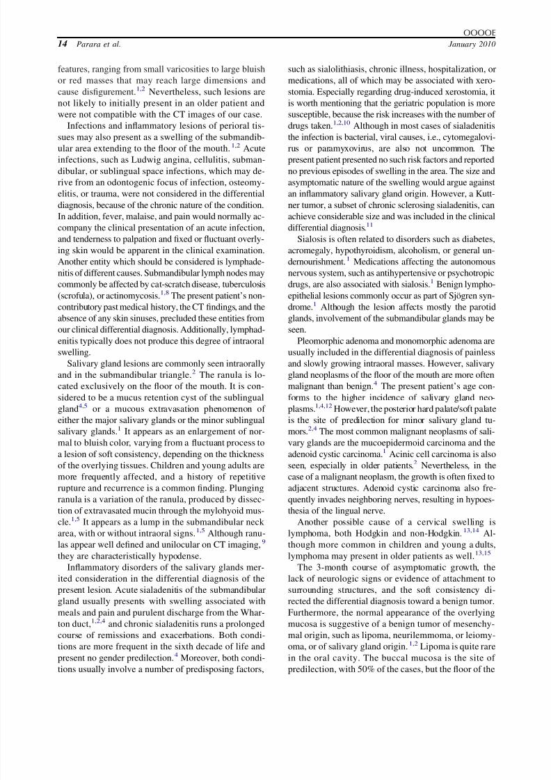
traorally, elevation of the floor of the mouth was noted,
which was covered by normal mucosa (Fig. 2). On pal-
pation, the nontender mass was soft to rubbery in consis-
tency. Although tongue movement was marginally re-
stricted by the physical presence of the swelling, the mass
per se was freely movable anteroposteriorly and medially
in relation to the tongue and the bone of the mandible. The
patient was totally edentulous and wearing complete
acrylic dentures. Normal saliva could be expressed from
the left submandibular salivary gland and there was no
sign of hypoesthesia or paresthesia of the area correspond-
ing to the ipsilateral lingual nerve.
A computerized tomography scan (CT) with en-hancement, which had been carried out before consul-
tation, confirmed the existence of a well demarcated
dense lesion lying between the mylohyoid and genio-
glossus muscles (Fig. 3). The lesion was solid, without
relation to the mandibular bone. Intravenous contrast
medium enhanced the image of the lesion. The remain-
der of the head and neck scan did not reveal any other
pathology or lymph node enlargement. Although it was
thought that magnetic resonance imaging could give
more precise and accurate information for the diagnosis
and management of the lesion, the patient refused any
further imaging studies.
DIFFERENTIAL DIAGNOSIS
The floor of the mouth and submandibular area may
be affected by numerous pathologic conditions, which
can be broadly classified as developmental, inflamma-
tory-obstructive, or neoplastic in origin. Lesions in this
area may be present for a prolonged period of time
before the patient seeks medical advice, usually as a
result of interference with swallowing or speech.
The floor of the mouth is the most common intraoral
location for developmental lesions of the oral soft tis-
aHospital Specialist, Oral and Maxillofacial Surgeon, Oral and Max-
illofacial Department, “Evangelismos” General Hospital.bLecturer, Department of Oral Pathology and Surgery, Dental School,
University of Athens; Oral and Maxillofacial Surgeon, Oral and
Maxillofacial Department, “Evangelismos” General Hospital.cAssistant Professor, Department of Oral Pathology and Surgery,Dental School, University of Athens.dHospital Specialist, Oral and Maxillofacial Surgeon, Department of
Oral and Maxillofacial Surgery, “Evangelismos” General Hospital.eSenior Hospital Specialist, Pathologist, Department of Pathology,
“Evangelismos” General Hospital.f Professor, Department of Oral Pathology and Surgery, Dental
School, University of Athens; Oral and Maxillofacial Department,
“Evangelismos” General Hospital.
Received for publication Feb 4, 2009; returned for revision Sep 12,
2009; accepted for publication Sep 14, 2009.
1079-2104/$ - see front matter
© 2010 Published by Mosby, Inc.
doi:10.1016/j.tripleo.2009.09.024
12
Vol. 109 No. 1 January 2010
CLINICOPATHOLOGIC CONFERENCE Editor: Paul C. Edwards

8/3/2019 Rhadomyoma Mouth Floor OOOE 2010
http://slidepdf.com/reader/full/rhadomyoma-mouth-floor-oooe-2010 2/5
sues, particularly dermoid cysts, branchial cleft cysts,
heterotopic gastrointestinal cysts, and thyroglossal duct
cysts.1-3 Dermoid cysts are considered to be a variation
of teratomas and are thought to arise from entrapmentof epithelial remnants during closure of the branchial
arches or as a result of trauma.4,5 Almost one-fifth of
the dermoid cysts that occur in the head and neck area
are located on the floor of the mouth,1,4 where they may
cause tongue elevation, submental protrusion, or
both.4,5 They are predominantly seen in young persons,
presenting as soft to rubbery swellings in the midline or
laterally.1 Branchial cleft cysts are developmental
anomalies which arise from incomplete closure of
branchial arches.1,2,4 They usually appear in relatively
young patients as fluctuant swellings located anteriorly
to the sternocleidomastoid muscle.1,6,7 Heterotopic gas-
trointestinal cysts are choristomas of head and neck and
mostly affect the sublingual area or the floor of themouth. They appear to have a male preponderance and
are usually lined with gastric mucosa.3 Thyroglossal
duct cysts arise from remnants of the embryonic thy-
roid.1,2,4 They typically present in the midline in close
contact to the hyoid bone, often producing a character-
istic movement during swallowing,3 which was not
seen in the present patient. In most cases, a fluctuant or
soft swelling is evident in the neck region, although
some lesions may appear intraorally1,2 and cause dys-
phagia.4 Half of the patients are younger than 20 years
at the time of diagnosis.1 Developmental lesions
present a relatively silent course before causing anysymptoms, as with the present patient. However, this
patient’s age made the probability of such an entity less
likely. Additionally, the CT findings were suggestive of
a solid rather than a cystic lesion.
Congenital lesions, such as vascular malformation or
lymphangioma (cystic hygroma), are generally in-
cluded in the differential diagnosis of neck masses
extending to the floor of the mouth. Both lesions are
most commonly seen in childhood, with 90% of
lymphangiomas of the head and neck diagnosed by the
age of 2 years.5 They demonstrate a variety of clinical
Fig. 1. Extraoral examination revealed a left submandibular
swelling with a diameter of 3 cm. The soft to rubbery swelling
was nontender and not firmly attached to overlying skin, the
lower jaw, or the tongue.
Fig. 2. Intraoral photograph showing ipsilateral elevation of the
floor of the mouth. The swelling was covered by normal mucosa
and was freely movable anteroposteriorly and medially.
Fig. 3. Transversal computerized tomography scan with con-
trast enhancement depicting a well delineated dense lesion of
the submandibular triangle.
OOOOE
Volume 109, Number 1 Parara et al. 13
8/3/2019 Rhadomyoma Mouth Floor OOOE 2010
http://slidepdf.com/reader/full/rhadomyoma-mouth-floor-oooe-2010 3/5
features, ranging from small varicosities to large bluish
or red masses that may reach large dimensions and
cause disfigurement.1,2 Nevertheless, such lesions are
not likely to initially present in an older patient and
were not compatible with the CT images of our case.
Infections and inflammatory lesions of perioral tis-
sues may also present as a swelling of the submandib-ular area extending to the floor of the mouth.1,2 Acute
infections, such as Ludwig angina, cellulitis, subman-
dibular, or sublingual space infections, which may de-
rive from an odontogenic focus of infection, osteomy-
elitis, or trauma, were not considered in the differential
diagnosis, because of the chronic nature of the condition.
In addition, fever, malaise, and pain would normally ac-
company the clinical presentation of an acute infection,
and tenderness to palpation and fixed or fluctuant overly-
ing skin would be apparent in the clinical examination.
Another entity which should be considered is lymphade-
nitis of different causes. Submandibular lymph nodes maycommonly be affected by cat-scratch disease, tuberculosis
(scrofula), or actinomycosis.1,8 The present patient’s non-
contributory past medical history, the CT findings, and the
absence of any skin sinuses, precluded these entities from
our clinical differential diagnosis. Additionally, lymphad-
enitis typically does not produce this degree of intraoral
swelling.
Salivary gland lesions are commonly seen intraorally
and in the submandibular triangle.2 The ranula is lo-
cated exclusively on the floor of the mouth. It is con-
sidered to be a mucus retention cyst of the sublingual
gland4,5
or a mucous extravasation phenomenon of either the major salivary glands or the minor sublingual
salivary glands.1 It appears as an enlargement of nor-
mal to bluish color, varying from a fluctuant process to
a lesion of soft consistency, depending on the thickness
of the overlying tissues. Children and young adults are
more frequently affected, and a history of repetitive
rupture and recurrence is a common finding. Plunging
ranula is a variation of the ranula, produced by dissec-
tion of extravasated mucin through the mylohyoid mus-
cle.1,5 It appears as a lump in the submandibular neck
area, with or without intraoral signs.1,5 Although ranu-
las appear well defined and unilocular on CT imaging,9
they are characteristically hypodense.
Inflammatory disorders of the salivary glands mer-
ited consideration in the differential diagnosis of the
present lesion. Acute sialadenitis of the submandibular
gland usually presents with swelling associated with
meals and pain and purulent discharge from the Whar-
ton duct,1,2,4 and chronic sialadenitis runs a prolonged
course of remissions and exacerbations. Both condi-
tions are more frequent in the sixth decade of life and
present no gender predilection.4 Moreover, both condi-
tions usually involve a number of predisposing factors,
such as sialolithiasis, chronic illness, hospitalization, or
medications, all of which may be associated with xero-
stomia. Especially regarding drug-induced xerostomia, it
is worth mentioning that the geriatric population is more
susceptible, because the risk increases with the number of
drugs taken.1,2,10 Although in most cases of sialadenitis
the infection is bacterial, viral causes, i.e., cytomegalovi-rus or paramyxovirus, are also not uncommon. The
present patient presented no such risk factors and reported
no previous episodes of swelling in the area. The size and
asymptomatic nature of the swelling would argue against
an inflammatory salivary gland origin. However, a Kutt-
ner tumor, a subset of chronic sclerosing sialadenitis, can
achieve considerable size and was included in the clinical
differential diagnosis.11
Sialosis is often related to disorders such as diabetes,
acromegaly, hypothyroidism, alcoholism, or general un-
dernourishment.1 Medications affecting the autonomous
nervous system, such as antihypertensive or psychotropicdrugs, are also associated with sialosis.1 Benign lympho-
epithelial lesions commonly occur as part of Sjögren syn-
drome.1 Although the lesion affects mostly the parotid
glands, involvement of the submandibular glands may be
seen.
Pleomorphic adenoma and monomorphic adenoma are
usually included in the differential diagnosis of painless
and slowly growing intraoral masses. However, salivary
gland neoplasms of the floor of the mouth are more often
malignant than benign.4 The present patient’s age con-
forms to the higher incidence of salivary gland neo-
plasms.1,4,12
However, the posterior hard palate/soft palateis the site of predilection for minor salivary gland tu-
mors.2,4 The most common malignant neoplasms of sali-
vary glands are the mucoepidermoid carcinoma and the
adenoid cystic carcinoma.1 Acinic cell carcinoma is also
seen, especially in older patients.2 Nevertheless, in the
case of a malignant neoplasm, the growth is often fixed to
adjacent structures. Adenoid cystic carcinoma also fre-
quently invades neighboring nerves, resulting in hypoes-
thesia of the lingual nerve.
Another possible cause of a cervical swelling is
lymphoma, both Hodgkin and non-Hodgkin.13,14 Al-
though more common in children and young adults,lymphoma may present in older patients as well.13,15
The 3-month course of asymptomatic growth, the
lack of neurologic signs or evidence of attachment to
surrounding structures, and the soft consistency di-
rected the differential diagnosis toward a benign tumor.
Furthermore, the normal appearance of the overlying
mucosa is suggestive of a benign tumor of mesenchy-
mal origin, such as lipoma, neurilemmoma, or leiomy-
oma, or of salivary gland origin.1,2 Lipoma is quite rare
in the oral cavity. The buccal mucosa is the site of
predilection, with 50% of the cases, but the floor of the
OOOOE
14 Parara et al. January 2010
8/3/2019 Rhadomyoma Mouth Floor OOOE 2010
http://slidepdf.com/reader/full/rhadomyoma-mouth-floor-oooe-2010 4/5
mouth may also be involved. It typically presents as a
slowly enlarging mass covered by normal appearing
mucosa of yellowish hue. Middle-aged persons are
more prone to develop lipomas, and no gender predi-
lection has been reported.1,2 The CT imaging of a
lipoma is that of a hypodense nonenhancing lesion, in
contrast to the present case.
Malignant soft tissue tumors were considered to be a
remote possibility. Rapid growth, lack of circumscrip-
tion, fixation to adjacent structures, and ulceration of
the overlying mucosa1,12 are common features of ma-
lignancy that were not noted in the present patient.
MANAGEMENT AND DIAGNOSIS
With the provisional diagnosis of a benign soft tissue
tumor, an incisional biopsy under local anesthesia was
planned. During the initial incision and identification of
Wharton duct, it became apparent that the growth was
encapsulated and clearly separated from nearby struc-
tures, allowing for the excision of the lesion in toto.
The surgical specimen, consisting of a brown encap-
sulated lobulated tumor measuring 3.0 1.5 cm, was
fixed in 10% buffered formalin and processed for rou-
tine histopathologic examination. Hematoxylin and eo-sin–stained sections showed a benign lesion encapsu-
lated by fibrous connective tissue. The lesion consisted
of large polygonal cells with eosinophilic to clear cy-
toplasm (Fig. 4), several presenting with discrete cross-
striations (Fig. 5). No nuclear or cellular pleomorphism
or mitoses were seen. The cells stained red with Mas-
son trichrome, and were immunohistochemically reac-
tive for desmin and myoglobin.
The final diagnosis was adult rhabdomyoma.
The patient’s postsurgical recovery was uneventful,
without salivary gland obstruction or lingual hypoes-
thesia. Further management included endoscopy of the
larynx and pharynx to exclude multifocal type of rhab-
domyoma.16 The patient’s stomach, prostate, and heart
were investigated with ultrasonography. Moreover, the
patient had no features associated with tuberous scle-
rosis, such as childhood seizures, hypomelanotic mac-
ules, facial angiofibromas, or bone cysts.17
At the time of writing, the patient had been under
regular follow-up for 2 years and remained free of
recurrence or new lesion.
DISCUSSION
Rhabdomyomas are rare benign tumors originating
from striated muscle18,19 and classified as cardiac and
extracardiac.18 Cardiac rhabdomyomas affect the myo-
cardial fibers and produce a diffuse distortion of the
heart muscle. They are generally considered hamarto-
mas rather than true neoplastic lesions and are usually
associated with tuberous sclerosis.17,18
The number of extracardiac rhabdomyomas reported
in the literature exceeds 160 cases, since the first report
in 1897.16,19,20 Extracardiac rhabdomyomas are of 3
types: adult, fetal, and genital. The majority of extra-cardiac rhabdomyomas are of the adult type and occur
in the head and neck.5,16,19 The fetal type, also occur-
ring more commonly in the head and neck region, is
considered to be a developmental abnormality.19,21 The
adult type usually presents at a mean age of 50 years
with a male preponderance of 4:1.16,20 The genital type,
in contrast, is a solitary lesion that occurs more com-
monly in women.20,22
On microscopic examination, the fetal rhabdomyoma
is composed of slightly differentiated polygonal cells
admixed with spindle-shaped cells.23 This type is typ-
Fig. 4. Histologic examination revealed a lesion consisting of
large polygonal cells with eosinophilic to clear cytoplasm
(hematoxylin and eosin stain, original magnification 200).
Fig. 5. Higher power histologic examination shows cross-
striations (arrow) in lesional cells (hematoxylin and eosin
stain, original magnification 400).
OOOOE
Volume 109, Number 1 Parara et al. 15

8/3/2019 Rhadomyoma Mouth Floor OOOE 2010
http://slidepdf.com/reader/full/rhadomyoma-mouth-floor-oooe-2010 5/5
ically more cellular than the adult type and often has a
myxoid stroma. Lesions with pleomorphic characteris-
tics and increased mitotic activity can rarely be mis-
taken for rhabdomyosarcomas.23 The adult type pre-
sents a simpler structure compared with the fetal
subtype, with large ovoid or polygonal cells with gran-
ular eosinophilic cytoplasm. Some cells present a vac-uolated cytoplasm due to the accumulation of glyco-
gen.16,23,24 There are usually a large number of blood
vessels, scant stroma, and a well defined capsule.18
Routine hematoxylin and eosin histologic features are
usually sufficiently characteristic. Immunohistochemi-
cal markers of skeletal muscle differentiation, such as
desmin, myoglobin, and muscle-specific actin, may oc-
casionally be of value in confirming the diagnosis.16,25
The treatment of choice for rhabdomyoma is surgical
excision. Multifocal presentation26,27 and local recur-
rence16,28 are possible, mandating long-term follow-up,
especially for lobulated tumors or tumors showing in-vasive growths.16,20 Recurrence is usually associated
with inadequate resection and not locally aggressive
biologic behavior.16,24,28
The authors express their gratitude to Dr. D. Rontogianni,
Head of the Pathology Department of “Evangelismos” General
Hospital, Athens, Greece, for permission to take pictures of
the tissue specimen.
REFERENCES
1. Neville BW, Damm DD, Allen CM, Bouquot JE. Oral and
maxillofacial pathology. 2nd edition. Philadelphia: Saunders;
2002.
2. Regezi JA, Sciubba J. Oral pathology, clinical-pathologic corre-
lations. 2nd edition. Philadelphia: Saunders; 1993.
3. Smith JE, Wren MK, Richardson MS, White DR. Colonic du-
plication in the submandibular region of the neck. Int J Pediat
Otorhinolaryngol Extra 2009;4:17-20.
4. Lian TS. Benign tumors and tumor-like lesions of the oral cavity.
In: Flint P, Haughey B, Robbins KT, Thomas JR, editors Cum-
mings’ otolaryngology: head and neck surgery. 4th ed. Philadel-
phia: Mosby; 2005. p. 1574-7.
5. Beil CM, Keberle M. Oral and oropharyngeal tumors. Eur J
Radiol 2008;66:448-59.
6. Glosser JW, Pires CA, Feinberg SE. Branchial cleft or cervical
lymphoepithelial cysts. Etiology and management. J Am Dent
Assoc 2003;134:81-6.
7. Tosios K, Rallis G, Vallianatou D, Vlachodimitropoulos D.Yellow-white tumor on the floor of the mouth. Oral Surg Oral
Med Oral Pathol Oral Radiol Endod 2006;101:701-4.
8. Philbert RF, Kim AK, Chung DP. Cervical tuberculosis (scrof-
ula): a case report. J Oral Maxillofac Surg 2004;62:94-7.
9. Mosier KM. Nononcologic imaging of the oral cavity and jaws.
Otolaryngol Clin North Am 2008;41:103-37.
10. Scully C. Drug effects on salivary glands: dry mouth. Oral Dis
2003;9:165-76.
11. Huang C, Damrose E, Bhuta S, Abemayor E. Kuttner tumor
(chronic sclerosing sialadenitis). Am J Otolaryngol 2002;23:
394-7.
12. Munir N, Bradley PJ. Diagnosis and management of neoplastic
lesions of the submandibular triangle. Oral Oncol 2008;44:
251-60.
13. Rockacy J, Viozzi CF, Zent CS. Mantle cell lymphoma present-
ing as a slowly enlarging lesion of the floor of the mouth in a
healthy 72-year-old female: report of a case. J Oral Maxillofacial
Surg 2007;65:333-7.
14. Vaughan ED. Primary tumors of the neck. In: Booth PW, Schen-del SA, Hausamen, JE, editors. Maxillofacial surgery. 2nd ed.
Edinburgh: Churchill Livingstone; 2007. p. 461-74.
15. Epstein JB, Epstein JD, Le ND, Gorsky M. Characteristics of
oral and paraoral malignant lymphoma: a population-based re-
view of 361 cases. Oral Surg Oral Med Oral Pathol Oral Radiol
Endod 2001;92:519-25.
16. Kapadia SB, Meis JM, Frisman DM, Ellis GL, Heffner DK,
Hyams VJ. Adult rhabdomyoma of the head and neck: a clini-
copathologic and immunophenotypic study. Hum Pathol 1993;
24:608-17.
17. Curatolo P, Bombardieri R, Jozwiak S. Tuberous sclerosis. Lan-
cet 2008;372:657-68.
18. Cleveland DB, Chen S, Allen CM, Ahing SI, Svirsky JA. Adult
rhabdomyoma—a light microscopic, ultrastructural, virologic,
and immunologic analysis. Oral Surg Oral Med Oral Pathol OralRadiol Endod 1994;77:147-53.
19. Liess BD, Zitsch RP, III Lane R, Bickel JT. Multifocal adult
rhabdomyoma: a case report and literature review. Am J Otol
2005;26:214-7.
20. Veziroglu F, Uckan S, Senguven B. Adult type rhabdomyoma in
a child. Oral Oncol Extra 2006;42:213-6.
21. Reid CO, Smith CJ. Rhabdomyoma of the floor of the mouth: a
new case and review of recently reported intra-oral rhabdomyo-
mas. Br J Oral Maxillofac Surg 1985;23:284-91.
22. Davies B, Noha P, Smaldone MC, Ranganathan S, Docimoa SG.
Paratesticular rhabdomyoma in a young adult: case study and
review of the literature. J Pediatr Surg 2007;42:E5-E7.
23. Kapadia SB, Meis JM, Frisman DM, Ellis GL, Heffner DK. Fetal
rhabdomyoma of the head and neck: a clinicopathologic and
immunophenotypic study of 24 cases. Human Pathol 1993;24:
754-65.
24. Favia G, Lo Muzio L, Serpico R, Maiorano E. Rhabdomyoma of
the head and neck: clinicopathologic features of two cases. Head
Neck 2003;23:700-4.
25. Garcia-Ruiz JA, Sanchez-Aniceto G, De La Mata-Pages R,
Gonzalez-Rex JA, Ballestin Carcavilla C. Submandibular rhab-
domyoma: a case report. Br J Oral Maxillofac Surg 1991;
29:123-6.
26. Koutsimpelas D, Weber A, Lippert BM, Mann WJ. Multifocal
adult rhabdomyoma of the head and neck: a case report and
literature review. Auris Nasus Larynx 2008;35:313-7.
27. Neville BW, McConnel FM. Multifocal adult rhabdomyoma:
report of a case and review of the literature. Arch Otolaryngol
1981;107:175-8.28. Valdez TA, Desai U, Volk MS. Recurrent fetal rhabdomyoma of
the head and neck. Int J Pediatr Otorhinolaryngol 2006;70:
1115-8.
Reprint requests:
Eleni Parara
16, Koimiseos Theotokou st
151 24 Athens
Greece
OOOOE
16 Parara et al. January 2010


![ddd.uab.cat · · 2011-05-21000E- OOOE- 000E- OOOE OCOE- 000E- OCOE- OCOE- OCOE- OCOE- ... 1.266E—04 I at a diameter of 5.423E+OO Surface Area Pore Number ... [PSI] 14. 25. 00](https://img.pdfslide.net/doc/110x75/5adcb1887f8b9aa5088bd040/ddduabcat-oooe-000e-oooe-ocoe-000e-ocoe-ocoe-ocoe-ocoe-1266e04.jpg)