Embed Size (px)
Citation preview
Rev Odonto Cienc 2012;27(4):339-343 339
Received: June 4, 2012Accepted: October 29, 2012
Conflict of Interests: The authors state that there are no financial and personal conflicts of interest that could have inappropriately influenced their work.
Copyright: © 2012 Lima et al.; licensee EDIPUCRS. This is an Open Access article distributed under the terms of the Creative Commons Attribution-Noncommercial-No Derivative Works 3.0 Unported License.
Case Report
Epithelial-myoepithelial carcinoma in the floor of mouth: Case report and review of the literature
Carcinoma epitelial-mioepitelial no assoalho bucal: relato de caso e revisão da literatura
Emeline das Neves de Araújo Lima a
Pedro Carlos da Rocha Neto a
Ana Miryam Costa de Medeiros a Roseana de Almeida Freitas a
a Department of Oral Pathology, Federal University of Rio Grande do Norte, Natal, RN, Brazil
Correspondence:Emeline das Neves de Araújo LimaUniversidade Federal do Rio Grande do NorteDepartamento de OdontologiaAv. Senador Salgado Filho, 1787 – Lagoa NovaNatal, RN – Brasil59056-000E-mail: [email protected]
Abstract
Purpose: to document the clinical, histopathological and immunohistochemical findings of an epithelial-myoepithelial carcinoma (EMC) in the floor of mouth and submandibular region. Furthermore, we intend to discuss the current literature about this tumor.
Case description: the biphasic pattern of neoplastic cells could be confirmed by immunohistochemical analysis, which showed positivity for cytokeratin (CK7) in luminal cells and for α-smooth muscle actin (α-SMA) in those non-luminal. All of the features were consistent with the diagnostic criteria required for EMC and the patient was referred for specialized treatment.
Conclusion: EMC is a rare type of malignant tumor, accounting for about 1% of all salivary gland tumors. Arising most frequently in the parotid gland (75%), approximately 10% of the tumors have their origin in the submandibular gland, and rarely affect the minor salivary glands. The diagnosis should be based on conventional light microscopy and confirmed by immunohistochemical analysis.
Key words: Adenocarcinoma; immunohistochemistry; submandibular gland
Resumo
Objetivo: documentar achados clínicos, histopatológicos e imuno-histoquímicos de um carcinoma epitelial-mioepitelial (CEM) no assoalho na boca e região submandibular. Além disso, pretendemos discutir a literatura atual sobre esse tumor.
Descrição do caso: o padrão bifásico das células neoplásicas pôde ser confirmado por análise imuno-histoquímica, que mostrou positividade para citoqueratina (CK7) nas células luminais e para α-actina de músculo liso (α-SMA) naquelas não-luminais. Todos os aspectos foram consistentes com os critérios diagnósticos requeridos para CEM e a paciente foi encaminhada para tratamento especializado.
Conclusão: CEM é um tipo de tumor maligno raro, somando aproximadamente 1% de todos os tumores de glândula salivar. Originando-se mais frequentemente na glândula parótida (75%), aproximadamente 10% dos tumores têm sua origem na glândula submandibular, e raramente afetam as glândulas salivares menores. O diagnóstico deve ser baseado na microscopia de luz convencional e confirmado por análise imuno-histoquímica.
Palavras-chave: Adenocarcinoma; imuno-histoquímica; glândula submandibular
340 Rev Odonto Cienc 2012;27(4):339-343
Epithelial-myoepithelial carcinoma
Introduction
The epithelial-myoepithelial carcinoma (EMC) is a rare low-grade malignant salivary gland neoplasm comprising approximately 1% to 2% of all primary salivary gland tumors (1). It was first described by Donath et al. in 1972 and recognized as a distinct entity by the World Health Organization (WHO) in 1991 (2-4).
Approximately 60% of the patients with EMC are female (4) and the majority of them are in the seventh and eighth decades of life. It affects the parotid gland in about 80% of cases (5-7), however, lesions arising in minor salivary glands and extraoral areas have also been reported (19% to 31%) (3,6). Soft palate seems to be the site of predilection of minor salivary gland EMCs (3,4).
Histologically, the tumor is characterized by a biphasic cytomorphology comprised of an inner layer of duct lining cells and an outer layer of clear myoepithelial cells (1,5,6). It can also show a multinodular growth pattern with tumor islands separated by a basement membrane and dense fibrous connective tissue bands. In many cases, the clear myoepithelial cell components are more predominant than the typical biphasic character (8). Based on the primary histological characteristics, the EMC can be classified in four categories: solid, tubular, pappilary and cribriform (5).
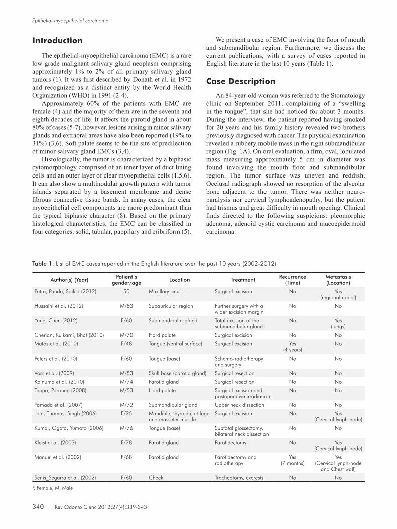
We present a case of EMC involving the floor of mouth and submandibular region. Furthermore, we discuss the current publications, with a survey of cases reported in English literature in the last 10 years (Table 1).
Case Description
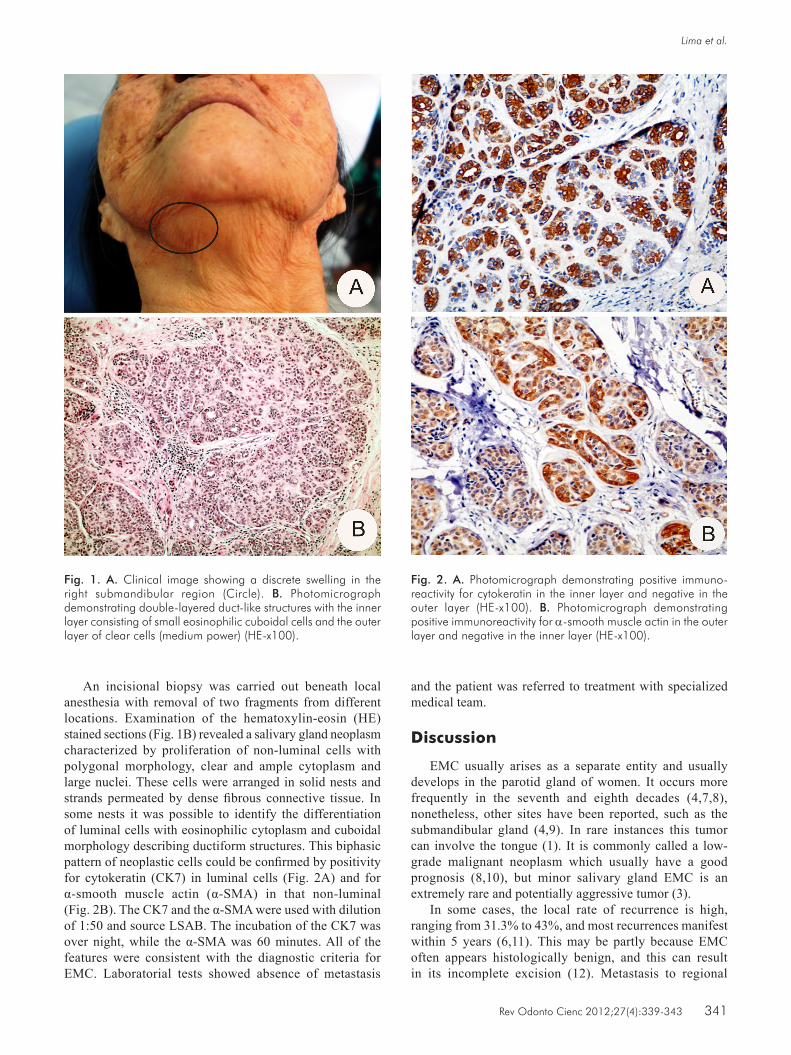
An 84-year-old woman was referred to the Stomatology clinic on September 2011, complaining of a “swelling in the tongue”, that she had noticed for about 3 months. During the interview, the patient reported having smoked for 20 years and his family history revealed two brothers previously diagnosed with cancer. The physical examination revealed a rubbery mobile mass in the right submandibular region (Fig. 1A). On oral evaluation, a firm, oval, lobulated mass measuring approximately 5 cm in diameter was found involving the mouth floor and submandibular region. The tumor surface was uneven and reddish. Occlusal radiograph showed no resorption of the alveolar bone adjacent to the tumor. There was neither neuro- paralysis nor cervical lymphoadenopathy, but the patient had trismus and great difficulty in mouth opening. Clinical finds directed to the following suspicions: pleomorphic adenoma, adenoid cystic carcinoma and mucoepidermoid carcinoma.
Table 1. List of EMC cases reported in the English literature over the past 10 years (2002-2012).
Author(s) (Year) Patient’s gender/age Location Treatment Recurrence
(Time)Metastasis (Location)
Patra, Panda, Saikia (2012) 50 Maxillary sinus Surgical excision No Yes (regional nodal)
Hussaini et al. (2012) M/83 Subauricular region Further surgery with a wider excision margin
No No
Yang, Chen (2012) F/60 Submandibular gland Total excision of the submandibular gland
No Yes (lungs)
Cherian, Kulkarni, Bhat (2010) M/70 Hard palate Surgical excision No No
Matos et al. (2010) F/48 Tongue (ventral surface) Surgical excision Yes(4 years)
No
Peters et al. (2010) F/60 Tongue (base) Schemo-radiotherapy and surgery
No No
Voss et al. (2009) M/53 Skull base (parotid gland) Surgical resection No No
Kainuma et al. (2010) M/74 Parotid gland Surgical resection No No
Teppo, Paronen (2008) M/53 Hard palate Surgical excision and postoperative irradiation
No No
Yamada et al. (2007) M/72 Submandibular gland Upper neck dissection No No
Jain, Thomas, Singh (2006) F/25 Mandible, thyroid cartilage and masseter muscle
Surgical excision No Yes (Cervical lynph-node)
Kumai, Ogata, Yumoto (2006) M/76 Tongue (base) Subtotal glossectomy, bilateral neck dissection
No No
Kleist et al. (2003) F/78 Parotid gland Parotidectomy No Yes (Cervical lynph-node)
Manuel et al. (2002) F/68 Parotid gland Parotidectomy and radiotherapy
Yes (7 months)
Yes (Cervical lynph-node
and Chest wall)
Senis_Segarra et al. (2002) F/60 Cheek Tracheotomy, exeresis No No
F, Female; M, Male
Rev Odonto Cienc 2012;27(4):339-343 341
Lima et al.
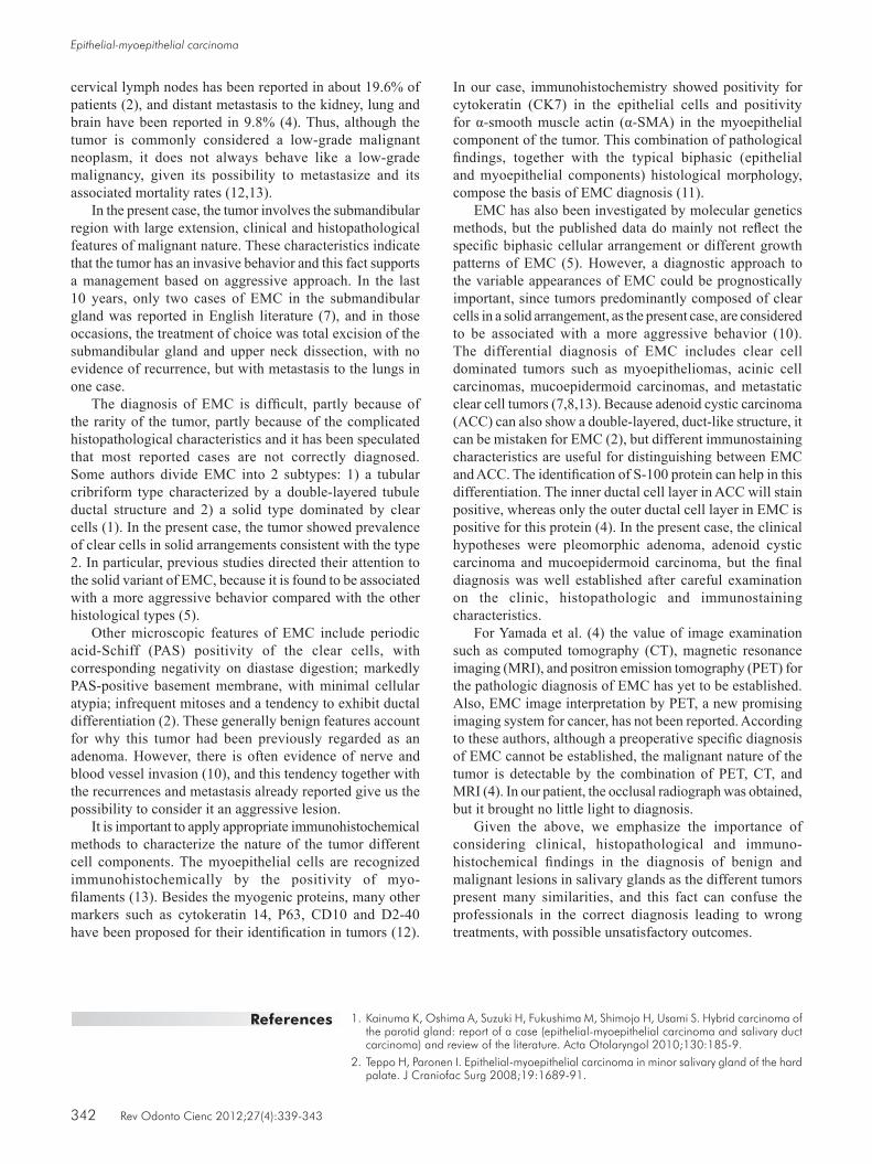
An incisional biopsy was carried out beneath local anesthesia with removal of two fragments from different locations. Examination of the hematoxylin-eosin (HE) stained sections (Fig. 1B) revealed a salivary gland neoplasm characterized by proliferation of non-luminal cells with polygonal morphology, clear and ample cytoplasm and large nuclei. These cells were arranged in solid nests and strands permeated by dense fibrous connective tissue. In some nests it was possible to identify the differentiation of luminal cells with eosinophilic cytoplasm and cuboidal morphology describing ductiform structures. This biphasic pattern of neoplastic cells could be confirmed by positivity for cytokeratin (CK7) in luminal cells (Fig. 2A) and for α-smooth muscle actin (α-SMA) in that non-luminal (Fig. 2B). The CK7 and the α-SMA were used with dilution of 1:50 and source LSAB. The incubation of the CK7 was over night, while the α-SMA was 60 minutes. All of the features were consistent with the diagnostic criteria for EMC. Laboratorial tests showed absence of metastasis
Fig. 1. A. Clinical image showing a discrete swelling in the right submandibular region (Circle). B. Photomicrograph demonstrating double-layered duct-like structures with the inner layer consisting of small eosinophilic cuboidal cells and the outer layer of clear cells (medium power) (HE-x100).
Fig. 2. A. Photomicrograph demonstrating positive immuno- reactivity for cytokeratin in the inner layer and negative in the outer layer (HE-x100). B. Photomicrograph demonstrating positive immunoreactivity for α-smooth muscle actin in the outer layer and negative in the inner layer (HE-x100).
and the patient was referred to treatment with specialized medical team.
Discussion
EMC usually arises as a separate entity and usually develops in the parotid gland of women. It occurs more frequently in the seventh and eighth decades (4,7,8), nonetheless, other sites have been reported, such as the submandibular gland (4,9). In rare instan ces this tumor can involve the tongue (1). It is commonly called a low- grade malignant neoplasm which usually have a good prognosis (8,10), but minor salivary gland EMC is an extremely rare and potentially aggressive tumor (3).
In some cases, the local rate of recurrence is high, ranging from 31.3% to 43%, and most recurrences manifest within 5 years (6,11). This may be partly because EMC often appears histologically benign, and this can result in its incomplete excision (12). Metastasis to regional
342 Rev Odonto Cienc 2012;27(4):339-343
Epithelial-myoepithelial carcinoma
cervical lymph nodes has been reported in about 19.6% of patients (2), and distant metastasis to the kidney, lung and brain have been reported in 9.8% (4). Thus, although the tumor is commonly considered a low-grade malignant neoplasm, it does not always behave like a low-grade malignancy, given its possibility to metastasize and its associated mortality rates (12,13).
In the present case, the tumor involves the submandibular region with large extension, clinical and histopathological features of malignant nature. These characteristics indicate that the tumor has an invasive behavior and this fact supports a management based on aggressive approach. In the last 10 years, only two cases of EMC in the submandibular gland was reported in English literature (7), and in those occasions, the treatment of choice was total excision of the submandibular gland and upper neck dissection, with no evidence of recurrence, but with metastasis to the lungs in one case.
The diagnosis of EMC is difficult, partly because of the rarity of the tumor, partly because of the complicated histopathological characteristics and it has been speculated that most reported cases are not correctly diagnosed. Some authors divide EMC into 2 subtypes: 1) a tubular cribriform type characterized by a double-layered tubule ductal structure and 2) a solid type dominated by clear cells (1). In the present case, the tumor showed prevalence of clear cells in solid arrangements consistent with the type 2. In particular, previous studies directed their attention to the solid variant of EMC, because it is found to be associated with a more aggressive behavior compared with the other histological types (5).
Other microscopic features of EMC include periodic acid-Schiff (PAS) positivity of the clear cells, with corresponding negativity on diastase digestion; markedly PAS-positive basement membrane, with minimal cellular atypia; infrequent mitoses and a tendency to exhibit ductal differentiation (2). These generally benign features account for why this tumor had been previously regarded as an adenoma. However, there is often evidence of nerve and blood vessel invasion (10), and this tendency together with the recurrences and metastasis already reported give us the possibility to consider it an aggressive lesion.
It is important to apply appropriate immunohistochemical methods to characterize the nature of the tumor different cell components. The myoepithelial cells are recognized immunohistochemically by the positivity of myo- filaments (13). Besides the myogenic proteins, many other markers such as cytokeratin 14, P63, CD10 and D2-40 have been proposed for their identification in tumors (12).
In our case, immunohistochemistry showed positivity for cytokeratin (CK7) in the epithelial cells and positivity for α-smooth muscle actin (α-SMA) in the myoepithelial component of the tumor. This combination of pathological findings, together with the typical biphasic (epithelial and myoepithelial components) histological morphology, compose the basis of EMC diagnosis (11).
EMC has also been investigated by molecular genetics methods, but the published data do mainly not reflect the specific biphasic cellular arrangement or different growth patterns of EMC (5). However, a diagnostic approach to the variable appearances of EMC could be prognostically important, since tumors predominantly composed of clear cells in a solid arrangement, as the present case, are considered to be associated with a more aggressive behavior (10). The differential diagnosis of EMC includes clear cell dominated tumors such as myoepitheliomas, acinic cell carcinomas, mucoepidermoid carcinomas, and metastatic clear cell tumors (7,8,13). Because adenoid cystic carcinoma (ACC) can also show a double-layered, duct-like structure, it can be mistaken for EMC (2), but different immunostaining characteristics are useful for distinguishing between EMC and ACC. The identification of S-100 protein can help in this differentiation. The inner ductal cell layer in ACC will stain positive, whereas only the outer ductal cell layer in EMC is positive for this protein (4). In the present case, the clinical hypotheses were pleomorphic adenoma, adenoid cystic carcinoma and mucoepidermoid carcinoma, but the final diagnosis was well established after careful examination on the clinic, histopathologic and immunostaining characteristics.
For Yamada et al. (4) the value of image examination such as computed tomography (CT), magnetic resonance imaging (MRI), and positron emission tomography (PET) for the pathologic diagnosis of EMC has yet to be established. Also, EMC image interpretation by PET, a new promising imaging system for cancer, has not been reported. According to these authors, although a preoperative specific diagnosis of EMC cannot be established, the malignant nature of the tumor is detectable by the combination of PET, CT, and MRI (4). In our patient, the occlusal radiograph was obtained, but it brought no little light to diagnosis.
Given the above, we emphasize the importance of considering clinical, histopathological and immuno- histochemical findings in the diagnosis of benign and malignant lesions in salivary glands as the different tumors present many similarities, and this fact can confuse the professionals in the correct diagnosis leading to wrong treatments, with possible unsatisfactory outcomes.
References 1. Kainuma K, Oshima A, Suzuki H, Fukushima M, Shimojo H, Usami S. Hybrid carcinoma of the parotid gland: report of a case (epithelial-myoepithelial carcinoma and salivary duct carcinoma) and review of the literature. Acta Otolaryngol 2010;130:185-9.
2. Teppo H, Paronen I. Epithelial-myoepithelial carcinoma in minor salivary gland of the hard palate. J Craniofac Surg 2008;19:1689-91.
Rev Odonto Cienc 2012;27(4):339-343 343
Lima et al.
3. Matos FR, Miranda JL, Mesquita AT, Santos CR, Freitas R de A. Epithelial-myoepithelial carcinoma in the ventral surface of the tongue. Braz J Otorhinolaryngol 2010;76:540.
4. Yamada H, Kawaguchi K, Yagi M, Morita Y, Mishima K, Uno K et al. Epithelial-myoepi thelial carcinoma of the submandibular gland with a high uptake of 18F-FDG: a case report and image diagnosis. Oral Surg Oral Med Oral Pathol Oral Radiol Endod 2007;104:243-8.
5. Seethala RR, Barnes EL, Hunt JL. Epithelial-myoepithelial carcinoma. A review of the clinicopathologic spectrum and immunopheno-typic characteristics in 61 tumors of the salivary glands and upper aerodigestive tract. Am J Surg Pathol 2007;31:44-57.
6. Yang S, Chen X. Epithelial-myoepithelial carcinoma with high grade transformation. Int J Oral Maxillofac Surg 2012; 41:810-3.
7. Cherian S, Kulkarni R, Bhat N. Epithelial-myoepithelial carcinoma in the hard palate: a case report. Acta Cytol 2010;54:835-9.
8. De Araújo VC, Altemani A, Furuse C, Martins MT, de Araújo NS. Immunoprofile of reactive salivary myoepithelial cells in intraductal areas of carcinoma ex-pleomorphic adenoma. Oral Oncol 2006;42:1011-16.
9. Furuse C, Sousa SO, Nunes FD, Magalhães MH, Araújo VC. Myoepithelial cell markers in salivary gland neoplasms. Int J Surg Pathol 2005;13:57-65.
10. Jain M, Thomas S, Singh S. Epithelial myoepitheial carcinoma of minor salivary gland--low grade malignant tumor presenting with nodal metastasis. Indian J Pathol Microbiol 2006;49:399-401.
11. Singh G, Sharma MC, Agarwal S, Prasad GL, Mishra S, Singh MM et al. Epithelial-myoepithelial carcinoma of the lacrimal gland: a rare case. Ann Diagn Pathol 2012;16: 292-7.
12. Peters P, Repanos C, Earnshaw J, Stark P, Burmeister B, McGuire L et al. Epithelial-myoepithelial carcinoma of the tongue base: a case for the case-report and review of the literature. Head Neck Oncol 2010;2:2-4.
13. Voss PJ, Leow AM, Schulze D, Metzger MC, Liebehenschel N, Schmelzeisen R. Navigation-guided resection with immediate functional reconstruction for high-grade malignant parotid tumour at skull base. Int J Oral Maxillofac Surg 2009;38:886-90.