Embed Size (px)
Citation preview
85
Right Thing at a Wrong Time? Adenosine A
3
Receptors and Cerebroprotection in Stroke
DAG K.J.E. VON LUBITZ,
a
KIMBERLY L. SIMPSON,
b
AND RICK C.S. LIN
b
a
Emergency Medicine Research Laboratories, Department of Emergency Medicine, University of Michigan Health System, Ann Arbor, Michigan, U.S.A.
b
Department of Anatomy, University of Mississippi Medical Center, Jackson, Mississippi, U.S.A.
A
BSTRACT
: The involvement of adenosine A
3
receptors in normal and patho-logic functions of the brain remains to be defined. Previous studies have shownthat chronic preischemic administration of the agonist [
N
6
-(3-iodobenzyl)-5
�
-
N
-methylcarboxoamidoadenosine or IB-MECA) results in a significant protec-tion of neurons in selectively vulnerable brain regions and in an equally signif-icant reduction of the subsequent mortality. Acute administration of the drug,on the other hand, resulted in a pronounced worsening of these parameters.We now report that the effect of administration of IB-MECA depends on thetiming of treatment with respect to the onset of the focal insult, and provide thefirst data supporting speculation that treatment with adenosine A
3
receptoragonists may decrease the infarct size following focal brain ischemia.
1,2
Treat-ment with IB-MECA administered 20 min prior to transient middle cerebralischemia (MCAO
t
= 30 min) resulted in a significant increase of the infarct size(
p
< 0.01), whereas administration 20 min after ischemia resulted in statistical-ly significant decrease of the infarct volume. Postischemic treatment results inimproved neuronal preservation, decreased intensity of reactive gliosis, andpronounced reduction of microglial infiltration. The data indicate that theeffects of adenosine A
3
receptor stimulation depend on the differential impactof these receptors on both neuronal and non-neuronal elements of the cerebraltissue, for example, astrocytes, microglia, and vasculature.
K
EYWORDS
: Adenosine A
3
Receptors; Stroke; Focal cerebral ischemia; Neuro-protection; Cerebral infarction; Physiology; Pharmacology; Treatment; Mice.
INTRODUCTION
Despite nearly a decade since the discovery of the latest member of the adenosinereceptor family,
3,4
the biological role of adenosine A
3
receptors is still ill defined.Studies have shown that stimulation of this receptor results in degranulation of mastcells, histamine release, vasoconstriction, and hypotension.
5–7
Other authors havedemonstrated the potent inhibitory effect of adenosine A
3
receptor activation on theprocesses accompanying inflammation, for example, neutrophil degranulation
8
or
Address for correspondence: Dag K.J.E. von Lubitz, Department of Emergency Medicine,TC/B1354/0303, University of Michigan Health System, 1500 E. Medical Center Drive, AnnArbor, MI 48109-0303, U.S.A. Voice:
+
1 734 936 6020; fax:
+
1 734 [email protected]

86 ANNALS NEW YORK ACADEMY OF SCIENCES
inhibition of eosinophil migration.
9
Adenosine A
3
receptors also appear to activatecertain types of Cl
−
channels of the non-pigmented epithelium.
10
The bulk of experimental work that has attempted to define the role of adenosineA
3
receptors in these pathological processes has concentrated on ischemia of theheart and brain. Several authors have shown that preischemic exposure of either car-diac myocytes or isolated hearts to adenosine A
3
receptor agonists results in protec-tion against ischemic damage,
11–13
indicating that these receptors may be involvedin the cardioprotective effects of preconditioning. These results are starkly opposedby the effects of the acute preischemic treatment on the outcome of global cerebralischemia, where both neuronal damage and postischemic mortality are very signifi-cantly aggravated by the agonists of adenosine A
3
receptor.
14
However, protectiveeffects of adenosine A
3
receptor agonist treatment have been reported that are con-sequent to chronic administration of
N
6
-(3-iodobenzyl)-5
′
-
N
-methylcarboxoami-doadenosine (IB-MECA).
14
Involvement of adenosine A
3
receptors in these effectshas been confirmed by preliminary reports of the intensely cerebroprotective effectof preischemic exposure to the adenosine A
3
receptor antagonist.
15,16
Although the damaging, involvement of adenosine A
3
receptors in globalischemia has been demonstrated,
14
nothing is known about the effects of either pre-or postischemic stimulation of these receptors in focal brain ischemia. Based largelyon the circumstantial evidence, it has recently been theorized that postischemic stim-ulation of adenosine A
3
receptors may actually result in cerebroprotection.
1,16
Theunquestionable yet rather confusing involvement of adenosine A
3
receptors inischemic brain damage warrants extension of previous studies to focal ischemia.Stimulation of adenosine A
3
receptors appears to affect at least two transducing sys-tems, and some of the affected pathways appear to be clearly involved in strokeinduced pathology.
17,18
Hence, the data obtained during studies of experimentalfocal ischemia may also shed further light on the pathology of stroke in humans.
MATERIALS AND METHODS
Animals and Drugs
CD-1 (
n
=
60) male mice (35 g; Charles River Laboratories, Wilmington, MA)were used. The animals were randomly separated into three experimental groups(
n
=
20/group). Treated animals were injected i.p. with 1.0 mg/kg
N
6
-(3-iodoben-zyl)-5
′
-
N
-methylcarboxoamidoadenosine (IB-MECA; RBI, Natick, MA) dissolvedin Emulphor (Rhône-Poulenc, Cranbury, NJ) and saline as described elsewhere.
14
The drug was administered either 20 min prior to, or 20 min following middle cere-bral artery occlusion. The control group received i.p. injections of the vehicle.
Surgery
Anesthesia and Temperature Maintenance
Presurgical anesthesia was initiated with 4
%
isoflurane carried in the 70:30mixture of nitrous oxide and oxygen, and maintained at 1.5
%
isoflurane. The gaswas administered through a face mask (Kent Scientific, Litchfield, CT). Body

87VON LUBITZ
et al.
: ADENOSINE A
3
RECEPTORS AND STROKE
temperature was maintained at 37.5
°
C throughout the surgery and during recoveryusing a rectal temperature probe and a heating pad (Kent Scientific, Litchfield, CT).
Surgical Procedure
The model of filament induced transient middle cerebral artery occlusion wasused. Following a ventromedial incision of the neck, the left common carotid arterywas exposed under surgical microscope (Vision Engineering, New Milford, CT).The occipital branches of the external carotid and the terminal lingual and maxillarybranches were then isolated and coagulated. Subsequently, the internal carotid wasexposed and its pterygopalatine branch ligated close to its origin using a 6-0 silksuture. The procedure ensured that only the intracranial ramus of the common carot-id artery remained patent. A 2-cm length of 5-0 blunt tipped (through heat applica-tion) nylon suture was then introduced into the external carotid and gently advancedinto the internal branch of the artery. Resistance and slight bending of the sutureindicated that the tip lodged in the proximal segment of the anterior cerebral artery.Typically, the distance traveled by the tip (i.e., between the tip and the bifurcation ofthe common carotid artery) was 10–11 mm. After ensuring that the tip was firmlylodged in the artery, the suture was anchored for 30 min. After withdrawing thesuture, the wound neck was sutured, and the animal was left to recover. To preventpostsurgical infection, a mixture of neomycin, polymycin, B-sulphate, and bacitra-cin (Triple-Antibiotic Ointment, E. Fougera & Co., Melville, NY) was topicallyapplied to the wound.
Verification of the Occlusion
The completeness of the artery occlusion was verified using a laser-Doppler tech-nique (Perimed, North Royalton, OH). Prior to the occlusion, the cortical blood flowwas measured transcranially by means of a 1-mm diameter probe apposed to theskull surface over the area of the expected infarct.
14
Five minutes after the occlusionthe probe was advanced to the same place, and the continuous flow measurement wasmade over the next five minutes. Consistent depression of the postischemic flow byat least 95
%
, compared to the preischemic values, indicated successful occlusion ofthe artery.
Determination of the Infarct Size
Seven days after the occlusion, the animals were anesthetized with Nembutal(50 mg/kg) and decapitated. Following removal, the brains were sectioned into1-mm thick coronal slices that were then immersed in a warm (37
°
C) 2
%
solution of2,3,5-triphenyl-tetrazolium chloride (TTC) in 1X PBS. After 20 min, the slices weregently washed with several rinses of 2
%
buffered (pH 7.4), 2
%
solution of paraform-aldehyde in PBS, followed by additional fixation in fresh paraformaldehyde solu-tion. The slices were then placed under an automated scanning system(EXPRESSION 636, Epson, Japan) and scanned at 720 d.p.i. The volume of the inf-arct was determined using NIH Image analyzing software. In the animals thatshowed signs of secondary cerebral hemorrhages (seen only in the pretreatmentgroup), the area affected was considered part of the total infarct zone.

88 ANNALS NEW YORK ACADEMY OF SCIENCES
Histology
Separate groups of animals (
n
=
20) were used for the assessment of histologicaldamage and immunocytochemistry. Seven days after ischemia, the animals wereanesthetized and perfused with buffered paraformaldehyde (4.5
%
, pH 7.4). Afterfixation, the brains were removed and cut on a freezing microtome into 25
µ
m slices.Slices from the penumbra zone (determined by means of laser-Doppler measurementduring ischemia) were subjected to either to Nissl or GFAP and lectin immuno-cytochemical
19
staining to indicate the extent of neuronal preservation in the hippoc-ampus, and the extent of astrocyte and microglia activation in the hippocampus andthe cortex of the penumbra zone.
Statistics
Infarct volume data were analyzed using GraphPad/Inplot (GraphPazd Software,San Diego, CA) software. Statistical parameters were determined by means of ANO-VA followed by the Student–Newman–Keuls test with
p
<
0.05 indicating significantdifference.
RESULTS
At seven days postischemia, the differences in the extent of the infarcted brain tis-sue were easily perceived (see F
IGURE
1). Apart from the clearly demarcated infarct,the gross appearance of coronally sectioned brains in controls and posttreated ani-mals was unremarkable. In the pretreated group, several brains (4/7) showed wide-spread intracerebral hemorrhages and partial collapse of the tissue (F
IG
. 1).Comparison of infarct volumes (see F
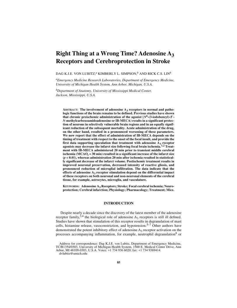
IGURE
2) showed significant reduction of inf-arct volume in the post treated animals.
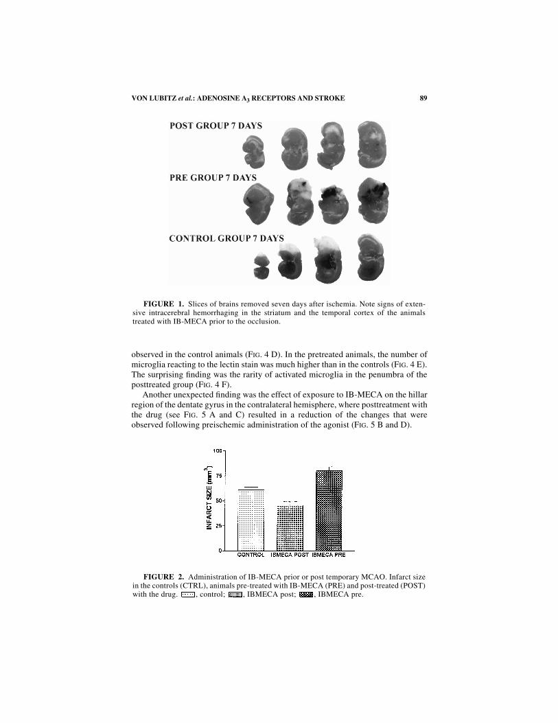
Even in the absence of quantitative determination of neuronal preservation, theextent of the hippocampal damage in the pretreated group was slightly worse than inthe controls (see F
IGURE
3 A and B). We doubt, however, that neuronal countingwould reveal any statistical differences. In the posttreated group, both the thicknessand the appearance of the pyramidal neurons in the lateral CA1 segment of the pen-umbral hippocampus appeared to be normal (F
IG
. 3 C). The impact of pre- versuspostischemic treatment with IB-MECA became fully apparent when GFAP and lec-tin stains were compared.
The extent of GFAP staining in the controls and pretreated animals (see F
IGURE
4A and B) was fully comparable to that observed in the gerbils exposed to IB-MECApreceding global cerebral ischemia.
19
As in the previous studies, the astrocytes in thepretreated animals were characterized by sturdy processes containing an intense loadof GFAP (F
IG
. 4 B). In the animals treated after ischemia, the appearance of astro-cytes changed dramatically. The GFAP containing processes were slender, theirelongated ramifications creating a delicate lattice (F
IG
. 4 C) rather than the coarsenetwork characteristic of the pretreated animals and, to a lesser degree, control miceas well (F
IG
. 4 A, B, and C).Microglial infiltration of the penumbral cortex showed similar pattern to that of
reactive gliosis. As expected, a significant presence of activated microglia was
89VON LUBITZ
et al.
: ADENOSINE A
3
RECEPTORS AND STROKE
observed in the control animals (F
IG
. 4 D). In the pretreated animals, the number ofmicroglia reacting to the lectin stain was much higher than in the controls (F
IG
. 4 E).The surprising finding was the rarity of activated microglia in the penumbra of theposttreated group (F
IG
. 4 F).Another unexpected finding was the effect of exposure to IB-MECA on the hillar
region of the dentate gyrus in the contralateral hemisphere, where posttreatment withthe drug (see F
IG
. 5 A and C) resulted in a reduction of the changes that wereobserved following preischemic administration of the agonist (F
IG
. 5 B and D).
FIGURE 1. Slices of brains removed seven days after ischemia. Note signs of exten-sive intracerebral hemorrhaging in the striatum and the temporal cortex of the animalstreated with IB-MECA prior to the occlusion.
FIGURE 2. Administration of IB-MECA prior or post temporary MCAO. Infarct sizein the controls (CTRL), animals pre-treated with IB-MECA (PRE) and post-treated (POST)with the drug. , control; , IBMECA post; , IBMECA pre.
90 ANNALS NEW YORK ACADEMY OF SCIENCES
FIGURE 3. The effect of focal ischemia (A), IB-MECA prior to focal ischemia (B),and IB-MECA after focal ischemia (C). Treatment following MCAO occlusion results in apronounced sparing of the hippocampal neurons in the hippocampus adjacent to the lesion.The damage in the two other groups is, essentially, identical.

91VON LUBITZ
et al.
: ADENOSINE A
3
RECEPTORS AND STROKE
FIGURE 4. Focal ischemia (A) and either pretreatment with IB-MECA (B) or admin-istration of the drug after removal of the occluding filament (C) and their effect on GFAPimmunostaining (A–C) or microglial infiltration (D, ischemia alone; E, pretreatment;F, posttreatment) within the penumbral zone of the cortex. Note that posttreatment withIB-MECA markedly changes the appearance of astrocytic processes (compare with A andB) and practically eliminates microglial elements from the affected volume of the brain.

92 ANNALS NEW YORK ACADEMY OF SCIENCES
FIG
UR
E5.
Hil
us o
f gy
rus
dent
atus
on
the
cont
rala
tera
l si
de.
A,
brai
n of
one
of
the
anim
als
post
trea
ted
wit
h IB
-ME
CA
(N
issl
sta
in).
The
thi
nar
row
poi
nts
at o
ne o
f th
e fe
w d
amag
ed n
euro
ns, t
he t
hick
arr
ow a
t an
astr
ocyt
e. C
, GF
AP
sta
in f
rom
ano
ther
bra
in s
lice
in th
e sa
me
anim
al. N
ote
that
the
appe
aran
ce o
f as
troc
ytes
is v
ery
sim
ilar
to th
ose
in F
IGU
RE 4
C. B
rep
rese
nts
the
sam
e re
gion
in th
e an
imal
pre
trea
ted
wit
h IB
-ME
CA
. The
inte
nsit
yof
the
coll
ater
al d
amag
e is
far
mor
e in
tens
e an
d th
e as
troc
ytes
res
embl
e (D
) th
ose
seen
in th
e ip
sila
tera
l cor
tex
(com
pare
to F
IG. 4
B).

93VON LUBITZ et al.: ADENOSINE A3 RECEPTORS AND STROKE
DISCUSSION
Although the relative importance of the adenosine A3 receptor in the operation ofseveral cell, tissue, and organ types is the subject of intense speculation,1,20,21 noneof the existing data allow definitive conclusions. To the contrary, many of the recent-ly published studies reveal consistencies within one system, merely to be contradict-ed by the results obtained in another (for recent reviews see Refs. 16, 20, and 21).The complex intracellular effects evoked by the stimulation of adenosine A3 recep-tors (e.g., see Refs. 21–24) are unquestionably among the underlying sources of theongoing mystery to which the present paper adds, rather than subtracts.
As in global ischemia,14 acute exposure to IB-MECA prior to middle cerebralartery occlusion (focal ischemia) results in a significant increase of the infarctedbrain volume. Since, contrary to our studies of global ischemia,14 we did not mea-sure postischemic blood flow during the experiments described here, it is unknownwhether drug related amplification of the regional disturbances of cerebral bloodperfusion contribute to the aggravation of damage in the animals pretreated with IB-MECA. It has been shown, however, that a 200 µg/kg dose of a more selective andpotent adenosine A3 receptor agonist 2-Cl-IB-MECA (2-chloro-N6-(3-iodoben-zyl)adenosine-5′-N-methylcarboxamide) administered i.v. in conscious rats resultsin the complete release of vascular histamine stores.6 Thus, since histamine plays acritical role in the formation of brain oedema following focal cerebral ischemia,25,26
we can not exclude the possibility that the increased infarct volume in animals pre-treated with IB-MECA is, at least in part, influenced by the stimulation of the ade-nosine A3 receptors located on mast cells.5,27 Histamine induced oedema involves anitric oxide component,28,29 and our previous studies15,16 demonstrated increasedimmunocytochemical reactivity of nitric oxide synthase in the pyramidal and radia-tum strata of the hippocampus following preischemic injection of IB-MECA in ger-bils. However, activation of nitric oxide synthase was entirely independent of theinteraction between adenosine A3 receptors and histamine releasing mechanisms,2,16
and the immunocytochemical reaction product appeared to concentrate at the neu-ronal rather than vascular components of the brain.16,19 Hence, it is quite likely thatthe destructive impact of preischemically administered IB-MECA involves both cir-culatory and neuronal components. Finally, astrocytes and microglia are also affect-ed by IB-MECA as shown by our latest studies,30 indicating that the degree of astro-and microglial activation depends on the timing of treatment with respect to theinsult itself.
The multiplicity of cellular and organs systems affected by stimulation of adenos-ine A3 receptors, and the powerful effects elicited by such stimulation,1,16,18,20,21
stand in contrast to the relative paucity of these receptors in all studied cell/organsystems.31 Yet, although an ever increasing number of adenosine A3 receptor medi-ated effects is being reported, their mechanistic aspects remain unclear.1,16,21 Inview of these uncertainties, any attempt at an explanation of the different outcomesbetween pre- and posttreated animals is also highly tentative and needs extensiveexperimental corroboration.
It has been suggested14 that worsening of the outcome of cerebral ischemiainduced by acute preischemic administration of adenosine A3 agonist may representthe cumulative effect of several adverse events triggered by the drug immediately
94 ANNALS NEW YORK ACADEMY OF SCIENCES
prior to the occlusion (“priming effect”). Events such as release of inflammatorymediators and concomitant degradation of the blood brain barrier integrity, and del-eterious attenuation of the cerebral blood flow,6,25–27,32 may be of primary impor-tance. Combined with the increased influx of Ca2+ (both passive and through thevoltage regulated calcium channels) and the liberation of internal calcium storeselicited by adenosine A3 receptor stimulation,22 the overall impact of these eventswould, indeed, predispose the brain to a significantly increased susceptibility toischemia. On the other hand, when administered following a focal insult, IB-MECAinduces cerebroportection in focal ischemia when the drug is given following tran-sient occlusion of the middle cerebral artery.30 Whether this cerebroprotectiveimpact of adenosine A3 receptor agonists is related to astrocyte activation, directneuroprotective effect, or both16,18,19,33,34 is unclear. The results of immunocy-tochemical studies presented in this paper confirm the presence of complex neuronaland glial effects induced by postischemic stimulation of adenosine A3 receptors.Moreover, the latter observations offer strong support for the recently publishedhypothesis on the role of cerebral adenosine A3 receptors as a part of the “adenosinebased cerebroprotective complex”.1,16 This paper, and also our previous studies ofadenosine A3 receptor impact on the outcome of focal and global ischemia,14,16,30
indicate significant involvement of these receptors in the pathology resulting fromthe arrested cerebral blood supply. However, the relationship between adenosine A3receptor stimulation and the outcome of an ischemic event is not straightforward.The presented data have confirmed initial hypothetical assumptions that the extentof the subsequent damage depends on the timing of adenosine A3 receptor activationversus the onset of ischemia.30 Most likely, the degree of receptor activation18,21,33
may be an important factor as well. In summary, although our results provide furtherillumination of the complexity of A3 receptor elicited effects, they do not provide adefinitive solution to the baffling role of these receptors in the generation of strokedamage. As in many other studies of this still untreatable disease, we too must meek-ly conclude that “further extensive experiments are necessary” in order to understandthe exact nature of the participating mechanisms and the factors determining the dualrole of adenosine A3 receptors in the brain.
REFERENCES
1. VON LUBITZ, D.K.J.E. 1999. Adenosine and cerebral ischemia: therapeutic future ordeath of a brave concept? Eur. J. Pharmacol. 371: 85–102.
2. VON LUBITZ, D.K.J.E., et al. 1999. J. Cereb. Blood Flow.
3. MEYERHOF, W.R., R. MÜLLER-BRECHLIN & D. RICHTER. 1991. Molecular cloning ofnovel putative G protein coupled receptor expressed during rat spermiogenesis.FEBS Lett. 284: 155–160.
4. ZHOU, Q.Y., C.Y. LI, M.E. OLAH, et al. 1992. Molecular cloning and characterizationof an adenosine receptor: the A3 receptor. Proc. Natl. Acad. Sci. U.S.A. 89: 7432–7436.
5. SHEPHERD, R.K., J. LINDEN & B.R. DULING. 1996. Adenosine-induced vasoconstrictionin vivo. Role of the mast cell and A3 adenosine receptor. Circ. Res. 78: 627–634.
6. VAN SCHAICK, E.A., K.A. JACOBSON, H.O. KIM, et al. 1996. Hemodynamic effects andhistamine release elicited by the selective adenosine A3 receptor agonist 2-Cl-IB-MECA in conscious rats. Eur. J. Pharmacol. 308: 311–314.

95VON LUBITZ et al.: ADENOSINE A3 RECEPTORS AND STROKE
7. FOZARD, J.R. & A.M. CARRUTHERS. 1993. Adenosine A3 receptors mediate hypoten-sion in the angiotensin II-supported circulation of the pithed rat. Br. J. Pharmacol.109: 3–5.
8. BOUMA, M.G., T.M. JEUNHOMME, D.L. BOYLE, et al. 1997. Adenosine inhibits neutro-phil degranulation in activated human whole blood: involvement of adenosine A2 andA3 receptors. J. Immunol. 158: 5400–5408.
9. KNIGHT, D., X. ZHENG, C. ROCCHINI, et al. 1997. Adenosine A3 receptor stimulationinhibits migration of human eosinophils. J. Leukoc. Biol. 62: 465–468.
10. MITCHELL, C.H., K. PETERSON-YANTORNO, D.A. CARRE, et al. 1999. A3 adenosinereceptors regulate Cl- channels of nonpigmented ciliary epithelial cells. Am. J. Phys-iol. 276(3-1): C659–666.
11. STAMBAUGH, K., K.A. JACOBSON, J.-L. JI & B.T. LIANG. 1997. A novel cardioprotectivefunction of adenosine A1 and A3 receptors during prolonged simulated ischemia. J.Physiol. (Heart Circ. Physiol.) 42: H501–H505.
12. DOUGHERTY, C., J. BARUCHA, P.R. SCHOFIELD, et al. 1998. Cardiac myocytes renderedischemia resistant by expressing the human adenosine A1 and A3 receptor. FASEB J.12: 1785–1792.
13. HILL, R.J., J.J. OLEYNEK, W. MAGEE, et al. 1998. Relative importance of adenosine A1and A3 receptors in mediating physiological or pharmacological protection fromischemic myocardial injury in the rabbit heart. J. Mol. Cell Cardiol. 30: 579–585.
14. VON LUBITZ, D.K.J.E., R.C.S. LIN, P. POPIK, et al. 1994. Adenosine A3 receptor stimu-lation and cerebral ischemia. Eur. J. Pharmacol. 263: 59–67.
15. VON LUBITZ, D.K.J.E., R.C.S. LIN & K.A. JACOBSON. 1997. Adenosine A3 receptorsand protection against cerebral ischemic damage in gerbils. Soc. Neurosci. Abstr.23/2, 745.16.
16. VON LUBITZ, D.K.J.E. 1999. Stimulation of adenosine A3 receptors in cerebralischemia: neuronal death, recovery, or both? Ann. N.Y. Acad. Sci. 890: 93–105.
17. ABBRACCHIO, M.P., R. BRAMBILLA, S. CERUTTI, et al. 1995. G-protein dependent acti-vation of phospholipase C by adenosine A3 receptors in the rat brain. Mol. Pharma-col. 48: 1038–1045.
18. ABBRACCHIO, M.P., S. CERUTI, R. BRAMBILLA, et al. 1997. Modulation of apoptosis byadenosine in the central nervous system: a possible role for the A3 receptor. Ann.N.Y. Acad. Sci. 825: 11–22.
19. VON LUBITZ, D.K.J.E., R.C.S. LIN, M.BOYD, et al. 1999. Chronic administration ofadenosine A3 receptor agonist and cerebral ischemia: neuronal and glial effects. Eur.J. Pharmacol. 367: 157–163.
20. JACOBSON, K.A. 1999. Adenosine A3 receptors: novel ligands and paradoxical effects.Trends Pharmacol. Sci. 19: 185–191.
21. JACOBSON, K.A., C. HOFFMANN, F. CATTABENI & M.P. ABBRACCHIO. 1999. Adenosine-induced cell death: evidence for receptor-mediated signaling. Apoptosis 4: 197–211.
22. KOHNO, Y., X.-D. JI, S.D. MAWHORTER, et al. 1996. Activation of A3 adenosine recep-tors on human eosinophils elevates intracellular calcium. Blood 88: 5569–3574.
23. CERUTI, S., D. BARBIERI, C. FRANCESCHI, et al. 1996. Effects of adenosine A3 receptoragonists on induction of cell protection at low and cell death at high concentrations.Drug Dev. Res. 37: 177.
24. ABBRACCHIO, M.P., G. RAINALDI, A.M. GIAMMARIOLI, et al. 1997. The A3 adenosinereceptor mediates cell spreading, reorganization of actin cytoskeleton, and distribu-tion of Bcl-XL: studies in human astroglioma cells. Biochem. Biophys. Res. Comm.24: 297–304.
25. JOO, F., J. KOVACS, P. SZRDAHELYIS, et al. 1994. The role of histamine in brain oedemaformation. Acta Neurochir. Suppl. 60: 76–78.
26. NEMETH, L., M.A. DELL, A. FALUS, et al. 1998. Cerebral ischemia reperfusion-inducedvasogenic brain edema formation in rats: effect of an intracellular histamine receptorantagonist. Eur. J. Pediatr. Surg. 8: 216–219.
27. FOZARD, J.R., H.J. PFANKUCHE & H.J. SCHUURMAN. 1996. Mast cell degranulation fol-lowing adenosine A3 receptor activation in rats. Eur. J. Pharmacol. 298: 293–297.
28. MAYHAN, W.G. 1996. Role of nitric oxide in histamine-induced increases in perme-ability of the blood-brain-barrier. Brain Res. 743: 70–76.

96 ANNALS NEW YORK ACADEMY OF SCIENCES
29. SZABO, A., J. KASZAKI, M. BOROS & S. NAGY. 1997, Possible relationship between his-tamine and nitric oxide release in the postischemic flow response following mesen-teric ischemia of different durations. Shock 7: 376–382.
30. YE, W., J. MCCLELLAN & D.K.J.E. VON LUBITZ. 1999. Involvement of adenosine A3receptor in generation of experimental brain damage following middle cerebralocclusion in mice. Acad. Emerg. Med. 6: 261.
31. JI, X.-D., D.K.J.E.VON LUBITZ, M.E. OLAH, et al. 1994. Species differences in ligandaffinity at central A3 adenosine receptors. Drug Dev. Res. 33: 51–59.
32. HALLENBECK, J.M. 1996. Significance of the inflammatory response in brain ischemia.Acta Neurochir. Supp. 66: 27–31.
33. SEI, Y., D.K.J.E. VON LUBITZ, M.P. ABBRACCHIO, et al. 1997. Adenosine A3 receptoragonist-induced neurotoxicity in rat cerebellar granule neurons. Drug Dev. Res. 40:267–273.
34. YAO, Y., Y. SEI, M.P. ABBRACCHIO, et al. 1997. Adenosine A3 receptor agonists pro-tect HL-60 and U-937 cells from apoptosis induced by A3 antagonists. Biochem.Biophys. Res. Comm. 232: 317–322