Embed Size (px)
Citation preview

AD-COR Program inovativ de formare in domeniul cardiologiei pediatricePOSDRU/179/3.2/S/152012
Data:23-11-2015
MODUL TEORETIC
Right ventricular physiology and Phatophysiology
Imputernicit: Prof. Dr. Tammam Youssef
Activitate prestata de I.R.C.C.S. POLICLINICO SAN DONATO – MILANO, ITALIA in baza contractului nr. 18/22144/29.07.2015

Acest material a fost documentat/ validat/ prezentat la sesiunile de formare în
cadrul proiectului „AD-COR Program inovativ de formare în domeniul cardiologiei
pediatrice” - POSDRU/179/3.2/S/152012, proiect cofinanțat din Fondul Social
Operațional Sectorial Dezvoltarea Resurselor Umane 2007-2013.
Beneficiar: Universitatea de Medicină și Farmacie „Carol Davila” București
Conținutul acestui material nu reprezintă în mod obligatoriu poziția oficială a Uniunii Europene sau a Guvernului României
RIGHT VENTRICULAR PHYSIOLOGY AND PHATOPHYSIOLOGY
Claudio Bussadori DVM, MD, PhD, Dipl. ECVIM (Cardiology)
Department of Pediatric Cardiology and Adult with Congenital Heart Disease San Donato Hospital Milano Italy

RIGHT VENTRICULAR STRUCTURE
Rudski LG, Afilalo J. The blind men of indostan and the elephant in the echo lab. J Am Soc Echocardiogr. 2012;25:714-717

RIGHT VENTRICULAR STRUCTURE

RV CONTRACTION1. Inlet and trabeculated myocardium
2. Infundibulum (approximately 25 to 50 ms apart)
Contraction of the infundibulum is of longer duration
than contraction of the inflow region
The RV contracts by 3 separate mechanisms
1. Inward movement of the free wall, which produces a bellows effect
2. Contraction of the longitudinal fibers, which shortens the long axis and draws the tricuspid annulus toward the apex
3. Traction on the free wall at the points of attachment secondary to LV contraction

RV CONTRACTION
Greater longitudinally than radially.
Twisting do not contribute significantly to RV contraction.
Moreover, because of the higher surface-to- volume ratio of the RV, a smaller inward motion is required to eject the same stroke volume.

RV SYSTOLIC EJECTION
• Right-sided pressures lower than left-sided.
• RV isovolumic contraction time is shorter.
• End-systolic flow may continue in the presence of a negative ventricular-arterial pressure gradient (hangout interval)

RV FLOW
Fredriksson AG, Zajac J, Eriksson J, Dyverfeldt P, Bolger AF, Ebbers T, Carlhall CJ. 4-d blood flow in the human right ventricle. Am J Physiol Heart Circ Physiol. 2011;301:H2344-2350

LONGITUDINAL RV CONTRACTION
4Chambers RVOT

VENTRICULAR INTERDEPENDENCE
- Mainly through the interventricular septum.
- The pericardium it is mostly relevant for diastolic ventricular interdependence.
- Approximately 20% to 40% of RV systolic pressure and volume outflow results from LV contraction.
- Moreover, in the presence of scarring of the RV or replacement with a non contractile patch, the septum is able to maintain circulatory stability as long as the RV is not dilated.
- Beyond the physiological range, excessive RV volume loading can compress the LV and global ventricular function.

VENTRICULAR INTERDIPENDENCE
Volume overload
Reversed Bernheim effect
Pressure overload

VENTRICULAR INTERDIPENDENCE
Bilateral Diameter/anteroposterior diameter
➢ 1 pressure and/or Volume overload
Normal: 1 both end-systole and end-diastole RV volume overload: +/- 1.0 at end-systole, >1.0 at end-diastole RV pressure overload: >1.0 at both end-systole and end-diastole

RV PHISIOLOGY OF THE CONTRACTION

LV Systole RV Systole
RV PHISIOLOGY OF THE CONTRACTION

XSTRAIN AND NORMAL RIGHT AND LEFT VENTRICLE COLOR MAP
Normal Left Ventricle Normal Right Ventricle

RV REFERENCE VALUESRV ENDO STRAIN (%)
BAS LAT MID LAT APIC LAT APIC SEP MID SEP BAS SEP Global
-25.12 -21.58 -16.00 -18.24 -19.25 -21.33 -20.77
RV ENDO STRAIN RATE s-1
BAS LAT MID LAT APIC LAT APIC SEP MID SEP BAS SEP Global
-2.00 -1.62 -1.08 -1.17 -1.40 -1.42 -1.47

RIGHT VENTRICULAR STRAIN

McConnell’s Sign
Walsh BM, Moore CL. Mcconnell's sign is not specific for pulmonary embolism: Case report and review of the literature. J Emerg Med. 2015;49:301-304

RV Change in ASD Closure
26 patients (16F; 10M) mean age 31
+/- 24.6 years, In ASD patient RV Strain
was significantly higher than in the
control group. 24 h. after closure, there
was a significant reduction in
longitudinal Strain in the RV free wall
and the right side of the septum.
Bussadori et al. Assessment of right and left ventricular
longitudinal, circumferential and radial strain and strain
rate after atrial septal defect closure through a new 2D
echocardiographic based method. ESC 2009
33 patients
20 F, 13 M, mean age 44.7+/-18.5
6 M post closure. Before percutaneous
closure, mean GLS was significantly increased in
comparison to control group, and significantlyreduced after closure. Analysis of regional PSS
showed significant decrease in the lateral
apical, lateral mid, and septal apical segments.
Jategaonkar, S. et al Two-dimensional strain and strain
rate imaging of the right ventricle in adult patients before
and after percutaneous closure of atrial septal defects
European Journal of Echocardiography (2009) 10, 499–502

Controls ASD
Global RVStrain -20.77±3.87 -23.95±5.24 P = 0,0438
ASD pre
ASDO
24 h after
ASDO
Global RV Strain -23.95±5.24 -17.04±5.94 0.00016
ASD pre
ASDO
ASD
post ASDO
Basal Circ.Strain -20.31±4.64 -25.39±5.22 0.00003
RV CHANGE IN ASD CLOSURE
Bussadori C, et al. Right and left ventricular strain and strain rate in young adults
before and after percutaneous atrial septal defect closure. Echocardiography.
2011;28:730-737

LV CHANGE IN ASD CLOSURELeft ventricular end diastolic volume and left ventricular cardiac output both increased significantly p = 0.002, and p=0.01.
Bussadori C, et al. Echocardiography. 2011;28:730-737

LV CHANGE IN ASD CLOSURE
Global circumferential strain at mitral level augmented significantly -20.3% ± 4.64 vs -25.39 % ±5.22 p = 0.00003.

IVCIVR
Emax = ESP/ESV

RV
Volume
Pre
ssu
re
LV

EFFECT OF PRELOAD VARIATION ON SV (WITH CONSTANT CONTRACTILITY)
↑ Preload ↑ Stroke Volume
↑ ESV
= FE
↑ EDV
↓ Preload
↓ Stroke Volume
↓ ESV
= FE
↓ EDV

EFFECT OF AFTERLOAD VARIATION ON SV (WITH CONSTANT CONTRACTILITY)
Increased Afterload
↑EDV ↑ESV
↓SV
Decreased Afterload:
↓ EDV ↓ ESV
↑ SV

EFFECT OF CONTRACTILITY VARIATIONS
Increased Contractility:
↑SV
↓EDV ↓ESV
EF ↑
Decreased Contractility:
↓ SV
↑ EDV ↑ ESV
EF ↓

LV P
ress
ure
LV VolumeSystolic function:maximum systolic elastance curve Diastolic function:diastolic elastance curve
End-systolic elastance curve
IVC Occlusion
Diastolic elastance curve

VԀ 10 20 30
Ven
tric
ula
rP
ress
ure
mm
Hg
EPINEPHRINE CONTROL
Ventricular Volume ml
Left Ventricular PV loops: rectangular, Emax = end-systoleSuga et al, Circ Res 1973; 32:314-322

0
60
60
Pre
ssu
re (
mm
Hg)
Volume ml
ESPVR
EDPVR
Maughan et al. Circ Res 1979; 44:309-15

RIGHT VENTRICULAR PV LOOPS AT INCREASING VENOUS RETURN
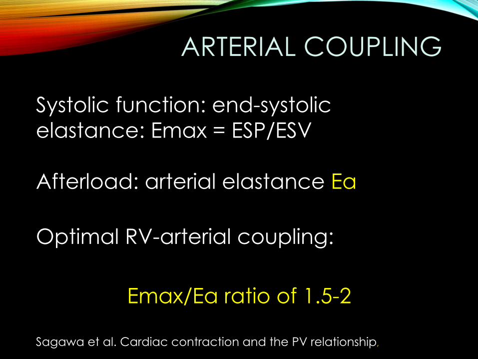
ARTERIAL COUPLING
Systolic function: end-systolic
elastance: Emax = ESP/ESV
Afterload: arterial elastance Ea
Optimal RV-arterial coupling:
Emax/Ea ratio of 1.5-2
Sagawa et al. Cardiac contraction and the PV relationship,

VA COUPLING

LV Arterial Coupling
Severe HTAfter 15 Days of treatment


RV FUNCTION AND RV REMODELING
- RV remodeling and adaptation to P/V overload is different depending on whether it occurs in early or later life
- In adults, evaluation of RV function needs to take into consideration:
1. The pathophysiology of the disease
2. The structural/functional changes caused by the treatment

RV FUNCTION AND RV REMODELING
RV has been considered as a chamber more adaptable to volume overload than LV but less able to tolerate pressure overload
Nevertheless, things are much more complex and the RV can remodel in a wide variety of ways

RIGHT VENTRICLE : VOLUME PUMP …
Increase of PreloadIncrease of afterload
ESP (mmHg) SV
VD
VS

VA COUPLING
To Each his own

RV FUNCTION AND RV REMODELING IN PUPPIES
Chronic pressure overload of RV in children results in ↑ ↑
hypertrophy more than in adult age such as in
pulmonary hypertension or pulmonary thromboembolism

VENTRICULAR PRESSURE–VOLUME RELATIONSHIPS AND ITS PERFORMANCE
❖The afterload reserve of the normal right ventricle is approximately one-third of that of the left ventricle.
❖Acute changes in acute pulmonary impedance are poorly tolerated by the right ventricle.
❖Slowly and early progressive rise in pulmonary arterial impedance may result in progressive change toward a ‘‘left ventricular’’ pattern of the pressure–volume loop.

Systole Systole
LV Coronary flow
RV Coronary flow
Coronary flow in the lower-pressure right ventricle
occurs mostly in systole

RIGHT VENTRICULAR FIBERS ORIENTATION AND REMODELING
Normal Normal

RV REMODELING IN PRESSURE OVERLOAD: ACQUIRED
VS CONGENITAL
EisenmengerPulmonary Thromboembolism

RIGHT VENTRICULAR REMODELING
Pulmonary Hypertension (PTH) Tricuspid dysplasia
Inadequate hypertrophy
Acquired pressure overload Volume overload

RIGHT VENTRICULAR FIBERS ORIENTATION AND REMODELING
Severe PSHuman ToF

RV RESTRICTIVE PHYSIOLOGY
Gross right ventricular hypertrophy, small RV cavity volumes, and fibrosis.
Systolic function become much more radial than longitudinal.
Bussadori C. Imaging evaluation, . in: Chessa, M. Giamberti, A. The Right Ventricle in Adults with Tetralogy of
Fallot Springer, Milan 2012, pp. 91-112. 2012

RIGHT VENTRICULAR FIBERS ORIENTATION AND REMODELING
Congenital Chronic
pressure overload (PS or
Eisenmenger) cause:
• Circumferential fibers
hypertrophy.
• Systolic function become
much more radial than
longitudinal.
• Adaptation to high systolic
pressure.
• Restrictive physiology.
Bussadori C, Salvo GD, Pluchinotta FR, Piazza L, Gaio G, Russo MG, Carminati M. Evaluation of right ventricular function in adults with congenital heart defects. Echocardiography. 2014

RV RESTRICTIVE PHYSIOLOGY
RAP > of PADP
• Transtricuspid flow will be transmitted to the pulmonary artery rather than translating to right ventricular filling.
End Diastolic Forward Flow (EDFF)
• Persistence of antegradediastolic flow in the pulmonary artery.
Cullen S, et al. Circulation. 1995 Mar 15;91(6):1782–9.
Gatzoulis MA et al Circulation. 1995 Mar 15;
91(6):1775–81.

• PS
• Eisenmenger,
• Systemic Right Ventricle
• Idiopathic PHT
• Pulmonary Thromboembolism

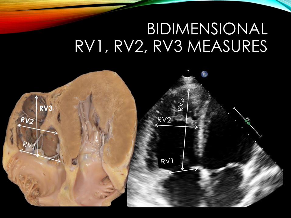
BIDIMENSIONALRV1, RV2, RV3 MEASURES
Apical 4 chamber view optimized for the RV:
3 standard Ø (measures in late diastole):
- 2 transverse Ø:
• RV1 tricuspid annulus (hum. reference values: 33-35 mm)
• RV2 apex of papillary muscles RV2 (hum. reference values: 23-33 mm)
- 1 longitudinal Ø:
• RV3 center of tricuspid plane to RV apex (hum. reference values: 67-75 mm)

ASE GUIDELINES FOR RVQUANTIFICATION
RV simplified RVOT measures
Rudski LG, et al. Guidelines for the echocardiographic assessment of the right heart in adults: A report from the american society of echocardiography. Journal of the American Society of Echocardiography. 2010;23:685-713

BIDIMENSIONALRV1, RV2, RV3 MEASURES
RV
3
BIDIMENSIONALRV1, RV2, RV3 MEASURES
RV3
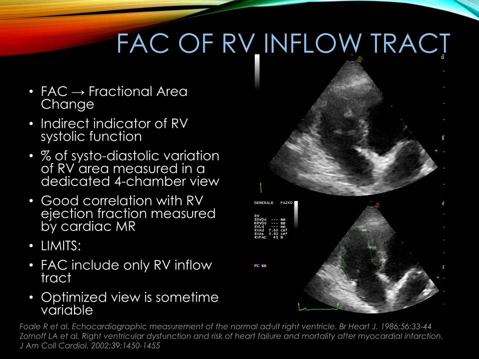
FAC OF RV INFLOW TRACT
• FAC → Fractional Area Change
• Indirect indicator of RV systolic function
• % of systo-diastolic variation of RV area measured in a dedicated 4-chamber view
• Good correlation with RV ejection fraction measured by cardiac MR
• LIMITS:
• FAC include only RV inflow tract
• Optimized view is sometime variable
Foale R et al. Echocardiographic measurement of the normal adult right ventricle. Br Heart J. 1986;56:33-44
Zornoff LA et al. Right ventricular dysfunction and risk of heart failure and mortality after myocardial infarction.
J Am Coll Cardiol. 2002;39:1450-1455
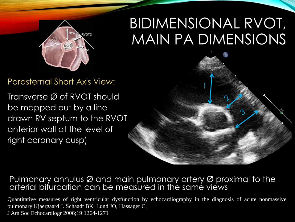
1Parasternal Short Axis View:
Transverse Ø of RVOT should
be mapped out by a line
drawn RV septum to the RVOT
anterior wall at the level of
right coronary cusp)
BIDIMENSIONAL RVOT, MAIN PA DIMENSIONS
Pulmonary annulus Ø and main pulmonary artery Ø proximal to the arterial bifurcation can be measured in the same views
Quantitative measures of right ventricular dysfunction by echocardiography in the diagnosis of acute nonmassive
pulmonary Kjaergaard J. Schaadt BK, Lund JO, Hassager C.
J Am Soc Echocardiogr 2006;19:1264-1271

BIDIMENSIONAL RVOT, MAIN PA DIMENSIONS
Pulmonary embolism PR

TAPSE TRICUSPID ANNULUS PEAK SYSTOLIC EXCURSION
• Widely used to study RV
systolic function
• BASED on the ASSUMPTION
that displacement of basal
and adjacent segments in
apical 4-chamber view is
representative of the
function of the entire RV
• Not valid in the regional RV
wall motion abnormalities
Tricuspid annular motion
Hammarstrom E, Wranne B, Pinto FJ, Puryear J, Popp RL.
J Am Soc Echocardiogr 1991 Mar-Apr4(2):131-9
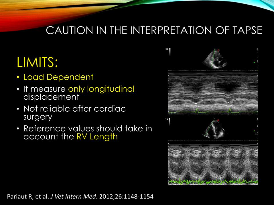
CAUTION IN THE INTERPRETATION OF TAPSE
LIMITS:• Load Dependent
• It measure only longitudinaldisplacement
• Not reliable after cardiac surgery
• Reference values should take in account the RV Length
Pariaut R, et al. J Vet Intern Med. 2012;26:1148-1154

TAPSE
Severe PS Severe PR

TAPSE
TAPSE 22 mm Severe PR
TAPSE 16 mm 24 H after PPVI

DTI OF THE TRICUSPID ANNULUS
- IVA: Isovolumic Myocardial Acceleration
- S’ Peak systolic velocity
- E: Early diastolic velocity
- A: Late Diastolic velocity
E
IC S
A
Characteristic of mitral and tricuspid annular velocities determined by pulsed wave Doppler tissue imaging in healthy subjects
Alam M. Wardell J, Andersson E, Samad BA, Nordlander R.
J Am Soc Echocardiogr 1999; 12 (8): 618-628

a
b
Index of Myocardial Performance (Tei)
a-b/b
IMP = (TCO-ET):ET = (ICT+IVRT):ET
Tissue Doppler method, Normal value <0.55 Pathological value >0.55
RIMP can be falsely low in conditions associated with elevated RA
pressures, which will decrease the IVRT.
ICT IVRT

Normal PHT
ICT 38±7 85±41
IVRT 49±9 135±43
ET 322±21 241±43
Tei 0.28±0.04 0.93±0.34

ISOVOLUMIC ACCELERATION (IVA)
Acceleration of the myocardium
during isovolumic contraction
(IVA)
• Index of contractile function,
comparing it to myocardial
acceleration, and velocities
measured during the ejection
phase.
• Several clinical ands
experimental validation.
Vogel M, et al. Circulation. 2002;105:1693–1699
Frigiola A, et al. Circulation. 2004; SuppI 110:I53–57.

ISOVOLUMICACCELERATION (IVA)
IVA
S’
IVC

VENA CAVA CONGESTION
• Normal IVC collapsibility• RAP 6 mmHg
• IVC collapsibility 35-45%• RAP 9 mmHg
• IVC collapsibility < 35%• RAP 16 mmHg
Pepi M. et al J Am Soc. Echoc. 1994
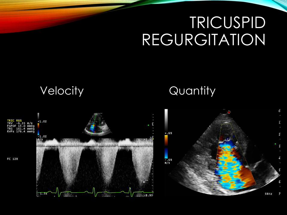
TRICUSPID REGURGITATION
Velocity Quantity

ANTEROGRADE PULMONARY FLOW
Severe Stenosis Dynamic Stenosis

PULMONARYREGURGITATION

PULMONARYREGURGITATION


RV dimensional and functional parameters
- RV Fractional area; TAPSE
- DTI (annular velocities)
- Pulmonary systolic pressure and RA Pressure
Routine evaluation in 1000
cases
6 + 1 minutesTamborini et al Int J Cardiol 2006