Embed Size (px)
Citation preview
ROLE OF GTP BINDING IN
YEAST SEPTINS
By
ASHOK RAJENDRAN
Master of Science
University of Madras
Chennai, India
April 2001
Submitted to the Faculty of the Graduate College of the
Oklahoma State University in partial fulfillment of
the requirements for the Degree of
MASTER OF SCIENCE May 2005
ii
ROLE OF GTP BINDING IN
YEAST SEPTINS
Thesis Approved:
MARK S. LONGTINE ________________________________________________
Thesis Adviser
ROBERT L. MATTS ________________________________________________
RICHARD C. ESSENBERG ________________________________________________
A. GORDON EMSLIE _______________________________________________
Dean of the Graduate College

iii
ACKNOWLEDGMENTS
This thesis is the result of the co-operation, support, and guidance of many
individuals. At this time, I would like to express my gratitude to those who have helped
me in this accomplishment.
First and foremost, I would like to thank Dr. Mark S. Longtine for providing me
an opportunity to work in this project and to pursue my Master’s degree in his lab. I am
grateful for his encouragement, kindness and his guidance. Were it not his unwavering
support and patience, I could not have become the scientist I am today.
I sincerely thank Dr. Robert L. Matts for his guidance, supervision, and expertise.
I am eternally grateful for his patience and invaluable suggestions. I would also like to
thank Dr. Richard C. Essenberg for being in my committee, for his suggestions and help
in completing my thesis.
I owe my sanity to my colleagues Satish Nagaraj, Manivannan Subramaniyan and
Lavanya Amirneni for being a great research partners and friends. I would like
to thank my friends Balaji Hariharasundaram, Palgunan Kalyanaraman for their
friendship and insight, which was most critical to the successful completion of this study.
Moreover, I wish to express my sincere gratitude to the Department of Biochemistry &
Molecular Biology for supporting me during this study.
Most of all, I wish to thank my parents to whom I am forever in debt for their
constant love and support. When I thought I could go no further, their faith gave me the

iv
strength to continue. None of this would have been possible without their support. They
are the best and I love them dearly. I would also extend my heartfelt thanks to my brother
for constantly motivating and encouraging me during my study.
v
TABLE OF CONTENTS
Chapter Page 1. INTRODUCTION……………………………………………………....... 1
Septin………………………………………………………………. 1 Functions of septins………………………………………………… 2 Cytokinesis…………………………….…………………… 2 Bud site selection…………………………………………... 5 Morphogenesis and cell cycle progression………………… 6 Spindle orientation…..…………………………………….. 6 Secretion…………………………………………………… 7 Mating……………………………………………………… 9 Filament formation and dynamics of septin complexes…………… 9 Importance of septin organization in its function………………….. 11 Septin in other organisms…………………………………….......... 11 Septins in D.melanogaster…………………………........... 11 Septins in mammals……………………………………….. 12 Septins in C.elegans………………………………………. 13
2. ROLE OF NUCLEOTIDE BINDING IN YEAST SEPTINS……………. 14
P-Loop in septins…………………………………………………... 14 Septin nucleotide binding………………………………………….. 14
3. MATERIALS AND METHODS……………………….……………........ 19
Preparation of protein extract and immunoprecipitation………………. 20 High pressure liquid chromatography…………………………………. 21 UV-crosslinking………………………………………........................... 21 Co-immunoprecipitation……………………………………………….. 22 In vitro transcription and translation (TnT) and metal affinity Purification…………………………………………………………….. 23
4. RESULTS………………………………………………………………….. 24
Co-immunoprecipitation………………..………………………………. 24 Affinity of septin towards the complex……………………………….... 33 Septin-septin interaction and septin complex formation in vitro ….….… 34
vi
Chapter Page
High pressure liquid chromatography………………………………….. 44 UV-crosslinking of septin……………………………………………… 47
5. DISCUSSION…………………………………………………..…………. 53 GTP binding is necessary for septin-septin interaction…………………. 53 BIBLIOGRAPHY……………………………………………………………….. 58
vii
LIST OF FIGURES Figure Page
1. Co-immunoprecipitation of septin complexes from CDC3 and cdc3pG129V, K132E, T133N mutant trains………………………………………………….. 26
2. Co-immunoprecipitation of septin complexes from CDC10 and cdc10K45T
mutant strains………………………………………………………………….. 28 3. Co-immunoprecipitation of septin complexes from CDC11 and cdc11 mutant
strains………………………………………………………………………….. 30 4. Co-immunoprecipitation of septin complexes from Cdc12p and its mutants…. 32
5. Effect of mutation on the affinity of Cdc11p towards the complex…………… 35
6. Pairwise in vitro septin-septin interactions……………………………………. 37
7. Ternary in vitro septin-septin interactions…………………………………….. 40
8. Quaternary in vitro septin-septin interactions…………………………………. 41
9. Interaction of Cdc11p and its P-loop mutants with the complex……………… 43
10. Analysis of nucleotides released from over expressed septin…………………. 46
11. Septins specifically bind GTP………………………………………………… 48
12. UV cross-linking of Cdc11p, cdc11pG32V and cdc11pR35T to GTP………… 50
13. UV cross-linking of Cdc11p, cdc11pASV and Cdc11pASIX to GTP…………. 51
14. UV cross-linking of Cdc3p, Cdc10p, Cdc12p P-loop mutants to GTP………… 52
viii
ABBREVIATIONS
2-D 2-Dimensional ATP Adenosine triphosphate BBF Bead beating buffer CFP Cyan fluorescent protein DTT Dithiothreitol DNA Deoxyribonucleic acid EB Elution buffer FRAP Fluorescence recovery after photo bleaching GAP GTPase activating protein GDP Guanosine diphosphate GEF Guanine nucleotide exchange factor GFP Green fluorescent protein GTP Guanosine triphosphate HPLC High pressure liquid chromatography kDa Kilo Dalton μl micro liter MLL Mixed lineage leukemia mM milli Molar NTP Nucleotide triphosphate pH -log [H+] PMSF Phenyl methyl sulfonyl fluoride PP2A Protein phosphatase 2A PVDF Polyvinylidene fluoride
ix
RNA Ribonucleic acid RPM Revolutions per minute RRL Rabbit reticulocyte lysate SA Specific activity SDS-PAGE Sodium dodecyl sulphate polyacrylamide gel electrophoresis TNT In vitro transcription and translation UV Ultraviolet
1
CHAPTER 1
INTRODUCTION
1.1 Septin:
Septins are a highly conserved family of GTP binding proteins found in all
eukaryotes, with the exception of plants. Septins were originally identified in
Saccharomyces cerevisiae in the Hartwell collection of temperature -sensitive mutants
defective in various stages of the cell division cycle (Hartwell, 1971). S. cerevisiae
encodes seven septins: CDC3, CDC10, CDC11, CDC12, SHS1, SPR28 and SPR3.
Among septins, there is about 35-90% sequence identity (Gladfelter et al., 2001;
Longtine et al., 1996). The size of septins ranges from ~40-75 kDa. The N-terminus of
septins contains a nucleotide-binding domain composed of G1 motif (P-loop domain), G3
and G4 motifs while the C-terminal of most septins contains a predicted coiled-coil
domain.
In S. cerevisiae, Cdc3p, Cdc10p, Cdc11p, Cdc12p, and Shs1p are expressed
during vegetative growth. These septins initially localize to the cell cortex at the
presumptive bud site (Longtine et al., 1996). As the bud emerges, septins rearrange from
a cortical ring to an hourglass-shaped collar spanning both sides of the mother-bud neck
just under the plasma membrane. This hourglass structure remains until right before
cytokinesis when the hourglass structure splits into two distinct rings. These rings persist
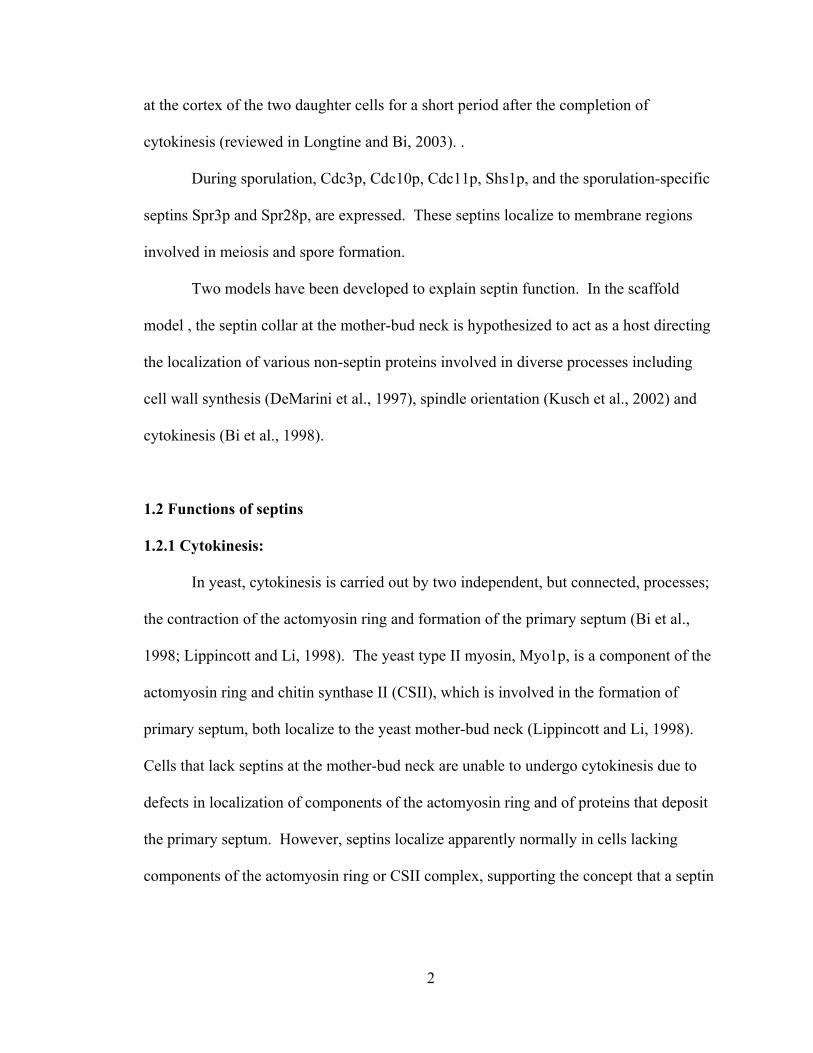
2
at the cortex of the two daughter cells for a short period after the completion of
cytokinesis (reviewed in Longtine and Bi, 2003). .
During sporulation, Cdc3p, Cdc10p, Cdc11p, Shs1p, and the sporulation-specific
septins Spr3p and Spr28p, are expressed. These septins localize to membrane regions
involved in meiosis and spore formation.
Two models have been developed to explain septin function. In the scaffold
model , the septin collar at the mother-bud neck is hypothesized to act as a host directing
the localization of various non-septin proteins involved in diverse processes including
cell wall synthesis (DeMarini et al., 1997), spindle orientation (Kusch et al., 2002) and
cytokinesis (Bi et al., 1998).
1.2 Functions of septins
1.2.1 Cytokinesis:
In yeast, cytokinesis is carried out by two independent, but connected, processes;
the contraction of the actomyosin ring and formation of the primary septum (Bi et al.,
1998; Lippincott and Li, 1998). The yeast type II myosin, Myo1p, is a component of the
actomyosin ring and chitin synthase II (CSII), which is involved in the formation of
primary septum, both localize to the yeast mother-bud neck (Lippincott and Li, 1998).
Cells that lack septins at the mother-bud neck are unable to undergo cytokinesis due to
defects in localization of components of the actomyosin ring and of proteins that deposit
the primary septum. However, septins localize apparently normally in cells lacking
components of the actomyosin ring or CSII complex, supporting the concept that a septin
3
"scaffold" localizes proteins involved in cytokinesis, thereby being involved in
cytokinesis
Recently, another model for the role of septins in cytokinesis has been
hypothesized, by demonstrating a role of septins in restricting the diffusion of membrane-
associated proteins. Takizawa and co-workers showed that Ist2p (a transmembrane
protein) specifically localized to the bud plasma membrane of the bud diffuses rapidly
within the bud. However, GFP-tagged Ist2p did not cross the bud neck and enter the
mother-cell membrane. However, after shift of a cdc12-6 septin mutant to 37˚C,
resulting in delocalization of septins from the neck, GFP-Ist2p now diffused across the
mother-bud neck and entered the plasma membrane of the mother-cell. These data
suggest that membrane-localized septins form a barrier to diffusion across the mother-
bud neck. Similarly, Lte1p, a guanine-nucleotide exchange (GEF) protein for the small G
protein Tem1p, localizes specifically to the cortex of the bud. However, in shs1∆/sep7∆
and cdc10∆ strains, with perturbed septin localization, Lte1p is also present in the mother
cell. Normally, after the switch to isotropic growth, actin patches and proteins involved
in polarized growth are restricted to the cortex of the bud. However, Barral et al. (2000)
found that if septins are delocalized then both actin patches and polarity proteins can now
be detected in the cortex of the mother cell. Together, these data strongly argue that in
yeast septins can serve as a barrier to the movement of transmembrane and membrane-
associated proteins.
Recently Schmidt and Nichols (2004) provided evidence for the role of septin in
forming a diffusion barrier in mammalian cell. Diffusion of the lipid analog
dialkylindocarbocyanine (DiI C18) and the membrane proteins (differing in topology and
4
lipid association) across the cleavage furrow was measured. The movement of proteins
with a cytosolic domain was restricted whereas there was no block in the diffusion of
proteins anchored in the outer leaflet of the plasma membrane or of DiI. The pattern of
distribution of septin was consistent with a functional role in limiting diffusion,
suggesting a possible role in the formation of diffusion barrier in the mammalian cells.
Septins are also found on either side of the contractile ring during cytokinesis in S.
pombe. These data indicate that a septin-dependent barrier to diffusion is a conserved
property of cortically localized septins.
Dobbelaere and Barral (2004) identified another function for a septin-based
diffusion barrier. During cytokinesis, after the splitting of the septin hourglass structure
into two discrete septin rings, they showed that proteins involved in cytokinesis,
including the polarizome complex involved in actin filament nucleation, the exocyst
involved in secretion, and chitin synthase II involved in formation of the primary septum,
all co-localize within the split septin rings. These data suggest that the septins do not
serve as a scaffold to recruit cortical proteins, but that septins instead formed a boundary
for the localization of these proteins during cytokinesis. To investigate whether or not
the proteins localized between the split septin rings were indeed compartmentalized,
Dobbelaere et al. did fluorescence recovery after photo bleaching (FRAP) analysis.
These data indicated that the proteins localized within the split septin rings indeed are
restricted from exchange with proteins outside the neck region. Furthermore, they
showed that intact septin rings are required for the restricted movement of the proteins
localized between the split septin rings. Thus, another role (at least in yeast) is to provide
5
a barrier that restricts the localization of proteins involved in cytokinesis to the proper
region of the cell.
1.2.2 Bud site selection:
Selection of the location of bud emergence ensures proper selection of an axis for
cell polarization. Haploid and diploid yeast cells have spatially distinct patterns of
budding. In haploid cells the new bud is formed in the axial pattern, being adjacent to the
previous bud site (which is also the site of cytokinesis in the preceding cell cycle). In
bipolar budding, a characteristic of diploid cells, buds are formed both at the distal and
proximal poles. The budding site in the subsequent generation in yeast is determined by
spatial landmark proteins localized at the cell poles, including at the mother-bud neck, in
the previous cycle. The BUD3 gene is required for the axial pattern of budding and any
mutations of BUD3 affect the axial pattern. Genes of BUD8 and BUD9 encode
components of the markers at the distal and proximal poles of the daughter cell and are
involved in bipolar budding pattern. When cdc12 mutant was shifted to restrictive
temperature, localization of Bud3p was rapidly lost (Chant, 1999; Schenkman et al.,
2002). Bud8p and Bud9p were also shown to be septin dependent for their localization
(Chant, 1999; Schenkman et al., 2002). These data indicate that the localization of
landmark proteins involved in bud-site selection is septin dependent. Conversely, septin
localization is normal in cells lacking bud-site selection proteins. Together, these data
are consistent with a model in which the septins serve as a scaffold to localize proteins
involved in bud-site selection.
6
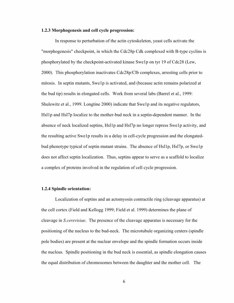
1.2.3 Morphogenesis and cell cycle progression:
In response to perturbation of the actin cytoskeleton, yeast cells activate the
"morphogenesis" checkpoint, in which the Cdc28p Cdk complexed with B-type cyclins is
phosphorylated by the checkpoint-activated kinase Swe1p on tyr 19 of Cdc28 (Lew,
2000). This phosphorylation inactivates Cdc28p/Clb complexes, arresting cells prior to
mitosis. In septin mutants, Swe1p is activated, and (because actin remains polarized at
the bud tip) results in elongated cells. Work from several labs (Barrel et al., 1999:
Shulewitz et al., 1999. Longtine 2000) indicate that Swe1p and its negative regulators,
Hsl1p and Hsl7p localize to the mother-bud neck in a septin-dependent manner. In the
absence of neck localized septins, Hsl1p and Hsl7p no longer repress Swe1p activity, and
the resulting active Swe1p results in a delay in cell-cycle progression and the elongated-
bud phenotype typical of septin mutant strains. The absence of Hsl1p, Hsl7p, or Swe1p
does not affect septin localization. Thus, septins appear to serve as a scaffold to localize
a complex of proteins involved in the regulation of cell cycle progression.
1.2.4 Spindle orientation:
Localization of septins and an actomyosin contractile ring (cleavage apparatus) at
the cell cortex (Field and Kellogg 1999; Field et al. 1999) determines the plane of
cleavage in S.cerevisiae. The presence of the cleavage apparatus is necessary for the
positioning of the nucleus to the bud-neck. The microtubule organizing centers (spindle
pole bodies) are present at the nuclear envelope and the spindle formation occurs inside
the nucleus. Spindle positioning in the bud neck is essential, as spindle elongation causes
the equal distribution of chromosomes between the daughter and the mother cell. The
7
proper position of the spindle, relative to the cleavage apparatus, depends on the capture
and shrinkage of the astral microtubules emanating from the spindle pole bodies. By
using a temperature sensitive cdc12-1 mutation, it was shown that septin function was
required for proper nuclear migration and positioning of the spindle relative to the
cleavage plane (Kusch et al., 2002). This work also found that both Tub1-GFP and Kar9-
GFP show transient interactions with the bud neck and that these interactions typically
resulted in movement of the nucleus towards the neck. Thus, it appears as if septins
interact (directly or indirectly) with microtubules and have a role in proper spindle
orientation and migration prior to mitosis.
1.2.5 Secretion:
In all cells, secretion of new material to the plasma membrane is essential for cell
growth prior to cell division. In S. cerevisiae, bud emergence occurs by the specific and
targeted delivery of new membrane and cell wall components to the growing bud. The
general pathway of secretion is fairly well characterized; in polarized secretion, vesicles
are carried along actin cables by Myo2p, a type V myosin, and are tethered to the sites of
secretion by a multiprotein complex called the exocyst (Novick et al., 1995). The final
vesicle fusion step, conserved in yeast, is mediated by formation of a SNARE complex,
which involves the vesicle-associated v-SNAREs Snc1p or Snc2p associating with the
plasma membrane- associated t-SNARES, Sso1p/Sso2p, and with the SNAP 25 homolog,
Sec9p (Sollner et al., 1993). The Munc-18 homolog, Sec1p, plays a positive role in
vesicle fusion by binding to productive preassembled trans-SNARE complexes (Carr et
al., 1999).
8
The current knowledge of septin function in secretion comes largely from the
studies of mammalian septins, especially in regards to function in neuronal secretions.
Several lines of evidence suggest the involvement of septins in secretion; the mammalian
septin CDC10 co-immunoprecipitates with the Sec6/8 (exocyst) complex (Hsu et al.,
1998); the neuronal septin Sept5 was shown to bind directly to the t-SNARE, syntaxin 1-
A (Beites et al., 1999); and cell fractionation and immunoelectron and
immunofluorescent microscopic studies indicate a possible association of septins with
secretory vesicles (Beites et al., 1999; Dent et al., 2002; Kinoshita et al., 2000). Secretion
assays using cultured HIT-T15 and platelets from mouse lacking Syntaxin-4 indicated a
negative role for septins in secretion (Beites et al., 1999; Blomberg et al., 1999). In
accordance with these results, it was recently observed that α-SNAP, which is involved
in disassembling cis-SNARE complexes present after vesicle fusion, shows competitive
binding to SNAREs with septins, suggesting that septin interactions with SNARE
proteins may play a negative role in secretion (Beites et al., 2005).
In yeast, the plasma membrane t-SNAREs Sso1p and Sso2p localize throughout
the periphery of the plasma membrane while several other components involved in
targeted secretion localize to the region of polarized growth (the bud tip, the bud
periphery, or to the mother-bud neck during cytokinesis). In a recent study Gladfelter et
al. (2005) observed that temperature sensitive cdc42V36T,K94EA cells displayed wide necks
with misorganized septins at the neck and mislocalized septins at the bud tip (Gladfelter
et al., 2002). However in cdc42 V36T, K94EA cdc12-6 double-mutant cells, which lack
localized septins wide bud necks were not observed. These results suggest that, at least
in yeast, septins may play a positive role in directing secretion. Recent work from our lab
9
(Manivannan Subramaniyan, personal communication) suggests that the yeast Cdc3p
septin may interact with the yeast plasma-membrane t-SNAREs, Sso1p and Sso2p. Thus,
it appears as if septin-SNARE interactions may be conserved, but the exact function of
these interactions may differ between mammalian cells and yeast.
1.2.6 Mating:
In response to mating pheromone, cells develop a characteristic pointed mating
projection, known as Shmoo. Shmoos form prior to fusion of cells of the opposite mating
type and formation of the diploid cell. Septins are found as diffused band at the base of
shmoos, (Ford and Pringle, 1991; Kim et al., 1991) and Giot and Konoka (1997) reported
that cells carrying a temperature-sensitive mutation (cdc12-6) were defective in
shmooing. In addition, Cdc12p interacts with Afr1p, the protein induced by mating
pheromone, and improves the ability of Afr1p to promote shmoo morphogenesis. Thus,
septins appear to have a role in mating, though further work needs to be done to clarify
this possible role.
1.3 Filament formation and dynamics of septin complexes:
Septins purified from S. cerevisiae (Frazier et al., 1998; Versele et al., 2004), D.
melanogaster (Field et al., 1996) and mammals (Hsu et al., 1998) form filaments in vitro.
However, the role of filament formation and nuclear organization of septin complexes
remain unclear. Septins purified from wild type S. cerevisiae cells under low salt
conditions, formed filaments measuring 7-9 nm in diameter. Extensive filament pairing
was also observed. However, cdc10Δ or cdc11Δ strains at 23˚C did not form long or
10
paired filaments in vitro and filamentous appearing structures were not visible in vivo
(Frazier et al., 1998). Nevertheless, in cdc10∆ strains septins are largely functional,
suggesting that long filaments are not necessary for septin function.
Dynamics of septin localization and assembly is important in understanding septin
function. Cdc42p, a rho-family GTPase, and its activating factor Cdc24p, are required
for septin delivery to the future bud site. Gladfelter et al. (2002), proposed a model in
which Cdc42 resembles the EF-Tu/EF-1α GTPase and hydrolysis of Cdc42p-bound GTP
has a direct role in septin ring formation. Caviston et al. (2003), proposed another model
in which Cdc42 acts as a Ras-like molecular switch along with its GTPase activity
protein (GAPs). According to the model of Caviston et al. (2003), septins are first
recruited to the presumptive bud after which assembly of the ring takes place. Cdc42 and
its GAPs are required for the above processes. Recently, a patch structure of septins was
shown to form before it transforms into the ring (Erfei Bi, personal communication). The
mechanism of transition of septin patch into a cortical ring in unbudded cells and into the
septin hourglass at the neck of budded cells is not clearly understood. It has been
suggested that GTP binding to Cdc10p and Cdc12p and septin phosphorylation of septin
by Cla4, a PAK kinase, may be necessary for the transition into the septin collar (Versele
and Thorner, 2004).
EM studies on yeast septins showed 10 nm striations at the bud neck but striations
were not seen in unbudded cells (Byers and Goetsch, 1976). The striations are absent
during late cell cycle, which may be due to the splitting of septin collar into a double ring
structure. Fluorescence Recovery After Photo bleaching (FRAP) analysis revealed that
septin rings are dynamic and unstable before bud emergence but that septins in the
11
hourglass are non-dynamic while septin rings after hourglass splitting are again dynamic
(Dobbelaere et al., 2003). These studies indicate that septin ring consists of less
organized septin assembly compared to the septin collar, which may have a more stable
and high order organization.
1.4 Importance of septin organization in its function:
At elevated temperatures, temperature sensitive yeast septin mutants have
elongated bud morphology and defective septin structure (Lew, 2000; McMillan et al.,
1999). Proteins encoded by BN15, CLA4, and GIN4 are important in septin organization
as deletion of any of these genes results in abnormal septin localization and organization
and in elongated buds (Longtine et al., 2000; Sreenivasan et al., 2003). Moreover,
perturbation in septin organization causes the arrest of cells in the G2/M phase of the cell
cycle indicating that septin mis-organization results in a cell cycle delay. The function of
septins as a barrier to diffusion likely requires highly complex organization. However,
Longtine et al. (1998), showed that for at least some septin functions, abnormal septin
organization at the neck can still result in significant septin function. In gin4Δ cells
septins functioned efficiently to localize proteins involved in bud-site selection and chitin
deposition, even though septin organization was clearly abnormal.
1.5 Septins in other organisms:
1.5.1 Septins in D. melanogaster:
The genome of D. melanogaster encodes five septins - Pnut, Sep1, Sep2, Sep4
and Sep5 (Fares et al., 1995; Field et al., 1996; Neufeld and Rubin, 1994) and (Adam et
12
al., 2000). Pnut, the first septin identified in D. melanogaster, is thought to be involved
in cytokinesis. Pnut mutants die as pupae with small imaginal discs due to the defects in
cytokinesis (Neufeld and Rubin, 1994). In addition to Pnut, Sep1 and Sep2 are also
involved in cytokinesis. In vitro, reconstitution of septin complexes isolated from D.
melanogaster embryos, Pnut, Sept1 and Sept7 were found in stoichiometric amounts
(Field et al., 1996). Though the role of Sep4 and Sep 5 in D. melanogaster is not clear, it
is believed that these septins may be involved in non-cytokinetic processes like vesicle
trafficking and exocytosis in non-dividing neuronal cells.
1.5.2 Septins in mammals:
Septins are a multigene family in mammals comprising of 12 septins (SEPT1-12),
some of which produce multiple splice variants (Macara et al., 2002; Surka et al., 2002).
Mammalian septins are predicted to be involved in vesicle transport and exocytosis
(Beites et al., 1999). Some septins, such as Sept 5/CDCrel-1, are expressed almost
exclusively in the brain (Caltagarone et al., 1998; Xue et al., 2000). Sep 5 is also a
substrate for Parkin, an E3 ubiquitin ligase involved in Parkinson’s disease.
Sept 2 and sept 5 were shown to interact with the SNARE protein syntaxin (Beites
et al., 1999). Sept 2, Sept 4, Sept 6 and Sept 7 associate with the exocyst complex, which
is involved in polarized secretion (Kinoshita et al., 1998). Septins were also presumed to
be involved in regulating exocytic events, but the mechanism is still not clear (Beites et
al., 1999).
Septins are also involved in various human diseases and neurodegenerative
disorders (Kartmann and Roth, 2001). Septin genes are found in genes encoding MLL
13
(mixed lineage leukemia). The experimental results of (Liauw et al., 2002) shows that
sept 7 expression is down-regulated in Alzheimer’s disease. Sept 5 over-expression in
the brain results in dopamine neurodegeneration (Dong, 2003). Sept 1, Sept2 and Sept 4
accumulate in filamentous deposits called neurofibrillary tangles (Kinoshita et al., 1998).
Sept 4 is found in the α-synuclein, a protein responsible for the formation of pathological
filamentous aggregates scientifically known as lewy bodies (Ihara et al., 2003). These
data indicate that septins play an essential role in human brain function.
1.5.3 Septins in C. elegans:
UNC-59 and UNC-61 are the two septins encoded in the C.elegans genome.
These septins are not involved in embryonic cytokinesis. These septins are reported to be
involved in post-embryonic neuroblast cell division (Nguyen et al., 2000). These septins
were first isolated as uncoordinated (unc) mutants in C. elegans (Brenner, 1974).
Mutations in septins caused uncoordination in newly hatched larvae in the absence of
cytokinetic defect (Finger et al., 2003) suggesting roles for septins in addition to
cytokinesis. Possible roles include roles in axon guidance and migration during C.
elegans embryogenesis (Choi et al., 2003). These data, and the fact that septins are not
expressed in neurons of the adult worm, led the authors to conclude that septins have a
developmental, rather than a functional, role in the C. elegans nervous system.
14
CHAPTER 2
ROLE OF NUCLEOTIDE BINDING IN YEAST SEPTINS
2.1 P-Loop in septins:
All septins possess an N-terminal P-loop nucleotide-binding domain, which is
characteristic of GTP and ATP binding proteins. It has been reported that D.
melanogaster (Field et al., 1996), human (Kinoshita et al., 1997b) and yeast septins (Field
et al., 1996) bind GTP. The conserved glycines in the P-loop are necessary for proper
folding of this nucleotide-binding pocket; the conserved lysine forms a bond with the
phosphate, and the conserved serine/threonine aids in the co-ordination with Mg2+.
Mutation of these residues disrupt nucleotide binding and/or hydrolysis of a wide variety
of nucleotide binding proteins (Bourne et al., 1991).
2.2 Septin nucleotide binding:
The role of septin nucleotide binding and/or hydrolysis is poorly understood.
Various models have been hypothesized on the role of GTP binding and hydrolysis. First
is the polymerization model, where GTP binding and hydrolysis is required for the
polymerization of septins into filaments, that GTP binding may play a solely structural
role, simply being required for proper septin folding. In yet another model it has been
proposed that GTP binding aids in the formation of heteromeric complex (complex
assembly model) rather than polymerization. Since all septins posses a conserved GTP-
15
binding domain, septins can be compared with GTPase-signaling proteins and thereby
septin function can be related to their ability to bind and hydrolyze GTP. In vitro studies
by Field et al., 1996 showed that purified D. melanogaster septin complexes strongly
bound to GTP/GDP and had GTPase-activity, but this GTP binding/exchange of D.
melanogaster septin was found to be very slow. Individual recombinant septins and
mammalian septins showed faster GTP binding/exchange kinetics in vitro. Cdc3p,
Cdc10p, Cdc11p and Cdc12p immunopurified from S. cerevisiae bind guanosine
nucleotides in stoichiometric amounts, with a GDP: GTP ratio of approximately 2.2:1.
Despite the different number of septin polypeptides in the septin complex, the nucleotide:
septin ratios and the GDP:GTP ratios are highly conserved in S. cerevisiae, D.
melanogaster and mammals. This evolutionary conservation suggests that GTP binding
may be necessary for stabilizing the septin polypeptides and that nucleotide binding plays
a structural role, analogous to the role of GTP for ∝-tubulin.
Versele et al. (2004) studied the GTPase function of yeast septins. In their work,
which used yeast septins expressed in E. coli, only Cdc10p and Cdc12p bound and
hydrolyzed in vitro. A cdc12 mutant (cdc12Δ339-407) that lacked the coiled-coil domain,
had a five-fold increase in the rate of GTP hyrolysis as compared to full-length Cdc12p.
They also studied the effect of GTP binding to CDC12 and CDC10. They made mutants
in which GTP-binding or GTP hydrolysis was specifically abrogated. Using these
mutants, the authors found that GTP hydrolysis might not play an important role in vivo
in septins. Furthermore, they have shown that GTP binding may be essential for the
proper assembly of the septin collar at the bud neck, although GTP binding did not
appear, in their conditions, to be required for efficient formation of heteromeric septin
16
complexes. A double-mutant strain with cdc10 cdc12 GTP-binding defective alleles
displayed a more dramatic defect in septin localization and function than either single
mutant, suggesting redundancy of function. By reconstituting wild-type and mutant
septin complexes in vitro and by examining the filament formation by electron
microscope the authors found that under low salt condition, wild-type septins formed
long filaments where as GTP-binding deficient mutant failed to form long filaments,
further indicating that GTP binding may be involved in the formation of higher order
septin complexes.
The role of GTP binding in mammalian septins has also been
demonstrated in forming hetero-oligomeric polymers. (Kinoshita et al., 1997a) showed
Nedd5 (SEPT2) mutants lacking GTP-binding activity disrupted Nedd5 containing fibers
and failed to organize into filamentous like structures, implying that GTP binding is
required for its assembly. They also demonstrated that immunological depletion of
SEPT2 interfered with cytokinesis. Kinoshita et al later demonstrated that SEPT2, 6 and
SEPT7 formed filaments in the presence of anillin (actin-binding protein). The
involvement of Sept5 (CDCrel-1) in exocytosis has been demonstrated in which it was
found that transfection of HIT-T15 cells with dominant negative mutation (defective in
GTP-binding) promoted secretion, where as wild type CDCrel-1 reduced secretion.
SEPT2, SEPT6 and SEPT9 have also been shown to bind GTP (Kinoshita et al., 1997;
Sheffield et al., 2003; Robertson et al., 2004).
The role of nucleotide binding in septin function has been complicated by
conflicting results; GTPase activity of bacterially expressed recombinant Sept2 was
reported to promote septin polymerization; Mendoza et al. (2002) expressed a single
17
recombinant septin polypeptide (xenopus Sept 2) in bacteria; septin complexes purified
from Drosophila bound to GTP but GTP hydrolysis did not influence septin
polymerization (Field et al., 1996); endogenously added guanine nucleotides had no
effect on in vitro assembly of filaments from multi-septin complexes (Kinoshita et al.,
2002; Sheffield et al., 2003). Future studies are necessary to resolve or explain these
contradicting results.
Zhang et al., (1999), showed that mammalian septin H5 (Sept4) is associated with
the plasma membrane and binds to the phospholipids phosphatidylinositol 4,5-
bisphosphate (Ptdlns(4,5)P2) and phosphatidylinositol 3,4,5-trisphosphate
(Ptdlns(3,4,5)P3). A conserved polybasic region in Sept4, which was essential for the
interaction with phospholipids, was found next to the GTP binding motif. GTP binding
and hydrolysis by Sept4 significantly reduced its Ptdlns (4,5) P2-binding capability,
indicating that septin-GTP binding regulates the interaction of septins with lipids thereby
playing an important role in the binding of septins to membranes.
There are various models hypothesized regarding septin GTP-binding/ hydrolysis
and septin function such as the “scaffold model” and the “diffusion barrier model”.
Complete knowledge about the architecture of the septin cortex and its regulation is
necessary to understand septin function. Though septin localization is essential for its
function, little is known about the mechanism of septin localization. There are many
questions regarding septin organization and function that remain unanswered. Do GTP
binding and hydrolysis play a role in septin-septin interaction or septin non-septin protein
interaction? Is GTP-binding/hydrolysis essential for septin localization? Answers to
above question are of great biological importance and will yield many insights in the near

18
future. The work presented here brings new insights about the role of nucleotide binding
in septin-septin interaction, septin localization and septin dependent processes.
19
CHAPTER 3
MATERIALS AND METHODS
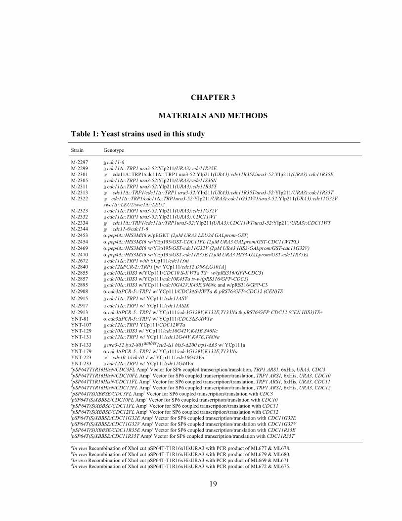
Table 1: Yeast strains used in this study Strain Genotype M-2297 a cdc11-6 M-2299 a cdc11∆::TRP1 ura3-52:YIp211(URA3):cdc11R35E M-2301 a/� cdc11∆::TRP1/cdc11∆:: TRP1 ura3-52:YIp211(URA3):cdc11R35E/ura3-52:YIp211(URA3):cdc11R35E M-2305 a cdc11∆::TRP1 ura3-52:YIp211(URA3):cdc11S36N M-2311 a cdc11∆::TRP1 ura3-52:YIp211(URA3):cdc11R35T M-2313 a/� cdc11∆::TRP1/cdc11∆::TRP1 ura3-52:YIp211(URA3):cdc11R35T/ura3-52:YIp211(URA3):cdc11R35T M-2322 a/�cdc11∆::TRP1/cdc11∆::TRP1ura3-52:YIp211(URA3):cdc11G32V#1/ura3-52:YIp211(URA3):cdc11G32V
swe1∆::LEU2/swe1∆::LEU2 M-2323 a cdc11∆::TRP1 ura3-52:YIp211(URA3):cdc11G32V M-2332 a cdc11∆::TRP1 ura3-52:YIp211(URA3):CDC11WT M-2334 a/� cdc11∆::TRP1/cdc11∆::TRP1ura3-52:YIp211(URA3):CDC11WT/ura3-52:YIp211(URA3):CDC11WT M-2344 a/� cdc11-6/cdc11-6 M-2453 α pep4∆::HIS3MX6 w/pEGKT (2µM URA3 LEU2d GALprom-GST) M-2454 α pep4∆::HIS3MX6 w/YEp195/GST-CDC11FL (2µM URA3 GALprom/GST-CDC11WTFL) M-2469 α pep4∆::HIS3MX6 w/YEp195/GST-cdc11G32V (2µM URA3 HIS3-GALprom/GST-cdc11G32V) M-2470 α pep4∆::HIS3MX6 w/YEp195/GST-cdc11R35E (2µM URA3 HIS3-GALprom/GST-cdc11R35E) M-2672 a cdc11∆::TRP1 with YCp111/cdc113nt M-2840 a cdc12∆PCR-2::TRP1 [w/ YCp111/cdc12 D98A,G101A] M-2855 a cdc10∆::HIS3 w/YCp111/CDC10 S-X WTa TS+ w/(pRS316/GFP-CDC3) M-2857 a cdc10∆::HIS3 w/YCp111/cdc10K45Ta ts-w/(pRS316/GFP-CDC3) M-2895 a cdc10∆::HIS3 w/YCp111/cdc10G42V,K45E,S46Nc and w/pRS316/GFP-C3 M-2908 α cdc3∆PCR-5::TRP1 w/ YCp111/CDC3∆S-XWTa & pRS76/GFP-CDC12 (CEN)TS M-2915 a cdc11∆::TRP1 w/ YCp111/cdc11ASV M-2917 a cdc11∆::TRP1 w/ YCp111/cdc11ASIX M-2913 α cdc3∆PCR-5::TRP1 w/ YCp111/cdc3G129V,K132E,T133Na & pRS76/GFP-CDC12 (CEN HIS3)TS+ YNT-81 α cdc3∆PCR-5::TRP1 w/ YCp111/CDC3∆S-XWTa YNT-107 a cdc12∆::TRP1 YCp111/CDC12WTa YNT-129 a cdc10∆::HIS3 w/ YCp111/cdc10G42V,K45E,S46Nc YNT-131 a cdc12∆::TRP1 w/ YCp111/cdc12G44V,K47E,T48Na YNT-133 a ura3-52 lys2-801amberleu2-∆1 his3-∆200 trp1-∆63 w/ YCp111a YNT-179 α cdc3∆PCR-5::TRP1 w/ YCp111/cdc3G129V,K132E,T133Na YNT-223 a/� cdc10-1/cdc10-1 w/ YCp111/ cdc10G42Va YNT-233 a cdc12∆::TRP1 w/ YCp111/cdc12G44Va apSP64TT1R16HisN/CDC3FL Ampr Vector for SP6 coupled transcription/translation, TRP1 ARS1, 6xHis, URA3, CDC3 bpSP64TT1R16HisN/CDC10FL Ampr Vector for SP6 coupled transcription/translation, TRP1 ARS1, 6xHis, URA3, CDC10 cpSP64TT1R16HisN/CDC11FL Ampr Vector for SP6 coupled transcription/translation, TRP1 ARS1, 6xHis, URA3, CDC11 dpSP64TT1R16HisN/CDC12FL Ampr Vector for SP6 coupled transcription/translation, TRP1 ARS1, 6xHis, URA3, CDC12 epSP64T(S)XBBSE/CDC3FL Ampr Vector for SP6 coupled transcription/translation with CDC3 fpSP64T(S)XBBSE/CDC10FL Ampr Vector for SP6 coupled transcription/translation with CDC10 gpSP64T(S)XBBSE/CDC11FL Ampr Vector for SP6 coupled transcription/translation with CDC11 hpSP64T(S)XBBSE/CDC12FL Ampr Vector for SP6 coupled transcription/translation with CDC12 ipSP64T(S)XBBSE/CDC11G32E Ampr Vector for SP6 coupled transcription/translation with CDC11G32E jpSP64T(S)XBBSE/CDC11G32V Ampr Vector for SP6 coupled transcription/translation with CDC11G32V kpSP64T(S)XBBSE/CDC11R35E Ampr Vector for SP6 coupled transcription/translation with CDC11R35E lpSP64T(S)XBBSE/CDC11R35T Ampr Vector for SP6 coupled transcription/translation with CDC11R35T aIn vivo Recombination of XhoI cut pSP64T-T1R16xHisURA3 with PCR product of ML677 & ML678. bIn vivo Recombination of XhoI cut pSP64T-T1R16xHisURA3 with PCR product of ML679 & ML680. cIn vivo Recombination of XhoI cut pSP64T-T1R16xHisURA3 with PCR product of ML669 & ML671 dIn vivo Recombination of XhoI cut pSP64T-T1R16xHisURA3 with PCR product of ML672 & ML675.
20
eSubcloned BamHI/SmaI fragment containing CDC3 from pGEX4T/CDC3 into BglII/SmaI digested pSP64T(S)XBSSE. fSubcloned XbaI/SacI digested PCR product (~1.2 kb) of ML477 & ML478 on pALTER/CDC10 into XbaI/SacI digested pSP64T(S)XBSSE. gSubcloned ~ 1.3kb SalI (blunted)/BglII fragment containing CDC11 from pSL301/CDC11 into BglII/SmaI digested pSP64T(S)XBSSE. hSubcloned ~ 1.3kb NotI (blunted)/BamHI fragment containing CDC12 from pGEX4T/CDC12 into BglII/SmaI digested pSP64T(S)XBSSE. iIn vivo Recombination of BglII/MfeI cut pSP64T(S)XBSSE/CDC11 with the BglII/MfeI digested PCR product of ML621 & ML566 on YCp111/cdc11G32E. jIn vivo Recombination of BglII/MfeI cut pSP64T(S)XBSSE/CDC11 with the BglII/MfeI digested PCR product of ML621 & ML566 on YCp111/cdc11G32V. kIn vivo Recombination of BglII/MfeI cut pSP64T(S)XBSSE/CDC11 with the BglII/MfeI digested PCR product of ML621 & ML566 on YCp111/cdc11R35E. lIn vivo Recombination of BglII/MfeI cut pSP64T(S)XBSSE/CDC11 with the BglII/MfeI digested PCR product of ML621 & ML566 on YCp111/cdc11R35T.
3.1 Preparation of protein extract and immunoprecipitation:
Unless indicated otherwise, all steps were carried out at 4˚C. 500 μl of bead
beating buffer (25 mM HEPES pH 7.4, 150 mM NaCl, 0.1% Triton X-100, 5 mM MgCl2,
10% glycerol, protease inhibitors, phosphatase inhibitors and PMSF) (BBF) was added to
a cell pellet corresponding to 25 OD600 of cells (~50 mg of cell pellet). 250 μl of glass
beads were added, followed by vortexing at 4˚C for 5 min at high speed, incubation on
ice for 2.5 min, then vortexing at high speed for an additional 5 min. The mixture was
then centrifuged at 14000 rpm for 10 min and the supernatant was transferred to a fresh
tube. Protein concentration was determined by Bio-Rad assay and ~5-25 mg of total
protein was isolated.
25 μl of protein-G beads was added to ~12 mg of total protein extract and ~2
mg/ml of anti-Cdc3p (Sigma Genosys) antibody was added and the mixture was gently
rocked for ~2.5 hrs at 4°C. The beads were then pelleted and washed four times with 500
µl of wash buffer (25 mM HEPES pH 7.4, 150 mM NaCl, 0.1% Triton X-100, 5 mM
MgCl2 10% glycerol, protease inhibitors, phosphatase inhibitors and PMSF). The washed
beads were then incubated overnight in 1.6 volumes (40 µl) of elution buffer [20 mM
21
Tris-HCl, pH 7.8 1 mM KCl, 2 mM MgCl2, 0.5 mM EGTA, 4% sucrose with 0.8 mg/ml
Cdc3 C-terminal peptide (Sigma Genosys)]. The beads were then pelleted at 500X g, and
the supernatant containing the septin complexes was used for the UV cross-linking assay.
3.2 High pressure liquid chromatography (HPLC):
The samples for HPLC were obtained by over-expressing the septins using
galactose induction, and purifying them over GA-beads (Glutathione agarose). After
washing the beads with BBF the protein was then denatured by the addition of 30 μl 8 M
urea, 100 mM Tris-HCl, pH7.2, followed by heating to 100ºC for 1min. 30 μl of water
was added and the mixture was transferred to 10 kDa cut-off Microcon filter and was
centrifuged at 13000 RPM for 15 minutes. This was followed by two more washes with
water (30 μl each). The total volume (120 μl) was then assayed by HPLC through a
mono-Q column (Amersham Biosciences) of 1ml bed volume with a loading capacity of
50mg. The column was prepared by equilibration with 100 mM ammonium bicarbonate
and the sample was eluted using a gradient of 100-500 mM ammonium bicarbonate over
30 min at a flow rate of 1 ml/min, was used for the sample elution. Standard (a mixture of
100pmol of ATP, GDP and GTP) was run separately using the identical conditions.
3.3 UV-crosslinking:
Septin complexes purified as described above were incubated with [α-32P] GTP
(2μM GTP SA, 800 Ci/mmol) and 300 μM non-radioactive ATP at 30°C with or without
competing non-radioactive GTP at 300 μM. Also [α-32P] GTP with no competitor
nucleotide and [α-32P] GTP with 1 mM EDTA were assayed. 5 μl of sample was taken

22
for SDS-PAGE analysis to assay for complex purification. 2-mercaptoethanol was added
to the remaining portion of the sample to 1%, followed by incubation on ice for 1 min
and then exposure to UV light on ice for five periods of 3 min each with 1min pause
between each UV exposure. UV-crosslinking was carried out using a Stratagene UV
Stratalinker 1800 with 254 nm bulbs. The samples were placed 7 cm from the UV lamps
and the power of the instrument was approximately1140μW/cm2. After UV exposure, 8
μl of 6X SDS-PAGE sample buffer was added and the sample heated for 30 s at 100°C.
200μl of cross-linking buffer (20 mM Tris-HCl, pH7.8, 100 mM KCl, 0.5 mM EGTA,
and 2 mM MgCl2) was added to each sample. The mixture was filtered through a 10-kDa
cut off microcon filter until 98% of the liquid went through (13000 rpm, 20 min). The top
of the filter was resuspended in 30μl of 1X sample buffer, heated for 1min at 100°C and
subjected to 10% SDS PAGE. The proteins from gel were then transferred to a PVDF
membrane by electroblotting and the membrane was used for autoradiography and
phosphoimaging.
3.4 Co-immunoprecipitation:
Yeast cells grown exponentially in appropriate media at room temperature were
shifted to 37ºC for 2 hours. The cells were then disrupted with glass beads by vortexing at
4º C in (25 mM HEPES pH 7.4, 150 mM NaCl, 0.1% Triton X-100, 5 mM MgCl2, 10%
glycerol, protease inhibitors, phosphatase inhibitors and PMSF). After centrifugation to
remove cell debris, the supernatant was used for immunoprecipitation. 30 µl of beads, ~2
mg of the total protein and 3-4 µg of antibody (anti-Cdc11p and anti-Cdc12p) were
mixed and incubated at 4ºC for 2 hrs. The beads were then washed four times, each with
23
350 µl BBF. Proteins bound to the beads were eluted with SDS sample buffer, analyzed
by SDS-PAGE and western blotting using anti-Cdc3p, anti-Cdc10p (Santa Cruz
Biotechnology), anti-Cdc11p and anti-Cdc12p (Sigma Genosys) antibodies.
3.5 In vitro transcription and translation (TNT) and metal affinity purification:
Fifty micro liters of TNT mixture in our laboratory contains 2.92 µl water, 9.58 µl
buffer (10 mM creatine phosphatase, 1 mM DTT, 3 mM magnesium acetate, 110 mM
potassium acetate, and 0.6 mM each amino acid except for methionine. 0.8 mM each
rNTPspH7.3) 1 µl plasmid (1 µg/µl), 0.5 µl of 35S-methionine (Perkin Elmer, SA, 1175.0
Ci/mmol), 35 µl of rabbit reticulocyte lysate and 1 µl of SP6 RNA polymerase. The
above reagents were aliquoted into a 1.5 ml microfuge tube and mixed well avoiding air
bubbles. After incubating for 30 min at 37ºC, the mixture was clarified by centrifugation.
The supernatants were then mixed and allowed to interact for 1 hr at 23ºC, followed by
affinity purification using 25 µl of Talon beads (BD Biosciences) at 23ºC for 1 hr. 5 µl of
the supernatant was taken as the unbound (UB) sample. The beads were washed four
times, with 350 µl of wash buffer (BBF). 30 µl of 1X sample buffer was added and
heated for 5 min at 100ºC. Bound (B) and unbound (UB) fractions were separated by
SDS-PAGE, electroblotted to PVDF membrane and detected by autoradiography.
24
CHAPTER 4
RESULTS
4.1 Co-immunoprecipitation
Purified septin complexes from S. cerevisiae form filaments in vitro (Frazier et
al., 1998; Versele et al., 2004). In addition to septin-septin interactions, septins also
interact with non-septin proteins (reviewed in the Introduction). Septins contain a
putative GTP binding domain and it has been reported that septins bind GTP (Vrabioiu et
al., 2003). Though septins bind GTP, the role of GTP binding is not yet clear.
To study the role of GTP binding in yeast septins, we constructed several septin
mutants by mutating residues at the P-loop using site directed mutagenesis. We then
asked if these mutations affected septin-septin interactions by carrying out a series of co-
immunoprecipitation experiments. The mutant alleles analyzed in these studies were
cdc3G129V,K132E,T133N,cdc10K45T, cdc 11-6 (cdc11G32E), cdc11R35T and cdc11R35E.
andcdc12G44V, cdc12G44V, K47E, T48N and cdc12D98A, G101A.
Cells were grown to mid-log phase at 23ºC and then shifted to 37ºC for 2 hr.
Protein extracts were then made from the cells grown at 23ºC and 37ºC (for Cdc3p and
Cdc10p experiments) or after shift to 37ºC (for Cdc11p and Cdc12p experiments) to
assay for septin-septin interactions. Septin proteins were then immunoprecipitated using
anti-Cdc11p or anti-Cdc12p antibodies and protein-G beads. Immunoprecipitated septin
25
complexes were separated by SDS-PAGE and analyzed by Western blotting, using four
different antibodies which recognize Cdc3p, Cdc10p, Cdc11p, and Cdc12p.
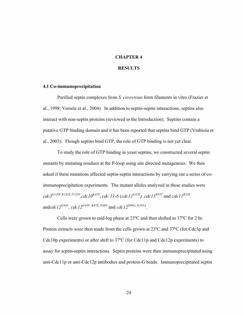
The results of these experiments for wild-type Cdc3p and cdc3pG129V, K132E,
T133N strains are shown in Figure 1. First, after a two hour shift to 37˚C, all four septins
are stable and present in wild-type amounts (Fig 1A), indicating no effect on protein
stability. As shown in Fig 1B, cdc3pG129V, K132E, T133N immunoprecipitated less
efficiently with Cdc11p than did wild-type Cdc3p . In contrast, in extracts from wild-
type cells Cdc3p efficiently immunoprecipitates with Cdc11p. Also note, that when
cdc3p levels are reduced, there is a concomitant reduction in the amount of
immunoprecipitated Cdc10p. Similar results were obtained with immunoprecipitation
using anti-Cdc12p antibodies (Fig. 1C): cdc3pG129V, K132E, T133N associated less
efficiently with Cdc12p than did wild-type Cdc3p, and there was a concomitant reduction
in the amount of Cdc10p in the complex. Thus, the triple-P-loop mutation of Cdc3p
reduces its interaction with Cdc11p and Cdc12p, consistent with the observed
temperature-sensitive viability of this strain. In addition, these results indicate that
Cdc10p association with Cdc11p and Cdc12p depends upon the presence of Cdc3p,
suggesting that Cdc10p association in the complex may be mediated by a direct
interaction with Cdc3p.
26
Figure 1. Co-immunoprecipitation of septin complexes from CDC3 and cdc3pG129V, K132E, T133N mutant strains. Yeast cells grown exponentially in SDC–His medium at 23˚C were shifted to 37ºC for 2 hr and protein extracts prepared. 80μg of total protein was taken (A) and ~2mg of total protein was used for immunoprecipitation with IgG beads bound to (B) anti-Cdc11p antibody or (C) anti-Cdc12p antibody. Bound proteins were eluted with SDS sample buffer, separated by SDS-PAGE and septins detected by immuno-blotting using anti-Cdc3p, anti-Cdc10p anti-Cdc11p and anti-Cdc12p antibodies, as indicated.
27
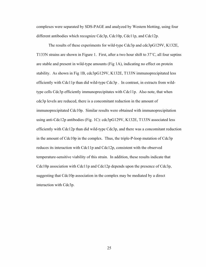
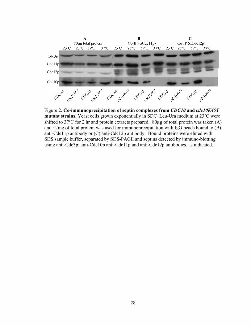
Investigation of the effect of cdc10K45T on its incorporation into a septin
complex is shown in Fig. 2. After a two hour shift to 37˚C, all four septins are stable and
present in wild-type amounts (Fig 2A), indicating no effect on protein stability.
Consistent with its full, or nearly full function in viability at 23˚C (Satish Nagaraj,
personal communication), cdc10pK45T efficiently interacts with both Cdc11p- and
Cdc12p-containing septin complexes at 23˚C (Fig. 2A-B). In agreement with the
temperature-sensitive viability of cdc10pK45T strains (Satish Nagaraj, personal
communication), at 37˚C cdc10p K45T is no longer associated with septin complexes
containing Cdc11p or Cdc12p (Fig. 2B,C). However, in contrast to observation that
Cdc10p association with Cdc11p or Cdc12p depends upon the presence of Cdc3p (Fig.
1), Cdc3p association with Cdc11p and Cdc12p occurs independently of the presence of
Cdc10p (Fig. 2B, C).
28
. Figure 2. Co-immunoprecipitation of septin complexes from CDC10 and cdc10K45T mutant strains. Yeast cells grown exponentially in SDC–Leu-Ura medium at 23˚C were shifted to 37ºC for 2 hr and protein extracts prepared. 80μg of total protein was taken (A) and ~2mg of total protein was used for immunoprecipitation with IgG beads bound to (B) anti-Cdc11p antibody or (C) anti-Cdc12p antibody. Bound proteins were eluted with SDS sample buffer, separated by SDS-PAGE and septins detected by immuno-blotting using anti-Cdc3p, anti-Cdc10p anti-Cdc11p and anti-Cdc12p antibodies, as indicated.
29
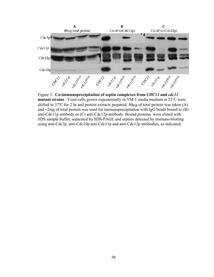
The effects of mutations on the P-loop of Cdc11p on its participation in septin-
complex formation are shown in Fig. 3. After a two hour shift to 37˚C, all septins are
stable and present in wild-type amounts (Fig 3A) in the mutant strains analyzed
(cdc11R35T, cdc11-6, and cdc11R35E) , indicating no affect on protein stability. cdc11-6
(cdc11G32E) strains show strong temperature-sensitive viability defects, being inviable at
32˚C and above (Satish Nagaraj, personal communication). cdc11pG32E does not
interact with cdc11p or Cdc12p-containing septin complexes at 37˚C (Fig. 3A, B),
suggesting that defects of cdc11pG32E in septin-septin interactions may cause the
observed inviability. I also assayed the defects in septin complex formation of
cdc11pR35T and cdc11pR35E. cdc11pR35E is expected to show a more dramatic defect
in nucleotide binding than cdc11pR35T, due to the presence of charge-charge repulsion
of the glutamate with the phosphates of the bound nucleotide. Consistent with this idea,
cdc11pR35E shows temperature sensitive viability while cdc11pR35T is wild-type for
viability at all temperatures (Satish Nagaraj, personal communication). As expected,
cdc11pR35T interacts well with the other septins, while cdc11pR35E shows clearly
reduced incorporation into a septin heteromeric complex (Fig. 3B, C). Interestingly, in
the absence of cdc11p (Fig. 3C, cdc11-6 lane) the remaining three septins interact
efficiently (though with reduced levels of Cdc10p). Also, we note that in the
intermediate cdc11 allele (cdc11R35E), cdc11p and Cdc12p can be found as a co-
immunoprecipitating sub-complex (Fig. 3B, cdc11R35E lane), suggesting a strong
interaction between Cdc11p and Cdc12p. Thus, these data suggest that a complex of
Cdc3p, Cdc10p, and Cdc12p forms in the absence of Cdc11p, and that Cdc11p and
Cdc12p may show a strong, and likely direct, interaction.,
30
Figure 3. Co-immunoprecipitation of septin complexes from CDC11 and cdc11 mutant strains. Yeast cells grown exponentially in YM-1 media medium at 23˚C were shifted to 37ºC for 2 hr and protein extracts prepared. 80μg of total protein was taken (A) and ~2mg of total protein was used for immunoprecipitation with IgG beads bound to (B) anti-Cdc11p antibody or (C) anti-Cdc12p antibody. Bound proteins were eluted with SDS sample buffer, separated by SDS-PAGE and septins detected by immuno-blotting using anti-Cdc3p, anti-Cdc10p anti-Cdc11p and anti-Cdc12p antibodies, as indicated.
31
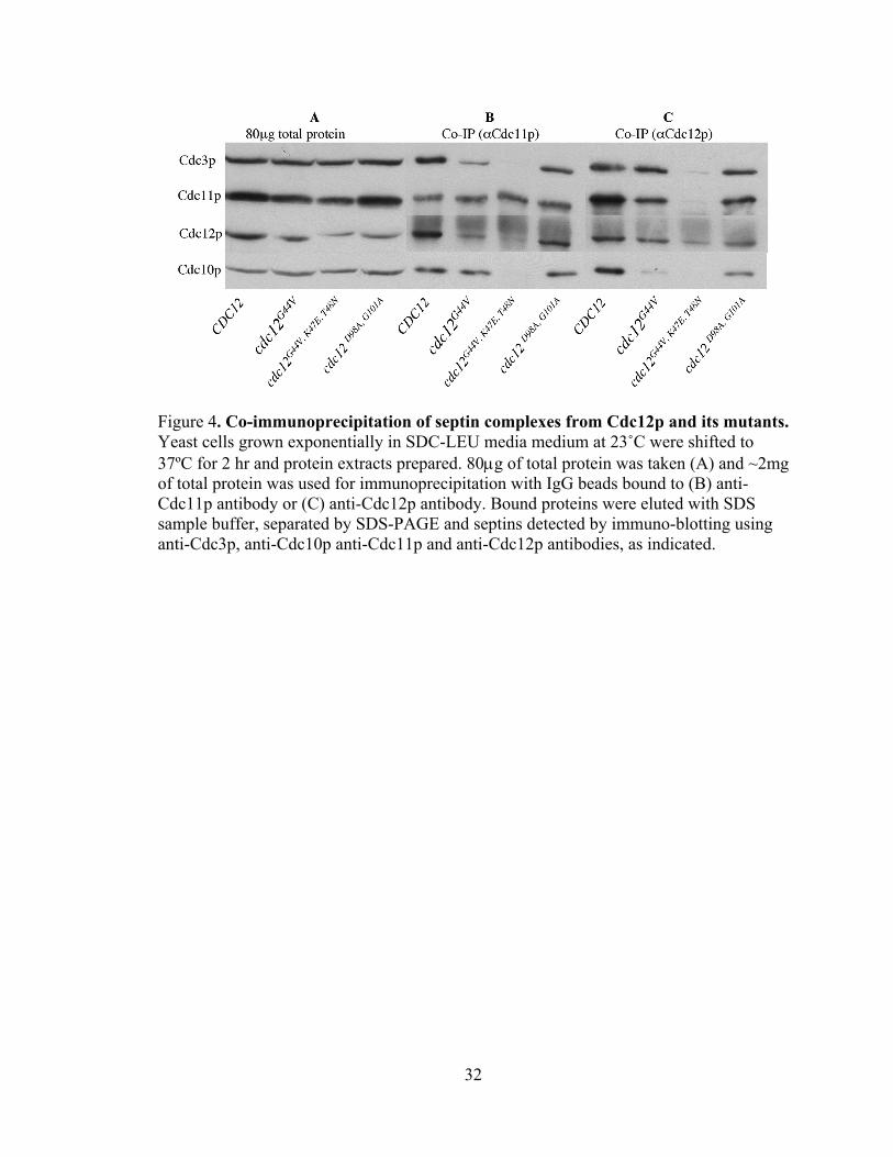
Finally, I assayed the effects of disrupting Cdc12p GTP binding or GTP hydrolysis
on its participation in septin-septin complexes. As expected from their full viability at all
temperatures (Satish Nagaraj, personal communication), both Cdc12p and cdc12pD98A,
G101A, efficiently formed a complex containing Cdc3p, Cdc11p, Cdc12p (or
cdc12pD98A, G101A), and Cdc10p (Fig. 4 B, C). cdc12G44V strains show only a slight
defect in viability at 37˚C (Satish Nagaraj, personal communication), consistent with
ability of cdc12pG44V to participate nearly as effectively as Cdc12p in septin complex
formation. However, in this strain there does appear to be slightly reduced levels of
Cdc11p and Cdc10p in the complex (Fig. 4C, cdc12G44V lane). In contrast, a cdc12G44V,
LK47E, T48N strain that shows clear viability defects at 37˚C (Satish Nagaraj) is unable to
participate in septin complex formation (Fig. 4B, C). Notably, if cdc12p is not associated
with the other septins (Fig. 4C, cdc12G44V, LK47E, T48N lane), Cdc11p is also unable to
associate with Cdc3p or Cdc10p (Fig. 4B cdc12G44V, LK47E, T48N lane).
32
Figure 4. Co-immunoprecipitation of septin complexes from Cdc12p and its mutants. Yeast cells grown exponentially in SDC-LEU media medium at 23˚C were shifted to 37ºC for 2 hr and protein extracts prepared. 80μg of total protein was taken (A) and ~2mg of total protein was used for immunoprecipitation with IgG beads bound to (B) anti-Cdc11p antibody or (C) anti-Cdc12p antibody. Bound proteins were eluted with SDS sample buffer, separated by SDS-PAGE and septins detected by immuno-blotting using anti-Cdc3p, anti-Cdc10p anti-Cdc11p and anti-Cdc12p antibodies, as indicated.
33
4.2 Affinity of septin towards the complex:
The above co-immunoprecipitation analyses suggested that cdc11 P-loop mutant
proteins assemble into heteromeric septin complexes at 23˚C as well as wild-type
Cdc11p. However, this conclusion seemed unlikely for several reasons. First, work by
our collaborators (Chris Field and Alina Vriabiou) suggested that septin filament
formation in vitro was defective in extracts from cdc11pR35T cells with a more dramatic
defect in cdc11pR35E cells. In addition, quantitation of coomassie-blue stained bands of
septin complexes purified from these strains suggested a mild reduction in the
stoichiometry of the mutant cdc11p proteins compared to that of wild-type Cdc11p.
Finally, two-hybrid and in vitro studies (Satish Nagaraj, personal communication)
indicate that there are detectable defects in septin-septin interactions in most P-loop
mutants even at 23˚C.
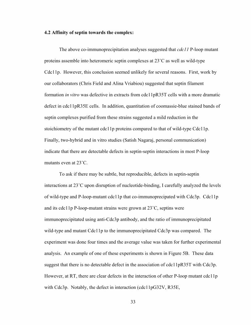
To ask if there may be subtle, but reproducible, defects in septin-septin
interactions at 23˚C upon disruption of nucleotide-binding, I carefully analyzed the levels
of wild-type and P-loop-mutant cdc11p that co-immunoprecipated with Cdc3p. Cdc11p
and its cdc11p P-loop-mutant strains were grown at 23˚C, septins were
immunoprecipitated using anti-Cdc3p antibody, and the ratio of immunoprecipitated
wild-type and mutant Cdc11p to the immunoprecipitated Cdc3p was compared. The
experiment was done four times and the average value was taken for further experimental
analysis. An example of one of these experiments is shown in Figure 5B. These data
suggest that there is no detectable defect in the association of cdc11pR35T with Cdc3p.
However, at RT, there are clear defects in the interaction of other P-loop mutant cdc11p
with Cdc3p. Notably, the defect in interaction (cdc11pG32V, R35E,

34
S36N>cdc11pG32V≥cdc11pG32E, cdc11pR35E >cdc11pS36N>cdc11pR35T) closely
parallels the defects in temperature-sensitive viability of these strains (Satish Nagaraj,
personal communication). Together, these data suggest that there are slight, but
quantifiable, defects in interactions of cdc11p P-loop mutant septins with Cdc3p at 23˚C.
35
0
0.2
0.4
0.6
0.8
1
1.2
1.4
Cdc1
1p W
T
cdc1
1pG32
E
cdc1
1pG32
V
cdc1
1pR3
5T
cdc1
1pR3
5E
cdc1
1pS3
6N
cdc1
1pG32
V,R3
5E,S
36N
Cdc11pWT and mutants
Rati
o o
f C
dc1
1p
Vs
Cd
c3p
ratio of Cdc11p Vs Cdc3p
Figure 5. Effect of mutation on the affinity of Cdc11p towards the complex. CDC11 and cdc11 P-loop mutant strains were grown to log phase in SDC–Ura media at 23˚C. Protein extracts were prepared, and septin complexes immunoprecipitated using anti-Cdc3p antibody and protein-G beads. Bound proteins were eluted with SDS sample buffer, separated by SDS-PAGE and Cdc3p and Cdc11p were detected by immunoblotting using anti-Cdc3p and anti-Cdc11p antibodies, respectively (A) Ratio of Cdc11p and cdc11p mutant proteins to Cdc3p, taken from the average of four separate experiments. (B) One of the experiments used to generate the graph shown in (A).
36
4.3 Septin-septin interaction and septin complex formation in vitro:
Septins are present as multi-septin complexes in vivo. My co-
immunoprecipitation studies gave some clue on septin-septin interactions. However, I
wanted to further dissect septin-septin interactions. In this work, I used rabbit
reticulolysate (RRL) coupled in vitro transcription and translation (TnT) assays to
investigate septin-septin interactions. For purification, one septin was tagged with six N-
terminal histidine residues, with the remaining septins untagged. The His6-tagged protein
and any associated septins were then purified by metal (cobalt) affinity chromatography
and subsequently analyzed by SDS-PAGE and autoradiography.
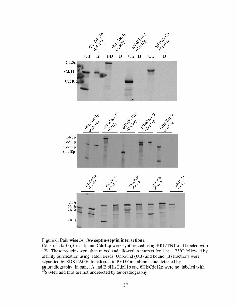
First, I assayed for direct interactions of septins in pair wise assays. The results of
these assays are shown in Figure 6. First, Cdc11p and Cdc12p interact directly and
efficiently, forming an ~1:1 complex (Fig. 6A, B, and C, 6HisCdc11p+Cdc12p lanes and
Fig. 6B 6HisCdc12p+Cdc11p lanes). In addition, these assays detected an interaction
between 6His-Cdc3p and Cdc12p (Fig. C, 6HisCdc3p+Cdc12p lane). It is unclear why
no detection was detected between 6HisCdc12p+Cdc3p (Fig. 6B).
.
37
Figure 6. Pair wise in vitro septin-septin interactions. Cdc3p, Cdc10p, Cdc11p and Cdc12p were synthesized using RRL/TNT and labeled with 35S. These proteins were then mixed and allowed to interact for 1 hr at 23ºC,followed by affinity purification using Talon beads. Unbound (UB) and bound (B) fractions were separated by SDS PAGE, transferred to PVDF membrane, and detected by autoradiography. In panel A and B 6HisCdc11p and 6HisCdc12p were not labeled with 35S-Met, and thus are not undetected by autoradiography.

38
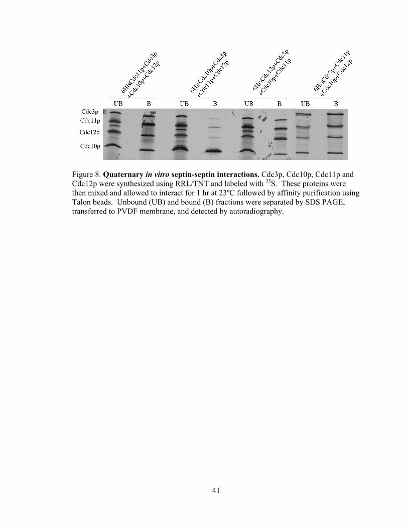
Next, I used the in vitro RRL/TnT assay to investigate formation of ternary and
quaternary septin-septin complexes. First, all four septins associate efficiently, forming
an apparently stoichiometric complex (Fig. 7 A,B 6HisCdc11p+Cdc3p+Cdc10p+Cdc12p
lanes). There was a decrease in the stoichiometry of purified Cdc3p, Cdc11p, and
Cdc12p when using 6His-tagged Cdc10p, suggesting the 6His tag may disrupt the ability
of Cdc10p to participate in complex formation. The detection of this complex is based
upon a specific interaction of the 6His-tagged protein with the talon resin, as a mixture of
the four untagged septins does not associate with the Talon beads (Fig. 7,
Cdc11p+Cdc12p+Cdc10p+Cdc3p lanes). This assay also indicates that N-terminal 6His-
tagged Cdc10p is defective, as 6HisCdc10p does not interact with the other septins (Fig.
8) while untagged Cdc10p interacts well. In contrast, N-terminally 6His-tagged Cdc11p,
Cdc12p, and Cdc3p all efficiently form the quaternary complex, indicating that the tags
do not affect protein-protein interactions. As the septins in this assay efficiently interact,
it appears to be a valid method to investigate ternary septin-septin interactions.
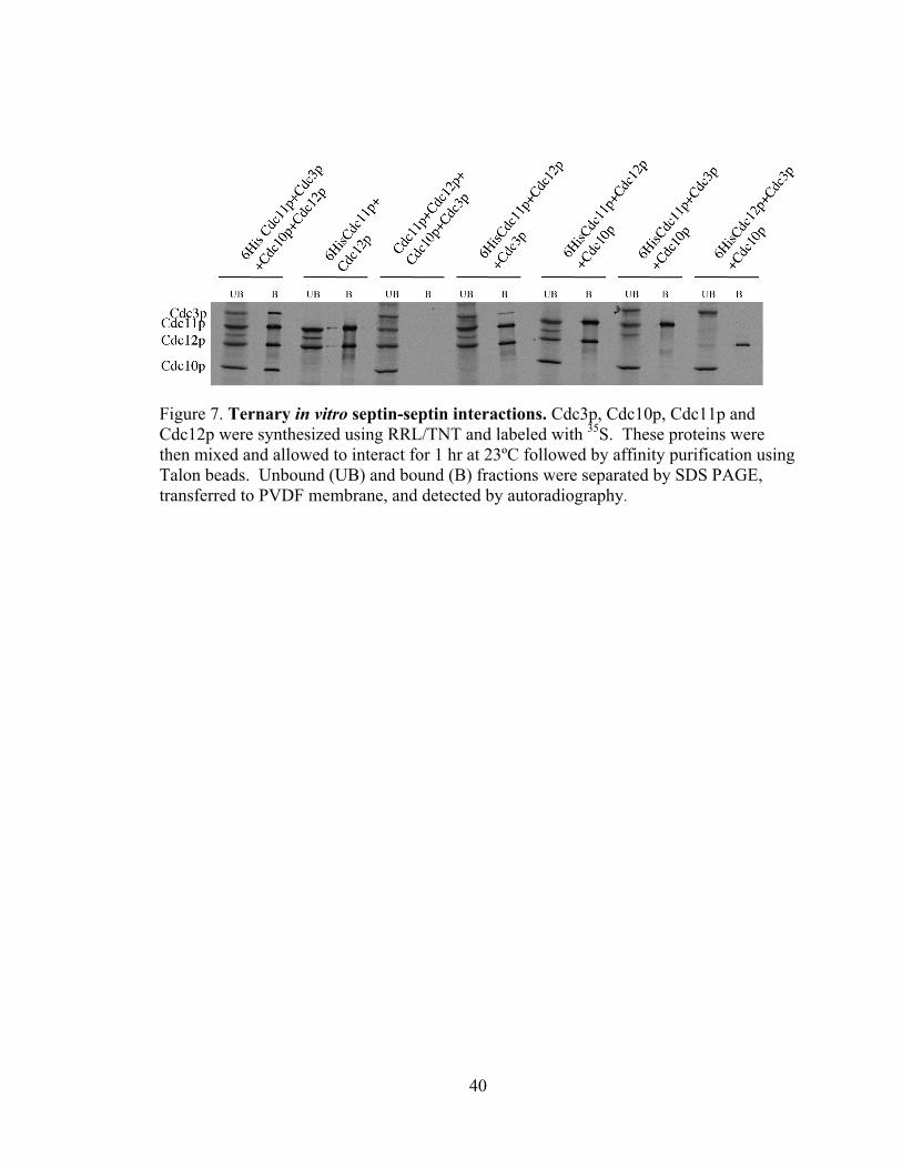
Interactions of three septins are investigated in Figure 7. As shown previously,
Cdc11p and Cdc12p interact stoichiometrically in pair wise interactions, but neither alone
interacts with Cdc3p (Fig. 6). In the presence of both Cdc11p and Cdc12p, however, a
sub-stoichiometric (but reproducible) amount of Cdc3p interacts to form a ternary
complex (Fig. 7, compare 6HisCdc11p+Cdc12p+Cdc3p bound lane with Fig. 6A 6
HisCdc11p+Cdc3p lanes and with Fig. 6B 6HisCdc12p+Cdc3p lanes). Thus, Cdc3p
interacts more efficiently with a complex of Cdc11p and Cdc12p than to either single
septin. Surprisingly, no other ternary complex was detected. Thus, these data suggest

39
while Cdc11p and Cdc12p may be involved in direct interaction and are required for the
efficient interaction of Cdc3p which may have a direct interaction with Cdc10p.
40
Figure 7. Ternary in vitro septin-septin interactions. Cdc3p, Cdc10p, Cdc11p and Cdc12p were synthesized using RRL/TNT and labeled with 35S. These proteins were then mixed and allowed to interact for 1 hr at 23ºC followed by affinity purification using Talon beads. Unbound (UB) and bound (B) fractions were separated by SDS PAGE, transferred to PVDF membrane, and detected by autoradiography.
41
Figure 8. Quaternary in vitro septin-septin interactions. Cdc3p, Cdc10p, Cdc11p and Cdc12p were synthesized using RRL/TNT and labeled with 35S. These proteins were then mixed and allowed to interact for 1 hr at 23ºC followed by affinity purification using Talon beads. Unbound (UB) and bound (B) fractions were separated by SDS PAGE, transferred to PVDF membrane, and detected by autoradiography.
42
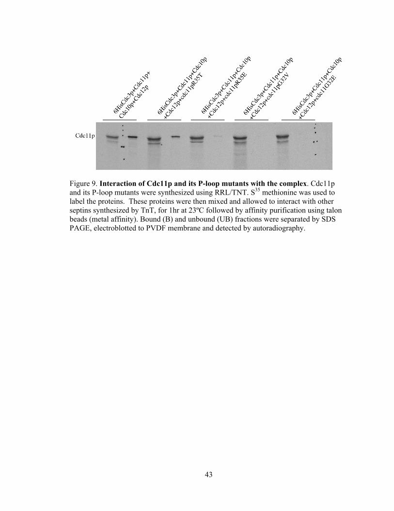
The RRL/TnT assay was then used to determine if cdc11p P-loop-mutant proteins
showed defects in septin-septin interactions. Consistent with the co-immunoprecipitation
results described earlier (Figs. 4 and 5), cdc11pR35T, cdc11pR35E, cdc11pG32V and
cdc11pG32E all displayed defects in assembly into septin complexes (Fig. 9). As
expected, cdc11R35T retained the ability to interact with the other septins (Fig. 9,
compare 6HisCdc3p+Cdc11p+Cdc3p+cdc11pR35T lanes with
6HisCdc3p+Cdc11p+Cdc10p+Cdc12p). However, this interaction was reproducibly
slightly reduced compared to wild-type Cdc11p. Similar reduction was observed in pair-
wise interactions of cdc11pR35T with Cdc12p (Satish Nagaraj, personal
communication). Consistent with their more dramatic defects in function in vivo (Satish
Nagaraj, personal communication), cdc11pR35E, cdc11pG32V and cdc11pG32E were all
unable to interact with the septin complex (Fig. 9) or with Cdc12p in pair wise
interactions (Satish Nagaraj, personal communication). Together, these data suggest that
disruption of nucleotide binding by Cdc11p disrupts its interaction with the septin
complex in vivo (Figs. 4 and 5) and in vitro (Fig. 9).
43
Figure 9. Interaction of Cdc11p and its P-loop mutants with the complex. Cdc11p and its P-loop mutants were synthesized using RRL/TNT. S35 methionine was used to label the proteins. These proteins were then mixed and allowed to interact with other septins synthesized by TnT, for 1hr at 23ºC followed by affinity purification using talon beads (metal affinity). Bound (B) and unbound (UB) fractions were separated by SDS PAGE, electroblotted to PVDF membrane and detected by autoradiography.
44
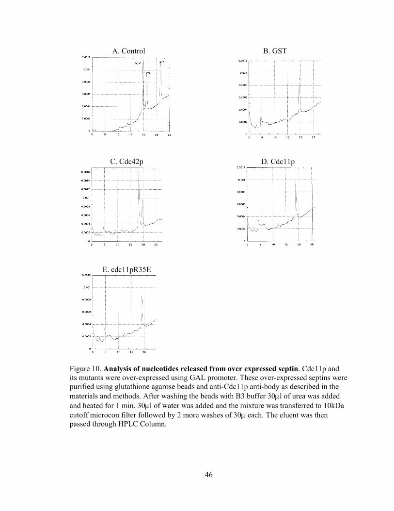
4.4 High pressure liquid chromatography (HPLC):
Co-immunoprecipitation and in vitro binding studies indicate that mutations in the
P-loop of septins disrupt septin-septin interactions. However, these studies do not
directly demonstrate that the introduced mutations affect nucleotide binding. As an
attempt to address this question, we over-expressed N-terminally GST-tagged
(glutathione-S-transferase) wild-type and P-loop-mutant Cdc11p under the control of the
galactose-inducible, GAL1 promoter. Cells were grown to mid-log phase at 23˚C in
raffinose containing medium and expression of the GST-tagged induced by the addition
of galactose to 2% final concentration, followed by a 12 hr period of induction at 23˚C.
Protein extracts were prepared, and ~5 mg of total protein was incubated with 30 µl bed
volume glutathione-agarose resin for 1 hr at 23˚C. Following 4X washes of 350 µl for
2.5 min each, the bound proteins and nucleotides were eluted by the addition of 30µl of
8M urea and heating at 100˚C for 1min. Typically, this resulted in the purification of ~25
µg of the GST-tagged septin. The supernatant was filtered through a 10 Kd microcon
filter by centrifuging for 15 min. After washing the filter thrice the filtrate was pooled
and was loaded on a 1 ml MonoQ column that was equilibrated in buffer containing 100
mM ammonium bicarbonate. The sample was assayed by running a 100 mM to 500 mM
gradient of ammonium bicarbonate, with control runs performed by using different
concentrations of GTP, GDP, and/or ATP (Fig 10A) to determine elution times of these
nucleotides and that the amount of nucleotide was directly correlated with the peak area
for the bound nucleotide, using absorbance at 260 nm. An example of a control run is
shown in Fig. 10A. In sample runs with purified septins, ATP was used as an internal
control. First, we assayed a positive control, expressing GST-tagged wild-type Cdc42p
45
with a C188S mutation so it is not tightly associated with membranes (Mol. Biol. Cell.
(1993) 4:1307–1316. Subcellular localization of Cdc42p, a Saccharomyces cerevisiae
GTP-binding protein involved in the control of cell polarity. M. Ziman, D. Preuss, J.
Mulholland, J. M. O'Brien, D Botstein, and D. I. Johnson). As expected, Cdc42p was
bound predominantly to GDP (Fig. 10C), which elutes just before the added ATP (Fig.
10A) under these conditions. Quantitation of the amount of purified proteins (not shown)
and the amount of purified GDP was in ~1:1 stoichiometry. In contrast, GST-Cdc11p
was almost entirely nucleotide free, with only ~5% of the protein containing bound GDP
and no detectable levels of GTP (Fig. 10D). Similar results were obtained for GST-
control and cdc11pR35E, with the exception of even lower levels of bound GDP. These
data are consistent with reduced GDP binding to GST-cdc11pR35E compared to GST-
Cdc11p wild-type protein. However, the most dramatic finding is that overexpressed
GST-tagged septins are largely nucleotide free. This contrasts with data from the Field
lab indicating that septins purified from yeast as a complex containing stoichiometric
amounts of bound guanine nucleotide |(Vrabioiu et al., 2003). Thus, we conclude that
grossly overexpressed Cdc11p or P-loop mutant cdc11p proteins display dramatic
reductions in nucleotide binding. It is possible that these overexpressed septins are not
folded properly (for example, the overexpression level overwhelms some aspect of the
protein folding machinery involved in septin folding), and thus are non-functional or only
poorly functional. Indeed, overexpressed GST-tagged Cdc11p does not interact with
Cdc12p at the expected levels (Angie Thomure, personal communication), suggesting it
is not folded properly and abnormaly nucleotide-free. Thus, it seems as if the
overexpression strategy is not useful for analyzing septin nucleotide binding.
46
A. Control B. GST
C. Cdc42p D. Cdc11p
E. cdc11pR35E
Figure 10. Analysis of nucleotides released from over expressed septin. Cdc11p and its mutants were over-expressed using GAL promoter. These over-expressed septins were purified using glutathione agarose beads and anti-Cdc11p anti-body as described in the materials and methods. After washing the beads with B3 buffer 30μl of urea was added and heated for 1 min. 30μl of water was added and the mixture was transferred to 10kDa cutoff microcon filter followed by 2 more washes of 30μ each. The eluent was then passed through HPLC Column.
47
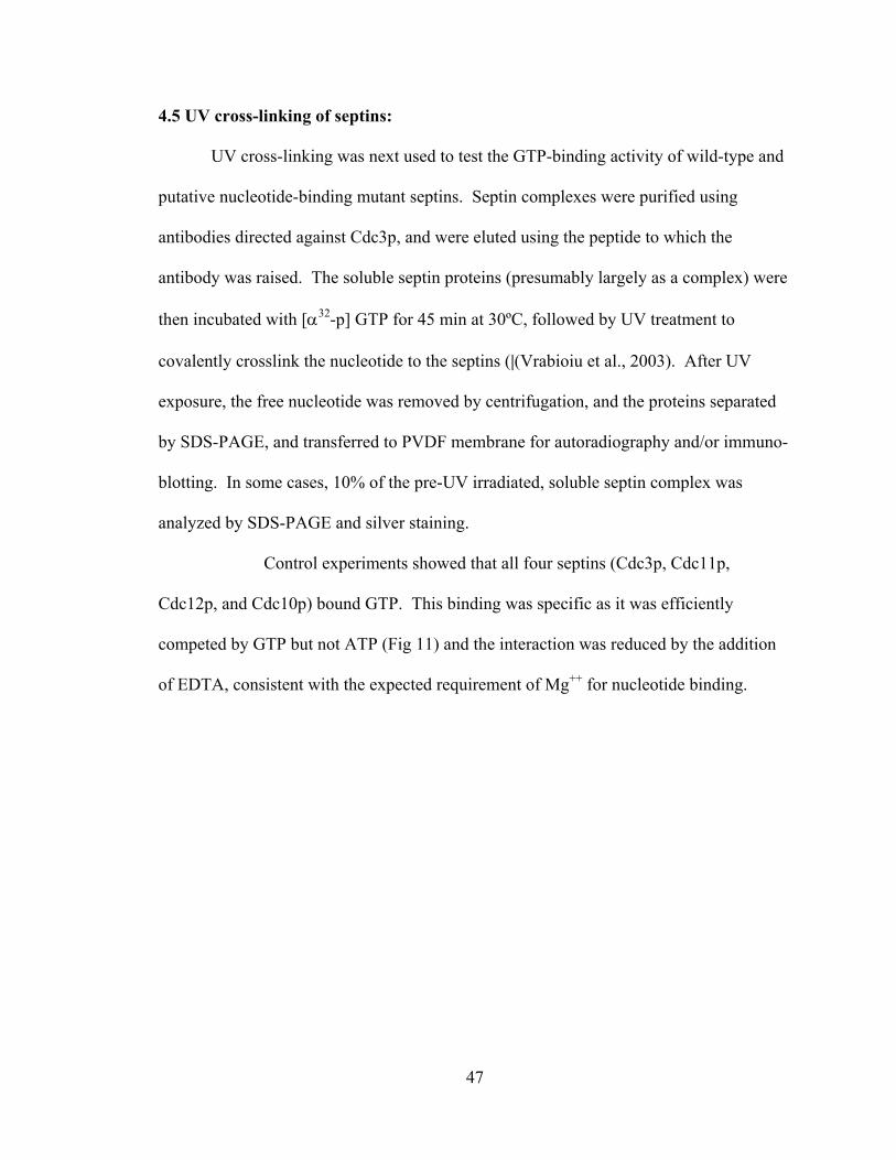
4.5 UV cross-linking of septins:
UV cross-linking was next used to test the GTP-binding activity of wild-type and
putative nucleotide-binding mutant septins. Septin complexes were purified using
antibodies directed against Cdc3p, and were eluted using the peptide to which the
antibody was raised. The soluble septin proteins (presumably largely as a complex) were
then incubated with [α32-p] GTP for 45 min at 30ºC, followed by UV treatment to
covalently crosslink the nucleotide to the septins (|(Vrabioiu et al., 2003). After UV
exposure, the free nucleotide was removed by centrifugation, and the proteins separated
by SDS-PAGE, and transferred to PVDF membrane for autoradiography and/or immuno-
blotting. In some cases, 10% of the pre-UV irradiated, soluble septin complex was
analyzed by SDS-PAGE and silver staining.
Control experiments showed that all four septins (Cdc3p, Cdc11p,
Cdc12p, and Cdc10p) bound GTP. This binding was specific as it was efficiently
competed by GTP but not ATP (Fig 11) and the interaction was reduced by the addition
of EDTA, consistent with the expected requirement of Mg++ for nucleotide binding.
48
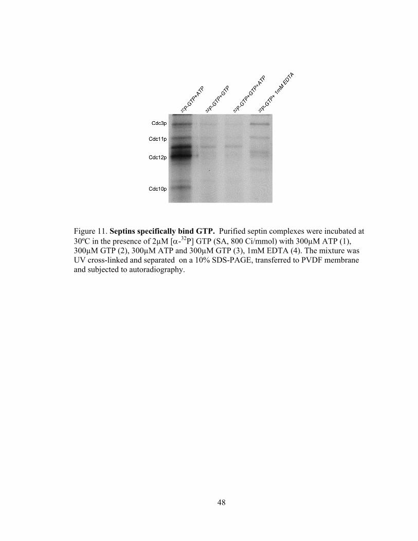
Figure 11. Septins specifically bind GTP. Purified septin complexes were incubated at 30ºC in the presence of 2µM [α-32P] GTP (SA, 800 Ci/mmol) with 300µM ATP (1), 300µM GTP (2), 300µM ATP and 300µM GTP (3), 1mM EDTA (4). The mixture was UV cross-linked and separated on a 10% SDS-PAGE, transferred to PVDF membrane and subjected to autoradiography.
49
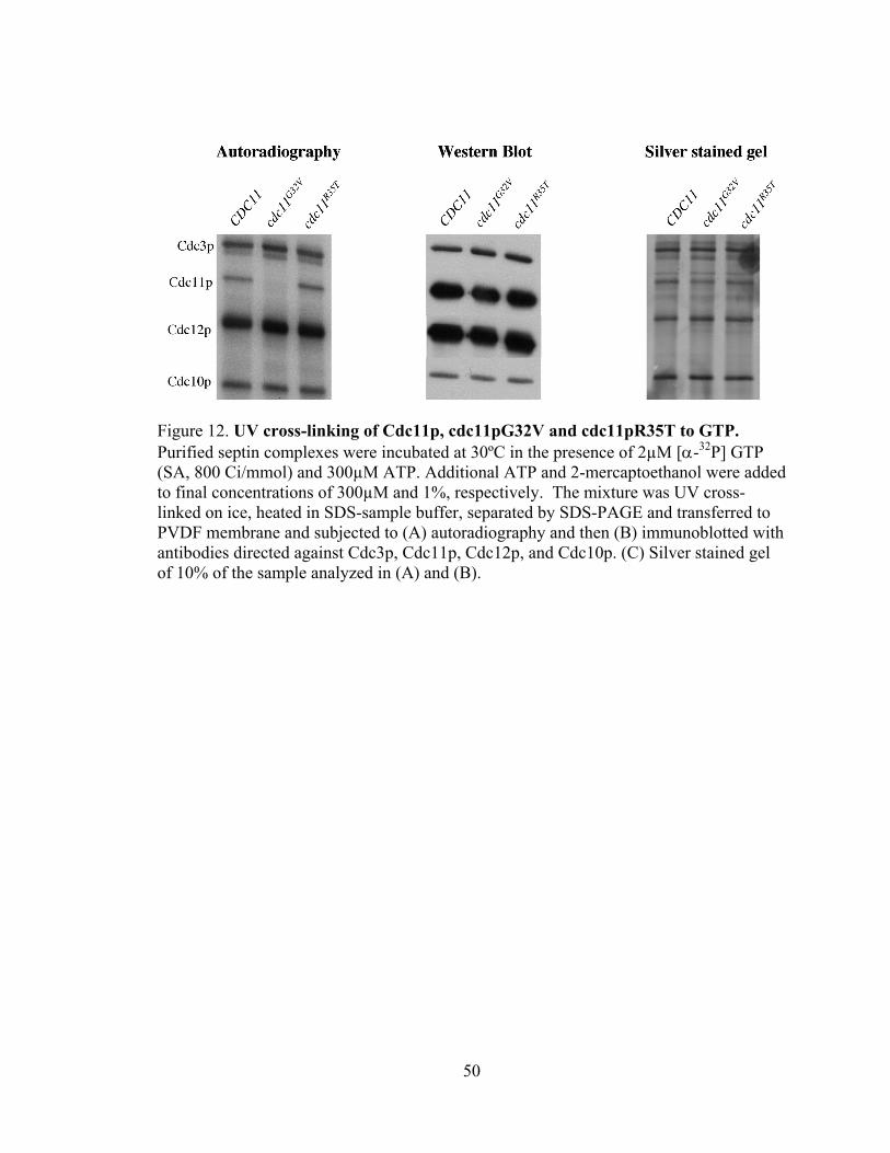
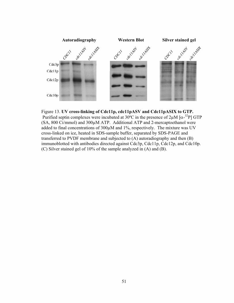
Figure 12 shows that wild-type Cdc11p and cdc11pR35T efficiently bind GTP
while cdc11pG32V has a dramatic defect in GTP binding. This decrease in GTP binding
by cdc11pG32V is much greater than the reduction in the levels of Cdc11p (Fig 12B, C)
indicating that cdc11pG32V has a reduced affinity for GTP. Similar results were
obtained for the cdc11pASIX protein, which contains a mutation in the G4 domain
(Satish Nagaraj, personal communication) and is thus predicted to also have reduced GTP
binding. In contrast, cdc11pASV, which contains a mutation in the G4 domain and is
predicted to efficiently bind GTP, shows efficient crosslinking to GTP (Fig. 13A)
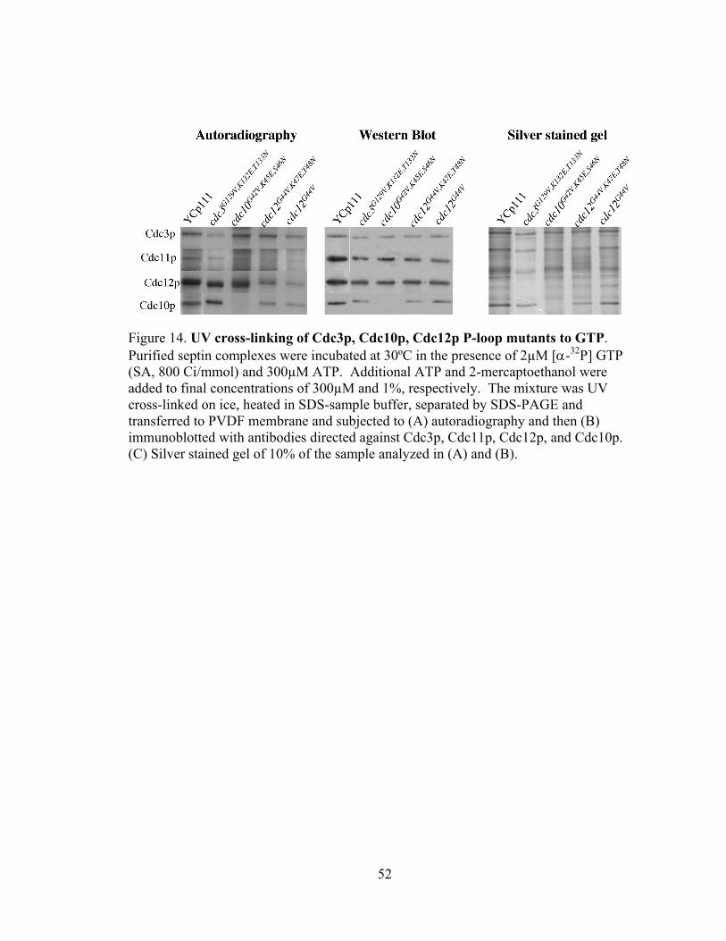
Consistent with the results obtained with Cdc11p, mutations in the Cdc3p P-loop
(cdc3G129V,K132E,T133N) or Cdc12p P-loop (cdc12 G44V,K47E,T48N) also showed reduced GTP
binding relative to their respective wild-type proteins (Fig14).
50
Figure 12. UV cross-linking of Cdc11p, cdc11pG32V and cdc11pR35T to GTP. Purified septin complexes were incubated at 30ºC in the presence of 2µM [α-32P] GTP (SA, 800 Ci/mmol) and 300µM ATP. Additional ATP and 2-mercaptoethanol were added to final concentrations of 300µM and 1%, respectively. The mixture was UV cross-linked on ice, heated in SDS-sample buffer, separated by SDS-PAGE and transferred to PVDF membrane and subjected to (A) autoradiography and then (B) immunoblotted with antibodies directed against Cdc3p, Cdc11p, Cdc12p, and Cdc10p. (C) Silver stained gel of 10% of the sample analyzed in (A) and (B).
51
Autoradiography Western Blot Silver stained gel
Figure 13. UV cross-linking of Cdc11p, cdc11pASV and Cdc11pASIX to GTP. Purified septin complexes were incubated at 30ºC in the presence of 2µM [α-32P] GTP (SA, 800 Ci/mmol) and 300µM ATP. Additional ATP and 2-mercaptoethanol were added to final concentrations of 300µM and 1%, respectively. The mixture was UV cross-linked on ice, heated in SDS-sample buffer, separated by SDS-PAGE and transferred to PVDF membrane and subjected to (A) autoradiography and then (B) immunoblotted with antibodies directed against Cdc3p, Cdc11p, Cdc12p, and Cdc10p. (C) Silver stained gel of 10% of the sample analyzed in (A) and (B).
52
Figure 14. UV cross-linking of Cdc3p, Cdc10p, Cdc12p P-loop mutants to GTP. Purified septin complexes were incubated at 30ºC in the presence of 2µM [α-32P] GTP (SA, 800 Ci/mmol) and 300µM ATP. Additional ATP and 2-mercaptoethanol were added to final concentrations of 300µM and 1%, respectively. The mixture was UV cross-linked on ice, heated in SDS-sample buffer, separated by SDS-PAGE and transferred to PVDF membrane and subjected to (A) autoradiography and then (B) immunoblotted with antibodies directed against Cdc3p, Cdc11p, Cdc12p, and Cdc10p. (C) Silver stained gel of 10% of the sample analyzed in (A) and (B).
53
CHAPTER 5
DISCUSSION
GTP binding is necessary for septin-septin interactions:
To test the hypothesis that GTP binding is necessary for septin-septin interactions,
we made a series of mutations in septin P-loop domains. The P-loop is a motif
(GxxGxGKS/T), which interacts with bound nucleotide and is essential for ATP or GTP
binding in a wide variety of proteins. The P-loop mutations commonly resulted in
temperature sensitive viability and defects in septin localization and corresponding
defects in bud morphology and cell division (Satish Nagaraj, personal communication).
However, at 23˚C where septins localized normally to the mother-bud neck, septin
dependent processes were not affected (Satish Nagaraj, personal communication),
suggesting that GTP binding is not required for septin dependent processes.
In my work, I focused on the effect of GTP binding on septin-septin interactions.
Coupled in vitro transcription and translation (TnT) assays revealed a strong and direct
interaction of Cdc11p with Cdc12p. In contrast, cdc11p P-loop mutant proteins showed
defects in interaction with Cdc12p in vitro, suggesting that GTP binding by Cdc11p is
important in its interaction with Cdc12p. In these assays, cdc11pR35T, which has only a
slight defected in septin function in vivo, showed a slight, but detectable defect in
interaction with Cdc12p. Alleles with stronger defects in vivo, including cdc11pR35E
54
and cdc11pG32E, showed dramatic defects in interaction with Cdc12p in vitro. The
correlation between the defects in function in vivo and defects in septin-septin
interactions, suggest that the primary defect in cdc11p GTP-binding mutant cells is a
defect in interaction of cdc11p with Cdc12p. It seems likely that this defect then results
in the observed septin localization defects, and consequent disruption of septin function,
in vivo.
To verify that disruption of septin GTP binding disrupts septin-septin interactions
in vivo, septin co-immunoprecipitation assays were carried out with extracts from septin
P-loop mutant strains grown at 23˚C or following a 2 hr shift to 37˚C. The level of
interaction detected in these assays correlates to the severity of the mutation on septin
function in vivo and on septin-septin interactions in vitro. In these assays, cdc11pR35T
interacted well with the Cdc3p, Cdc10p, and Cdc12p septins at 37˚C, while a clear, but
not complete, interaction defect was observed with cdc11pR35E. Consistent with its
relatively low restrictive temperature for viability, cdc11pG32E (encoded by cdc11-6)
shows a clear defect in association with the septin complex. In addition, we noted that in
the absence of associated cdc11p, Cdc3p, Cdc10p, and Cdc12p still efficiently associate
in a heteromeric complex, indicating that Cdc11p is not required for formation of a
complex containing these three other septins.
Similar results were obtained with co-immunoprecipitation experiments using
extracts from cdc12, cdc10, and cdc3 septin P-loop-mutant strains. After shift of a cdc12
P-loop mutant strain to 37˚C, cdc12p was no longer present in a complex with Cdc11p,
and, in the absence of cdc12p, Cdc11p was unable to associate with Cdc10p or Cdc3p.
Thus, the presence of Cdc11p in a heteromeric septin-septin complex likely depends on
55
the presence of Cdc12p. cdc3p P-loop mutant proteins only poorly formed a complex
with the other septins. In the absence of Cdc3p, Cdc10p complex association was absent
suggesting that association of Cdc10p with a heteromeric septin-septin complex is
dependent upon the presence of Cdc3p. Finally, a P-loop mutant cdc10p no longer
associated with the other septins after a shift to 37˚C. However, all three other septins
(Cdc3p, Cdc11p, and Cdc12p) remained associated.
Together, these data confirm that defects in GTP binding affect septin-septin
interactions in vivo. In addition, my data may explain why cells lacking Cdc11p or
Cdc10p are viable while cells lacking Cdc3p or Cdc12p are inviable. I found that in the
absence of Cdc10p or Cdc11p, a trimeric complex is nonetheless efficiently formed by
the remaining septins. In contrast, in the absence of Cdc3p or Cdc12p, a trimeric
complex of the remaining septins is not formed. Thus, I suggest that a complex of three,
but not two, septins is required for viability. Versele et al (2004) reported the in vitro
interaction of Cdc11p with Cdc12p and of Cdc12p with Cdc3p. They also report that
Cdc10p has a higher affinity for the Cdc3p-Cdc12p complex, whereas our data suggest
that Cdc10p interacts preferably with Cdc3p and does not interact with Cdc12p. This
discrepancy may be due to the fact that septins in their study were expressed in E. coli
and also in their assay one of the septins was N-terminally tagged with GST and the
interacting partner had a 6XHis tag (N/C- terminal).
At 23˚C, septin P-loop mutants do not have any observable phenotype (Satish,
Nagaraj, personal communication). They are completely viable, do not have any defect
in morphology and the mutant septins localize normally. They show abnormalities only
at elevated temperature. However, analysis of double-mutant strains (Satish Nagaraj
56
personal communication) suggests that these mutants may not be fully wild type at 23˚C.
Consistent with this possibility, I found that there was reproducibly reduced cdc11p to
Cdc3p ratios obtained after immunoprecipitation using anti-Cdc3p antibodies in extracts
from cells grown at 23˚C. This ratio varied in proportion to the severity of mutation in
the P-loop; e.g. cdc11G32V,R35E,S36N had a lower ratio compared to cdc11R35T . This result
indicates that septin P-loop mutants do have defects in GTP binding, even at 23˚C, but
the compromised GTP binding and slight reduction in septin-septin interactions is not
sufficient to yield a visible phenotype.
Our hypothesis that bound GTP affects septin-septin interactions was partly
addressed by the biochemical analysis I have described. However, it is important that we
demonstrate that the P-loop mutations have the expected biochemical effects on GTP
binding. As one way to address this I carried out HPLC experiments to identify the
nucleotide bound to over expressed Cdc11p wild type and P-loop-mutant proteins. I
tagged CDC11 and its P-loop mutants with GST and over-expressed these fusion proteins
using GAL promoter. However, in contrast to the Cdc42p small GTPase control, most of
the over expressed wild-type Cdc11p was nucleotide free. As work by others has
indicated that yeast septins (|Vrabioiu et al., 2003) are bound in an ~1:1 stoichiometry
with guanine nucleotides, we suspect that there was perhaps a folding defect in the over
expressed Cdc11p protein. Thus, we concluded that this assay was not going to be useful
for its desired purpose. Since the HPLC analysis was inconclusive, I did a UV cross-
linking analysis, where I used UV light to covalently cross-link α32P-GTP to septin
complexes purified from yeast. Cdc11p in the septin complex bound to GTP quite firmly
where as cdc11pG32V and cdc11pASIX, both of which are predicted to disrupt GTP
57
binding, showed greatly reduced GTP binding in this assay. This was not due to reduced
amounts of cdc11p in the immunopurified complex, as the reduction in nucleotide
binding was very large, while very similar levels of cdc11p and wild-type Cdc11p were
present in the isolated complexes. Furthermore, cdc11AS-V, a mutation predicted to
reduce GTP hydrolysis but to not affect GTP binding, retained the ability to bind GTP,
indicates that the reduced GTP binding was not due to reduced protein levels, suggesting
a biochemical defect in GTP binding by the cdc11p P-loop mutant septin proteins. This
assay also indicated the expected GTP-binding defects in cdc3p, cdc10p, and cdc12p P-
loop mutant septin proteins. Thus, these results indicate that the P-loop mutations
resulted in the expected defects in GTP binding.
These results support the “filament formation" model for septin GTP binding and
hydrolysis. According to this model, the status of bound nucleotide regulates septin-
septin interactions. We do not have any concrete data to prove that GTP binding and
hydrolysis does not have a regulatory role in S.cerevisiae but because of the slow rate of
GTP hydrolysis in S.cerevisiae septin reported by |Vrabioiu et al., 2000 , we speculate
that GTP binding may play an structural role in yeast septins. However, the function of
GTP hydrolysis is of interest, and it is important to note that the conserved biochemical
ability to hydrolyze GTP and the purification of septins as complexes containing GTP
and GDP suggest that there is still likely to be a (as yet undiscovered) role for GTP
hydrolysis. In future work, it will be important to characterize the effect of GTP binding
(and hydrolysis) on septin protein structure and how this structure affects septin-septin
interactions.
58
BIBLIOGRAPHY
|Adam, J.C., J.R. Pringle, and M. Peifer. 2000. Evidence for functional differentiation among Drosophila septins in cytokinesis and cellularization. Mol Biol Cell. 11:3123-35.
Beites, C.L., K.A. Campbell, and W.S. Trimble. 2005. The septin Sept5/CDCrel-1
competes with alpha-SNAP for binding to the SNARE complex. Biochem J. 385:347-53.
Beites, C.L., H. Xie, R. Bowser, and W.S. Trimble. 1999. The septin CDCrel-1 binds
syntaxin and inhibits exocytosis. Nat Neurosci. 2:434-9. Bi, E., P. Maddox, D.J. Lew, E.D. Salmon, J.N. McMillan, E. Yeh, and J.R. Pringle.
1998. Involvement of an actomyosin contractile ring in Saccharomyces cerevisiae cytokinesis. J Cell Biol. 142:1301-12.
Blomberg, N., E. Baraldi, M. Nilges, and M. Saraste. 1999. The PH superfold: a
structural scaffold for multiple functions. Trends in Biochemical Sciences. 24:441-5.
Bourne, H.R., D.A. Sanders, and F. McCormick. 1991. The GTPase superfamily:
conserved structure and molecular mechanism. Nature. 349:117-27. Brenner, S. 1974. The genetics of Caenorhabditis elegans. Genetics. 77:71-94. Byers, B., and L. Goetsch. 1976. A highly ordered ring of membrane-associated filaments
in budding yeast. Journal of Cell Biology. 69:717-721. Caltagarone, J., J. Rhodes, W.G. Honer, and R. Bowser. 1998. Localization of a novel
septin protein, hCDCrel-1, in neurons of human brain. Neuroreport. 9:2907-12. Carr, C.M.E.G., Mary Munson, Frederick M. Hughson, and a.P.J. Novick. 1999. Sec1p
Binds to SNARE Complexes and Concentrates at Sites of Secretion. Journal of cell biology. 146:333 - 344.
Caviston, J.P., M. Longtine, J.R. Pringle, and E. Bi. 2003. The role of Cdc42p GTPase-
activating proteins in assembly of the septin ring in yeast. Mol Biol Cell. 14:4051-66.
59
Chant, J. 1999. Cell polarity in yeast. Annu Rev Cell Dev Biol. 15:365-91. Choi, P., H. Snyder, L. Petrucelli, C. Theisler, M. Chong, Y. Zhang, K. Lim, K.K.
Chung, K. Kehoe, L. D'Adamio, J.M. Lee, E. Cochran, R. Bowser, T.M. Dawson, and B. Wolozin. 2003. SEPT5_v2 is a parkin-binding protein. Brain Res Mol Brain Res. 117:179-89.
DeMarini, D.J., A.E. Adams, H. Fares, C. De Virgilio, G. Valle, J.S. Chuang, and J.R.
Pringle. 1997. A septin-based hierarchy of proteins required for localized deposition of chitin in the Saccharomyces cerevisiae cell wall. Journal of Cell Biology. 139:75-93.
Dent, J., K. Kato, X.R. Peng, C. Martinez, M. Cattaneo, C. Poujol, P. Nurden, A. Nurden,
W.S. Trimble, and J. Ware. 2002. A prototypic platelet septin and its participation in secretion. Proc Natl Acad Sci U S A. 99:3064-9.
Dobbelaere, J., and Y. Barral. 2004. Spatial coordination of cytokinetic events by
compartmentalization of the cell cortex. Science. 305:393-6. Dobbelaere , J., M.S. Gentry, R.L. Hallberg, and Y. Barral 2003. Phosphorylation-
dependent regulation of septin dynamics during the cell cycle. Developmental Cell. 4:345-357.
Dong, Z. 2003. Dopamine-dependent neurodegeneration in rats induced by viral vector-
mediated overexpression of the parkin target protein, CDCrel-1. PNAS. 100:12438–12443.
Fares, H., M. Peifer, and J.R. Pringle. 1995. Localization and possible functions of
Drosophila septins. Mol Biol Cell. 6:1843-59. Field, C.M., O. Al-Awar, J. Rosenblatt, M.L. Wong, B. Alberts, and T.J. Mitchison.
1996. A purified Drosophila septin complex forms filaments and exhibits GTPase activity. Journal of Cell Biology. 133:605-16.
Finger, F.P., K.R. Kopish, and J.G. White. 2003. A role for septins in cellular and axonal
migration in C. elegans. Dev Biol. 261:220-34. Ford, S.K., and J.R. Pringle. 1991. Cellular morphogenesis in the Saccharomyces
cerevisiae cell cycle: localization of the CDC11 gene product and the timing of events at the budding site. Developmental Genetics. 12:281-92.
Frazier, J.A., M.L. Wong, M.S. Longtine, J.R. Pringle, M. Mann, T.J. Mitchison, and C. Field. 1998. Polymerization of purified yeast septins: evidence that organized filament arrays may not be required for septin function. J Cell Biol. 143:737-49.
Gladfelter, A.S., I. Bose, T.R. Zyla, E.S. Bardes, and D.J. Lew. 2002. Septin ring
assembly involves cycles of GTP loading and hydrolysis by Cdc42p. J Cell Biol.
60
156:315-26. Gladfelter, A.S., L. Kozubowski, T.R. Zyla, and D.J. Lew. 2005. Interplay between
septin organization, cell cycle and cell shape in yeast. J Cell Sci. 118:1617-28. Gladfelter, A.S., J.J. Moskow, T.R. Zyla, and D.J. Lew. 2001. Isolation and
characterization of effector-loop mutants of CDC42 in yeast. Molecular Biology of the Cell. 12:1239-55.
Hartwell, L.H. 1971. Genetic control of the cell division cycle in yeast: IV. Genes
controlling bud emergence and cytokinesis. Experimental Cell Research. 69:265-276.
Hsu, S.C., C.D. Hazuka, R. Roth, D.L. Foletti, J. Heuser, and R.H. Scheller. 1998.
Subunit composition, protein interactions, and structures of the mammalian brain sec6/8 complex and septin filaments. Neuron. 20:1111-22.
Ihara, M., H. Tomimoto, H. Kitayama, Y. Morioka, I. Akiguchi, H. Shibasaki, M. Noda,
and M. Kinoshita. 2003. Association of the cytoskeletal GTP-binding protein Sept4/H5 with cytoplasmic inclusions found in Parkinson's disease and other synucleinopathies. J Biol Chem. 278:24095-102.
Kartmann, B., and D. Roth. 2001. Novel roles for mammalian septins: from vesicle
trafficking to oncogenesis. J Cell Sci. 114:839-44. Kim, H.B., B.K. Haarer, and J.R. Pringle. 1991. Cellular morphogenesis in the
Saccharomyces cerevisiae cell cycle: localization of the CDC3 gene product and the timing of events at the budding site. Journal Of Cell Biology. 112:535-44.
Kinoshita, A., M. Kinoshita, H. Akiyama, H. Tomimoto, I. Akiguchi, S. Kumar, M.
Noda, and J. Kimura. 1998. Identification of septins in neurofibrillary tangles in Alzheimer's disease. Am J Pathol. 153:1551-60.
Kinoshita, A., M. Noda, and M. Kinoshita. 2000. Differential localization of septins in
the mouse brain. J Comp Neurol. 428:223-39. Kinoshita, M., C.M. Field, M.L. Coughlin, A.F. Straight, and T.J. Mitchison. 2002. Self-
and actin-templated assembly of Mammalian septins. Dev Cell. 3:791-802. Kinoshita, M., S. Kumar, A. Mizoguchi, C. Ide, A. Kinoshita, T. Haraguchi, Y. Hiraoka,
and M. Noda. 1997a. Nedd5, a mammalian septin, is a novel cytoskeletal component interacting with actin-based structures. Genes and Development. 11:1535-47.
Kinoshita, M., S. Kumar, A. Mizoguchi, C. Ide, A. Kinoshita, T. Haraguchi, Y. Hiraoka,
and M. Noda. 1997b. Nedd5, a mammalian septin, is a novel cytoskeletal
61
component interacting with actin-based structures. Genes Dev. 11:1535-47. Kusch, J., A. Meyer, M.P. Snyder, and Y. Barral. 2002. Microtubule capture by the
cleavage apparatus is required for proper spindle positioning in yeast. Genes Dev. 16:1627-39.
Lew, D.J. 2000. Cell-cycle checkpoints that ensure coordination between nuclear and
cytoplasmic events in Saccharomyces cerevisiae. Curr Opin Genet Dev. 10:47-53. Liauw, J., V. Nguyen, J. Huang, P. St George-Hyslop, and R. Rozmahel. 2002.
Differential display analysis of presenilin 1-deficient mouse brains. Brain Res Mol Brain Res. 109:56-62.
Lippincott, J., and R. Li. 1998. Sequential assembly of myosin II, an IQGAP-like protein,
and filamentous actin to a ring structure involved in budding yeast cytokinesis. Journal of Cell Biology. 140:355-66.
Longtine, M.S., D.J. DeMarini, M.L. Valencik, O.S. Al-Awar, H. Fares, C. De Virgilio,
and J.R. Pringle. 1996. The septins: roles in cytokinesis and other processes. Current Opinion In Cell Biology. 8:106-19.
Longtine, M.S., H. Fares, and J.R. Pringle. 1998. Role of the yeast Gin4p protein kinase
in septin assembly and the relationship between septin assembly and septin function. Journal of Cell Biology. 143:719-36.
Longtine, M.S., C.L. Theesfeld, J.N. McMillan, E. Weaver, J.R. Pringle, and D.J. Lew.
2000. Septin-dependent assembly of a cell cycle-regulatory module in Saccharomyces cerevisiae. Mol Cell Biol. 20:4049-61.
Macara, I.G., R. Baldarelli, C.M. Field, M. Glotzer, Y. Hayashi, S.C. Hsu, M.B.
Kennedy, M. Kinoshita, M. Longtine, C. Low, L.J. Maltais, L. McKenzie, T.J. Mitchison, T. Nishikawa, M. Noda, E.M. Petty, M. Peifer, J.R. Pringle, P.J. Robinson, D. Roth, S.E. Russell, H. Stuhlmann, M. Tanaka, T. Tanaka, W.S. Trimble, J. Ware, N.J. Zeleznik-Le, and B. Zieger. 2002. Mammalian septins nomenclature. Mol Biol Cell. 13:4111-3.
McMillan, J.N., M.S. Longtine, R.A. Sia, C.L. Theesfeld, E.S. Bardes, J.R. Pringle, and
D.J. Lew. 1999. The morphogenesis checkpoint in Saccharomyces cerevisiae: cell cycle control of Swe1p degradation by Hsl1p and Hsl7p. Molecular and Cellular Biology. 19:6929-39.
Mendoza, M., A.A. Hyman, and M. Glotzer. 2002. GTP binding induces filament
assembly of a recombinant septin. Current Biology. 12:1858-63.
Neufeld, T.P., and G.M. Rubin. 1994. The Drosophila peanut gene is required for cytokinesis and encodes a protein similar to yeast putative bud neck filament
62
proteins. Cell. 77:371-9. Nguyen, T.Q., H. Sawa, H. Okano, and J.G. White. 2000. The C. elegans septin genes,
unc-59 and unc-61, are required for normal postembryonic cytokineses and morphogenesis but have no essential function in embryogenesis. Journal of Cell Science. 113:3825-37.
Novick, P., M.D. Garrett, P. Brennwald, A. Lauring, F.P. Finger, R. Collins, and D.R.
TerBush. 1995. Control of exocytosis in yeast. Cold Spring Harbor Symposia On Quantitative Biology. 60:171-7.
Schenkman, L.R., C. Caruso, N. Page, and J.R. Pringle. 2002. The role of cell cycle-
regulated expression in the localization of spatial landmark proteins in yeast. J Cell Biol. 156:829-41.
Schmidt, K., and B.J. Nichols. 2004. A barrier to lateral diffusion in the cleavage furrow
of dividing mammalian cells. Curr Biol. 14:1002-6. Sheffield, P.J., C.J. Oliver, B.E. Kremer, S. Sheng, Z. Shao, and I.G. Macara. 2003.
Borg/septin interactions and the assembly of mammalian septin heterodimers, trimers, and filaments. J Biol Chem. 278:3483-8.
Sollner, T., S.W. Whiteheart, M. Brunner, H. Erdjument-Bromage, S. Geromanos, P.
Tempst, and J.E. Rothman. 1993. SNAP receptors implicated in vesicle targeting and fusion. Nature. 362:318-24.
Sreenivasan, A., A.C. Bishop, K.M. Shokat, and D.R. Kellogg. 2003. Specific inhibition of Elm1 kinase activity reveals functions required for early G1 events. Mol Cell Biol. 23:6327-37.
Surka, M.C., C.W. Tsang, and W.S. Trimble. 2002. The mammalian septin MSF
localizes with microtubules and is required for completion of cytokinesis. Mol Biol Cell. 13:3532-45.
Takizawa, P.A., J.L. DeRisi, J.E. Wilhelm, and R.D. Vale. 2000. Plasma membrane
compartmentalization in yeast by messenger RNA transport and a septin diffusion barrier. Science. 290:341-4.
Versele, M., B. Gullbrand, M.J. Shulewitz, V.J. Cid, S. Bahmanyar, R.E. Chen, P. Barth, T. Alber, and J. Thorner. 2004. Protein-protein interactions governing septin heteropentamer assembly and septin filament organization in Saccharomyces cerevisiae. Mol Biol Cell. 15:4568-83.
Versele, M., and J. Thorner. 2004. Septin collar formation in budding yeast requires GTP
binding and direct phosphorylation by the PAK, Cla4. J Cell Biol. 164:701-15. Vrabioiu, A.M., S.A. Gerber, S.P. Gygi, C.M. Field, and T.J. Mitchison. 2003. The
63
majority of the S. cerevisiae septin complexes do not exchange guanine nucleotide. J Biol Chem. 279:3111-3118.
Walikonis, R.S., O.N. Jensen, M. Mann, D.W. Provance, Jr., J.A. Mercer, and M.B.
Kennedy. 2000. Identification of proteins in the postsynaptic density fraction by mass spectrometry. J Neurosci. 20:4069-80.
Xue, J., X. Wang, C.S. Malladi, M. Kinoshita, P.J. Milburn, I. Lengyel, J.A. Rostas, and
P.J. Robinson. 2000. Phosphorylation of a new brain-specific septin, G-septin, by cGMP-dependent protein kinase. Journal of Biological Chemistry. 275:10047-56.
Zhang, J., C. Kong, H. Xie, P.S. McPherson, S. Grinstein, and W.S. Trimble. 1999.
Phosphatidylinositol polyphosphate binding to the mammalian septin H5 is modulated by GTP. Curr Biol. 9:1458-67.
VITA
Ashok Rajendran
Candidate for the Degree of
Master of Science
Thesis: ROLE OF GTP BINDING IN YEAST SEPTINS Major Field: Biochemistry Biographical:
Personal Data: Born in Chennai, India, on March 28, 1979, son of Rajendran Subbiah and Vijayalakshmi Rajendran
Education: Graduated from Kendriya Vidyalaya, Chennai, India, in May 1996;
received Bachelor of Science from New College, Chennai, India, in May 1999; Obtained Master of Science from University of Madras, Chennai, India in April 2001;Completed the requirements for the Master of Science degree with a major in Biochemistry at Oklahoma State University in May 2005.
Experience: Employed by Oklahoma State University, Department of Biochemistry and Molecular Biology, as a research assistant, 2003-present.