Embed Size (px)
Citation preview

Developmental Cell
Article
Role of Lipid Metabolism in SmoothenedDerepression in Hedgehog SignalingAmir Yavari,1,5 Raghavendra Nagaraj,1,5 EdwardOwusu-Ansah,1 Andrew Folick,1 Kathy Ngo,1 Tyler Hillman,2 Gerald Call,3
Rajat Rohatgi,4 Matthew P. Scott,2 and Utpal Banerjee1,*1Department of Molecular, Cell, and Developmental Biology, Department of Biological Chemistry, Molecular Biology Institute,
University of California Los Angeles, Los Angeles, CA 90095, USA2Department of Developmental Biology, Genetics, and Bioengineering, HHMI, Stanford University, Stanford, CA 94305, USA3Department of Pharmacology, Midwestern University, Glendale, AZ 85308, USA4Division of Oncology, Beckman Center, Stanford University, Stanford, CA 94305, USA5These authors contributed equally to this work*Correspondence: [email protected]
DOI 10.1016/j.devcel.2010.06.007
SUMMARY
The binding of Hedgehog (Hh) to its receptor Patchedcauses derepression of Smoothened (Smo), result-ing in the activation of the Hh pathway. Here, weshow that Smo activation is dependent on the levelsof the phospholipid phosphatidylinositol-4 phos-phate (PI4P). Loss of STT4 kinase, which is requiredfor the generation of PI4P, exhibits hh loss-of-func-tion phenotypes, whereas loss of Sac1 phosphatase,which is required for the degradation of PI4P,results in hh gain-of-function phenotypes in multiplesettings during Drosophila development. Further-more, loss of Ptc function, which results in the activa-tion of Hh pathway, also causes an increase in PI4Plevels. Sac1 functions downstream of STT4 and Ptcin the regulation of Smo membrane localization andHh pathway activation. Taken together, our resultssuggest a model in which Ptc directly or indirectlyfunctions to suppress the accumulation of PI4P.Binding of Hh to Ptc derepresses the levels of PI4P,which, in turn, promotes Smo activation.
INTRODUCTION
The Hedgehog (Hh) signaling pathway, which was discovered
through genetic analysis during Drosophila development, is
highly conserved across evolution and functions in a diverse
array of developmental decisions (Jiang and Hui, 2008). The
embryonic functions of the Hh pathway are recapitulated in the
adult, where it is required for the maintenance of stem cell fate
and for tissue repair (Beachy et al., 2004). Consistent with its
diverse role, loss of Hh signaling results in developmental
defects, and its overactivation has been implicated in the
etiology of cancer (Jiang and Hui, 2008).
Activation of the signaling cascade occurs upon binding of Hh
to its receptor, the 12 trans-membrane protein Patched (Ptc),
which differs from conventional receptors for intercellular signals
in that it functions as a pathway inhibitor (Hooper and Scott,
54 Developmental Cell 19, 54–65, July 20, 2010 ª2010 Elsevier Inc.
1989; Jiang and Hui, 2008; Stone et al., 1996). Upon ligand
binding, Ptc is inactivated, which in turn leads to a relief of
repression of Smoothened (Smo), a seven trans-membrane
protein similar to G-coupled receptors, that transduces the
signal inside the cell (Alcedo et al., 1996; van den Heuvel and
Ingham, 1996). The specific details of the intracellular relay
mechanism activated by Smo differs somewhat between verte-
brates and flies but culminates in the activation of a transcrip-
tional effector, Cubitis Interruptus (Ci), in Drosophila and a group
of Gli proteins in vertebrates (Ruiz i Altaba et al., 2002). In the
absence of the Hh signal, the full-length (155 kDa) Ci protein is
processed by the proteasome-ubiquitin pathway to generate
a 75 kDa product that functions as a repressor of Hh pathway
target genes. Upon activation of the signal, the proteasome-
ubiquitin-mediated processing of Ci is blocked and full-length
Ci is stabilized and migrates to the nucleus, functioning as
an activator of Hh target genes. Ptc, the receptor for Hh, and
also the negative regulator of the pathway, is transcriptionally
up-regulated by Ci, setting up a feedback loop to control the
amplitude and duration of the signal.
A major unresolved issue in the Hedgehog signal transduction
pathway is the mechanism by which Ptc inhibits Smo to block
the activation of the pathway, and this issue constitutes a primary
focus of this study. Biochemical analysis suggests that Smo can
exist either in an active or an inactive state and that Ptc down-
regulates Smo function by stabilizing the inactive state of Smo.
This process appears to be catalytic, rather than through stoi-
chiometric interactions, because Ptc inhibits Smo even when
the latter is present in molar excess (Taipale et al., 2002). As
a further complication, in spite of multiple binding studies, Ptc
has never been found to be physically associated with Smo
(Denef et al., 2000; Taipale et al., 2002). Genetic studies in flies
suggest that Ptc regulates the membrane localization of Smo,
and, in ptc mutants, Smo is localized constitutively to the mem-
brane, activating the pathway. Likewise, in vertebrate model
systems, localization of Smo to the primary cilium of the cell is
essential for Hh pathway activation, and Ptc inhibits its transport
to these primary cilia (Huangfu et al., 2003; Rohatgi et al., 2007).
Finally, the Ptc-Smo interaction appears to be one of the most
important steps in the Hh pathway because it is one of the
most frequently disrupted steps in Hh-pathway-related human
cancers (Rohatgi and Scott, 2007).

Developmental Cell
PI4P Promotes Hedgehog Signaling
Apart from Ptc-mediated relief of repression, Smo activation
has also been proposed to require the binding of a small mole-
cule, rather than a protein ligand. Cyclopamine, a plant alkaloid
that is teratogenic in sheep, binds Smo and inhibits its function.
Likewise, studies in tissue culture cells suggest that sterollike
molecules can bind Smo, and also the Ptc protein has a sterol
sensor domain, a 180 amino acid module present in proteins
involved in sterol metabolism and vesicular transport (Eaton,
2008). Finally, sequence homology studies suggest that Ptc
protein shows similarity to the Bacterial Resistance, Nodulation
Division family of proteins, which function as homotrimeric small
molecule pumps (Taipale et al., 2002). In this scenario, Ptc is
proposed to function as a pump to change the concentration
of a small molecule involved in Smo activation. The nature of
the proposed small molecule and its relationship to the Ptc/
Smo interaction remains unresolved.
Phosphoinositiols (PIs) are lipid constituents of the plasma and
organelle membranes of all cells and occur as a collection of
seven different phosphorylated versions. The interconversion
between different PI moieties is regulated by multiple kinases
and phosphatases (Blero et al., 2007; Skwarek and Boulianne,
2009). Different versions of PI show specific localization to
membranes of different subcellular compartments and regulate
cytoskeletal organization, signal transduction, and membrane
and protein trafficking (Skwarek and Boulianne, 2009). In yeast,
the generation of phosphatidylinositol-4 phosphate (PI4P), the
first step in the synthesis of all PIs, is regulated by two genes,
PIK1 and STT4, which are required for cell viability and are highly
conserved across evolution (Audhya et al., 2000; Flanagan et al.,
1993; Yoshida et al., 1994). These two genes function in a nonre-
dundant manner because overexpression of STT4 in PIK1
mutant background does not rescue its phenotype and suggests
that STT4 and PIK1 generate distinct nonoverlapping pools of
PI4P (Foti et al., 2001). Localization studies further suggest that
PIK1 is primarily present in the nucleus and the Golgi (Audhya
et al., 2000; Walch-Solimena and Novick, 1999), whereas STT4
is primarily localized to the cytoplasm. In yeast, the cytoplasmic
STT4 is recruited to the plasma membrane by trans-membrane
proteins for localized synthesis of PI4P on the membrane and
is required for PKC-1-dependent activation of the MAP kinase
cascade (Audhya and Emr, 2002). Inhibition of the mammalian
STT4 homolog, PI4III kinase a, delocalizes PH-domain-based
reporters from the plasma membrane, suggesting that its
function in providing PI4P at the cell surface is evolutionarily
conserved with yeast (Balla et al., 2005). A PIK1 homolog, PI4III
kinase b, is also present in mammals but studies suggest that it
functions primarily to produce PI4P in the Golgi, where it is used
to regulate trafficking to the cell surface (Godi et al., 2004; Wong
et al., 1997). To a great extent, however, the precise degree to
which STT4 and PIK1 function has been conserved and appor-
tioned among their mammalian orthologs remains unclear.
In yeast, suppressor of actin-1 (sac1) phosphatase has been
shown to dephosphorylate PI4P to PI, and its loss results in an
8- to 10-fold increase in the levels of PI4P (Foti et al., 2001).
Sac1 is localized to the Golgi and the Endoplasmic Reticulum
and is highly conserved in its sequence between Drosophila,
mice, and humans. In Drosophila, sac1 was first identified as
a lethal mutation with embryonic defects showing a puckering
phenotype similar to that seen upon an increase of Jun Kinase
D
signaling (JNK) (Wei et al., 2003). Loss of sac1 function causes
an increase in Jun Kinase activity during dorsal closure and
results in ectopic expression of decapentaplegic (dpp), the
signal for the TGF-b pathway.
In this article, we show that loss of sac1 function causes
ectopic activation of Hedgehog signaling. Our studies further
show that this increased signaling upon loss of sac1 is due to
PI4P accumulation and occurs at the level of Ptc and Smo inter-
action. Consistent with this observation, our results show that
loss of sac1 and the gene encoding the kinase required for
PI4P production generate Hh gain and loss of function pheno-
type, respectively, in multiple settings during Drosophila devel-
opment. We propose a model in which control of lipid metabo-
lism by Ptc plays a novel and critical role in transducing the Hh
pathway signal.
RESULTS
Activation of Jun Kinase, Wg, and Dpp in sac1 MutantClones During Imaginal Disc DevelopmentThe previously studied function of Drosophila sac1 relates to its
role in the activation of JNK during embryonic dorsal closure (Wei
et al., 2003). Consistent with these studies, sac1 mutant clones
generated in eye imaginal discs also show increased JNK
signaling exemplified by a significant increase in the level of
pJUN staining (Figure 1A), as well as by the increased expression
of a JNK reporter gene (Figures 1B and 1C). Activation of the JUN
kinase pathway in imaginal discs has been shown to cause cell
death mediated by activation of Caspase-3 (Igaki et al., 2002).
Indeed, Caspase-3 is activated in sac1 mutant clones (Fig-
ure 1D), and the resulting apoptotic signal is suppressed by
mutations in the genes encoding JNK (basket [bsk]) and in the
gene encoding the upstream kinase that activates the JNKKKK
(misshapen [msn]) (Figures 1E and 1F). Overexpression of such
antiapoptotic genes as DIAP-1 also blocks cell death in sac1
mutant tissue (Figure 1G). This causally links Caspase activation
in sac1 clones to the activation of the JNK pathway. A second
major target of JNK signaling is wingless (wg) (Ryoo et al.,
2004), and we found that Wg protein expression is also
increased in sac1 mutant clones and that this ectopic Wg
expression is also suppressed in msn/msn, sac1/ sac1 double
mutant clones (Figures 1H and 1I).
In addition to these phenotypes, which largely confirm
previous findings at other developmental stages where sac1
function was studied, we found that sac1 mutant clones also
show a dramatic increase in the expression of the gene encoding
the BMP ortholog in Drosophila, decapentaplegic (dpp) (Figures
2A–2B0). UnlikeWg and Caspase-3, increased dpp expression is
independent of JNK signaling because it is maintained in msn,
sac1 double mutant clones (Figures 2Cand 2C0). Because dpp
is a target of Hedgehog (Hh) in the eye imaginal disc, we investi-
gated whether sac1 is associated with increased output from
Hh signaling.
In the third instar eye disc, Ci, the transcriptional effector of Hh
signaling, is activated in a narrow stripe of cells immediately
anterior to the morphogenetic furrow that marks the advancing
front of the differentiation wave (Figure 2D). In sac1 mutant
clones, expression of activated Ci is dramatically increased
both ahead and behind the furrow (Figures 2E and 2E0) and is
evelopmental Cell 19, 54–65, July 20, 2010 ª2010 Elsevier Inc. 55

Figure 1. Activation of Jun Kinase Cascade
and Caspase-3 in sac1 Mutant Clones
All tissues are third instar eye discs, posterior is to
the left.
(A) In a disc containing sac1L2F/ sac1L2Fmutant
clones, anti-phospho-JNK staining (red) is up-
regulated in mutant tissue (nongreen).
(B) Control, mock clones in which both green and
nongreen tissue are wild-type show normal weak
wild-type expression of msn-lacZ reporter (red)
in cells posterior to the furrow (marked by
arrowhead).
(C) In a disc containing sac1L2F/ sac1L2F mutant
clones (nongreen),msn-lacZ reporter (red) is over-
expressed in the mutant tissue.
(D) In a disc containing sac1L2F / sac1L2F mutant
clone (nongreen), Caspase-3 (red) is activated in
the mutant tissue.
(E) In a disc containing double mutant sac1L2F/
sac1L2F and bsk1/ bsk1 clones (noncolored) sacIL2F
clones with a single copy loss of bsk (blue) and
wild-type cells (green), Caspase-3 (red) expres-
sion is fully suppressed in cells doubly mutant for
sacI/bsk and partially suppressed in sacI-only
mutant clones (blue), which are heterozygous for
bsk (compare with D). bsk1 is a null allele of JUN
kinase in Drosophila.
(F) In a disc containing double mutant sac1L2F,
msn172/ sac1L2F, msn172 clones (nongreen), Cas-
pase-3 (red) expression is suppressed in the
mutant tissue (compare with D).
(G) DIAP1 was overexpressed using the Ay-Gal4
system (see Experimental Procedures) in a disc
containing sac1 mutant clones (non-green). Cells
mutant for sac1L2F / sac1L2F, which also overex-
press DIAP1, show a reduction in Caspase-3
(red) activation (compare with D).
(H) Wg (red) is overexpressed in mutant tissue in a disc containing sac1L2F/ sac1L2F mutant clones (nongreen).
(I) Wg (red) expression is suppressed in the mutant tissue in a disc containing sac1L2F / sac1L2F, msn172/ msn172 double mutant clones (nongreen) (compare
with H).
Developmental Cell
PI4P Promotes Hedgehog Signaling
independent of JNK signaling because it is maintained in msn,
sac1 double mutant clones (Figures 2F and 2F0). Additionally,loss of sac1 function causes high levels of membrane localized
Smo to accumulate in the mutant tissue (Figures 2G–2H0), andthis phenotype is also independent of JNK signaling because it
is maintained in msn, sac1 double mutant clones (Figures 2I
and 2I0). Ptc is a downstream target of Hh signaling and is also
expressed at higher levels in sac1 mutant clones (Figures 2J–
2K0), and its expression is also maintained in sac1, msn double
mutant combination (Figures 2L and 2L0). Thus, transducers of
Hh pathway and its downstream targets dpp and ptc are ectop-
ically activated in sac1 mutant tissue in a JNK-independent
manner. Furthermore, membrane-associated receptors and
ligands of other signaling pathways, such as Notch and Delta,
whose localization and trafficking has been reported to require
PI function (Skwarek and Boulianne, 2009), show no increase
upon loss of sac1 function (see Figures S1A–S1F available
online). Likewise, localization of proteins associated with
plasma membrane, such as Crumbs, PDGF/VEGF receptor
(PVR), and Armadillo, is unaffected in sac1 mutant clones
(Figure S1G–S1O). These observations suggests that the loss
of sac1 function does not result in general defects in protein
56 Developmental Cell 19, 54–65, July 20, 2010 ª2010 Elsevier Inc.
transport to the membranes but specifically increases output
from Hh signaling.
Interestingly, the expression of Hh protein remains unaltered in
sac1 mutant clones (Figures 3A and 3A0). Also, accumulation of
ectopic Smo in sac1 is not altered in hhts genetic background
under conditions nonpermissive for hhts function (Figures 3D
and 3D0). Thus, the Hh pathway is activated in sac1mutant tissue
downstream of the ligand binding event.
Sac1 Is Required for the Membrane Localization of SmoIn wild-type cells, Smo is largely localized to vesicles and trans-
locates to the plasma membrane upon activation by Hh (Denef
et al., 2000; Jia et al., 2004; Zhu et al., 2003). We further investi-
gated the membrane localization of Smo in a sac1mutant back-
ground using the previously described Drosophila salivary gland
model (Zhu et al., 2003). In this system, all components of
Hh signaling cascade, except the Hh protein, are normally
expressed. Ectopic expression of Hh in these cells using antp-
Gal4 as a salivary-gland-specific driver causes membrane local-
ization of Smo and activation of the pathway (Figures 3E and 3F).
In the antp-Gal4 UAS-sac1RNAi combination, where sac1 func-
tion is attenuated in the salivary gland, Smo is relocalized to

Figure 2. Activation of Hedgehog Signaling Pathway in sac1 Mutant Clones Is Independent of JNK Signaling
(A) dpp-lacZ reporter (red) is expressed in cells at the morphogenetic furrow (arrow) in a wild-type disc with mock clones (both green and nongreen tissue are
wild-type).
(B and B0) In a disc containing sac1L2F/ sac1L2F mutant clones (nongreen), dpp-lacZ reporter (red) is overexpressed in mutant tissue.
(C and C0) In a disc containing double mutant sac1L2F / sac1L2F,msn172/ msn172 clones (nongreen), dpp-lacZ reporter (red) continues to be overexpressed and is
similar to that seen in sac1L2F / sac1L2F mutant clones (compare with B and B0).(D) Wild-type eye disc stained with Ci antibody. Ci (red) is expressed in a band of cells anterior to the morphogenetic furrow (arrow).
(E and E0) Ci (red) is increased in the mutant tissue in a disc containing sac1L2F / sac1L2F mutant clones (nongreen).
(F and F0) In a disc containing double mutant sac1L2F / sac1L2F, msn172/ msn172 clones (nongreen), Ci (red) continues to be overexpressed and is similar to that
seen in sac1L2F / sac1L2F mutant clones (compare with E and E0).(G) Wild-type eye disc stained with anti-Smo antibody. Smo (red) is only expressed in cells posterior to the morphogenetic furrow (arrow).
(H and H0) In a disc containing sac1L2F/ sac1L2F clones (nongreen), Smo (red) expression is enhanced in mutant cells even anterior to the furrow.
(I and I0) In a disc containing double mutant sac1L2F / sac1L2F,msn172/ msn172 clones (nongreen), Smo (red) is overexpressed and is similar to that seen in sac1L2F /
sac1L2F mutant clones (compare with H and H0).(J) Wild-type eye disc stained with anti-Ptc antibody. Ptc (red) is very weakly expressed in cells of the eye disc.
(K and K0) In a disc containing sac1L2F/ sac1L2F clones (nongreen), Ptc (red) is overexpressed in mutant cells.
(L and L0) In a disc containing double mutant sac1L2F / sac1L2F,msn172/ msn172 clones (nongreen), Ptc (red) continues to be overexpressed similar to that seen in
sac1L2F / sac1L2F mutant clones (compare with K and K0).
Developmental Cell
PI4P Promotes Hedgehog Signaling
the cell membrane even in the absence of Hh (Figure 3G).
Furthermore, this membrane localization of Smo promoted by
the loss of sac1 function is sufficient to cause expression of
the Hedgehog pathway target, dpp-lacZ (Figures 3H and 3H0)and is suppressed when sac1RNAi and smoRNAi are coexpressed
D
in the same cells (Figures 3I and 3I0). These results clearly estab-
lish, in a system where Hh is not normally expressed, that loss
of sac1 alone is sufficient to promote both the membrane local-
ization of Smo and its ability to activate downstream compo-
nents of the pathway.
evelopmental Cell 19, 54–65, July 20, 2010 ª2010 Elsevier Inc. 57
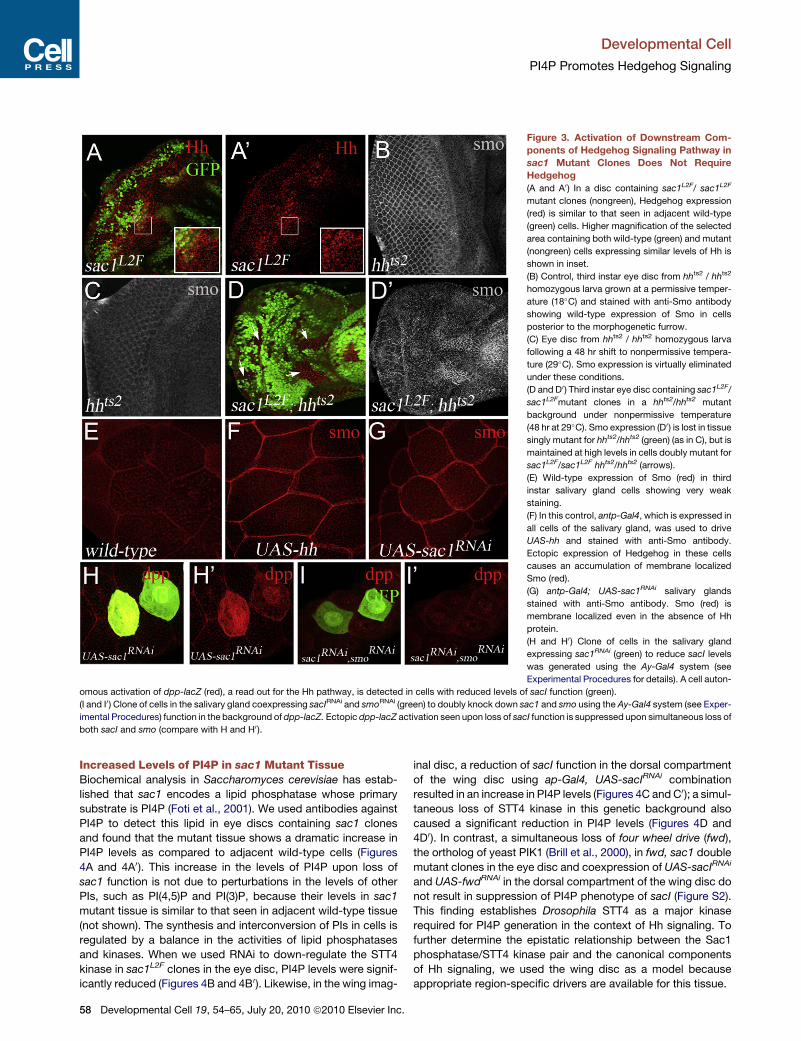
Figure 3. Activation of Downstream Com-
ponents of Hedgehog Signaling Pathway in
sac1 Mutant Clones Does Not Require
Hedgehog
(A and A0) In a disc containing sac1L2F/ sac1L2F
mutant clones (nongreen), Hedgehog expression
(red) is similar to that seen in adjacent wild-type
(green) cells. Higher magnification of the selected
area containing both wild-type (green) and mutant
(nongreen) cells expressing similar levels of Hh is
shown in inset.
(B) Control, third instar eye disc from hhts2 / hhts2
homozygous larva grown at a permissive temper-
ature (18�C) and stained with anti-Smo antibody
showing wild-type expression of Smo in cells
posterior to the morphogenetic furrow.
(C) Eye disc from hhts2 / hhts2 homozygous larva
following a 48 hr shift to nonpermissive tempera-
ture (29�C). Smo expression is virtually eliminated
under these conditions.
(D and D0) Third instar eye disc containing sac1L2F/
sac1L2Fmutant clones in a hhts2/hhts2 mutant
background under nonpermissive temperature
(48 hr at 29�C). Smo expression (D0) is lost in tissuesingly mutant for hhts2/hhts2 (green) (as in C), but is
maintained at high levels in cells doubly mutant for
sac1L2F/sac1L2F hhts2/hhts2 (arrows).
(E) Wild-type expression of Smo (red) in third
instar salivary gland cells showing very weak
staining.
(F) In this control, antp-Gal4, which is expressed in
all cells of the salivary gland, was used to drive
UAS-hh and stained with anti-Smo antibody.
Ectopic expression of Hedgehog in these cells
causes an accumulation of membrane localized
Smo (red).
(G) antp-Gal4; UAS-sac1RNAi salivary glands
stained with anti-Smo antibody. Smo (red) is
membrane localized even in the absence of Hh
protein.
(H and H0) Clone of cells in the salivary gland
expressing sac1RNAi (green) to reduce sacI levels
was generated using the Ay-Gal4 system (see
Experimental Procedures for details). A cell auton-
omous activation of dpp-lacZ (red), a read out for the Hh pathway, is detected in cells with reduced levels of sacI function (green).
(I and I0) Clone of cells in the salivary gland coexpressing sacIRNAi and smoRNAi (green) to doubly knock down sac1 and smo using the Ay-Gal4 system (see Exper-
imental Procedures) function in the background of dpp-lacZ. Ectopic dpp-lacZ activation seen upon loss of sacI function is suppressed upon simultaneous loss of
both sacI and smo (compare with H and H0 ).
Developmental Cell
PI4P Promotes Hedgehog Signaling
Increased Levels of PI4P in sac1 Mutant TissueBiochemical analysis in Saccharomyces cerevisiae has estab-
lished that sac1 encodes a lipid phosphatase whose primary
substrate is PI4P (Foti et al., 2001). We used antibodies against
PI4P to detect this lipid in eye discs containing sac1 clones
and found that the mutant tissue shows a dramatic increase in
PI4P levels as compared to adjacent wild-type cells (Figures
4A and 4A0). This increase in the levels of PI4P upon loss of
sac1 function is not due to perturbations in the levels of other
PIs, such as PI(4,5)P and PI(3)P, because their levels in sac1
mutant tissue is similar to that seen in adjacent wild-type tissue
(not shown). The synthesis and interconversion of PIs in cells is
regulated by a balance in the activities of lipid phosphatases
and kinases. When we used RNAi to down-regulate the STT4
kinase in sac1L2F clones in the eye disc, PI4P levels were signif-
icantly reduced (Figures 4B and 4B0). Likewise, in the wing imag-
58 Developmental Cell 19, 54–65, July 20, 2010 ª2010 Elsevier Inc.
inal disc, a reduction of sacI function in the dorsal compartment
of the wing disc using ap-Gal4, UAS-sacIRNAi combination
resulted in an increase in PI4P levels (Figures 4C and C0); a simul-
taneous loss of STT4 kinase in this genetic background also
caused a significant reduction in PI4P levels (Figures 4D and
4D0). In contrast, a simultaneous loss of four wheel drive (fwd),
the ortholog of yeast PIK1 (Brill et al., 2000), in fwd, sac1 double
mutant clones in the eye disc and coexpression of UAS-sacIRNAi
and UAS-fwdRNAi in the dorsal compartment of the wing disc do
not result in suppression of PI4P phenotype of sacI (Figure S2).
This finding establishes Drosophila STT4 as a major kinase
required for PI4P generation in the context of Hh signaling. To
further determine the epistatic relationship between the Sac1
phosphatase/STT4 kinase pair and the canonical components
of Hh signaling, we used the wing disc as a model because
appropriate region-specific drivers are available for this tissue.

Figure 4. Mutation in sac1 Leads to Increased PI4P
(A–B0) Third instar eye discs. (A and A0) In a third eye disc containing, PI4P (red) levels are significantly up-regulated in sac1L2F/ sac1L2F mutant clone (nongreen).
(B and B0) In sac1L2F / sac1L2F mutant clones that have also lost STT4 function, PI4P (red) levels are significantly reduced as compared to loss of sacI function
alone (compare with A and A0 ).(C andD0) Third instar wing discs. The wing pouch is demarcated by awhite dotted line. sacI function was reduced in the dorsal compartment of thewing disc (that
expresses Apterous; see schematic in Figure 5A) by a combination of ap-Gal4, UAS-GFP; UAS-sac1RNAi (green).
(C and C0) PI4P (red) levels are significantly increased in the green dorsal compartment (C0) compared to the adjacent nongreen tissue that corresponds to the
ventral compartment.
(D and D0) A simultaneous loss of STT4 kinase and sacI in the dorsal compartment of the wing disc (green) causes PI4P (red) levels to be significantly reduced as
compared to loss of sacI function alone (compare with C and C0 ).
Developmental Cell
PI4P Promotes Hedgehog Signaling
In the third instar wing imaginal disc, Hh is expressed in cells of
theposterior compartment andactivates the signalingcascade in
the anterior compartment in a graded fashion (Figure 5A). Ci, the
transcriptional effector of Hh signaling, is expressed in all cells of
the anterior compartment, whereas the transcription of Dpp,
which requires high levels of Hh signaling, is seen in a narrow
stripe of cells adjacent to the A/P boundary (Figure 5B). Smo, in
contrast, accumulates in all cells of the posterior compartment
(Figure 5B). Reduction of sac1 function using ap-gal4, a dorsal
compartment-specific driver in thewing disc, using the combina-
tion ap-gal4, UAS-sac1RNAi results in an expansion of the region
in which Smo protein accumulates to include the entire dorsal
compartment including the anterior half (Figures 5C and 5C0).This increase in Smo localization on the membrane is not due
to an increase in the transcription of smo because quantitative
PCR comparison between ap-gal4, UAS-sac1RNAi and its sibling
control shownodifference in the level of smo transcripts (data not
shown). Therefore, as in theeyedisc, lossof sac1 function causes
increased membrane localization of Smo. Reduction of the
STT4 kinase function in the dorsal compartment using ap-gal4,
UAS-STT4RNAi causes reduced Smo protein accumulation in
the posterior half of the dorsal compartment (Figures 5D and
5D0). A simultaneous reduction of STT4 and sac1 function was
achieved in the dorsal compartment of the wing imaginal
disc using the combined genotype ap-gal4, UAS-sac1RNAi,
UAS-STT4 RNAi. This causes a reduction in Smo protein accumu-
lation in the posterior half of the dorsal compartment (Figures 5E
and 5E0), a phenotype identical to that seen upon loss of STT4
function (Figures 5D and 5D0). These results strongly argue for
a requirement of the STT4 kinase that gives rise to PI4P in the
membrane localization of Smo and the activation of the pathway.
D
Finally, loss of STT4 function in the dorsal compartment of the
wing disc causes a strong reduction in the expression of Ptc,
a Hh target gene (Figures 5F–5G0) and to a lesser extent the
expression of activated Ci in the dorsal-anterior compartment
indicating an attenuation of Hh signaling (Figures S3A and S3B).
The Ptc protein plays perhaps the most prominent role in the
regulation of Smo membrane localization during Hh signaling
(Jiang and Hui, 2008; Zhu et al., 2003). For example, overexpres-
sion of Ptc in the dorsal compartment of the wing disc using
the genetic combination ap-gal4, UAS-ptc causes a loss of
membrane associated Smo in the posterior half of the dorsal
compartment (Figures 5H and 5H0). We show that overexpres-
sion of Ptc can also override the Smo membrane localization
phenotype and PI4P accumulation seen as a result of down-
regulation of sacI (Figures 5I and 5I0; Figures S3C–S3F). Thus,
ptc functions along with sac1 in the regulation of membrane
localization of Smo.
In wild-type third instar wing disc, dpp-lacZ expression is
dependent on high levels of Hh signaling and is seen as a stripe
of cells in the anterior compartment along the A/P boundary
(Figure 5B). This expression is expanded in the dorsal-anterior
compartment when sac1 function is reduced in the dorsal
compartment of the wing disc (Figures 5J and 5J0) indicatingincreased Hh signaling in cells that normally respond to Hh.
However, a simultaneous loss of STT4 and sac1 in the dorsal
compartment of the wing disc prevents this expansion of dpp-
lacZ expression and causes a suppression of the sac1 pheno-
type (Figures 5K and 5K0). In this combination, lower sac1 levels
would increase PI4P levels, but the STT4 kinase is required for
the generation of PI4P in the first place. Thus, the simultaneous
block of STT4 and sac1 will cause a reduction in PI4P leading to
evelopmental Cell 19, 54–65, July 20, 2010 ª2010 Elsevier Inc. 59

Figure 5. sac1 Phosphatase and stt4 PI4 Kinase Cause Elevated Membrane Levels of Smoothened Protein
The wing pouch is marked by a dotted line. Red channel only shown in gray scale in (C0), (D0), (E0), (F0), and (G0) for clarity.(A) A schematic representation of the third instar wing disc. The wing pouch is marked by white dotted line. The A/P compartment boundary (A/P) demarcates the
anterior and posterior compartments. The D/V compartment (D/V) boundary demarcates the dorsal and the ventral compartments. ap-Gal4 is expressed
throughout the dorsal compartment (shown in blue). Smo is expressed in the posterior compartment of the pouch (red dots)
(B) In wild-type, Smo (red) is expressed in the posterior compartment, whereas the Hh target reporter dpp-lacZ (green) is activated along the A/P compartment
boundary.
(C andC0) Knockdownof sac1 in the dorsal compartment of thewing disc using the combination ap-Gal4, UAS-GFP; UAS-sac1RNAi (green cells), causes Smo (red)
membrane localization to expand to the dorsal-anterior compartment of the wing (arrow) (compare with B). Red channel only shown in gray scale for clarity (C0 ).(D andD0) Knockdownof stt4 kinase in thedorsal compartment of thewingdisc using thecombination ap-Gal4,UAS-stt4RNAi UAS-GFP (green) is shown. Smo (red)
membrane localization is reduced in the dorsal posterior compartment (arrow). Red channel is only shown in gray scale for clarity (D0).(E and E0) Double knockdown of sac1 and stt4 in the dorsal compartment of the wing disc using ap-Gal4, UAS-GFP; UAS-stt4RNAi; UAS-sac1RNAi (green) genetic
combinations is shown. Smo (red) accumulation in thedorsal anterior compartment (arrow) anddorsal posterior compartment (arrowhead) is significantly reduced.
Red channel is only shown in gray scale for clarity (E0).(F andF0) As a control,wild-typePtc (red) is expressedalong theA/Pboundary at similar levels in cells of thedorsal (green) and ventral (nongreen) regionsof thewing
pouch.
(G and G0) Knockdown of stt4 kinase in the dorsal (green) compartment of the wing disc using the genetic combination ap-Gal4, UAS-stt4RNAiUAS-GFP causes
significant reduction in Ptc (red) expression.
Developmental Cell
PI4P Promotes Hedgehog Signaling
60 Developmental Cell 19, 54–65, July 20, 2010 ª2010 Elsevier Inc.

Developmental Cell
PI4P Promotes Hedgehog Signaling
a reduction in dpp-lacZ expression, a readout for Hh signaling.
These results link phospholipid metabolism with the ability of
Smo to signal and activate Hh signaling. To directly test whether
PI4P levels increase upon activation of Hh signaling, we once
again used the salivary gland as a model. In wild-type, salivary
gland cells contain low PI4P that increases dramatically when
sac1 is down-regulated (Figures 6A–6A00). Likewise, activation
of Hh signaling by removing ptc in flip-out clones using the
Ay-gal4 system in the salivary gland (see Experimental Proce-
dures) using the combination Ay-Gal4 UAS-ptcRNAi, which
causes a loss of ptc expression in cells expressingGal4 (marked
by GFP) and causes a cell-autonomous increase in its PI4P
content (Figures 6B–6B00). Similarly, an increase in PI4P content
is seen when ptc function is attenuated in the dorsal compart-
ment of the wing imaginal disc using the combination ap-gal4,
UAS-ptcRNAi or when mutant clones of ptc are analyzed in the
eye imaginal disc (Figures S3G and S3H). The increase in PI4P
levels upon loss of ptc function is dependent on STT4 function
because simultaneous loss of ptc and STT4 function in the
combination ap-gal4, UAS-ptcRNAi and UAS-SST4RNAi results
in the suppression of the phenotype (Figures S3I–S3L). This
establishes a causal relationship between the strength of the
Hh signal and the level PI4P. Additionally, the change in level
of PI4P is not a feedback from a posttranscriptional target of
Hh signaling because overexpression of activated Ci in the
dorsal compartment does not result in an increase in PI4P levels,
nor does a simultaneous loss of ptc and smo in the dorsal
compartment of the wing disc using the combination ap-gal4,
UAS-ptcRNAi, UAS-smoRNAi suppress the increased levels of
PI4P (Figures S3M–S3P).
STT4 and Sac1 Are Conserved Componentsof the Hedgehog Signaling PathwayResults presented thus far suggest that STT4 functions as a
positive modulator of Hh signaling, whereas Sac1 is required
to attenuate this signal. To test whether loss of STT4 and sac1
in the embryo results in the signature hedgehog segment polarity
phenotypes, we microinjected hairpin versions of STT4 RNA
(because no loss of function alleles are available) and for sac1,
we isolated homozygous loss of function sac1L2F/ sac1L2F
mutants and monitored their denticles belt phenotypes. In
wild-type late stage embryos, there are 14 denticles belts with
a distinct segmental polarity pattern (Figures 6C–6C0). As posi-
tive controls, microinjection of a short hairpin version of Smo
RNA exhibits overspecification of denticle belts and disruption
of polarity (Figures 6D and 6D0), and Ptc mutant embryos show
reduction in the denticle belts (Hooper and Scott, 1989). sac1L2F
homozygous mutant embryos show a reduction in the width of
the denticle belts, a phenotype similar to that seen in ptc, indic-
(H and H0) Overexpression of Ptc in the dorsal compartment of the wing disc usin
mulation is significantly reduced in the dorsal posterior compartment (arrow). Not
channel. Red channel is only shown in gray scale for clarity (H0).(I and I0) Overexpression of Ptc in a sac1 knockdown background in the dorsal co
ization in the posterior dorsal compartment is reduced (compare with C) and C0).(J and J0 ) sac1 knockdown in the dorsal compartment of the wing disc using ap-G
Ectopic activation of dpp-lacZ (red) is seen the dorsal anterior compartment (comp
the A/P boundary and lower near the distal edges of the pouch.
(K and K0) Double knockdown of sac1 and stt4 in the dorsal compartment of thewi
(green) is shown. Ectopic dpp-lacZ as a read out for activation of the Hh pathway
D
ative of increased Hh signaling (Figures 6E and 6E0), whereas
loss of STT4 results in overspecification of denticle belts (Fig-
ures 6F and 6F0), a phenotype similar to that seen in hh and
smo mutant embryos (compare Figures 6F and 6F0 with Fig-
ures 6D and 6D0), further establishing that STT4 functions as
a positive modulator of Hh signaling. Because of the nature of
feedback loop between Hh and Wg at the segmental boundary,
this effect of sac1/STT4 on Hh is likely to trigger a change in
Wg levels as well.
Many aspects of Hedgehog signal transduction, though not all,
are evolutionarily conserved between Drosophila and mammals.
We explored the possibility that PI4P signaling modulates Hh
signal transduction in mammalian cells. The mouse genome
encodes an ortholog of STT4, called PIKIII kinase a, as estab-
lished by domain architecture and catalytic domain primary
sequence alignment. A second mouse kinase, PI4III kinase b,
has a domain architecture that appears to be more closely
related to PIK1 (Balla and Balla, 2006). The effect of kinase
subunit RNA interference was tested in Shh-LIGHT2 cells, a
mouse fibroblast cell line engineered to possess aGli-dependent
luciferase-based reporter system (Taipale et al., 2000). Treat-
ment of these cells with Shh gives robust (>10-fold) increases
in reporter activity. In a control experiment, Shh-LIGHT2 cells
were treated with RNAi that inactivates smo RNA. Loss of Smo
function reduced Shh-induced reporter activation, demon-
strating the effectiveness of this system in measuring effects
on Hh pathway transduction (Figure 6G). Next, we tested RNAi
treatments designed to reduce kinase functions. RNAi against
PI3 kinase or PI4II kinases did not affect Shh-driven reporter
induction to a significant extent. In contrast RNAi against the
mammalian STT4 homolog, PI4III kinase a, strongly reduced
Shh-stimulated reporter activity (Figure 6G). RNAi against the
mammalian PIK1 homolog, PI4PIII kinase b, also affected
reporter induction in these cells. The RNAi sequences used
were each highly specific for the kinase target, so the results
are unlikely to be explained by cross-reactivity. Taken together,
these results suggest a nonredundant role for PI4III kinases in
mammalian Hh pathway transduction. Note that RNAi against
the mammalian ortholog of Sac1 did not affect reporter activity,
either in basal or Shh-stimulated conditions (data not shown).
However, Shh-LIGHT2 cells are relatively insensitive to Hh
pathway derepression caused by the removal of negative regula-
tors such as Suppressor of Fused (data not shown).
DISCUSSION
A major regulatory step in the modulation of Hedgehog signaling
occurs at the level of the twomultipass transmembrane proteins,
Patched and Smoothened. Genetic and biochemical studies
g the combination ap-gal4, UAS-ptc-YFP (green). Smo (red) membrane accu-
e that the fully functional Ptc-YFP fusion can be directly visualized in the green
mpartment of the wing disc using ap-gal4 (green). Smo (red) membrane local-
al4, UAS-GFP; UAS-sac1RNAi (green) in the background of dpp-lacZ is shown.
arewithB). It is currently unclear why the expression of dpp-lacZ is highest near
ng disc using the combination ap-Gal4, UAS-GFP/ UAS-stt4RNAi; UAS-sac1RNAi
seen upon loss of sacI alone is suppressed (compare with J and J0).
evelopmental Cell 19, 54–65, July 20, 2010 ª2010 Elsevier Inc. 61
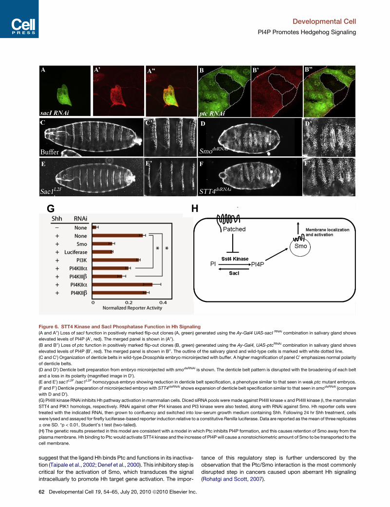
Figure 6. STT4 Kinase and SacI Phosphatase Function in Hh Signaling
(A and A00) Loss of sacI function in positively marked flip-out clones (A, green) generated using the Ay-Gal4 UAS-sacI RNAi combination in salivary gland shows
elevated levels of PI4P (A0, red). The merged panel is shown in (A00).(B and B00) Loss of ptc function in positively marked flip-out clones (B, green) generated using the Ay-Gal4, UAS-ptcRNAi combination in salivary gland shows
elevated levels of PI4P (B0, red). The merged panel is shown in B00. The outline of the salivary gland and wild-type cells is marked with white dotted line.
(C and C0) Organization of denticle belts in wild-type Drosophila embryo microinjected with buffer. A higher magnification of panel C0 emphasizes normal polarity
of denticle belts.
(D and D0) Denticle belt preparation from embryo microinjected with smodsRNAi is shown. The denticle belt pattern is disrupted with the broadening of each belt
and a loss in its polarity (magnified image in D0 ).(E and E0) sac1L2F /sac1L2F homozygous embryo showing reduction in denticle belt specification, a phenotype similar to that seen in weak ptcmutant embryos.
(F and F0 ) Denticle preparation of microinjected embryo with STT4dsRNAi shows expansion of denticle belt specification similar to that seen in smodsRNAi (compare
with D and D0).(G) PI4III kinase RNAi inhibits Hh pathway activation in mammalian cells. Diced siRNA pools were made against PI4III kinase a and PI4III kinase b, the mammalian
STT4 and PIK1 homologs, respectively. RNAi against other PI4 kinases and PI3 kinase were also tested, along with RNAi against Smo. Hh reporter cells were
treated with the indicated RNAi, then grown to confluency and switched into low-serum growth medium containing Shh. Following 24 hr Shh treatment, cells
were lysed and assayed for firefly luciferase-based reporter induction relative to a constitutiveRenilla luciferase. Data are reported as themean of three replicates
± one SD. *p < 0.01, Student’s t test (two-tailed).
(H) The genetic results presented in this model are consistent with a model in which Ptc inhibits PI4P formation, and this causes retention of Smo away from the
plasmamembrane. Hh binding to Ptc would activate STT4 kinase and the increase of PI4P will cause a nonstoichiometric amount of Smo to be transported to the
cell membrane.
Developmental Cell
PI4P Promotes Hedgehog Signaling
suggest that the ligand Hh binds Ptc and functions in its inactiva-
tion (Taipale et al., 2002; Denef et al., 2000). This inhibitory step is
critical for the activation of Smo, which transduces the signal
intracelluarly to promote Hh target gene activation. The impor-
62 Developmental Cell 19, 54–65, July 20, 2010 ª2010 Elsevier Inc.
tance of this regulatory step is further underscored by the
observation that the Ptc/Smo interaction is the most commonly
disrupted step in cancers caused upon aberrant Hh signaling
(Rohatgi and Scott, 2007).

Developmental Cell
PI4P Promotes Hedgehog Signaling
In this article, we show that phospholipid metabolism plays an
important role in the modulation of Hh signaling at the level of
Ptc/Smo interaction. In particular, our results show that an
increase in the level of PI4P by the inactivation of Sac1 phospha-
tase leads to Smo protein relocalization to the membrane and an
increase in Hh signaling in multiple tissues during Drosophila
development. Furthermore the kinase (STT4), which is required
for the generation of PI4P, is also required for the proper trans-
duction of Hh signaling as indicated by its effects on Hh target
gene expression. PI4P accumulation in the cell is a hallmark of
sac1 mutations and is also seen upon loss of ptc activity.
Furthermore, in sac1 mutant tissue, we find both increased
membrane localization of Smo and accumulation of PI4P,
whereas reduction in the PI4P kinase function leads to an hh-like
loss of function phenotype. These results establish that phos-
pholipid metabolism provides a critical regulatory input in the
modulation of Hh signaling.
Recent studies have proposed that Smo activation requires an
input from a nonprotein small molecule. Cholesterol and its
derivatives (oxysterols) are likely candidates for the small mole-
cules required directly or indirectly for Ptc inhibition or Smo acti-
vation, because they also promote the translocation of Smo to
the cilium (Dwyer et al., 2007). Because oxysterols are known
to bind to vesicular transport proteins that also interact with
phospholipids (Xu et al., 2001), further studies on possible coop-
eration between these two lipid types could further shed light the
mechanism of Smo activation.
Inactivation of Smo by Ptc occurs in a catalytic fashion
(Taipale et al., 2002) in that a small number of Ptc molecules
can inactivate many more Smo molecules. Our results provide
an explanation for this nonstoichiometric inhibitory mechanism.
The finding that inactivation of Ptc increases PI4P suggests
that Ptc normally functions in keeping PI4P levels low within
a cell. This could be achieved either by the down-regulation of
the STT4 kinase or by the up-regulation of the Sac1 phospha-
tase. It is less likely that Ptc modulates Sac1 activity because
in vivo localization studies in multiple models system have
shown that Sac1 is predominantly localized to the Golgi and,
as a result of proximity arguments alone, it seems a more likely
possibility that Ptc modulates PI4P levels by down-regulating
the lipid kinase. In this model, during normal Hh signaling,
binding of Hh to Ptc will relieve repression of the kinase by Ptc
and cause an increase in PI4P. As with all genetic analysis in
Drosophila, our results do not imply direct protein interactions;
currently unknown transduction components could exist, and
future biochemical analyses will reveal which, if any, of the inter-
actions is direct. However, our genetic analysis does allow us to
propose how an increase in the levels of this lipid can activate Hh
signaling. Studies from both flies and vertebrate model system
have suggested that the localization of Smo protein to the
plasma membrane is essential for the activation of the pathway,
and studies in multiple model systems have shown that PI4P
function is essential in the vesicular transport of cargo proteins
from the Golgi to the plasma membrane (Skwarek and Bou-
lianne, 2009). We therefore propose that Hh binding to Ptc
releases inhibition of a lipid kinase such as STT4, resulting in
high PI4P levels. This aids vesicular transport of Smo to the
membrane and causes its activation. A schematic representing
the genetic model that is consistent with past and present data
D
is shown in Figure 6H. Our results using Shh-responsive mouse
fibroblasts indicate that mammalian Hh signal transduction is
dependent on the activity of the murine STT4 ortholog, PI4III
kinase a. Previous localization studies suggest PI4III kinase
a contributes to plasma membrane PI4P pools, an observation
consistent with a conserved role for PI4P metabolites in the
control of Smo by mammalian Ptc1 (Balla et al., 2005; Wong
et al., 1997). The observation that RNAi against the mammalian
PIK1 homolog, PI4III kinase b, also reduces Hh signal transduc-
tion could suggest it has diverged in function between flies and
mammals. Alternatively, PI4P pools could be exchanged more
readily between membrane-bound subcellular compartments
and the cell surface in mammalian cells, making the removal of
either of the PI4III kinases affect global availability of PI4P deriv-
atives. In mammalian cells, Smo activation is associated with
translocation of the molecule to the primary cilium, a ubiquitous
microtubule-based cell surface protrusion (Corbit et al., 2005;
Huangfu et al., 2003). Given that Drosophila cells appear to
lack primary cilia, it will be of interest to determine whether PI4III
kinase activity is required for Smo translocation.
EXPERIMENTAL PROCEDURES
Fly Stocks
The following stocks were used in this study: FRT40A and FRT80B (Blooming-
ton), w; msn172 P{neoFRT}80B/TM6B (J. Treisman), yw; msn j1E2/TM3, Sb
(Y. N. Jan), sac1L2F (H. Wei), sac1BG02228(Bloomington), bsk1 /Cyo (M. Seeger),
UAS-DIAP1 (B. Hay), Pk61CEP837 (Szeged), hhts2 (K. Moses), dpp-lacZ (Bloo-
mington), UAS-sac1RNAi (VDRC 37217), UAS-stt4RNAi (VDRC 15993), UAS-
fwdRNAi (VDRC 27785), fwdEY05397 (Bloomington), puc-lacZ (Bloomington),
UAS-ptcRNAi (NIG-FLY 2411R-1), UAS-smoRNAi (NIG-FLY 1156R-1), UAS-
ptc-yfp, UAS-ci (K.Basler), UAS-hh (P. Ingham), and Ay-Gal4 (Bloomington).
Immunohistochemistry and Embryonic ds-RNA Injections
We used the following antibodies: mouse anti-b-galactosidase (1:100; Prom-
ega); rat anti-Ci (1:50; R. Holmgren); rabbit anti–cleaved caspase-3 (1:200;
Cell Signaling); rabbit anti-Hh (1:1,000 ; Ingham,P.); mouse anti-Ptc andmouse
anti-Smo, anti-Crumbs, anti-Dl, and anti-Notch (1:50; 1:20, 1:100, 1:20, and
1:20; Hybridoma Bank, Iowa); mouse anti-PI4P (1:100; Echelon); mouse
anti-Wg (1:100;Hybridoma); and rabbit anti-phosphoJNK (1:50;Cell Signaling).
Antibody stainingwere performed as described elsewhere (Rogge et al., 1995),
except that anti-PI4P staining (Blagoveshchenskaya et al., 2002) was per-
formed using .3% Saponin in 1X TBS (Sigma S7900) during washes and
antibody incubation. ds-RNA and denticle preparation experiments were per-
formed as described elsewhere (Kennerdell and Carthew, 2000).
Ay-Gal4 Flip-Out Clones
Flip-out clones were generated in the eye and in the salivary gland. In the eye
discs, ey-flp was used to flip out the stuffer cassette from act-FRTyFRT-Gal4
to generate act FRTGal4, which expresses Gal4 in all cells of the eye imaginal
disc (Ito et al., 1997). In the salivary gland, hs-flp was used as a source for flp.
The respective crosses were maintained at 18�C; when the larvae reached
midsecond instar, a brief heat shock (10 min) was given at 37�C, and when
the larvae reached third instar, salivary glands were dissected out, fixed,
and stained with appropriate antibodies.
Imaging
Samples were imaged using a BioRad Radiance 2000 confocal with Laser-
Sharp 2000 acquisition software. Fluorescent intensity quantifications were
analyzed by use of ImageJ software.
Shh Reporter Assay
Diced siRNA pools were generated using previously described methods
(Myers et al., 2003). Gene-specific PCR primers were used to amplify
evelopmental Cell 19, 54–65, July 20, 2010 ª2010 Elsevier Inc. 63
Developmental Cell
PI4P Promotes Hedgehog Signaling
�550 bp segments from the coding region of each of the gene indicated in
Table S1. A second round of PCR was used to attach forward and reverse
T7 polymerase binding sites to the gene-specific PCR products. This DNA
was used as template for the production of long dsRNA using in vitro transcrip-
tion (Ambion). The dsRNA products were processed into �21-bp fragments
using recombinant RNaseIII enzyme, resulting in diced siRNA pools directed
against each target gene (NEB).
To conduct mammalian Hh pathway reporter assays, diced siRNA pools
were introduced into Shhh-LIGHT2 cells using a reverse transfection proce-
dure. For each well of a 96-well plate, Lipofectamine 2000 (Invitrogen) was
complexed with 10 pmol of siRNAs in 50 ml of Opti-MEM (GIBCO) in individual
wells. Early-passage Shh-LIGHT2 cells in log-phase growth were trypsinized,
counted, and adjusted to a concentration of 200 cells/ml. One hundred micro-
liters of culturemedium containing 23 104 cells was gently added to eachwell,
atop the transfection mixture. One day after transfection, the culture medium
was changed to DMEM containing 0.5% FBS with or without the addition of
Shh-conditioned medium. After 24 hr incubation with Shh-conditioned
medium, cells were lysed and luciferase signals were read using the Dual
Luciferase Reporter Assay System (Promega). Data are reported as ratios of
Hh-dependent firefly luciferase signal to constitutive Renilla luciferase signal.
SUPPLEMENTAL INFORMATION
Supplemental information includes three figures and one table and can be
found with this article online at doi:10.1016/j.devcel.2010.06.007.
ACKNOWLEDGMENTS
This project was initiated as an undergraduate research project in the HHMI
Professors program. A.Y., A.F., and K.N are undergraduate research students
in the UCLA Undergraduate Research Consortium in Functional Genomics,
which is supported by a Howard Hughes Medical Institute Professor’s Award
to U.B. We would like to acknowledge the contribution of Emil Kohan and
Ji-Eun Lee to early parts of this project. We thank members of the Banerjee
laboratory for helpful suggestions during the course of this work. R.N and
G.C are instructors in the UCLA Undergraduate Research Consortium in Func-
tional Genomics. T.H. was supported by a Stanford Bio-X fellowship. M.P.S. is
an Investigator of the Howard HughesMedical Institute.Wewould like to thank
J. Treisman, H. Wei, K. Moses, P. Ingham, M. Seeger, A. J. Zhu, K. Basler, and
the stock centers of Bloomington, Szeged, NIG (Tokyo), and VDRC (Vienna) for
providing Drosophila stocks and reagents. U.B. is supported by National
Institutes of Health grants RO1EY008152 and R01HL067395.
Received: November 18, 2009
Revised: March 26, 2010
Accepted: May 4, 2010
Published: July 19, 2010
REFERENCES
Alcedo, J., Ayzenzon, M., Von Ohlen, T., Noll, M., and Hooper, J.E. (1996). The
Drosophila smoothened gene encodes a seven-pass membrane protein,
a putative receptor for the hedgehog signal. Cell 86, 221–232.
Audhya, A., and Emr, S.D. (2002). Stt4 PI 4-kinase localizes to the plasma
membrane and functions in the Pkc1-mediated MAP kinase cascade. Dev.
Cell 2, 593–605.
Audhya, A., Foti, M., and Emr, S.D. (2000). Distinct roles for the yeast phospha-
tidylinositol 4-kinases, Stt4p and Pik1p, in secretion, cell growth, and organelle
membrane dynamics. Mol. Biol. Cell 11, 2673–2689.
Balla, A., and Balla, T. (2006). Phosphatidylinositol 4-kinases: old enzymes
with emerging functions. Trends Cell Biol. 16, 351–361.
Balla, A., Tuymetova, G., Tsiomenko, A., Varnai, P., and Balla, T. (2005).
A plasma membrane pool of phosphatidylinositol 4-phosphate is generated
by phosphatidylinositol 4-kinase type-III alpha: studies with the PH domains
of the oxysterol binding protein and FAPP1. Mol. Biol. Cell 16, 1282–1295.
Beachy, P.A., Karhadkar, S.S., and Berman, D.M. (2004). Mending and malig-
nancy. Nature 431, 402.
64 Developmental Cell 19, 54–65, July 20, 2010 ª2010 Elsevier Inc.
Blagoveshchenskaya, A.D., Hannah, M.J., Allen, S., and Cutler, D.F. (2002).
Selective and signal-dependent recruitment of membrane proteins to secre-
tory granules formed by heterologously expressed von Willebrand factor.
Mol. Biol. Cell 13, 1582–1593.
Blero, D., Payrastre, B., Schurmans, S., and Erneux, C. (2007). Phosphoinosi-
tide phosphatases in a network of signalling reactions. Pflugers Arch. 455,
31–44.
Brill, J.A., Hime, G.R., Scharer-Schuksz, M., and Fuller, M.T. (2000). A phos-
pholipid kinase regulates actin organization and intercellular bridge formation
during germline cytokinesis. Development 127, 3855–3864.
Corbit, K.C., Aanstad, P., Singla, V., Norman, A.R., Stainier, D.Y., and Reiter,
J.F. (2005). Vertebrate Smoothened functions at the primary cilium. Nature
437, 1018–1021.
Denef, N., Neubuser, D., Perez, L., andCohen, S.M. (2000). Hedgehog induces
opposite changes in turnover and subcellular localization of patched and
smoothened. Cell 102, 521–531.
Dwyer, J.R., Sever, N., Carlson, M., Nelson, S.F., Beachy, P.A., and Parhami,
F. (2007). Oxysterols are novel activators of the hedgehog signaling pathway in
pluripotent mesenchymal cells. J. Biol. Chem. 282, 8959–8968.
Eaton, S. (2008). Multiple roles for lipids in the Hedgehog signalling pathway.
Nat. Rev. Mol. Cell Biol. 9, 437–445.
Flanagan, C.A., Schnieders, E.A., Emerick, A.W., Kunisawa, R., Admon, A.,
and Thorner, J. (1993). Phosphatidylinositol 4-kinase: gene structure and
requirement for yeast cell viability. Science 262, 1444–1448.
Foti, M., Audhya, A., and Emr, S.D. (2001). Sac1 lipid phosphatase and Stt4
phosphatidylinositol 4-kinase regulate a pool of phosphatidylinositol 4-phos-
phate that functions in the control of the actin cytoskeleton and vacuole
morphology. Mol. Biol. Cell 12, 2396–2411.
Godi, A., Di Campli, A., Konstantakopoulos, A., Di Tullio, G., Alessi, D.R., Kular,
G.S., Daniele, T., Marra, P., Lucocq, J.M., and De Matteis, M.A. (2004). FAPPs
control Golgi-to-cell-surface membrane traffic by binding to ARF and PtdIns(4)
P. Nat. Cell Biol. 6, 393–404.
Hooper, J.E., and Scott, M.P. (1989). The Drosophila patched gene encodes
a putative membrane protein required for segmental patterning. Cell 59,
751–765.
Huangfu, D., Liu, A., Rakeman, A.S., Murcia, N.S., Niswander, L., and Ander-
son, K.V. (2003). Hedgehog signalling in the mouse requires intraflagellar
transport proteins. Nature 426, 83–87.
Igaki, T., Kanda, H., Yamamoto-Goto, Y., Kanuka, H., Kuranaga, E., Aigaki, T.,
and Miura, M. (2002). Eiger, a TNF superfamily ligand that triggers the
Drosophila JNK pathway. EMBO J. 21, 3009–3018.
Ito, K., Awano, W., Suzuki, K., Hiromi, Y., and Yamamoto, D. (1997). The
Drosophila mushroom body is a quadruple structure of clonal units each of
which contains a virtually identical set of neurones and glial cells. Development
124, 761–771.
Jia, J., Tong, C., Wang, B., Luo, L., and Jiang, J. (2004). Hedgehog signalling
activity of Smoothened requires phosphorylation by protein kinase A and
casein kinase I. Nature 432, 1045–1050.
Jiang, J., and Hui, C.C. (2008). Hedgehog signaling in development and
cancer. Dev. Cell 15, 801–812.
Kennerdell, J.R., and Carthew, R.W. (2000). Heritable gene silencing in
Drosophila using double-stranded RNA. Nat. Biotechnol. 18, 896–898.
Myers, J.W., Jones, J.T., Meyer, T., and Ferrell, J.E., Jr. (2003). Recombinant
Dicer efficiently converts large dsRNAs into siRNAs suitable for gene silencing.
Nat. Biotechnol. 21, 324–328.
Rogge, R., Green, P.J., Urano, J., Horn-Saban, S., Mlodzik, M., Shilo, B.Z.,
Hartenstein, V., andBanerjee, U. (1995). The role of yan inmediating the choice
between cell division and differentiation. Development 121, 3947–3958.
Rohatgi, R., Milenkovic, L., and Scott, M.P. (2007). Patched1 regulates
hedgehog signaling at the primary cilium. Science 317, 372–376.
Rohatgi, R., and Scott, M.P. (2007). Patching the gaps in Hedgehog signalling.
Nat. Cell Biol. 9, 1005–1009.

Developmental Cell
PI4P Promotes Hedgehog Signaling
Ruiz i Altaba, A., Sanchez, P., and Dahmane, N. (2002). Gli and hedgehog in
cancer: tumours, embryos and stem cells. Nat. Rev. Cancer 2, 361–372.
Ryoo, H.D., Gorenc, T., and Steller, H. (2004). Apoptotic cells can induce
compensatory cell proliferation through the JNK and the Wingless signaling
pathways. Dev. Cell 7, 491–501.
Skwarek, L.C., and Boulianne, G.L. (2009). Great expectations for PIP: phos-
phoinositides as regulators of signaling during development and disease.
Dev. Cell 16, 12–20.
Stone, D.M., Hynes, M., Armanini, M., Swanson, T.A., Gu, Q., Johnson, R.L.,
Scott, M.P., Pennica, D., Goddard, A., Phillips, H., et al. (1996). The tumour-
suppressor gene patched encodes a candidate receptor for Sonic hedgehog.
Nature 384, 129–134.
Taipale, J., Chen, J.K., Cooper, M.K., Wang, B., Mann, R.K., Milenkovic, L.,
Scott, M.P., and Beachy, P.A. (2000). Effects of oncogenic mutations in
Smoothened and Patched can be reversed by cyclopamine. Nature 406,
1005–1009.
Taipale, J., Cooper, M.K., Maiti, T., and Beachy, P.A. (2002). Patched acts
catalytically to suppress the activity of Smoothened. Nature 418, 892–897.
D
van den Heuvel, M., and Ingham, P.W. (1996). smoothened encodes
a receptor-like serpentine protein required for hedgehog signalling. Nature
382, 547–551.
Walch-Solimena, C., and Novick, P. (1999). The yeast phosphatidylinositol-4-
OH kinase pik1 regulates secretion at the Golgi. Nat. Cell Biol. 1, 523–525.
Wei, H.C., Sanny, J., Shu, H., Baillie, D.L., Brill, J.A., Price, J.V., and Harden, N.
(2003). The Sac1 lipid phosphatase regulates cell shape change and the JNK
cascade during dorsal closure in Drosophila. Curr. Biol. 13, 1882–1887.
Wong, K., Meyers, R., and Cantley, L.C. (1997). Subcellular locations of phos-
phatidylinositol 4-kinase isoforms. J. Biol. Chem. 272, 13236–13241.
Xu, Y., Liu, Y., Ridgway, N.D., and McMaster, C.R. (2001). Novel members of
the human oxysterol-binding protein family bind phospholipids and regulate
vesicle transport. J. Biol. Chem. 276, 18407–18414.
Yoshida, S., Ohya, Y., Goebl, M., Nakano, A., and Anraku, Y. (1994). A novel
gene, STT4, encodes a phosphatidylinositol 4-kinase in the PKC1 protein
kinase pathway of Saccharomyces cerevisiae. J. Biol. Chem. 269, 1166–1172.
Zhu, A.J., Zheng, L., Suyama, K., and Scott, M.P. (2003). Altered localization of
Drosophila Smoothened protein activates Hedgehog signal transduction.
Genes Dev. 17, 1240–1252.
evelopmental Cell 19, 54–65, July 20, 2010 ª2010 Elsevier Inc. 65


![Welcome [med.stanford.edu]med.stanford.edu/.../aboutus/2017-MIPS-brochure.pdf · via sound (ultrasound, photoacoustic), magnetism (MRI or magnetic resonance imaging, MPI or magnetic](https://img.pdfslide.net/doc/110x75/5f0c64747e708231d4352ce6/welcome-med-med-via-sound-ultrasound-photoacoustic-magnetism-mri-or-magnetic.jpg)