Embed Size (px)
Citation preview
Role of lymphotoxin-a in cigarette smoke-
induced inflammation and lymphoid
neogenesisT. Demoor*, K.R. Bracke*, T. Maes*, B. Vandooren#, D. Elewaut#, C. Pilette",G.F. Joos* and G.G. Brusselle*
ABSTRACT: In chronic obstructive pulmonary disease (COPD), chronic inflammation is
accompanied by peribronchial lymphoid aggregates. Lymphotoxin (LT)-a, crucial in secondary
lymphoid organogenesis, may be involved in lymphoid neogenesis.
We examined cigarette smoke (CS)-induced pulmonary lymphoid neogenesis and inflammation
in vivo in LTa knockout (LTa-/-) and wild-type (WT) mice and studied the expression of lymphoid
chemokines by lung fibroblasts in vitro.
T-cell numbers (in bronchoalveolar lavage fluid (BALF) and lungs) and lymphoid aggregate
numbers were significantly higher in air-exposed LTa-/- mice than in WT animals, and increased
upon chronic CS exposure in both genotypes. In contrast, local immunoglobulin A responses
upon chronic CS exposure were attenuated in LTa-/- mice. CXC chemokine ligand (CXCL) 13 and
CC chemokine ligand (CCL) 19 mRNA in total lung and CXCL13 protein level in BALF increased
upon CS exposure in WT, but not in LTa-/- mice. In vitro lymphotoxin-b receptor (LTbR) stimulation
induced CXCL13 and CCL19 mRNA in WT lung fibroblasts. Furthermore, in vitro exposure to CS
extract upregulated CXCL13 mRNA expression in WT, but not in LTbR-/-, lung fibroblasts.
In this murine model of COPD, CS induces pulmonary expression of lymphoid chemokines
CXCL13 and CCL19 in a LTab–LTbR-dependent fashion. However, LTa is not required for CS-
induced pulmonary lymphocyte accumulation and neogenesis of lymphoid aggregates.
KEYWORDS: Chronic obstructive pulmonary disease, cigarette smoking, cytokines and
chemokines, lymphotoxin-a, mouse models
Chronic obstructive pulmonary disease(COPD) is characterised by a slowlyprogressive airflow limitation that is
poorly reversible [1]. COPD is one of the leadingcauses of death with cigarette smoking as themain risk factor [2]. The molecular and cellularmechanisms responsible for the development ofCOPD are poorly understood. For that reason, amurine smoke model of COPD was developed,showing a pulmonary pathology comparable tothe disease in COPD patients [3], includinginflammation and remodelling of the small air-ways as well as destruction of the lung paren-chyma and emphysema.
Recently, BRACKE et al. [4], as well as others [5],reported the appearance of lymphoid aggregatesin murine lung tissue upon chronic cigarettesmoke (CS) exposure. Similar lymphoid follicleswith clearly delineated T- and B-cell areas can beseen in lung sections of patients with severeCOPD [6, 7]. The underlying mechanisms and
functional properties of lymphoid follicle forma-tion in the lung remain to be elucidated.
Lymphotoxin (LT)-a, essential in organogenesisof secondary lymphoid organs, may induce thedevelopment of tertiary lymphoid tissue. LTaknockout (LTa-/-) mice have no lymph nodes orPeyer’s patches and aberrant splenic architecture[8, 9], while site-specific LTa expression intransgenic animals causes local inflammationwith a cellular composition and organisationsimilar to those of lymph nodes [10].
LTa exists as a soluble homotrimer (LTa3) or as amembrane-bound heterotrimer with lymphoto-xin-b (LTa1b2). LTa3, produced by activatedT-cells and early B-cells, is functionally redun-dant with tumour necrosis factor (TNF)-a [11].LTa1b2 on activated T- and B-lymphocytes andnatural killer (NK) cells binds exclusively to theTNF receptor (TNFR)-like LTb receptor (LTbR),on stromal and myeloid lineage cells [12]. TheLTab–LTbR pathway is crucial in lymphoid
AFFILIATIONS
*Dept of Respiratory Medicine,
Laboratory for Translational Research
in Obstructive Pulmonary Diseases,
Ghent University Hospital,#Dept of Rheumatology, Laboratory
of Molecular Immunology and
Inflammation, Ghent University
Hospital, Ghent, and"Unit of Pneumology and
Microbiology, Dept of Pneumology,
Cliniques Universitaires St Luc,
Universite Catholique de Louvain,
Brussels, Belgium.
CORRESPONDENCE
G.G. Brusselle
Dept of Respiratory Medicine
Ghent University Hospital 7K12E
De Pintelaan 185
9000 Ghent
Belgium
E-mail: [email protected]
Received:
July 04 2008
Accepted after revision:
Dec 29 2008
First published online:
Jan 22 2009
European Respiratory Journal
Print ISSN 0903-1936
Online ISSN 1399-3003
EUROPEAN RESPIRATORY JOURNAL VOLUME 34 NUMBER 2 405
Eur Respir J 2009; 34: 405–416
DOI: 10.1183/09031936.00101408
Copyright�ERS Journals Ltd 2009
c

organ development and triggers the expression of lymphoidchemokines, such as CXC chemokine ligand (CXCL) 12,CXCL13, CC chemokine ligand (CCL) 19 and CCL21 [13, 14].Furthermore, the LTab–LTbR pathway has been implicated inthe thymic emigration of (Va14) invariant NK T (iNKT) cells [15].
To reveal the in vivo role of LTa in CS-induced inflammation,pulmonary emphysema, lymphoid neogenesis and mucosalimmunoglobulin (Ig) A production, we subjected wild-type(WT) and LTa-/- mice to subacute (4 weeks) or chronic(24 weeks) CS exposure. Moreover, we studied in vitro theexpression of lymphoid chemokines by lung fibroblasts underbaseline conditions and upon stimulation with agonistic LTbRantibody (Ab) and/or cigarette smoke extract (CSE).
MATERIALS AND METHODSAnimalsHomozygous male C57Bl/6 LTa-/- (LTatm1Dch) mice andC57Bl/6 WT mice (8 weeks old) were obtained from theJackson Laboratory (Bar Harbor, ME, USA) [8]. The local ethicscommittee for animal experimentation of the faculty ofMedicine and Health Sciences (Ghent University Hospital,Ghent, Belgium) approved all in vivo manipulations.
Smoke exposureMice (n58 per group) were exposed to CS, as describedpreviously [16]. Briefly, groups of eight mice were exposedwhole body to the tobacco smoke of five cigarettes (ReferenceCigarette 2R4F without filter; University of Kentucky,Lexington, KY, USA), four times a day with 30 min smoke-free intervals, 5 days a week for 4 weeks (subacute exposure)or 24 weeks (chronic exposure). During the exposure anoptimal smoke:air ratio of 1:6 was obtained. The controlgroups were exposed to air. Carboxyhaemoglobin in the serumof smoke-exposed mice reached a nontoxic level of 8.3¡1.4%(compared with 1.0¡0.2% in air-exposed mice, n57 for bothgroups), similar to carboxyhaemoglobin blood concentrationsof human smokers [17].
Bronchoalveolar lavage24 h after the last exposure, mice were euthanised with anoverdose of pentobarbital and bronchoalveolar lavage fluid(BALF) was collected, as described previously [16]. A total cellcount was performed in a Burker chamber, and differential cellcounts (on o400 cells) were performed on cytocentrifugepreparations using standard morphologic criteria after May–Grunwald–Giemsa staining. Flow cytometric analysis of BALcells was performed to enumerate dendritic cells (DCs) andCD4+ and CD8+ T-lymphocytes.
Preparation of lung single-cell suspensionsAfter rinsing of pulmonary and systemic circulation, the leftlung was used for histology, and the right lung for thepreparation of a single-cell suspension, as detailed previously[16]. Cell counting was performed with a Z2 Beckman Coulterparticle counter (Beckman Coulter, Ghent, Belgium).
Labelling of BAL cells and single-cell suspensions for flowcytometryThe following monoclonal Abs (mAbs) were used to identifymouse DC populations: anti-CD11c-allophycocyanin (APC; HL3)and anti-I-Ab-phycoerythrin (PE; AF6-120.1). We discriminated
between macrophages and myeloid DCs using the methodologydescribed by VERMAELEN and PAUWELS [18]. The following mAbswere used to stain mouse T-cell subpopulations: anti-CD4-fluorescein isothiocyanate (FITC; GK1.5), anti-CD8-FITC(53-6.7), anti-CD3-APC (145-2C11) and anti-CD69-PE (H1.2F3).Using anti-CD19-PE (1D3) and anti-CD11c, B-lymphocytes wereidentified as the CD11c-low and CD19-positive population. AllmAbs were obtained from BD Pharmingen (San Diego, CA,USA). iNKT cells were stained with anti-CD3 and PE-conjugatedCD1d tetramer loaded with a-galactosylceramide (aGalCer) [19].In a last step before analysis, cells were incubated with 7-aminoactinomycin D (or viaprobe; BD Pharmingen) to check cellviability. Flow cytometry data acquisition was performed on aFACScaliburTM running CellQuestTM software (BD Biosciences,San Jose, CA, USA). FlowJo software was used for data analysis(TreeStar Inc., Ashland, OR, USA).
HistologyThe left lung was fixated by intratracheal infusion of fixative(4% paraformaldehyde), as described previously [16]. The lunglobe was embedded in paraffin and cut into 3 mm transversalsections. Photomicrographs were captured using a Zeiss KS400image analyser platform (KS400, Zeiss, Oberkochen,Germany).
Quantification of pulmonary emphysemaTo evaluate pulmonary emphysema, we determined theenlargement of the alveolar spaces by measuring the meanlinear intercept (Lm), as described previously [4, 16]. Usingimage analysis software (ImageJ 1.34s; National Institutes ofHealth, Bethesda, MD, USA) a 1006100 mm grid was placedover images of haematoxylin and eosin-stained lung sections,acquired and scored in a blinded fashion. The total length ofeach line of the grid divided by the number of alveolarintercepts gives the average distance between alveolatedsurfaces, the Lm.
Morphometric quantification of lymphoid infiltratesTo evaluate the presence of lymphoid infiltrates in lung tissues,sections obtained from formalin-fixed, paraffin-embeddedlung lobes were subjected to an immunohistological CD3/B220 double-staining as described previously [4]. Infiltrates inthe proximity of airways and blood vessels were counted.Dense accumulations of o50 cells were defined as lymphoidaggregates. Counts were normalised for the number ofbronchovascular bundles per lung section.
Immunohistochemistry for CXCL13Paraffin sections were incubated with primary anti-CXCL13Ab (R&D Systems, Minneapolis, MN, USA), followed bybiotinylated rabbit anti-goat-Ab from the Vectastain Elite ABCkit (Vector Laboratories, Burlingame, CA, USA). After incuba-tion with streptavidin-horseradish peroxidase, slides werecoloured with diaminobenzidine (Dako, Carpinteria, CA, USA)and counterstained with Mayer’s haematoxylin (Sigma–Aldrich, St Louis, MO, USA).
ELISACommercially available ELISA kits were used to determineCXCL13 and CCL21 protein levels in BALF (R&D Systems) aswell as IgM (ZeptoMetrix, Buffalo, NY, USA) and IgA (Alpha
CELL AND ANIMAL STUDIES T. DEMOOR ET AL.
406 VOLUME 34 NUMBER 2 EUROPEAN RESPIRATORY JOURNAL
Diagnostic International, San Antonio, TX, USA) titers inserum and BALF. Secretory-IgA (S-IgA) was measured with asandwich ELISA, developed in the laboratory of co-author C.Pilette. BALF samples were assayed using a polyclonal goat Abto rat secretory component (SC), cross-reactive with mouse SC,as capture antibody and biotinylated anti-mouse IgA (Sigma–Aldrich) as detection antibody. S-IgA data were expressed ascorrected optical density, in the absence of a murine standard.
Reverse transcriptase PCR analysisTotal lung RNA was extracted with the RNeasy Mini Kit(Qiagen, Hilden, Germany). RNA from cultured cells wasextracted with the ChargeSwitch Total RNA Cell Kit (InvitrogenCorp, Carlsbad, CA, USA). Expression of CXCL13, CCL19,CCL20, CXCR5 and CC chemokine receptor (CCR) 7 mRNA,relative to hypoxanthine guanine phosphoribosyltranferasemRNA, was analysed with the Assays-on-Demand GeneExpression Products (Applied Biosystems, Foster City, CA,USA). RT-PCR was performed on a LightCycler 480 Instrument(Roche Diagnostics, Basel, Switzerland) with murine leukemiavirus reverse transcriptase (Applied Biosystems) under pre-viously described conditions [4].
Lung fibroblast cultureLungs from a WT and LTbR-/- mouse (C57Bl/6) [20] weredigested, as described previously, into single-cell suspensions.Cells were seeded in Dulbecco’s modified Eagle mediumsupplemented with 10% fetal bovine serum, L-glutamine andpenicillin/streptomycin (all from Gibco BRL; Invitrogen Corp)and incubated in a humidified 37uC incubator with 5% CO2.24 h after plating, nonadherent cells were removed by mediumchange. Cells were passaged at subconfluency. At passage 5,fibroblastic phenotype was checked on Lab-Tek chamber slides(Nalge Nunc, Rochester, NY, USA). In addition to positivestaining of the cells for vimentin with monoclonal anti-vimentinAb (clone VIM 13.2, Sigma–Aldrich), fibroblast morphology wasconfirmed by inverted phase-contrast microscopy.
Stimulation of cultured lung fibroblastsAt passage 6, cells were plated onto 24-well plates at a density of46104 cells?well-1 and grown to confluency. Cells were stimu-lated for 48 h with 1 mL culture medium, isotype control(2 mg?mL-1), agonistic LTbR Ab (2 mg?mL-1), 5% CSE, or thecombination of agonistic LTbR Ab with CSE. 100% CSE wasprepared as described previously [21]. Agonistic LTbR Abconsisted of a 9:1 mix of 4H8 and 3C8 (C.F. Ware, La JollaInstitute, San Diego, CA, USA). Isotype control consisted of a 9:1mix of rat IgG2a (A110-2) and rat IgG1 (R3-34; BD Pharmingen).
Statistical analysisReported values are expressed as mean¡SEM. Statisticalanalysis was performed with Sigma Stat software (SPSS 15.0Inc., Chicago, IL, USA) using nonparametric tests (Kruskall–Wallis and the Mann–Whitney U-test). A p-value pf0.05 wasconsidered significant.
RESULTS
Modulation of CS-induced inflammation in BALF of LTa-/-
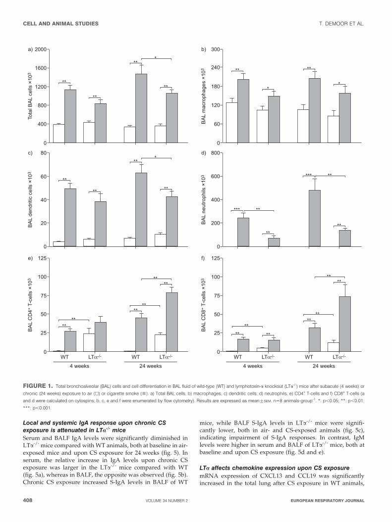
miceSubacute and chronic CS exposure increased the number of totalcells, alveolar macrophages, DCs, neutrophils and lymphocytes
in the BALF of both WT and LTa-/- mice, compared with air-exposed animals (fig. 1). Neutrophilic inflammation was sig-nificantly attenuated in LTa-/- mice at both time points (fig. 1d).In contrast, baseline levels of BAL lymphocytes, more specifi-cally both CD4+ and CD8+ T-cells, were significantly higher inair-exposed LTa-/- mice than in WT mice, and increased furtherupon CS exposure (fig. 1e and f).
CS-induced pulmonary inflammation and emphysemaThe CS-induced increase in lung macrophages and DCs wascomparable between WT and LTa-/- mice (fig. 2a and b).Baseline B- and T-cell numbers were higher in the lungs of air-exposed LTa-/- mice versus WT mice. Contrary to the B-cellnumbers, T-cell numbers increased strongly upon chronic CSexposure in the lung compartment of both WT and LTa-/- mice(fig. 2c and d).
In both WT and LTa-/- mice, chronic CS exposure inducedsignificantly higher numbers of activated CD4+CD69+ andCD8+CD69+ T-cells. Yet again, both cell types were signifi-cantly increased in air-exposed LTa-/- mice in comparison withWT mice (fig. 2e and f). These steady state differences inlymphocyte numbers, between air-exposed WT and LTa-/-
mice, were confirmed in the short-term experiment (data notshown).
The Lm increased significantly upon chronic CS exposure inWT mice (air 34.55¡0.35 versus smoke 37.35¡0.71 mm; 8.1%increase; p50.037) as in LTa-/- mice (air 33.71¡0.50 versussmoke 36.71¡0.82 mm; 8.9% increase; p50.009). There was nosignificant difference in Lm between CS-exposed WT andLTa-/- mice (p50.505).
Increase of iNKT cells in the lung upon subacute CSexposure is LTa-dependentiNKT cell numbers were determined through high specificitybinding to CD1d-tetramer loaded with aGalCer [19] (fig. 3e).Subacute CS exposure caused a significant increase of iNKTcells in WT but not in LTa-/- mice (fig. 3a and b). In contrast,chronic CS exposure did not change iNKT cell numbers in theWT and knockout mice, but LTa-/- mice had lower percentagesof iNKT cells compared with WT mice (fig. 3c and d).
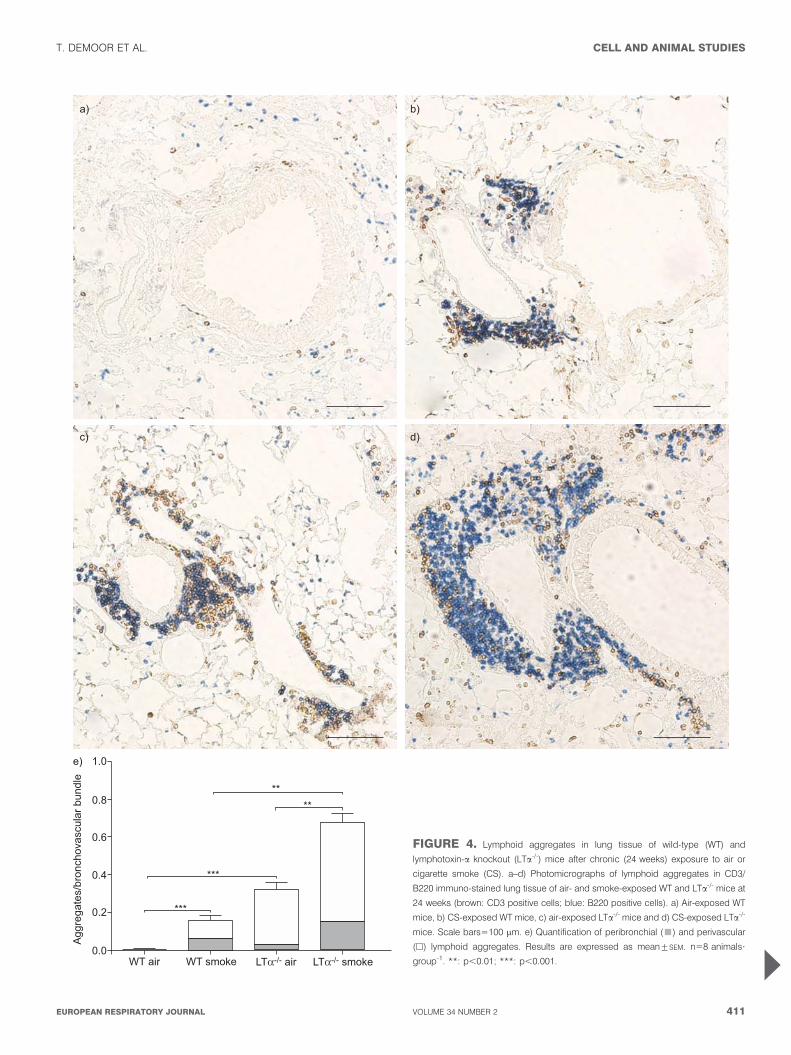
LTa is not required for lymphoid neogenesis upon chronicCS exposureLymphoid aggregates were scarce in lung sections of air-exposed WT mice (fig. 4a and e), whereas the lungs of air-exposed LTa-/- mice were strongly infiltrated with lymphocytes(fig. 4c and 5). Chronic (24 weeks) CS exposure significantlyincreased the number of peribronchial and perivascularlymphoid aggregates in the lungs of both WT and LTa-/- mice(fig. 4). Lymphoid aggregates were absent in the lungs of WTmice after subacute (4 weeks) exposure to air or CS (data notshown). In contrast, lungs of air-exposed LTa-/- mice alreadyshowed a high degree of lymphocyte infiltration, but short-term CS exposure did not induce additional aggregates(aggregates/bronchovascular bundles: air-exposed LTa-/- mice:0.31¡0.059, CS-exposed LTa-/- mice: 0.28¡0.054; mean¡SEM,n55 animals?group-1).
T. DEMOOR ET AL. CELL AND ANIMAL STUDIES
cEUROPEAN RESPIRATORY JOURNAL VOLUME 34 NUMBER 2 407
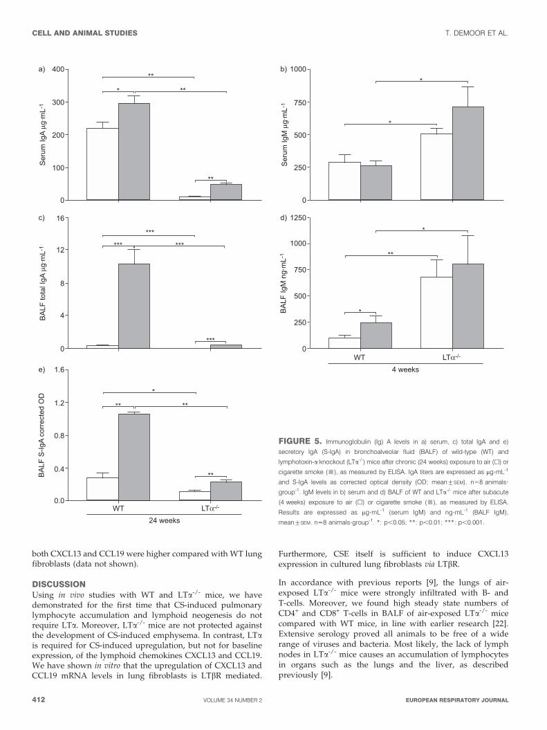
Local and systemic IgA response upon chronic CSexposure is attenuated in LTa-/- miceSerum and BALF IgA levels were significantly diminished inLTa-/- mice compared with WT animals, both at baseline in air-exposed mice and upon CS exposure for 24 weeks (fig. 5). Inserum, the relative increase in IgA levels upon chronic CSexposure was larger in the LTa-/- mice compared with WT(fig. 5a), whereas in BALF, the opposite was observed (fig. 5b).Chronic CS exposure increased S-IgA levels in BALF of WT
mice, while BALF S-IgA levels in LTa-/- mice were signifi-cantly lower, both in air- and CS-exposed animals (fig. 5c),indicating impairment of S-IgA responses. In contrast, IgMlevels were higher in serum and BALF of LTa-/- mice, both atbaseline and upon CS exposure (fig. 5d and e).
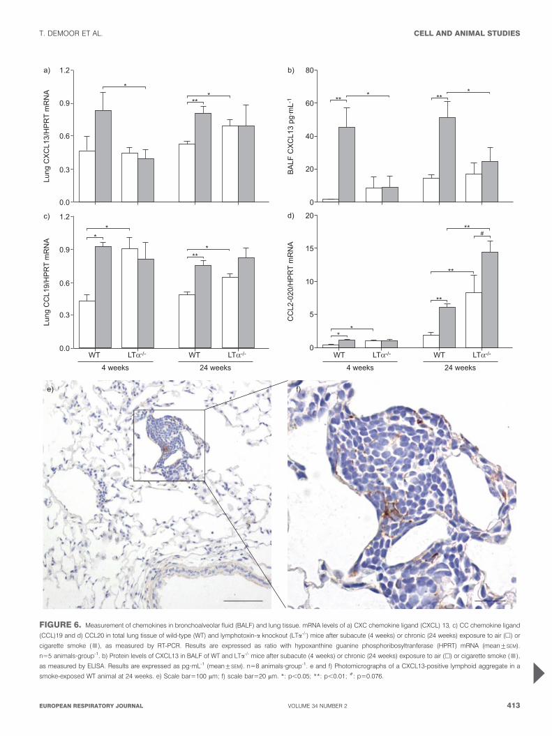
LTa affects chemokine expression upon CS exposuremRNA expression of CXCL13 and CCL19 was significantlyincreased in the total lung after CS exposure in WT animals,
2000
1600
1200
800
400
0
Tota
l BA
L ce
lls ×
103
**
a) 300
180
240
120
60
0
BA
L m
acro
phag
es ×
103
b)
**
**
**
**
*
**
*
*
80
60
40
20
0
BA
L de
ndrit
ic c
ells
×10
3
**
c) 800
400
600
200
0
BA
L ne
utro
phils
×10
3
d)
**
**
**
*** **
**
*** **
**
*
125
100
75
50
25
0
BA
L C
D4+
T-c
ells
×10
3
**
e) 125
75
50
100
25
0
BA
L C
D8+
T-c
ells
×10
3
f)
**
**
**
** **
**
****
**
**
**
**
WT LTα-/-
4 weeks
WT LTα-/-
24 weeks
WT LTα-/-
4 weeks
WT LTα-/-
24 weeks
FIGURE 1. Total bronchoalveolar (BAL) cells and cell differentiation in BAL fluid of wild-type (WT) and lymphotoxin-a knockout (LTa-/-) mice after subacute (4 weeks) or
chronic (24 weeks) exposure to air (h) or cigarette smoke (&). a) Total BAL cells, b) macrophages, c) dendritic cells, d) neutrophils, e) CD4+ T-cells and f) CD8+ T-cells (a
and d were calculated on cytospins; b, c, e and f were enumerated by flow cytometry). Results are expressed as mean¡SEM. n58 animals?group-1. *: p,0.05; **: p,0.01;
***: p,0.001.
CELL AND ANIMAL STUDIES T. DEMOOR ET AL.
408 VOLUME 34 NUMBER 2 EUROPEAN RESPIRATORY JOURNAL
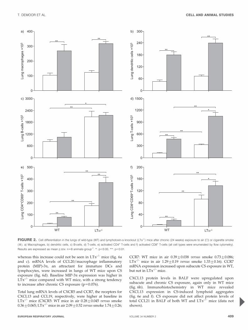
whereas this increase could not be seen in LTa-/- mice (fig. 6aand c). mRNA levels of CCL20/macrophage inflammatoryprotein (MIP)-3a, an attractant for immature DCs andlymphocytes, were increased in lungs of WT mice upon CSexposure (fig. 6d). Baseline MIP-3a expression was higher inLTa-/- mice compared with WT mice, with a strong tendencyto increase after chronic CS exposure (p50.076).
Total lung mRNA levels of CXCR5 and CCR7, the receptors forCXCL13 and CCL19, respectively, were higher at baseline inLTa-/- mice (CXCR5: WT mice in air 0.28¡0.045 versus smoke0.36¡0.065; LTa-/- mice in air 2.09¡0.52 versus smoke 1.74¡0.26;
CCR7: WT mice in air 0.39¡0.038 versus smoke 0.73¡0.086;LTa-/- mice in air 1.29¡0.19 versus smoke 1.33¡0.16). CCR7mRNA expression increased upon subacute CS exposure in WT,but not in LTa-/- mice.
CXCL13 protein levels in BALF were upregulated uponsubacute and chronic CS exposure, again only in WT mice(fig. 6b). Immunohistochemistry in WT mice revealedCXCL13 expression in CS-induced lymphoid aggregates(fig. 6e and f). CS exposure did not affect protein levels oftotal CCL21 in BALF of both WT and LTa-/- mice (data notshown).
400
300
200
100
0
Lung
mac
roph
ages
×10
3
****
a) 300
180
240
120
60
0
Lung
den
driti
c ce
lls ×
103
**
**
b)
3000
2400
1800
600
1200
0
Lung
B-c
ells
×10
3
***
c) 1500
1200
900
600
300
0
Lung
T-c
ells
×10
3
***
**
d)
500
400
300
100
WT LTα-/- WT LTα-/-
200
0
Lung
CD
4+C
D69
+ T-
cells
×10
3 **
**
e) 200
160
120
80
40
0
Lung
CD
8+C
D69
+ T-c
ells
×10
3
*
**
**
f)
**
**
**
**
FIGURE 2. Cell differentiation in the lungs of wild-type (WT) and lymphotoxin-a knockout (LTa-/-) mice after chronic (24 weeks) exposure to air (h) or cigarette smoke
(&). a) Macrophages, b) dendritic cells, c) B-cells, d) T-cells, e) activated CD4+ T-cells and f) activated CD8+ T-cells (all cell types were enumerated by flow cytometry).
Results are expressed as mean¡SEM. n58 animals?group-1. *: p,0.05; **: p,0.01.
T. DEMOOR ET AL. CELL AND ANIMAL STUDIES
cEUROPEAN RESPIRATORY JOURNAL VOLUME 34 NUMBER 2 409

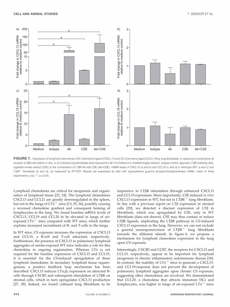
In vitro expression of CXCL13 and CCL19 in lung fibroblastsis LTbR mediatedTo reveal the mechanism by which CXCL13 and CCL19 areinduced in the lung, we stimulated cultured WT and LTbR-/-
lung fibroblasts with agonistic LTbR Ab and/or CSE.
In WT lung fibroblasts, in vitro stimulation with agonistic LTbRAb or CSE resulted in a 60-fold and five-fold increase in
expression of CXCL13 mRNA, respectively. Furthermore, thecombination of LTbR Ab with CSE induced a 100-foldupregulation in CXCL13 mRNA levels (fig. 7a). A similarCCL19 mRNA response was seen upon stimulation with LTbRAb or LTbR Ab with CSE, but there was no significant effect ofCSE exposure alone (fig. 7c). The different stimulations did notaffect CXCL13 and CCL19 mRNA expression in LTbR-/- lungfibroblasts (fig. 7b and d), however, baseline mRNA levels of
80
60
40
20
0
Lung
iNK
T ce
lls ×
103
*
b)
0.5
0.4
0.3
0.1
WT LTα-/- WT LTα-/-
0.2
0.0
Lung
iNK
T ce
lls %
c) 45
30
15
0
Lung
iNK
T ce
lls ×
103
d)
*
*
24 weeks24 weeks
WT LTα-/-
4 weeks
0.8
0.6
0.4
0.2
0.0
Lung
iNK
T ce
lls %
*
a)
WT LTα-/-
4 weeks
e)
CD
3-A
PC
αGalCer-CD1d Tetra-PE
FIGURE 3. a–d) Invariant natural killer T (iNKT) cells in lung digests of wild-
type (WT) and lymphotoxin-a knockout (LTa-/-) mice after subacute (4 weeks) or
chronic (24 weeks) exposure to air (h) or cigarette smoke (&), enumerated by
flow cytometry. e) Gating for iNKT cells were stained with allophycocyanin (APC)-
conjugated anti-CD3 antibody and phycoerythrin (PE)-conjugated CD1d-
tetramer loaded with a-galactosylceramide (aGalCer), as indicated by the
ellipse. Results are expressed as percentage of total cells (a and c) and as
absolute cell number (b and d), mean¡SEM. n58 animals?group-1. *: p,0.05.
CELL AND ANIMAL STUDIES T. DEMOOR ET AL.
410 VOLUME 34 NUMBER 2 EUROPEAN RESPIRATORY JOURNAL
1.0e)
****
***
***
0.8
0.6
0.4
0.2
0.0
Agg
rega
tes/
bron
chov
ascu
lar b
undl
e
WT air WT smoke LTα-/- smokeLTα-/- air
c)
a)
d)
b)
FIGURE 4. Lymphoid aggregates in lung tissue of wild-type (WT) and
lymphotoxin-a knockout (LTa-/-) mice after chronic (24 weeks) exposure to air or
cigarette smoke (CS). a–d) Photomicrographs of lymphoid aggregates in CD3/
B220 immuno-stained lung tissue of air- and smoke-exposed WT and LTa-/- mice at
24 weeks (brown: CD3 positive cells; blue: B220 positive cells). a) Air-exposed WT
mice, b) CS-exposed WT mice, c) air-exposed LTa-/- mice and d) CS-exposed LTa-/-
mice. Scale bars5100 mm. e) Quantification of peribronchial (&) and perivascular
(h) lymphoid aggregates. Results are expressed as mean¡SEM. n58 animals?
group-1. **: p,0.01; ***: p,0.001.
T. DEMOOR ET AL. CELL AND ANIMAL STUDIES
cEUROPEAN RESPIRATORY JOURNAL VOLUME 34 NUMBER 2 411
both CXCL13 and CCL19 were higher compared with WT lungfibroblasts (data not shown).
DISCUSSIONUsing in vivo studies with WT and LTa-/- mice, we havedemonstrated for the first time that CS-induced pulmonarylymphocyte accumulation and lymphoid neogenesis do notrequire LTa. Moreover, LTa-/- mice are not protected againstthe development of CS-induced emphysema. In contrast, LTais required for CS-induced upregulation, but not for baselineexpression, of the lymphoid chemokines CXCL13 and CCL19.We have shown in vitro that the upregulation of CXCL13 andCCL19 mRNA levels in lung fibroblasts is LTbR mediated.
Furthermore, CSE itself is sufficient to induce CXCL13expression in cultured lung fibroblasts via LTbR.
In accordance with previous reports [9], the lungs of air-exposed LTa-/- mice were strongly infiltrated with B- andT-cells. Moreover, we found high steady state numbers ofCD4+ and CD8+ T-cells in BALF of air-exposed LTa-/- micecompared with WT mice, in line with earlier research [22].Extensive serology proved all animals to be free of a widerange of viruses and bacteria. Most likely, the lack of lymphnodes in LTa-/- mice causes an accumulation of lymphocytesin organs such as the lungs and the liver, as describedpreviously [9].
400
300
100
200
0
Ser
um Ig
A µg
·mL-
1
**
*
a)
750
1000
500
250
0
Ser
um Ig
M µ
g·m
L-1
*
b)
16
12
4
8
0
BA
LF to
tal I
gA µ
g·m
L-1 ***
***
c) 1250
1000
750
500
250
0
BA
LF Ig
M n
g·m
L-1
*
*
d)
1.6
1.2
0.4
WT LTα-/-
0.8
0.0
BA
LF S
-IgA
corr
ecte
d O
D
**
**
e)
**
**
*
**
** *
***
***
24 weeks
WT LTα-/-
4 weeks
FIGURE 5. Immunoglobulin (Ig) A levels in a) serum, c) total IgA and e)
secretory IgA (S-IgA) in bronchoalveolar fluid (BALF) of wild-type (WT) and
lymphotoxin-a knockout (LTa-/-) mice after chronic (24 weeks) exposure to air (h) or
cigarette smoke (&), as measured by ELISA. IgA titers are expressed as mg?mL-1
and S-IgA levels as corrected optical density (OD; mean¡SEM). n58 animals?
group-1. IgM levels in b) serum and d) BALF of WT and LTa-/- mice after subacute
(4 weeks) exposure to air (h) or cigarette smoke (&), as measured by ELISA.
Results are expressed as mg?mL-1 (serum IgM) and ng?mL-1 (BALF IgM),
mean¡SEM. n58 animals?group-1. *: p,0.05; **: p,0.01; ***: p,0.001.
CELL AND ANIMAL STUDIES T. DEMOOR ET AL.
412 VOLUME 34 NUMBER 2 EUROPEAN RESPIRATORY JOURNAL
1.2
0.9
0.6
0.3
0.0
Lung
CX
CL1
3/H
PR
T m
RN
A
*
a) 80
40
60
20
0
BA
LF C
XC
L13
pg·m
L-1
b)
** ** *** *
1.2
0.9
0.3
0.6
0.0
Lung
CC
L19/
HP
RT
mR
NA *
c)
***
*
20
15
10
5
0
CC
L2-0
20/H
PR
T m
RN
A
*
d)
*
**
#**
**
WT LTα-/-
4 weeks
WT LTα-/-
24 weeks
*
WT LTα-/-
4 weeks
WT LTα-/-
24 weeks
e) f)
FIGURE 6. Measurement of chemokines in bronchoalveolar fluid (BALF) and lung tissue. mRNA levels of a) CXC chemokine ligand (CXCL) 13, c) CC chemokine ligand
(CCL)19 and d) CCL20 in total lung tissue of wild-type (WT) and lymphotoxin-a knockout (LTa-/-) mice after subacute (4 weeks) or chronic (24 weeks) exposure to air (h) or
cigarette smoke (&), as measured by RT-PCR. Results are expressed as ratio with hypoxanthine guanine phosphoribosyltranferase (HPRT) mRNA (mean¡SEM).
n55 animals?group-1. b) Protein levels of CXCL13 in BALF of WT and LTa-/- mice after subacute (4 weeks) or chronic (24 weeks) exposure to air (h) or cigarette smoke (&),
as measured by ELISA. Results are expressed as pg?mL-1 (mean¡SEM). n58 animals?group-1. e and f) Photomicrographs of a CXCL13-positive lymphoid aggregate in a
smoke-exposed WT animal at 24 weeks. e) Scale bar5100 mm; f) scale bar520 mm. *: p,0.05; **: p,0.01; #: p50.076.
T. DEMOOR ET AL. CELL AND ANIMAL STUDIES
cEUROPEAN RESPIRATORY JOURNAL VOLUME 34 NUMBER 2 413
Lymphoid chemokines are critical for neogenesis and organi-sation of lymphoid tissue [23, 24]. The lymphoid chemokinesCXCL13 and CCL21 are greatly downregulated in the spleen,but not in the lungs of LTa-/- mice [13, 25, 26], possibly causinga reversed chemokine gradient and consequent homing oflymphocytes to the lung. We found baseline mRNA levels ofCXCL13, CCL19 and CCL20 to be elevated in lungs of air-exposed LTa-/- mice compared with WT mice, which furtherexplains increased recruitment of B- and T-cells to the lungs.
In WT mice, CS exposure increases the expression of CXCL13and CCL19, a B-cell and T-cell attractant, respectively.Furthermore, the presence of CXCL13 in pulmonary lymphoidaggregates of smoke-exposed WT mice indicates a role for thischemokine in ongoing organisation. Whereas LTa is notrequired for the baseline expression of CXCL13 and CCL19,it is essential for the CS-induced upregulation of theselymphoid chemokines. In secondary lymphoid tissue organo-genesis, a positive feedback loop mechanism has beendescribed: CXCL13 induces LTa1b2 expression on attracted B-cells through CXCR5 and subsequent stimulation of LTbR onstromal cells, which in turn upregulates CXCL13 production[27, 28]. Indeed, we found cultured lung fibroblasts to be
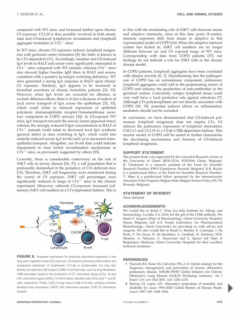
responsive to LTbR stimulation through enhanced CXCL13and CCL19 expression. More importantly, CSE induced in vitroCXCL13 expression in WT, but not in LTbR-/- lung fibroblasts.In line with a previous report on LTb expression in stromalcells [29], we detected a discreet expression of LTb infibroblasts, which was upregulated by CSE, only in WTfibroblasts (data not shown). CSE may thus contain or induceLTbR ligands, implicating the LTbR pathway in CS-inducedCXCL13 expression in the lung. However, we can not excludea general nonresponsiveness of LTbR-/- lung fibroblaststowards the different stimuli. In figure 8, we propose amechanism for lymphoid chemokine expression in the lungupon CS exposure.
Interestingly, CXCR5 and CCR7, the receptors for CXCL13 andCCL19, respectively, appear to be important for lymphoidneogenesis in chronic inflammatory autoimmune disease [30].In contrast, the inability of LTa-/- mice to generate a CXCL13-and CCL19-response does not prevent the development ofpulmonary lymphoid aggregates upon chronic CS exposure,suggesting other chemokines are involved. We demonstratedthat CCL20, a chemokine that attracts immature DCs andlymphocytes, was higher in lungs of air-exposed LTa-/- mice
200
100
150
1050
0
5
Fold
cha
nge
in C
XC
L13
mR
NA
rela
tive
to m
ediu
m c
ontro
l
#
#
#
#
#
a)
2
1
3
0
Fold
cha
nge
in C
XC
L13
mR
NA
rela
tive
to m
ediu
m c
ontro
l
b)
2
1
3
0
Fold
cha
nge
in C
CL1
9 m
RN
Are
lativ
e to
med
ium
con
trol
d)25
20
10
5
15
0
Fold
cha
nge
in C
CL1
9 m
RN
Are
lativ
e to
med
ium
con
trol
c)
Medium Isotype Ab CSE Ab+CSE Medium Isotype Ab CSE Ab+CSE
FIGURE 7. Expression of lymphoid chemokines CXC chemokine ligand (CXCL) 13 and CC chemokine ligand (CCL) 19 by lung fibroblasts, in response to lymphotoxin-b
receptor (LTbR) stimulation in vitro. a–d) Cultured lung fibroblasts were exposed for 48 h to Dulbecco’s modified Eagle medium, isotype control, agonistic LTbR antibody (Ab),
cigarette smoke extract (CSE) or the combination of LTbR Ab with CSE (Ab+CSE). mRNA levels of CXCL13 (a and b) and CCL19 (c and d) in wild-type (WT; a and c) and
LTbR-/- fibroblasts (b and d), as measured by RT-PCR. Results are expressed as ratio with hypoxanthine guanine phosphoribosyltranferase mRNA; mean of three
experiments¡SEM.#: pf0.05.
CELL AND ANIMAL STUDIES T. DEMOOR ET AL.
414 VOLUME 34 NUMBER 2 EUROPEAN RESPIRATORY JOURNAL
compared with WT mice, and increased further upon chronicCS exposure. CCL20 is thus possibly involved in both steadystate and CS-induced lymphocyte recruitment and lymphoidaggregate formation in LTa-/- mice.
In WT mice, chronic CS exposure induces lymphoid neogen-esis with germinal centre formation [5]; the latter is known tobe LTa dependent [31]. Accordingly, baseline and CS-inducedIgA levels in BALF and serum were significantly attenuated inLTa-/- mice compared with WT animals, whereas LTa-/- micealso showed higher baseline IgM titers in BALF and serum,consistent with a putative Ig isotype switching deficiency. WTmice generated a strong IgA response in BALF upon chronicCS exposure. Similarly, IgA appears to be increased inbronchial secretions of chronic bronchitis patients [32, 33].However, when IgA levels are corrected for albumin, toexclude diffusion effects, there is in fact a decrease of indices oflocal active transport of IgA across the epithelium [32, 33],which could relate to reduced expression of epithelialpolymeric immunoglobulin receptor/transmembrane secre-tory component in COPD airways [34]. In CS-exposed WTmice, IgA transport towards the airway lumen appeared intact,whereas the strongly reduced S-IgA concentrations in BALF ofLTa-/- animals could relate to decreased local IgA synthesis(general defect in class switching to IgA, which could alsounderlie reduced serum IgA levels) and/or to decreased trans-epithelial transport. Altogether, our B-cell data could indicateimpairment in class switch recombination mechanisms inLTa-/- mice, as previously suggested by others [35].
Currently, there is considerable controversy on the role ofiNKT cells in airway disease [36, 37], a cell population that isprofoundly diminished in the periphery of LTa deficient mice[15]. Therefore, iNKT cell frequencies were monitored duringthe course of CS exposure. iNKT cell percentages weresignificantly reduced in lungs of LTa-/- mice in the chronicexperiment. Moreover, subacute CS-exposure increased pul-monary iNKT cell numbers in a LTa-dependent fashion. This is
in line with the modulating role of iNKT cells between innateand adaptive immunity, since at this time point (4 weeks),immune responses shift from innate to adaptive in thisexperimental model of COPD [16]. When the adaptive immunesystem has kicked in, iNKT cell numbers are no longerdifferent between air and CS exposed lungs of WT mice.Corresponding with data from COPD patients [37], ourfindings do not indicate a role for iNKT cells in the chronicdisease model.
In COPD patients, lymphoid aggregates have been correlatedwith disease severity [6, 7]. Hypothesising that the pathogen-esis of COPD has an autoimmune component, pulmonarylymphoid aggregates could add to the perpetuating nature ofCOPD and enhance the production of auto-antibodies in thegerminal centres. Conversely, ectopic lymphoid tissue couldvery well have a local protective role against infections [7].Although LTa polymorphisms are not directly associated withCOPD [38, 39], potential indirect effects on inflammatorymediators should not be excluded.
In conclusion, we have demonstrated that CS-induced pul-monary lymphoid neogenesis does not require LTa. CSinduces the pulmonary expression of lymphoid chemokinesCXCL13 and CCL19 in a LTab–LTbR-dependent fashion. Thismurine model of COPD will be useful to further characterisethe developing mechanisms and function of CS-inducedlymphoid neogenesis.
SUPPORT STATEMENTThe present study was supported by the Concerted Research Action ofthe University of Ghent (BOF/GOA 01251504; Ghent, Belgium).B. Vandooren is a research assistant of the Fund for ScientificResearch Flanders (FWO-Vlaanderen; Brussels, Belgium). K.R. Brackeis a postdoctoral fellow of the Fund for Scientific Research Flanders.T. Maes is a postdoctoral fellow sponsored by the InteruniversityAttraction Poles Program/Belgian State/Belgian Science Policy (P6/35;Brussels, Belgium).
STATEMENT OF INTERESTNone declared.
ACKNOWLEDGEMENTSWe would like to thank C. Ware (La Jolla Institute for Allergy andImmunology, La Jolla, CA, USA) for the gift of the LTbR antibody. Wethank P. Jacques (Dept of Rheumatology, Ghent University Hospital,Ghent, Belgium) and A-S. Franki (Laboratory for PharmaceuticalBiotechnology, Ghent University) for providing us with advice andreagents. We also would like to thank G. Barbier, E. Castrique, I. DeBorle, P. De Gryze, K. De Saedeleer, A. Goethals, N. Hertsens, M-R.Mouton, A. Neessen, C. Snauwaert and E. Spruyt (all Dept ofRespiratory Medicine, Ghent University Hospital) for their excellenttechnical assistance.
REFERENCES1 Pauwels RA, Buist AS, Calverley PM, et al. Global strategy for the
diagnosis, management, and prevention of chronic obstructivepulmonary disease. NHLBI/WHO Global Initiative for ChronicObstructive Lung Disease (GOLD) Workshop summary. Am JRespir Crit Care Med 2001; 163: 1256–1276.
2 Murray CJ, Lopez AD. Alternative projections of mortality anddisability by cause 1990–2020: Global Burden of Disease Study.Lancet 1997; 349: 1498–1504.
Fibroblast DC
CCR7
CXCR5
CXCL13LTβR
LTαβ
CCL19
Lymphocyte B-cell
T-cell
FIGURE 8. Proposed mechanism for lymphoid chemokine expression in the
lung upon cigarette smoke (CS) exposure. CS induces pulmonary inflammation and
subsequent expression of lymphotoxin (LT)-ab on lymphocytes, but may also
directly stimulate the LTb receptor (LTbR) on stromal cells, such as lung fibroblasts.
LTbR stimulation leads to the production of CC chemokine lignad (CCL) 19 and
CXC chemokine ligand (CXCL) 13 which attract dendrite cells (DCs) and T- and B-
cells, respectively. Finally, CXCL13 may induce LTab on B-cells, creating a positive
feedback loop mechanism. CXCR: CXC chemokine receptor; CCR: CC chemokine
receptor.
T. DEMOOR ET AL. CELL AND ANIMAL STUDIES
cEUROPEAN RESPIRATORY JOURNAL VOLUME 34 NUMBER 2 415
3 Hautamaki RD, Kobayashi DK, Senior RM, et al. Requirement formacrophage elastase for cigarette smoke-induced emphysema inmice. Science 1997; 277: 2002–2004.
4 Bracke KR, D’hulst AI, Maes T, et al. Cigarette smoke-inducedpulmonary inflammation and emphysema are attenuated inCCR6-deficient mice. J Immunol 2006; 177: 4350–4359.
5 van der Strate BW, Postma DS, Brandsma C-A, et al. Cigarettesmoke-induced emphysema: a role for the B cell? Am J Respir Crit
Care Med 2006; 173: 751–758.6 Hogg JC, Chu F, Utokaparch S, et al. The nature of small-airway
obstruction in chronic obstructive pulmonary disease. N Engl J
Med 2004; 350: 2645–2653.7 Hogg JC, Chu FS, Tan WC, et al. Survival after lung volume
reduction in chronic obstructive pulmonary disease: insights fromsmall airway pathology. Am J Respir Crit Care Med 2007; 176: 454–459.
8 De Togni P, Goellner J, Ruddle NH, et al. Abnormal developmentof peripheral lymphoid organs in mice deficient in lymphotoxin.Science 1994; 264: 703–707.
9 Banks TA, Rouse BT, Kerley MK, et al. Lymphotoxin-a-deficientmice. Effects on secondary lymphoid organ development andhumoral immune responsiveness. J Immunol 1995; 155: 1685–1693.
10 Kratz A, Campos-Neto A, Hanson MS, et al. Chronic inflammationcaused by lymphotoxin is lymphoid neogenesis. J Exp Med 1996;183: 1461–1472.
11 Sacca R, Cuff CA, Lesslauer W, et al. Differential activities ofsecreted lymphotoxin-a3 and membrane lymphotoxin-a1b2 inlymphotoxin-induced inflammation: critical role of TNF receptor1 signaling. J Immunol 1998; 160: 485–491.
12 Crowe PD, VanArsdale TL, Walter BN, et al. A lymphotoxin-b-specific receptor. Science 1994; 264: 707–710.
13 Ngo VN, Korner H, Gunn MD, et al. Lymphotoxin a/b and tumornecrosis factor are required for stromal cell expression of homingchemokines in B- and T-cell areas of the spleen. J Exp Med 1999;189: 403–412.
14 Dejardin E, Droin NM, Delhase M, et al. The lymphotoxin-breceptor induces different patterns of gene expression via two NF-kB pathways. Immunity 2002; 17: 525–535.
15 Franki AS, Van BK, Dewint P, et al. A unique lymphotoxin-ab-dependent pathway regulates thymic emigration of Va14 invariantnatural killer T-cells. Proc Natl Acad Sci USA 2006; 103: 9160–9165.
16 D’hulst AI, Vermaelen KY, Brusselle GG, et al. Time course ofcigarette smoke-induced pulmonary inflammation in mice. EurRespir J 2005; 26: 204–213.
17 Macdonald G, Kondor N, Yousefi V, et al. Reduction ofcarboxyhaemoglobin levels in the venous blood of cigarettesmokers following the administration of carbogen. RadiotherOncol 2004; 73: 367–371.
18 Vermaelen K, Pauwels R. Accurate and simple discrimination ofmouse pulmonary dendritic cell and macrophage populations byflow cytometry: methodology and new insights. Cytometry A 2004;61: 170–177.
19 Matsuda JL, Naidenko OV, Gapin L, et al. Tracking the response ofnatural killer T-cells to a glycolipid antigen using CD1d tetramers.J Exp Med 2000; 192: 741–754.
20 Futterer A, Mink K, Luz A, et al. The lymphotoxin b receptorcontrols organogenesis and affinity maturation in peripherallymphoid tissues. Immunity 1998; 9: 59–70.
21 Bracke K, Cataldo D, Maes T, et al. Matrix metalloproteinase-12and cathepsin D expression in pulmonary macrophages and
dendritic cells of cigarette smoke-exposed mice. Int Arch Allergy
Immunol 2005; 138: 169–179.
22 Kang HS, Blink SE, Chin RK, et al. Lymphotoxin is required formaintaining physiological levels of serum IgE that minimizes Th1-mediated airway inflammation. J Exp Med 2003; 198: 1643–1652.
23 Luther SA, Lopez T, Bai W, et al. BLC expression in pancreaticislets causes B-cell recruitment and lymphotoxin-dependentlymphoid neogenesis. Immunity 2000; 12: 471–481.
24 Luther SA, Bidgol A, Hargreaves DC, et al. Differing activities ofhomeostatic chemokines CCL19, CCL21, and CXCL12 in lympho-cyte and dendritic cell recruitment and lymphoid neogenesis.J Immunol 2002; 169: 424–433.
25 Lo JC, Chin RK, Lee Y, et al. Differential regulation of CCL21 inlymphoid/nonlymphoid tissues for effectively attracting T cells toperipheral tissues. J Clin Invest 2003; 112: 1495–1505.
26 Moyron-Quiroz JE, Rangel-Moreno J, Kusser K, et al. Role ofinducible bronchus associated lymphoid tissue (iBALT) inrespiratory immunity. Nat Med 2004; 10: 927–934.
27 Ansel KM, Ngo VN, Hyman PL, et al. A chemokine-drivenpositive feedback loop organizes lymphoid follicles. Nature 2000;406: 309–314.
28 Mebius RE. Organogenesis of lymphoid tissues. Nat Rev Immunol
2003; 3: 292–303.
29 Cui CY, Hashimoto T, Grivennikov SI, et al. Ectodysplasinregulates the lymphotoxin-b pathway for hair differentiation.Proc Natl Acad Sci USA 2006; 103: 9142–9147.
30 Wengner AM, Hopken UE, Petrow PK, et al. CXCR5- and CCR7-dependent lymphoid neogenesis in a murine model of chronicantigen-induced arthritis. Arthritis Rheum 2007; 56: 3271–3283.
31 Matsumoto M, Mariathasan S, Nahm MH, et al. Role oflymphotoxin and the type I TNF receptor in the formation ofgerminal centers. Science 1996; 271: 1289–1291.
32 Stockley RA, Burnett D. Local IgA production in patients withchronic bronchitis: effect of acute respiratory infection. Thorax
1980; 35: 202–206.
33 Atis S, Tutluoglu B, Salepci B, et al. Serum IgA and secretory IgAlevels in bronchial lavages from patients with a variety ofrespiratory diseases. J Investig Allergol Clin Immunol 2001; 11:112–117.
34 Pilette C, Godding V, Kiss R, et al. Reduced epithelial expression ofsecretory component in small airways correlates with airflowobstruction in chronic obstructive pulmonary disease. Am J Respir
Crit Care Med 2001; 163: 185–194.
35 Eugster HP, Muller M, Karrer U, et al. Multiple immuneabnormalities in tumor necrosis factor and lymphotoxin-a double-deficient mice. Int Immunol 1996; 8: 23–36.
36 Akbari O, Faul JL, Hoyte EG, et al. CD4+ invariant T-cell-receptor+natural killer T-cells in bronchial asthma. N Engl J Med 2006; 354:1117–1129.
37 Vijayanand P, Seumois G, Pickard C, et al. Invariant natural killerT cells in asthma and chronic obstructive pulmonary disease. N
Engl J Med 2007; 356: 1410–1422.
38 Ruse CE, Hill MC, Tobin M, et al. Tumour necrosis factor genecomplex polymorphisms in chronic obstructive pulmonary dis-ease. Respir Med 2007; 101: 340–344.
39 Tanaka G, Sandford AJ, Burkett K, et al. Tumour necrosis factorand lymphotoxin-a polymorphisms and lung function in smokers.Eur Respir J 2007; 29: 34–41.
CELL AND ANIMAL STUDIES T. DEMOOR ET AL.
416 VOLUME 34 NUMBER 2 EUROPEAN RESPIRATORY JOURNAL