Embed Size (px)
Citation preview
International Journal of
Molecular Sciences
Review
Role of Non-Myocyte Gap Junctions and ConnexinHemichannels in Cardiovascular Health and Disease:Novel Therapeutic Targets?
Robert D. Johnson * ID and Patrizia Camelliti *
School of Biosciences and Medicine, University of Surrey, Guildford GU2 7XH, UK* Correspondence: [email protected] (R.D.J.); [email protected] (P.C.)
Received: 10 February 2018; Accepted: 12 March 2018; Published: 15 March 2018
Abstract: The heart is a complex organ composed of multiple cell types, including cardiomyocytes anddifferent non-myocyte populations, all working closely together to determine the hearts propertiesand maintain normal cardiac function. Connexins are abundantly expressed proteins that formplasma membrane hemichannels and gap junctions between cells. Gap junctions are intracellularchannels that allow for communication between cells, and in the heart they play a crucial rolein cardiac conduction by coupling adjacent cardiomyocytes. Connexins are expressed in bothcardiomyocytes and non-myocytes, including cardiac fibroblasts, endothelial cells, and macrophages.Non-myocytes are the largest population of cells in the heart, and therefore it is important to considerwhat roles connexins, hemichannels, and gap junctions play in these cell types. The aim of thisreview is to provide insight into connexin-based signalling in non-myocytes during health anddisease, and highlight how targeting these proteins could lead to the development of novel therapies.We conclude that connexins in non-myocytes contribute to arrhythmias and adverse ventricularremodelling following myocardial infarction, and are associated with the initiation and developmentof atherosclerosis. Therefore, therapeutic interventions targeting these connexins represent an excitingnew research avenue with great potential.
Keywords: connexin; hemichannel; gap junction; cardiovascular disease; fibroblast; endothelial;macrophage; non-myocyte; therapeutic; inflammation
1. Introduction
The heart is a complex multicellular organ composed of cardiomyocytes (CMs) and non-myocytes,including cardiac fibroblasts (CFs), endothelial cells (ECs), smooth muscle cells (SMCs), pericytes,resident stem cells and immune cells. Each cell population has distinct features and functions, and theywork closely together to determine the structural, biochemical, mechanical and electrophysiologicalproperties essential for maintaining effective myocardial function [1]. CMs are muscle cells responsiblefor generating contractile force [2]; CFs produce and remodel the extracellular matrix (ECM) in responseto physiological or pathological stimuli [3]; whilst ECs form the cardiac endothelium, the interiorlining of blood vessels and cardiac valves [1]. Immune cells, such as monocytes and macrophages,are also found in the heart, where they are recruited from the blood following cardiac injury toaid wound-healing [4], but recent evidence has shown that populations of cardiac tissue-residentmacrophages also exist, and that they are involved in tissue homeostasis [5,6]. Interestingly, CMsoccupy 70–85% of the myocardial tissue volume [7], but only constitute around 30% of the actual cellnumbers in the heart [8,9], with non-myocytes comprising the remaining 70% of cells [9]. CFs aregenerally believed to be the largest population of non-myocytes [10], contributing up to two thirdsof total cells in rat [11] and human [12] hearts. However, a recent study has argued that ECs are thedominant non-myocyte population, accounting for 60% of total non-myocytes, whilst CFs constitute
Int. J. Mol. Sci. 2018, 19, 866; doi:10.3390/ijms19030866 www.mdpi.com/journal/ijms
Int. J. Mol. Sci. 2018, 19, 866 2 of 26
under 20% of non-myocyte cells [13]. This has led to debate over the cellular composition of thenon-myocyte population in the heart, and the relevance of each cell type in cardiac homeostasis andcardiovascular disease.
In the adult heart, CMs are arranged in highly organised muscle sheets and are interconnectedby intercalated discs, specialised cell junctions responsible for maintaining cardiac tissue structureintegrity and allowing synchronised contraction [14]. Intercalated discs contain three distinctcomponents; desmosomes, fascia adherens, and gap junctions. Desmosomes and fascia adherensare mechanical linkages, anchoring cell membranes to the intermediate filament network and theactin cytoskeleton respectively, whilst gap junctions form dynamic intracellular communicationchannels [14]. Gap junctions mediate cell-to-cell movement of ions and are crucial for impulseconduction through the cardiac conduction system and ventricular myocardium [15]. Gap junctionsare also involved in the transfer of metabolites/second messengers between cells and allow sharingof metabolic demands across groups of cells [16]. Gap junctions are formed when a hemichannel inthe plasma membrane of one cell docks with a hemichannel in the plasma membrane of an adjacentcell [17], with hemichannels made up of six connexin protein subunits [18]. A variety of connexinsare expressed in the cardiovascular system, including connexin (Cx)31.9, Cx32, Cx37, Cx40, Cx43,and Cx45, although Cx37, Cx40, Cx43, and Cx45 are the predominant connexin isoforms [19]. Connexinexpression also shows regional differences both in the heart and in the vasculature. For example,in the heart, Cx43 is found mainly between atrial and ventricular myocytes, as well as in parts ofthe conduction system, Cx40 is expressed in atrial myocytes, the atrioventricular node, bundle ofHis and ventricular conduction system, and Cx45 is mainly expressed in the sinoatrial node (SAN),atrioventricular node (AVN), bundle of His and bundle branches [15].
Normal heart rhythm is dependent on the coupling of CMs by gap junctions, with connexinand gap junction remodelling leading to potentially serious, and often fatal, cardiac arrhythmias [20].The possible roles of Cx43 and Cx40 remodelling have been well-studied, but Cx45 remodelling isless understood [21]. Down-regulation of Cx43 has been observed in the failing human heart [22,23],whilst knockout of Cx43 in mice increases the incidence of sudden cardiac death resulting fromspontaneous ventricular arrhythmia [24], and increases the frequency and length of ventriculartachycardia induced by ischemia [25]. Further, dephosphorylation and translocation of Cx43 fromgap junctions to the cell cytosol has been shown to contribute to electrical uncoupling after ischemiain a Langendorff-perfused rat heart [26]. Mutations in Cx40 have been linked to patients with atrialfibrillation [27–29], whilst transgenic mice harbouring a human loss-of-function Cx40 mutation displayreduced atrial conduction and prolonged incidence of atrial fibrillation as a result of reduced gapjunctional conductance [30]. However, investigations in to the possible role of Cx45 in cardiac diseasehave been hampered by Cx45 knockout in animals causing an endocardial cushion defect in earlycardiogenesis, and death shortly after birth [31]. A mouse model with Cx45 deletion specifically inadult CMs has been developed and has shown that Cx45 is important in AVN conduction, but is notessential for the survival of adult mice [32].
Dysfunctional hemichannels have also been associated with cardiac disease. Specifically, two atrialfibrillation-linked Cx40 mutations, V85I and L221I, were shown to cause increased hemichannelconductance without changing gap junction function [33]. This gain-of-function mutation is thought tocontribute to atrial fibrillation by increasing the influx and outflux of sodium (Na+) and potassium (K+)ions respectively, leading to membrane depolarisation, Na+ channel inactivation, and reduced CMexcitability [33]. In addition, the G38D Cx40 mutation, another atrial fibrillation-associated mutation,is also thought to increase hemichannel function [34]. Cx43 hemichannels in CMs have been implicatedin exacerbating ischemia-reperfusion injury, with Cx43 hemichannel blockade by the peptide Gap19improving CM viability both in vitro and in vivo [35].
Therefore, connexins are crucial for the maintenance of normal cardiac function. However,as CMs are not the only cell type to express connexins in the heart and not the dominant cardiac cellpopulation in terms of cell numbers, it is important to consider what roles non-myocyte connexins
Int. J. Mol. Sci. 2018, 19, 866 3 of 26
play in cardiovascular function. This review highlights the role of CF, EC, and macrophage connexinsin the healthy and diseased heart, and discusses how modifying their function may lead to newtherapeutic benefits.
2. Connexins and Cardiac Fibroblasts
2.1. Fibroblast Identification and Function
CFs are widely distributed cells of mesenchymal origin that are traditionally defined as cellsthat secrete ECM components, such as different types of collagen and fibronectin [36]. However,collagen production is not used as a defining characteristic of CFs, with morphological identifiersused instead. Morphologically, CFs are flat, spindle-shaped cells that lack a basement membrane,but display irregular folding and multiple elongated cytoplasmic processes/sheet-like extensions [37].However, the main issue in the identification of CFs is the lack of a cell specific marker. Commonly usedmarkers for identification of CFs include the discoidin domain receptor 2 (DDR2), periostin (POSTN),vimentin, platelet derived growth factor receptor (PDGFR), CD90/Thy-1 and fibroblast-specificprotein 1 (FSP1) [38]. However, none of these proteins are uniquely or continuously expressed in CFs.For example, DDR2 is also expressed in leukocytes [39], POSTN expression is low in non-diseasedhearts, and FSP1 expression in mouse models of cardiac fibrosis is not primarily seen in CFs, but ratherhaematopoietic and vascular cells [40]. The identification of a definitive CF cell-specific marker iscrucial for future fibroblast research.
The main role of CFs is to maintain and regulate the ECM, which is important for structuralsupport, signal transduction, and fluid movement [41]. CFs are also involved in fibrotic scar maturationduring wound healing post-myocardial injury. Following acute myocardial infarction (MI), CFs becomeactivated, proliferate rapidly, migrate to the site of injury, and express contractile proteins such asα-smooth muscle actin (α-SMA) [42]. These activated CFs are called myofibroblasts and have anincreased ability to produce and deposit ECM components, leading to fibrotic accumulation [43].ECM secretion by myofibroblasts initially causes adaptive fibrosis, which is crucial to protect thestructural integrity of the heart, before the fibrotic scar later matures due to the stiffening of secretedcollagen at the site of injury [44]. CFs also synthesize and secrete bioactive molecules that areinvolved in paracrine/autocrine signalling during fibrosis [45]. Pro-inflammatory cytokines interleukin(IL)-6, IL-1β and tumour necrosis factor-α (TNF-α) have diverse effects on CFs. TNF-α increases CFproliferation, migration and pro-inflammatory cytokine expression, but decreases collagen synthesis;IL-1β stimulates ECM degradation and CF migration, but reduces CF proliferation; while IL-6reduces CF collagen synthesis [45]. The pro-fibrotic cytokine transforming growth factor-β (TGF-β)increases fibrillar collagen, fibronectin and proteoglycan synthesis, and has been reported as a specificstimulus needed for CF differentiation into myofibroblasts. Angiotensin II (Ang II) can also induce CFdifferentiation [45], whilst the vasoactive peptide endothelin-1 (ET-1) is also a pro-fibrotic mediatorthat enhances CF proliferation and ECM deposition [46]. Vascular endothelial growth factor (VEGF),secreted by CFs in response to hypoxic or inflammatory stimuli, acts on endothelial cells to induceangiogenesis [45].
2.2. Fibroblast Connexins and Cell-Cell Coupling
CFs express several connexin isoforms, with distinct expression patterns in different cardiacregions, disease conditions, and stage of development. Normal adult mouse ventricular CFs expressCx40 and Cx43 in culture [47], while neonatal rat CF cultures express Cx43 and Cx45 [48]. Cx43 has alsobeen observed in adult rabbit ventricular CFs in situ [49]. CFs have been shown to express Cx40 andCx45 in rabbit SAN tissue [50], Cx40 and Cx43 in the rabbit atrium [49], and Cx40 in the rabbit AVN [51].Fibroblast connexin expression has also been investigated following cardiac injury, with Cx43 and Cx45identified in sheep infarct ventricular tissue [52] and Cx43 detected in CFs isolated from post-MI rat
Int. J. Mol. Sci. 2018, 19, 866 4 of 26
hearts [53]. Importantly, CF connexin expression increases following cardiac injury [51,53], indicatinga potential role for CF connexins in myocardial remodelling.
Several studies have shown that CFs establish functional gap junctional channels with adjacentCFs as well as with neighbouring CMs, both in vitro and in situ. Functional coupling betweenventricular CFs in culture occurs through Cx40, Cx43, or Cx40/43 heterotypic gap junctions [47];while CF-CF coupling in the SAN is mediated by Cx40 in fibroblast-rich areas devoid of CMs andby Cx45 in regions of the node where fibroblasts intermingle with CMs [50]. Growing evidenceindicates that CFs directly communicate with CMs through connexins, with Cx43 [53] and Cx45 [48]identified as the isoforms involved in coupling between neonatal CMs and CFs in co-cultures, and Cx45supporting CF-CM coupling in SAN tissue [50]. In situ, functionality of this coupling was confirmedby dye transfer assays using the gap-junction-permeable dye Lucifer yellow [50], while in vitro wasconfirmed by fluorescence recovery after photobleaching of calcein-AM [53], and the observationof successful conduction of electrical impulses between groups of CMs interconnected by fibroblastinserts up to 300 µm in length [48]. Despite the above evidence, whether CF-CM coupling existsin vivo remains subject of debate. A recent study by Quinn et al. [54] provides the first direct proof offunctional coupling between non-myocytes and CMs in the whole heart. Using optogenetic tools, theymonitored the electrical activity of non-myocytes in Langendorff-perfused mouse hearts and found thatnon-myocytes displayed CM-like action potentials at the border zone of cryo-injured hearts, indicatingthe presence of electrotonic coupling between non-myocytes and CMs in native myocardium [54].This study also supports previous indirect evidence of CF-CM coupling in post-infarct rabbit hearts,where cardiac electrical impulses propagated into scar tissue despite the chemical ablation of survivingendocardial CMs [55]. Further, the presence of connexins in CFs within the infarct suggested CFs maybe involved in impulse conduction through gap junction-mediated electrical coupling with CMs [52,56].These studies highlight the possibility for CF-CM coupling in the ventricles, specifically post-MI toelectrically link infarct and non-infarct regions of the heart.
2.3. Role of Fibroblasts in Cardiac Electrophysiology
In addition to their biochemical and structural functions, CFs participate in mechano-electricalfeedback and can directly contribute to cardiac electrophysiology [49]. CFs can affect electrophysiologypassively, for example by acting as obstacles to the spread of electrical activity. Following myocardialinjury, CF proliferation and collagen accumulation leads to densely fibrotic ventricular scars that bothmechanically and electrically separate surviving bundles of CMs [57]. This can cause arrhythmiathrough the dense fibrosis first creating areas of conduction block, whilst second, fibrosis can forceelectrical impulses to take a convoluted path through the separated CM bundles, and even reduceconductivity between CMs, slowing conduction [57]. Further, the development of new treatmentstargeting fibrosis, reducing collagen deposition and myofibroblast proliferation, show promise forreducing cardiac fibrosis-based arrhythmia [58]. However, the discovery that CFs express connexinsand form functional gap junctions with CMs supports the hypothesis that CFs may play a more activerole in modulating cardiac electrophysiology. CFs can affect CM electrophysiology through bothacting as long-range conductors and actively affecting the action potential properties of CMs [59].The high membrane resistance and low capacitance of CFs make them suitable to act as conductors forlong-range signal transmission [60], whilst CFs can modulate CM electrophysiology due to having amore positive resting membrane potential [59].
Active CF modulation of CM electrophysiology through CF-CM gap junctional coupling hasmainly been demonstrated in vitro and in silico. Miragoli et al. [61] demonstrated that coating strandsof neonatal rat CMs with myofibroblasts caused slowed conduction as a result of CM depolarisation,Na+ channel inactivation, and a slower inward upstroke current [61]. Further, a follow-up studyshowed co-culturing myofibroblasts and CMs led to ectopic CM automaticity [62], whilst inhibitionof myofibroblast-CM coupling in vitro through myofibroblast Cx43 knockdown was found to beanti-arrhythmic [63]. Cx43 knockdown reduced CM-myofibroblast coupling, and prevented the
Int. J. Mol. Sci. 2018, 19, 866 5 of 26
increase in CM maximum diastolic potential, CM automaticity, changes to conduction velocity andprolonged repolarisation, reducing the incidence of re-entrant tachycardia as a result [63]. This isparticularly important as myofibroblasts isolated from infarct adult rat hearts have been shown toexhibit higher Cx43 expression and functional coupling with CMs than normal fibroblasts in culture,suggesting they have greater arrhythmogenic potential [53]. A study combining myofibroblast-CMco-cultures and computer simulations looked at the effect of myofibroblast density and levelof CM-myofibroblast coupling on conduction velocity. They found that increasing the densityof myofibroblasts, or increasing the level of CM-myofibroblast coupling, increased conductionvelocity [64]. Together, these results suggested that at low levels of coupling myofibroblasts actas charge sinks to slow conduction velocity, but at higher levels of coupling myofibroblasts instead actas conductors to facilitate conduction to downstream CMs [64].
There has also been indirect evidence of CFs acting as conductors in vivo. Mahoney et al. [65]identified Cx43 gap junctions between CMs and non-myocytes as a possible route for electrical signaltransmission in the ventricles, after optically mapped electrical signals were observed in both theventricular scar region and surrounding healthy myocardium of wild-type mice following cardiacinjury [65]. However, electrical signals in the ventricular scar of fibroblast-specific protein-1 drivenconditional Cx43 knockout (Cx43FSP1KO) mice (a model where Cx43 is only knocked out in FSP1positive cells to reduce Cx43 expression in non-myocytes but not CMs) were significantly reducedcompared to wild-type mice, suggesting that deletion of Cx43 from FSP1 positive cells reduced thecoupling between the scar and uninjured myocardium [65]. Direct current delivery to the scar ofwild-type mice was shown to electrically excite the uninjured myocardium, whilst electrical impulsescould also conduct across scars [65]. Although these findings support an active role of non-myocytesin cardiac electrophysiology, interpretation of the results is difficult due to the lack of specificity ofFSP1 expression. FSP1 can be expressed in CFs, haematopoietic and vascular cells [40], and multiplecell types, aside from CFs and myofibroblasts were present in the injured myocardium of the mice [65].Therefore, whilst demonstrating that CMs and non-myocytes in ventricular scars are electricallycoupled in a Cx43-dependent manner, identification of cell specific markers is needed to provide moreinformation on which non-myocytes couple to CMs.
Whereas gap junctional coupling of ventricular CFs and CMs has been extensively investigatedin vitro and recently confirmed in vivo, coupling between atrial CFs and CMs is less understood. Atrialfibrillation (AF) is the most common sustained form of clinical arrhythmia, with atrial fibrosis a clinicalhallmark of AF and myofibroblast content increasing more in the atria than the ventricles followingcongestive heart failure [66]. Computer models have been used to show the potential effect of couplingbetween atrial CFs and CMs. At the single-cell level, atrial CF coupling with CMs increases the restingmembrane potential of CMs, decreases upstroke velocity, and prolongs the action potential duration.Further, when more than 10 fibroblasts are coupled to a CM, the CM is unable to attain the activationthreshold for the upstroke Na+ current and fails to generate an action potential [67]. A 2D-model ofCF-CM coupling has shown CF coupling could disturb the wavefront of atrial excitation and lead tothe formation of re-entrant arrhythmias [67]. Targeting of atrial CF-CM coupling for catheter ablationhas also been shown to terminate spiral-wave re-entry earlier in AF [68]. Electromechanical coupling ofatrial CMs with CFs and a stretch-activated ion channel current has also been shown to modulate atrialCM excitability and action potential morphology [69], whilst heart failure (HF), a clinical syndromewhere ventricular filling and blood ejection is impaired due to structural and functional defects in theheart [70], has been shown to cause K+ ion channel remodelling in CFs and alter spiral-wave dynamicswhen CFs are coupled to CMs [71]. Therefore, coupling between CFs and CMs in the atria couldcontribute to AF, but requires further research in biologically relevant samples.
Altogether, these studies show the potential of CFs/myofibroblasts to contribute to electricalactivity in the heart through direct gap junctional coupling with CMs, acting as both conductors andmodulators of cardiac electrophysiology. This could be particularly important for treatment of post-MIventricular arrhythmia, the most common cause of sudden cardiac death post-infarction [72], as CMs
Int. J. Mol. Sci. 2018, 19, 866 6 of 26
and myofibroblasts come in to close contact at areas such as the infarct border zone [53], and in AFwhich is the most common form of clinical arrhythmia.
2.4. Fibroblasts, Connexins and Myocardial Remodelling
CFs play important roles in the wound healing and remodelling response to inflammation inthe heart. Several studies have shown Cx43 contributes to CF differentiation, proliferation andmigration post-MI. CFs can differentiate in to α-SMA positive myofibroblasts during cell culture, or beinduced to differentiate by TGF-β [73], a central mediator of the inflammatory and fibrotic responsepost-infarction [74]. TGF-β treatment of cultured CFs increases their expression of α-SMA and Cx43,whilst knockdown or overexpression of Cx43 decreases or increases the expression of α-SMA in CFsrespectively [73]. Therefore, these results suggest that Cx43 plays a role in the phenotypic conversionof CFs to myofibroblasts in response to TGF-β signalling. This has also been seen in vivo in left anteriorcoronary artery ligation MI model mice. Cx43-deficient mice show reduced differentiation of CFsto myofibroblasts compared to wild-type mice following injury, as well as delayed scar formationand fibrotic remodelling [75]. Interestingly, Cx43-deficient mice also displayed reduced fibrosis andadverse dilatation 4-weeks post-MI, and were said to have received beneficial post-MI remodelling.These beneficial effects of Cx43 knockout on post-MI remodelling were also attributed to decreasedTGF-β signalling [75].
CF proliferation and migration are important parts of the wound-healing process post-MI.An inverse relationship between Cx43 expression and CF proliferation has been shown in vitro,with reduced Cx43 expression increasing CF proliferation, and increased Cx43 expression reducingCF proliferation [76]. Connexin involvement in fibroblast migration and wound healing has beenwell established outside the heart [77–80], although little evidence exists for connexin involvementin CF migration. One study has shown Cx43 can affect CF migration in vitro. Treatment of CFssurrounding a spheroid of CMs with α-connexin carboxyl-terminus (αCT1), a mimetic peptide of thecarboxyl-terminus (CT) region of Cx43, caused the CFs to migrate to the centre of the spheroid anddisplace CMs [81]. The αCT1 peptide interferes with the interaction between the Cx43 CT and zonulaoccludens-1 (ZO-1), an anchoring protein that controls the cellular distribution of Cx43 [82]. This Cx43CT and ZO-1 interaction also facilitates hemichannel function, as disassociation of this interactionincreases the contribution of Cx43 hemichannels to Cx43 gap junctions [83]. Therefore, increasing gapjunctional communication between CFs or inhibiting Cx43 hemichannels may induce CF migrationduring wound healing post-MI.
Fibroblast connexin hemichannels have also been implicated in contributing to myocardialremodelling following cardiac injury. Blockade of Cx43 hemichannels with the peptide Gap26 hasbeen shown to be protective during ischemia-reperfusion injury [84], with further research specificallyimplicating myofibroblast Cx43 hemichannels [85]. Neonatal rat heart myofibroblasts cultured inischemia-like conditions were shown to display reduced gap junction communication but increasedhemichannel opening and cell death, with Gap26 preventing this hemichannel opening and reducinginfarct size [85]. Finally, connexin hemichannels also facilitate adenosine triphosphate (ATP) releasefrom CFs. ATP is released in the heart in response to myocardial injury in order to promote phagocyterecruitment and initiate pro-fibrotic responses [86]. Physical perturbation of CFs by culturing themin hypotonic medium leads to increased ATP release, although this can be prevented by increasingextracellular Ca2+, which blocks hemichannel permeability, or by knockdown of Cx43 and Cx45 [86].This suggests that ATP released from CFs via Cx43 or Cx45 hemichannels can contribute to pro-fibroticresponses following myocardial injury, such as CF differentiation to myofibroblasts and increasedcollagen synthesis, through activation of P2Y2 receptors and stimulating ERK signalling [86].
Another potentially important role of CF connexin hemichannels is the regulation of theinflammasome pathway. HF is associated with low-grade chronic inflammation that is mediated bydanger-associated molecular patterns (DAMPs), molecules indicating cell damage, and the nucleotideoligomerisation domain (NOD)-like receptor protein-3 (NLRP3) inflammasome pathway [87].
Int. J. Mol. Sci. 2018, 19, 866 7 of 26
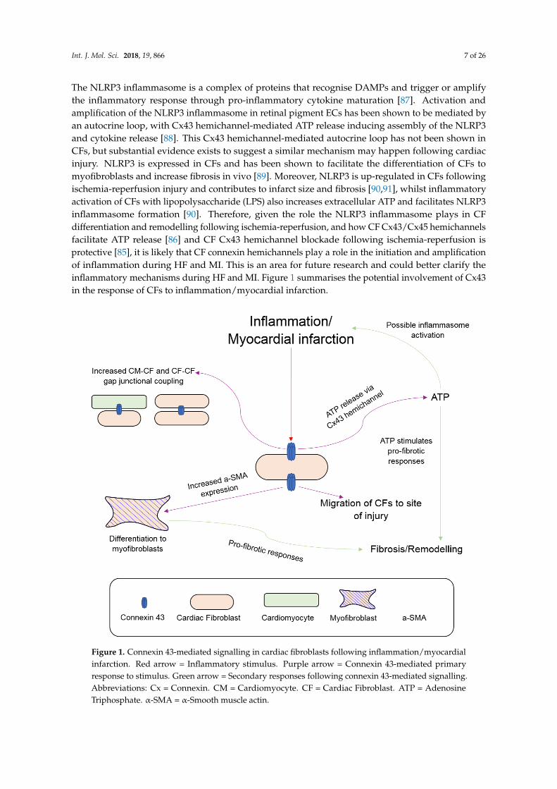
The NLRP3 inflammasome is a complex of proteins that recognise DAMPs and trigger or amplifythe inflammatory response through pro-inflammatory cytokine maturation [87]. Activation andamplification of the NLRP3 inflammasome in retinal pigment ECs has been shown to be mediated byan autocrine loop, with Cx43 hemichannel-mediated ATP release inducing assembly of the NLRP3and cytokine release [88]. This Cx43 hemichannel-mediated autocrine loop has not been shown inCFs, but substantial evidence exists to suggest a similar mechanism may happen following cardiacinjury. NLRP3 is expressed in CFs and has been shown to facilitate the differentiation of CFs tomyofibroblasts and increase fibrosis in vivo [89]. Moreover, NLRP3 is up-regulated in CFs followingischemia-reperfusion injury and contributes to infarct size and fibrosis [90,91], whilst inflammatoryactivation of CFs with lipopolysaccharide (LPS) also increases extracellular ATP and facilitates NLRP3inflammasome formation [90]. Therefore, given the role the NLRP3 inflammasome plays in CFdifferentiation and remodelling following ischemia-reperfusion, and how CF Cx43/Cx45 hemichannelsfacilitate ATP release [86] and CF Cx43 hemichannel blockade following ischemia-reperfusion isprotective [85], it is likely that CF connexin hemichannels play a role in the initiation and amplificationof inflammation during HF and MI. This is an area for future research and could better clarify theinflammatory mechanisms during HF and MI. Figure 1 summarises the potential involvement of Cx43in the response of CFs to inflammation/myocardial infarction.
Figure 1. Connexin 43-mediated signalling in cardiac fibroblasts following inflammation/myocardialinfarction. Red arrow = Inflammatory stimulus. Purple arrow = Connexin 43-mediated primaryresponse to stimulus. Green arrow = Secondary responses following connexin 43-mediated signalling.Abbreviations: Cx = Connexin. CM = Cardiomyocyte. CF = Cardiac Fibroblast. ATP = AdenosineTriphosphate. α-SMA = α-Smooth muscle actin.
Int. J. Mol. Sci. 2018, 19, 866 8 of 26
In summary, connexins appear to modulate the function of CFs during post-MI wound healing.CFs isolated from the infarct heart have previously been shown to display upregulated Cx43expression and increased intracellular communication [92], with this increased Cx43 possibly leadingto myofibroblast differentiation, CF migration, and adverse remodelling seen post-MI. However,increased Cx43 expression was shown to reduce CF proliferation, the opposite of what happenspost-MI, suggesting further mechanisms need to be elucidated. There is also potential for ATP releasedvia Cx43 hemichannels to initiate and amplify the inflammatory response during HF or post-MI, andfurther stimulate pro-fibrotic responses.
3. Connexins and Endothelial Cells
3.1. Roles of Endothelial Cells in Cardiac Muscle and the Vasculature
Endothelial cells form the cardiac endothelium, the lining in the chambers of the heart andthe coronary vasculature [9]. The cardiac endothelium is involved in signal transduction ofneurotransmitters, mechanical stimuli and hormones, and has been reported to modulate thecontractile function of CMs through paracrine signalling of nitric oxide (NO) and ET-1, amongothers [9]. NO in ECs is produced by endothelial NO synthase (eNOS), with NO affecting theonset of myocardial relaxation [93]. ET-1 has the opposite effect, binding to ETA receptors on CMscausing CM constriction [93]. In the normal myocardium, the dense capillary network guarantees closeproximity between ECs and CMs, facilitating interactions between the two cell types [93]. The cardiacendothelium also has roles in control of heart size and angiogenesis, with suggestions that these twoprocesses may be linked [94].
Endothelial cells also form a lining along every blood vessel in the human body, the vascularendothelium. The vascular endothelium is involved in regulation of vascular contraction, throughsimilar NO and ET-1 paracrine signalling to that seen in the cardiac endothelium [95]. The vascularendothelium also plays roles in regulating thrombosis and thrombolysis, as well as platelet/leukocyteinteraction with the vessel wall [96]. Under normal conditions, ECs work to prevent thrombosisby means of anti-coagulant and anti-platelet mechanisms, but upon injury this is changed to helprestore vascular integrity. Induction of pro-coagulant factors is important in the formation of fibrin,whilst platelet and leukocyte adhesion are important for recruitment of inflammatory cells to sites ofinjury [96].
3.2. Endothelial Cell-Cardiomyocyte Gap Junctional Signalling
ECs express three types of connexion, Cx40, Cx43, and Cx37 [97], with the first evidence for EC-CMgap junctions provided by Narmoneva et al. [98]. This study found ECs contributed to both the spatialorganisation and survival of CMs, as well as synchronising CM contraction as cultures containing bothCMs, and ECs showed a larger area of synchronised contraction than cultures of CMs only [98]. ECswere also shown to increase the expression of Cx43 in CMs in a VEGF-dependent manner, and increaseboth CM-CM and EC-CM coupling [98]. EC-CM uncoupling has been suggested to contribute tocardiac arrhythmia and failure in patients with hyperhomocysteinemia (HHcy). HHcy leads to elevatedlevels of homocysteine (Hcy), and has been associated with altered myocardial conduction and suddencardiac death [99]. Elevated levels of Hcy increased Cx43 degradation and peri-capillary fibrosis inmice, leading to disconnection between ECs and CMs. The elevated Hcy also caused arrhythmogenesis,and impaired ET-1 and NO-mediated regulation of cardiac contraction. These effects were amelioratedwith MK-801 treatment, a NMDA-R1 antagonist, suggesting a role for the NMDA-R1 in EC-CMuncoupling and arrhythmogenesis [99]. However, as both Cx43 degradation and fibrosis contributed tothe EC-CM uncoupling, it is unknown what the contribution of Cx43-mediated uncoupling is towardsarrhythmogenesis in HHcy, and should be investigated further.
Endothelial Cx40 has been shown to be beneficial after ischemia-reperfusion injury. The harmfuleffects of Cx43 hemichannels in CFs during ischemia-reperfusion have been explored earlier,
Int. J. Mol. Sci. 2018, 19, 866 9 of 26
but interestingly Cx40 in ECs is believed to confer cardioprotective effects. EC-specific deletionof Cx40 in mice causes neutrophil infiltration, increased infarct size, and increased cell deathfollowing ischemia-reperfusion injury, although no deleterious effects have been associated withCx37 deletion [100]. The Cx40-mediated protective effect is thought to be mediated by CD73 signalling,as methotrexate activation of CD73 in Cx40-knockout mice reduced neutrophil infiltration and infarctsize [100]. This effect is believed to arise from anti-adhesion signalling by CD73, which propagatesthrough Cx40 gap junctions between ECs to reduce leukocyte adhesion [101]. Similar effects ofCx40 deletion on recovery have been seen after post-ischemic hindlimb injury, where deletion ofCx40 reduced limb recovery and survival after ischemia [102] as a result of reduced tissue perfusion,arteriogenesis, and increased inflammatory response [103].
3.3. Endothelial Cells, Connexins, and Vasomotor Control
Gap junctions between ECs are important for allowing communication between cells to coordinateEC function in the vasculature. For example, knockdown of Cx37, Cx40, or Cx43 leads to reducedintercellular communication between human umbilical vein endothelial cells (HUVECs), impairingangiogenesis in matrigels as a result [104]. Gap junctional coupling between ECs has been shownto play a central role in coordinating the Ca2+ response of the endothelium to vasoactive agonists,with histamine stimulation of HUVECs causing an initial rapid increase in intracellular Ca2+ thatslowly spreads to neighbouring cells, a spread that is prevented by gap junctional blockade. The sameeffects have also been observed in mouse aorta after ATP stimulation [105]. Endothelium Ca2+ increaseplays an important role in NO production by eNOS, with gap junctional blockade in HUVECs alsoshown to reduce NO production in cells after histamine treatment [106]. Further to this, knockout of ECCx43 in mice has caused hypotension and elevated NO plasma levels, suggesting Cx43 also regulatesNO production [97]. Direct interaction between EC Cx37/40 and eNOS has been suggested, with Cx37and eNOS found to colocalise at points of cell-cell contact in mouse and human EC lines, and Cx37antisense treatment increasing NO production [107]. Co-localisation of Cx37, Cx40, and eNOS at pointsof cell-cell contact has also been reported in the aortic endothelium of Cx40+/+ mice. Deletion of Cx40reduced NO production, ATP-induced endothelium relaxation, and eNOS expression [107]. However,whilst only Cx40 was knocked out, a concurrent reduction in Cx37 was also observed, suggestingalterations in both Cx40 and Cx37 contributed to the reduced eNOS expression [107]. Interestingly,the above studies show opposing effects to NO production when Cx37 or Cx40 is reduced. However,given the different origin of ECs used, with venous ECs investigated in the first study and arterial ECsin the second study, this suggests that connexins could have different roles in the vasomotor control ofveins and arteries and is a possible area of future research.
ECs have been reported to affect SMCs relaxation via direct gap junctional coupling. Electronmicroscopy images have revealed areas of close contact between vascular endothelium and smoothmuscle that contain myoendothelial gap junctions [108]. The role of these heterocellular gap junctionsin the electrical and chemical coupling between ECs and SMCs has been demonstrated in the vascularwall of arteries. Simultaneous microelectrode recordings of ECs and SMCs in the vessel wall revealedboth cells had identical resting membrane potentials, and in response to current injection showedsimultaneous, equivalent changes in membrane potential [109]. Importantly, bidirectional electricalcoupling was observed, with depolarising and hyperpolarising currents injected in one cell leading todepolarisation or hyperpolarisation of the other [109]. Li et al. [110] however reported unidirectionalcoupling, with signals flowing from ECs to SMCs in spiral modiolar arteries isolated from the guineapig cochlea [110]. Therefore, whilst electrical coupling between ECs and SMCs appears to be presentand able to transmit signals to regulate vasomotor control, the co-ordination of this coupling is notwell understood.
Int. J. Mol. Sci. 2018, 19, 866 10 of 26
3.4. Endothelial Cells, Connexins and Atherosclerosis
Atherosclerosis is a disease which can eventually lead to thrombosis and possible stroke, MI,or sudden cardiac death [111]. Atherosclerosis begins with subendothelial accumulation of lipoproteinswhich triggers an inflammatory response, and subsequent activation of cells in the vascular wall andrecruitment of monocytes and macrophages, forming atherosclerotic plaques (atheromas) [112]. Mostof these atherosclerotic plaques do not cause vascular disease, but some vulnerable plaques havenecrotic cores which can lead to plaque rupture and promotion of vessel thrombosis [112].
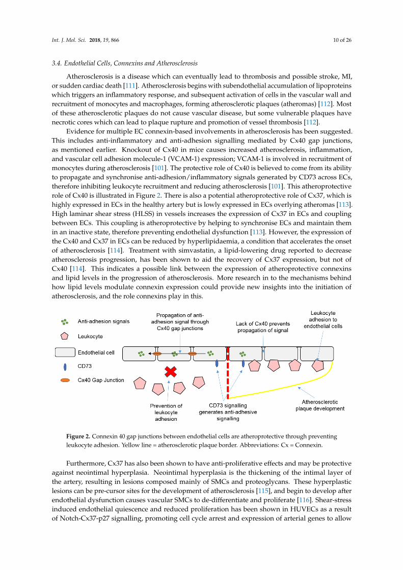
Evidence for multiple EC connexin-based involvements in atherosclerosis has been suggested.This includes anti-inflammatory and anti-adhesion signalling mediated by Cx40 gap junctions,as mentioned earlier. Knockout of Cx40 in mice causes increased atherosclerosis, inflammation,and vascular cell adhesion molecule-1 (VCAM-1) expression; VCAM-1 is involved in recruitment ofmonocytes during atherosclerosis [101]. The protective role of Cx40 is believed to come from its abilityto propagate and synchronise anti-adhesion/inflammatory signals generated by CD73 across ECs,therefore inhibiting leukocyte recruitment and reducing atherosclerosis [101]. This atheroprotectiverole of Cx40 is illustrated in Figure 2. There is also a potential atheroprotective role of Cx37, which ishighly expressed in ECs in the healthy artery but is lowly expressed in ECs overlying atheromas [113].High laminar shear stress (HLSS) in vessels increases the expression of Cx37 in ECs and couplingbetween ECs. This coupling is atheroprotective by helping to synchronise ECs and maintain themin an inactive state, therefore preventing endothelial dysfunction [113]. However, the expression ofthe Cx40 and Cx37 in ECs can be reduced by hyperlipidaemia, a condition that accelerates the onsetof atherosclerosis [114]. Treatment with simvastatin, a lipid-lowering drug reported to decreaseatherosclerosis progression, has been shown to aid the recovery of Cx37 expression, but not ofCx40 [114]. This indicates a possible link between the expression of atheroprotective connexinsand lipid levels in the progression of atherosclerosis. More research in to the mechanisms behindhow lipid levels modulate connexin expression could provide new insights into the initiation ofatherosclerosis, and the role connexins play in this.
Figure 2. Connexin 40 gap junctions between endothelial cells are atheroprotective through preventingleukocyte adhesion. Yellow line = atherosclerotic plaque border. Abbreviations: Cx = Connexin.
Furthermore, Cx37 has also been shown to have anti-proliferative effects and may be protectiveagainst neointimal hyperplasia. Neointimal hyperplasia is the thickening of the intimal layer ofthe artery, resulting in lesions composed mainly of SMCs and proteoglycans. These hyperplasticlesions can be pre-cursor sites for the development of atherosclerosis [115], and begin to develop afterendothelial dysfunction causes vascular SMCs to de-differentiate and proliferate [116]. Shear-stressinduced endothelial quiescence and reduced proliferation has been shown in HUVECs as a resultof Notch-Cx37-p27 signalling, promoting cell cycle arrest and expression of arterial genes to allow
Int. J. Mol. Sci. 2018, 19, 866 11 of 26
arterial specification [117]. Additionally, the overexpression of Cx37 in vascular SMCs has alsobeen shown to inhibit proliferation, whilst vascular SMCs isolated from Cx37-deficient (Cx37−/−)mice proliferate faster than those from wild-type mice [116]. Knockout of Cx37 was also shown toexacerbate, but delay the onset of, neointimal hyperplasia, whilst the development of neointimalhyperplasia correlated with increased cell proliferation in Cx37−/− mice [116]. This anti-proliferativeeffect of Cx37 was first shown by Burt et al. [118], where induced expression of Cx37 delayed cellcycle progression and reduced proliferation in rat insulinoma cells [118]. A possible mechanismexplaining this anti-proliferative effect is through differential phosphorylation of Cx37 CT regulatinggap junctional conductance, with reduced gap junctional conductance being associated with bothreduced proliferation and apoptosis in rat insulinoma cells [119].
There has also been evidence that Cx32 can provide protection against vascular inflammation.Overexpression of Cx32 in HUVECs reduces IL-6 and monocyte chemotactic protein-1 (MCP-1)secretion following TNF-α treatment, and knockout of Cx32 in mice leads to increased serumconcentrations of inflammatory cytokines following LPS injection [120]. These results suggest Cx32can regulate inflammatory cytokine production both in vitro and in vivo, and therefore targetingCx32 could provide a novel treatment route to prevent atherosclerosis and other inflammatoryvascular disorders [120]. Finally, Cx32 and Cx43 have both been implicated in arterial stiffening,a cholesterol-independent risk factor for cardiovascular events following atherosclerosis [121].This stiffening is thought to arise from gap junctional blockade increasing focal adhesion formationfollowing TNF-α treatment. Integrin-dependent focal adhesion formation leads to cell stiffening dueto the re-arrangement and contraction of actin [121].
Altogether, these studies support the idea that connexins and gap junctions play a role inatherosclerosis and the associated endothelial dysfunction, and could be suitable therapeutic targets inthe treatment of atherosclerosis and other vascular diseases.
4. Connexins and Macrophages
4.1. Roles of Macrophages in the Heart and Vasculature
Macrophages are one of the main cells involved in the innate immune response, with bothblood monocyte-derived macrophages and tissue-resident macrophages existing [122]. Tissue-residentmacrophages are present in tissues from embryogenesis, whilst blood monocyte-derived macrophagesrespond to injury and infection, infiltrating to sites of injury [123]. Tissue-resident macrophages can befound in both the heart and larger arteries, and under normal physiological conditions are involvedin phagocytosis of bacteria and debris. Cardiac macrophages are also involved in neonatal cardiacregeneration following injury due to their ability to promote angiogenesis [123]. However, the roleof macrophages following injury is much greater, specifically after MI and in the development ofatherosclerosis. Following MI and cell death, inflammatory signals recruit macrophages and monocytesto the site of injury, where they contribute to early and late stage MI healing. Early-stage healinginvolves clearing damaged tissue and secreting proteolytic enzymes, whilst late-stage healing involvespromoting myofibroblast accumulation, collagen deposition, and angiogenesis [124]. Infiltratingmonocytes may also cause fibronectin release from the ECM, fibronectin being important in scarstabilisation [124]. However, whilst aiding wound healing post-MI, macrophages contribute to theprogression and worsening of atherosclerosis. Macrophages contribute to every stage of atherosclerosis,from ingestion of deposited lipoproteins to form foam cells which persist in plaques, to the secretionand production of pro-inflammatory cytokines and matrix metalloproteinases, which de-stabilise andeventually rupture atherosclerotic plaques [125].
4.2. Macrophages, Connexins and Electrical Conduction in the Heart
In addition to the above functions, macrophages have recently been shown to play a role incardiac conduction in the AVN. The AVN is the only electrical connection between the atria and
Int. J. Mol. Sci. 2018, 19, 866 12 of 26
ventricles and is therefore crucial in cardiac electrical conduction. Hulsmans et al. [126] found thatcardiac macrophages were abundant in both mice and human AVNs, exhibiting spindle-shapedappearances and long protrusions which formed connections with stromal cells [126]. Real-time qPCRand immunofluorescence analysis of the AVN macrophages found them to express Cx43 at points ofcontact between macrophages and CMs. Co-cultures of these AVN macrophages and neonatal mouseCMs also showed Cx43 localised at points of cell interaction, therefore suggesting gap junctionalcoupling between the two cell types [126]. Patch-clamp analysis revealed 23% of these coupledmacrophages displayed rhythmic action-potential like depolarisation in culture, whilst a further 23%displayed irregular depolarisation, and the final 54% no depolarisation. Optogenetic stimulation anddepolarisation of macrophages was then found to increase AVN conduction, whilst deletion of Cx43from AVN macrophages slowed conduction, and complete macrophage ablation induced conductionblock. This data suggests that Cx43 coupling of macrophages and CMs only partly influences AVNconduction, and macrophages have Cx43-independent roles influencing conduction [126]. However,AVN connexin expression is different between mice and humans, as mice also express Cx30.2,a connexin that forms ultra-small-conductance channels, whilst humans either do not express orexpress at very low levels Cx31.9, the human orthologue of Cx30.2 [127]. As a result, these differencesin connexin expression could lead to differences in AVN conduction between mice and humans,suggesting macrophages may not be involved in human AVN conduction. Therefore, while this isthe first study to show direct involvement of macrophages in cardiac conduction, the exact role ofconnexin-mediated macrophage-CM coupling in normal and pathological conditions is unknown andrepresents an exciting area for future research.
Macrophages have also previously been suggested to affect conduction through altering Cx43expression post-MI. Atherosclerotic mice who undergo MI show slowed conduction and post-MIarrhythmias, as well as macrophage infiltration and reduced Cx43 expression. This macrophageinfiltration to infarcted areas was shown to correlate with Cx43 degradation, whilst CMs bordered bymacrophages showed clear Cx43 internalisation and degradation [128]. Mice also displayed elevatedserum IL-1β, a cytokine secreted by macrophages which has been known to degrade Cx43, suggestingsecretion of IL-1β by macrophages was responsible for the observed Cx43 degradation [128]. Therefore,macrophages appear to have direct and indirect roles in modulating cardiac conduction, both of whichare associated with Cx43 gap junctions.
4.3. Macrophages, Connexins, and Atherosclerosis
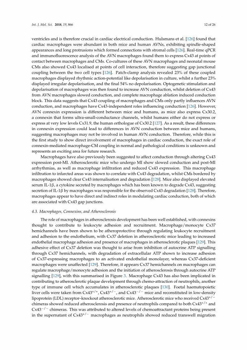
The role of macrophages in atherosclerosis development has been well established, with connexinsthought to contribute to leukocyte adhesion and recruitment. Macrophage/monocyte Cx37hemichannels have been shown to be atheroprotective through regulating leukocyte recruitmentand adhesion to the endothelium, with Cx37 deletion in atherosclerotic mice leading to increasedendothelial macrophage adhesion and presence of macrophages in atherosclerotic plaques [129]. Thisadhesive effect of Cx37 deletion was thought to arise from inhibition of autocrine ATP signallingthrough Cx37 hemichannels, with degradation of extracellular ATP shown to increase adhesionof Cx37-expressing macrophages to an activated endothelial monolayer, whereas Cx37-deficientmacrophages were unaffected [129]. Therefore, it appears Cx37 hemichannels on macrophages canregulate macrophage/monocyte adhesion and the initiation of atherosclerosis through autocrine ATPsignalling [129], with this summarised in Figure 3. Macrophage Cx43 has also been implicated incontributing to atherosclerotic plaque development through chemo-attraction of neutrophils, anothertype of immune cell which accumulates in atherosclerotic plaques [130]. Foetal haematopoieticliver cells were taken from Cx43+/+, Cx43+/−, and Cx43−/− mice and reconstituted in low-densitylipoprotein (LDL) receptor–knockout atherosclerotic mice. Atherosclerotic mice who received Cx43+/−
chimeras showed reduced atherosclerosis and presence of neutrophils compared to both Cx43+/+ andCx43−/− chimeras. This was attributed to altered levels of chemoattractant proteins being presentin the supernatant of Cx43+/− macrophages as neutrophils showed reduced transwell migration
Int. J. Mol. Sci. 2018, 19, 866 13 of 26
when assayed towards Cx43+/− macrophage supernatant [130]. Therefore, the expression level ofCx43 appears to influence the production of chemoattractant and chemotactic proteins, and futureresearch should be conducted to determine why and how specific levels of Cx43 lead to this irregularsecretion [130].
Figure 3. Autocrine ATP signalling via connexin 37 hemichannels prevents macrophage adhesionto endothelial cells. Yellow line = atherosclerotic plaque border. Abbreviations: Cx = Connexin.ATP = Adenosine Triphosphate.
Finally, a novel role of macrophage Cx43 in atherosclerotic development through favouringmacrophage phagocytosis of oxidised-LDLs has been suggested [131]. Cx43 has been shown toregulate phagocytosis in macrophages through Fc receptor-induced Ras homolog gene family, memberA (RhoA)-dependent rearrangement of the actin cytoskeleton [132]. Therefore, the suggestion is thatother receptors could drive phagocytosis, such as scavenger receptors, and this could lead to favoureduptake of oxidised-LDL [131]. LDL has previously been shown to enhance monocyte/macrophagephagocytosis of A Streptococcus, with increasing evidence that infection may also contribute to thedevelopment of atherosclerosis [133]. However, this has yet to be definitively established, and if Cx43directs phagocytic uptake via LDL is unknown.
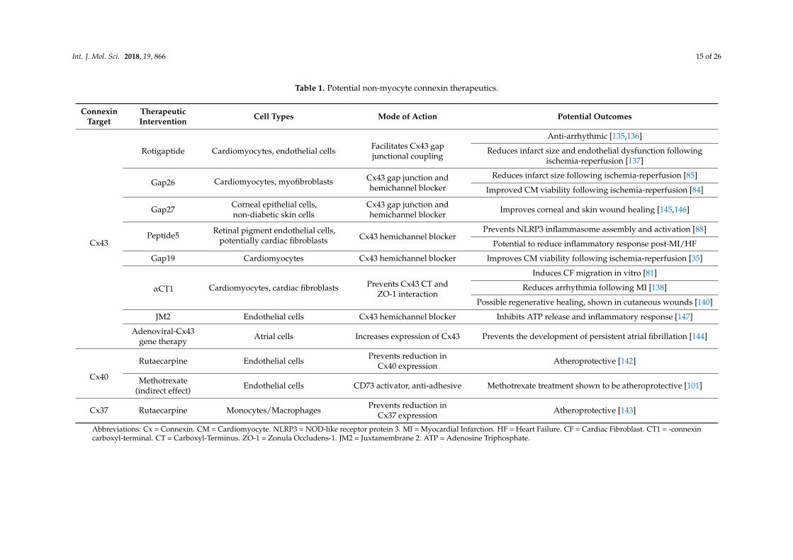
5. Non-Myocyte Gap Junctions and Hemichannels as Novel Therapeutic Targets
This review has so far described the roles of non-myocyte connexins, hemichannels,and homocellular/heterocellular gap junctions in the cardiovascular system during health anddisease. From potential influence over cardiac electrophysiology to atherosclerotic development,the possibility of developing therapeutics targeting connexins retains large interest. Substantial recentwork has been done to develop peptides that specifically target individual connexins, or target gapjunctions and hemichannels independently of each other [134]. Examples of these peptides includerotigaptide and αCT1. Rotigaptide works by promoting gap junctional coupling between cells andwas originally developed as an anti-arrhythmic therapy [135,136]. Rotigaptide has been shown toboth reduce myocardial infarct size and prevent impairment of endothelium-dependent vasomotorfunction following ischemia-reperfusion injury [137]. The Cx43 CT mimetic peptide αCT1, which wasmentioned earlier to promote CF migration, has been shown to reduce arrhythmia following cardiacinjury in mice by increasing the number of gap junctions at the infarct border zone [138]. Further to this,αCT1 treatment of submuscular biomedical implants reduces type 1 collagen deposition and number ofmyofibroblasts around the implant [139], whilst αCT1 treatment of skin wounds leads to faster woundclosure, reduced inflammation, swelling, neutrophil recruitment, and granulomas tissue, as wellas improved mechanical properties of the skin following closure [140]. Although this has not beeninvestigated in CFs, if similar results are seen then treatment with the αCT1 peptide could improve
Int. J. Mol. Sci. 2018, 19, 866 14 of 26
post-MI healing through reducing fibrosis and inflammation, whilst improving the mechanical strengthof the damaged tissue. Therefore, targeting the Cx43 CT could be a useful therapeutic target to improverecovery following MI by reducing the associated arrhythmia and adverse myocardial remodelling.
There is also potential to repurpose drugs currently used for other clinical purposes totarget connexins and connexin-based signalling. Two examples of this include simvastatin andmethotrexate. Simvastatin, a lipid-lowering drug, has been shown to improve Cx37 expressionduring hyperlipidaemia and be atheroprotective [113], and could therefore find additional use astreatment to prevent atherosclerotic plaque build-up, although more research is needed to determinehow lipid levels and statin drugs regulate connexin expression. Methotrexate is a widely useddrug in the treatment of cancer and inflammatory conditions [141], but has also been shown toreduce infarct size following ischemia-reperfusion in Cx40 knockout mice [100]. Down-regulationof Cx40 has been implicated in atherosclerosis as well [101]. Therefore, this suggests methotrexatecould find additional uses aiding recovery after ischemia-reperfusion and as an anti-atherosclerotictherapy through bypassing the need for Cx40 gap junctions to propagate anti-inflammatory signalsbetween ECs.
Another possible anti-atherosclerotic treatment involves rutaecarpine, a major quinazolinocarbolinealkaloid that is isolated from the traditional Chinese herbal medicine Evodia. Rutaecarpine hasbeen shown to be protective against atherosclerosis through regulating connexin expression in bothECs and monocytes. Pre-treatment of HUVECs with rutaecarpine prevents oxidised-LDL mediatedendothelial damage, the reduction in Cx37 and Cx40 expression, and monocyte adhesion [142], whilsta later follow-up study found rutaecarpine pre-treatment of monocytes recovered Cx37 expressionand anti-adhesive hemichannel ATP signalling following oxidised-LDL treatment [143]. Preventingendothelial oxidised-LDL damage and monocyte adhesion would therefore be useful to prevent theinitiation of atherosclerotic plaque build-up, halting disease progression at its early stages.
Gene therapy could also hold massive potential for treating connexin-related diseases, especiallythose where loss of connexin expression is associated with disease initiation/progression. For example,AF has been associated with down regulation of Cx43 and slowed conduction. However, atrial deliveryof adenoviral vectors containing Cx43 has been shown to increase atrial Cx43 expression and atrialconduction, consequently preventing the development of persistent AF [144]. Therefore, this highlightsthe ability of gene therapy to restore cardiac connexin expression in disease and reduce arrhythmogenicrisk. Some of the potential therapies targeting non-myocyte connexins are summarised in Table 1.
Int. J. Mol. Sci. 2018, 19, 866 15 of 26
Table 1. Potential non-myocyte connexin therapeutics.
ConnexinTarget
TherapeuticIntervention Cell Types Mode of Action Potential Outcomes
Cx43
Rotigaptide Cardiomyocytes, endothelial cells Facilitates Cx43 gapjunctional coupling
Anti-arrhythmic [135,136]
Reduces infarct size and endothelial dysfunction followingischemia-reperfusion [137]
Gap26 Cardiomyocytes, myofibroblasts Cx43 gap junction andhemichannel blocker
Reduces infarct size following ischemia-reperfusion [85]
Improved CM viability following ischemia-reperfusion [84]
Gap27 Corneal epithelial cells,non-diabetic skin cells
Cx43 gap junction andhemichannel blocker Improves corneal and skin wound healing [145,146]
Peptide5 Retinal pigment endothelial cells,potentially cardiac fibroblasts Cx43 hemichannel blocker
Prevents NLRP3 inflammasome assembly and activation [88]
Potential to reduce inflammatory response post-MI/HF
Gap19 Cardiomyocytes Cx43 hemichannel blocker Improves CM viability following ischemia-reperfusion [35]
αCT1 Cardiomyocytes, cardiac fibroblasts Prevents Cx43 CT andZO-1 interaction
Induces CF migration in vitro [81]
Reduces arrhythmia following MI [138]
Possible regenerative healing, shown in cutaneous wounds [140]
JM2 Endothelial cells Cx43 hemichannel blocker Inhibits ATP release and inflammatory response [147]
Adenoviral-Cx43gene therapy Atrial cells Increases expression of Cx43 Prevents the development of persistent atrial fibrillation [144]
Cx40
Rutaecarpine Endothelial cells Prevents reduction inCx40 expression Atheroprotective [142]
Methotrexate(indirect effect) Endothelial cells CD73 activator, anti-adhesive Methotrexate treatment shown to be atheroprotective [101]
Cx37 Rutaecarpine Monocytes/Macrophages Prevents reduction inCx37 expression Atheroprotective [143]
Abbreviations: Cx = Connexin. CM = Cardiomyocyte. NLRP3 = NOD-like receptor protein 3. MI = Myocardial Infarction. HF = Heart Failure. CF = Cardiac Fibroblast. CT1 = -connexincarboxyl-terminal. CT = Carboxyl-Terminus. ZO-1 = Zonula Occludens-1. JM2 = Juxtamembrane 2. ATP = Adenosine Triphosphate.
Int. J. Mol. Sci. 2018, 19, 866 16 of 26
This review has also highlighted possible roles of connexins in non-myocyte populations that havetherapeutic value which has yet to be investigated. An example of this is in the possible role of connexinmodulation of NO production. We previously mentioned how Cx37 and Cx40 are thought to interactdirectly with eNOS, and how reduced Cx37/40 expression reduces NO production [107], and thereforetargeting this interaction could provide therapeutic benefit. Patients with hypertension have beenshown to have reduced plasma NO levels [148], whilst the expression of Cx37 and Cx40 is also reducedin hypertensive animal models [149]. Therefore, investigations in to how Cx37 and Cx40 modulate NOproduction could lead to the development of treatments which target and facilitate this interaction,increasing NO production and reducing hypertension as a result. Further to this, glyceryl trinitrate(GTN; nitroglycerin) is a common treatment for patients with angina pectoris, MI or HF. The therapeuticbenefit of GTN arises from its ability to generate NO and cause vasodilation, restoring blood flow toischemic hearts [150]. However, sustained GTN treatment has been associated with increased risk ofexacerbating cardiac damage, possibly through inactivating the cardioprotective enzyme aldehydedehydrogenase 2 [150]. Therefore, targeting Cx37 and Cx40 to increase NO production could also bebeneficial in the treatment of angina pectoris, MI, and HF through providing the same vasodilatoryeffect, but avoiding aldehyde dehydrogenase 2 inactivation.
The recent discovery that macrophages contribute to AVN conduction could also be usefulfor treating arrhythmias and conduction abnormalities that result from inflammation of the heart.The inflammatory diseases lyme carditis, viral myocarditis, and cardiac sarcoidosis have all beenassociated with cardiac conduction abnormalities [151–153], whilst there has also been a reportedcase of a patient suffering from complete atrioventricular block after the regression of infectiousmyocarditis [154]. Macrophages also change phenotype and numbers in response to MI and HF, both ofwhich are associated with ventricular arrhythmia and sudden cardiac death [126]. More research isneeded to look at the contribution of macrophages to cardiac conduction during health and disease,if macrophages contribute to conduction block or conduction disorders associated with inflammatorydiseases, and if macrophages couple with CMs outside of the AVN. Targeting macrophage-CM couplingcould prove to be a useful target when treating inflammation-associated conduction abnormalities.
However, the biggest problem to overcome with connexin-based therapeutics is being ableto target individual connexins, gap junctions, or hemichannels, in a specific tissue and cell type,as connexins are widely expressed in different tissues. For example, Cx43 is abundantly expressed inmore than 35 tissues [155], and in the cardiovascular system alone is expressed in CMs, CFs, ECs andmacrophages. Therefore, targeting cell-specific connexins is important so as to not disrupt connexinfunctions in other cell types and tissues.
6. Conclusions
In conclusion, the evidence for non-myocyte gap junctions and hemichannels contributing tocardiovascular disease grows and is summarised schematically in Figure 4. However, targeting thesegap junctions or hemichannels is a promising new research avenue and has potential to develop newtherapeutics to reduce arrhythmias, aid recovery from cardiac injury, and prevent the development ofinflammatory vascular diseases such as atherosclerosis.
Int. J. Mol. Sci. 2018, 19, 866 17 of 26
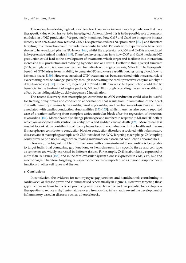
Figure 4. Schematic summary of connexin-based signalling in cardiac non-myocyte populations.Abbreviations: CM = Cardiomyocyte. CF = Cardiac Fibroblast. Cx = Connexin. SAN = Sinoatrial node.AVN = Atrioventricular node. ATP = Adenosine triphosphate.
Acknowledgments: We thank Daniel Stuckey, Centre for Advanced Biomedical Imaging, University CollegeLondon, for his helpful comments on the manuscript. This work was supported by the British Heart Foundation(grant FS/17/33/32931).
Author Contributions: Robert D. Johnson and Patrizia Camelliti composed the review paper.
Conflicts of Interest: The authors declare no conflict of interest.
Abbreviations
CMs CardiomyocytesCFs Cardiac FibroblastsECs Endothelial CellsSMCs Smooth Muscle CellsECM Extracellular MatrixCx ConnexinSAN Sinoatrial NodeAVN Atrioventricular NodeNa+ SodiumK+ PotassiumATP Adenosine triphosphateCa2+ CalciumDDR2 Discoidin Domain Receptor 2POSTN PeriostinFSP1 Fibroblast Specific Protein 1α-SMA α-Smooth Muscle Actin
Int. J. Mol. Sci. 2018, 19, 866 18 of 26
MI Myocardial InfarctionCx43FSP1KO Fibroblast-Specific Protein-1 Driven Conditional Cx43 KnockoutTGF-β Transforming Growth Factor-βNO Nitric OxideET-1 Endothelin-1eNOS Endothelial Nitric Oxide SynthaseVEGF Vascular Endothelial Growth FactorHHcy HyperhomocysteinemiaHcy HomocysteineHUVECs Human Umbilical Vein Endothelial CellsVCAM-1 Vascular Cell Adhesion Molecule-1IL InterleukinMCP-1 Monocyte Chemotactic Protein-1TNF-α Tumour Necrosis Factor-αLPS LipopolysaccharideRhoA Ras homolog gene family, member AHLSS High laminar shear stressLDL Low-Density LipoproteinDAMPs Danger-associated Molecular PatternsNOD Nucleotide Oligomerisation DomainNLRP3 NOD-like Receptor Protein-3αCT1 α-Connexin Carboxyl-TerminusZO-1 Zonula Occludens-1CT Carboxyl-TerminusAF Atrial FibrillationPDGFR Platelet Derived Growth Factor ReceptorGTN Glyceryl TrinitrateJM2 Juxtamembrane 2
References
1. Xin, M.; Olson, E.N.; Bassel-Duby, R. Mending broken hearts: Cardiac development as a basis for adult heartregeneration and repair. Nat. Rev. Mol. Cell Biol. 2013, 14, 529–541. [CrossRef] [PubMed]
2. Woodcock, E.A.; Matkovich, S.J. Cardiomyocytes structure, function and associated pathologies. Int. J.Biochem. Cell Biol. 2005, 37, 1746–1751. [CrossRef] [PubMed]
3. Lajiness, J.D.; Conway, S.J. The Dynamic Role of Cardiac Fibroblasts in Development and Disease.J. Cardiovasc. Transl. Res. 2012, 5, 739–748. [CrossRef] [PubMed]
4. Sager, H.B.; Kessler, T.; Schunkert, H. Monocytes and macrophages in cardiac injury and repair. J. Thorac.Dis. 2017, 9 (Suppl. 1), S30–S35. [CrossRef] [PubMed]
5. Epelman, S.; Lavine, K.J.; Beaudin, A.E.; Sojka, D.K.; Carrero, J.A.; Calderon, B.; Brija, T.; Gautier, E.L.;Ivanov, S.; Satpathy, A.T.; et al. Embryonic and adult-derived resident cardiac macrophages are maintainedthrough distinct mechanisms at steady state and during inflammation. Immunity 2014, 40, 91–104. [CrossRef][PubMed]
6. Pinto, A.R.; Paolicelli, R.; Salimova, E.; Gospocic, J.; Slonimsky, E.; Bilbao-Cortes, D.; Godwin, J.W.;Rosenthal, N.A. An Abundant Tissue Macrophage Population in the Adult Murine Heart with a DistinctAlternatively-Activated Macrophage Profile. PLoS ONE 2012, 7, e36814. [CrossRef] [PubMed]
7. Zhou, P.; Pu, W.T. Recounting cardiac cellular composition. Circ. Res. 2016, 118, 368–370. [CrossRef][PubMed]
8. Walsh, S.; Pontén, A.; Fleischmann, B.K.; Jovinge, S. Cardiomyocyte cell cycle control and growth estimationin vivo—An analysis based on cardiomyocyte nuclei. Cardiovasc. Res. 2010, 86, 365–373. [CrossRef] [PubMed]
9. Zhang, M.; Shah, A.M. ROS signalling between endothelial cells and cardiac cells. Cardiovasc. Res. 2014, 102,249–257. [CrossRef] [PubMed]
10. Camelliti, P.; Borg, T.K.; Kohl, P. Structural and functional characterisation of cardiac fibroblasts. Cardiovasc.Res. 2005, 65, 40–51. [CrossRef] [PubMed]
Int. J. Mol. Sci. 2018, 19, 866 19 of 26
11. Banerjee, I.; Fuseler, J.W.; Price, R.L.; Borg, T.K.; Baudino, T.A. Determination of cell types and numbersduring cardiac development in the neonatal and adult rat and mouse. Am. J. Physiol. Heart Circ. Physiol.2007, 293, H1883–H1891. [CrossRef] [PubMed]
12. Bergmann, O.; Zdunek, S.; Felker, A.; Salehpour, M.; Alkass, K.; Bernard, S.; Sjostrom, S.L.; Szewczykowska, M.;Jackowska, T.; dos Remedios, C.; et al. Dynamics of Cell Generation and Turnover in the Human Heart. Cell2015, 161, 1566–1575. [CrossRef] [PubMed]
13. Pinto, A.R.; Ilinykh, A.; Ivey, M.J.; Kuwabara, J.T.; Antoni, M.; Debuque, R.J.; Chandran, A.; Wang, L.;Arora, K.; Rosenthal, N.; et al. Revisiting Cardiac Cellular Composition. Circ. Res. 2015. [CrossRef]
14. Sheikh, F.; Ross, R.S.; Chen, J. Cell-Cell Connection to Cardiac Disease. Trends Cardiovasc. Med. 2009, 19,182–190. [CrossRef] [PubMed]
15. Jansen, J.A.; van Veen, T.A.B.; de Bakker, J.M.T.; van Rijen, H.V.M. Cardiac connexins and impulsepropagation. J. Mol. Cell. Cardiol. 2010, 48, 76–82. [CrossRef] [PubMed]
16. Goodenough, D.A.; Paul, D.L. Gap Junctions. Cold Spring Harb. Perspect. Biol. 2009, 1, a002576. [CrossRef][PubMed]
17. Söhl, G.; Willecke, K. Gap junctions and the connexin protein family. Cardiovasc. Res. 2004, 62, 228–232.[CrossRef] [PubMed]
18. Lo, C.W. Role of Gap Junctions in Cardiac Conduction and Development. Circ. Res. 2000, 87, 346. [CrossRef][PubMed]
19. Leybaert, L.; Lampe, P.D.; Dhein, S.; Kwak, B.R.; Ferdinandy, P.; Beyer, E.C.; Laird, D.W.; Naus, C.C.;Green, C.R.; Schulz, R. Connexins in Cardiovascular and Neurovascular Health and Disease:Pharmacological Implications. Pharmacol. Rev. 2017, 69, 396. [CrossRef] [PubMed]
20. Severs, N.J.; Bruce, A.F.; Dupont, E.; Rothery, S. Remodelling of gap junctions and connexin expression indiseased myocardium. Cardiovasc. Res. 2008, 80, 9–19. [CrossRef] [PubMed]
21. Desplantez, T. Cardiac Cx43, Cx40 and Cx45 co-assembling: Involvement of connexins epitopes in formationof hemichannels and Gap junction channels. BMC Cell Biol. 2017, 18, 3. [CrossRef] [PubMed]
22. Kostin, S.; Rieger, M.; Dammer, S.; Hein, S.; Richter, M.; Klovekorn, W.P.; Bauer, E.P.; Schaper, J. Gap junctionremodeling and altered connexin43 expression in the failing human heart. Mol. Cell. Biochem. 2003, 242,135–144. [CrossRef] [PubMed]
23. Dupont, E.; Matsushita, T.; Kaba, R.A.; Vozzi, C.; Coppen, S.R.; Khan, N.; Kaprielian, R.; Yacoub, M.H.;Severs, N.J. Altered connexin expression in human congestive heart failure. J. Mol. Cell. Cardiol. 2001, 33,359–371. [CrossRef] [PubMed]
24. Gutstein, D.E.; Morley, G.E.; Tamaddon, H.; Vaidya, D.; Schneider, M.D.; Chen, J.; Chien, K.R.; Stuhlmann, H.;Fishman, G.I. Conduction slowing and sudden arrhythmic death in mice with cardiac-restricted inactivationof connexin43. Circ. Res. 2001, 88, 333–339. [CrossRef] [PubMed]
25. Lerner, D.L.; Yamada, K.A.; Schuessler, R.B.; Saffitz, J.E. Accelerated onset and increased incidence ofventricular arrhythmias induced by ischemia in Cx43-deficient mice. Circulation 2000, 101, 547–552.[CrossRef] [PubMed]
26. Beardslee, M.A.; Lerner, D.L.; Tadros, P.N.; Laing, J.G.; Beyer, E.C.; Yamada, K.A.; Kleber, A.G.;Schuessler, R.B.; Saffitz, J.E. Dephosphorylation and intracellular redistribution of ventricular connexin43during electrical uncoupling induced by ischemia. Circ. Res. 2000, 87, 656–662. [CrossRef] [PubMed]
27. Gollob, M.H.; Jones, D.L.; Krahn, A.D.; Danis, L.; Gong, X.Q.; Shao, Q.; Liu, X.; Veinot, J.P.; Tang, A.S.;Stewart, A.F.; et al. Somatic mutations in the connexin 40 gene (GJA5) in atrial fibrillation. N. Engl. J. Med.2006, 354, 2677–2688. [CrossRef] [PubMed]
28. Yang, Y.Q.; Zhang, X.L.; Wang, X.H.; Tan, H.W.; Shi, H.F.; Jiang, W.F.; Fang, W.Y.; Liu, X. Connexin40 nonsensemutation in familial atrial fibrillation. Int. J. Mol. Med. 2010, 26, 605–610. [CrossRef] [PubMed]
29. Yang, Y.Q.; Liu, X.; Zhang, X.L.; Wang, X.H.; Tan, H.W.; Shi, H.F.; Jiang, W.F.; Fang, W.Y. Novel connexin40missense mutations in patients with familial atrial fibrillation. EP Europace 2010, 12, 1421–1427. [CrossRef][PubMed]
30. Lubkemeier, I.; Andrie, R.; Lickfett, L.; Bosen, F.; Stockigt, F.; Dobrowolski, R.; Draffehn, A.M.; Fregeac, J.;Schultze, J.L.; Bukauskas, F.F.; et al. The Connexin40A96S mutation from a patient with atrial fibrillationcauses decreased atrial conduction velocities and sustained episodes of induced atrial fibrillation in mice.J. Mol. Cell. Cardiol. 2013, 65, 19–32. [CrossRef] [PubMed]
Int. J. Mol. Sci. 2018, 19, 866 20 of 26
31. Kumai, M.; Nishii, K.; Nakamura, K.; Takeda, N.; Suzuki, M.; Shibata, Y. Loss of connexin45 causes a cushiondefect in early cardiogenesis. Development 2000, 127, 3501–3512. [PubMed]
32. Frank, M.; Wirth, A.; Andrie, R.P.; Kreuzberg, M.M.; Dobrowolski, R.; Seifert, G.; Offermanns, S.; Nickenig, G.;Willecke, K.; Schrickel, J.W. Connexin45 provides optimal atrioventricular nodal conduction in the adultmouse heart. Circ. Res. 2012, 111, 1528–1538. [CrossRef] [PubMed]
33. Sun, Y.; Hills, M.D.; Ye, W.G.; Tong, X.; Bai, D. Atrial Fibrillation-Linked Germline GJA5/Connexin40Mutants Showed an Increased Hemichannel Function. PLoS ONE 2014, 9, e95125. [CrossRef] [PubMed]
34. Patel, D.; Gemel, J.; Xu, Q.; Simon, A.R.; Lin, X.; Matiukas, A.; Beyer, E.C.; Veenstra, R.D. Atrialfibrillation-associated Connexin40 mutants make hemichannels and synergistically form gap junctionchannels with novel properties. FEBS Lett. 2014, 588, 1458–1464. [CrossRef] [PubMed]
35. Wang, N.; De Vuyst, E.; Ponsaerts, R.; Boengler, K.; Palacios-Prado, N.; Wauman, J.; Lai, C.P.; De Bock, M.;Decrock, E.; Bol, M.; et al. Selective inhibition of Cx43 hemichannels by Gap19 and its impact on myocardialischemia/reperfusion injury. Basic Res. Cardiol. 2013, 108, 309. [CrossRef] [PubMed]
36. Souders, C.A.; Bowers, S.L.K.; Baudino, T.A. Cardiac Fibroblast: The Renaissance Cell. Circ. Res. 2009, 105,1164–1176. [CrossRef] [PubMed]
37. Rog-Zielinska, E.A.; Norris, R.A.; Kohl, P.; Markwald, R. The Living Scar—Cardiac Fibroblasts and theInjured Heart. Trends Mol. Med. 2016, 22, 99–114. [CrossRef] [PubMed]
38. Gourdie, R.G.; Dimmeler, S.; Kohl, P. Novel therapeutic strategies targeting fibroblasts and fibrosis in heartdisease. Nat. Rev. Drug Discov. 2016, 15, 620–638. [CrossRef] [PubMed]
39. Baum, J.; Duffy, H.S. Fibroblasts and Myofibroblasts: What Are We Talking About? J. Cardiovasc. Pharmacol.2011, 57, 376–379. [CrossRef] [PubMed]
40. Kong, P.; Christia, P.; Saxena, A.; Su, Y.; Frangogiannis, N.G. Lack of specificity of fibroblast-specific protein 1in cardiac remodeling and fibrosis. Am. J. Physiol. Heart Circ. Physiol. 2013, 305, H1363–H1372. [CrossRef][PubMed]
41. Baudino, T.A.; Carver, W.; Giles, W.; Borg, T.K. Cardiac fibroblasts: Friend or foe? Am. J. Physiol. Heart Circ.Physiol. 2006, 291, H1015–H1026. [CrossRef] [PubMed]
42. Deb, A.; Ubil, E. Cardiac Fibroblast in Development and Wound Healing. J. Mol. Cell. Cardiol. 2014, 70, 47–55.[CrossRef] [PubMed]
43. Fan, D.; Takawale, A.; Lee, J.; Kassiri, Z. Cardiac fibroblasts, fibrosis and extracellular matrix remodeling inheart disease. Fibrogenesis Tissue Repair 2012, 5, 15. [CrossRef] [PubMed]
44. Travers, J.G.; Kamal, F.A.; Robbins, J.; Yutzey, K.E.; Blaxall, B.C. Cardiac Fibrosis: The Fibroblast Awakens.Circ. Res. 2016, 118, 1021–1040. [CrossRef] [PubMed]
45. Porter, K.E.; Turner, N.A. Cardiac fibroblasts: At the heart of myocardial remodeling. Pharmacol. Ther. 2009,123, 255–278. [CrossRef] [PubMed]
46. Talman, V.; Ruskoaho, H. Cardiac fibrosis in myocardial infarction—From repair and remodeling toregeneration. Cell Tissue Res. 2016, 365, 563–581. [CrossRef] [PubMed]
47. Louault, C.; Benamer, N.; Faivre, J.-F.; Potreau, D.; Bescond, J. Implication of connexins 40 and 43 in functionalcoupling between mouse cardiac fibroblasts in primary culture. Biochim. Biophys. Acta Biomembr. 2008, 1778,2097–2104. [CrossRef] [PubMed]
48. Gaudesius, G.; Miragoli, M.; Thomas, S.P.; Rohr, S. Coupling of cardiac electrical activity over extendeddistances by fibroblasts of cardiac origin. Circ. Res. 2003, 93, 421–428. [CrossRef] [PubMed]
49. Kohl, P.; Camelliti, P. Fibroblast-myocyte connections in the heart. Heart Rhythm 2012, 9, 461–464. [CrossRef][PubMed]
50. Camelliti, P.; Green, C.R.; LeGrice, I.; Kohl, P. Fibroblast network in rabbit sinoatrial node: Structural andfunctional identification of homogeneous and heterogeneous cell coupling. Circ. Res. 2004, 94, 828–835.[CrossRef] [PubMed]
51. Nisbet, A.M.; Camelliti, P.; Walker, N.L.; Burton, F.L.; Cobbe, S.M.; Kohl, P.; Smith, G.L. Prolongation ofatrio-ventricular node conduction in a rabbit model of ischaemic cardiomyopathy: Role of fibrosis andconnexin remodelling. J. Mol. Cell. Cardiol. 2016, 94, 54–64. [CrossRef] [PubMed]
52. Camelliti, P.; Devlin, G.P.; Matthews, K.G.; Kohl, P.; Green, C.R. Spatially and temporally distinct expressionof fibroblast connexins after sheep ventricular infarction. Cardiovasc. Res. 2004, 62, 415–425. [CrossRef][PubMed]
Int. J. Mol. Sci. 2018, 19, 866 21 of 26
53. Vasquez, C.; Mohandas, P.; Louie, K.L.; Benamer, N.; Bapat, A.C.; Morley, G.E. Enhanced fibroblast-myocyteinteractions in response to cardiac injury. Circ. Res. 2010, 107, 1011–1020. [CrossRef] [PubMed]
54. Quinn, T.A.; Camelliti, P.; Rog-Zielinska, E.A.; Siedlecka, U.; Poggioli, T.; O’Toole, E.T.; Knöpfel, T.; Kohl, P.Electrotonic coupling of excitable and nonexcitable cells in the heart revealed by optogenetics. Proc. Natl.Acad. Sci. USA 2016, 113, 14852–14857. [CrossRef] [PubMed]
55. Walker, N.L.; Burton, F.L.; Kettlewell, S.; Smith, G.L.; Cobbe, S.M. Mapping of epicardial activation in arabbit model of chronic myocardial infarction. J. Cardiovasc. Electrophysiol. 2007, 18, 862–868. [CrossRef][PubMed]
56. Kohl, P.; Camelliti, P.; Burton, F.L.; Smith, G.L. Electrical coupling of fibroblasts and myocytes: Relevance forcardiac propagation. J. Electrocardiol. 2005, 38 (Suppl. 4), 45–50. [CrossRef] [PubMed]
57. Stevenson, W.G. Ventricular Scars and Ventricular Tachycardia. Trans. Am. Clin. Climatol. Assoc. 2009, 120,403–412. [PubMed]
58. Morita, N.; Mandel, W.J.; Kobayashi, Y.; Karagueuzian, H.S. Cardiac fibrosis as a determinant of ventriculartachyarrhythmias. J. Arrhythm. 2014, 30, 389–394. [CrossRef] [PubMed]
59. Thomsen, M.B.; Calloe, K. Human atrial fibroblasts and their contribution to supraventricular arrhythmia.Physiol. Rep. 2016, 4, e12711. [CrossRef] [PubMed]
60. Kohl, P.; Gourdie, R.G. Fibroblast-myocyte electrotonic coupling: Does it occur in native cardiac tissue?J. Mol. Cell. Cardiol. 2014, 70, 37–46. [CrossRef] [PubMed]
61. Miragoli, M.; Gaudesius, G.; Rohr, S. Electrotonic Modulation of Cardiac Impulse Conduction byMyofibroblasts. Circ. Res. 2006, 98, 801. [CrossRef] [PubMed]
62. Miragoli, M.; Salvarani, N.; Rohr, S. Myofibroblasts Induce Ectopic Activity in Cardiac Tissue. Circ. Res.2007, 101, 755. [CrossRef] [PubMed]
63. Askar, S.F.; Bingen, B.O.; Swildens, J.; Ypey, D.L.; van der Laarse, A.; Atsma, D.E.; Zeppenfeld, K.; Schalij, M.J.;de Vries, A.A.; Pijnappels, D.A. Connexin43 silencing in myofibroblasts prevents arrhythmias in myocardialcultures: Role of maximal diastolic potential. Cardiovasc. Res. 2012, 93, 434–444. [CrossRef] [PubMed]
64. Zlochiver, S.; Muñoz, V.; Vikstrom, K.L.; Taffet, S.M.; Berenfeld, O.; Jalife, J. Electrotonic Myofibroblast-to-Myocyte Coupling Increases Propensity to Reentrant Arrhythmias in Two-Dimensional Cardiac Monolayers.Biophys. J. 2008, 95, 4469–4480. [CrossRef] [PubMed]
65. Mahoney, V.M.; Mezzano, V.; Mirams, G.R.; Maass, K.; Li, Z.; Cerrone, M.; Vasquez, C.; Bapat, A.; Delmar, M.;Morley, G.E. Connexin43 contributes to electrotonic conduction across scar tissue in the intact heart. Sci. Rep.2016, 6, 26744. [CrossRef] [PubMed]
66. Yue, L.; Xie, J.; Nattel, S. Molecular determinants of cardiac fibroblast electrical function and therapeuticimplications for atrial fibrillation. Cardiovasc. Res. 2011, 89, 744–753. [CrossRef] [PubMed]
67. Davies, L.; Jin, J.; Shen, W.; Tsui, H.; Shi, Y.; Wang, Y.; Zhang, Y.; Hao, G.; Wu, J.; Chen, S.; et al. Mkk4 Is aNegative Regulator of the Transforming Growth Factor Beta 1 Signaling Associated With Atrial Remodelingand Arrhythmogenesis With Age. J. Am. Heart Assoc. Cardiovasc. Cerebrovasc. Dis. 2014, 3, e000340. [CrossRef][PubMed]
68. Ashihara, T.; Haraguchi, R.; Nakazawa, K.; Namba, T.; Ikeda, T.; Nakazawa, Y.; Ozawa, T.; Ito, M.; Horie, M.;Trayanova, N.A. The Role of Fibroblasts in Complex Fractionated Electrograms During Persistent/PermanentAtrial Fibrillation: Implications for Electrogram-Based Catheter Ablation. Circ. Res. 2012, 110, 275–284.[CrossRef] [PubMed]
69. Zhan, H.; Xia, L. Excitation-Contraction Coupling between Human Atrial Myocytes with Fibroblasts andStretch Activated Channel Current: A Simulation Study. Comput. Math. Methods Med. 2013, 2013, 238676.[CrossRef] [PubMed]
70. Inamdar, A.A.; Inamdar, A.C. Heart Failure: Diagnosis, Management and Utilization. J. Clin. Med. 2016,5, 62. [CrossRef] [PubMed]
71. Aguilar, M.; Qi, X.Y.; Huang, H.; Nattel, S. Fibroblast Electrical Remodeling in Heart Failure and PotentialEffects on Atrial Fibrillation. Biophys. J. 2014, 107, 2444–2455. [CrossRef] [PubMed]
72. Bunch, T.J.; Hohnloser, S.H.; Gersh, B.J. Mechanisms of Sudden Cardiac Death in Myocardial InfarctionSurvivors. Circulation 2007, 115, 2451. [CrossRef] [PubMed]
73. Asazuma-Nakamura, Y.; Dai, P.; Harada, Y.; Jiang, Y.; Hamaoka, K.; Takamatsu, T. Cx43 contributes toTGF-β signaling to regulate differentiation of cardiac fibroblasts into myofibroblasts. Exp. Cell Res. 2009, 315,1190–1199. [CrossRef] [PubMed]
Int. J. Mol. Sci. 2018, 19, 866 22 of 26
74. Bujak, M.; Frangogiannis, N.G. The role of TGF-β Signaling in Myocardial Infarction and Cardiac Remodeling.Cardiovasc. Res. 2007, 74, 184–195. [CrossRef] [PubMed]
75. Zhang, Y.; Wang, H.; Kovacs, A.; Kanter, E.M.; Yamada, K.A. Reduced expression of Cx43 attenuatesventricular remodeling after myocardial infarction via impaired TGF-β signaling. Am. J. Physiol. Heart Circ.Physiol. 2010, 298, H477–H487. [CrossRef] [PubMed]
76. Zhang, Y.; Kanter, E.M.; Laing, J.G.; Aprhys, C.; Johns, D.C.; Kardami, E.; Yamada, K.A. Connexin43Expression Levels Influence Intercellular Coupling and Cell Proliferation of Native Murine CardiacFibroblasts. Cell Commun. Adhes. 2008, 15, 289–303. [CrossRef] [PubMed]
77. Mendoza-Naranjo, A.; Cormie, P.; Serrano, A.E.; Wang, C.M.; Thrasivoulou, C.; Sutcliffe, J.E.; Gilmartin, D.J.;Tsui, J.; Serena, T.E.; Phillips, A.R.; et al. Overexpression of the gap junction protein Cx43 as found in diabeticfoot ulcers can retard fibroblast migration. Cell Biol. Int. 2012, 36, 661–667. [CrossRef] [PubMed]
78. Wright, C.S.; van Steensel, M.A.; Hodgins, M.B.; Martin, P.E. Connexin mimetic peptides improve cellmigration rates of human epidermal keratinocytes and dermal fibroblasts in vitro. Wound Repair Regen. 2009,17, 240–249. [CrossRef] [PubMed]
79. Mori, R.; Power, K.T.; Wang, C.M.; Martin, P.; Becker, D.L. Acute downregulation of connexin43 at woundsites leads to a reduced inflammatory response, enhanced keratinocyte proliferation and wound fibroblastmigration. J. Cell Sci. 2006, 119 Pt 24, 5193–5203. [CrossRef] [PubMed]
80. Wright, C.S.; Pollok, S.; Flint, D.J.; Brandner, J.M.; Martin, P.E. The connexin mimetic peptide Gap27 increaseshuman dermal fibroblast migration in hyperglycemic and hyperinsulinemic conditions in vitro. J. Cell.Physiol. 2012, 227, 77–87. [CrossRef] [PubMed]
81. Ongstad, E.L.; Gourdie, R.G. Abstract 14887: A Connexin43 CT-Mimetic Peptide Increases FibroblastMigration in an in vitro Model of the Injury Border Zone. Circulation 2013, 128 (Suppl. 22), A14887.
82. Obert, E.; Strauss, R.; Brandon, C.; Grek, C.; Ghatnekar, G.; Gourdie, R.; Rohrer, B. Targeting the tight junctionprotein, zonula occludens-1, with the connexin43 mimetic peptide, αCT1, reduces VEGF-dependent RPEpathophysiology. J. Mol. Med. 2017, 95, 535–552. [CrossRef] [PubMed]
83. Rhett, J.M.; Jourdan, J.; Gourdie, R.G. Connexin 43 connexon to gap junction transition is regulated by zonulaoccludens-1. Mol. Biol. Cell 2011, 22, 1516–1528. [CrossRef] [PubMed]
84. Hawat, G.; Benderdour, M.; Rousseau, G.; Baroudi, G. Connexin 43 mimetic peptide Gap26 confers protectionto intact heart against myocardial ischemia injury. Pflüg. Arch. Eur. J. Physiol. 2010, 460, 583–592. [CrossRef][PubMed]
85. Johansen, D.; Cruciani, V.; Sundset, R.; Ytrehus, K.; Mikalsen, S.O. Ischemia Induces Closure of Gap JunctionalChannels and Opening of Hemichannels in Heart-derived Cells and Tissue. Cell. Physiol. Biochem. 2011, 28,103–114. [CrossRef] [PubMed]
86. Lu, D.; Soleymani, S.; Madakshire, R.; Insel, P.A. ATP released from cardiac fibroblasts via connexinhemichannels activates profibrotic P2Y(2) receptors. FASEB J. 2012, 26, 2580–2591. [CrossRef] [PubMed]
87. Butts, B.; Gary, R.A.; Dunbar, S.B.; Butler, J. The importance of NLRP3 inflammasome in heart failure. J. Card.Fail. 2015, 21, 586–593. [CrossRef] [PubMed]
88. Mugisho, O.O.; Green, C.R.; Kho, D.T.; Zhang, J.; Graham, E.S.; Acosta, M.L.; Rupenthal, I.D.The inflammasome pathway is amplified and perpetuated in an autocrine manner through connexin43hemichannel mediated ATP release. Biochim. Biophys. Acta Gen. Subj. 2018, 1862, 385–393. [CrossRef][PubMed]
89. Bracey, N.A.; Gershkovich, B.; Chun, J.; Vilaysane, A.; Meijndert, H.C.; Wright, J.R.; Fedak, P.W.; Beck, P.L.;Muruve, D.A.; Duff, H.J. Mitochondrial NLRP3 Protein Induces Reactive Oxygen Species to Promote SmadProtein Signaling and Fibrosis Independent from the Inflammasome. J. Biol. Chem. 2014, 289, 19571–19584.[CrossRef] [PubMed]
90. Sandanger, O.; Ranheim, T.; Vinge, L.E.; Bliksoen, M.; Alfsnes, K.; Finsen, A.V.; Dahl, C.P.; Askevold, E.T.;Florholmen, G.; Christensen, G.; et al. The NLRP3 inflammasome is up-regulated in cardiac fibroblasts andmediates myocardial ischaemia-reperfusion injury. Cardiovasc. Res. 2013, 99, 164–174. [CrossRef] [PubMed]
91. Kawaguchi, M.; Takahashi, M.; Hata, T.; Kashima, Y.; Usui, F.; Morimoto, H.; Izawa, A.; Takahashi, Y.;Masumoto, J.; Koyama, J.; et al. Inflammasome activation of cardiac fibroblasts is essential for myocardialischemia/reperfusion injury. Circulation 2011, 123, 594–604. [CrossRef] [PubMed]
Int. J. Mol. Sci. 2018, 19, 866 23 of 26
92. Zhang, Y.; Kanter, E.M.; Yamada, K.A. Remodeling of Cardiac Fibroblasts Following Myocardial InfarctionResults in Increased Gap Junction Intercellular Communication. Cardiovasc. Pathol. 2010, 19, e233–e240.[CrossRef] [PubMed]
93. Hsieh, P.C.H.; Davis, M.E.; Lisowski, L.K.; Lee, R.T. Endothelial-Cardiomyocyte Interactions in CardiacDevelopment and Repair. Ann. Rev. Physiol. 2006, 68, 51–66. [CrossRef] [PubMed]
94. Fountoulaki, K.; Dagres, N.; Iliodromitis, E.K. Cellular Communications in the Heart. Card. Fail. Rev. 2015, 1,64–68. [CrossRef] [PubMed]
95. Sandoo, A.; van Zanten, J.J.C.S.V.; Metsios, G.S.; Carroll, D.; Kitas, G.D. The Endothelium and Its Role inRegulating Vascular Tone. Open Cardiovasc. Med. J. 2010, 4, 302–312. [CrossRef] [PubMed]
96. Rajendran, P.; Rengarajan, T.; Thangavel, J.; Nishigaki, Y.; Sakthisekaran, D.; Sethi, G.; Nishigaki, I.The Vascular Endothelium and Human Diseases. Int. J. Biol. Sci. 2013, 9, 1057–1069. [CrossRef] [PubMed]
97. Liao, Y.; Day, K.H.; Damon, D.N.; Duling, B.R. Endothelial cell-specific knockout of connexin 43 causeshypotension and bradycardia in mice. Proc. Natl. Acad. Sci. USA 2001, 98, 9989–9994. [CrossRef] [PubMed]
98. Narmoneva, D.A.; Vukmirovic, R.; Davis, M.E.; Kamm, R.D.; Lee, R.T. Endothelial Cells Promote CardiacMyocyte Survival and Spatial Reorganization: Implications for Cardiac Regeneration. Circulation 2004, 110,962–968. [CrossRef] [PubMed]
99. Givvimani, S.; Qipshidze, N.; Tyagi, N.; Mishra, P.K.; Sen, U.; Tyagi, S.C. Synergism between arrhythmia andhyperhomo-cysteinemia in structural heart disease. Int. J. Physiol. Pathophysiol. Pharmacol. 2011, 3, 107–119.[PubMed]
100. Morel, S.; Braunersreuther, V.; Chanson, M.; Bouis, D.; Rochemont, V.; Foglia, B.; Pelli, G.; Sutter, E.;Pinsky, D.J.; Mach, F.; et al. Endothelial Cx40 limits myocardial ischaemia/reperfusion injury in mice.Cardiovasc. Res. 2014, 102, 329–337. [CrossRef] [PubMed]
101. Chadjichristos, C.E.; Scheckenbach, K.E.L.; van Veen, T.A.B.; Richani Sarieddine, M.Z.; de Wit, C.; Yang, Z.;Roth, I.; Bacchetta, M.; Viswambharan, H.; Foglia, B.; et al. Endothelial-Specific Deletion of Connexin40Promotes Atherosclerosis by Increasing CD73-Dependent Leukocyte Adhesion. Circulation 2010, 121, 123–131.[CrossRef] [PubMed]
102. Fang, J.S.; Angelov, S.N.; Simon, A.M.; Burt, J.M. Cx40 Is Required for, and Cx37 Limits, PostischemicHindlimb Perfusion, Survival and Recovery. J. Vasc. Res. 2011, 49, 2–12. [CrossRef] [PubMed]
103. Fang, J.S.; Angelov, S.N.; Simon, A.M.; Burt, J.M. Compromised regulation of tissue perfusion andarteriogenesis limit, in an AT1R-independent fashion, recovery of ischemic tissue in Cx40−/− mice. Am. J.Physiol. Heart Circ. Physiol. 2013, 304, H816–H827. [CrossRef] [PubMed]
104. Gärtner, C.; Ziegelhöffer, B.; Kostelka, M.; Stepan, H.; Mohr, F.-W.; Dhein, S. Knock-down of endothelialconnexins impairs angiogenesis. Pharmacol. Res. 2012, 65, 347–357. [CrossRef] [PubMed]
105. Kameritsch, P.; Pogoda, K.; Ritter, A.; Münzing, S.; Pohl, U. Gap junctional communication controls theoverall endothelial calcium response to vasoactive agonists. Cardiovasc. Res. 2012, 93, 508–515. [CrossRef][PubMed]
106. Pfenniger, A.; Derouette, J.-P.; Verma, V.; Lin, X.; Foglia, B.; Coombs, W.; Roth, I.; Satta, N.;Dunoyer-Geindre, S.; Sorgen, P.; et al. Gap Junction Protein Cx37 Interacts With Endothelial Nitric OxideSynthase in Endothelial Cells. Arterioscler. Thromb. Vasc. Biol. 2010, 30, 827. [CrossRef] [PubMed]
107. Alonso, F.; Boittin, F.-X.; Bény, J.-L.; Haefliger, J.-A. Loss of connexin40 is associated with decreasedendothelium-dependent relaxations and eNOS levels in the mouse aorta. Am. J. Physiol. Heart Circ. Physiol.2010, 299, H1365–H1373. [CrossRef] [PubMed]
108. Sandow, S.L.; Hill, C.E. Incidence of Myoendothelial Gap Junctions in the Proximal and Distal MesentericArteries of the Rat Is Suggestive of a Role in Endothelium-Derived Hyperpolarizing Factor—MediatedResponses. Circ. Res. 2000, 86, 341. [CrossRef] [PubMed]
109. Emerson, G.G.; Segal, S.S. Electrical Coupling Between Endothelial Cells and Smooth Muscle Cells inHamster Feed Arteries. Circ. Res. 2000, 87, 474. [CrossRef] [PubMed]
110. Li, L.; Ma, K.-T.; Zhao, L.; Li, X.-Z.; Zhang, Z.-S.; Shi, W.-Y.; Zhu, H.; Wei, L.-L.; Si, J.-Q. Myoendothelialcoupling is unidirectional in guinea pig spiral modiolar arteries. Microvasc. Res. 2012, 84, 211–217. [CrossRef][PubMed]
111. Moore, K.J.; Tabas, I. Macrophages in the pathogenesis of atherosclerosis. Cell 2011, 145, 341–355. [CrossRef][PubMed]
Int. J. Mol. Sci. 2018, 19, 866 24 of 26
112. Tabas, I.; García-Cardeña, G.; Owens, G.K. Recent insights into the cellular biology of atherosclerosis. J. CellBiol. 2015, 209, 13. [CrossRef] [PubMed]
113. Pfenniger, A.; Wong, C.; Sutter, E.; Cuhlmann, S.; Dunoyer-Geindre, S.; Mach, F.; Horrevoets, A.J.; Evans, P.C.;Krams, R.; Kwak, B.R. Shear stress modulates the expression of the atheroprotective protein Cx37 inendothelial cells. J. Mol. Cell. Cardiol. 2012, 53, 299–309. [CrossRef] [PubMed]
114. Yeh, H.-I.; Lu, C.-S.; Wu, Y.-J.; Chen, C.-C.; Hong, R.-C.; Ko, Y.-S.; Shiao, M.-S.; Severs, N.J.; Tsai, C.-H.Reduced Expression of Endothelial Connexin37 and Connexin40 in Hyperlipidemic Mice. Arterioscler.Thromb. Vasc. Biol. 2003, 23, 1391. [CrossRef] [PubMed]
115. Cizek, S.M.; Bedri, S.; Talusan, P.; Silva, N.; Lee, H.; Stone, J.R. Risk Factors for Atherosclerosis and theDevelopment of Pre-Atherosclerotic Intimal Hyperplasia. Cardiovasc. Pathol. 2007, 16, 344–350. [CrossRef][PubMed]
116. Allagnat, F.; Dubuis, C.; Lambelet, M.; Le Gal, L.; Alonso, F.; Corpataux, J.-M.; Déglise, S.; Haefliger, J.-A.Connexin37 reduces smooth muscle cell proliferation and intimal hyperplasia in a mouse model of carotidartery ligation. Cardiovasc. Res. 2017, 113, 805–816. [CrossRef] [PubMed]
117. Fang, J.S.; Coon, B.G.; Gillis, N.; Chen, Z.; Qiu, J.; Chittenden, T.W.; Burt, J.M.; Schwartz, M.A.; Hirschi, K.K.Shear-induced Notch-Cx37-p27 axis arrests endothelial cell cycle to enable arterial specification. Nat. Commun.2017, 8, 2149. [CrossRef] [PubMed]
118. Burt, J.M.; Nelson, T.K.; Simon, A.M.; Fang, J.S. Connexin 37 profoundly slows cell cycle progression in ratinsulinoma cells. Am. J. Physiol. Cell Physiol. 2008, 295, C1103–C1112. [CrossRef] [PubMed]
119. Jacobsen, N.L.; Pontifex, T.K.; Li, H.; Solan, J.L.; Lampe, P.D.; Sorgen, P.L.; Burt, J.M. Regulation of Cx37channel and growth suppressive properties by phosphorylation. J. Cell Sci. 2017, 130, 3308–3321. [CrossRef][PubMed]
120. Okamoto, T.; Akiyama, M.; Takeda, M.; Akita, N.; Yoshida, K.; Hayashi, T.; Suzuki, K. Connexin32 protectsagainst vascular inflammation by modulating inflammatory cytokine expression by endothelial cells.Exp. Cell Res. 2011, 317, 348–355. [CrossRef] [PubMed]
121. Okamoto, T.; Kawamoto, E.; Takagi, Y.; Akita, N.; Hayashi, T.; Park, E.J.; Suzuki, K.; Shimaoka, M. Gapjunction-mediated regulation of endothelial cellular stiffness. Sci. Rep. 2017, 7, 6134. [CrossRef] [PubMed]
122. Harari, E.; Guo, L.; Smith, S.L.; Braumann, R.E.; Virmani, R.; Finn, A.V. Heart-resident macrophages: Arethey involved in the rhythm of every beat? J. Thorac. Dis. 2017, 9, 2264–2267. [CrossRef] [PubMed]
123. Swirski, F.K.; Robbins, C.S.; Nahrendorf, M. Development and Function of Arterial and Cardiac Macrophages.Trends Immunol. 2016, 37, 32–40. [CrossRef] [PubMed]
124. Dutta, P.; Nahrendorf, M. Monocytes in Myocardial Infarction. Arterioscler. Thromb. Vasc. Biol. 2015, 35,1066–1070. [CrossRef] [PubMed]
125. Shirai, T.; Hilhorst, M.; Harrison, D.G.; Goronzy, J.J.; Weyand, C.M. Macrophages in VascularInflammation—From Atherosclerosis to Vasculitis. Autoimmunity 2015, 48, 139–151. [CrossRef] [PubMed]
126. Hulsmans, M.; Clauss, S.; Xiao, L.; Aguirre, A.D.; King, K.R.; Hanley, A.; Hucker, W.J.; Wulfers, E.M.;Seemann, G.; Courties, G.; et al. Macrophages Facilitate Electrical Conduction in the Heart. Cell 2017, 169,510–522.e20. [CrossRef] [PubMed]
127. Temple, I.P.; Inada, S.; Dobrzynski, H.; Boyett, M.R. Connexins and the atrioventricular node. Heart Rhythm2013, 10, 297–304. [CrossRef] [PubMed]
128. De Jesus, N.M.; Wang, L.; Herren, A.W.; Wang, J.; Shenasa, F.; Bers, D.M.; Lindsey, M.L.; Ripplinger, C.M.Atherosclerosis exacerbates arrhythmia following myocardial infarction: Role of myocardial inflammation.Heart Rhythm 2015, 12, 169–178. [CrossRef] [PubMed]
129. Wong, C.W.; Christen, T.; Roth, I.; Chadjichristos, C.E.; Derouette, J.-P.; Foglia, B.F.; Chanson, M.;Goodenough, D.A.; Kwak, B.R. Connexin37 protects against atherosclerosis by regulating monocyte adhesion.Nat. Med. 2006, 12, 950. [CrossRef] [PubMed]
130. Morel, S.; Chanson, M.; Nguyen, T.D.; Glass, A.M.; Richani Sarieddine, M.Z.; Meens, M.J.; Burnier, L.;Kwak, B.R.; Taffet, S.M. Titration of the gap junction protein Connexin43 reduces atherogenesis. Thromb.Haemost. 2014, 112, 390–401. [CrossRef] [PubMed]
131. Pfenniger, A.; Chanson, M.; Kwak, B.R. Connexins in atherosclerosis. Biochim. Biophys. Acta Biomembr. 2013,1828, 157–166. [CrossRef] [PubMed]
Int. J. Mol. Sci. 2018, 19, 866 25 of 26
132. Anand, R.J.; Dai, S.; Gribar, S.C.; Richardson, W.; Kohler, J.W.; Hoffman, R.A.; Branca, M.F.; Li, J.; Shi, X.-H.;Sodhi, C.P.; et al. A Role for Connexin43 in Macrophage Phagocytosis and Host Survival after BacterialPeritoneal Infection. J. Immunol. 2008, 181, 8534–8543. [CrossRef] [PubMed]
133. Zhou, L.; Liu, L.; Yang, J.; Li, Y.; Bai, W.; Liu, N.; Li, W.; Gao, Y.; Xu, L.; Liu, Z.; et al. LDL acts as an opsoninenhancing the phagocytosis of group A Streptococcus by monocyte and whole human blood. Med. Microbiol.Immunol. 2016, 205, 155–162. [CrossRef] [PubMed]
134. Schulz, R.; Görge, P.M.; Görbe, A.; Ferdinandy, P.; Lampe, P.D.; Leybaert, L. Connexin 43 is an emergingtherapeutic target in ischemia/reperfusion injury, cardioprotection and neuroprotection. Pharmacol. Ther.2015, 153, 90–106. [CrossRef] [PubMed]
135. Kjølbye, A.L.; Haugan, K.; Hennan, J.K.; Petersen, J.S. Pharmacological Modulation of Gap Junction Functionwith the Novel Compound Rotigaptide: A Promising New Principle for Prevention of Arrhythmias. BasicClin. Pharmacol. Toxicol. 2007, 101, 215–230. [CrossRef] [PubMed]
136. Shiroshita-Takeshita, A.; Sakabe, M.; Haugan, K.; Hennan, J.K.; Nattel, S. Model-dependent effects of the gapjunction conduction-enhancing antiarrhythmic peptide rotigaptide (ZP123) on experimental atrial fibrillationin dogs. Circulation 2007, 115, 310–318. [CrossRef] [PubMed]
137. Pedersen, C.M.; Venkatasubramanian, S.; Vase, H.; Hyldebrandt, J.A.; Contractor, H.; Schmidt, M.R.;Bøtker, H.E.; Cruden, N.L.; Newby, D.E.; Kharbanda, R.K.; et al. Rotigaptide protects the myocardium andarterial vasculature from ischaemia reperfusion injury. Br. J. Clin. Pharmacol. 2016, 81, 1037–1045. [CrossRef][PubMed]
138. O’Quinn, M.P.; Palatinus, J.A.; Harris, B.S.; Hewett, K.W.; Gourdie, R.G. A Peptide Mimetic of the Connexin43Carboxyl-Terminus Reduces Gap Junction Remodeling and Induced Arrhythmia Following VentricularInjury. Circ. Res. 2011, 108, 704–715. [CrossRef] [PubMed]
139. Soder, B.L.; Propst, J.T.; Brooks, T.M.; Goodwin, R.L.; Friedman, H.I.; Yost, M.J.; Gourdie, R.G. The connexin43carboxyl-terminal peptide ACT1 modulates the biological response to silicone implants. Plast. Reconstr. Surg.2009, 123, 1440–1451. [CrossRef] [PubMed]
140. Ghatnekar, G.S.; O’Quinn, M.P.; Jourdan, L.J.; Gurjarpadhye, A.A.; Draughn, R.L.; Gourdie, R.G. Connexin43carboxyl-terminal peptides reduce scar progenitor and promote regenerative healing following skinwounding. Regen. Med. 2009, 4, 205–223. [CrossRef] [PubMed]
141. Figueiró, F.; de Oliveira, C.P.; Bergamin, L.S.; Rockenbach, L.; Mendes, F.B.; Jandrey, E.H.F.; Moritz, C.E.J.;Pettenuzzo, L.F.; Sévigny, J.; Guterres, S.S.; et al. Methotrexate up-regulates ecto-5′-nucleotidase/CD73 andreduces the frequency of T lymphocytes in the glioblastoma microenvironment. Purinergic Signal. 2016, 12,303–312. [CrossRef] [PubMed]
142. Peng, W.-J.; Liu, Y.; Yu, Y.-R.; Fu, Y.-Q.; Zhao, Y.; Kuang, H.-B.; Huang, Q.-R.; He, M.; Luo, D. Rutaecarpineprevented dysfunction of endothelial gap junction induced by Ox-LDL via activation of TRPV1. Eur. J.Pharmacol. 2015, 756, 8–14. [CrossRef] [PubMed]
143. Liu, Y.; Fu, Y.Q.; Peng, W.J.; Yu, Y.R.; Wu, Y.S.; Yan, H.; Huang, Q.R.; He, M.; Luo, D. Rutaecarpine Reversesthe Altered Connexin Expression Pattern Induced by Oxidized Low-density Lipoprotein in Monocytes.J. Cardiovasc. Pharmacol. 2016, 67, 519–525. [CrossRef] [PubMed]
144. Bikou, O.; Thomas, D.; Trappe, K.; Lugenbiel, P.; Kelemen, K.; Koch, M.; Soucek, R.; Voss, F.; Becker, R.;Katus, H.A.; et al. Connexin 43 gene therapy prevents persistent atrial fibrillation in a porcine model.Cardiovasc. Res. 2011, 92, 218–225. [CrossRef] [PubMed]
145. Elbadawy, H.M.; Mirabelli, P.; Xeroudaki, M.; Parekh, M.; Bertolin, M.; Breda, C.; Cagini, C.; Ponzin, D.;Lagali, N.; Ferrari, S. Effect of connexin 43 inhibition by the mimetic peptide Gap27 on corneal woundhealing, inflammation and neovascularization. Br. J. Pharmacol. 2016, 173, 2880–2893. [CrossRef] [PubMed]
146. Pollok, S.; Pfeiffer, A.-C.; Lobmann, R.; Wright, C.S.; Moll, I.; Martin, P.E.M.; Brandner, J.M. Connexin 43mimetic peptide Gap27 reveals potential differences in the role of Cx43 in wound repair between diabeticand non-diabetic cells. J. Cell. Mol. Med. 2011, 15, 861–873. [CrossRef] [PubMed]
147. Calder, B.W.; Rhett, J.M.; Bainbridge, H.; Fann, S.A.; Gourdie, R.G.; Yost, M.J. Inhibition of Connexin 43Hemichannel-Mediated ATP Release Attenuates Early Inflammation During the Foreign Body Response.Tissue Eng. Part A 2015, 21, 1752–1762. [CrossRef] [PubMed]
148. Shiekh, G.A.; Ayub, T.; Khan, S.N.; Dar, R.; Andrabi, K.I. Reduced nitrate level in individuals withhypertension and diabetes. J. Cardiovasc. Dis. Res. 2011, 2, 172–176. [CrossRef] [PubMed]
Int. J. Mol. Sci. 2018, 19, 866 26 of 26
149. Yeh, H.-I.; Lee, P.-Y.; Su, C.-H.; Tian, T.-Y.; Ko, Y.-S.; Tsai, C.-H. Reduced Expression of Endothelial Connexins43 and 37 in Hypertensive Rats Is Rectified After 7-Day Carvedilol Treatment. Am. J. Hypertens. 2006, 19,129–135. [CrossRef] [PubMed]
150. Ferreira, J.C.B.; Mochly-Rosen, D. Nitroglycerin Use in Myocardial Infarction Patients: Risks and Benefits.Circ. J. 2012, 76, 15–21. [CrossRef] [PubMed]
151. Scheffold, N.; Herkommer, B.; Kandolf, R.; May, A.E. Lyme Carditis—Diagnosis, Treatment and Prognosis.Deutsch. Ärzteblatt Int. 2015, 112, 202–208. [CrossRef]
152. Dennert, R.; Crijns, H.J.; Heymans, S. Acute viral myocarditis. Eur. Heart J. 2008, 29, 2073–2082. [CrossRef][PubMed]
153. Sekhri, V.; Sanal, S.; DeLorenzo, L.J.; Aronow, W.S.; Maguire, G.P. Cardiac sarcoidosis: A comprehensivereview. Arch. Med. Sci. 2011, 7, 546–554. [CrossRef] [PubMed]
154. Maury, P.; Chilon, T.; Dumonteil, N.; Fontan, A. Complete atrioventricular block persisting after regressionof infectious myocarditis. J. Electrocardiol. 2008, 41, 665–667. [CrossRef] [PubMed]
155. Banerjee, D. Connexin’s Connection in Breast Cancer Growth and Progression. Int. J. Cell Biol. 2016, 2016,9025905. [CrossRef] [PubMed]
© 2018 by the authors. Licensee MDPI, Basel, Switzerland. This article is an open accessarticle distributed under the terms and conditions of the Creative Commons Attribution(CC BY) license (http://creativecommons.org/licenses/by/4.0/).