Embed Size (px)
Citation preview
Role of succinate dehydrogenase in pheochromocytomas and paragangliomas
PhD thesis
Nikoletta Katalin Lendvai
Doctoral School of Clinical Medicine Semmelweis University
Supervisor: Attila Patócs MD, Ph.D, M.Sc Official reviewers: Attila Bokor, MD, Ph.D
Judit Berenténé Bene, MD, Ph.D
Head of the Final Examination Committee: Edit Búzás, MD, D.Sc Members of the Final Examination Committee: Nóra Hosszúfalusi, MD, Ph.D
Zsolt Rónai MD, Ph.D
Budapest 2016
1
APPENDIX
Abbreviation
1. INTRODUCTION 6
1.1. Succinate dehydrogenase 6
1.1.1. The function of succinate dehydrogenase and the role of succinate
in cell metabolism 6
1.1.2. The structure of succinate dehydrogenase 8
1.1.3. Chromosomal localization of SDH subunits encoding genes 10
1.2. Pheochromocytoma and paraganglioma 11
1.2.1. Definition, anatomical localisation 11
1.2.2 Clinical features 14
1.2.3. Etiology and genetic background of hereditary
pheochromocytomas and paragangliomas 15
1.2.3.1. Familial pheochromocytoma/ paraganglioma syndrome 15
1.2.3.2. Multiple endocrine neoplasia type 2A and 2B 18
1.2.3.3. von Hippel-Lindau syndrome 20
1.2.3.4. Neurofibromatosis type 1 21
1.2.3.5. Recently identified genes causing
pheochromocytoma/ paraganglioma 21
1.2.4. Possible genetic modifiers 24
1.2.5. Biochemical characteristics of pheochromocytomas/paragangliomas 24
1.2.6. Diagnosis 26
1.2.6.1. Physical examination and family history 26
1.2.6.2. Biochemical testing 26
1.2.6.3. Imaging studies 28
1.2.6.4. Molecular genetic testing 28
1.2.7. Treatment of pheochromocytoma and paraganglioma 29
2. OBJECTIVES 31
3. METHODS 32
2
3.1. Germline mutation prevalence in Hungarian patients with
pheochromocytoma and/or paraganglioma 32
3.1.1. Patients 32
3.1.2. The first Hungarian case with extra adrenal pheochromocytoma associated
with SDHD gene mutation 33
3.1.3. Genetic testing of the RET, VHL, SDHB, SDHC, SDHD, SDHAF2, MAX
and TMEM127 genes using Sanger sequencing 34
3.2. The G12S polymorphism of the SDHD gene as a phenotype modifier in
patients with MEN2A syndrome 35
3.2.1. Patients
3.2.1.1. Patients with MEN2 syndrome 35
3.2.1.2. Patients with sporadic MTC 36
3.2.1.3. Patients with apparently sporadic PHEO 36
3.2.2. Germline mutation screening of the RET, VHL, SDHB, and SDHD genes 37
3.2.3. Restriction fragment length polymorphism (RFLP) analysis for
identification the G12S polymorphism of the SDHD gene 37
3.2.4. Statistical analysis 38
3.3. Biochemical consequences of SDHx mutations,
succinate to fumarate ratio in SDHB/D associated paragangliomas 39
3.3.1. Materials 39
3.3.2. Silencing of SDHB in MPC and MTT cells 40
3.3.3. Western blotting 40
3.3.4. Metabolic measurements 41
3.3.5. Statistical analysis 42
4. RESULTS 43
4.1. Germline mutation prevalence in Hungarian patients with
pheochromocytoma and/or paraganglioma 43
4.1.1. Genotype-phenotype associations 44
4.1.2. The first Hungarian case with extra adrenal pheochromocytoma associated
with SDHD gene mutation 48
3
4.2. The G12S polymorphism of the SDHD gene as a phenotype modifier in
patients with MEN2A syndrome 52
4.3. Biochemical consequences of SDHx mutations,
succinate to fumarate ratio in SDHB/D associated paragangliomas 54
4.3.1. Succinate concentration 54
4.3.2. Fumarate concentration 54
4.3.3. Succinate to fumarate ratio 57
4.3.4. Succinate to fumarate ratio in plasma samples 58
4.3.5. Succinate to fumarate ratio in MPC and MPP cells 58
5. DISCUSSION 59
5.1. Germline mutation prevalence in Hungarian patients with
pheochromocytoma and/or paraganglioma 59
5.2. The G12S polymorphism of the SDHD gene as a phenotype modifier in
patients with MEN2A syndrome 63
5.3. Biochemical consequences of SDHx mutations,
succinate to fumarate ratio in SDHB/D associated paragangliomas 65
6. CONCLUSION 67
7. SUMMARY/ÖSSZEFOGLALÁS 69
8. BIBLIOGRAPHY 71
9. BIBLIOGRAPHY OF THE CANDIDATE’S PUBLICATIONS 87
10. ACKNOWLEDGEMENTS 88
4
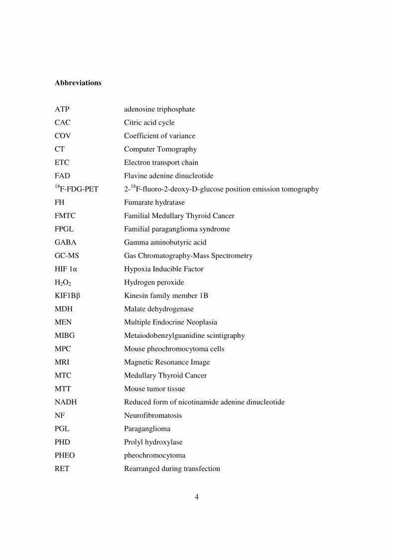
Abbreviations
ATP adenosine triphosphate
CAC Citric acid cycle
COV Coefficient of variance
CT Computer Tomography
ETC Electron transport chain
FAD Flavine adenine dinucleotide 18F-FDG-PET 2-18F-fluoro-2-deoxy-D-glucose position emission tomography
FH Fumarate hydratase
FMTC Familial Medullary Thyroid Cancer
FPGL Familial paraganglioma syndrome
GABA Gamma aminobutyric acid
GC-MS Gas Chromatography-Mass Spectrometry
HIF 1α Hypoxia Inducible Factor
H2O2 Hydrogen peroxide
KIF1Bβ Kinesin family member 1B
MDH Malate dehydrogenase
MEN Multiple Endocrine Neoplasia
MIBG Metaiodobenzylguanidine scintigraphy
MPC Mouse pheochromocytoma cells
MRI Magnetic Resonance Image
MTC Medullary Thyroid Cancer
MTT Mouse tumor tissue
NADH Reduced form of nicotinamide adenine dinucleotide
NF Neurofibromatosis
PGL Paraganglioma
PHD Prolyl hydroxylase
PHEO pheochromocytoma
RET Rearranged during transfection
5
RFLP Restriction fragment length polymorphism
ROS Reactive oxygen species
SD Standard deviation
SDH Succinate dehydrogenase
SDHA Succinate dehydrogenase subunit A
SDHB Succinate dehydrogenase subunit B
SDHC Succinate dehydrogenase subunit C
SDHD Succinate dehydrogenase subunit D
SDHx Succinate dehydrogenase subunits
VHL von Hippel-Lindau
6
1. INTRODUCTION
1.1. Succinate dehydrogenase and function
Succinate dehydrogenase (SDH) described first by Albert Szent-Györgyi in the middle of
the 1930’s is part of both the citric acid cycle (CAC) and respiratory electron transfer chain
(ETC)/oxidative phosphorylation. 1 SDH catalyzes the oxidation of succinate to fumarate in
the mitochondrial matrix and transfers electrons to ubiquinone without pumping protons
across the mitochondrial inner membrane. 1
1.1.1. The function of succinate dehydrogenase and the role of succinate
The Krebs cycle consists of chain of chemical reactions in order to generate energy for
cells. The whole CAC takes place in the mitochondrial matrix. It uses carbohydrates, fats,
amino acids and proteins to oxidase acetyl-coenzyme A (acetyl CoA). The main source of
acetyl CoA comes from the glycolysis, but can be derived from fatty acid oxidation as well.
The intermediates of the CAC serve as substrate for biosynthetic pathways.
The CAC result in a total of four molecules of ATP, ten molecules of NADH, and two
molecules of FADH2. Electrons from NADH and FADH2 are then transferred to molecular
oxygen through the oxidative phosphorylation. 2
Succinate dehydrogenase catalyzes the 7th step of the CAC (tricarboxylic acid cycle or
Krebs cycle). It catalyzes the oxidation of succinate to fumarate along with the reduction of
ubiquinone (Coenzyme Q) to ubiquinole, by transferring electrons thru FAD-FADH2
(Figure 1).
Succinate dehydrogenase or complex II is also involved in the oxidative phosphorylation
(OXPHOS) or electron transport chain, representing the major source of cellular energy.
The OXPHOS takes place in the inner membrane of the mitochondria and consists of four
complexes; complex I, II, III and IV. The fifth complex is the ATP synthase, which uses the
proton gradient to synthesize 32 to 34 ATP molecules. The flow of electrons from NADH
and FADH2 thru the protein complexes is associated with pumping protons to the
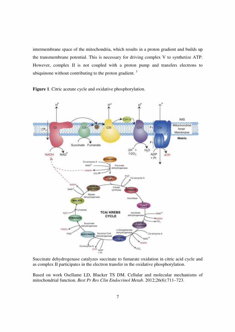
7
intermembrane space of the mitochondria, which results in a proton gradient and builds up
the transmembrane potential. This is necessary for driving complex V to synthetize ATP.
However, complex II is not coupled with a proton pump and transfers electrons to
ubiquinone without contributing to the proton gradient. 3
Figure 1. Citric acetate cycle and oxidative phosphorylation.
Succinate dehydrogenase catalyzes succinate to fumarate oxidation in citric acid cycle and as complex II participates in the electron transfer in the oxidative phosphorylation. Based on work Osellame LD, Blacker TS DM. Cellular and molecular mechanisms of mitochondrial function. Best Pr Res Clin Endocrinol Metab. 2012;26(6):711–723.
8
The substrate of SDH enzyme, succinate, is a distant product of the α-ketoglutarate
dehydrogenase complex. It is involved in several metabolic pathways including a
macrophage-specific metabolic pathway generating itaconate 4, it is connected with the
metabolism of branched-chain amino acids, heme synthesis, ketone bodies utilization and
the GABA shunt. 5,6 7
Regarding tumorigenesis, succinate is considered as a critical mediator of the hypoxic
response, and it has also been suggested that SDH plays an important role in ROS
homeostasis of cells producing superoxide and H2O2. 8
Succinate was also involved in posttranslational protein modification called succinylation
by this mechanism succinate might be involved in stabilization of certain proteins. In
specific tumours it was demonstrated that succinate stabilizes hypoxia inducible factor 1α, a
key transcription factor for regulating molecules which are involved in adaptation to
hypoxia and in facilitating blood vessel genesis (vascular endothelial growth factor, platelet
growth factor etc.). 9
In addition, succinate was discovered to exert its effects outside of cells in para- and
autocrine manners, mediated by the expression of at least one plasmalemmal succinate
receptor type. 10
Based on these fundamental processes it is not surprising that mutations of genes encoding
the subunits of SDH complex have been implicated in the pathogenesis of various diseases
including oxidative stress, tumour formation, neurodegeneration, hypoxia or “just simple
energy deficiency”.
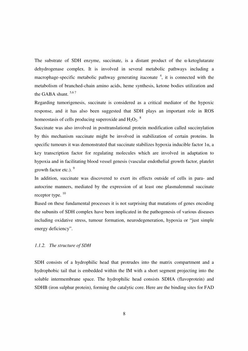
1.1.2. The structure of SDH
SDH consists of a hydrophilic head that protrudes into the matrix compartment and a
hydrophobic tail that is embedded within the IM with a short segment projecting into the
soluble intermembrane space. The hydrophilic head consists SDHA (flavoprotein) and
SDHB (iron sulphur protein), forming the catalytic core. Here are the binding sites for FAD
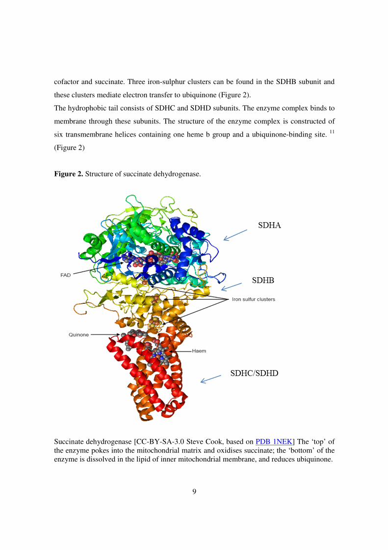
9
cofactor and succinate. Three iron-sulphur clusters can be found in the SDHB subunit and
these clusters mediate electron transfer to ubiquinone (Figure 2).
The hydrophobic tail consists of SDHC and SDHD subunits. The enzyme complex binds to
membrane through these subunits. The structure of the enzyme complex is constructed of
six transmembrane helices containing one heme b group and a ubiquinone-binding site. 11
(Figure 2)
Figure 2. Structure of succinate dehydrogenase.
Succinate dehydrogenase [CC-BY-SA-3.0 Steve Cook, based on PDB 1NEK] The ‘top’ of the enzyme pokes into the mitochondrial matrix and oxidises succinate; the ‘bottom’ of the enzyme is dissolved in the lipid of inner mitochondrial membrane, and reduces ubiquinone.
10
1.1.3. Chromosomal localization of genes encoding the SDH subunits
All four subunits of SDH or complex II are encoded by genes located in the nuclear
genome. SDHA encoding gene is mapped to the p arm of chromosome 5 at locus 15, SDHB
gene is localized on the p arm of chromosome 1 at locus 36. SDHC gene is encoded on the
q arm of chromosome 1 at locus 23. SDHD and SDHAF2 genes are encoded on the q arm
of chromosome 11 at locus 23.1 and 13, respectively. Number of exons and number of
amino acid residues are included in Table 1.
Genetic mutations of these genes are associated with familial paraganglioma syndrome,
childhood T-cell acute leukaemia and gastric stromal tumours. 12–16
Table 1. Chromosomal localization of succinate dehydrogenase subunits.
Gene Ensemble ID Chromosomal
localization Number of
exons Number of amino acids
SDHA ENSG00000073578 5p15 15 644 SDHB ENSG00000117118 1p36 8 280 SDHC ENSG00000143252 1q23 6 169 SDHD ENSG00000204370 11q23.1 4 159
SDHAF2 ENSG00000167985 11q13 4 166 SDHA - succinate dehydrogenase subunit A, SDHB - succinate dehydrogenase subunit B, SDHC - succinate dehydrogenase subunit C, SDHD - succinate dehydrogenase subunit D, SDHAF2 – succinate dehydrogenase complex assembly factor 2
11
1.2. Pheochromocytoma and paraganglioma
1.2.1. Definition, anatomical distribution
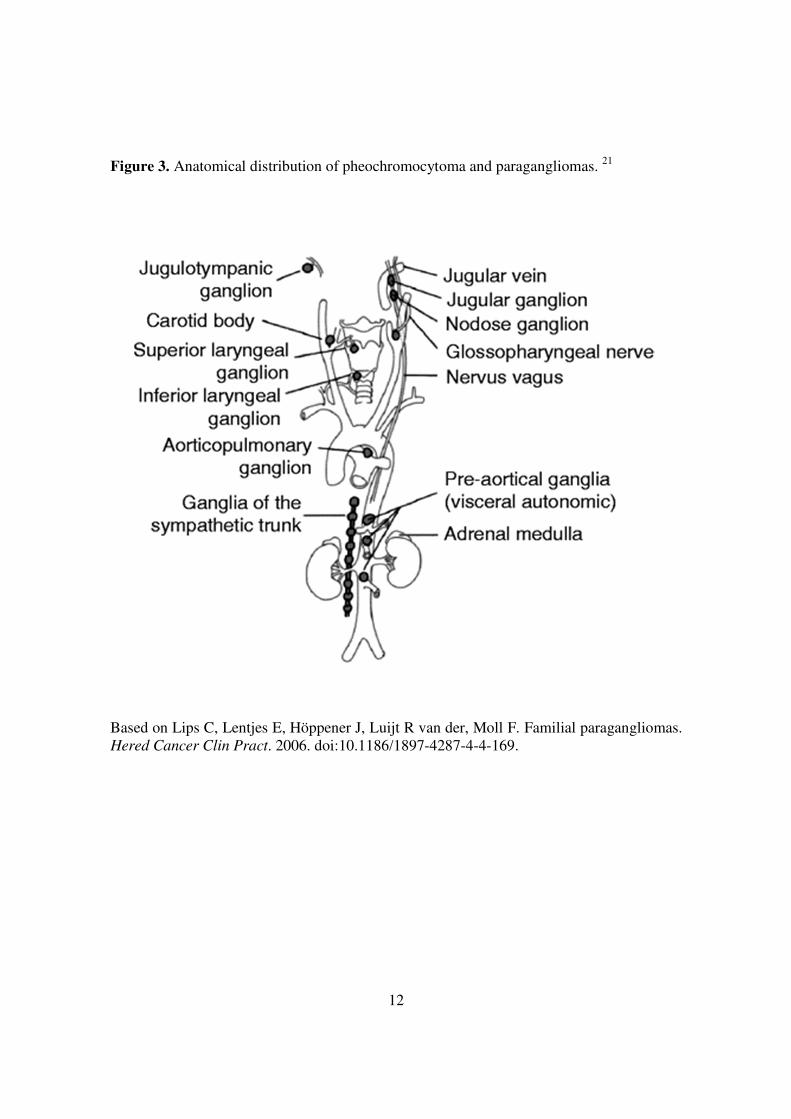
The term pheochromocytoma means “dusky-colored tumour” and it was historically
derived from the color change that occurs when the tumour tissue was immersed in
chromate salts. 17 Pheochromocytomas are rare catecholamine-producing tumours arising
from neural crest-derived chromaffin cells in the adrenal gland. Chromaffin cells are also
found in the sympathetic ganglions; sympathetic extra-adrenal paragangliomas are
generally confined to the lower mediastinum, abdomen, and pelvis (e.g. the aortic
chemoreceptors and the Zuckerkandl-organ), and are typically hormone secreting. In
contrast, parasympathetic paragangliomas are located predominantly on the skull base,
neck, and upper mediastinum (e.g. carotid artery/body). 18,19 20 (Figure 3)
12
Figure 3. Anatomical distribution of pheochromocytoma and paragangliomas. 21
Based on Lips C, Lentjes E, Höppener J, Luijt R van der, Moll F. Familial paragangliomas. Hered Cancer Clin Pract. 2006. doi:10.1186/1897-4287-4-4-169.
13
The prevalence of this tumour is estimated between 1:6500 to 1:2500 in the United States 22
but the autopsy reports suggest a prevalence of 1:2000 and suggest that many of these
tumours remain undiagnosed. 23,24 An increased frequency is noted in people subjected to
chronic hypoxia, living at higher-altitude regions or in the presence of respiratory or heart
diseases. 25 The incidence of pheochromocytoma is 2 to 8 per million persons per year. 26
Other tumours, as head-and-neck, abdominal, and pelvic PGL have an incidence of 0.5 per
million per year. 27 Paragangliomas in the Zuckerkandl-organ are the most common
sympathetic and carotid body tumours are the most common parasympathetic extra adrenal
paragangliomas. 28
Pheochromocytoma and paraganglioma can occur at all ages, but have a peek incidence at
the 4th and 5th decade, with an almost equal distribution between men and women. 29 30
Pheochromocytoma is present in 0.1% to 1% of patients with hypertension 31,32 and it is
present in approximately 5% of patients with incidentally discovered adrenal masses. 33
About 90% of PHEOs are unilateral, but bilateral tumours are seen in higher proportion in
syndromic cases. 27
The risk for malignant transformation is greater for extra-adrenal sympathetic
paragangliomas than for pheochromocytomas or skull base and neck paragangliomas. 20,25
It is difficult to determine the malignancy of PHEOs and PGLs as only the metastasis to
lymph node, bone, liver, or lung confirms malignancy. 19,34 However, pathological criteria,
as size, weight, presence of tumour necrosis, a greater than 4% of Ki-67 index and the
absence of S100 by immunohistochemistry have been shown to be associated with
malignancy. 35
Early diagnosis and resection of the tumour can cure most of the cases. The diagnosis is
difficult because clinical features/symptoms can mimic other diseases or can be very
unspecific or uncharacteristic.
14
1.2.2. Clinical features
Sympathetic paragangliomas secrete catecholamines, mainly adrenalin and noradrenalin;
parasympathetic paragangliomas are most often (ca. 95%) hormonally silent or have low
catecholamine production. Symptoms of PGL/PCC result either from the mass effects or
catecholamine hypersecretion. The main symptoms related to hormone hypersecretion are
the following:
• Hypertension (paroxysmal or sustained)
• Palpitation and tachycardia
• Sweating attacks
• Headache
• Facial flushing
• Chest and abdominal pain
• Anxiety and panic attacks
• Nausea
• Tremor
• Pallor
• Elevated fasting plasma glucose concentration
The symptoms are usually paroxysmal, but 50-60% of the patients have high blood
pressure between the episodes. 36 In some cases the symptoms are more severe and cause
cardiovascular and neurological manifestations. 37
Parasympathetic paragangliomas show a slow-growing, painless mass and patients develop
symptoms due to pressure of surrounding tissue or nerves: chocking, hoarseness, tickling
cough, Horner’s syndrome due to interruption of nervous tissue. 38
15
1.2.3. Etiology/Genetic background and associated hereditary syndromes
Pheochromocytomas and paragangliomas are mainly sporadic tumours, formerly about
10% of all tumours associated with hereditary syndromes, including multiple endocrine
neoplasia type 2 (MEN2), von Hippel-Lindau syndrome (VHL) and neurofibromatosis type
1 (NF1). 39 A small percent of PHEO/PGL associated with Carney-triad, Carney-Stratakis
syndrome and more rarely with MEN type 1. In the last decade several genes were
discovered as genetic causes of pheochromocytoma and paraganglioma, making the
prevalence of hereditary PHEO/PGL 30%–35% of all cases.
The common feature of hereditary syndromes is that they show an autosomal dominant
inheritance, meaning that the affected individual receives one mutant gene from one of
his/her parents. In some cases a de novo mutation in the germline of the patient may occur.
In sporadic (no germline disease-causing mutation) cases somatic, inactivating mutations
can also be identified. The most common genes and the associated syndromes are
summarized below.
1.2.3.1. Familial paraganglioma syndrome
Familial paraganglioma (FPGL) syndrome is caused by the germline heterozygous
mutations of the SDHx genes (SDHB, SDHC, SDHD, encoding subunits B, C and D,
respectively) 12,15,40 and the newly identified SDH5 gene. 16 SDHx genes encode subunits of
the mitochondrial complex II (succinate dehydrogenase, SDH), an enzyme involved in
oxidative phosphorylation and intracellular oxygen sensing and signalling.
Some differences between the mutation types have been observed. Most of the SDHB and
SDHC mutations are mutations which result in truncated protein or leading to amino acid
change of the iron sulphur clusters in the SDHB, while mutations of the SDHD gene are
more likely nonsense mutations and deletions/insertions. The genotype-phenotype
associations have been summarized in Table 2.
Biallelic mutation of the SDHA leads to Leigh syndrome, characterized by mitochondrial
encephalopathy and optic atrophy. In 2010 the first SDHA gene mutation was described
16
associating with abdominal PGL and head and neck PGL. 41 This latter finding added
SDHA to genes representing genetic susceptibility for PHEO/PGL.
SDHB mutations cause hereditary paraganglioma syndrome type 4. SDHB-related PGLs are
associated with abdominal, pelvic tumours, and show single presentation in two third of the
cases. Mutations in the SDHB gene are associated with a high risk of developing metastases 29 and about 20%–30% of patients already have metastatic disease at the time of the initial
diagnosis 42, while mean age at onset is typically 25-30 years. 43 The diagnosis of SDHB-
related PHEOs/PGLs is often delayed, most likely because of the less typical catecholamine
excess-related clinical presentations compared with other apparently sporadic or hereditary
PHEOs/PGLs. This is partially due to the fact that these tumours can have either a
biochemically silent phenotype, a low intratumoural catecholamine content, or a purely
dopaminergic phenotype. 42,44
SDHC gene mutations cause hereditary paraganglioma syndrome type 3 with
autosomal dominant inheritance. Classical clinical presentation for these tumours is solitary
head-neck paraganglioma with a low risk of malignancy. The mean age at diagnosis is 38
years. 45
Mutations of the SDHD gene are causing hereditary paraganglioma syndrome type
1. Its clinical presentation shows multiple abdominal and head-neck paragangliomas with
an age of onset at 28-31 years. 47, 48 SDHD-related PHEOs/PGLs, especially those derived
from the parasympathetic nervous system of the head and neck, are much less aggressive
than SDHB-related PGLs. It has to be mentioned that SDHD gene is maternally imprinted,
meaning that disease-causing mutations are inherited exclusively from the paternal side.
17
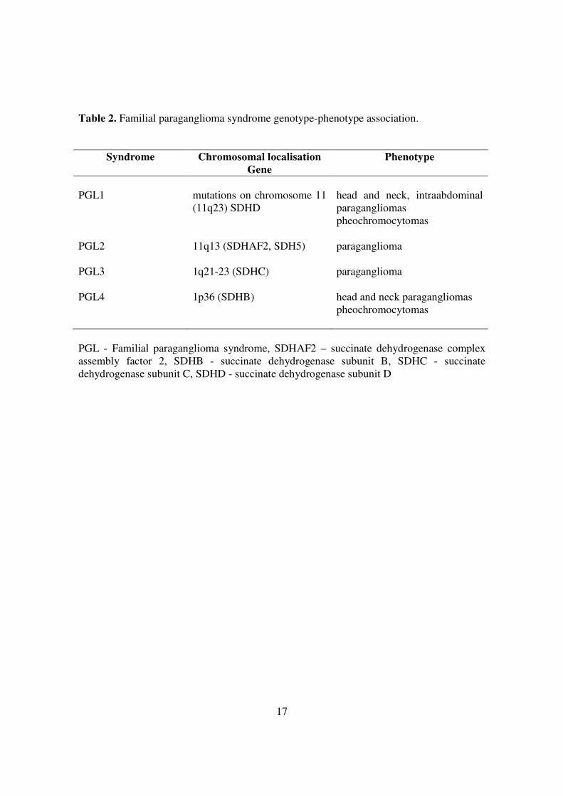
Table 2. Familial paraganglioma syndrome genotype-phenotype association.
Syndrome Chromosomal localisation Gene
Phenotype
PGL1
mutations on chromosome 11 (11q23) SDHD
head and neck, intraabdominal paragangliomas pheochromocytomas
PGL2 11q13 (SDHAF2, SDH5) paraganglioma
PGL3 1q21-23 (SDHC) paraganglioma
PGL4 1p36 (SDHB) head and neck paragangliomas pheochromocytomas
PGL - Familial paraganglioma syndrome, SDHAF2 – succinate dehydrogenase complex assembly factor 2, SDHB - succinate dehydrogenase subunit B, SDHC - succinate dehydrogenase subunit C, SDHD - succinate dehydrogenase subunit D
18
1.2.3.2. Multiple endocrine neoplasia type 2A and 2B
The RET (rearranged during transfection) proto-oncogene located on chromosomal region
10q11.2 consists of 21 exons encodes a receptor tyrosine kinase. 48 Germline, gain of
function mutations (mainly missense) of the RET proto-oncogene cause multiple endocrine
neoplasia type 2 (MEN2), an autosomal dominantly inherited disease with an approximate
prevalence of 2.5 per 100.000 in the general population. 48 The gene mutation are found in
specific, hot spot regions (exons: 10, 11, 13, 14, 15 and 16) of the RET proto-oncogene. 49
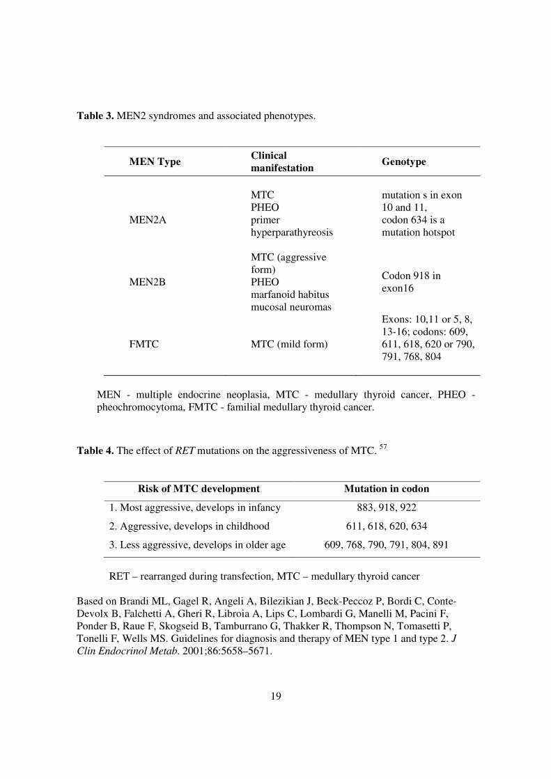
MEN2 has three subtypes: i) MEN2A, characterized by medullary thyroid carcinoma
(MTC), pheochromocytoma (PHEO) and primary hyperparathyroidism; ii) MEN2B, which
presents with the most aggressive MTC, pheochromocytoma, neuromas and marfanoid
phenotype; and iii) familial MTC (FMTC), the mildest form of MTC. (Table 3, Table 4)
Pheochromocytoma occurs in approximately 50% of the MEN2 patients. They are bilateral
in most of the cases and have a low rate for malignancy. 50,51 Usually, pheochromocytoma
is diagnosed at the same time as the medullary thyroid cancer, 52,53 while some more recent
studies showed that PHEO was diagnosed after the diagnosis of MTC 54,55, and very rarely
PHEO precedes MTC.
Analysis of the RET gene showed that missense mutations in codon 634 in exon 1 were the
most frequent mutations (mutation hot-spot) and the TGC634CGC (Cys-Arg) mutation
frequently associated with PHEO. 45,53,56
19
Table 3. MEN2 syndromes and associated phenotypes.
MEN Type Clinical manifestation
Genotype
MEN2A
MTC PHEO primer hyperparathyreosis
mutation s in exon 10 and 11, codon 634 is a mutation hotspot
MEN2B
MTC (aggressive form) PHEO marfanoid habitus mucosal neuromas
Codon 918 in exon16
FMTC MTC (mild form)
Exons: 10,11 or 5, 8, 13-16; codons: 609, 611, 618, 620 or 790, 791, 768, 804
MEN - multiple endocrine neoplasia, MTC - medullary thyroid cancer, PHEO - pheochromocytoma, FMTC - familial medullary thyroid cancer.
Table 4. The effect of RET mutations on the aggressiveness of MTC. 57
Risk of MTC development Mutation in codon
1. Most aggressive, develops in infancy 883, 918, 922
2. Aggressive, develops in childhood 611, 618, 620, 634
3. Less aggressive, develops in older age 609, 768, 790, 791, 804, 891
RET – rearranged during transfection, MTC – medullary thyroid cancer
Based on Brandi ML, Gagel R, Angeli A, Bilezikian J, Beck-Peccoz P, Bordi C, Conte-Devolx B, Falchetti A, Gheri R, Libroia A, Lips C, Lombardi G, Manelli M, Pacini F, Ponder B, Raue F, Skogseid B, Tamburrano G, Thakker R, Thompson N, Tomasetti P, Tonelli F, Wells MS. Guidelines for diagnosis and therapy of MEN type 1 and type 2. J
Clin Endocrinol Metab. 2001;86:5658–5671.
20
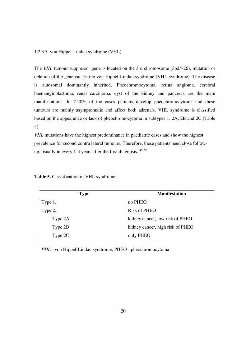
1.2.3.3. von Hippel-Lindau syndrome (VHL)
The VHL tumour suppressor gene is located on the 3rd chromosome (3p25-26), mutation or
deletion of the gene causes the von Hippel-Lindau syndrome (VHL-syndrome). The disease
is autosomal dominantly inherited. Pheochromocytoma, retina angioma, cerebral
haemangioblastoma, renal carcinoma, cyst of the kidney and pancreas are the main
manifestations. In 7-20% of the cases patients develop pheochromocytoma and these
tumours are mainly asymptomatic and affect both adrenals. VHL syndrome is classified
based on the appearance or lack of pheochromocytoma in subtypes 1, 2A, 2B and 2C (Table
5).
VHL mutations have the highest predominance in paediatric cases and show the highest
prevalence for second contra lateral tumours. Therefore, these patients need close follow-
up, usually in every 1-3 years after the first diagnosis. 45 58
Table 5. Classification of VHL syndrome.
Type Manifestation
Type 1. no PHEO
Type 2. Risk of PHEO
Type 2A
Type 2B
Type 2C
kidney cancer, low risk of PHEO
kidney cancer, high risk of PHEO
only PHEO
VHL - von Hippel-Lindau syndrome, PHEO - pheochromocytoma
21
1.2.3.4. Neurofibromatosis 1
Neurofibromatosis type 1 (von Recklinghausen disease) is characterized by skin lesions
(café-au-lait spots), growth of tumours along the nerve in the skin (neurofibromas) and
nodules in the iris (Lisch-nodules). Patients with NF1 syndrome have an increased risk for
developing optic glioma, leukemia and gastrointestinal tumours. The frequency of NF1 is
about 1:3000 to 1:4000 and therefore is one of the most frequent autosomal dominant
tumour syndrome, however about 50% of the cases result from new mutations.
Pheochromocytoma appears in 1% - 5.7% of all cases, although 50% of the patients with
high blood pressure have pheochromocytoma. 59
Inactivating, somatic mutations of NF1 were described in 41% of sporadic PHEO where
LOH at the NF1 locus were also described 60, suggesting that lack of function contributes to
the pathogenesis of PHEO.
The NF1 gene is located on chromosome 17 (17q11.2), the NF1 protein participates in the
regulation of the ras oncogene and its function resembles to tumour suppressor function.
Patients with NF1 syndrome are suggested clinical and laboratory surveillances for PHEO
every 3 years. 61
1.2.3.5. Recently identified, novel genes causing PHEO/PGL
TMEM127 (2q11) was identified in 2010 as a new pheochromocytoma susceptibility gene. 62 The encoded protein is a transmembrane protein which is involved in the mammalian
target of rapamycin signalling pathway. 46 The TMEM127 associated syndrome is
autosomal dominantly inherited. Clinical presentation is characterised by a presence of a
mainly unilateral PHEO in patients with no prior family history, however a recent study
showed that bilateral, extra-adrenal PGL cases were also described. 63,64
Pathogenic MAX (myc-associated factor X) (14q23) gene variants were shown to
predispose to PHEO/PGL. 65 MAX is a transcription factor involved in cellular
22
proliferation, differentiation and apoptosis. It seems that the phenotype associated with
MAX mutation shows the appearance of early onset (age <30 years), bilateral PHEO. 66
KIF1B was reported in a single case to be responsible for PHEO. This gene is frequently
implicated in inherited and sporadic neural crest tumours such as neuroblastomas. 20,67
EGLN1 (formerly known as PHD2), IDH1, HIF2A and FH genes have been reported to be
associated with hereditary PGL/PHEO, but their clinical significance is still unclear (Table
6). 68,69
23
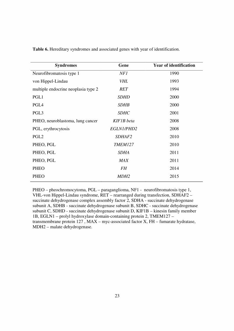
Table 6. Hereditary syndromes and associated genes with year of identification.
Syndromes Gene Year of identification
Neurofibromatosis type 1 NF1 1990
von Hippel-Lindau VHL 1993
multiple endocrine neoplasia type 2 RET 1994
PGL1 SDHD 2000
PGL4 SDHB 2000
PGL3 SDHC 2001
PHEO, neuroblastoma, lung cancer KIF1B beta 2008
PGL, erythrocytosis EGLN1/PHD2 2008
PGL2 SDHAF2 2010
PHEO, PGL TMEM127 2010
PHEO, PGL SDHA 2011
PHEO, PGL MAX 2011
PHEO FH 2014
PHEO MDH2 2015
PHEO – pheochromocytoma, PGL – paraganglioma, NF1 - neurofibromatosis type 1, VHL-von Hippel-Lindau syndrome, RET – rearranged during transfection, SDHAF2 – succinate dehydrogenase complex assembly factor 2, SDHA - succinate dehydrogenase subunit A, SDHB - succinate dehydrogenase subunit B, SDHC - succinate dehydrogenase subunit C, SDHD - succinate dehydrogenase subunit D, KIF1B – kinesin family member 1B, EGLN1 – prolyl hydroxylase domain-containing protein 2, TMEM127 – transmembrane protein 127 , MAX – myc-associated factor X, FH – fumarate hydratase, MDH2 – malate dehydrogenase.
24
1.2.4. Possible genetic modifiers
Germline gain of function mutations of the RET proto-oncogene cause MEN2. Several
important genotype–phenotype associations have been determined; the most commonly
affected codon, the codon 634 (nearly 85% of MEN2A cases), frequently associates with
PHEO and hyperparathyroidism, whereas mutations of codons 609, 611, 618, and 620
(accounting for 10–15% of MEN2A) usually associate with the milder form of MEN2. 70 53,56
However, the phenotypic heterogeneity observed even in members of the same family
suggests that other factors, for example genetic modifiers, may influence the clinical
manifestation of the disease. 71–74 As summarized earlier, mutations of SDHx genes are
causing PHEO/PGL syndromes. 75 Because both MTC and PHEO/PGL arise from neural
crest-derived precursor cells it may be hypothesised that same genetic factors may be
involved in both tumour types. Accumulation of amino-acid coding polymorphisms
(S163P in SDHB, G12S, and H50R in SDHD) have been found among patients with MTC,
especially in those with familial tumours. 76 In addition, these rare genetic variants have
been identified in patients with Cowden-like syndrome 77 and the H50R polymorphism has
been described in six members of a family with non-RET-associated C-cell hyperplasia and
hypercalcitoninemia. 78 These previous data may suggest a possible connection between
SDHx polymorphisms and familial MTC and/or C-cell hyperplasia/hypercalcitoninemia.
1.2.5. Biochemical characteristics
The diagnosis of SDHB-related PHEOs/PGLs is often delayed, most likely because of the
less typical catecholamine excess-related clinical presentations compared with other
apparently sporadic or hereditary PHEOs/PGLs. This is partially due to the fact that these
tumours can have either a biochemically silent phenotype, a low intratumoural
catecholamine content, or a purely dopaminergic phenotype. 42,44 In contrast, SDHD-related
25
PHEOs/PGLs, especially those derived from the parasympathetic nervous system of the
head and neck, are much less aggressive. The presence of SDH mutations impairs oxidative
phosphorylation and the Krebs cycle, resulting in metabolic abnormalities, including
succinate accumulation. 79
The Warburg effect
The Warburg effect is the 7th hallmark of most cancer types, along with persistent growth
signals, evasion of apoptosis, angiogenesis, insensitivity to anti-growth signals, unlimited
replication potential, invasion and metastasis. 80
In 1956 Dr. Otto Warburg described the effect that tumour cells show a high
glucose uptake in the presence of oxygen accompanied by lactic acid production, aerobic
glycolysis. 81 Dr. Warburg’s suspect that functional defect in the mitochondria causes
impaired respiration 82 has been proved in the past years. The increasing knowledge of the
Warburg effect lead us to new treatment 83 and diagnostic approaches, e.g. 2-18F-fluoro-2-
deoxy-D-glucose position emission tomography. The aerobic glycolysis yields only 2 ATP
molecules, but/and the tumour cells show an increased glycolysis.
Mutations in SDH genes lead to loss of function in SDH enzyme, which then lead to
succinate accumulation. Succinate inhibits prolyl hydroxylases (PHD), which has a role to
modify and degrade hypoxia inducible factor 1α (HIF1α). Increasing the levels of HIF1α
triggers tumourigenesis 84 85 79.
Presently, the ultimate diagnosis of these tumours is based on
immunohistochemistry to detect the presence or absence of the SDHB protein or genetic
testing for an SDH mutation or deletion 86,87. Although next-generation sequencing methods
will significantly reduce the costs of such testing, currently this genetic testing is still costly
and therefore limited or even unavailable in many countries. Neither method can be used
e.g., to predict therapeutic responses of these tumours, their resistance to various therapies,
for follow-up after a therapy is completed, or to assess their progression over time.
26
1.2.6. Diagnosis
The diagnosis of hereditary pheochromocytomas and paragangliomas is complex, meaning
physical examination, family history, biochemical and molecular genetic testing and
imaging studies.
Why is it important to suspect PHEO or PGL?
The consequences of catecholamine hypersecretion can lead to serious lesions or even
death. As 25-30% of the sporadic cases are caused by a germline mutation, family
screening can help to diagnose and treat the tumour earlier.
Malignancy is defined by existence of metastases; and patients with mutation in the SDHB
gene have a high risk for metastasis.
1.2.6.1. Physical examination and family history
A detailed family history and personal medical history is very important in cases with
PHEO/PGL. Personal medical history should cover the following: symptoms of
catecholamine excess, paroxysmal symptoms that may be triggered and enlarging masses.
1.2.6.2. Biochemical testing
In the diagnosis of PHEO and PGL we can use the biochemical characteristics of the
tumour, by measuring the levels of the secreted hormones and their major metabolites.
Plasma free and fractionated metanephrines are the first test to perform when PHEO/PGL is
suspected. (Table 7)
27
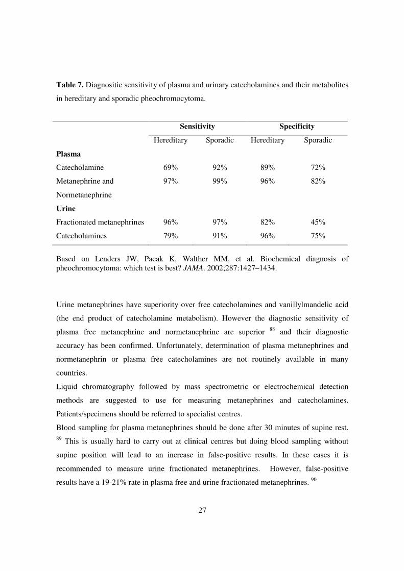
Table 7. Diagnositic sensitivity of plasma and urinary catecholamines and their metabolites
in hereditary and sporadic pheochromocytoma.
Sensitivity Specificity
Hereditary Sporadic Hereditary Sporadic
Plasma
Catecholamine
69%
92%
89%
72%
Metanephrine and
Normetanephrine
97% 99% 96% 82%
Urine
Fractionated metanephrines
96%
97%
82%
45%
Catecholamines 79% 91% 96% 75%
Based on Lenders JW, Pacak K, Walther MM, et al. Biochemical diagnosis of pheochromocytoma: which test is best? JAMA. 2002;287:1427–1434.
Urine metanephrines have superiority over free catecholamines and vanillylmandelic acid
(the end product of catecholamine metabolism). However the diagnostic sensitivity of
plasma free metanephrine and normetanephrine are superior 88 and their diagnostic
accuracy has been confirmed. Unfortunately, determination of plasma metanephrines and
normetanephrin or plasma free catecholamines are not routinely available in many
countries.
Liquid chromatography followed by mass spectrometric or electrochemical detection
methods are suggested to use for measuring metanephrines and catecholamines.
Patients/specimens should be referred to specialist centres.
Blood sampling for plasma metanephrines should be done after 30 minutes of supine rest. 89 This is usually hard to carry out at clinical centres but doing blood sampling without
supine position will lead to an increase in false-positive results. In these cases it is
recommended to measure urine fractionated metanephrines. However, false-positive
results have a 19-21% rate in plasma free and urine fractionated metanephrines. 90
28
In 50% of the patients with pheochromocytoma, both normetanephrine and metanephrine
are elevated; by at least 3 fold or more above the upper cut-off.
The clonidine test is recommended to distinguish the false-positive cases from the true-
positive ones.
Some medications can interfere with these measurements or with the catecholamine
disposition, not to mention physiological stress with severe conditions.
All positive cases should be followed up, and sometimes second, confirmatory
determinations are required.
1.2.6.3. Imaging studies
Imaging studies are recommended to locate the tumour in patients with positive
biochemical tests. However, only imaging studies can identify/locate the tumour in patients
with biochemically negative results.
CT is the first choice imaging modality for the thorax, abdomen and pelvis, its sensitivity is
88-100%. 91 Tumours greater than 5mm can already be detected by CT.
MRI has a better sensitivity in patients with extra-adrenal, recurrent and metastatic tumours
and head and neck paragangliomas. 91–93 MRI is preferred in children, pregnant women and
has a sensitivity of almost 100%. Both methods are used for tumour staging as well. 91,94 123I-metaiodobenzylguanidine (123I-MIBG) scintigraphy is a functional imaging modality in
metastatic cases and is used when radiotherapy with 131I-MIBG is planned.
In SDHx-related tumours 2-18F-fluoro-2-deoxy-D-glucose positron emission tomography
(18F-FDG-PET) has superiority to other imaging techniques. 94
1.2.6.4. Molecular genetic testing
Due to the large number of genes responsible for the development of PHEO/PGL the
genetic testing remains a diagnostic challenge. Since 1990, 14 different susceptibility genes
have been reported. Both laboratory workload and cost of testing of all genes are still
significant despite of the lower price of molecular biological reagents. Phenotype oriented
29
guidelines allow us some priorization in the order of genes tested but after a negative result
the remaining genes should also be examined. Therefore, it would be ideal that after
exclusion of some syndrome-associated genes based on the obvious phenotype features (i.e.
because of typical manifestation the NF1 gene is rarely tested) all of the remaining genes
would be tested at the same time. Recent technical improvements in sequencing
technology, the next generation sequencing (NGS) platforms - allow us to use whole exome
or targeted resequencing of all these genes. 95 The usefulness of NGS has been
demonstrated not only in resequencing of already known genes, but also in discoveries of
novel genes associated with PHEO/PGL. Confirmation of results and a negative NGS result
does not exclude the possibility of mutations especially the presence of large deletions.
Therefore, the gold standard methodology for identification of pathogenic mutation is the
PCR amplification of the coding region of target genes followed by Sanger sequencing. For
large deletion analysis multiple ligation probe amplification (MLPA) should be also
performed. In addition, the Endocrine Society clinical practice guideline recommend the
use of a clinical feature-driven diagnostic algorithm to establish the priorities for specific
genetic testing in PHEO/PGL patients with suspected germline mutations delivered within
the framework of health-care. 91,96
1.2.7. Treatment of pheochromocytoma and paraganglioma
Surgery is the definitive treatment of PHEO and PGL, if the tumour location allows
resection. To prevent cardiovascular complications patients with hormonally functional
PHEO/PGL should receive preoperative blockade, the first choice should be α-adrenergic
receptor blockers. Calcium channel blockers are the most common add on drugs and β-
adrenergic receptor blockers are used in co-administration to control tachycardia. These
latter two drugs are not recommended to us in single medication. α-adrenergic receptor
blocker treatment should be administered at least 7 days before surgery. 97
For surgery, recommendations suggest minimally invasive techniques. Laparoscopic
adrenalectomy is the first choice, but invasive tumours or tumours with size over 6 cm are

30
recommended for open resection. Paragangliomas are suggest for open resection, although
in some cases (e.g.: small, non-invasive, location) laparoscopic resection can be done.
After surgery patient personalized follow up is necessary depending on the genetic results.
In syndromic cases, where often bilateral tumours develop, a minimal invasive
tumourectomy (adrenal sparing surgery) with left adrenal cortex tissue is advised. 98
31
2. OBJECTIVES
During my PhD training I aimed to collect and to summarize the evidence of the role of
SDHx variants in the pathogenesis of PHEO/PGL. My specific aims were:
2.1. to evaluate the role of Mutations of SDHx genes in Hungarian patients with
PHEO/PGL
• to determine the prevalence of germline mutations in the SDHx, SDHAF2, MAX and
TMEM127 genes in Hungarian patients with apparently sporadic PHEO/PGL.
to describe the detailed phenotype of the first Hungarian case with SDHD gene
mutation.
• to collect and to report the genotype-phenotype association in patients with
PHEO/PG.
• to identify novel mutations among Hungarian patients with Pheo/PGL
2.2. to test whether polymorphisms of SDHx genes are phenotype modifiers in patients
with MEN2A syndrome, therefore I aimed to
• to determine the prevalence of SDHx polymorphisms in patients with RET mutations
(MEN2 patietns), in patients with sporadic medullary thyroid cancer (MTC),
sporadic PHEO, healthy subjects
2.3. to identify the metabolic consequences of SDHx mutations/deletions in tumour
tissues and cell lines. In order to fulfil this aim I aimed:
• to measure the levels of the two Krebs cycle metabolites, succinate and fumarate, in
tumour tissue and in human plasma samples obtained from patients with Pheo/PGL
• to determine the succinate to fumarate ratio in mouse pheochromocytoma (MPC)
and mouse tumour tissue (MTT) cells
• to test whether this difference propose the implementation of succinate/fumarate
measurements in clinical diagnosis.
32
3. METHODS
3.1. Germline mutation prevalence in Hungarian patients with pheochromocytoma
and/or paraganglioma
3.1.1. Patients
Our database containing the clinical and laboratory data of 129 patients diagnosed and
followed up at the 2nd Department of Medicine, Faculty of Medicine, Semmelweis
University with clinical diagnosis of PHEO/PGL between 1998 and 2014 was reviewed in
order to select cases for comprehensive genetic testing. All patients underwent genetic
counselling and written informed consent was obtained before genetic analysis.
Of these patients, the clinical diagnosis was confirmed by pathological examination of the
surgically removed tumour tissues in 92 cases. Mutation screening of the RET and VHL
genes identified 4 RET mutation carriers and 4 patients with germline VHL mutations. 99–101
In two cases the specific phenotype features indicated neurofibromatosis type 1. These
patients were excluded from this current analysis and SDHB, SDHC, SDHD, SDHAF2,
MAX and TMEM127 mutation analysis was performed in 82 cases. The main demographic
and pathological data are summarized in Table 8.
33
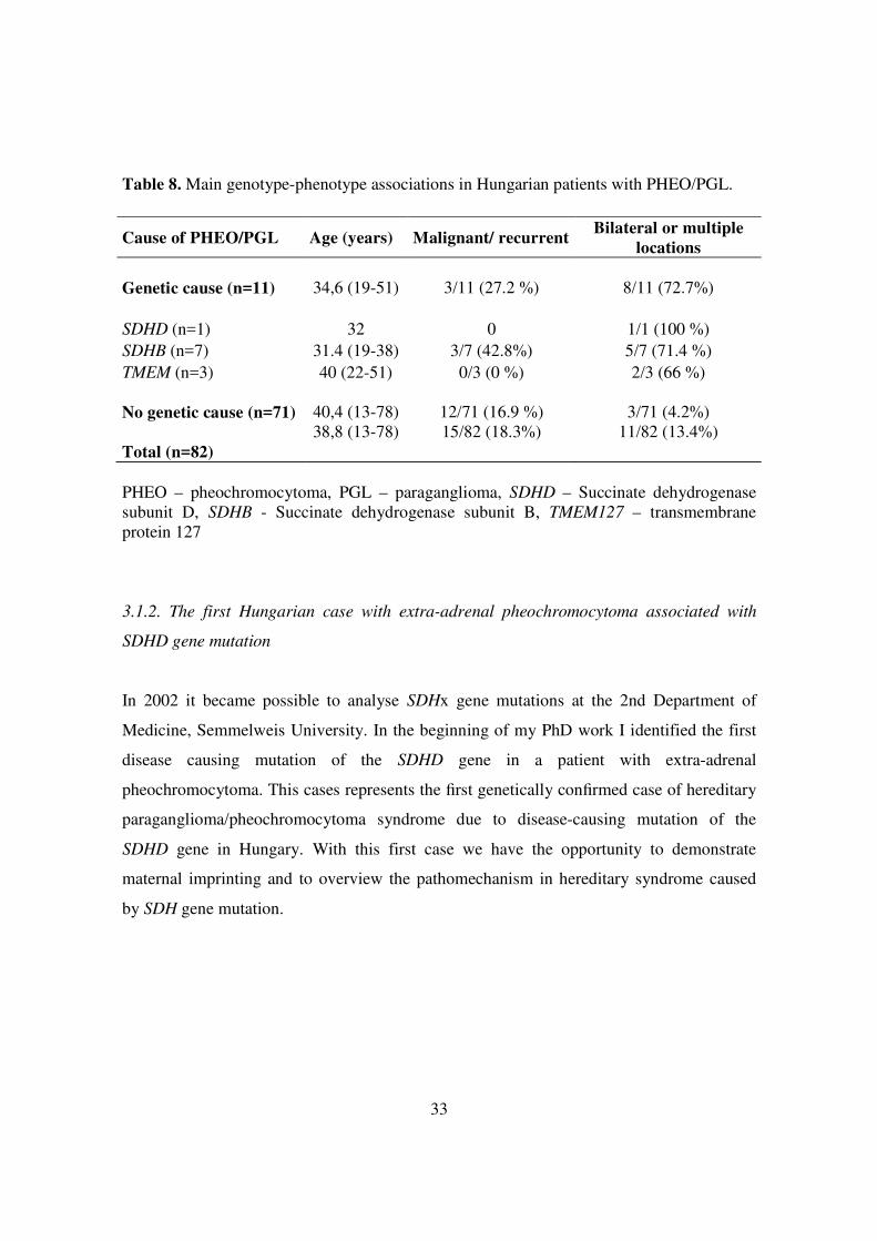
Table 8. Main genotype-phenotype associations in Hungarian patients with PHEO/PGL.
PHEO – pheochromocytoma, PGL – paraganglioma, SDHD – Succinate dehydrogenase subunit D, SDHB - Succinate dehydrogenase subunit B, TMEM127 – transmembrane protein 127
3.1.2. The first Hungarian case with extra-adrenal pheochromocytoma associated with
SDHD gene mutation
In 2002 it became possible to analyse SDHx gene mutations at the 2nd Department of
Medicine, Semmelweis University. In the beginning of my PhD work I identified the first
disease causing mutation of the SDHD gene in a patient with extra-adrenal
pheochromocytoma. This cases represents the first genetically confirmed case of hereditary
paraganglioma/pheochromocytoma syndrome due to disease-causing mutation of the
SDHD gene in Hungary. With this first case we have the opportunity to demonstrate
maternal imprinting and to overview the pathomechanism in hereditary syndrome caused
by SDH gene mutation.
Cause of PHEO/PGL Age (years) Malignant/ recurrent Bilateral or multiple
locations Genetic cause (n=11)
34,6 (19-51)
3/11 (27.2 %)
8/11 (72.7%)
SDHD (n=1) 32 0 1/1 (100 %) SDHB (n=7) 31.4 (19-38) 3/7 (42.8%) 5/7 (71.4 %) TMEM (n=3) 40 (22-51) 0/3 (0 %) 2/3 (66 %) No genetic cause (n=71) 40,4 (13-78) 12/71 (16.9 %) 3/71 (4.2%) Total (n=82)
38,8 (13-78)
15/82 (18.3%)
11/82 (13.4%)
34
3.1.3. Genetic testing of the RET, VHL, SDHB, SDHC, SDHD, SDHAF2, MAX and
TMEM127 Genes Using Sanger Sequencing
After genetic counselling and obtaining informed consent of all 82 patients, underwent
genetic testing for the SDHB, SDHC, SDHD, SDHAF2, MAX and TMEM127 using
conventional methods including PCR followed by Sanger sequencing. 99–101 Genomic DNA
was extracted from peripheral blood using commercially available DNA extraction kits
(DNA isolation from mammalian blood, Roche, or DNA isolation kit from blood, Qiagen
LTD). Bidirectional DNA sequencing of all these genes and large deletion analysis of the
SDHB, SDHC and SDHD genes were performed using multiplex ligation probe
amplification. 101
35
3.2. The G12S polymorphism of the SDHD gene as a phenotype modifier in patients
with MEN2A syndrome
3.2.1. Patients
Written informed consent was obtained from all patients and family members who
participated in the study. Patients underwent a complete clinical examination, laboratory
testing, including serum basal calcitonin measurement [hCalcitonin IRMA kit (Diagnostic
Systems Laboratories, Inc., Budapest, Hungary), reference range: male, < 15 pg/ml, female,
< 10 pg/ml until December 2007; and Liaison (Diasorin SPA, Stillwater, MN, USA),
reference value: male, < 18.9 pg/ml and female, < 6 pg/ml after January 2008], plasma
parathyroid hormone (Elecsys; Roche Diagnostics, Basel, Switzerland), urinary
catecholamine metabolites (high pressure liquid chromatography with electrochemical
detection), and imaging studies, including cervical ultrasonography, thoracal and abdominal
computed tomography (CT), and whole-body metaiodobenzylguanidine scintigraphy
(MIBG).
3.2.1.1. Patients with MEN2 syndrome
In total, 77 patients with germline RET proto-oncogene mutations who were members of 21
unrelated families with MEN2 syndrome were identified by genetic screening at our centre.
Of the 77 patients, 55 had MEN2A (mean age at diagnosis: 33.4±17 years; range: 7–76
years), three had MEN2B (mean age at diagnosis: 15.6±5 years; range: 10–20 years), and
19 had FMTC (mean age at diagnosis: 23.7±16.8 years; range: 2–57 years). The presence
of PHEO and MTC were confirmed by histological examination of surgically removed
tumours. Total thyroidectomy was performed in all patients with germline RET mutation in
the symptomatic group and was also offered to all individuals from the asymptomatic
group.
36
3.2.1.2. Patients with sporadic MTC
The study included 47 unrelated patients with histologically confirmed MTC evaluated
consecutively at the 2nd Department of Medicine, Faculty of Medicine, Semmelweis
University between 1998 and 2010. There were 15 men (age, mean ± SD, 44.7±13.3; range:
28–82 years) and 32 women (age, mean ± SD, 47.7±12,3; range: 23–76 years).
Preoperative evaluation included medical history, physical examination, thyroid and
abdominal ultrasonography, CT or magnetic resonance imaging (MRI), MIBG-
scintigraphy, routine biochemical testing, serum calcitonin measurements, and mutation
analysis of exons 10–14 of the RET gene.
3.2.1.3. Patients with apparently sporadic PHEO
The study included 48 unrelated patients with histologically confirmed sporadic adrenal
pheochromocytomas evaluated consecutively at the 2nd Department of Medicine, Faculty of
Medicine, Semmelweis University between 1998 and 2010. There were 16 men (age, mean
± SD, 36±14; range: 13–66 years) and 32 women (age, mean ± S.D, 42±14; range: 19–64
years). Pre-operative evaluation included medical history, physical examination, abdominal
ultrasonography, CT or MRI, MIBG-scintigraphy, routine biochemical testing, and 24 h
urinary catecholamine metabolite determination. The mutation analysis of RET exons 10–
14 and the entire VHL, SDHB, and SDHD genes revealed no disease-causing mutations.
Patients with confirmed VHL (five patients), SDHB (one patient), or SDHD (one patient)
mutations were excluded from the study. Five patients were initially thought to have
sporadic pheochromocytoma, but were later identified as having a disease-causing RET
mutation and were included in the study as RET mutation carriers. MTC, either by elevated
serum calcitonin or by postoperative histology, had been diagnosed in all of these patients.
Genetic counselling and genetic screening for all first-degree relatives have been offered.
37
3.2.2. Germline mutation screening of the RET, VHL, SDHB, and SDHD genes
Genomic DNA was isolated from peripheral blood using the Roche DNA Isolation Kit
(Roche Diagnostics GmbH, Mannheim, Germany) and QIAamp DNA Blood Mini Kit
(Qiagen Inc., Valencia, CA, USA) in accordance with the manufacturers’ instructions. RET
proto-oncogene mutations were detected by direct sequencing as previously reported 99 102.
Mutation analysis of the VHL, SDHB, and SDHD genes in cases of apparently sporadic
PHEO were performed by direct sequencing of the entire coding region of the VHL, SDHB,
and SDHD genes, as previously reported 99,101 , and large deletion analysis of the VHL,
SDHB, SDHC, and SDHD genes performed using multiplex ligation probe amplification. 101
3.2.3. Restriction fragment length polymorphism (RFLP) analysis for identification the
G12S polymorphism of the SDHD gene
The nucleotide change of G to A, which corresponds to the G12S polymorphism, results in
the preservation of the BanI restriction cleavage site. Therefore, digestion with the BanI
restriction enzyme (New England BioLabs Inc., Ipswich, MA, USA) for 90 min at 37°C
was performed after polymerase chain reaction (PCR) amplification of exon 1 of the SDHD
gene for genotyping of RET mutation carriers, sporadic MTC patients, and 100 controls
(Figure 4). Samples from patients with positive results were examined by direct DNA
sequencing. The results obtained with both methods were the same in all cases.
38
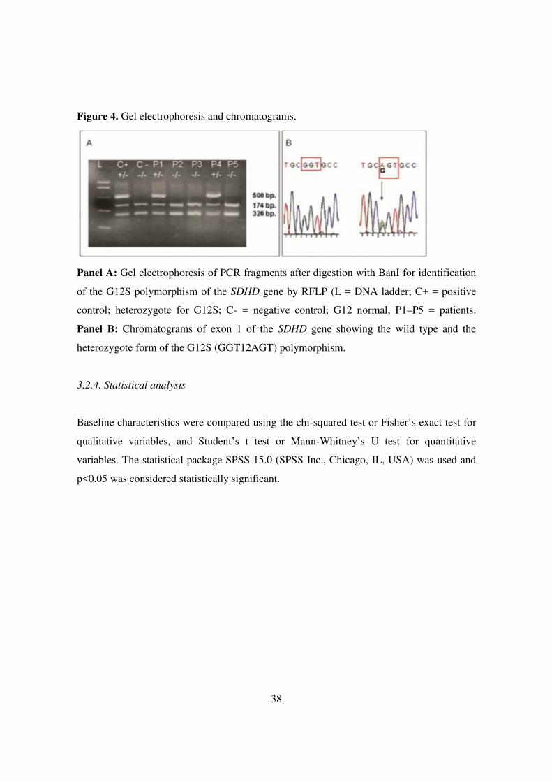
Figure 4. Gel electrophoresis and chromatograms.
Panel A: Gel electrophoresis of PCR fragments after digestion with BanI for identification
of the G12S polymorphism of the SDHD gene by RFLP (L = DNA ladder; C+ = positive
control; heterozygote for G12S; C- = negative control; G12 normal, P1–P5 = patients.
Panel B: Chromatograms of exon 1 of the SDHD gene showing the wild type and the
heterozygote form of the G12S (GGT12AGT) polymorphism.
3.2.4. Statistical analysis
Baseline characteristics were compared using the chi-squared test or Fisher’s exact test for
qualitative variables, and Student’s t test or Mann-Whitney’s U test for quantitative
variables. The statistical package SPSS 15.0 (SPSS Inc., Chicago, IL, USA) was used and
p<0.05 was considered statistically significant.
39
3.3. Biochemical consequences of SDHx mutations, succinate to fumarate ratio in SDHB/D associated paragangliomas
3.3.1. Materials
3.3.1.1. Human PHEOs/PGLs
PHEO/PGL tissue samples were collected at the National Institutes of Health (NIH) under
clinical protocol 00-CH-0093, approved by the Institutional Review Board of the Eunice
Kennedy Shriver National Institute of Child Health and Human Development (NICHD).
Tissue samples were frozen in liquid nitrogen shortly after surgical removal of a tumour.
All patients underwent genetic testing for known PHEO/PGL susceptibility genes except
SDHAF2 and neurofibromatosis 1 (NF1); the diagnosis of the latter was based on clinical
grounds.
In the present study, we included four groups of tumours: SDHB (10 PGLs), SDHD (5
PGLs), apparently sporadic (6 PHEOs, 4 PGLs), and NF1 (2 PHEOs). NF1-related PHEOs
were included because of the genetic background of the mouse PHEO (MPC) and mouse
tumour tissue (MTT) cells used in the in vitro experiments. A detailed summary of clinical
and patient characteristics is described in Table 1.
3.3.1.2. Plasma samples
Patient blood samples were collected at the NIH under clinical protocol (00-CH-0093),
approved by the Institutional Review Board of the NICHD. Blood samples were
centrifuged at 3500 rpm at 4°C for 20 minutes, and the plasma was stored at - 80°C until
further processing. In the present study we selected three samples for plasma measurements
from each group (SDHB, SDHD, and apparently sporadic PHEOs/PGLs).
40
3.3.1.3. MPC and MTT cells
The MPC and MTT cell lines were used as described previously. 103,104 MTT cells are
known to be more aggressive than MPC cells and show aggressiveness similar to human
disease. 104 MPC and MTT cells were maintained at 21%O2, 5%CO2, 37°C in DMEM
(4.5g/L D-glucose, L-glutamine, 110 mg/L sodium pyruvate; Life Technologies
Corporation) supplemented with 10% fetal bovine serum (Gibco), 5%heat-inactivated horse
serum (Gibco), and Anti-Anti 100_ (Penicillin/Streptomycin, Amphotericin B; Gibco). The
medium was changed every 2 to 3 days and cells were passaged when 80%–90%
confluence was reached. 103
3.3.2. Silencing of SDHB in MPC and MTT cells
Early passages of MPC and MTT cells were transduced with lentiviral particles carrying
either shRNA targeted against mSDHB or control shRNA (Thermo Fisher Scientific Inc).
The cells were transduced at multiplicity of infection = 1 and maintained according to the
manufacturer’s instructions. Medium containing 1 µg/mL puromycin was used to select
positive cells.
For the metabolic analysis we seeded 1.5 x 10⁶ cells on a 6-cm dish. After 24 hours, cells
were harvested in 1.5 mL PBS and snap-frozen in liquid nitrogen.
3.3.3. Western blotting
To evaluate the degree of SDHB silencing in MPC and MTT cells, Western blot analysis
was performed. On 35-mm dishes, 1.0 x 10⁶ cells were plated. The following day, they
were lysed and the protein concentration was determined using the Micro BCA Protein
Assay Kit (Thermo Fisher Scientific). Thirty micrograms of total protein per well was
loaded into a Criterion TGX
41
Precast Gel, 4%–12% (Bio-Rad Laboratories) and transferred to an Immobilon-P
membrane (EMD Millipore Corporation). The membrane was blocked in 5% nonfat dry
milk in 0.1% Tween in PBS for 1 hour. It was incubated with anti-SDHB antibody (Sigma-
Aldrich Co) for 1 hour. β-Actin (Cell Signaling Technology Inc.) was used as a loading
control. Proteins were detected using SuperSignal West Pico Chemiluminescent Substrate
(Thermo Fisher Scientific), and blots were exposed to High Performance
Chemiluminescence film (GE Healthcare) and analyzed with Image J 1.42q software
(NIH).
3.3.4. Metabolic measurements
Procedures for the determination of succinate and fumarate have been described elsewhere 105 and are briefly related here. The organic acids were analyzed as their tertiary butyl
dimethylsilyl ether derivatives using gas chromatography-mass spectrometry (GC-MS) in
the electron impact mode and quantified using the 13C-labeled internal standards for each
analyte. The N-methyl-N-(tert-butylmethylsilyl) trifluoroacetamide with 1% tert-
butyldimethylchlorosilane reagent was purchased from Pierce Chemical Co, and the 13C-
labeled organic acids were procured from Sigma Chemical Co. Samples for GC-MS
analysis were prepared by perchloric acid extraction as previously described. 105 The 13C-
labeled internal standards were added in two-fold excess of the concentrations of the
individual analytes in the tumour tissue to the neutralized PCA extracts. Extracts (5.0 µL)
were evaporated under a stream of nitrogen to dryness and were immediately reacted with 5
µL of the sylilating reagent in 15 µL of acetonitrile in 1.5 mL screw capped glass vials and
heated to 60°C for 5 minutes. Samples were analyzed on an Agilent 5973 quadrupole GC-
MS (Agilent). One microliter of the sample solution was injected onto a 250-µm x 30 m
capillary DB-1 (Agilent) column in the splitless injection mode. The mass spectrometer
was operated in the electron impact mode (70 eV) and the quadrupole mass analyzer
scanned for ions, which corresponded to a loss of 15 mass units (–CH3) from the molecular
42
ion and the base peak of each analyte and its corresponding 13C-labeled internal standard
using selected ion monitoring.
3.3.5. Statistical analysis
Data are expressed as means ± SD with coefficient of variation (COV). Student’s t test was
applied to determine the significance between the groups, with a P value of less than .05
considered significant. Grubbs’ test was performed using GraphPad to determine whether
there were any outliers among the values. The NF1 PHEOs were not statistically analyzed
due to the small sample size.
43
4. RESULTS
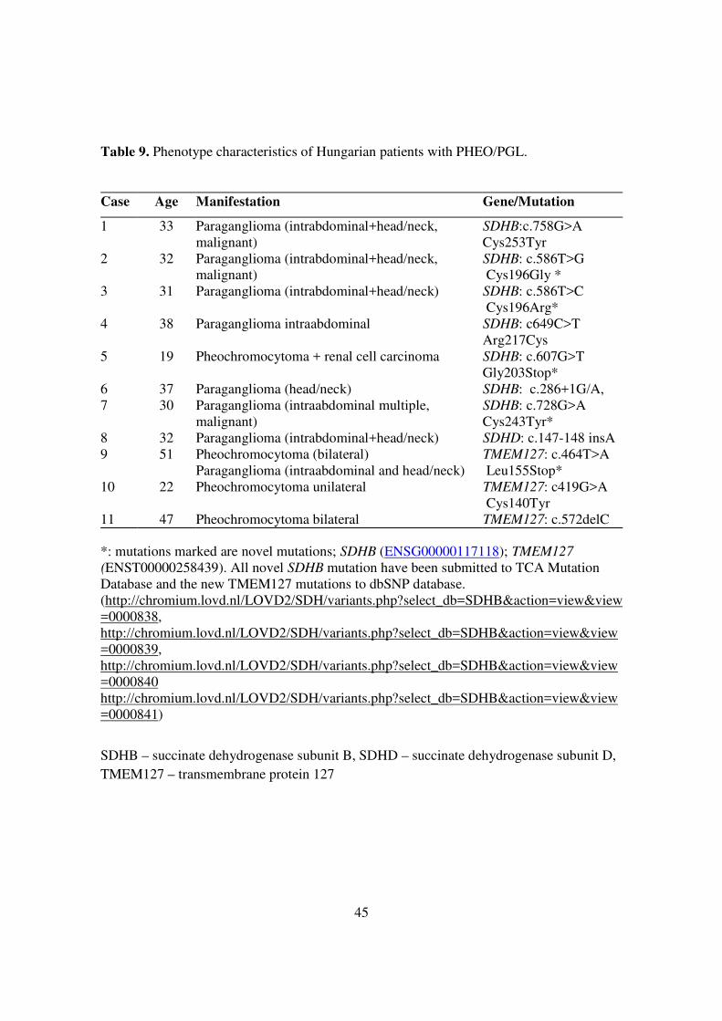
4.1. Germline mutation prevalence in Hungarian patients with PHEO and/or PGL
Eleven patients were identified to carry mutation in one of the PHEO/PGL associated
genes. Together with our previous data demonstrating mutations in RET (n=4) and VHL
(n=4) genes, the prevalence of germline disease-causing mutations in Hungarian patients
with apparently sporadic, non-syndromic PHEO/PGL was 21.1% (19/90; 11 of 82 cases, 4
RET and 4 VHL mutation carriers). For mutation detection bilateral involvement and
multiple tumours had the most positive predictive value. The prevalence of bilateral
tumours was significantly higher in mutation carriers than in genetically negative cases (8
of 11, 72.8% vs. 3 of 71, 2.1%; p<0.001).
The mutation spectrum observed in our patients was heterogeneous, the most frequent
mutations were detected in the SDHB gene (7 different of which 4 were novel mutations),
Three patients had TMEM127 mutations (two novel) and one had mutation in the SDHD
gene (Table 9). The chromatograms of all novel mutations identified are presented in
Figure 5. All novel SDHB mutation have been submitted to TCA Mutation Database and
the new TMEM127 mutations to dbSNP database (http://chromium.lovd.nl
/LOVD2/SDH/variants.php?select_db=SDHB&action= view&view=0000838,
http://chromium.lovd.nl/LOVD2/SDH/variants.php?select_db=SDHB&action=view& view
=0000839; http://chromium.lovd.nl/LOVD2/SDH/variants.php?select_ db=SDHB
&action=view&view=0000840; http://chromium.lovd.nl/LOVD2/SDH/ variants.php?
select_db=SDHB& action= view&view=0000841).
No mutations in SDHC, SDHAF2, and MAX were identified in our patients.
44
4.1.1. Genotype-phenotype associations
Comparison of the main demographic and clinical data of the genetically positive and
negative cases indicated that genetically positive patients were younger, their PHEO/PGL
was more frequently malignant, and 72% of cases had bilateral or multiple tumours (Table
8). As expected the malignancy was the highest (3 out of 7 cases) in patients with SDHB
mutations. Two patients with mutations SDHB:c758G>A -Cys253Tyr- and the novel
SDHB: c.586T>G -Cys196Gly- were lost because of metastatic disease by the age of 35
years. In these patients multiple metastases in bone and liver were observed. In the third
case with malignant PGL the novel SDHB: c728G>A Cys243Tyr mutation was identified.
In this patient an intraabdominal PGL with multiple bone metastases was diagnosed.
Another important finding was that the SDHB associated tumours were mainly
intraabdominal PGLs (6 out of the 7 cases). In one case with the novel SDHB c607G>T
Gly203Stop mutation pheochromocytoma and renal cell carcinoma with oncocytic feature
was detected at age of 19 years. The solid architecture, cytoplasmic inclusions of flocculent
material and intratumoural mast cells as the main characteristics for SDHB associated renal
cell carcinomas could be identified (Figure 6).
Head-neck PGLs were detected in a patient harbouring the SDHB: c286+1G/A
mutation, and in a patient with SDHD c.147-148 insA frameshift mutation. In the latter case
an intraabdominal PGL was also removed. After 4-8 years follow-up no malignancy was
observed in these cases.
TMEM127 mutations were detected in three patients. Two of them had PHEO (one
bilateral) while in the third patient with the novel mutation (TMEM127: c467T>A, -
Leu155Stop) PHEO and PGL of the head-neck region was also observed. These tumours
showed no malignancy. The youngest patient harbouring TMEM127 associated tumour was
22 years old.
45
Table 9. Phenotype characteristics of Hungarian patients with PHEO/PGL.
Case Age Manifestation Gene/Mutation
1 33 Paraganglioma (intrabdominal+head/neck, malignant)
SDHB:c.758G>A Cys253Tyr
2 32 Paraganglioma (intrabdominal+head/neck, malignant)
SDHB: c.586T>G Cys196Gly *
3 31 Paraganglioma (intrabdominal+head/neck) SDHB: c.586T>C Cys196Arg*
4 38 Paraganglioma intraabdominal SDHB: c649C>T Arg217Cys
5 19 Pheochromocytoma + renal cell carcinoma SDHB: c.607G>T Gly203Stop*
6 37 Paraganglioma (head/neck) SDHB: c.286+1G/A, 7 30 Paraganglioma (intraabdominal multiple,
malignant) SDHB: c.728G>A Cys243Tyr*
8 32 Paraganglioma (intrabdominal+head/neck) SDHD: c.147-148 insA 9 51 Pheochromocytoma (bilateral)
Paraganglioma (intraabdominal and head/neck) TMEM127: c.464T>A Leu155Stop*
10 22 Pheochromocytoma unilateral TMEM127: c419G>A Cys140Tyr
11 47 Pheochromocytoma bilateral TMEM127: c.572delC *: mutations marked are novel mutations; SDHB (ENSG00000117118); TMEM127
(ENST00000258439). All novel SDHB mutation have been submitted to TCA Mutation Database and the new TMEM127 mutations to dbSNP database. (http://chromium.lovd.nl/LOVD2/SDH/variants.php?select_db=SDHB&action=view&view=0000838, http://chromium.lovd.nl/LOVD2/SDH/variants.php?select_db=SDHB&action=view&view=0000839, http://chromium.lovd.nl/LOVD2/SDH/variants.php?select_db=SDHB&action=view&view=0000840 http://chromium.lovd.nl/LOVD2/SDH/variants.php?select_db=SDHB&action=view&view=0000841)
SDHB – succinate dehydrogenase subunit B, SDHD – succinate dehydrogenase subunit D, TMEM127 – transmembrane protein 127
46
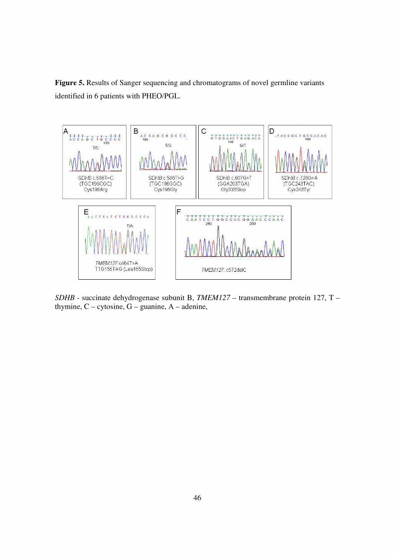
Figure 5. Results of Sanger sequencing and chromatograms of novel germline variants
identified in 6 patients with PHEO/PGL.
SDHB - succinate dehydrogenase subunit B, TMEM127 – transmembrane protein 127, T – thymine, C – cytosine, G – guanine, A – adenine,
47
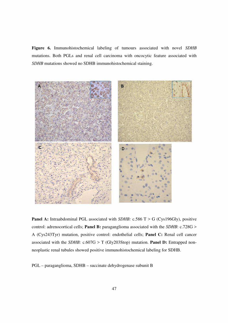
Figure 6. Immunohistochemical labeling of tumours associated with novel SDHB
mutations. Both PGLs and renal cell carcinoma with oncocytic feature associated with
SDHB mutations showed no SDHB immunohistochemical staining.
Panel A: Intraabdominal PGL associated with SDHB: c.586 T > G (Cys196Gly), positive
control: adrenocortical cells; Panel B: paraganglioma associated with the SDHB: c.728G >
A (Cys243Tyr) mutation, positive control: endothelial cells; Panel C: Renal cell cancer
associated with the SDHB: c.607G > T (Gly203Stop) mutation. Panel D: Entrapped non-
neoplastic renal tubules showed positive immunohistochemical labeling for SDHB.
PGL – paraganglioma, SDHB – succinate dehydrogenase subunit B
48
4.1.2. The first Hungarian case with extraadrenal pheochromocytoma associated with
SDHD gene mutation
Case report
A 33 years-old patient, diagnosed with hypertension 4 years ago (systolic: 120–167 Hgmm,
diastolic: 87–110 Hgmm, after 5 minutes of cycling was 251/117 Hgmm), had dizziness,
increased perspiration and palpitation. Physical examination showed no abnormalities. The
patient had mild hypokalaemia (serum potassium 3,5 mmol/l), mild hypocalcaemia (serum
calcium 2,16 mmol/l), all other parameters (glucose level, serum creatinine, cholesterine,
triglyceride, liver function, blood count) were normal. The 24 hour fractionated urine
metanephrine analysis showed elevated vanillylmandelic-acid (VMA, 18,2 mg/24 h, normal
range: 1,8–6,7 mg/24 h) and normetanephrine (2812,9 μg/24 h, normal range: 105–354
μg/24 h), together with normal metanephrine level (101,3 μg/24 h, normal range: 74–297
μg/24 h). Ultrasound, CT and MRI of the adrenal gland showed no abnormalities. The 18-
fluoro-DOPA positron emission tomography (18-F-DOPA PET CT) detected a tumour
mass resembling for a neuroendocrine tumours at the height of the III. lumbar vertebra
before the abdominal aorta. (Figure 7) MRI, later, confirmed the 18-F-DOPA PET
identified tumour mass. After preoperative drug administration (α- and β adrenergic
receptor blockers) the tumour with a diameter of 3,5cm was laparoscopically resected from
the front of the aortic bifurcation. Histological findings confirmed paraganglioma. After
tumour resection the symptoms disappeared and the blood pressure normalized. The patient
did not present the skin signs typical for neurofibromatosis and MEN2 syndrome was ruled
out by normal serum calcitonin level. Family history was unspecific for hereditary
syndrome. Regarding the young age he received genetic counselling. After signing
informed consent mutation screening was performed for genes causing hereditary
syndromes.
49
Mutation screening
Genomic DNA was isolated from peripheral blood using commercially available DNA
extraction kits (Boehringer Mannheim Corp., Indianapolis, USA) in accordance with the
manufacturers’ instructions. Mutation analysis of the VHL, SDHB, and SDHD genes in
were performed by bidirectional DNA sequencing of the entire coding region of the VHL,
SDHB, and SDHD genes, as previously reported. 99 There were no mutations in the exons
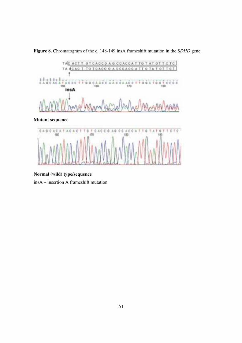
and near exons of the VHL, SDHC and SDHB genes, but there was a heterozygous c. 148-
149 insA frameshift mutation found in exon 2 of the SDHD gene (Figure 8). The frameshift
mutation results in a truncated protein; therefore its pathogeneity is expected.
Family screening
Genetic analysis of family members revealed the presence of the same mutation in his
asymptomatic father (c. 148-149 insA frameshift mutation) while the mutation was not
present in his mother and brother. Despite detailed physical examination, biochemical
(plasma, urine catecholamine) and imaging tests (abdominal CT scan) nor PHEO neither
PGL was confirmed in his father.
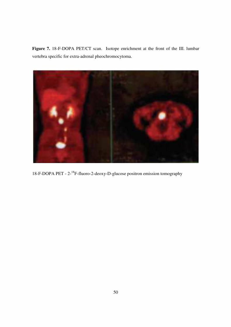
50
Figure 7. 18-F-DOPA PET/CT scan. Isotope enrichment at the front of the III. lumbar
vertebra specific for extra-adrenal pheochromocytoma.
18-F-DOPA PET - 2-18F-fluoro-2-deoxy-D-glucose positron emission tomography
51
Figure 8. Chromatogram of the c. 148-149 insA frameshift mutation in the SDHD gene.
Mutant sequence
Normal (wild) type/sequence
insA – insertion A frameshift mutation
52
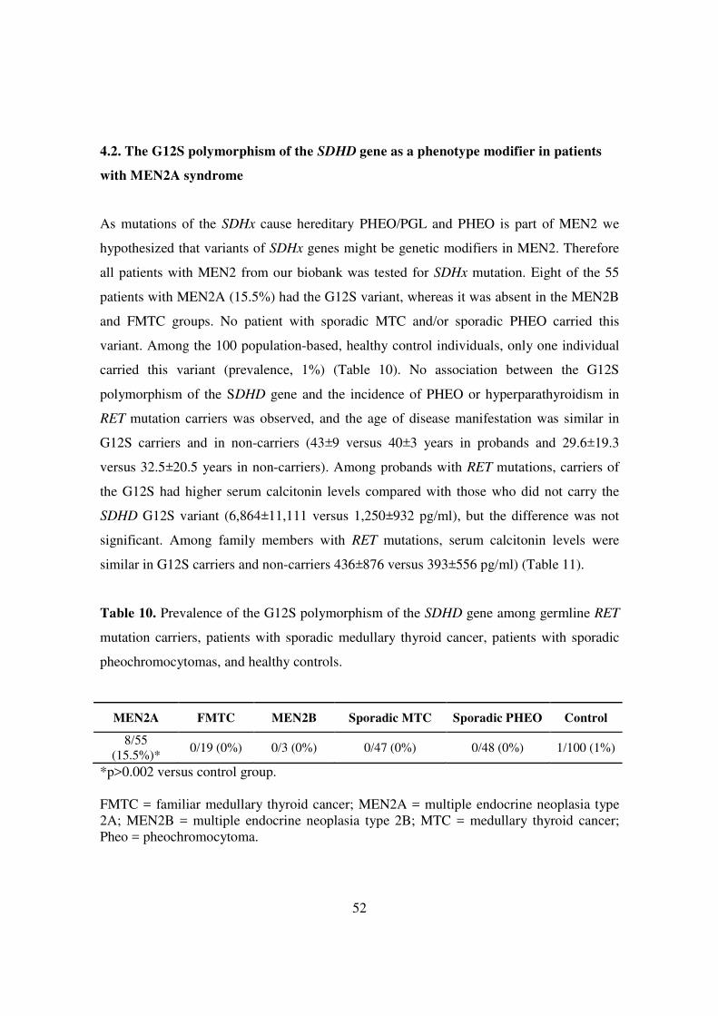
4.2. The G12S polymorphism of the SDHD gene as a phenotype modifier in patients
with MEN2A syndrome
As mutations of the SDHx cause hereditary PHEO/PGL and PHEO is part of MEN2 we
hypothesized that variants of SDHx genes might be genetic modifiers in MEN2. Therefore
all patients with MEN2 from our biobank was tested for SDHx mutation. Eight of the 55
patients with MEN2A (15.5%) had the G12S variant, whereas it was absent in the MEN2B
and FMTC groups. No patient with sporadic MTC and/or sporadic PHEO carried this
variant. Among the 100 population-based, healthy control individuals, only one individual
carried this variant (prevalence, 1%) (Table 10). No association between the G12S
polymorphism of the SDHD gene and the incidence of PHEO or hyperparathyroidism in
RET mutation carriers was observed, and the age of disease manifestation was similar in
G12S carriers and in non-carriers (43±9 versus 40±3 years in probands and 29.6±19.3
versus 32.5±20.5 years in non-carriers). Among probands with RET mutations, carriers of
the G12S had higher serum calcitonin levels compared with those who did not carry the
SDHD G12S variant (6,864±11,111 versus 1,250±932 pg/ml), but the difference was not
significant. Among family members with RET mutations, serum calcitonin levels were
similar in G12S carriers and non-carriers 436±876 versus 393±556 pg/ml) (Table 11).
Table 10. Prevalence of the G12S polymorphism of the SDHD gene among germline RET
mutation carriers, patients with sporadic medullary thyroid cancer, patients with sporadic
pheochromocytomas, and healthy controls.
MEN2A FMTC MEN2B Sporadic MTC Sporadic PHEO Control
8/55 (15.5%)*
0/19 (0%) 0/3 (0%) 0/47 (0%) 0/48 (0%) 1/100 (1%)
*p>0.002 versus control group. FMTC = familiar medullary thyroid cancer; MEN2A = multiple endocrine neoplasia type 2A; MEN2B = multiple endocrine neoplasia type 2B; MTC = medullary thyroid cancer; Pheo = pheochromocytoma.
53
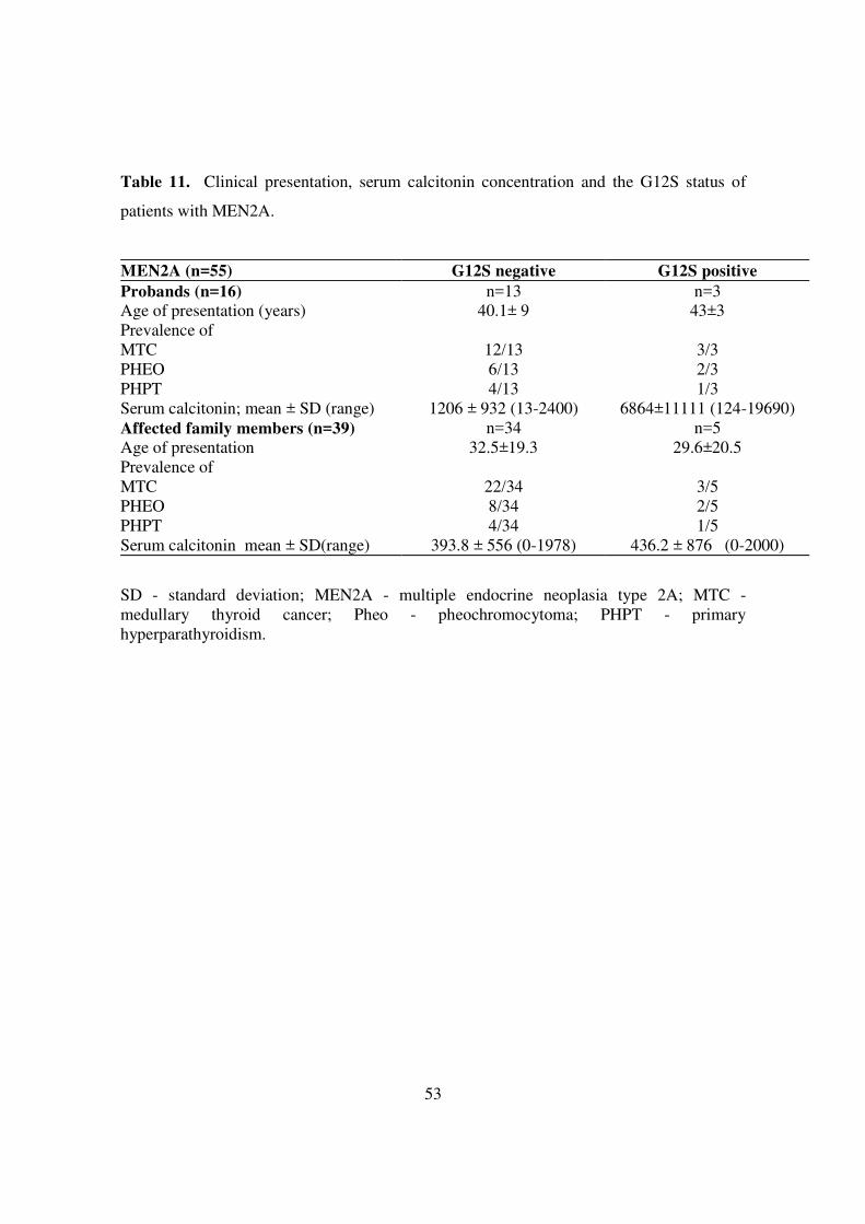
Table 11. Clinical presentation, serum calcitonin concentration and the G12S status of
patients with MEN2A.
MEN2A (n=55) G12S negative G12S positive Probands (n=16) n=13 n=3 Age of presentation (years) 40.1± 9 43±3 Prevalence of MTC 12/13 3/3 PHEO 6/13 2/3 PHPT 4/13 1/3 Serum calcitonin; mean ± SD (range) 1206 ± 932 (13-2400) 6864±11111 (124-19690) Affected family members (n=39) n=34 n=5 Age of presentation 32.5±19.3 29.6±20.5 Prevalence of MTC 22/34 3/5 PHEO 8/34 2/5 PHPT 4/34 1/5 Serum calcitonin mean ± SD(range) 393.8 ± 556 (0-1978) 436.2 ± 876 (0-2000)
SD - standard deviation; MEN2A - multiple endocrine neoplasia type 2A; MTC - medullary thyroid cancer; Pheo - pheochromocytoma; PHPT - primary hyperparathyroidism.
54
4.3. Biochemical consequences of SDHx mutations, succinate to fumarate ratio in
SDHB/D associated paragangliomas
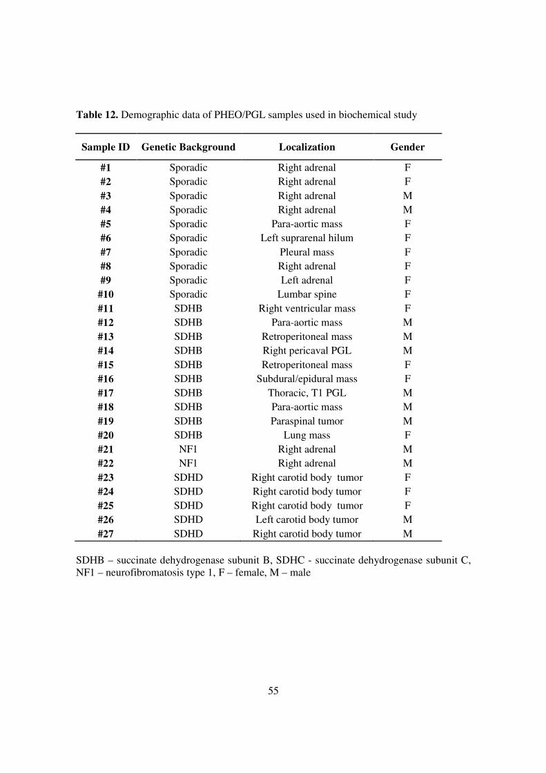
The present study included 27 tumour samples and SDHB silenced and control MPC and
MTT cells. The samples included 10 SDHB PGLs, 5 SDHD PGLs, 2 NF1 PHEOs, and 10
apparently sporadic PHEOs/PGLs (Table 12).
4.3.1. Succinate concentration in tumour tissues
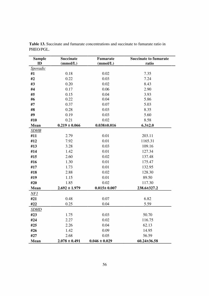
In SDHB-related PGLs, the results showed a mean succinate concentration of 2.7 ± 1.9
mmol/L with the COV of 0.71, in comparison with the apparently sporadic PHEO/PGL
group (0.22 ± 0.06 mmol/L; COV of 0.3; P = .0009). One SDHB-related PGL showed a
much higher succinate level than the other SDHB tumours (7.9 mmol/L) (Table 13). This
sample is a significant outlier (P < .05 with a Z value of 2.3). In the SDHD group, the mean
succinate concentration was 2.1 ± 0.5 mmol/L (COV of 0.24); this value was higher (P <
.05) than in the apparently sporadic PHEO/PGL group. The NF1 PHEOs showed a similar
mean succinate concentration (0.36 ± 0.16 mmol/L; COV of 0.45) to apparently sporadic
PHEOs/PGLs (Table 13).
4.3.2. Fumarate concentration
In SDHB-related PGLs, the results showed a mean fumarate concentration of 0.015 ± 0.007
mmol/L with the COV of 0.46, in SDHD-related PGLs 0.04 ± 0.03 (COV of 0.62), and in
apparently sporadic PHEOs/PGLs 0.038 ± 0.016 mmol/L (COV of 0.43). Fumarate
concentrations were significantly lower in the SDHB-related compared with SDHD-related
and apparently sporadic PHEOs/PGLs (P = .005, P = .0008, respectively). The NF1 PHEO
group showed a mean fumarate concentration of 0.06 ± 0.02 mmol/L (COV of 0.324)
(Table 13).
55
Table 12. Demographic data of PHEO/PGL samples used in biochemical study
Sample ID Genetic Background Localization Gender
#1 Sporadic Right adrenal F #2 Sporadic Right adrenal F #3 Sporadic Right adrenal M #4 Sporadic Right adrenal M #5 Sporadic Para-aortic mass F #6 Sporadic Left suprarenal hilum F #7 Sporadic Pleural mass F #8 Sporadic Right adrenal F #9 Sporadic Left adrenal F #10 Sporadic Lumbar spine F #11 SDHB Right ventricular mass F #12 SDHB Para-aortic mass M #13 SDHB Retroperitoneal mass M #14 SDHB Right pericaval PGL M #15 SDHB Retroperitoneal mass F #16 SDHB Subdural/epidural mass F #17 SDHB Thoracic, T1 PGL M #18 SDHB Para-aortic mass M #19 SDHB Paraspinal tumor M #20 SDHB Lung mass F #21 NF1 Right adrenal M #22 NF1 Right adrenal M #23 SDHD Right carotid body tumor F #24 SDHD Right carotid body tumor F #25 SDHD Right carotid body tumor F #26 SDHD Left carotid body tumor M #27 SDHD Right carotid body tumor M
SDHB – succinate dehydrogenase subunit B, SDHC - succinate dehydrogenase subunit C, NF1 – neurofibromatosis type 1, F – female, M – male
56
Table 13. Succinate and fumarate concentrations and succinate to fumarate ratio in PHEO/PGL.
Sample ID
Succinate (mmol/L)
Fumarate (mmol/L)
Succinate to fumarate ratio
Sporadic #1 0.18 0.02 7.35 #2 0.22 0.03 7.24 #3 0.20 0.02 8.43 #4 0.17 0.06 2.90 #5 0.15 0.04 3.93 #6 0.22 0.04 5.86 #7 0.37 0.07 5.03 #8 0.28 0.03 8.35 #9 0.19 0.03 5.60 #10 0.21 0.02 8.58 Mean 0.219 ± 0.066 0.038±0.016 6.3±2.0 SDHB #11 2.79 0.01 203.11 #12 7.92 0.01 1165.31 #13 3.28 0.03 109.16 #14 1.42 0.01 127.34 #15 2.60 0.02 137.48 #16 1.30 0.01 175.47 #17 1.73 0.01 132.95 #18 2.88 0.02 128.30 #19 1.15 0.01 89.50 #20 1.85 0.02 117.30 Mean 2.692 ± 1.979 0.015± 0.007 238.6±327.2 NF1 #21 0.48 0.07 6.82 #22 0.25 0.04 5.59 SDHD #23 1.75 0.03 50.70 #24 2.27 0.02 116.75 #25 2.26 0.04 62.13 #26 1.42 0.09 14.95 #27 2.68 0.05 56.59 Mean 2.078 ± 0.491 0.046 ± 0.029 60.24±36.58
57
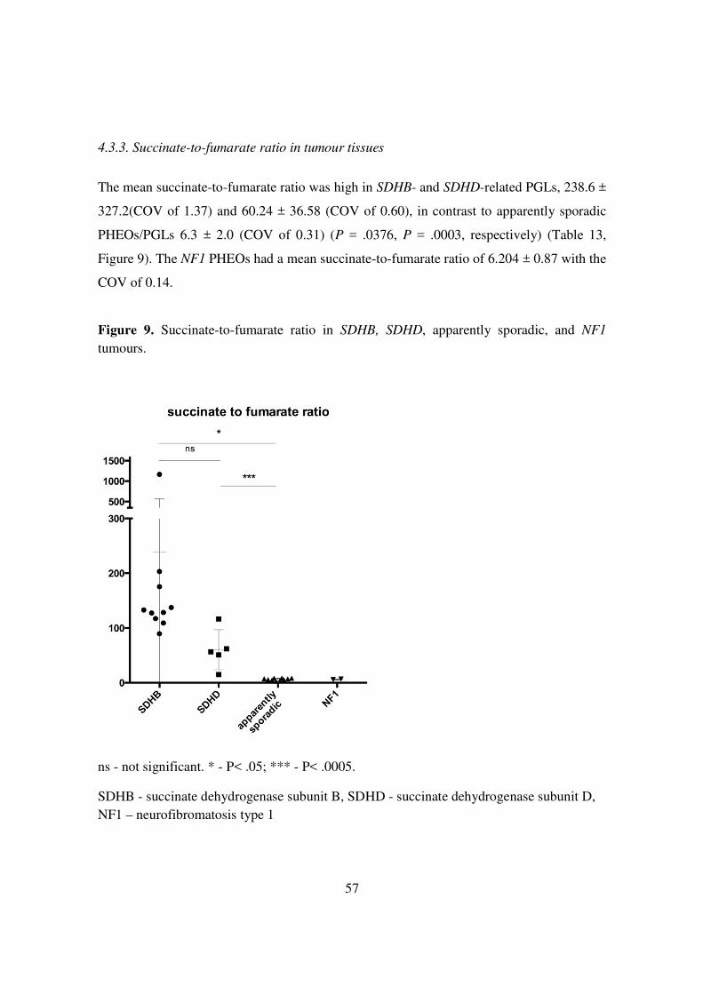
4.3.3. Succinate-to-fumarate ratio in tumour tissues
The mean succinate-to-fumarate ratio was high in SDHB- and SDHD-related PGLs, 238.6 ±
327.2(COV of 1.37) and 60.24 ± 36.58 (COV of 0.60), in contrast to apparently sporadic
PHEOs/PGLs 6.3 ± 2.0 (COV of 0.31) (P = .0376, P = .0003, respectively) (Table 13,
Figure 9). The NF1 PHEOs had a mean succinate-to-fumarate ratio of 6.204 ± 0.87 with the
COV of 0.14.
Figure 9. Succinate-to-fumarate ratio in SDHB, SDHD, apparently sporadic, and NF1 tumours.
ns - not significant. * - P< .05; *** - P< .0005.
SDHB - succinate dehydrogenase subunit B, SDHD - succinate dehydrogenase subunit D, NF1 – neurofibromatosis type 1
58
4.3.4. Succinate-to-fumarate ratio in plasma samples
The plasma mean succinate-to-fumarate ratio showed a slight increase in the SDHB and
SDHD groups (3.15 ± 1.63 and 2.75 ± 1.65) compared with the apparently sporadic group
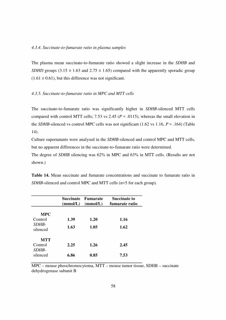
(1.61 ± 0.61), but this difference was not significant.
4.3.5. Succinate-to-fumarate ratio in MPC and MTT cells
The succinate-to-fumarate ratio was significantly higher in SDHB-silenced MTT cells
compared with control MTT cells; 7.53 vs 2.45 (P = .0115), whereas the small elevation in
the SDHB-silenced vs control MPC cells was not significant (1.62 vs 1.16, P = .164) (Table
14).
Culture supernatants were analysed in the SDHB-silenced and control MPC and MTT cells,
but no apparent differences in the succinate-to-fumarate ratio were determined.
The degree of SDHB silencing was 62% in MPC and 63% in MTT cells. (Results are not
shown.)
Table 14. Mean succinate and fumarate concentrations and succinate to fumarate ratio in
SDHB-silenced and control MPC and MTT cells (n=5 for each group).
Succinate (mmol/L)
Fumarate (mmol/L)
Succinate to fumarate ratio
MPC
Control 1.39 1.20 1.16 SDHB-silenced
1.63 1.05 1.62
MTT
Control 2.25 1.26 2.45 SDHB-silenced
6.86 0.85 7.53
MPC – mouse pheochromocytoma, MTT – mouse tumor tissue, SDHB – succinate dehydrogenase subunit B
59
5. DISCUSSION
5.1. Germline mutation prevalence in Hungarian patients with pheochromocytoma
and/or paraganglioma
PHEO/PGLs are rare catecholamine producing tumours. To date 14 genes have been
implicated the genetic susceptibility of PHEO/PGL, which are responsible for the 25–30%
of all cases. The American Society of Clinical Oncology have suggested that for patients
with a ≥ 10 % chance for carrying a germline mutation genetic testing should be offered 91,106. Patients with PHEO/PGL are in this group. Currently the genetic analysis of patients
with PHEO/PGL includes molecular genetic analysis of RET, VHL, SDHx, MAX and
TMEM127 genes. MAX and TMEM127 were identified in 2010, and to date only few
studies have been published about the prevalence of mutations of these genes in apparently
sporadic cases and only few studies reported genotype-phenotype associations. 45,62107–109
Our current study was initiated to comprehensively analyze the prevalence of germline
mutations in our cohort of histologically confirmed non-syndromic patients with
PHEO/PGL. Using conventional molecular biological methods we identified 11 germline
mutation carriers (SDHB=8, TMEM127=3), including six novel mutations. These results,
together with our previous data on RET (n = 4) and VHL mutations (n = 4) in Hungarian
patients with apparently sporadic, non-syndromic PHEO/PGL shows that 21.1 % of our
patients carry mutation in one of the PHEO/PGL susceptibility genes. 99,101 This finding is
in line with previously reported data in other populations 45,109 and with a recent review by
Brito et al. 108 The mutation spectrum observed in our cohort suggests that no founder
mutation is present in the Hungarian population. Genetic studies performed in the past did
not include the mutation testing of KIF1B, EGLN1, TMEM127, MAX or the recently
identified MDH2 and their prevalence in apparently sporadic PHEO/PGL cases are lacking.
Therefore, our findings are also important from this aspect and the results demonstrated that
in a population with heterogeneous genetic background the genetic screening should be
performed for all of these genes. The novel mutations identified in our cases are considered
as disease-causing mutations, because they are either protein truncating mutations
60
(TMEM127. c572delC, SDHB Gly203Stop and TMEM127 Leu155Stop) or they affect
residues which are important for protein function and in the same codon other mutations
have already been reported as pathogenic (SDHB p.Cys196Gly and p.Cys243Tyr)
according to the TCA Cycle Gene Mutation Database (http://chromium.liacs.nl). These
novel mutations are not listed in any database including dbSNP database
(http://www.ncbi.nih.gov/SNP), exome variant server (http://evs.gs. washington.edu/EVS/,
version v.0.0.30) and Exac variant (exac.broadinstitute.org/) databases. In addition a
negative SDHB immunostaining of tumours associated with SDHB p.Cys196Gly,
p.Cys243Tyr and Gly203Stop (Fig. 6.) further supports the pathogenic role of SDHB
mutations in these patients. Genotype-phenotype associations confirmed that the malignant
potential is frequently associates with SDHB mutations. The presentation and the course of
the disease of our case with the SDHB Cys196Gly mutation were unique. In this case
malignant PGL presenting as a primary PGL in the occipital bone was found. By reviewing
the literature only one similar case was found. Kanai et al. presented a 61-year-old male
patient diagnosed with multiple paragangliomas including intracranial PGL and osteolytic
lesion in the occipital bone. Despite surgical interventions and chemotherapy, the patient
died in the fourth year after the diagnosis. No data about the genetic background of this
case was reported but the similar behaviour observed in these two cases may raise the
pathogenic role of SDHB. 110 In addition, a more complex phenotype, including a rare
concomitant tumour (PHEO/PGL and renal cell carcinoma) was found in another patient
with SDHB mutation. Renal cell carcinoma with oncocytic feature has been reported as a
hallmark of the SDHB associated renal cell carcinomas. 96,111 In our patient the lack of
SDHB staining confirmed the loss of SDHB protein in tumour tissue while it was kept in
renal tubular cells. Based on our and Williamson’s results genetic testing of the SDHB gene
should be offered for patients presenting with renal cell carcinoma with oncocytic features. 96,111
The lack of mutation of SDHC gene is not entirely unexpected among our patients because
our patient group consisted of patients having mostly intraabdominal PGLs and PHEOs
whereas SDHC mutations have been identified exclusively in tumours located at the head
61
and neck regions. 45,91,107 In addition, sporadic head and neck PGLs may present with less
symptoms and maybe they are possibly underdiagnosed. Mutations in SDHAF2, MAX and
TMEM127 genes have been reported only in a very few cases. 65,108 In our study no
SDHAF2 and MAX mutations were found but TMEM127 mutations were detected in 3
patients of which 2 mutations proved to be novel. It seems particularly important that
TMEM127 mutations were previously reported only in patients with adrenal PHEOs, but in
one of our patient having a novel TMEM127 mutation bilateral adrenal PHEOs as well as
glomus caroticum PGL were detected. This new phenotype, confirmed by later studies
indicates that mutations of TMEM127 can also associate with head and neck PGLs. 62 112,113
In our study the two novel TMEM127 mutations were truncating mutations strongly
suggesting their deleterious nature. The third TMEM127 mutation was detected in a 22-
years-old female patient presenting with unilateral adrenal PHEO. This mutation was
already reported by Yao et al. and, surprisingly this seems to be the only TMEM127
mutation associated with malignant phenotype. 112,113 Toledo et al. reported a six generation
family with TMEM127 mutation and suggested that clinical surveillance in TMEM127
carriers should be started at the age of 22 years. Our findings indicate that clinical
surveillance should be started at earlier age. In our mutation-negative patients only three
cases were presented with bilateral or multiple tumours. These patients, together with the
12 patients with malignant phenotype (5 PGL and 7 PHEO) may have mutations of genes
which were not investigated in the present study. Testing the KIF1B, EGLN1, FH, IDH2
and MDH2 genes by classical methods represents a significant work load and cost;
therefore, next generation sequencing based methods would be desired. The clinical follow-
up of patients identified with pathogenic, germline mutation and their first-degree relatives
is challenging. First of all, in the affected families for the first degree relatives genetic
counselling followed by genetic testing should be offered. These tumour syndromes are
inherited in an autosomal dominant manner, therefore the chance of inheriting the
pathogenic variant is 50%. The SDHD gene is maternally imprinted therefore the
pathogenic variant is inherited from the paternal side, hence in children inheriting mutation
from their mother the development of the disease is extremely unexpected. The penetrance
of PHEO/PGL varies significantly between these syndromes. It seems to be very low for
62
SDHA, SDHB, SDHC, SDHD and TMEM127 mutations but it is higher for RET, VHL and
NF1 alterations. Of course the typical manifestations associating with RET, VHL and NF1
mutations are highly penetrant and several times precede the development of PHEO (ie.
medullary thyroid cancer in RET mutation carriers, renal cell cancer, hemangioblastoma
and retina angiomatosis in VHL carriers and skin lesions in NF1 mutation carriers). In these
families the routine clinical follow-up includes regular checking for manifestation using
laboratory and imaging techniques (summarized by Lenders, 91).
Investigations show abnormalities in oxygen sensing, HIF1 stabilization 85,114, apoptosis 77;
and increased formation of reactive oxygen species in tumour development associated to
SDHx mutations.
In the first Hungarian patient with extra-adrenal pheochromocytoma related to SDHD gene
mutation the heterozygous frameshift c.148-149 insA mutation was detected. This mutation
was previously reported in a Turkish family. (www. chromium.liacs.nl/lovd_sdh) 115 The
typical transmission for SDHD gene mutation and PGL/PHEO syndrome was also seen in
our patient, the mutation carrier father was asymptomatic (clinical, biochemical and
imaging test showed no abnormalities), therefore he inherited the mutation from his mother.
This phenomenon is referred maternal imprinting. 116 In these families the symptoms
appear in the children of the asymptomatic or symptomatic mutation carrier males; however
the children of the mutation carrier females are usually asymptomatic. The pathogenesis of
genomial imprinting is still unclear, which makes it difficult to recognize the inheritance of
the disease.
The characteristics of hereditary syndromes with PHEO include manifestation at young age
and the increased incidence of these tumours among family members. In addition, in
contrast to sporadic cases, hereditary PHEO are mainly bilateral or are located in multiple,
extra-adrenal localization compared to non-syndromic, sporadic cases.
In 25-30% of the apparently sporadic pheochromocytoma germline mutations can be
identified. In children and young patients mostly mutation of the RET and VHL genes are
affected. 117 However, our case with apparently sporadic extra-adrenal pheochromocytoma
with early onset due to mutation in the SDHD gene highlighted that even in these younger
cases other genes may be mutated. In a family where a mutation carrier was identified
63
genetic counselling for family members and in mutation carriers clinical testing following
the international recommendations are recommended. These recommendations include
specific laboratory and imaging testing.
5.2. The G12S polymorphism of the SDHD gene as a phenotype modifier in patients
with MEN2A syndrome
The phenotypic heterogeneity seen in families with different RET mutations, the variation
of clinical course within families with the same RET mutation, and the results from RET
transgenic mouse models suggest a potential role of genetic components in phenotype
modulation. 56,118
Polymorphisms of the RET gene have been analyzed as such genetic modifiers, but the
results from these studies are conflicting. Robledo et al. showed that two RET variants
(G691S and S904S) may modify the age of onset of MTC in family members 71; and
Tamanaha et al. reported that two intronic polymorphisms of RET may modify the
phenotype in a large family with G533C RET mutation 73, while Baumgartner-Parzer found
that the L769L and the IVS14-24 may act as modifiers in some forms of hereditary and
sporadic MTC. 119 However, Lesueur et al. were unable to replicate this association in a
large cohort of 384 members of MEN2 families from four different European populations.
This latter study showed that of the several polymorphisms of RET, its co-receptors and
ligands, only the synonymous polymorphism (A432A) of the RET gene associated weakly
with tumour spectra in patients with MEN2A. 74 In MEN2-related MTC RET variants have
been proposed as genetic susceptibility factors for the development of sporadic MTC:
polymorphisms located in coding regions of RET; G691S, L769L, S836S, and S904S have
been shown to be over-represented in patients with sporadic MTC 120–122 compared with the
general population, but others were unable to confirm these associations 123,124 suggesting
that variants of RET may be involved in the pathogenesis of sporadic MTC as well.
Germline mutations of SDHx genes encoding subunits of the mitochondrial complex II
represent a genetic susceptibility for PHEO/PGL. These tumours are derived from cells of
64
the neural crest, similar to MTC. RET mutations also cause PHEO, again suggesting a link
between the genetic background of PHEO and MTC. Therefore, it has been assumed that
mutations of these genes may be involved in the pathogenesis of MTC. Lima et al. reported
a family with C-cell hyperplasia, a pre-cancerous state of MTC, who were proved to have
the H50R variant of the SDHD gene. 78
Montani et al. demonstrated an increased frequency of amino acid-coding SDHx
polymorphisms in patients with sporadic and familial MTC. 76 In addition, a systemic
evaluation of genetic variants of the SDHx genes among patients with sporadic MTC
showed a significant association between the H50R variant and sporadic MTC in Spanish
patients, although this observation was absent in an English cohort. 125 Variants of the
SDHx genes have been implicated in the pathogenesis of various endocrine and non-
endocrine tumours, such as Merkel cell carcinoma, carcinoid, papillary thyroid cancer,
pituitary tumours and renal cell cancer found in patients with Cowden-like syndrome. 77
During my PhD thesis work I found that the G12S variant was significantly over-
represented among RET mutation carriers compared with sporadic MTC, sporadic PHEO,
or control individuals. This variant occurred mainly in patients with MEN2A, while
Montani et al. detected G12S in a patient with MEN2B harbouring the M918T mutation of
the RET gene. 76 Interestingly, the prevalence of alterations of the SDHx genes in patients
with RET mutations was similar in our study and the study of Montani et al.. 76 The
prevalence of the G12S in the general population is between 2.5% and 5% 126 according to
the Leiden Open Variation Database (http://chromium.liacs.nl) 127, which is somewhat
higher than in our control population (1%). This difference may be due to differences in the
selection criteria applied for controls. Our control group were evaluated for endocrine
dysfunction; none of them had signs or symptoms characteristic of thyroid cancer or PHEO.
By contrast, population-based controls, frequently anonymous blood donors, have been
never tested for these rare conditions. Alternatively, the difference between the studies in
prevalence of G12S can also be attributed to the ethnic background of the different
populations tested. Our patients and controls were of Hungarian origin, representing an
independent entity among Caucasian populations. More importantly, in our study, the high
65
incidence of the G12S variant among RET carriers, especially in those with the MEN2A
phenotype, raised the possibility that this variant may have a role in the phenotypic
modulation of the disease. However, we were unable to detect significant differences in the
clinical presentation between G12S carriers and non-carriers. Whether this failure was a
result of the relatively small size of our patient cohort remains to be further investigated.
Interestingly, Waldmann et al. reported an increased prevalence of intronic SDHB
polymorphisms among patients with malignant PHEO compared with patients with benign
tumors. 128
5.3. Biochemical consequences of SDHx mutations, succinate to fumarate ratio in
SDHB/D associated paragangliomas
Using tumour tissue homogenates I found that the tumor tissue succinate-to-fumarate ratio
was significantly higher in SDHB- and SDHD-related PGLs compared to apparently
sporadic and NF1-related PHEOs/PGLs. Furthermore, SDHB-silenced MTT cells showed a
similar trend of increased succinate-to-fumarate ratio compared with control MTT cells.
These results suggested for the first time that the succinate-to-fumarate ratio can be used as
a new metabolic marker for SDHB/D-related PHEOs/PGLs.
SDH is the crucial enzyme in energy metabolism that links the tricarboxylic acid cycle, also
called the Krebs cycle, to oxidative phosphorylation. 129 In the Krebs cycle, SDH catalyzes
the oxidation of succinate to fumarate, whereas as mitochondrial complex II, it transfers
electrons to the quinone pool, supporting the reduction of ubiquinone. 129 More than a
decade ago, mutations in genes encoding SDH subunits B, C, and D, and more recently
mutations in SDHAF2 and SDHA, were discovered to be involved in the pathogenesis of
PHEOs/PGLs. 12,15,16,40,41 Mutations in these genes result in impaired function of the SDH
enzyme associated with succinate accumulation and loss of fumarate 79. Succinate
accumulation has been shown to result in the inhibition of prolyl hydroxylases and
consequently in the impaired degradation of hypoxia-inducible factor α (HIF1-, 2- α). 114
HIF1-, 2- α stabilization affects the activation of many genes promoting tumorigenesis and
66
cancer development with accelerated aerobic glycolysis (the so-called Warburg effect). 130,131 Reactive oxygen species, which also accumulate due to SDH mutations, were found
to stabilize HIF-α. 8,132 These and other findings suggest that, indeed, SDHx-related
PHEOs/PGLs could be viewed as a metabolic disease. 133 Thus, the assessment of
metabolic intermediates in these tumours could bring new discoveries, including the
introduction of novel biomarkers specifically used in the clinical diagnosis of these unique
metabolic tumours. Metabolomics encompasses the characterization of metabolite profiles
to genetic or environmental changes in biological samples. 134 There are several different
separation and detection methods for analytical procedures of the samples, including
nuclear magnetic resonance spectroscopy and GC-MS. Metabolomic analysis is fast and
reliable in the identification of metabolite changes in specific tissues, including tumours. 134
Genetic testing and immunohistochemistry are currently excellent methods for the
diagnosis of SDHx mutations. 86,87 Unfortunately, these methods cannot assess any response
of these tumours (eg, to chemo- or radiotherapy, their therapeutic resistance, or follow-up
after a therapy is completed). Moreover, these methods cannot detect acute changes in the
activity of these tumours; thus, they cannot predict the sudden aggressive behavior and
metastatic spread that is often seen in patients with SDHB mutations.
By introducing the succinate-to-fumarate ratio as a new marker in these tumours may
provide a new opportunity to not only diagnose but also monitor their behaviour and
therapeutic responses. Currently such monitoring would require a tumour sample to be
obtained; we predict that in the near future plasma samples could also be used to assess
these tumours as described above. This will be based on large prospective studies, as well
as the introduction of more sensitive GC-MS methods. Because the pathogenesis of these
tumours is primarily based on mitochondrial damage tightly linked to the Krebs cycle and
the Warburg effect, we predict that other important metabolites will soon be introduced and
used in clinical assessment with the succinate-to-fumarate ratio.
67
6. CONCLUSION
I summarized clinical, demographic and genetic data of Hungarian patients with apparently
sporadic PHEO/PGL. Using a comprehensive mutational screening of a large series of
patients with PHEO/PGL, I determined the prevalence of disease-causing mutations in this
patient group. The most frequent mutations were detected in the SDHB, TMEM127, RET
and VHL gene. This heterogeneous genetic background with six novel mutations observed
in Hungarian patients was similar to other populations where no founder mutations are
present. The genetic screening offered for PHEO/PGL patients in this population should
cover all of the genes identified to date but the first gene for testing should be the SDHB for
patients with intraabdominal PGL especially with malignant phenotype. The novel
genotype-phenotype associations revealed may contribute to improvement of diagnostic
approaches and may help to achieve a better clinical follow up of patients with PHEO/PGL.
Both laboratory workload and cost of testing of all genes are still significant, but phenotype
oriented guidelines allow us to set up an order of genes tested, after a negative result the
remaining genes should be also examined. For most effective work the optimum would be
to exclude some of the syndrome-associated genes based on the obvious phenotype features
(i.e. because of typical manifestation the NF1 gene is rarely tested) and all remaining genes
would be tested at the same time. Testing KIF1B, EGLN1, FH, IDH2 and MDH2 genes by
next generation sequencing based methods would also be desired. The clinical follow-up of
patients identified with pathogenic, germline mutations and their first-degree relatives is
challenging. First of all, in the affected families for all first degree relatives genetic
counselling followed by genetic testing should be offered.
Beside the disease-causing SDHx mutation I found a significantly higher prevalence of the
G12S variant of the SDHD gene among germline RET mutation carriers presenting with
MEN2A compared to the control group. The high prevalence of the G12S variant in these
patients supports its genetic modifier role, however, we were unable to detect significant
differences in the clinical presentation between G12S carriers and non-carriers. This
proposal remains to be established.
68
For the first time I was able to demonstrate that the succinate-to-fumarate ratio could be
used as a new metabolic marker for the presence of SDHB/D-related PGLs. Accumulation
of succinate result in the inhibition of prolyl hydroxylases and consequently in the impaired
degradation of hypoxia-inducible factor α (HIF1-, 2- α). 114 HIF1-, 2-α stabilization has an
impact on genes promoting tumorigenesis and cancer development with accelerated aerobic
glycolysis. 130,131 Based on the literature and my results, through a large prospective clinical
study including other SDH PHEOs/PGLs, it would be possible to determine the diagnostic
accuracy of succinate-to-fumarate ratio in the diagnosis of PHEO/PGL. Furthermore,
following the confirmation of our initial results, we may hypothesize that intratumoural and
perhaps plasma changes in the succinate-to-fumarate ratio will serve as an important
indicator of potential therapies directed toward mutated SDH proteins. 135
69
7. SUMMARY
Succinate dehydrogenase links the citric acid cycle and the oxidative phosphorylation. It
consists of four subunits (SDHA, SDHB, SDHC, SDHD), which are encoded in the nuclear
genome. Mutations can occur in all subunit genes causing familial paraganglioma
syndromes.
Pheochromocytomas and paragangliomas are tumours deriving from the chromaffin cells of
the adrenal gland and the sympathetic and parasympathetic ganglions, respectively. The
symptoms are due to the extreme catecholamine secretion and/or the pressure of the
surrounding tissue. In 25-30% of the apparently sporadic cases germline mutations of the
RET, VHL, NF1, SDHx, SDHAF2, TMEM127 and MAX genes are identified.
In the mutation screening analysis of the Hungarian population mutations in the RET, VHL,
SDHx, TMEM127, MAX genes were identified, which showed the same distribution as
described in literature. Six novel, possible disease causing mutations were identified in
SDHB and TMEM127 genes and it has been confirmed that SDHB-related tumours have a
high risk of malignancy and are mostly associated with abdominal paragangliomas. The
prevalence of the G12S variant of SDHD gene is high in multiple endocrine neoplasia type
2A patients harbouring RET mutation. The presence of G12S variant seems to play a role as
phenotype modifier in MEN2 patients, which needs to be clarified. Due to mutations in the
SDHx genes the enzyme function is disturbed and succinate can accumulate. In SDHB/D-
related paragangliomas the succinate-to-fumarate ratio was significantly higher compared
to NF1 tumours and controls. This is the first time to present that succinate-to-fumarate
ratio can be a new marker in the diagnosis of SDHB/D-related paragangliomas. This current
investigation hypothesizes that plasma succinate-to-fumarate ratio could be a marker for
tumour follow up and treatment in the future.
70
7. ÖSSZEFOGLALÁS
A citrát ciklust és az oxidatív foszforilációt egy négy alegységből álló enzim, a szukcinát
dehidrogenáz köti össze. Mind a négy alegységet (SDHA, SDHB, SDHC, SDHD) kódoló
gén a nukleáris genomban kódolt és az esetlegesen előforduló mutáció esetén hibás enzim
jön létre, mely familiáris paraganglióma szindrómák kialakulásához vezethet.
A phaeochromocytómák és paragangliómák (PHEO/PGL) a mellékvese velőállományának
kromaffin sejtjeiből illetve ritkábban a szimpatikus vagy paraszimpatikus ganglionsejtekből
kiinduló daganatok. A klinikai tüneteket a daganatban képződő katecholaminok okozzák,
de bizonyos esetekben nyomási tüneteket is jelentkezhetnek. Általában sporadikusan
fordulnak elő, de 25-30%-ukban ki lehet mutatni a RET, VHL, NF1, SDHx, SDHAF2,
TMEM127és MAX gének csírasejtes mutációit.
A hazai sporadikus PHEO/PGL populációban végzett mutáció analízis segítségével
bebizonyosodott, hogy a RET, VHL, SDHx, TMEM127, MAX génmutációk előfordulása
megegyezik az irodalomban közölt adatokkal. Hat új, feltehetően betegség okozó mutáció
került bemutatásra az SDHB és TMEM127 génekben, valamint igazolódott, sporadikus
intraabdominális PGL betegekben az SDHB mutáció a leggyakoribb, és az SDHB-hez
társult betegségek malignusak. Az SDHD gén G12S variánsa nagyobb gyakorisággal
fordult elő a RET mutációt hordozó multiplex endokrin neoplázia 2A szindrómában
szenvedő betegekben. A G12S variáns előfordulása ezen betegekben feltételezi genetikus
módosító szerepét, mely jelenleg még tisztázásra vár. Az SDHx gén mutációk
következtében sérült funkciójú enzim jön létre, ami a szukcinát szintjének emelkedésével
jár. Az SDHB/D-hez társult PGL szövetmintáiban a szukcinát-fumarát aránya
szignifikánsan magasabb volt a sporadikus és NF1 PGL-hoz képest. Első alkalommal
sikerült bemutatni, hogy a szukcinát-fumarát arány, mint lehetséges új metabolikus marker
alkalmazható lenne a SDHB/D génmutációhoz társult PGL jelenlétének kimutatásában. A
jelenlegi vizsgálat feltételezi, hogy a későbbiekben a plazma szukcinát-fumarát arány a
daganat nyomonkövetésében, esetlegesen a kezelésében nyújthat segítséget.
71
8. BIBLIOGRAPHY
1. Huang S, Millar A. (2013) Succinate dehydrogenase: the complex roles of a simple
enzyme. Curr Opin Plant Biol. 16:344-349.
2. Cooper G, Hausman R. (2000) The Cell: A Molecular Approach. 2nd Edition.
Sinauer Assocites; 2000.
3. Hatefi Y. (1985) The mitochondrial electron transport and oxidative phosphorylation
system. Annu Rev Biochem. 54:1015-1069.
4. Cordes T, Wallace M, Michelucci A, Divakaruni AS, Sapcariu SC, Sousa C, Koseki
H, Cabrales P, Murphy AN, Hiller K, Metallo CM. (2016) Immunoresponsive Gene
1 and Itaconate Inhibit Succinate Dehydrogenase to Modulate Intracellular Succinate
Levels. J Biol Chem. 291:14274-14284.
5. Keefe AD, Lazcano A, Miller SL. (1995) Evolution of the biosynthesis of the
branched-chain amino acids. Orig Life Evol Biosph. 25:99-110.
6. Furuyama K, Sassa S. (2000) Interaction between succinyl CoA synthetase and the
heme-biosynthetic enzyme ALAS-E is disrupted in sideroblastic anemia. J Clin
Invest. 105:757-764.
7. Hernandez-Fisac I, Fernandez-Pascual S, Ortsater H, Pizarro-Delgado J, Martin del
Rio R, Bergsten P, Tamarit-Rodriguez J. (2006) Oxo-4-methylpentanoic acid directs
the metabolism of GABA into the Krebs cycle in rat pancreatic islets. Biochem J.
400:81-89.
8. Ishii T, Yasuda K, Akatsuka A, Hino O, Hartman P, Ishii N. (2005) A mutation in
the SDHC gene of complex II increases oxidative stress, resulting in apoptosis and
tumorigenesis. Cancer Res. 65:203–209.
9. Mills E, O’Neill LAJ. (2014) Succinate: a metabolic signal in inflammation. Trends
Cell Biol. 24:313-320.
10. Qutub AA, Popel AS. (2007) Three autocrine feedback loops determine HIF1 alpha
expression in chronic hypoxia. Biochim Biophys Acta. 1773:1511-1525.
11. Yankovskaya V, Horsefield R, Törnroth S, Luna-Chavez C, Miyoshi H, Léger C,
Byrne B, Cecchini G, Iwata S. (2003) Architecture of Succinate Dehydrogenase and
72
Reactive Oxygen Species Generation. Science (80- ). 299:700-704.
12. Baysal B, Ferrell R, Willett-Brozick J, Lawrence E, Myssiorek D, Bosch A, van der
Mey A, Taschner P, Rubinstein W, Myers E, Richard C 3rd, Cornelisse C, Devilee
P, Devlin B. (2000) Mutations in SDHD, a Mitochondrial Complex II Gene, in
Hereditary Paraganglioma. Science (80- ). 287:848-851.
13. Baysal B. (2007) A recurrent stop-codon mutation in succinate dehydrogenase
subunit B gene in normal peripheral blood and childhood T-cell acute leukemia.
PLoS One. 2:e436.
14. Barletta JA, Hornick JL. (2012) Succinate dehydrogenase-deficient tumors:
diagnostic advances and clinical implications. Adv Anat Pathol. 19:193-203.
15. Astuti D, Latif F, Dallol A, Dahia P, Douglas F, George E, Sköldberg F, Husebye E,
Eng C, Maher E. (2001) Gene mutations in the succinate dehydrogenase subunit
SDHB cause susceptibility to familial pheochromocytoma and to familial
paraganglioma. Am J Hum Genet. 69:49-54.
16. Hao H, Khalimonchuk O, Schraders M, Dephoure N, Bayley J, Kunst H, Devilee P,
Cremers C, Schiffman J, Bentz B, Gygi S, Winge D, Kremer H, Rutter J. (2009)
SDH5, a gene required for flavination of succinate dehydrogenase, is mutated in
paraganglioma. Science (80- ). 28:1139–42.
17. Welander J, Söderkvist P, Gimm O. (2011) Genetics and clinical characteristics of
hereditary pheochromocytomas and paragangliomas. Endocr Relat Cancer. 18:R253-
276.
18. McNichol A. (2001) Differential diagnosis of pheochromocytomas and
paragangliomas. Endocr Pathol. 12:407-415.
19. DeLellis R, Lloyd R, Heitz P, Lloyd R, Tischler A, Kimura N, McNicol A, Young J.
(2004) World Health organization classification of tumours Pathology and Genetics:
Tumours of Endocrine Organ. In: IARC Press, Lyon. ; 2004.
20. Kirmani S, Young W. Hereditary Paraganglioma-Pheochromocytoma Syndromes.
(Pagon R, Adam M, Ardinger H, Wallace S, Amemiya A, Bean L, Bird T, Fong C,
Mefford H, Smith R, Stephens K, eds.). Seattle, WA: University of Washington
21. Lips C, Lentjes E, Höppener J, Luijt R, Moll F. (2006) Familial paragangliomas.
73
Hered Cancer Clin Pract. 4:169-176.
22. Chen H, Sippel R, Pacak K. (2010) The NANETS Consensus Guideline for the
Diagnosis and Management of Neuroendocrine Tumors: Pheochromocytoma,
Paraganglioma & Medullary Thyroid Cancer. Pancreas. 39:775-783.
23. Sutton M, Sheps S, Lie J. (1981) Prevalence of clinically unsuspected
pheochromocytoma. Review of a 50-year autopsy series. Mayo Clin Proc. 56:354-
360.
24. McNeil AR, Blok BH, Koelmeyer TD, Burke MP, Hilton JM. (2000)
Phaeochromocytomas discovered during coronial autopsies in Sydney, Melbourne
and Auckland. Aust N Z J Med. 30:648-652.
25. Pasini B, Stratakis CA. (2009) SDH mutations in tumorigenesis and inherited
endocrine tumours: lesson from the phaeochromocytoma-paraganglioma syndromes.
J Intern Med. 266:19-42.
26. Stenström G, Svärdsudd K. (1986) Pheochromocytoma in Sweden 1958-1981. An
analysis of the National Cancer Registry Data. Acta Med Scand. 220:225-232.
27. Lefebvre M, Foulkes WD. (2014) Pheochromocytoma and paraganglioma
syndromes: genetics and management update. Curr Oncol. 21:e8-e17.
28. Patocs A. (2011) Az endokrin és anyagcsere-betegségek gyakorlati kézikönyve. In:
Leövey A, Nagy V, Paragh G, Rácz K, eds. Budapest: Medicina Könyvkiadó;
2011:447-464.
29. Gimenez-Roqueplo A, Favier J, Rustin P, Rieubland C, Crespin M, Nau V, Khau
Van Kien P, Corvol P, Plouin P, Jeunemaitre X, Network COMETE. (2003)
Mutations in the SDHB gene are associated with extra-adrenal and/or malignant
phaeochromocytomas. Cancer Res. 63:5615–5621.
30. Favia G, Lumachi F, Polistina F, D’Amico D. (1998) Pheochromocytoma, a rare
cause of hypertension: long-term follow-up of 55 surgically treated patients. World J
Surg. 22:689-93; discussion 694.
31. Sinclair A, Isles C, Brown I, Cameron H, Murray G, Robertson J. (1987) Secondary
hypertension in a blood pressure clinic. Arch Intern Med. 147:1289-1293.
32. Anderson GJ, Blakeman N, Streeten D. (1994) The effect of age on prevalence of
74
secondary forms of hypertension in 4429 consecutively referred patients. J
Hypertens. 12:609-615.
33. Young WJ. (2000) Management approaches to adrenal incidentalomas. A view from
Rochester, Minnesota. Endocrinol Metab Clin North Am. 29:159-185.
34. Erlic Z, Neumann HPH. (2009) Familial pheochromocytoma. Hormones (Athens).
8:29-38.
35. de Wailly P, Oragano L, Rade F, Beaulieu A, Arnault V, Levillain P, Kraimps JL.
(2012) Malignant pheochromocytoma: new malignancy criteria. Langenbecks Arch
Surg. 397:239-246.
36. Lenders JWM, Eisenhofer G, Mannelli M, Pacak K. (2005) Phaeochromocytoma.
Lancet (London, England). 366:665-675.
37. Spencer E, Pycock C, Lytle J. (1993) Phaeochromocytoma presenting as acute
circulatory collapse and abdominal pain. Intensive Care Med. 19:356-357.
38. Benn DE, Robinson BG, Clifton-Bligh RJ. (2015) 15 YEARS OF
PARAGANGLIOMA: Clinical manifestations of paraganglioma syndromes types 1-
5. Endocr Relat Cancer. 22:T91-103.
39. Maher ER, Eng C. (2002) The pressure rises: update on the genetics of
phaeochromocytoma. Hum Mol Genet. 11:2347-2354.
40. Niemann S, Müller U. (2000) Mutations in SDHC cause autosomal dominant
paraganglioma, type 3. Nat Genet. 26:268–270.
41. Burnichon N, Brière J, Libé R, Vescovo L, Rivière J, Tissier F, Jouanno E,
Jeunemaitre X, Bénit P, Tzagoloff A, Rustin P, Bertherat J, Favier J, Gimenez-
Roqueplo A. (2010) SDHA Is a Tumor Suppressor Gene Causing Paraganglioma.
Hum Mol Genet. 19:3011–3020.
42. Timmers H, Kozupa A, Eisenhofer G, Raygada M, Adams K, Solis D, Lenders J,
Pacak K. (2007) Clinical presentations, biochemical phenotypes, and genotype-
phenotype correlations in patients with SDHB-associated pheochromocytomas and
paragangliomas. J Clin Endocrinol Metab. 92:779–786.
43. Kantorovich V, King KS, Pacak K. (2010) SDH-related pheochromocytoma and
paraganglioma. Best Pract Res Clin Endocrinol Metab. 24:415-424.
75
44. Eisenhofer G, Lenders J, Siegert G, Bornstein S, Friberg P, Milosevic D, Mannelli
M, Linehan W, Adams K, Timmers H, Pacak K. (2012) Plasma methoxytyramine: a
novel biomarker of metastatic pheochromocytoma and
paragangliomainrelationtoestablishedriskfactorsoftumoursize, location and SDHB
mutation status. Eur J Cancer. 48:1739– 1749.
45. Neumann H, Bausch B, McWhinney S, Bender B, Gimm O, Franke G, Schipper J,
Klisch J, Altehoefer C, Zerres K, Januszewicz A, Eng C, Smith W, Munk R, Manz
T, Glaesker S, Apel T, … Group.. F-W-CPS. (2002) Germ-line mutations in
nonsyndromic pheochromocytoma. N Engl J Med. 346:1459–1466.
46. Jafri M, Maher ER. (2012) The genetics of phaeochromocytoma: using clinical
features to guide genetic testing. Eur J Endocrinol. 166:151-158.
47. Boedeker CC, Neumann HPH, Offergeld C, Maier W, Falcioni M, Berlis A,
Schipper J. (2009) Clinical features of paraganglioma syndromes. Skull Base. 19:17-
25.
48. Toledo R, Dahia P. (2015) Next-generation sequencing for the diagnosis of
hereditary pheochromocytoma and paraganglioma syndromes. Curr Opin Endocrinol
Diabetes Obes. 22:169–179.
49. Raue F, Frank-Raue K. (2010) Update multiple endocrine neoplasia type 2. Fam
Cancer. 9:449-457.
50. Webb TA, Sheps SG, Carney JA. (1980) Differences between sporadic
pheochromocytoma and pheochromocytoma in multiple endocrime neoplasia, type 2.
Am J Surg Pathol. 4:121-126.
51. Modigliani E, Vasen HM, Raue K, Dralle H, Frilling A, Gheri RG, Brandi ML,
Limbert E, Niederle B, Forgas L. (1995) Pheochromocytoma in multiple endocrine
neoplasia type 2: European study. The Euromen Study Group. J Intern Med.
238:363-367.
52. Howe J, Norton J, Wells SJ. (1993) Prevalence of pheochromocytoma and
hyperparathyroidism in multiple endocrine neoplasia type 2A: results of long-term
follow-up. Surgery. 114:1070-1077.
53. Mulligan L, Eng C, Attie T, Lyonnet S, Marsh D, Hyland V, Robinson B, Frilling A,
76
Verellen-Dumoulin C, Safar A. (1994) Diverse phenotypes associated with exon 10
mutations of the RET protooncogene. Hum Mol Genet. 3:2163–2167.
54. Nguyen L, Niccoli-Sire P, Caron P, Bastie D, Maes B, Chabrier G, Chabre O,
Rohmer V, Lecomte P, Henry JF, Conte-Devolx B. (2001) Pheochromocytoma in
multiple endocrine neoplasia type 2: a prospective study. Eur J Endocrinol. 144:37-
44.
55. Rodriguez JM, Balsalobre M, Ponce JL, Rios A, Torregrosa NM, Tebar J, Parrilla P.
(2008) Pheochromocytoma in MEN 2A syndrome. Study of 54 patients. World J
Surg. 32:2520-2526.
56. Eng C, Clayton D, Schuffenecker I, Lenoir G, Cote G, Gagel R, van Amstel H, Lips
C, Nishisho I, Takai S, Marsh D, Robinson B, Frank-Raue K, Raue F, Xue F, Noll
W, Romei C, … Mulligan L. (1996) The relationship between specific RET proto-
oncogene mutations and disease phenotype in multiple endocrine neoplasia type 2.
International RET mutation consortium analysis. JAMA. 276:1575–1579.
57. Brandi M, Gagel R, Angeli A, Bilezikian J, Beck-Peccoz P, Bordi C, Conte-Devolx
B, Falchetti A, Gheri R, Libroia A, Lips C, Lombardi G, Mannelli M, Pacini F,
Ponder B, Raue F, Skogseid B, … Marx S. (2001) Guidelines for diagnosis and
therapy of MEN type 1 and type 2. J Clin Endocrinol Metab. 86:5658–5671.
58. Glasker S, Neumann HPH, Koch CA, Vortmeyer AO. (2000) Von Hippel-Lindau
Disease. In: De Groot LJ, Beck-Peccoz P, Chrousos G, Dungan K, Grossman A,
Hershman JM, Koch C, McLachlan R, New M, Rebar R, Singer F, Vinik A,
Weickert MO, eds. South Dartmouth (MA); 2000.
59. Walther MM, Herring J, Enquist E, Keiser HR, Linehan WM. (1999) von
Recklinghausen’s disease and pheochromocytomas. J Urol. 162:1582-1586.
60. Burnichon N, Buffet A, Parfait B, Letouze E, Laurendeau I, Loriot C, Pasmant E,
Abermil N, Valeyrie-Allanore L, Bertherat J, Amar L, Vidaud D, Favier J, Gimenez-
Roqueplo A-P. (2012) Somatic NF1 inactivation is a frequent event in sporadic
pheochromocytoma. Hum Mol Genet. 21:5397-5405.
61. Gruber LM, Erickson D, Babovic-Vuksanovic D, Thompson GB, Young WFJ,
Bancos I. (2016) Pheochromocytoma and paraganglioma in patients with
77
neurofibromatosis type 1. Clin Endocrinol (Oxf). July 2016.
62. Qin Y, Yao L, King E, Buddavarapu K, Lenci R, Chocron E, Lechleiter J, Sass M,
Aronin N, Schiavi F, Boaretto F, Opocher G, Toledo R, Toledo S, Stiles C, Aguiar
R, Dahia P. (2010) Germline mutations in TMEM127 confer susceptibility to
pheochromocytoma. Nat Genet. 42:229–233.
63. Yao L, Schiavi F, Cascon A, Qin Y, Inglada-Perez L, King EE, Toledo RA, Ercolino
T, Rapizzi E, Ricketts CJ, Mori L, Giacche M, Mendola A, Taschin E, Boaretto F,
Loli P, Iacobone M, … Schiavi, F, Cascon A, Qin Y, Inglada-Pérez L, King EE,
Toledo RA, Ercolino T, Rapizzi E, Ricketts CJ, Mori L, Giacchè M, Mendola A,
Taschin E, Boaretto F, Loli P, Iacobone M, Rossi GP, Biondi B, Lima-Junior JV KC
et al. (2010) Spectrum and prevalence of FP/TMEM127 gene mutations in
pheochromocytomas and paragangliomas. JAMA. 304:2611-2619.
64. Neumann HPH, Sullivan M, Winter A, Malinoc A, Hoffmann MM, Boedeker CC,
Bertz H, Walz MK, Moeller LC, Schmid KW, Eng C. (2011) Germline mutations of
the TMEM127 gene in patients with paraganglioma of head and neck and
extraadrenal abdominal sites. J Clin Endocrinol Metab. 96:E1279-82.
65. Comino-Méndez I, Gracia-Aznárez F, Schiavi F, Landa I, Leandro-García L, Letón
R, Honrado E, Ramos-Medina R, Caronia D, Pita G, Gómez-Graña A, de Cubas A,
Inglada-Pérez L, Maliszewska A, Taschin E, Bobisse S, Pica G, … Cascón A. (2011)
Exome sequencing identifies MAX mutations as a cause of hereditary
pheochromocytoma. Nat Genet. 43:663–667.
66. Burnichon N, Cascon A, Schiavi F, Morales NP, Comino-Mendez I, Abermil N,
Inglada-Perez L, de Cubas AA, Amar L, Barontini M, de Quiros SB, Bertherat J,
Bignon Y-J, Blok MJ, Bobisse S, Borrego S, Castellano M, … Robledo M. (2012)
MAX mutations cause hereditary and sporadic pheochromocytoma and
paraganglioma. Clin Cancer Res. 18:2828-2837.
67. Schlisio S, Kenchappa RS, Vredeveld LCW, George RE, Stewart R, Greulich H,
Shahriari K, Nguyen N V, Pigny P, Dahia PL, Pomeroy SL, Maris JM, Look AT,
Meyerson M, Peeper DS, Carter BD, Kaelin WGJ. (2008) The kinesin KIF1Bbeta
acts downstream from EglN3 to induce apoptosis and is a potential 1p36 tumor
78
suppressor. Genes Dev. 22:884-893.
68. Ladroue C, Carcenac R, Leporrier M, Gad S, Le Hello C, Galateau-Salle F, Feunteun
J, Pouyssegur J, Richard S, Gardie B. (2008) PHD2 mutation and congenital
erythrocytosis with paraganglioma. N Engl J Med. 359:2685-2692.
69. Bento C, Percy MJ, Gardie B, Maia TM, van Wijk R, Perrotta S, Della Ragione F,
Almeida H, Rossi C, Girodon F, Astrom M, Neumann D, Schnittger S, Landin B,
Minkov M, Randi ML, Richard S, … Ugo V. (2014) Genetic basis of congenital
erythrocytosis: mutation update and online databases. Hum Mutat. 35:15-26.
70. Mulligan L, Kwok J, Healey C, Elsdon M, Eng C, Gardner E, Love D, Mole S,
Moore J, Papi L. (1993) Germ-line mutations of the RET proto-oncogene in multiple
endocrine neoplasia type 2A. Nature. 363:458–460.
71. Robledo M, Gil L, Pollán M, Cebrián A, Ruíz S, Azañedo M, Benitez J, Menárguez
J, Rojas J. (2003) Polymorphisms G691S/S904S of RET as genetic modifiers of
MEN 2A. Cancer Res. 63:1814–1817.
72. Borrego S, Saez M, Ruiz A, Gimm O, Lopez-Alonso M, Antinolo G, Eng C. (1999)
Polymorphisms in the RET proto-oncogene are over-represented in patients with
Hirschsprung disease and may represent loci modifying phenotypic expression. J
Med Genet. 36:771–774.
73. Tamanaha R, Camacho C, Pereira A, da Silva A, Maciel R, Cerutti J. (2009)
Evaluation of RET polymorphisms in a six-generation family with G533C RET
mutation: specific RET variants may modulate age at onset and clinical presentation.
ClinEndocrinol (Oxf). 71:56–64.
74. Lesueur F, Cebrian A, Robledo M, Niccoli-Sire P, Svensson K, Pinson S, Leyland J,
Whittaker J, Pharoah P, Ponder B. (2006) Polymorphisms in RET and its coreceptors
and ligands as genetic modifiers of multiple endocrine neoplasia type 2A. Cancer
Res. 66:1177–1180.
75. Hirawake H, Taniwaki M, Tamura A, Amino H, Tomitsuka E, Kita K. (1999)
Characterization of the human SDHD gene encoding the small subunit of
cytochrome b (cybS) in mitochondrial succinate–ubiquinone oxidoreductase.
Biochim Biophys Acta. 1412:295–300.
79
76. Montani M, Schmitt AM, Schmid S, Locher T, Saremaslani P, Heitz PU,
Komminoth P, Perren A. (2005) No mutations but an increased frequency of SDHx
polymorphisms in patients with sporadic and familial medullary thyroid carcinoma.
Endocr Relat Cancer. 12:1011-1016.
77. Ni Y, Zbuk K, Sadler T, Patocs A, Lobo G, Edelman E, Platzer P, Orloff M, Waite
K, Eng C. (2008) Germline mutations and variants in the succinate dehydrogenase
genes in Cowden and Cowden-like syndromes. Am J Hum Genet. 83:261–268.
78. Lima J, Teixeira-Gomes J, Soares P, Maximo V, Honavar M, Williams D, Sobrinho-
Simões M. (2003) Germline succinate dehydrogenase subunit D mutation
segregating with familial non-RET C cell hyperplasia. J Clin Endocrinol Metab.
88:4932–4937.
79. Pollard P, Brière J, Alam N, Barwell J, Barclay E, Wortham N, Hunt T, Mitchell M,
Olpin S, Moat S, Hargreaves I, Heales S, Chung Y, Griffiths J, Dalgleish A,
McGrath J, Gleeson M, … Tomlinson I. (2005) Accumulation of Krebs cycle
intermediates and over-expression of HIF 1alpha in tumours which result from
germline FH and SDH mutations. Hum Mol Genet. 14:2231–2239.
80. Hanahan D, Weinberg RA. (2000) The hallmarks of cancer. Cell. 100:57-70.
81. WARBURG O. (1956) On the origin of cancer cells. Science. 123:309-314.
82. WARBURG O. (1956) On respiratory impairment in cancer cells. Science. 124:269-
270.
83. Elf SE, Chen J. (2014) Targeting glucose metabolism in patients with cancer.
Cancer. 120:774-780.
84. King A, Selak MA, Gottlieb E. (2006) Succinate dehydrogenase and fumarate
hydratase: linking mitochondrial dysfunction and cancer. Oncogene. 25:4675-4682.
85. Dahia P, Ross K, Wright M, Hayashida C, Santagata S, Barontini M, Kung A, Sanso
G, Powers J, Tischler A, Hodin R, Heitritter S, Moore F, Dluhy R, Sosa J, Ocal I,
Benn D, … Stiles C. (2005) A HIF 1alpha regulatory loop links hypoxia and
mitochondrial signals in pheochromocytomas. PLoS Genet. 1:72-80.
86. Pawlu C, Bausch B, Reisch N, Neumann H. (2005) Genetic testing for
pheochromocytoma-associated syndromes. Ann Endocrinol. 66:178–185.
80
87. van Nederveen F, Gaal J, Favier J, Korpershoek E, Oldenburg R, de Bruyn E,
Sleddens H, Derkx P, Rivière J, Dannenberg H, Petri B, Komminoth P, Pacak K,
Hop W, Pollard P, Mannelli M, Bayley J, … Dinjens W. (2009) An
immunohistochemical procedure to detect patients with paraganglioma and
phaeochromocytoma with germline SDHB, SDHC, or SDHD gene mutations: a
retrospective and prospective analysis. Lancet Oncol. 10:764–771.
88. Lenders JW, Keiser HR, Goldstein DS, Willemsen JJ, Friberg P, Jacobs MC,
Kloppenborg PW, Thien T, Eisenhofer G. (1995) Plasma metanephrines in the
diagnosis of pheochromocytoma. Ann Intern Med. 123:101-109.
89. Lenders JWM, Willemsen JJ, Eisenhofer G, Ross HA, Pacak K, Timmers HJLM,
Sweep CGJF. (2007) Is supine rest necessary before blood sampling for plasma
metanephrines? Clin Chem. 53:352-354.
90. Yu R, Wei M. (2010) False positive test results for pheochromocytoma from 2000 to
2008. Exp Clin Endocrinol Diabetes. 118:577-585.
91. Lenders J, Duh Q, Eisenhofer G, Gimenez-Roqueplo A, Grebe S, Murad M, Naruse
M, Pacak K, Young WJ. (2014) Pheochromocytoma and paraganglioma: an
Endocrine Society clinical practice guideline. J Clin Endocrinol Metab. 99:1915–
1942.
92. Gimenez-Roqueplo A-P, Caumont-Prim A, Houzard C, Hignette C, Hernigou A,
Halimi P, Niccoli P, Leboulleux S, Amar L, Borson-Chazot F, Cardot-Bauters C,
Delemer B, Chabolle F, Coupier I, Libe R, Peitzsch M, Peyrard S, … Rohmer V.
(2013) Imaging work-up for screening of paraganglioma and pheochromocytoma in
SDHx mutation carriers: a multicenter prospective study from the PGL.EVA
Investigators. J Clin Endocrinol Metab. 98:E162-73.
93. Sahdev A, Sohaib A, Monson JP, Grossman AB, Chew SL, Reznek RH. (2005) CT
and MR imaging of unusual locations of extra-adrenal paragangliomas
(pheochromocytomas). Eur Radiol. 15:85-92.
94. Timmers HJLM, Kozupa A, Chen CC, Carrasquillo JA, Ling A, Eisenhofer G,
Adams KT, Solis D, Lenders JWM, Pacak K. (2007) Superiority of
fluorodeoxyglucose positron emission tomography to other functional imaging
81
techniques in the evaluation of metastatic SDHB-associated pheochromocytoma and
paraganglioma. J Clin Oncol. 25:2262-2269.
95. Cao M, Sun F, Huang X, Dai J, Cui B, Ning G. (2013) Analysis of the inheritance
pattern of a Chinese family with phaeochromocytomas through whole exome
sequencing. Gene. 526:164-169.
96. Ricketts C, Woodward E, Killick P, Morris M, Astuti D, Latif F, Maher E. Germline
SDHB mutations and familial renal cell carcinoma. J Natl Cancer Inst. 100:1260–
1262.
97. Mannelli M. (2006) Management and treatment of pheochromocytomas and
paragangliomas. Ann N Y Acad Sci. 1073:405-416.
98. Castinetti F, Qi X-P, Walz MK, Maia AL, Sanso G, Peczkowska M, Hasse-Lazar K,
Links TP, Dvorakova S, Toledo RA, Mian C, Bugalho MJ, Wohllk N, Kollyukh O,
Canu L, Loli P, Bergmann SR, … Neumann HPH. (2014) Outcomes of adrenal-
sparing surgery or total adrenalectomy in phaeochromocytoma associated with
multiple endocrine neoplasia type 2: an international retrospective population-based
study. Lancet Oncol. 15:648-655.
99. Patócs A, Karádi E, Tóth M, Varga I, Szücs N, Balogh K, Majnik J, Gláz E, Rácz K.
(2004) Clinical and biochemical features of sporadic and hereditary
phaeochromocytomas: an analysis of 41 cases investigated in a single endocrine
centre. Eur J Cancer Prev. 13:403–409.
100. Igaz P, Patócs A, Rácz K. (2009) Uncommon MEN2A phenotype in a patient with a
RET protooncogene exon 10, codon 611 mutation. Clin Endocrinol. 71:304–305.
101. Gergics P, Patocs A, Toth M, Igaz P, Szucs N, Liko I, Fazakas F, Szabo I, Kovacs B,
Glaz E, Racz K. (2009) Germline VHL gene mutations in Hungarian families with
von Hippel-Lindau disease and patients with apparently sporadic unilateral
pheochromocytomas. Eur J Endocrinol. 161:495–502.
102. Patócs A, Valkusz Z, Igaz P, Balogh K, Tóth M, Varga I, Rácz K. (2003)
Segregation of the V804L mutation and S836S polymorphism of exon 14 of the RET
gene in an extended kindred with familial medullary thyroid cancer. Clin Genet.
63:219–223.
82
103. Powers J, Evinger M, Tsokas P, Bedri S, Alroy J, Shahsavari M, Tischler A. (2000)
Pheochromocytoma cell lines from heterozygous neurofibromatosis knockout mice.
Cell Tissue Res. 302:309–320.
104. Martiniova L, Lai E, Elkahloun A, Abu-Asab M, Wickremasinghe A, Solis D, Perera
S, Huynh T, Lubensky I, Tischler A, Kvetnansky R, Alesci S, Morris J, Pacak K.
(2009) Characterization of an animal model of aggressive metastatic
pheochromocytoma linked to a specific gene signature. Clin Exp Metastasis.
26:239–250.
105. Pawlosky R, Kashiwaya Y, Srivastava S, King M, Crutchfield C, Volkow N, Kunos
G, Li T, Veech R. (2010) Alterations in brain glucose utilization accompanying
elevations in blood ethanol and acetate concentrations in the rat. Alcohol Clin Exp
Res. 34:375–381.
106. Fishbein L, Merrill S, Fraker D, Cohen D, Nathanson K. (2013) Inherited mutations
in pheochromocytoma and paraganglioma: why all patients should be offered genetic
testing. Ann Surg Oncol. 20:1444–1450.
107. Welander J, Andreasson A, Juhlin C, Wiseman R, Bäckdah L, Höög A, Larsson C,
Gimm O, Söderkvist P. (2014) Rare germline mutations identified by targeted next-
generation sequencing of susceptibility genes in pheochromocytoma and
paraganglioma. J Clin Endocrinol Metab. 99:E1352–E1360.
108. Brito J, Asi N, Bancos I, Gionfriddo M, Zeballos-Palacios C, Leppin A, Undavalli C,
Wang Z, Domecq J, Prustsky G, Elraiyah T, Prokop L, Montori V, Murad M. (2015)
Testing for germline mutations in sporadic pheochromocytoma/ paraganglioma: a
systematic review. Clin Endocrinol. 82:338– 345.
109. Pęczkowska M, Kowalska A, Sygut J, Waligórski D, Malinoc A, Janaszek-
Sitkowska H, Prejbisz A, Januszewicz A, Neumann H. (2013) Testing new
susceptibility genes in the cohort of apparently sporadic
phaeochromocytoma/paraganglioma patients with clinical characteristics of
hereditary syndromes. Clin Endocrinol. 79:817–823.
110. Kanai H, Yamashita N, Hashimoto N, Ogawa K, Suzuki S. (2012) A case of
malignant paraganglioma presenting with skull metastases. No Shinkei Geka.

83
40:711–716.
111. Williamson S, Eble J, Amin M, Gupta N, Smith S, Sholl L, Montironi R, Hirsch M,
Hornick J. (2015) Succinate dehydrogenase-deficient renal cell carcinoma: detailed
characterization of 11 tumors defining a unique subtype of renal cell carcinoma.
Mod Pathol. 28:80–94.
112. Yao L, Schiavi F, Cascon A, Qin Y, Inglada-Pérez L, King E, Toledo R, Ercolino T,
Rapizzi E, Ricketts C, Mori L, Giacchè M, Mendola A, Taschin E, Boaretto F, Loli
P, Iacobone M, … Dahia P. (2010) Spectrum and prevalence of FP/TMEM127 gene
mutations in pheochromocytomas and paragangliomas. JAMA. 304:2611–2619.
113. Toledo S, Lourenço DJ, Sekiya T, Lucon A, Baena M, Castro C, Bortolotto L,
Zerbini M, Siqueira S, Toledo R, Dahia P. (2015) Penetrance and clinical features of
pheochromocytoma in a six-generation family carrying a germline TMEM127
mutation. J Clin Endocrinol Metab. 100:E308–E31.
114. Selak M, Armour S, MacKenzie E, Boulahbel H, Watson D, Mansfield K, Pan Y,
Simon M, Thompson C, Gottlieb E. (2005) Succinate links TCA cycle dysfunction
to oncogenesis by inhibiting HIF-alpha prolylhydroxylase. Cancer Cell. 7:77–85.
115. Astrom K, Cohen JE, Willett-Brozick JE, Aston CE, Baysal BE. (2003) Altitude is a
phenotypic modifier in hereditary paraganglioma type 1: evidence for an oxygen-
sensing defect. Hum Genet. 113:228-237.
116. van der Mey AG, Maaswinkel-Mooy PD, Cornelisse CJ, Schmidt PH, van de Kamp
JJ. (1989) Genomic imprinting in hereditary glomus tumours: evidence for new
genetic theory. Lancet (London, England). 2:1291-1294.
117. Neumann HPH, Bausch B, McWhinney SR, Bender BU, Gimm O, Franke G,
Schipper J, Klisch J, Altehoefer C, Zerres K, Januszewicz A, Eng C, Smith WM,
Munk R, Manz T, Glaesker S, Apel TW, … Eng C. (2002) Germ-line mutations in
nonsyndromic pheochromocytoma. N Engl J Med. 346:1459-1466.
118. Cranston A, Ponder B. (2003) Modulation of medullary thyroid carcinoma
penetrance suggests the presence of modifier genes in a RET transgenic mouse
model. Cancer Res. 63:4777–4780.
119. Baumgartner-Parzer S, Lang R, Wagner L, Heinze G, Niederle B, Kaserer K,
84
Waldhäusl W, Vierhapper H. (2005) Polymorphisms in exon 13 and intron 14 of the
RET protooncogene: genetic modifiers of medullary thyroid carcinoma? J Clin
Endocrinol Metab. 90:6232–6236.
120. Elisei R, Cosci B, Romei C, Bottici V, Sculli M, Lari R, Barale R, Pacini F, Pinchera
A. (2004) RET exon 11 (G691S) polymorphism is significantly more frequent in
sporadic medullary thyroid carcinoma than in the general population. J Clin
Endocrinol Metab. 89:3579–3584.
121. Gimm O, Neuberg D, Marsh D, Dahia P, Hoang-Vu C, Raue F, Hinze R, Dralle H,
Eng C. (1999) Over-representation of a germline RET sequence variant in patients
with sporadic medullary thyroid carcinoma and somatic RET codon 918 mutation.
Oncogene. 18:1369–23.
122. Cebrian A, Lesueur F, Martin S, Leyland J, Ahmed S, Luccarini C, Smith P, Luben
R, Whittaker J, Pharoah P, Dunning A, Ponder B. (2005) Polymorphisms in the
initiators of RET (rearranged during transfection) signaling pathway and
susceptibility to sporadic medullary thyroid carcinoma. J Clin Endocrinol Metab.
90:6268–6274.
123. Berard I, Kraimps J, Savagner F, Murat A, Renaudin K, Nicolli-Sire P, Bertrand G,
Moisan J, Bezieau S. (2004) Germline-sequence variants S836S and L769L in the
RE arranged during Transfection (RET) proto-oncogene are not associated with
predisposition to sporadic medullary carcinoma in the French population. Clin
Genet. 65:150–152.
124. Wohllk G, Soto C, Bravo A, Becker C. (2005) G691S, L769L and S836S ret proto-
oncogene polymorphisms are not associated with higher risk to sporadic medullary
thyroid carcinoma in Chilean patients. Rev Med Chil. 133:397–402.
125. Cascon A, Cebrian A, Pollan M, Ruiz-Llorente S, Montero-Conde C, Leton R,
Gutierrez R, Lesueur F, Milne R, Gonzalez-Albarran O, Lucas-Morante T, Benitez J,
Ponder B, Robledo M. (2005) Succinate dehydrogenase D variants do not constitute
a risk factor for developing C cell hyperplasia or sporadic medullary thyroid
carcinoma. J Clin Endocrinol Metab. 90:2127–2130.
126. Cascon A, Ruiz-Llorente S, Cebrian A, Leton R, Telleria D, Benitez J, Robledo M.
85
(2003) G12S and H50R variations are polymorphisms in the SDHD gene. Genes
Chromosom Cancer. 37:220-221.
127. Taschner P, Jansen J, Baysal B, Bosch A, Rosenberg E, Bröcker-Vriends A, van Der
Mey A, van Ommen G, Cornelisse C, Devilee P. (2001) Nearly all hereditary
paragangliomas in the Netherlands are caused by two founder mutations in the
SDHD gene. Genes Chromosom Cancer. 31:274–281.
128. Waldmann J, Langer P, Habbe N, Fendrich V, Ramaswamy A, Rothmund M,
Bartsch D, Slater E. (2009) Mutations and polymorphisms in the SDHB, SDHD,
VHL, and RET genes in sporadic and familial pheochromocytomas. Endocrine.
35:347–355.
129. Gottlieb E, Tomlinson I. (2005) Mitochondrial tumour suppressors: a genetic and
biochemical update. Nat Rev Cancer. 5:857–866.
130. Semenza G, Wang G. (1992) A nuclear factor induced by hypoxia via de novo
protein synthesis binds to the human erythropoietin gene enhancer at a site required
for transcriptional activation. Mol Cell Biol. 12:5447–5454.
131. Semenza G, Roth P, Fang H, Wang G. (1994) Transcriptional regulation of genes
encoding glycolytic enzymes by hypoxia-inducible factor 1. J Biol Chem.
269:23757–23763.
132. Guzy R, Sharma B, Bell E, Chandel N, Schumacker P. (2008) Loss of the SdhB, but
not the SdhA, subunit of complex II triggers reactive oxygen species-dependent
hypoxia-inducible factor activation and tumorigenesis. Mol Cell Biol. 28:718–731.
133. Linehan W, Srinivasan R, Schmidt L. (2010) The genetic basis of kidney cancer: a
metabolic disease. Nat Rev Urol. 7:277–285.
134. Fiehn O. (2002) Metabolomics—the link between genotypes and phenotypes. Plant
Mol Biol. 48:155–171.
135. Yang C, Matro J, Huntoon K, Ye D, Huynh T, Fliedner S, Breza J, Zhuang Z, Pacak
K. (2012) Missense mutations in the human SDHB gene increase protein
degradation without altering intrinsic enzymatic function. FASEB J. 26:4506–4516.
86
9. BIBLIOGRAPHY OF THE CANDIDATE’S PUBLICATIONS
9.1. Publications related to the theme of the PhD thesis
Lendvai N, Szabó I, Butz H, Bekő G, Horányi J, Tarjányi M, Alföldi S, Szabó I, Rácz K,
Patócs A. (2009) SDHD génmutációhoz társult extraadrenalis phaeochromocytoma.
Orvosi Hetilap. 150:645-649.
Lendvai N, Tóth M, Valkusz Zs, Bekő G, Szücs N, Csajbók É, Igaz P, Kriszt B, Kovács B,
Rácz K, Patócs A. (2012) Over-representation of the G12S polymorphism of the SDHD
gene in patients with MEN2A syndrome. Clinics. 67:85-89.
Lendvai NK, Pawlosky R, Bullova P, Eisenhofer G, Patocs A, Veech RL, Pacak K. (2014)
Succinate-to-Fumarate Ratio as a New Metabolic Marker to Detect the Presence of
SDHB/D-related Paraganglioma: Initial Experimental and Ex Vivo Findings.
Endocrinology. 155:27-32.
Patócs A, Lendvai NK, Butz H, Liko I, Sapi Y, Szucs N, Toth G, Grolmusz VK, Igaz P,
Toth M, Rácz K. (2016) Novel SDHB and TMEM127 Mutations in Patients with
Pheochromocytoma/Paraganglioma Syndrome. Pathol. Oncol. Res. 22:673-679 .
87
9.2. Publications not related to the theme of the PhD thesis Fliedner SM, Kaludercic N, Jiang XS, Hansikova H, Hajkova Z, Sladkova J, Limpuangthip
A, Backlund PS, Wesley R, Martiniova L, Jochmanova I, Lendvai NK, Breza J, Yergey
AL, Paolocci N, Tischler AS, Zeman J, Porter FD, Lehnert H, Pacak K. (2012). Warburg
effect's manifestation in aggressive pheochromocytomas and paragangliomas: insights from
a mouse cell model applied to human tumor tissue. PLoS One. 7:e40949.
Fliedner SM, Engel T, Lendvai NK, Shankavaram U, Nölting S, Wesley R, Elkahloun AG,
Ungefroren H, Oldoerp A, Lampert G, Lehnert H, Timmers H, Pacak K. (2014). Anti-
cancer potential of MAPK pathway inhibition in paragangliomas-effect of different statins
on mouse pheochromocytoma cells. PLoS One. 9:e97712.
Schovanek J, Bullova P, Tayem Y, Giubellino A, Wesley R, Lendvai N, Nölting S,
Kopacek J, Frysak Z, Pommier Y, Kummar S, Pacak K. (2014). Inhibitory Effect of the
Noncamptothecin Topoisomerase I Inhibitor LMP-400 on Female Mice Models and
Human Pheochromocytoma Cells. Endocrinology. 156:4094-4104.

88
10. AKNOWLEDGEMENTS
I am grateful to Professor Károly Rácz, for giving me the opportunity to work and conduct
this research at the 2nd Department of Medicine, Semmelweis University, Budapest.
I would like to give special thanks to Dr. Attila Patócs for his endless patience and support
during my scientific studies and for his friendship.
I am grateful to Dr. Karel Pacak, who tutored and supported me during my stay at NIH.
I would like to thank to Dr. Péter Igaz for tutoring and teaching me during my clinical
practice.
I would like to thank Maria Krauszné Vaczula for teaching me all laboratory skills, the
encouragement during my PhD studies.
I would like to express my gratitude to all of the collegues and tutors, who helped and
participated in my work: Dr. Karolina Feldman, Dr. Zsófia Tömböl, Dr. Nikolette Szücs,
Dr. Ildikó Adler, Dr. Katalin Balogh, Dr. Miklós Tóth, Dr. Péter Pusztai, dr. Vince
Grolmusz.
I am thankful to Dr. Judit Tőke and Dr. Péter Reismann, who were the in-house opponents.
Their interesting and valuable comments improved the dissertation.
I would like to express my gratitude to all of my friends, parents and family for their
endless encouragement. And last but not least, I am thankful to my husband, Dr. Miklós
Krepuska, and to our sons, Mate and Levente who supported me and who give me
tremendous love and patience.


![Succinate Dehydrogenase-a Comparative Review · Membrane-bound succinate dehydrogenase [SDH; E.C.1.3.99.1 succinate:(acceptor) oxido-reductase] is present in all aerobic cells. Ever](https://img.pdfslide.net/doc/110x75/5e54371d86904d694572eef0/succinate-dehydrogenase-a-comparative-review-membrane-bound-succinate-dehydrogenase.jpg)