Embed Size (px)
Citation preview
This article is available online at http://www.jlr.org Journal of Lipid Research Volume 52, 2011 1617
Copyright © 2011 by the American Society for Biochemistry and Molecular Biology, Inc.
been used as a chemical inducer of fatty liver ( 2, 3 ). Al-though somewhat inconclusive, the reported mechanisms of OA-induced fatty liver include impairment of fatty acid oxidation, stimulation of lipogenesis, and reduction in lipid transport from the liver ( 4, 5 ). OA-induced fatty liver is species specifi c, and thus far, only the rat has been found to be susceptible. Durschlag and Robinson demonstrated that pharmacokinetic factors determined species specifi c-ity in OA-induced hepatotoxicity ( 6 ). However, the precise molecular mechanism by which OA induces fat accumula-tion, specifi cally in the rat liver, has remained unclear. Identifi cation of molecular targets of OA effects and sus-ceptibility are of great importance in terms of scientifi c as well as practical aspects. The major source of OA in a typi-cal adult diet is milk and dairy products, which contribute about 0.005% of the total solids, a level at which rats do not show hepatic changes. However, considering the high content of OA in many dietary supplements, vitamin and mineral complexes, and muscle-building products widely sold on the market, we should be wary of possible health effects posed to humans.
AMP-activated protein kinase (AMPK), a heterotrimeric enzyme complex, is the key regulator of energy metabo-lism in cells. The energy-sensing motif of the enzyme mon-itors the energy status of cells and controls its activity by phosphorylation at T172 ( 7 ). In the liver, activation of AMPK phosphorylates and inactivates the rate-limiting en-zymes of lipogenesis, such as acetyl-CoA carboxylase (ACC) ( 8 ). AMPK is classically regulated by various metabolic stresses that cause an increase in the AMP/ATP ratio or
Abstract Orotic acid (OA), an intermediate in pyrimidine metabolism, has been used for a variety of purposes, such as dietary supplements. Although it is well documented that OA induces fatty liver in a species-specifi c manner, the pre-cise molecular mechanisms remain unclear. The present study investigated the role of the adenosine monophos-phate-activated protein kinase (AMPK)-sterol regulatory element-binding protein-1 (SREBP-1) pathway in the OA-induced fatty liver. Treatment with OA suppressed the phosphorylation of AMPK via proteasomal degradation of upstream kinase LKB1 and induced activation of SREBP-1 in both human hepatoma cell lines and primary rat hepato-cytes. OA-induced SREBP-1 transcriptional activity was sup-pressed by cotreatment with aminoimidazole carboxamide ribonucleotide (AICAR) or metformin, or by overexpres-sion of constitutively active AMPK (CA-AMPK) in the human hepatoma cell line. Importantly, in vivo data cor-roborated these results. Feeding 1% OA with diet decreased the phosphorylation of AMPK and increased the maturation of SREBP-1 and the expression of SREBP-responsive genes in the rat liver. OA-induced lipid accumulation was also completely inhibited by rapamycin. Mouse hepatocytes and mice were resistant to OA-induced lipogenesis because of little if any response in AMPK and downstream effectors. In conclusion, OA induces hepatic lipogenesis, mediated predominantly by the AMPK/SREBP-1 pathway in rat hepa-tocytes and human hepatoma cell lines. —Jung, E-J., S-W. Kwon, B-H. Jung, S-H. Oh, and B-H. Lee. Role of the AMPK-SREBP-1 pathway in the development of orotic acid-induced fatty liver. J. Lipid Res. 2011. 52: 1617–1625.
Supplementary key words lipids • toxicology • pharmacology • ade-nosine monophosphate-activated protein kinase • sterol regulatory ele-ment-binding protein-1
Induction of fatty liver in rats by orotic acid (OA; 6-carboxyuracil) administration was fi rst observed in 1955 ( 1 ). Since then, OA has been investigated by many and has
This work was supported by the Korea Healthcare Technology R&D Project, Ministry for Health, Welfare & Family Affairs (Grant A100096); and by the National Research Foundation, Ministry of Education, Science and Technology (Grant 20100001706).
Manuscript received 2 March 2011 and in revised form 13 July 2011.
Published, JLR Papers in Press, July 14, 2011 DOI 10.1194/jlr.M015263
Role of the AMPK/SREBP-1 pathway in the development of orotic acid-induced fatty liver
Eun-Jeong Jung , * Sung-Won Kwon , * Byung-Hwa Jung , † Seon-Hee Oh , § and Byung-Hoon Lee * ,1
College of Pharmacy and Research Institute of Pharmaceutical Sciences,* Seoul National University ; Integrated Omics Center, † Korea Institute of Science and Technology, Seoul; and Research Center for Resistant Cells, § College of Medicine, Chosun University , Gwangju, Republic of Korea
Abbreviations: ACC, acetyl-CoA carboxylase; AICAR, aminoimida-zole carboxamide ribonucleotide; AMPK, adenosine monophosphate-activated protein kinase; CA-AMPK, constitutively active AMPK; DN-AMPK, dominant-negative AMPK; HE, hematoxylin-eosin; LKB1(STK11), serine/threonine kinase 11; mTOR, mammalian target of rapamycin; OA, orotic acid; PRPP, phosphoribosyl pyrophosphate; SCD1, stearoyl-CoA desaturase-1; SREBP, sterol regulatory element-binding protein; TG, triglyceride.
1 To whom correspondence should be addressed. e-mail: [email protected]
The online version of this article (available at http://www.jlr.org) contains supplementary data in the form of two fi gures and one table.
by guest, on July 11, 2018w
ww
.jlr.orgD
ownloaded from
.html http://www.jlr.org/content/suppl/2011/07/14/jlr.M015263.DC1Supplemental Material can be found at:

1618 Journal of Lipid Research Volume 52, 2011
anti-LKB1, anti-c-Myc, siLKB1 and anti-Flag were prepared from Santa Cruz Biotechnology, Inc. (Santa Cruz, CA). Nile red, orotic acid monohydrate, aminoimidazole carboxamide ribonucleotide (AICAR), and metformin were obtained from Sigma Chemical Co. (St. Louis, MO). We purchased lactacystin and radicicol from Calbiochem (San Diego, CA). Rapamycin were obtained from Selleck Chemicals, LLC (Houston, TX).
Cell culture SK-Hep1and HepG2 human hepatoma, H4IIEC3 rat hepa-
toma, and HeLa cells were cultured in Dulbecco’s Minimal Essen-tial Medium (DMEM; Gibco-BRL, Rockville, MD), supplemented with 10% fetal bovine serum, 3.7 mg/ml sodium bicarbonate, 100 units/ml penicillin, and streptomycin . Cells were incubated in a 37°C incubator in an atmosphere of 5% in air. SK-Hep1, an immortal human cell line derived from the ascetic fl uid of a pa-tient with adenocarcinoma of the liver, is used frequently as a representative hepatoma line. Rat and mouse hepatocytes were isolated from SD rats and C57/BL6 mice by collagenase perfu-sion as described ( 23, 24 ).
Real-time RT-PCR Total RNA from livers and cells with or without OA were iso-
lated using EASY BLUE total RNA extraction kit (INTRON Bio-technology, Korea), then reverse transcribed using SuperScript TM III First-Strand Synthesis System (Invitrogen, Carlsbad, CA). Ex-pression of mRNA was quantifi ed by using SYBR Green PCR Mas-ter Mix and performed with ABI Prism 7000 Sequence Detection System (Applied Biosystems, Foster City, CA) with cDNA synthe-sis for FAS, LXR � , SREBP-1a, SREBP-1c, ACC, SCD1, L-PK, and GAPDH. The primers used for these genes are listed in Table 1 . The data were analyzed using ABI Prism Software version 1.5 (Applied Biosystems) and were normalized with glyceraldehyde 3-phosphate dehydrogenase (GAPDH) levels.
Western blot analysis Chemicals were dissolved in DMSO or distilled water, and cells
were incubated in serum-free or 1% serum media with or without chemicals. The fi nal concentration of vehicle in media was main-tained at 0.1% (v/v), and appropriate vehicle controls were used in all cases. Immunoblotting for SREBP-1, phosphor-AMPK (Thr172), AMPK, phosphor-ACC (Ser79), ACC, LKB1, phosphor-S6K1 (Thr389), S6K1, mTOR, phosphor-Raptor (Ser792), Rap-tor, and GAPDH were performed by established procedures. Bands were visualized using a chemiluminescence detection system (ChemiDoc TM XRS system, Bio-Rad Laboratories, Inc., Hercules, CA) according to the manufacturer’s instructions. Protein con-tents were determined using the method for DC protein assay (Bio-Rad Laboratories) using BSA as a standard.
Transient transfection and reporter gene assay The luciferase reporter constructs, SRE-TK-Luc, were kind
gifts from Dr. M. O. Lee (Seoul National University, Korea). The expression constructs constitutively active AMPK (CA-AMPK) and dominant-negative AMPK (DN-AMPK; T172A), which were provided by Dr. S. G. Kim (Seoul National University, Korea), were originally from Dr. J. Ha (Kyung Hee University, Korea) and Dr. Y. Kanno (Toho University, Japan), respectively. The plasmids of pCMV-RL were purchased from Promega Co. (Madi-son, WI). The expression plasmids of pEGFP-LKB1 were obtained from Dr. J. Ha. Cells were transfected with appropriate plasmid constructs (SRE-TK-Luc, CA-AMPK, DN-AMPK, and pEGFP-LKB1) by using the LipofectAMINE 2000 (Invitrogen) as recom-mended by the manufacturer, and they were incubated in 1% serum media with or without OA. pCMV-RL plasmids were
other cellular energy and metabolic states, such as glyco-gen, lipid, and NAD/NADH redox potential ( 9–11 ). How-ever, recent studies have suggested that other unidentifi ed pathways can regulate AMPK, regardless of cellular energy status. Metformin and rosiglitazone phosphorylate AMPK without any change in cellular energy state ( 12 ). Leptin has been found to stimulate AMPK through both AMP-dependent and AMP-independent mechanisms ( 13 ). AMPK is phosphorylated by upstream kinases, among which serine/threonine kinase 11 (LKB1) has been identi-fi ed as a major AMPK kinase in the liver ( 14 ). LKB1 is con-sidered to be constitutively active; however, in some cases, altered expression or activity of LKB1 is associated with the phosphorylation of AMPK ( 15 ). It is well documented that AMPK phosphorylation inhibits sterol regulatory ele-ment-binding protein-1 (SREBP-1), the key transcription factor responsible for fatty acid synthesis, through mam-malian target of rapamycin (mTOR) and liver X receptor- � (LXR � ) ( 16 ). Conversely, inhibition of AMPK has been suggested as a central event causing the development of chemical-induced fatty liver. Repressed AMPK activates anabolic pathways, such as fatty acid synthesis, and inhibits catabolic pathways, such as fatty acid oxidation. In studies performed with hepatocytes and in the livers of ethanol-fed mice, You et al. demonstrated that inhibition of AMPK leads to the activation of lipogenesis mediated by SREBP-1 ( 17 ). Similar results were obtained in studies with cigarette smoking and the saturated fatty acid palmitate ( 18, 19 ). AMPK positively regulated fatty acid oxidation by activat-ing peroxisome proliferator-activated receptor- � (PPAR � ) and PPAR � coactivator (PGC)-1 ( 20 ). Thus, inhibition of AMPK by chemicals or physiological state may contribute to the intracellular accumulation of lipids.
The fi rst step in OA metabolism is the reaction with phosphoribosyl pyrophosphate (PRPP) to form orotidine-5 ′ -monophosphate. Depletion of PRPP by an OA dose is followed by a reduction in purine biosynthesis, including ATP, and defects in lipid transport from the liver ( 21 ). Re-cent studies have suggested an alternative mechanism of OA toxicity in which altered transcription of genes in-volved in fatty acid metabolism contribute to fatty liver; however, the exact mechanism of the gene expression changes remains to be elucidated ( 22 ). To understand the molecular mechanism of OA-induced fatty liver, we tested the effects of OA on the AMPK/SREBP-1 pathway in hu-man hepatoma cell lines and rat or mouse hepatocytes as well as in rats and mice in vivo. Here, we report that OA activates SREBP-1 by AMPK inhibition in rat hepatocytes and human hepatoma cell lines in vitro and in rats in vivo.
MATERIALS AND METHODS
Materials Anti-AMPK, anti-phosphor-AMPK (Thr172), anti-ACC, anti-
phosphor-ACC (Ser79), anti-p70S6Kinase, anti-phosphor-p70S6Ki-nase (Thr389), anti-GAPDH, and anti-mTOR were purchased from Cell Signaling Technology, Inc. (Beverly, MA). Anti-SREBP-1,
by guest, on July 11, 2018w
ww
.jlr.orgD
ownloaded from
.html http://www.jlr.org/content/suppl/2011/07/14/jlr.M015263.DC1Supplemental Material can be found at:

Role of AMPK in orotic acid-induced fatty liver 1619
was performed using rabbit anti-SREBP-1 polyclonal antibody as previously described ( 26 ). Sections were counterstained with Meyer’s hematoxylin. Negative controls with normal rabbit se-rum were processed in parallel, and no positive staining was observed.
Oil Red O staining For Oil Red O staining, frozen liver tissues were cut into 7 µm
with Probe-On-Plus Slides (Thermo Fisher Scientifi c) and affi xed to microscope slides. Sections were reacted with Oil Red O solu-tion buffer for 7 min at 60°C, and then incubated with 85% pro-pylene glycol for 3 min. After rinsing with water, sections were stained with Harris hematoxylin for counterstaining.
Liver triglyceride content analysis Liver TG content was determined by a modifi ed Folch method
described previously ( 27 ). Briefl y, 50 mg liver tissue was homog-enized in chloroform/methanol (2/1; v/v). After extraction at room temperature overnight, the organic phase was used for the determination of hepatic TG content using the serum triglycer-ide determination kit (Sigma-Aldrich).
Electrophoretic mobility shift assay The labeled probe for the SRE of the FAS genes (5 ′ -CATCCG-
GCATCACCCCACCGACGGCG-3 ′ ) were incubated with nuclear extract obtained from OA-treated cells. Electrophoretic mobility shift assay (EMSA) was carried out using Liteshift Chemilumines-cent EMSA kit (Thermo Fisher Scientifi c, Inc., Rockford, IL) as recommended by the manufacturer. Reaction complexes were visualized using a chemiluminescence detection system (Chemi-Doc TM XRS system, Bio-Rad Laboratories).
Statistical analysis All values are presented as the means ± SE of at least three in-
dependent experiments. Signifi cant differences were obtained using Student’s t -test. A difference was considered signifi cant at P < 0.05.
RESULTS
OA activates SREBP-1 in rat hepatocytes and human hepatoma cell lines
SREBP-1 is a membrane-bound transcription factor that regulates lipid homeostasis by controlling the expression of genes for lipid metabolism. Increased hepatic lipogen-esis by activation of SREBP-1 may contribute to the develop-ment of chemical-induced fatty liver ( 28 ). We fi rst examined the effects of OA on SREBP-1 activation in various hepatic
cotransfected as an internal control. Luciferase activities were measured by using the Dual-Luciferase assay detection kit according to manufacturer’s instructions (Promega). Luciferase activity was detected using a Centro LB960 luminometer (Berthold Tech-nologies, Oak Ridge, TN). Transfection of siRNAs specifi c to LKB1 into SK-Hep1 cells was performed with LipofectAMINE 2000 (Invitrogen) according to the manufacturer’s instructions. The cells were further incubated for 24 h prior to treatment with OA for 24 h.
Determination of adenine nucleotides Frozen liver tissues of rat or mice and SK-Hep1, HepG2, pri-
mary rat, or mouse hepatocytes were washed with PBS and ho-mogenized with perchloric acid (PCA). The samples were subjected to HPLC analysis for AMP and ATP as described previ-ously ( 25 ).
Nile-red analysis For Nile-red assay, SK-Hep1 cells were seeded in fl uorescence
plates. On the following day, the cells were incubated with or without OA. Cells were washed with PBS and stained with 100 ng/ml Nile-red fl uorescent dye (Sigma-Aldrich, St. Louis, MO) in PBS. The stained cells were washed twice with PBS and then analyzed by fl uorescence detection system (Molecular Devices, Sunnyvale, CA).
Animal experiments Male Sprague-Dawley rats and C57BL/6 mice ages 4-5 weeks
were purchased from Orient Bio, Korea, and housed in an air-conditioned room (24°C) with a 12 h light/dark cycle. After one week adaption period, animals were divided to three groups. Ani-mals were given normal purifi ed rodent diet (AIN-93G) or OA diet prepared by supplementation of 0.5% and 1% OA to the normal diet for seven days. They were euthanized after being fasted for 18 h. Livers were removed quickly and frozen at � 70°C until subsequent protein analysis. In the separate study of mTOR effects on OA-fed rats, rats were divided into three groups. The rapamycin groups were given daily by gavage 2 mg/kg rapamycin dissolved in vehicle (DMSO/polyethylene glycol 400) with 1% OA mixed in diet as described above for seven days. The experi-ments using animals were carried out in accordance with animal experiment guidelines with approval of the Animal Care and Use Committee of Seoul National University.
Histological analysis and immunohistochemistry The tissues were fi xed with 4% paraformaldehyde in PBS and
then transferred in paraffi n. The tissues were embedded in paraf-fi n blocks, cut into 5 µm sections with Probe-On-Plus Slides (Thermo Fisher Scientifi c, Inc., Waltham, MA), and stained with hematoxylin and eosin (H and E). Immunohistochemistry assay
TABLE 1. Sequences of real-time RT-PCR primers
Gene Forward Sequences Reverse Sequences Source
hSREBP-1a 5 ′ -CTGCTGACCGACATCGAAGAC-3 ′ 5 ′ -GATGCTCAGTGGCACTGACTCTTC-3 ′ Ref. 46 hSREBP-1c 5 ′ -GGAGGGGTAGGGCCAACG-3 ′ 5 ′ -AGGGGTGGAGCTCAACTG-3 ′ Ref. 47 hFASN 5 ′ -CGCGTGGCCGGCTACTCCTAC-3 ′ 5 ′ -CGGCTGCCACACGCTCCTCT-3 ′ NM_004104hSCD1 5 ′ -TGGTGATGTTCCAGAGGAGGTA-3 ′ 5 ′ -AATGTGGTGAAGTTGATGTGCC-3 ′ NM_005063hACC1 5 ′ -TTTGACCTCACTGCCATTCC-3 ′ 5 ′ -GCGACTTCCATACCGCATTAC-3 ′ NM_198834hLPK 5 ′ -ATCATGCTGTCAGGGGAGAC-3 ′ 5 ′ -AGTGGGATCACGGCTTAGTG-3 ′ NM_181871hGAPDH 5 ′ -GAGTCAACGGATTTGGTCGT-3 ′ 5 ′ -TTGATTTTGGAGGGATCTCG-3 ′ NM_002046rSREBP-1 5 ′ -CATGGATTGCACATTTGAAGAC-3 ′ 5 ′ -GCAGGAGAAGAGAAGCTCTCAG-3 ′ Ref. 48 rFAS 5 ′ -GGCCACCTCAGTCCTGTTAT-3 ′ 5 ′ -AGGGTCCAGCTAGAGGGTACA-3 ′ NM_017332rSCD1 5 ′ -TCCCCTCCTCCAAGGTCTAC-3 ′ 5 ′ -GCTCCACAAGCGATGAGC-3 ′ NM_139192rLXR � 5 ′ -CAGGAAGAGATGTCCTTGTGG-3 ′ 5 ′ -TCTTCCACAACTCCGTTGC-3 ′ NM_031627rACC1 5 ′ -GGACAACACCTGTGTGGTAGAA-3 ′ 5 ′ 0CGTGGGGATGTTCCCTCT-3 ′ NM_022193
by guest, on July 11, 2018w
ww
.jlr.orgD
ownloaded from
.html http://www.jlr.org/content/suppl/2011/07/14/jlr.M015263.DC1Supplemental Material can be found at:

1620 Journal of Lipid Research Volume 52, 2011
and inhibition of ACC. We found that ACC phosphoryla-tion was suppressed by OA treatment. The decreased phos-phorylation of AMPK and ACC by OA was largely prevented by addition of metformin or AICAR ( Fig. 1D ).
Whereas rats are susceptible to OA-induced fatty liver, mice are known to be resistant ( 6 ). To understand the mo-lecular mechanisms responsible for the species difference in OA effects, we examined the effects of OA on AMPK and SREBP-1 proteins in primary cultured mouse hepato-cytes. Neither the maturation of SREBP-1 protein nor the phosphorylation of AMPK was affected by OA in these cells (supplementary Fig. I).
Activation of AMPK attenuates OA-induced expression of lipogenic genes
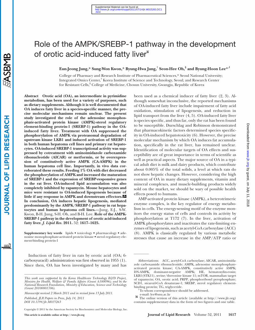
Phosphorylation of AMPK suppresses expression of SREBP-1 in vitro and in vivo ( 30, 31 ), whereas inhibition of AMPK augments expression of SREBP-1 and thus induces fatty liver. To identify the role of AMPK in the activation of SREBP-1 by OA, we transfected SK-Hep1 cells with a eukary-otic expression vector expressing constitutively active AMPK protein tagged with Myc epitope, and then the level of ma-ture SREBP-1 was determined following OA treatment. Re-sults are summarized in Fig. 2A . Basal and OA-induced expression of mature SREBP-1 protein was completely an-tagonized by CA-AMPK. Transfection with DN-AMPK tagged with Flag signifi cantly increased the basal mature SREBP-1 expression, whereas no effect was shown in OA-treated cells ( Fig.2A ). We next examined whether OA increased the ex-pression of SREBP-1 target genes that are involved in fatty acid synthesis. When the levels of Fas, Srebp-1a, Srebp-1c, Acc , Scd-1 , and L-Pk gene transcripts were measured by quantita-tive real-time PCR using gene-specifi c primers, we found that their mRNA expression was increased dose depend-ently in OA-treated cells. Combination treatment with AICAR prevented the OA-associated increase in the mRNA levels ( Fig. 2B ). Consequently, AICAR completely abrogated the effects of OA on intracellular lipid accumulation mea-sured using Nile-red staining ( Fig. 2C ).
We next performed reporter gene analysis using a re-porter gene containing SRE sequences and a gel-shift assay in SK-Hep1 and HepG2 cells. Treatment of cells with OA resulted in a dose-dependent increase in luciferase activity ( Fig. 3A ). OA-induced luciferase activity was considerably diminished by cotreatment with metformin or AICAR or by cotransfection of CA-AMPK. Conversely, introduction of DN-AMPK to cells markedly augmented the OA effect on SRE luciferase activity ( Fig. 3A ). We also observed an in-crease in the nuclear SREBP-1-dependent gel-shift activity. Supershift analysis confi rmed the DNA binding of SREBP-1 ( Fig. 3B ). Together, these data suggested that OA induces the maturation and transcriptional activity of SREBP-1 through AMPK inhibition in human and rat hepatocytes.
OA induces proteasomal degradation of LKB1 Control of protein stability via the proteasomal degrada-
tion pathway plays an important role in regulating many cellular processes. We asked whether the inhibition of AMPK phosphorylation by OA might be associated with
cells by Western blot analysis. Treatment with OA resulted in a substantial and dose-dependent increase in the amount of mature SREBP-1 protein in both human hepa-toma SK-Hep1 and HepG2 cell lines. Similar results were obtained in primary rat hepatocytes in which the expres-sion of mature SREBP-1 was increased, although a de-crease in the precursor form was observed ( Fig. 1A ). A dose-dependent increase in the mRNA for Srebp-1c was observed in the SK-Hep1, HepG2, and primary rat hepato-cytes ( Fig. 1B ).
OA inhibits AMPK phosphorylation in rat hepatocytes and human hepatoma cell lines
AMPK is a key energy sensor in the cells that ultimately control the lipid metabolism machinery. Although AMPK is classically regulated by the cellular energy status, it has been reported that chemicals or chemical mixtures can also regulate the activity of AMPK. To understand the po-tential mechanisms by which OA induces SREBP-1 activa-tion, we examined the effects of OA on the phosphorylation of AMPK, because phosphorylation on the Thr172 residue is essential for its activation ( 29 ). Incubation of human and rat hepatoma cell lines with OA for 24 h resulted in the dose-dependent reduction in phosphorylation of AMPK despite increased expression of AMPK, resulting in the de-crease in the pAMPK/AMPK ratio. Similar results were ob-tained when primary rat hepatocytes were treated with OA ( Fig. 1C ). Activation of AMPK leads to the phosphorylation
Fig. 1. OA induces the activation of SREBP-1 and suppression of AMPK in human and rat liver cell lines and primary rat hepato-cytes. Cells were treated with OA for 24 h in 1% serum media (A, B) and in serum-free condition, following incubation in serum-free media for 16 h (C, D). Cells were treated with 100 � M OA in the absence or presence of 500 � M metformin (Met) or AICAR for 24 h (D). Expression of proteins and mRNA was analyzed by West-ern blotting and by real-time PCR, respectively. Each bar represents the mean ± SE of three independent experiments. Results are ex-pressed as fold changes over vehicle control. One representative blot of at least three independent experiments with similar results is shown. * P < 0.05, ** P < 0.01, *** P < 0.001 compared with vehicle control.
by guest, on July 11, 2018w
ww
.jlr.orgD
ownloaded from
.html http://www.jlr.org/content/suppl/2011/07/14/jlr.M015263.DC1Supplemental Material can be found at:
Role of AMPK in orotic acid-induced fatty liver 1621
bility of LKB1 is decreased by the hsp90 inhibitor radicicol and that cotreatment with the proteasomal inhibitor lacta-cystin prevents its degradation ( Fig. 4D , left panel) ( 32 ). We next treated OA to HeLa cells expressing wild-type LKB1 in the absence or presence of lactacystin, and then examined intracellular levels of LKB1 by immunoblotting. These experiments revealed that the decrease in expres-sion of LKB1 by OA was completely recovered by combina-tion treatment with lactacystin ( Fig. 4D ). These data suggested that OA inhibits AMPK phosphorylation at least partly by proteasomal degradation of the upstream kinase LKB1.
OA activates SREBP-1 in rats in vivo To test whether the data from our in vitro experiments
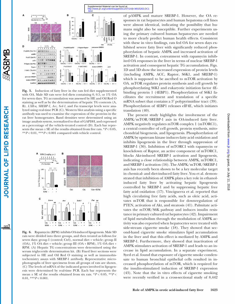
could be reproduced in vivo, rats and mice were fed OA in the diet for seven days. Consistent with a previous study ( 33 ), rats fed OA showed increased hepatic TG levels and hepatic steatosis. Hematoxylin-eosin (HE) and Oil Red O staining of liver sections obtained from OA-treated rats clearly showed that rats were susceptible to OA-induced fatty liver ( Fig. 5A ). OA treatment signifi cantly and dose-dependently increased the hepatic TG level by 2- to
the proteasomal degradation of AMPK kinase. We exam-ined the effects of OA on the expression levels of various AMPK kinases. Treatment of SK-Hep1 cells with OA de-creased the expression of LKB1, one of the major AMPK kinases expressed in the liver ( Fig. 4A ). Other AMPK ki-nases, such as CaMKK � or CaMKK � , were unaffected by OA (data not shown). To determine whether LKB1 could mediate the OA effects on AMPK phosphorylation, we compared the results of LKB1-defi cient cells with that of LKB1-expressing cells in their response to OA. To that end, we compared AMPK phosphorylation and SREBP-1 ac-tivation in HeLa cells defi cient or reconstituted with LKB1 as well as in SK-Hep1 cells wild-type or silenced with LKB1 siRNA. Western blot analysis of AMPK is shown in Fig. 4B . AMPK phosphorylation was not affected by OA in either LKB1-defi cient HeLa cells or LKB-silenced SK-Hep1 cells, whereas overexpression of LKB1 to HeLa cells restored the effect. Similar results were observed in SK-Hep1 cells. OA induced expression and activation of SREBP-1 and its target genes in SK-Hep1 cells. However, knockdown of LKB1 abolished the effects of OA ( Fig. 4C ).
The stability of LKB1 is regulated by the interaction with hsp90, which prevents its degradation by the proteasome. Our results and those of other groups suggest that the sta-
Fig. 2. Activation of AMPK attenuates OA-induced expression of lipogenic genes. SK-Hep1 cells were transfected with mock, CA-AMPK, or DN-AMPK for 18 h, and treated with OA for 24 h. West-ern blot analysis using a specifi c antibody was used to examine the expression of the proteins in the whole cell lysates. Band densities were determined using an image analysis system, normalized to that of GAPDH, and expressed as fold changes over vehicle con-trol. One representative blot of at least three independent experi-ments with similar results is shown (A). Cells were treated with OA (0, 1, 10, 100 � M) in the absence or presence of 500 � M AICAR for 24 h. Total RNA was extracted, and the mRNA for each gene was analyzed by real-time PCR using gene-specifi c primers. Results are expressed as fold changes over vehicle control (B). Cells were treated with OA, and Nile-red fl uorescence was observed after 48 h using a Molecular Device Gemini EM fl uorometer (excitation 485 nm/emission 580 nm). The relative fl uorescence intensity ratio for each sample was calculated by using a value of 1 for the vehicle control (C). Each bar represents the mean ± SE of three independent experiments. * P < 0.01, ** P < 0.01, *** P < 0.001 com-pared with vehicle control.
Fig. 3. Activation of AMPK attenuates OA-induced SREBP-1 trans-activation. SRE luciferase transactivation was determined from the lysates of SK-Hep1 and HepG2 cells transfected with the pSRE-luc construct alone or with mock, CA-AMPK, or DN-AMPK and treated with OA in the absence or presence of 500 � M metformin or AICAR. Results are expressed as fold changes over vehicle control. Each bar represents the mean ± SE of three independent experi-ments. * P < 0.05, ** P < 0.01 compared with mock-transfected vehicle control; # P < 0.05, ## P < 0.01 compared with DN-AMPK-transfected vehicle control (A). Gel shift analysis of SREBP-1 binding to the SRE was performed by the reaction of nuclear extracts with biotin-labeled SRE probe. For the competition assay, the nuclear extract was incubated with SREBP-1 antibody mixed with a labeled SRE probe. SS indicates a supershift of the SREBP1-DNA complex (B).
by guest, on July 11, 2018w
ww
.jlr.orgD
ownloaded from
.html http://www.jlr.org/content/suppl/2011/07/14/jlr.M015263.DC1Supplemental Material can be found at:

1622 Journal of Lipid Research Volume 52, 2011
signal transduction pathway, and its kinase activity regu-lates LXR � activation and subsequent lipogenic gene ex-pression by SREBP-1 ( 34, 35 ). Inhibition of mTORC1 with rapamycin or knockdown of Raptor, an active component of mTORC1, blocks Akt-induced SREBP-1 activation and lipogenesis, indicating a close relationship between AMPK, mTORC1, and SREBP-1 activation ( 16 ). We asked whether activation of the mTOR/S6K1 pathway was linked to SREBP-1 activity, which then contributed to fat accumula-tion in the OA-fed rats. As expected, OA increased the phosphorylation of S6K1 and decreased the phosphoryla-tion of Raptor ( Fig. 5C ). To determine the effect of ra-pamycin, an mTOR inhibitor, in the OA-induced fatty liver, rats were fed 1% OA and administered rapamycin for seven days. OA-induced lipid accumulation was com-pletely inhibited by the treatment with rapamycin ( Fig. 6A ). HE and Oil Red O staining of liver sections clearly confi rmed the results ( Fig. 6B ). Immunohistochemistry analyses showed signifi cantly repressed SREBP-1 in the ra-pamycin-cotreatment liver ( Fig. 6B ). All the downstream effectors of SREBP-1, such as Lxr- � , Acc, Scd-1 and Fas, were consistently suppressed by rapamycin ( Fig. 6C ). These re-sults support OA-induced fatty liver via mTOR-dependent SREBP-1 activation.
DISCUSSION
OA is an intermediate in pyrimidine metabolism that is readily synthesized in mammals and is also found in large quantities in cow’s milk ( 36 ). Owing to the benefi cial ef-fects on the absorption of essential nutrients as well as on heart function ( 37 ), OA has been used as a dietary supple-ment and in vitamin and mineral complexes. OA has also been used worldwide as a tonic and for anabolic purposes. However, administration of large amounts of OA has been reported to induce fatty liver in the rat ( 1 ). The reported mechanisms of OA-induced fatty liver include impairment of fatty acid oxidation, stimulation of lipogenesis, and re-duction in lipid transport from the liver ( 4, 5 ). Although liver fatty change by OA is evident and severe, the precise molecular mechanisms remain poorly understood. Addi-tionally, the effects of OA are species specifi c; thus, the possible human health effects of OA have not been clearly identifi ed ( 38 ).
Here, we describe the novel fi nding that the AMPK/mTOR/SREBP-1 pathway is involved in the OA-induced fatty liver. Our study demonstrated that OA inhibited the phosphorylation of AMPK and increased the activation of SREBP-1 in rat hepatocytes and human hepatoma cell lines. Activation of SREBP-1 and SREBP-regulated pro-moter by OA was mediated through AMPK inhibition, as verifi ed by cotreatment with AMPK activators metformin and AICAR or by cotransfection of the constitutively active form of AMPK mutant in a SRE-luciferase assay. The de-crease in the AMPK phosphorylation by OA can be as-cribed, at least in part, to the proteasomal degradation of LKB1. Primary cultured mouse hepatocytes were resistant to OA treatment, as shown by the unchanged expression
4-fold compared with the control ( Fig. 5B ). Next we deter-mined the expression and phosphorylation of the protein levels in the liver tissue. In line with the in vitro data shown above, the expression of AMPK and ACC were increased and the phosphorylation of the proteins were decreased, resulting in the signifi cant decrease in the pAMPK/AMPK ratio and the pACC/ACC ratio ( Fig. 5D ). Similar results were obtained when we analyzed the expression and phos-phorylation of Raptor in which we observed a net decrease in the pRaptor/Raptor ratio. The expression and phosphory-lation of S6K1 were increased by OA treatment, leading to an increase in the pS6K1/S6K1 ratio. The level of ma-ture SREBP-1 was increased in rat liver ( Fig. 5D ). Increases in protein expression, such as AMPK, ACC, and S6K, by OA in vitro and in vivo can be ascribed to the activation of mTOR, which remains to be explained in later experi-ments. However, little if any change was observed in mice in terms of fatty liver and phosphorylation of AMPK. More-over, expression of mature SREBP-1 was even decreased in mouse liver (supplementary Fig. II). The AMP/ATP ratio was not changed in vitro or in vivo (supplementary Table I). To confi rm that nuclear SREBP-1 is functional in vivo, we examined the mRNA levels of Lxr- � , SREBP-1, Acc, Scd-1 , and Fas using real-time PCR. Increased expression of the genes was observed in the OA-fed group ( Fig. 5C ).
Rapamycin inhibits OA-induced fatty liver S6K1, a member of the serine/threonine protein kinase
family, is a downstream effector of the PI3K/Akt/mTOR
Fig. 4. OA induces proteasomal degradation of LKB1. SK-Hep1, siLKB1-transfected SK-Hep1, HeLa, and HeLa cells transfected with mock or pEGFP-LKB1 expression vector were treated with OA for 24 h (A, B, C). HeLa cells transfected with pEGFP-LKB1 expres-sion vector were treated with OA in the absence or presence of 3 h pretreatment with 5 � M lactacystin, a proteasomal degradation in-hibitor. The radicicol and lactacystin treatment served as a control for proteasomal degradation and inhibition (D). Western blot analysis using a specifi c antibody was used to examine the expres-sion of the proteins in the whole cell lysates. Band densities were determined using an image analysis system, normalized to that of GAPDH, and expressed as a percentage of the vehicle-treated con-trol. Total RNA was extracted, and the mRNA for each gene was analyzed by real-time PCR using gene-specifi c primers. Results are expressed as fold changes over vehicle control. Each bar represents the mean ± SE of three independent experiments. ** P < 0.01, *** P < 0.001 compared with vehicle control.
by guest, on July 11, 2018w
ww
.jlr.orgD
ownloaded from
.html http://www.jlr.org/content/suppl/2011/07/14/jlr.M015263.DC1Supplemental Material can be found at:
Role of AMPK in orotic acid-induced fatty liver 1623
of pAMPK and mature SREBP-1. However, the OA re-sponses in rat hepatocytes and human hepatoma cell lines were almost identical, indicating the possibility that hu-mans might also be susceptible. Further experiments us-ing the primary cultured human hepatocytes are needed to more clearly predict human health effects. Consistent with these in vitro fi ndings, rats fed OA for seven days ex-hibited severe fatty liver with signifi cantly reduced phos-phorylation of hepatic AMPK and increased activation of SREBP-1. In contrast, cotreatment with rapamycin inhib-ited OA responses in the liver in terms of nuclear SREBP-1 activation and consequent hepatic TG accumulation. Figs. 1D and 5D show the increased expression of protein levels (including AMPK, ACC, Raptor, S6K1, and SREBP-1) which is supposed to be ascribed to mTOR activation by OA. mTOR regulates protein synthesis and cell growth by phosphorylating S6K1 and eukaryotic initiation factor 4E-binding protein 1 (4EBP1). Phosphorylation of S6K1 fa-cilitates the recruitment and translation of a specifi c mRNA subset that contains a 5 ′ polypyrimidine tract ( 39 ). Phosphorylation of 4EBP1 releases eIF4E, which initiates translation ( 40 ).
The present study highlights the involvement of the AMPK/mTOR/SREBP-1 axis in OA-induced fatty liver. AMPK negatively regulates mTOR complex 1 (mTORC1), a central controller of cell growth, protein synthesis, mito-chondrial biogenesis, and lipogenesis. Phosphorylation of AMPK by upstream kinase induces fatty acid oxidation and inhibits lipogenesis in the liver through suppression of SREBP-1 ( 30 ). Inhibition of mTORC1 with rapamycin or knockdown of Raptor, an active component of mTORC1, blocks Akt-induced SREBP-1 activation and lipogenesis, indicating a close relationship between AMPK, mTORC1, and SREBP-1 activation ( 16 ). The AMPK/mTOR/SREBP-1 axis has recently been shown to be a key molecular target in chemical- and diet-induced fatty liver. You et al. demon-strated that inhibition of AMPK plays a key role in ethanol-induced fatty liver by activating hepatic lipogenesis controlled by SREBP-1 and by suppressing hepatic free fatty acid oxidation ( 17 ). Vinciguerra et al. reported that high circulating free fatty acids, such as oleic acid, acti-vates mTOR that is responsible for downregulation of PTEN, activation of Akt, and steatosis ( 41 ). Palmitate acti-vates the mTOR/S6K pathway and induces insulin resis-tance in primary cultured rat hepatocytes ( 42 ). Impairment of lipid metabolism through the modulation of AMPK ac-tivity was also reported when hepatocytes were treated with side-stream cigarette smoke ( 18 ). They showed that sec-ond-hand cigarette smoke stimulates lipid accumulation in the liver and that this effect is mediated by AMPK and SREBP-1. Furthermore, they showed that inactivation of AMPK stimulates activation of SREBP-1 and leads to an in-crease in lipid accumulation. In a separate experiment, Syed et al. found that exposure of cigarette smoke conden-sate to human bronchial epithelial cells resulted in in-creased phosphorylation of mTOR, which is required for the insulin-stimulated induction of SREBP-1 expression ( 43 ). Note that the in vitro effects of cigarette smoking were recently verifi ed in a cross-sectional study of 8,442
Fig. 5. Induction of fatty liver in the rats fed diet supplemented with OA. Male SD rats were fed diets containing 0, 0.5, or 1% OA for seven days. TG accumulation was assessed by HE and Oil Red O staining as well as by the determination of hepatic TG contents (A, B). LXR- a , SREBP-1, Acc, Scd-1 , and Fas transcript levels were ana-lyzed using real-time PCR (C). Western blot analysis using a specifi c antibody was used to examine the expression of the proteins in the rat liver homogenates. Band densities were determined using an image analysis system, normalized to that of GAPDH, and expressed as a percentage of the vehicle-treated control (D). Each bar repre-sents the mean ± SE of the results obtained from fi ve rats. * P < 0.05, ** P < 0.01, *** P < 0.001 compared with vehicle control.
Fig. 6. Rapamycin (RPM) inhibits OA-induced lipogenesis. Male SD rats were divided into three groups, and then treated as follows for seven days: group I (control; Ctrl), normal diet + vehicle; group II (OA), 1% OA diet + vehicle; group III (OA + RPM), 1% OA diet + RPM. (A) Hepatic TG concentrations were determined using the serum triglyceride determination kit. (B) Fixed liver sections were subjected to HE and Oil Red O staining as well as immunohis-tochemistry assays with SREBP-1 antibody. Representative micro-photographs of liver specimens from all groups of rats are shown. (C) The levels of mRNA of the indicated genes involved in lipogen-esis were determined by real-time PCR. Each bar represents the mean ± SE of the results obtained from six rats. * P < 0.05, ** P < 0.01, *** P < 0.001.
by guest, on July 11, 2018w
ww
.jlr.orgD
ownloaded from
.html http://www.jlr.org/content/suppl/2011/07/14/jlr.M015263.DC1Supplemental Material can be found at:

1624 Journal of Lipid Research Volume 52, 2011
pharmacokinetic studies will be needed to defi ne human health effects. The effects of OA could be even greater in obese and insulin-resistant diabetic patients if we consider the poor sensitivity of these patients to insulin-induced downstream effectors, including AMPK and SREBP-1. Reg-ular ethanol consumption and cigarette smoke may also synergistically affect OA-induced fatty liver.
In summary, we demonstrated that OA induces the SREBP-1-induced lipogenesis pathway through mTOR-p70S6K1 activation and that reduced AMPK phosphoryla-tion contributed to the activation of S6K1 phosphorylation. Moreover, we discovered that phosphorylation of AMPK is partially decreased by OA-induced LKB1 proteasomal deg-radation ( Fig. 7 ). Our study provides some critical infor-mation needed for an understanding of the molecular mechanisms of OA-induced fatty liver and of species differ-ences in susceptibility. OA induces lipogenesis, mediated predominantly by the AMPK/mTOR/SREBP-1 pathway in rat hepatocytes and human hepatoma cell lines.
REFERENCES
1 . Standerfer , S. B. , and P. Handler . 1955 . Fatty liver induced by oro-tic acid feeding. Proc. Soc. Exp. Biol. Med. 90 : 270 – 271 .
2 . Ferreira , A. V. , G. G. Parreira , L. C. Porto , E. G. Mario , H. L. Delpuerto , A. S. Martins , and L. M. Botion . 2008 . Fenofi brate prevents orotic acid-induced hepatic steatosis in rats. Life Sci. 82 : 876 – 883 .
3 . Buang , Y. , Y. M. Wang , J. Y. Cha , K. Nagao , and T. Yanagita . 2005 . Dietary phosphatidylcholine alleviates fatty liver induced by orotic acid. Nutrition . 21 : 867 – 873 .
4 . Miyazawa , S. , S. Furuta , and T. Hashimoto . 1982 . Reduction of beta-oxidation capacity of rat liver mitochondria by feeding orotic acid. Biochim. Biophys. Acta . 711 : 494 – 502 .
5 . Hebbachi , A. M. , M. C. Seelaender , B. W. Baker , and G. F. Gibbons . 1997 . Decreased secretion of very-low-density lipoprotein triacyl-glycerol and apolipoprotein B is associated with decreased intracel-lular triacylglycerol lipolysis in hepatocytes derived from rats fed orotic acid or n-3 fatty acids. Biochem. J. 325 : 711 – 719 .
6 . Durschlag , R. P. , and J. L. Robinson . 1980 . Species specifi city in the metabolic consequences of orotic acid consumption. J. Nutr. 110 : 822 – 828 .
7 . Hawley , S. A. , M. Davison , A. Woods , S. P. Davies , R. K. Beri , D. Carling , and D. G. Hardie . 1996 . Characterization of the AMP-activated protein kinase kinase from rat liver and identifi cation of threonine 172 as the major site at which it phosphorylates AMP-activated protein kinase. J. Biol. Chem. 271 : 27879 – 27887 .
8 . Hardie , D. G. , J. Corton , Y. P. Ching , S. P. Davies , and S. Hawley . 1997 . Regulation of lipid metabolism by the AMP-activated protein kinase. Biochem. Soc. Trans. 25 : 1229 – 1231 .
9 . Kawanaka , K. , L. A. Nolte , D. H. Han , P. A. Hansen , and J. O. Holloszy . 2000 . Mechanisms underlying impaired GLUT-4 trans-location in glycogen-supercompensated muscles of exercised rats. Am. J. Physiol. Endocrinol. Metab. 279 : E1311 – E1318 .
10 . Taylor , E. B. , W. J. Ellingson , J. D. Lamb , D. G. Chesser , and W. W. Winder . 2005 . Long-chain acyl-CoA esters inhibit phosphorylation of AMP-activated protein kinase at threonine-172 by LKB1/STRAD/MO25. Am. J. Physiol. Endocrinol. Metab. 288 : E1055 – E1061 .
11 . Rafaeloff-Phail , R. , L. Ding , L. Conner , W. K. Yeh , D. McClure , H. Guo , K. Emerson , and H. Brooks . 2004 . Biochemical regulation of mammalian AMP-activated protein kinase activity by NAD and NADH. J. Biol. Chem. 279 : 52934 – 52939 .
12 . Fryer , L. G. D. , A. Parbu-Patel , and D. Carling . 2002 . The anti-dia-betic drugs rosiglitazone and metformin stimulate AMP-activated protein kinase through distinct signaling pathways. J. Biol. Chem. 277 : 25226 – 25232 .
13 . Minokoshi , Y. , Y. B. Kim , O. D. Peroni , L. G. Fryer , C. Müller , D. Carling , and B. B. Kahn . 2002 . Leptin stimulates fatty-acid oxi-dation by activating AMP-activated protein kinase. Nature . 415 : 339 – 343 .
subjects. The prevalence rate of nonalcoholic fatty liver disease was signifi cantly higher among cigarette smokers versus nonsmokers. Moreover, the prevalence ratio in-creases with the amount of cigarettes smoked per day ( 44 ). Although the precise effects of OA in humans are un-known, we must not exclude the possibility that OA con-sumption by diet and dietary supplements can induce fatty liver in humans. A quantitative risk assessment is needed to address the issue.
One of the novel fi ndings in the present study is the identifi cation of a molecular target that responds differ-ently between the rat and mouse. Previously, the likely cause of the interspecies differences in OA hepatotoxicity has been thought to be metabolic differences. Mice accu-mulated less of the OA dose in the liver and excreted much more in the urine than did rats, thereby rendering them resistant to OA toxicity ( 6 ). Feeding an arginine-defi cient diet to rats leads to a marked fatty liver by an elevated plasma concentration of OA. Additionally, mice, hamsters, and rabbits were resistant to fatty liver largely because of their high urinary excretion rate of OA ( 45 ). Apart from this pharmacokinetic issue, no other information has been available concerning species-specifi c mechanisms of OA hepatotoxicity. Our results showed that the most striking difference between the rat and mouse hepatocytes in pri-mary culture is AMPK phosphorylation and SREBP-1 acti-vation in response to OA exposure. Whereas decreased phosphorylation of AMPK and modulation of a series of downstream events leading to fatty acid synthesis and lipo-genesis were observed in rat hepatocytes and human hepa-toma cell lines, mouse hepatocytes were resistant to OA with regard to the AMPK/SREBP-1-dependent lipogenic pathway. Similar results were observed in animal studies us-ing SD rats and C57BL/6 mice. These two species showed completely different responses to OA in terms of AMPK/SREBP-1 signaling and development of steatosis, indicating that not only pharmacokinetic but also pharmacodynamic factors participate in determining interspecies differences. Considering the very similar responses of human hepatoma cell lines and rat hepatocytes to the AMPK/SREBP-1 path-way, humans could be a species that may be sensitive to OA-induced fatty liver. Further experiments using primary cultured human hepatocytes and comprehensive human
Fig. 7. Proposed mechanism of OA-induced lipogenesis by the AMPK/SREBP-1 pathway.
by guest, on July 11, 2018w
ww
.jlr.orgD
ownloaded from
.html http://www.jlr.org/content/suppl/2011/07/14/jlr.M015263.DC1Supplemental Material can be found at:

Role of AMPK in orotic acid-induced fatty liver 1625
14 . Hawley , S. A. , J. Boudeau , J. L. Reid , K. J. Mustard , L. Udd , T. P. Mäkelä , D. R. Alessi , and D. G. Hardie . 2003 . Complexes between the LKB1 tumor suppressor, STRADalpha/beta and MO25alpha/beta are upstream kinases in the AMP-activated protein kinase cas-cade. J. Biol. 2 : 28 – 32 .
15 . Branvold , D. J. , D. R. Allred , D. J. Beckstead , H. J. Kim , N. Fillmore , B. M. Condon , J. D. Brown , S. N. Sudweeks , D. M. Thomson , and W. W. Winder . 2008 . Thyroid hormone effects on LKB1, MO25, phospho-AMPK, phospho-CREB, and PGC-1 � in rat muscle. J. Appl. Physiol. 105 : 1218 – 1227 .
16 . Porstmann , T. , C. R. Santos , B. Griffi ths , M. Cully , M. Wu , S. Leevers , J. R. Griffi ths , Y. L. Chung , and A. Schulze . 2008 . SREBP activity is regulated by mTORC1 and contributes to Akt-dependent cell growth. Cell Metab. 8 : 224 – 236 .
17 . You , M. , M. Matsumoto , C. M. Pacold , W. K. Cho , and D. W. Crabb . 2004 . The role of AMP-activated protein kinase in the action of ethanol in the liver. Gastroenterology . 127 : 1798 – 1808 .
18 . Yuan , H. , J. Y. J. Shy , and M. Martins-Green . 2009 . Second-hand smoke stimulates lipid accumulation in the liver by modulating AMPK and SREBP-1. J. Hepatol. 51 : 535 – 547 .
19 . Wu , Y. , P. Song , J. Xu , M. Zhang , and M. H. Zou . 2007 . Activation of protein phosphatase 2A by palmitate inhibits AMP-activated pro-tein kinase. J. Biol. Chem. 282 : 9777 – 9788 .
20 . Lee , W. J. , M. Kim , H. S. Park , H. S. Kim , M. J. Jeon , K. S. Oh , E. H. Koh , J. C. Won , M. S. Kim , G. T. Oh , et al . 2006 . AMPK activa-tion increases fatty acid oxidation in skeletal muscle by activating PPAR � and PGC-1. Biochem. Biophys. Res. Commun. 340 : 291 – 295 .
21 . Kelley , W. N. , M. L. Greene , I. H. Fox , F. M. Rosenbloom , R. I. Levy , and J. E. Seegmiller . 1970 . Effects of orotic acid on purine and lipo-protein metabolism in man. Metabolism . 19 : 1025 – 1035 .
22 . Griffi n , J. L. , S. A. Bonney , C. Mann , A. M. Hebbachi , G. F. Gibbons , J. K. Nicholson , C. C. Shoulders , and J. Scott . 2004 . An integrated reverse functional genomic and metabolic approach to understand-ing orotic acid-induced fatty liver. Physiol. Genomics . 17 : 140 – 149 .
23 . French , C. T. , W. H. Hanneman , L. S. Chubb , R. E. Billings , and M. E. Andersen . 2004 . Induction of CYP1A1 in primary rat hepatocytes by 3,3 ′ ,4,4’,5-pentachlorobiphenyl: evidence for a switch circuit ele-ment. Toxicol. Sci. 78 : 276 – 286 .
24 . Honkakoski , P. , and M. Negishi . 1998 . Protein serine/threonine phos-phatase inhibitors suppress phenobarbital-induced Cyp2b10 gene transcription in mouse primary hepatocytes. Biochem. J. 330 : 889 – 895 .
25 . Wynants , J. , and H. Van Belle. Single-run high-performance liquid chromatography of nucleotides, nucleosides, and major purine bases and its application to different tissue extracts. 1985 . Anal. Biochem. 144 : 258 – 266 .
26 . Wang , L. H. , Y. L. Choi , X. Y. Hua , Y. K. Shin , Y. J. Song , S. J. Youn , H. Y. Yun , S. M. Park , W. J. Kim , H. J. Kim , et al . 2006 . Increased expres-sion of sonic hedgehog and altered methylation of its promoter region in gastric cancer and its related lesions. Mod. Pathol. 19 : 675 – 683 .
27 . Bruce , C. R. , M. J. Anderson , A. L. Carey , D. G. Newman , A. Bonen , A. D. Kriketos , G. J. Cooney , and J. A. Hawley . 2003 . Muscle oxida-tive capacity is a better predictor of insulin sensitivity than lipid status. J. Clin. Endocrinol. Metab. 88 : 5444 – 5451 .
28 . You , M. , M. Fischer , M. A. Deg , and D. W. Crabb . 2002 . Ethanol induces fatty acid synthesis pathways by activation of sterol regulatory element-binding protein (SREBP). J. Biol. Chem. 277 : 29342 – 29347 .
29 . Stein , S. C. , A. Woods , N. A. Jones , M. D. Davison , and D. Carling . 2000 . The regulation of AMP-activated protein kinase by phospho-rylation. Biochem. J. 345 : 437 – 443 .
30 . Zhou , G. , R. Myers , Y. Li , Y. Chen , X. Shen , J. Fenyk-Melody , M. Wu , J. Ventre , T. Doebber , N. Fujii , et al . 2001 . Role of AMP-activated
protein kinase in mechanism of metformin action. J. Clin. Invest. 108 : 1167 – 1174 .
31 . Shklyaev , S. , G. Aslanidi , M. Tennant , V. Prima , E. Kohlbrenner , V. Kroutov , M. Campbell-Thompson , J. Crawford , E. W. Shek , P. J. Scarpace , et al . 2003 . Sustained peripheral expression of transgene adiponectin offsets the development of diet-induced obesity in rats. Proc. Natl. Acad. Sci. USA . 100 : 14217 – 14222 .
32 . Boudeau , J. , M. Deak , M. A. Lawlor , N. A. Morrice , and D. R. Alessi . 2003 . Heat-shock protein 90 and Cdc37 interact with LKB1 and regulate its stability. Biochem. J . 370(Pt. 3): 849–857.
33 . Creasey , W. A. , L. Hankin , and R. E. Handschumacher . 1961 . Fatty livers induced by orotic acid. J. Biol. Chem. 236 : 2064 – 2070 .
34 . Brown , E. J. , P. A. Beal , C. T. Keith , J. Chen , T. B. Shin , and S. L. Schreiber . 1995 . Control of p70S6 kinase by kinase activity of FRAP in vivo. Nature . 377 : 441 – 446 .
35 . Hwahng , S. H. , S. H. Ki , E. J. Bae , H. E. Kim , and S. G. Kim . 2009 . Role of adenosine monophosphate-activated protein kinase-p70 ribosomal S6 kinase-1 pathway in repression of liver X receptor-alpha-dependent lipogenic gene induction and hepatic steatosis by a novel class of dithiolethiones. Hepatology . 49 : 1913 – 1925 .
36 . Hurlbert , R. B. , and V. R. Potter . 1954 . Nucleotide metabolism. J. Biol. Chem. 209 : 1 – 21 .
37 . Richards , S. M. , R. A. J. Conyers , J. L. Fisher , and F. L. Rosenfeldt . 1997 . Cardioprotection by orotic acid: metabolism and mechanism of action. J. Mol. Cell. Cardiol. 29 : 3239 – 3250 .
38 . Robinson , J. L. 1980 . Bovine milk orotic acid: variability and signifi -cance for human nutrition. J. Dairy Sci. 63 : 865 – 871 .
39 . Dufner , A. , and G. Thomas . 1999 . Ribosomal S6 kinase signaling and the control of translation. Exp. Cell Res. 253 : 100 – 109 .
40 . Gingras , A. C. , B. Raught , and N. Sonenberg . 2004 . mTOR signal-ing to translation. Curr. Top. Microbiol. Immunol. 279 : 169 – 197 .
41 . Vinciguerra , M. , C. Veyrat-Durebex , M. A. Moukil , L. Rubbia-Brandt , F. Rohner-Jeanrenaud , and M. Foti . 2008 . PTEN down-regulation by unsaturated fatty acids triggers hepatic steatosis via an NF-kappaBp65/mTOR-dependent mechanism. Gastroenterology . 134 : 268 – 280 .
42 . Mordier , S. , and P. B. Iynedjian . 2007 . Activation of mammalian target of rapamycin complex 1 and insulin resistance induced by palmitate in hepatocytes. Biochem. Biophys. Res. Commun. 362 : 206 – 211 .
43 . Syed , D. N. , F. Afaq , M. H. Kweon , N. Hadi , N. Bhatia , V. S. Spiegelman , and H. Mukhtar . 2007 . Green tea polyphenol EGCG suppresses cigarette smoke condensate-induced NF-kappaB acti-vation in normal human bronchial epithelial cells. Oncogene . 26 : 673 – 682 .
44 . Xu , C. F. , C. H. Yu , L. Xu , M. Miao , and Y. M. Li . 2010 . Is cigarette smoking an independent risk factor or a cofactor for nonalcoholic fatty liver disease? Hepatology . 52 : 803 – 804 .
45 . Milner , J. A. , and A. S. Hassan . 1981 . Species specifi city of arginine defi ciency-induced hepatic steatosis. J. Nutr. 111 : 1067 – 1073 .
46 . Harrison , W. J. , J. J. Bull , H. Seltmann , C. C. Zouboulis , and M. P. Philpott . 2007 . Expression of lipogenic factors galectin-12, resistin, SREBP-1, and SCD in human sebaceous glands and cultured sebo-cytes. J. Invest. Dermatol. 127 : 1309 – 1317 .
47 . Sekiya , M. , A. Hiraishi , M. Touyama , and K. Sakamoto . 2008 . Oxidative stress induced lipid accumulation via SREBP1c activation in HepG2 cells. Biochem. Biophys. Res. Commun. 375 : 602 – 607 .
48 . Howell 3rd , G. , X. Deng , C. Yellaturu , E. A. Park , H. G. Wilcox , R. Raghow , and M. B. Elam . 2009 . N-3 polyunsaturated fatty ac-ids suppress insulin-induced SREBP-1c transcription via reduced trans-activating capacity of LXR � . Biochim. Biophys. Acta . 1791: 1190 – 1196 .
by guest, on July 11, 2018w
ww
.jlr.orgD
ownloaded from
.html http://www.jlr.org/content/suppl/2011/07/14/jlr.M015263.DC1Supplemental Material can be found at:

![friday, may 31, 2013004e2d1.netsolhost.com/2013OralAbstracts.pdfthrough activation of Sterol Regulatory Element-Binding Protein (SREBP) transcription factors [2]. A recent study found](https://img.pdfslide.net/doc/110x75/601e5c12ded03932f17de27b/friday-may-31-through-activation-of-sterol-regulatory-element-binding-protein.jpg)