Embed Size (px)
Citation preview
Running Head: Carotenoid metabolism in soybean root nodules Corresponding author: Choong-Ill Cheon
Mailing address: Department of Biological Science,
Sookmyung Women’s University,
Hyochangwongil 52, Yongsan-gu,
Seoul 140-742, Korea
Phone: 82-2-710-9396
Fax: 82-2-2077-7322
E-mail address: [email protected]
Journal research area: Plants Interacting with Other Organisms
Plant Physiology Preview. Published on May 22, 2013, as DOI:10.1104/pp.113.215020
Copyright 2013 by the American Society of Plant Biologists
www.plantphysiol.orgon May 14, 2018 - Published by Downloaded from Copyright © 2013 American Society of Plant Biologists. All rights reserved.

Kim et al.
2
Functional implication of β-carotene hydroxylases in soybean nodulation
Yun-Kyoung Kim2, Sunghan Kim2, Ji-Hyun Um, Kyunga Kim, Sun-Kang Choi, Byung-Hun Um, Suk-Woo Kang, Jee-Woong Kim, Shinichi Takaichi, Seok-Bo Song, Choon-Hwan Lee, Ho-Seung Kim, Ki Woo Kim, Kyoung Hee Nam, Suk-Ha Lee, Yul-Ho Kim, Hyang-Mi Park, Sun-Hwa Ha, Desh Pal S. Verma and Choong-Ill Cheon*
Department of Biological Science, Sookmyung Women’s University, Seoul, Korea (Y.-K.K., S.K., J.-H.U., K.H.N. C.-I.C.), Department of Statistics, Sookmyung Women’s University, Seoul, Korea (K.K.), Gangneung Science Industry Foundation, Gangneung (S.-K.C.), Natural Products Research Center, KIST Gangneung Institute, Gangneung (B.-H.U., S.-W.K.), Electron Microscopy Lab, Dental Research Institute, Seoul National University (J.-W.K.), Department of Biology, Nippon Medical School, Kosugi-cho 2, Nakahara, Kawasaki, Japan (S.T.), Department of Functional Crop, National Institute of Crop Science, Milyang (S.-B.S.), Department of Molecular Biology, Pusan National University, Busan (C.-H.L., H.-S.K.), School of Ecological and Environmental System, Kyungpook National University, Sangju (K.W.K.), School of Plant Science, Seoul National University, Seoul (S.-H.L.), National Institute of Crop Science, Suwon (Y.-H.K., H.-M.P.), National Institute of Agricultural Science and Technology, Suwon (S.-H.H.), Ohio State Biotechnology Center, Ohio State University, Columbus, OH43210, USA (D.P.S.V.)
One–Sentence Summary
: Carotenoids are essential for nodule development in soybean
www.plantphysiol.orgon May 14, 2018 - Published by Downloaded from Copyright © 2013 American Society of Plant Biologists. All rights reserved.
Kim et al.
3
Footnotes
1This research was supported by the Basic Science Research Program through the
National Research Foundation of Korea (NRF) funded by the Ministry of Education,
Science and Technology (2-1205-0037) and by grants from the Next-Generation
BioGreen 21 Program (No. PJ009076012013), Rural Development Administration,
Republic of Korea
2These authors contributed equally to this work
*Corresponding author: [email protected]; fax: 82-2-2077-7322
www.plantphysiol.orgon May 14, 2018 - Published by Downloaded from Copyright © 2013 American Society of Plant Biologists. All rights reserved.
Kim et al.
4
ABSTRACT Legume-Rhizobium symbiosis requires signaling between the symbiotic
partners, and differential expression of plant genes during nodule development.
Previously we cloned a gene encoding a putative β-carotene hydroxylase (GmBCH1)
from soybean (Glycine max) whose expression increased during nodulation with
Bradyrhizobium japonicum. In the present work we extended our study to three
GmBCHs to examine their possible role(s) in nodule development as they were
additionally identified as nodule-specific, along with the completion of the soybean
genome. In situ hybridization revealed the expression of three GmBCHs (GmBCH1,
GmBCH2, and GmBCH3) in the infected cells of root nodules, and their enzymatic
activities were confirmed by functional assays in E. coli. Localization of GmBCHs by
transfecting Arabidopsis protoplasts with GFP fusions and by EM immunogold
detection in soybean nodules indicated that GmBCH2 and GmBCH3 were present in
plastids while GmBCH1 appeared to be cytosolic. RNAi of the GmBCHs severely
impaired nitrogen fixation as well as nodule development. Surprisingly, we failed to
detect zeaxanthin, a product of GmBCH, or any other carotenoids, in nodules. We
therefore examined the possibility that most of the carotenoids in nodules are converted
or cleaved to other compounds. We detected the expression of some carotenoid cleavage
dioxygenases (GmCCDs) in wild-type nodules, and also a reduced amount of
zeaxanthin in GmCCD8-expressing E. coli, suggesting cleavage of the carotenoid. In
view of these findings we propose that carotenoids such as zeaxanthin synthesized in
root nodules are cleaved by GmCCDs, and we discuss the possible roles of the
carotenoid cleavage products in nodulation.
Keywords: rhizobial symbiosis, β-carotene hydroxylase, carotenoid cleavage
dioxygenases, apocarotenoid, soybean, RNAi
www.plantphysiol.orgon May 14, 2018 - Published by Downloaded from Copyright © 2013 American Society of Plant Biologists. All rights reserved.
Kim et al.
5
INTRODUCTION Legume-Rhizobium symbiosis results in the formation of root nodule, in which
rhizobia fix atmospheric nitrogen. Nodule development requires diverse events such as
Nod factor synthesis in the rhizobia, perception of the Nod factor on plant roots by
receptor-like kinases, endocytosis of rhizobia into plant cells, and so on (Stacey et al.,
2006; Oldroyd et al., 2011; Singh and Parniske, 2012). Sequential expression of
numerous plant genes occurs during nodulation, contributing to different stages
including nitrogen fixation. Arbuscular mycorrhizal (AM) symbiosis exhibits many
similarities to the nodulation process (Oldroyd et al., 2009). For example, SymRK, the
receptor-like kinase gene, is required for both rhizobial and AM symbioses (Stracke et
al., 2002). Similarly, the signal transduction pathways following perception are also in
part the same, and the genes common to the two pathways have been referred to as the
common SYM genes (Kistner et al., 2005). These similarities may reflect common
mechanisms for host plant cells to respond to symbionts although the commonality is
not globally defined yet.
Plant carotenoids are mostly C40 tetraterpenoid pigments with a series of
double bonds (DellaPenna and Pogson, 2006; Lu and Li, 2008). They play essential
roles in photosynthesis. The phytohormone abscisic acid is synthesized from
xanthophylls, oxygenated derivatives of carotenoids. The beneficial effects of
carotenoids for human disease prevention and health promotion are well-established and
are based on their antioxidant activities (Kopsell and Kopsell, 2006; Rao and Rao, 2007;
von Lintig, 2010). Metabolic engineering approaches have produced crop plants with
enhanced carotenoid contents and improved nutritional value (Giuliano et al., 2006).
For example, enhancement of β–carotene, provitamin A, by engineering the carotenoid
biosynthetic pathway resulted in the development of golden rice (Ye et al., 2000; Paine
et al., 2005: Ha et al., 2010).
The initial step of carotenoid biosynthesis is the production of phytoene by the
www.plantphysiol.orgon May 14, 2018 - Published by Downloaded from Copyright © 2013 American Society of Plant Biologists. All rights reserved.
Kim et al.
6
enzyme phytoene synthase (PSY) (Fig. 1) (DellaPenna and Pogson, 2006; Cazzonelli
and Pogson, 2010). The subsequent activities of desaturases, isomerase, and cyclase
convert phytoene into lycopene, and further into β-carotene. Xanthophyll synthesis
begins with the action of β-carotene hydroxylase (BCH) on β-carotene, producing
initially β-cryptoxanthin and thereafter zeaxanthin (Kim et al., 2009). Overexpression of
BCH has been found to confer tolerance to light stress (Davison et al., 2002). The
subsequent steps catalyzed by zeaxanthin epoxidase (ZEP) and neoxanthin synthase
(NSY) lead to the synthesis of ABA (Takaichi and Mimuro, 1998).
Various carotenoid cleavage dioxygenases (CCDs) catalyze the formation of
apocarotenoids with functions as hormones, flavors, and pigments (Auldridge et al.,
2006b; Strack and Fester, 2006; Tsuchiya and McCourt, 2009; Walter et al., 2010).
Recently, CCD7 and CCD8 were shown to control the synthesis of strigolactones, newly
discovered hormones that inhibit shoot branching (Gomez-Roldan et al., 2008; Umehara
et al., 2008; Vogel et al., 2010; Ruyter-Spira et al., 2013). In addition, carotenoid
cleavage products have been discovered in plant roots colonized by arbuscular
mycorrhizal (AM) fungi (Strack and Fester, 2006). During AM symbiosis, roots
synthesize apocarotenoids at the same time as activating plant genes for carotenoid
metabolism. Although RNA interference (RNAi)-mediated inhibition of apocarotenoid
synthesis suggests that apocarotenoids are functionally significant (Snowden et al.,
2005; Floss et al., 2008), their role in AM symbiosis is unknown.
In a search for genes differentially induced during soybean-Rhizobium
symbiosis, several antioxidant genes including a gene encoding a putative β-carotene
hydroxylase were identified. In this report, we describe genes (GmBCHs) encoding a
putative β-carotene hydroxylase whose expression increase in soybean root nodules.
The biochemical activities of BCHs were therefore investigated. RNAi inhibition of
GmBCH expression interfered with nitrogen fixation as well as nodule development.
Subsequent analysis of the expression and biochemical activities of GmCCDs in root
www.plantphysiol.orgon May 14, 2018 - Published by Downloaded from Copyright © 2013 American Society of Plant Biologists. All rights reserved.

Kim et al.
7
nodules led us to hypothesize that GmCCD8 could be involved in the synthesis of
apocarotenoids from zeaxanthin in these nodules.
RESULTS
Expression of genes encoding putative β-carotene hydroxylases in Glycine max
We isolated from root nodules of Glycine max a cDNA with strong homology
to β-carotene hydroxylase (GmBCH1), whose expression was higher in nodules than in
roots (Fig. 2A; Lee et al., 2005). In a BLAST search with the GmBCH1 sequence
against the soybean genome (http://www.phytozome.net/soybean.php; Schmutz et al.,
2010), we found several open reading frames encoding products with amino-acid
sequences highly homologous to that of GmBCH1; they were designated GmBCH2 -
GmBCH5 (GenBank accession numbers: GmBCH1 [AY575953], GmBCH2
[BT093388], GmBCH3 [BT098487], GmBCH4 [JF970190], GmBCH5 [JF970191]).
The ORFs of GmBCH1 and GmBCH2 are very similar to functionally confirmed β-
carotene hydroxylases, e.g., they have, respectively, 74 % and 75 % sequence identity to
the β-carotene hydroxylase from Coffea arabica (CaCRTR-B; Simkin et al., 2008)
(Supplemental Fig. S1A). They have divergent N-terminal regions, like most of
previously reported β-carotene hydroxylases, but carry four histidine-containing motifs,
in which spacing of the histidine residues were conserved such as HXXXXH and
HXXHH. Presence of the histidines in these motifs has been confirmed to be essential
for the enzymatic activity as the mutagenesis abolishing these histidine residues resulted
in no enzymatic conversion of β-carotene into zeaxanthin (Supplemental Fig. S1A;
Bouvier et al., 1998). GmBCH2 appears to possess a plastid transit sequence (see
below) (Yu et al., 2007), and GmBCH3 has a sequence almost identical to GmBCH1
except for its N-terminal 33 amino acids, so also contains the above-mentioned motifs
common to β-carotene hydroxylases. The 5’-untranslated regions of GmBCH1 and
www.plantphysiol.orgon May 14, 2018 - Published by Downloaded from Copyright © 2013 American Society of Plant Biologists. All rights reserved.
Kim et al.
8
GmBCH3 differ. Whereas the GmBCH3 locus could be identified in the present version
of soybean genome database, the unique 5' region of GmBCH1 sequence was not
detected in the database. Therefore we attempted to establish the presence of GmBCH1
in the soybean genome by cloning its specific 5’ DNA region (see Materials and
Methods). A 432-bp DNA fragment, which included the upstream promoter region of
GmBCH1, was cloned (Supplemental Fig. S2A) and this 5’ region as well as the coding
region of GmBCH1 were also detected by genomic PCR (Supplemental Fig. S2B, lanes
3 and 6). GmBCH4 and GmBCH5 also contain the motifs mentioned above and thus are
regarded as additional BCH paralogs (Supplemental Fig. S1A).
We investigated the expression of the GmBCHs in various tissues by real-time
RT-PCR. To optimize the PCR, we examined critical aspects of the primers
(Supplemental Fig. S3). Expression of GmBCH2 was higher than that of GmBCH1 and
GmBCH3 in most tissues, particularly in leaf, and relatively low in nodules, whereas
expression of GmBCH1 and GmBCH3 was high in leaf or flower and noticeable in root
nodules (Fig. 2). Expression of GmBCH4 and GmBCH5 was high in leaf and quite low
in roots and nodules. These results generally match with the RNA-Seq Atlas data
(Libault et al., 2010; Severin et al., 2010; Supplemental Table S1). GmBCH3 expression
in the transcriptomic data might actually reflect the expression of both GmBCH1 and
GmBCH3 because their coding regions of both genes are almost identical and GmBCH1
is not identified in the current version of the soybean genome database (Supplemental
Table S1).
In order to examine GmBCH expression during nodulation, we performed real-
time RT-PCR and in situ hybridization. For the in situ hybridization, specific regions of
each gene were used to make probes for GmBCH1/3 and GmBCH2 (Supplemental Figs.
S1 and S4, A and B; see Materials and methods). The probes were checked for
hybridization specificity by whole-mount in situ hybridization to young leaves
(Supplemental Fig. S4, C and D). The relative expression levels of GmBCH1/3 and
www.plantphysiol.orgon May 14, 2018 - Published by Downloaded from Copyright © 2013 American Society of Plant Biologists. All rights reserved.
Kim et al.
9
GmBCH2 obtained from whole-mount in situ hybridization in young leaves was found
to be comparable to those from the real-time RT-PCR in Figure 2, implying that the
GmBCHs probes are not likely to cross-hybridize. Sections from nodules at different
stages of development were hybridized with a probe for GmBCH1 / GmBCH3 together
as they have almost identical DNA sequences. GmBCH1 and GmBCH3 were found to
be induced as nodules matured, suggesting that both are involved in the nodulation
process (Fig. 3, A, C, D to G). Their expression was seen in the root pericycle in the
early stages of nodulation and became high in the central infected zones in the early and
mature nodule stages. GmBCH2 was also expressed during nodulation, especially
strongly in 7-day-old nodules (Fig. 3, B, I to L). Its expression was also strong in root
pericycle. The GmBCH expression levels during nodulation were confirmed by the
fluorometric measurement of GUS activities using transgenic roots and nodules
expressing GmBCH2/3 promoter (1.6-kb upstream regions of each) – GUS fusions
(Supplemental Fig. S5). These observations could mean that expression of the putative
β-carotene hydroxylase genes, especially GmBCH1, GmBCH2 and GmBCH3 may be
involved in nodulation.
Enzymatic activities of GmBCHs
The similarity of GmBCH1, GmBCH2 and GmBCH3 to β-carotene
hydroxylases of other plants prompted us to test whether they were functionally active
in converting β-carotene into zeaxanthin. E. coli carrying pACCAR16∆crtX for the
production of β-carotene (Misawa et al., 1990) was transformed with GmBCH1 or
GmBCH2 cDNA cloned in pUC19 (pGmBCH1 or pGmBCH2) or with pUC19 alone as
negative control. While HPLC analysis of the carotenoids extracted from E. coli
transformed with pUC19 alone identified only β-carotene (Fig. 4A), E. coli
transformants harboring the GmBCH genes yielded β-cryptoxanthin and zeaxanthin, as
defined by their retention times and the absorption spectra of the eluants, as well as their
www.plantphysiol.orgon May 14, 2018 - Published by Downloaded from Copyright © 2013 American Society of Plant Biologists. All rights reserved.
Kim et al.
10
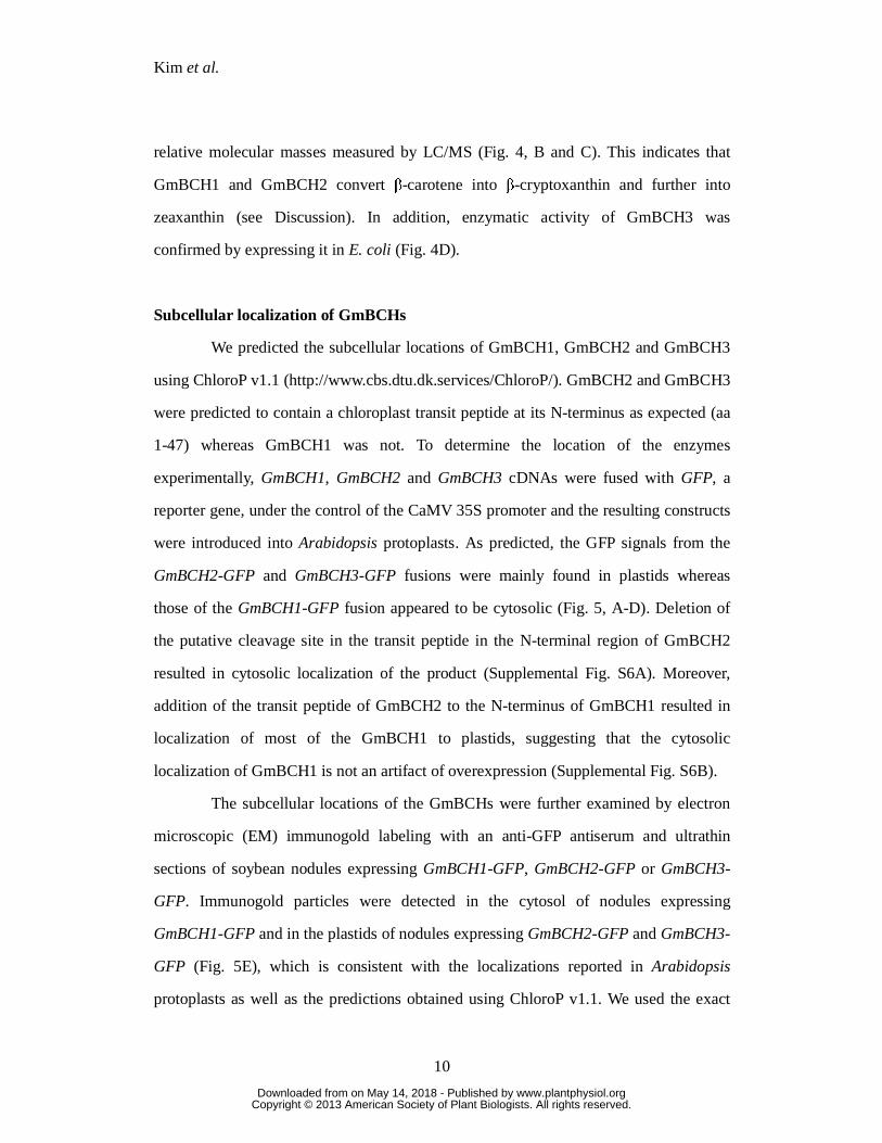
relative molecular masses measured by LC/MS (Fig. 4, B and C). This indicates that
GmBCH1 and GmBCH2 convert β-carotene into β-cryptoxanthin and further into
zeaxanthin (see Discussion). In addition, enzymatic activity of GmBCH3 was
confirmed by expressing it in E. coli (Fig. 4D).
Subcellular localization of GmBCHs
We predicted the subcellular locations of GmBCH1, GmBCH2 and GmBCH3
using ChloroP v1.1 (http://www.cbs.dtu.dk.services/ChloroP/). GmBCH2 and GmBCH3
were predicted to contain a chloroplast transit peptide at its N-terminus as expected (aa
1-47) whereas GmBCH1 was not. To determine the location of the enzymes
experimentally, GmBCH1, GmBCH2 and GmBCH3 cDNAs were fused with GFP, a
reporter gene, under the control of the CaMV 35S promoter and the resulting constructs
were introduced into Arabidopsis protoplasts. As predicted, the GFP signals from the
GmBCH2-GFP and GmBCH3-GFP fusions were mainly found in plastids whereas
those of the GmBCH1-GFP fusion appeared to be cytosolic (Fig. 5, A-D). Deletion of
the putative cleavage site in the transit peptide in the N-terminal region of GmBCH2
resulted in cytosolic localization of the product (Supplemental Fig. S6A). Moreover,
addition of the transit peptide of GmBCH2 to the N-terminus of GmBCH1 resulted in
localization of most of the GmBCH1 to plastids, suggesting that the cytosolic
localization of GmBCH1 is not an artifact of overexpression (Supplemental Fig. S6B).
The subcellular locations of the GmBCHs were further examined by electron
microscopic (EM) immunogold labeling with an anti-GFP antiserum and ultrathin
sections of soybean nodules expressing GmBCH1-GFP, GmBCH2-GFP or GmBCH3-
GFP. Immunogold particles were detected in the cytosol of nodules expressing
GmBCH1-GFP and in the plastids of nodules expressing GmBCH2-GFP and GmBCH3-
GFP (Fig. 5E), which is consistent with the localizations reported in Arabidopsis
protoplasts as well as the predictions obtained using ChloroP v1.1. We used the exact
www.plantphysiol.orgon May 14, 2018 - Published by Downloaded from Copyright © 2013 American Society of Plant Biologists. All rights reserved.
Kim et al.
11
binomial test in order to determine whether gold particles were preferentially located in
a specific organelle (Conover, 1971). For GmBCH1, more than 78% of the gold
particles were found in the cytosol (p-value = 0.0478). For GmBCH2/GmBCH3, more
than 64%/70% of the gold particles were found in the plastid (p-values = 0.0492/0.0329,
respectively). These results suggest that GmBCH2 and GmBCH3 function in the
plastids of root nodules, while GmBCH1 resides in the cytosol, unlike other β-carotene
hydroxylases. It is not clear how a cytosolic β-carotene hydroxylase can participate in
carotenoid metabolism.
Drastically reduced nitrogen fixation in nodules expressing RNAi against GmBCHs
To see if GmBCH1/3 were essential for nodule growth we made an RNAi
construct against them and subcloned it downstream of the leghemoglobin (Lbc3)
promoter. This RNAi construct contained a part of the N-terminal region of GmBCH1
and GmBCH3 which includes the 5’-untranslated region of GmBCH3 (Supplemental
Fig. S7). The sequence of the RNAi construct was 99% identical to GmBCH1 and
GmBCH3, and had significant identity to GmBCH4, but not to GmBCH2 or GmBCH5.
The resulting cassette, GmBCH(1+3)-RNAi, was introduced into pCAMBIA1304,
which harbors 35S-GUS (the CaMV 35S promoter fused to GUS) as a reporter. This
construct was introduced into Agrobacterium rhizogenes to generate transgenic hairy
roots (Lee et al., 2005).
The formation of root nodules was markedly reduced on the GmBCH(1+3)-
RNAi hairy roots (Fig. 6A), and similar defective nodulation was observed when a
transgenic hairy root formed along with a non-transgenic (GUS-negative) hairy root on
the same plant (Fig. 6B). In the meantime, there appeared to be no difference in root
growth between the transgenic and non-transgenic hairy roots. The impairment of
nodulation on GmBCH(1+3)-RNAi hairy roots resulted in lower nodule weight as
measured in 24 GmBCH(1+3)-RNAi plants and 19 controls (Fig. 6C). When we
www.plantphysiol.orgon May 14, 2018 - Published by Downloaded from Copyright © 2013 American Society of Plant Biologists. All rights reserved.
Kim et al.
12
checked the expression levels of GmBCH1 and GmBCH3 in the transgenic nodules, we
found that these varied; hence as representatives with different nodule weights, we
chose RNAi plants #2, #20 and #21 for further expression analysis. RNAi plants #20
and #21, as expected, had significantly reduced expression of GmBCH1 and GmBCH3
together with decreased nodule weights (RNAi type 2), whereas GmBCH1 and
GmBCH3 expression was less affected in the RNAi plants with almost the same nodule
weights as the control, such as RNAi plant #2 (RNAi type 1) (Fig. 6D). Although the
relationship between expression levels of GmBCH(1+3) and nodule weight is only
shown for the three representative transgenic plants (i.e., #2, #20 and #22) in Fig. 6D,
we actually examined the expression levels and nodule weights in 12 transgenic plants
and 4 controls. To establish the statistical significance of the RNAi-plant groups (i.e.,
control vs RNAi-type 1 vs RNAi-type 2), we conducted an analysis of variance
(ANOVA) with Tukey’s multiple comparison procedure, and found a significant
difference in nodule weight between control and RNAi-type 2, as well as between
RNAi-type 1 and RNAi-type 2 (Supplemental Fig. S8A); there was also a significant
difference in the expression of GmBCH(1+3) between these pair of groups
(Supplemental Fig. S8B) (Maritz, 1981). In addition, using all 16 plants making up the
three groups, we examined the correlation between gene expression level and nodule
weights based on the Spearman rank correlation coefficient. This yielded a strong
positive correlation of 0.76 (p-value = 0.001) (Supplemental Fig. S8C). Meanwhile,
GmBCH4 expression, which was quite low in root nodules, was somewhat affected in
most of the RNAi plants. Expression of GmBCH2 as well as GmBCH5 was not altered
in any of the plants (Fig. 6D). These observations imply that the effect of the RNAi on
GmBCHs transcript levels was specific to GmBCH1 and GmBCH3. Expression of
GmVDE, a gene contributing positively to the accumulation of zeaxanthin from the
opposite direction of GmBCH in the xanthophyll cycle (Fig. 1; Cazzonelli and Pogson,
2010), was also unaffected, indicating that silencing of the GmBCHs in the RNAi
www.plantphysiol.orgon May 14, 2018 - Published by Downloaded from Copyright © 2013 American Society of Plant Biologists. All rights reserved.

Kim et al.
13
nodules was not compensated, at least at transcription level, by the induction of GmVDE
(Fig. 6D). In agreement with the observed retardation of nodule development, on the
other hand, expression of Lbc3, which encodes leghemoglobin, an oxygen carrier for
symbiosis with rhizobia, and is one of the hallmark genes for the development of
nitrogen-fixing nodule (Ott et al., 2005), was decreased in RNAi plants #20 and #21
(Fig. 6D). In addition, acetylene reduction assays showed that nitrogen-fixing ability
was also lower in the RNAi nodules (Fig. 6F). The RNAi nodules examined by electron
microscopy contained empty vesicles rather than symbiosomes with rhizobia in about
60% of the cells examined. In addition, the rhizobia were not even enclosed by
symbiosome membranes in about 5% of the cells of the RNAi nodules with strongly
repressed GmBCH(1+3) (Fig. 6E; Supplemental Fig. S9). These data indicate that
expression of GmBCH1 and/or GmBCH3 may be essential for nodule development.
Expression of GmBCH2 was prominent during nodulation; hence we also
silenced GmBCH2 in nodules using the leghemoglobin promoter-driven RNAi approach.
When we compared 9 GmBCH2-RNAi plants with 8 control plants, we found that the
reduction in GmBCH2 expression resulted in decreased nodule weight and nitrogenase
activity (Fig. 6, G to K). GmBCH5 expression was also reduced in the GmBCH2-RNAi
nodules, probably due to its strong homology with GmBCH2, while expression of
GmBCH1 and GmBCH3 was not affected and GmBCH4 expression was somewhat
increased (Fig. 6I). About 65% of the infected cells examined contained empty vesicles
and most of them exhibited the presence of bacteroids outside symbiosomes (Fig. 6J). It
is not clear whether the phenotypic difference observed by electron microscopy of
GmBCH(1+3)-RNAi nodules and GmBCH2-RNAi nodules indicates their different
roles in nodulation. In addition, expression of GmBCH4 and GmBCH5 was also
observed in nodules, albeit at a low level (Supplemental Fig. S1, B and C). Taken
together, these results suggest that the GmBCHs, comprising GmBCH1 to GmBCH5,
may be essential for nodule development.
www.plantphysiol.orgon May 14, 2018 - Published by Downloaded from Copyright © 2013 American Society of Plant Biologists. All rights reserved.
Kim et al.
14
Decreased expression of the putative zeaxanthin epoxidase gene during nodulation
The impairment of nodulation in the RNAi hairy roots suggested to us that a
product of β-carotene hydroxylase action, or some other derivatives of the carotenoid
metabolic pathway, plays an important role in nodulation. This led us to examine the
expression of genes encoding two enzymes of the xanthophyll cycle; zeaxanthin
epoxidase (ZEP) and violaxanthin de-epoxidase (VDE). Since we identified three
GmZEPs (Supplemental Fig. S1D) and two GmVDEs (Supplemental Fig. S1E) in the
soybean genome sequence, we examined their expression. Actually, we came across a
few more DNA sequences homologous to the ZEP and VDE genes in other plants, but
they showed only partial identity and were not further studied (Supplemental Fig. S1, D
and E). The primers used to examine the expression of GmZEPs and GmVDEs were
designed using DNA sequences highly conserved among other plants. The transcript
levels of both genes were considerably lower in roots and nodules than in the aerial
parts of the plant such as leaves, stems, and flowers (Fig. 7, A and B; Supplemental Fig.
S10). As nodules matured, GmZEP expression decreased and was almost undetectable
in mature 27-day-old nodules (Fig. 7D). Expression of the GmVDEs remained relatively
constant throughout nodule development (Fig. 7E). In addition, we examined the
expression of two soybean NCED1 orthologs (GmNCED1a and GmNCED1b;
Supplemental Fig. S1F), and found that their expression was high in flowers, but quite
low in roots and nodules, although expression in 7-day-old nodules was a little higher
than in other stages of nodulation (Fig. 7, C and F). The expression levels of GmZEPs,
GmVDEs and GmNCED1s generally matched with those in the soybean RNA-Seq
Atlases, especially the expression in the aerial parts of the soybean (Libault et al., 2010;
Severin et al., 2010; Supplemental Table S1). These expression data appear to indicate
that GmZEPs, GmVDEs, and GmNCED1s do not play major roles, if any, in further
carotenoid metabolism after β-carotene hydroxylase during nodulation although we
www.plantphysiol.orgon May 14, 2018 - Published by Downloaded from Copyright © 2013 American Society of Plant Biologists. All rights reserved.
Kim et al.
15
cannot rule out the possibility that their protein levels are higher than suggested by their
transcript levels.
Expression of CCDs in soybean root nodules
We hypothesized that zeaxanthin might be synthesized in root nodules by β-carotene
hydroxylases and be converted to other carotenoids, though not via xanthophyll to ABA,
because ZEP expression was very low (Fig. 7D). Therefore we extracted and quantified
carotenoids from root nodules. Contrary to our expectation, carotenoids including
zeaxanthin were almost undetectable in root nodules (data not shown). This raised the
question of what biochemical reactions occur in root nodules subsequent to zeaxanthin
production by the β-carotene hydroxylases. Since carotenoid cleavage products have
been found in roots infected with mycorrhizal fungi and are regarded as important in
arbuscular mycorrhizal symbiosis (Strack and Fester, 2006), we reasoned that the
carotenoids synthesized in root nodules might be depleted if carotenoid cleavage
dioxygenases (CCDs) were active. The white-colored petals of chrysanthemum express
high levels of CmCCD4a, with the result that no carotenoid can be detected (Ohmiya et
al., 2006), and RNAi-mediated suppression of CmCCD4a expression was found to lead
to accumulation of carotenoids and yellow petal color, confirming the relationship
between the amount of carotenoid and expression of CmCCD4a. Thus, we considered
the possibility that conversion of carotenoids into apocarotenoids by CCDs might
explain our failure to detect carotenoids in root nodules.
We tested this possibility by examining the expression of carotenoid cleavage
dioxygenases (CCDs) in root nodules of soybean. We chose to examine the expression
of CCD1, CCD7, and CCD8 in nodules and excluded CCD4 due to its primary
expression in aerial tissues. From the soybean genome sequence
(http://www.phytozome.net/soybean.php), we obtained the DNA sequences of putative
versions of CCD7 and CCD8, which are reported to cleave carotenoids. The deduced
www.plantphysiol.orgon May 14, 2018 - Published by Downloaded from Copyright © 2013 American Society of Plant Biologists. All rights reserved.
Kim et al.
16
amino acid sequences of CCD7 and CCD8 (designated GmCCD7 and GmCCD8,
hereafter) were very similar to previously reported CCD7 and CCD8 sequences, and
were named GmCCD7a, GmCCD7b, GmCCD8a, and GmCCD8b (Supplemental Fig.
S1, G and H). In addition, we also encountered putative GmCCD1a and GnCCD1b
sequences and examined their expression (Supplemental Fig. S1I). We found a few
more DNA sequences, but their deduced amino acid sequences were only partially
identical to previously reported CCDs and not studied further (Supplemental Fig. S1, G
and H). Using appropriate primers, expression of GmCCD7s and GmCCD8s proved to
be relatively high in roots and nodules whereas GmCCD1s expression was in all tissues,
in agreement with the data of Libault et al. (2010) and Severin et al. (2010) (Fig. 8, A to
C; Supplemental Table S1). Although expression of GmCCD7s was rather low, it, as
well as GmCCD8s, appeared to be induced upon rhizobial infection (Fig. 8, E and F).
We therefore tested whether GmCCD7/GmCCD8 could actually cleave
zeaxanthin. E. coli transformed with pAC-zeaxanthin produced zeaxanthin as shown by
HPLC analysis as well as by the yellow color of colonies (Fig. 9, A and B). GmCCD7a-
expressing E. coli were also yellow and a protein of the expected size corresponding to
GmCCD7 (70 kDa) was detected by SDS-PAGE (Fig. 9C), but lysates gave a strong
peak of zeaxanthin, implying that the zeaxanthin produced was not cleaved. In contrast,
E. coli transformed with the GmCCD8a-expressing plasmid were no longer yellow
while producing a protein of the expected molecular weight (60 kDa). This recombinant
E. coli strain gave only a very small zeaxanthin peak (asterisk; Fig. 9, A and B). The
integrity of GmCCD7a was also tested by another E. coli functional assay, in which the
β-carotene cleaving activity of GmCCD7 was examined and compared with other CCDs
in E. coli carrying the plasmid pACCAR16∆crtX that enables the accumulation of β-
carotene (Supplemental Fig. S11). β-carotene cleaving activity was consistently
detected in GmCCD7a-expressing E. coli although it was about 3-fold lower than that
of an AtCCD7-expressing E. coli (Supplemental Fig. S11, C and D). Moreover, this
www.plantphysiol.orgon May 14, 2018 - Published by Downloaded from Copyright © 2013 American Society of Plant Biologists. All rights reserved.
Kim et al.
17
apparent gap in the activity between GmCCD7a and the other two CCDs in this assay
(Supplemental Fig. S11, C to E) could be further reduced if the relative protein
expression level of these proteins was taken into account, as measured by western blot
of these cell extracts (Supplemental Fig. S11A). Therefore, it seems likely that both
GmCCD7a and GmCCD8a are functional enzymes involved in the β-carotene
metabolism in soybean nodule. These data also imply that zeaxanthin is cleaved by
GmCCD8a, but not by GmCCD7a although we cannot exclude the possibility that
GmCCD7a synthesized in root nodules may have cleavage activity on zeaxanthin.
Taken together, our observations suggest that carotenoids such as zeaxanthin are
synthesized but are cleaved by GmCCD8 during soybean nodule development.
DISCUSSION We have investigated the significance of carotenoid metabolism in nodule
development. This work was prompted by the isolation of a gene encoding β-carotene
hydroxylase (BCH) as a gene differentially expressed in root nodules (Lee et al., 2005).
GmBCH1, GmBCH2 and GmBCH3 expression was found to increase during nodulation
and was especially localized to the infected region of nodules (Figs. 2 and 3). In
addition, RNAi-mediated repression of these genes resulted in the retardation of nodule
development including impairment in symbiosome formation (Fig. 6; Supplemental Fig.
S9). In the RNAi nodules, nitrogen fixation appeared to be damaged judging from
nitrogenase assays as well as the expression of lbc3, an essential gene for nitrogen
fixation. We have thus presented evidence for the first time that carotenoid metabolism
by GmBCHs is required for proper nodule development.
We confirmed that the GmBCHs that we isolated from root nodules encoded
active enzymes. cDNAs were expressed in E.coli harboring a vector for producing β-
carotene (Misawa et al., 1990), and HPLC analyses of E. coli expressing the GmBCHs
revealed the accumulation of zeaxanthin and also of large amounts of β-cryptoxanthin
www.plantphysiol.orgon May 14, 2018 - Published by Downloaded from Copyright © 2013 American Society of Plant Biologists. All rights reserved.
Kim et al.
18
(Fig. 4). Similar results have been reported from functional assays in E. coli (Yu et al.,
2007), while the expression of other BCH orthologues resulted in the synthesis of more
zeaxanthin than cryptoxanthin (Sun et al., 1996; Galpaz et al., 2006). The synthesis of
β-cryptoxanthin, that is, the asymmetric addition of hydroxyl groups to the β-end group
of β-carotene, could be due to a slightly different conformation of the BCHs in E. coli or
the failure of the BCHs to form a stable dimer (Sun et al., 1996; Yu et al., 2007). In any
case the results confirm that the putative GmBCHs are, indeed, functional β-carotene
hydroxylases.
To our surprise we failed to detect any carotenoids in root nodules. To account
for this, we tested the possibility that all the carotenoids were cleaved to synthesize
ABA, since ABA is able to coordinate some aspects of nodulation (Ding et al., 2008).
However, expression of putative GmZEPs and putative GmNCED1s was found to be
quite low in nodules (Fig. 7, A and C; Supplemental Fig. S10A) and furthermore,
GmZEP expression became almost undetectable in mature nodules (Fig. 7D;
Supplemental Fig. S10B) while the expression of putative GmVDEs in root nodules was
similar to the level in roots (Fig. 7E). In fact, nodule number is decreased by ABA
treatment (Suzuki et al., 2004), and a lotus mutant with reduced endogenous ABA
exhibited enhanced nodulation and nitrogen fixation (Tominaga et al., 2009). Since
ABA was suggested to be a negative regulator of nodulation, it is possible that its
concentration is not high in effective nodules. However, we cannot exclude the
possibility that a certain amount of carotenoid is metabolized to ABA during specific
stages of nodulation.
Despite its high sequence homology with other plant CCD8s (Supplemental
Fig. S1H), GmCCD8a apparently exhibited activity on both β-carotene and zeaxanthin
in our E. coli functional assay, contradicting the current view of the substrate specificity
and proposed role for CCD8 in the sequential cleavage reactions of C40-carotenoids
(Alder et al., 2008; Walter et al., 2010). On the other hand, this result is consistent with
www.plantphysiol.orgon May 14, 2018 - Published by Downloaded from Copyright © 2013 American Society of Plant Biologists. All rights reserved.
Kim et al.
19
an earlier report that showed direct cleavage activity of AtCCD8 on a few C40-
carotenoids (Auldridge et al., 2006a). In addition, it was reported that CCD8 interfered
with carotenoid biosynthesis when it was overexpressed in E. coli (Alder et al., 2008).
Therefore, our current result, together with the previous data of Auldridge et al. (2006a),
appears to suggest that more in-depth studies on the reaction catalyzed by CCD8,
including the nature of the substrate and the cleavage product, are needed. Before
drawing a conclusion that GmCCD8a is able to cleave zeaxanthin it will be necessary to
identify the cleavage products generated in the assay. The data in Fig. 9 also show that
GmCCD7a did not alter the HPLC profile of zeaxanthin-producing E. coli. Since
recombinant AtCCD7 has broad substrate specificity and cleaves C40-carotenoids
including β-carotene into C27- and C13-apocarotenoids (Booker et al., 2004; Schwartz et
al., 2004) and GmCCD7 cleaved β-carotene albeit less efficiently than AtCCD7
(Supplemental Fig. S11), we cannot exclude the possibility that the GmCCD7
synthesized in root nodules can cleave diverse C40-carotenoids including zeaxanthin.
Based on these observations, we propose that GmCCD7, and possibly GmCCD8, cleave
C40-carotenoids and that the cleavage products are further cleaved by GmCCD8 (Alder
et al., 2008) inside nodule tissue.
The carotenoids synthesized inside the chloroplasts of leaves play essential
roles in photosynthesis. On the other hand, those present in flowers, fruits or roots are
not needed for photosynthesis and accumulate in special subcellular compartments such
as chromoplasts and cytoplasmic lipid vesicles. For example, a β-carotene oxygenase in
a unicellular green alga was localized to the lipid vesicles outside plastids (Grünewald
et al., 2001). In our study, both in silico prediction and actual experiment showed that
GmBCH2 and GmBCH3 were present in plastids whereas GmBCH1 was present,
unexpectedly, in the cytosol (Fig. 5). Interestingly, the localization of GmBCH1 in the
cytosol seems to be closely associated with a short stretch of N-terminal sequences
present only in GmBCH1 in addition to the putative transit-peptide sequence which are
www.plantphysiol.orgon May 14, 2018 - Published by Downloaded from Copyright © 2013 American Society of Plant Biologists. All rights reserved.
Kim et al.
20
present in all of the plastidial GmBCH isoform sequences. Perhaps adding this sequence
makes the transit peptide nonfunctional. Although no corresponding locus to GmBCH1
was found in the soybean genome database (http://www.phytozome.net), we have been
able to clone GmBCH1 repeatedly by RT-PCR and we also could isolate its 0.45-kb
upstream sequences from the cultivar Williams 82, from which the soybean sequence
database was generated, as well as the cultivar Sinpaldal 2, which has been used as the
material of our present study, by DNA walking (Supplemental Fig. S2). Therefore, it is
likely that GmBCH1 may be present in an unsequenced gap of the present soybean
genome sequence. The substrate of GmBCH1 in the cytosol of infected cells, and the
significance of its cytosolic location for the symbiotic interaction between soybean and
Rhizobium need to be studied in the future.
Carotenoid cleavage products such as β-ionone, or dihydroactinidiolide, are
synthesized in conditions of stress, being involved in plant protection mechanisms
(Bouvier et al., 2005). It is thought that the synthesis of carotenoids and their cleavage
products promotes symbiosis between plants and arbuscular mycorrhizal (AM) fungi
(Strack and Fester, 2006; Walter et al., 2010). However, since little attention has been
paid to the presence of (apo)carotenoids in root nodules, it remains unclear what role the
former plays in root nodule symbiosis, given that our work points to the presence of
apocarotenoids as well as carotenoids in the nodules. Since nodulation and nitrogen
fixation were severely inhibited in the GmBCHs-RNAi root nodules (Fig. 6) and
expression of GmBCHs and GmCCDs was induced during nodulation, the biosynthesis
of carotenoid and presumably apocarotenoids appears to play a significant role in
nodulation. A possible role of carotenoid cleavage products is to protect the infected
cells from oxidative stress. The symbiosomes enclosing rhizobia must produce
tremendous amounts of reactive oxygen species since a nitrogen-fixing infected cell
contains about 20,000 rhizobia. Alternatively, apocarotenoids may act as signaling
molecules during the maturation of nodules. A further possibility is that C13 and C14
www.plantphysiol.orgon May 14, 2018 - Published by Downloaded from Copyright © 2013 American Society of Plant Biologists. All rights reserved.
Kim et al.
21
apocarotenoids are essential for rhizobial symbiosis, as proposed for AM symbiosis
(Walter et al., 2010). While there seems to be no clear indication as to which
apocarotenoid(s) is(are) effective in AM symbiosis, identification of the apocarotenoids
present in root nodules may offer a key to understanding the establishment and
functioning of legume-Rhizobium symbiosis.
MATERIALS AND METHODS
Plants, rhizobia and growth conditions
Soybean (Glycine max cv. Sinpaldal 2) seeds were sterilized and grown in
darkness on moist, absorbent paper at 28°C for 3 days. Three-day-old seedlings were
inoculated with rhizobia (Bradyrhizobium japonicum USDA110), transferred to
sterilized vermiculites, and grown at 28°C for 1 month. Tissues harvested from Glycine
max were frozen immediately in liquid nitrogen and stored at -70°C until used for RNA
extraction. For real time PCR, mature leaves (fully expanded), stems, flowers (including
flower buds and mature flowers), roots, and the mature nodule (27-day-old) were
collected separately.
Gene isolation and vector construction
A partial cDNA clone of GmBCH2 (BE607999) was identified by a BLAST
search at the NCBI EST database. Because this clone did not contain the 5’ end of the
ORF, RACE PCR was performed to recover the missing 5’ DNA sequence, and the
coding sequence was extended using a CapFishing kit (SEEGENE). Full-length first-
strand cDNA synthesized with oligo(dT)-ACP was amplified using the primers listed in
Supplemental Table S2. The degenerate primers for GmZEP and the primers for the full-
length cDNA are shown in that Table. To generate the constructs for bacterial
expression, the coding regions of the GmBCHs were amplified with Pfu DNA
polymerase (COREBIO) (Supplemental Table S2). Full-length cDNAs encoding the
www.plantphysiol.orgon May 14, 2018 - Published by Downloaded from Copyright © 2013 American Society of Plant Biologists. All rights reserved.
Kim et al.
22
putative CCD7 and CCD8 in Glycine max were obtained from the soybean genome
sequence (http://www.phytozome.net/soybean.php), and full-length GmCCD7 and
GmCCD8 cDNAs were amplified from nodule RNA by RT-PCR (Supplemental Table
S2). The resulting amplified products were cloned into pUC19 to make pGmBCH1,
pGmBCH2, pGmBCH3, p3HA-GmCCD7, p3HA-GmCCD8, respectively and their
sequences were confirmed by DNA sequencing.
A DNA fragment including the 5’ region as well as the coding region of
GmBCH1 was isolated from soybean genomic DNA using a DNA Walking SpeedUPTM
Premix Kit (SEEGENE). PCR was performed with an adaptor provided in the kit and
gene-specific primers; GmBCH1 primer: 5’- GAGAGTGTTTGTGTTCGCCTGCG-3’,
second nested GmBCH1 primer: 5’- AGTAAGGAATGTGATGATCCC-3’, third nested
GmBCH1 primer: 5’- CTATCCCCCATGAAGCGAATGCC-3’. The PCR products were
cloned into the pGEM-T easy vector (PROMEGA) and sequenced.
To make GmBCH promoter-GUS fusions, the 5’ upstream sequences of
GmBCH2 and GmBCH3 were identified in the soybean genome
(http://www.phytozome.net/soybean.php) as shown in Supplemental Fig. S5. The 1.5-
kb 5’ upstream regions of GmBCH3 and GmBCH2, respectively, were amplified by
PCR with Pfu DNA polymerase (COREBIO) using the primers shown in Supplemental
Table S2, and cloned upstream of GUS in pCAMBIA3301. Fluorometric assays of GUS
activity were performed as described by Jefferson et al. (1987) with modifications.
Real time RT-PCR
Total RNA was extracted with an RNeasy Plant Mini Kit (QIAGEN) and
cDNAs were synthesized with M-MLV Reverse Transcriptase (PROMEGA) after
treatment with DNase I to remove contaminating genomic DNA. One μl of first-strand
cDNA was used as template, and the primers used are listed in Supplemental Table S2.
Real-time RT-PCR was performed with SYBR Green PCR Master Mix (TAKARA)
www.plantphysiol.orgon May 14, 2018 - Published by Downloaded from Copyright © 2013 American Society of Plant Biologists. All rights reserved.
Kim et al.
23
using a Rotor-Gene 3000 (CORBETT RESEARCH) and an ABI Prism 5700 sequence
detection system. All RT-PCR transcript levels were normalized with the geometric
mean of three reference genes (GmELF1b, GmActin2/7 and ubiquitin) (Vandesompele
et al., 2002).
In Situ RNA hybridization
In situ RNA hybridization was performed according to Oh et al. (2001).
Nodules were harvested 2, 7 and 27 days after rhizobial inoculation. Each nodule was
processed by microtechniques, hybridized with digoxigenin-labeled antisense and sense
RNA probes under standard condition, and washed with low-stringency and high-
stringency buffers for longer than under standard conditions. Hybridization stringency
was established by the washing steps in order to avoid cross-hybridization. The probes
for GmBCH(1+3) and GmBCH2 were made using the N-terminal regions of the
GmBCHs, which have no significant sequence similarity to each other (Supplemental
Fig. S4); we used a 400-bp region for the GmBCH(1+3) probe and a 300-bp region for
the GmBCH2 probe, as indicated in Supplemental Fig. S1A. Whole-mount in situ
hybridization of soybean leaves was performed with the GmBCH(1+3) and GmBCH2
probes according to Weigel and Glazebrook (2002), and images from whole-mount in
situ hybridization were quantified with Image J (National Institutes of Health, Bethesda,
MD, USA) as in Ubuka and Bentley (2009).
HPLC analysis of carotenoids
To measure their activities, pGmBCH1, pGmBCH2, pGmBCH3, and pUC19
as negative control, were introduced into E. coli JM109 carrying pACCAR16∆crtX,
which expresses genes for the production of β-carotene (Misawa et al., 1990). E. coli
transformants were grown overnight at 28°C in 2 ml LB liquid medium containing 50
μg/ml ampicillin and 35 μg/ml chloramphenicol. The overnight cultures were used to
www.plantphysiol.orgon May 14, 2018 - Published by Downloaded from Copyright © 2013 American Society of Plant Biologists. All rights reserved.
Kim et al.
24
inoculate 50 ml LB medium with the same antibiotics. After 3 h, 0.1 mM IPTG was
added, and the E. coli were further incubated in darkness at 28°C for 72 h. For
expression of GmCCD7 or GmCCD8, p3HA-GmCCD7 or p3HA-GmCCD8 were
introduced into E. coli (BL21) containing a carotenoid-producing construct (pAC-
zeaxanthin or pACCAR16∆crtX). The transformants were grown as described above
and incubated for 24 h after adding IPTG. Carotenoid cleavage activity was inferred
from the absence of accumulating carotenoids, that is, the absence of yellow color.
Cell pellets of E. coli cultures were resuspended in 80% acetone and
concentrated. After re-dissolving in methanol, 10 μl samples were used for HPLC.
Assays were performed in ambient conditions using a Prostar 230 ternary gradient
pump, a Prostar 430 autosampler and a Prostar 335 photodiode array detector
(VARIAN). Separation was carried out on a 4.6×150 mm carotenoid column (YMC
CO.) with a particle size of 3 mm. The mobile phase consisted of solvent A (methanol-
tert-butyl methyl ether, 10:90, v/v) and solvent B (water-methanol, 5:95, v/v). A linear
gradient was used (10% solvent A at 0 min, 65% solvent A at 40-45 min, 95% solvent A
at 45-50 min). The flow-rate was maintained at 1 ml/min, and the chromatographic
profile was recorded at 470 nm. MS data for the carotenoids were obtained using a
1200L LC/MS (VARIAN). Mass spectrometry conditions were as follows; APCI
positive ion mode; mass range, m/z 200-800; corona current, 2.0 μA; nebulizing gas
pressure (N2), 60 psi; drying gas (N2) flow rate, 4 L/min; drying gas temperature, 300°C.
Subcellular localization of GmBCHs using Arabidopsis protoplasts and EM
immunogold labeling
To make GFP fusion constructs, the full-length cDNAs of the GmBCHs were
amplified by PCR with Pfu DNA polymerase (COREBIO) using primers (Supplemental
Table S2) containing XbaI and BamHI sites, and fused in-frame to GFP. To make a
truncated GmBCH2-GFP, a part of GmBCH2 (corresponding to amino acids 48 to 314)
www.plantphysiol.orgon May 14, 2018 - Published by Downloaded from Copyright © 2013 American Society of Plant Biologists. All rights reserved.
Kim et al.
25
was used. To make a fusion of GmBCH1-GFP with the transit peptide of GmBCH2, we
used a region of GmBCH2 corresponding to amino acids 1 to 47. Protoplasts isolated
from Arabidopsis were transfected by the polyethylene glycol method as described in
Yoo et al. (2007). After 16 h incubation, fluorescence was examined with a fluorescence
microscope.
To make the transgenic plants expressing GmBCH1-GFP, GmBCH2-GFP or
GmBCH3-GFP for EM immunogold labeling, fusions of GmBCHs-GFP under the
CaMV 35S promoter were inserted between the HindIII/EcoRI sites of pCAMBIA3301.
Transgenic nodules were produced according to Lee et al. (2005). Immunoelectron
microscopic studies were performed according to Lin et al. (2011). Sections of 27-day-
old nodule on copper grids were labeled with anti-GFP rabbit antibody (ABCAM) and
then with 10 nm gold-conjugated goat anti-rabbit antibody (ABCAM). The sections
were viewed in a JSM-1200EX II transmission electron microscope (JEOL).
Generation of transgenic root nodules
To make GmBCH(1+3)-RNAi and GmBCH2-RNAi constructs, a 230 bp-
fragment targeting both GmBCH1 and GmBCH3 and a 180 bp-fragment of GmBCH2
were amplified by PCR with Pfu DNA polymerase (COREBIO), using primers shown
in Supplemental Table S2, respectively. The amplified fragments were inserted into the
HindIII/XbaI and XhoI/KpnI sites of pKANNIBAL (Wesley et al., 2001). The
GmBCH(1+3)-RNAi construct was transferred into the binary plasmid pCAMBIA1304,
and the resulting plasmid was introduced into Agrobacterium rhizogenes (K599) by the
freeze-thaw method. Hairy roots emerging after infection with the agrobacteria were
examined for GUS expression in order to identify the transgenic hairy roots, and only
one transgenic hairy root in each plant was spared to be used for nodulation, removing
the all others (Lee et al., 2005).
www.plantphysiol.orgon May 14, 2018 - Published by Downloaded from Copyright © 2013 American Society of Plant Biologists. All rights reserved.
Kim et al.
26
Acetylene reduction assay
Ethylenes produced per g (fresh weight) of nodules were determined as
previously described (Oh et al., 2001).
Transmission electron microscopy
Nodule specimens (ca. 1×3 mm2 with 1 mm-thick underlying tissues) from
transgenic roots containing the GmBCH(1+3)-RNAi construct were excised with a
razor blade and processed as reported previously (Kim, 2008). After metal staining, the
sections were examined with a transmission electron microscope (JEM-1010; JEOL)
operated at an accelerating voltage of 80 kV.
The following materials are available in the online version of this article.
Supplemental Figure S1. Comparison of the amino acid sequences of β-carotene
hydroxylases, zeaxanthin epoxidases (ZEPs), violaxanthin de-epoxidases (VDEs), 9-cis-
epoxycarotenoid dioxygenase 1s (NCED1s), carotenoid cleavage dioxygenases 7s,
carotenoid cleavage dioxygenases 8s, and carotenoid cleavage dioxygenases 1s.
Supplemental Figure S2. The upstream region of GmBCH1 isolated by DNA walking.
Suppelemental Figure S3. Assessment of PCR efficiency of primer sets for GmBCHs.
Suppelemental Figure S4. Assessment of probes for in situ hybridization.
Suppelemental Figure S5. Expression of GmBCH2-GUS and GmBCH3-GUS in
transgenic soybean nodules measured by fluorometric assay of GUS.
Supplemental Figure S6. Subcellular localization of N-terminally deleted GmBCH2
and a fusion of GmBCH1 with the transit peptide of GmBCH2.
Supplemental Figure S7. DNA sequences used in preparing for GmBCH(1+3)-RNAi
and GmBCH2-RNAi constructs.
Supplemental Figure S8. Statistical analyses of nodule weight and gene expression in
www.plantphysiol.orgon May 14, 2018 - Published by Downloaded from Copyright © 2013 American Society of Plant Biologists. All rights reserved.
Kim et al.
27
GmBCH(1+3)-RNAi nodules.
Supplemental Figure S9. Electron microscopic analysis of GmBCH(1+3)-RNAi
nodules.
Supplemental Figure S10. Expression of GmZEP3 in Glycine max.
Supplemental Figure S11. Functional assay of GmCCD7 and GmCCD8 in β–carotene
accumulating E. coli.
Supplemental Table S1. RNA-seq expression data for soybean genes in various tissues
Supplemental Table S2. Primers used for gene cloning and gene expression by real
time RT-PCR
ACKNOWLEDGEMENTS We thank Drs. Norihiko Misawa and Francis X. Cunningham, Jr. for generously
providing pACCAR16∆crtX and pAC-zeaxanthin, respectively.
LITERATURE CITED Alder A, Holdermann I, Beyer P, Al-Babili S (2008) Carotenoid oxygenases involved
in plant branching catalyse a highly specific conserved apocarotenoid cleavage reaction.
Biochem J 416: 289-296
Auldridge ME, Block A, Vogel JT, Dabney-Smith C, Mila I, Bouzayen M,
Magallanes-Lundback M, DellaPenna D, McCarty DR, Klee HJ (2006a)
Characterization of three members of the Arabidopsis carotenoid cleavage dioxygenase
family demonstrates the divergent roles of this multifunctional enzyme family. Plant J
45: 982–993
www.plantphysiol.orgon May 14, 2018 - Published by Downloaded from Copyright © 2013 American Society of Plant Biologists. All rights reserved.
Kim et al.
28
Auldridge ME, McCarty DR, Klee HJ (2006b) Plant carotenoid cleavage oxygenases
and their apocarotenoid products. Curr Opin Plant Biol 9: 315–321
Booker J, Auldridge M, Wills S, McCarty D, Klee H, Leyser O (2004) MAX3/CCD7
is a carotenoid cleavage dioxygenase required for the synthesis of a novel plant
signaling molecule. Curr Biol 14: 1232-1238
Bouvier F, Keller Y, d'Harlingue A, Camara B (1998) Xanthophyll biosynthesis:
molecular and functional characterization of carotenoid hydroxylases from pepper fruits
(Capsicum annuum L.). Biochim Biophys Acta 1391: 320-328
Bouvier F, Isner JC, Dogbo O, Camara B (2005) Oxidative tailoring of carotenoids: a
prospect towards novel functions in plants. Trends Plant Sci 10: 187-194
Cazzonelli CI, Pogson BJ (2010) Source to sink: regulation of carotenoid biosynthesis
in plants. Trends Plant Sci 15: 266-274
Conover WJ (1971) Practical nonparametric statistics. John Wiley & Sons, New York,
pp 97-104
Davison PA, Hunter CN, Horton P (2002) Overexpression of β-carotene hydroxylase
enhances stress tolerance in Arabidopsis. Nature 418: 203-206
DellaPenna D, Pogson BJ (2006) Vitamin synthesis in plants: tocopherols and
carotenoids. Annu Rev Plant Biol 57: 711–738
Ding Y, Kalo P, Yendrek C, Sun J, Liang Y, Marsh JF, Harris JM, Oldroyd GE
www.plantphysiol.orgon May 14, 2018 - Published by Downloaded from Copyright © 2013 American Society of Plant Biologists. All rights reserved.
Kim et al.
29
(2008) Abscisic acid coordinates nod factor and cytokinin signaling during the
regulation of nodulation in Medicago truncatula. Plant Cell 20: 2681-2695
Floss DS, Schliemann W, Schmidt J, Strack D, Walter MH (2008) RNA interference-
mediated repression of MtCCD1 in mycorrhizal roots of Medicago truncatula causes
accumulation of C27 apocarotenoids, shedding light on the functional role of CCD1.
Plant Physiol 148: 1267–1282
Galpaz N, Ronen G, Khalfa Z, Zamir D, Hirschberg J (2006) A chromoplast-specific
carotenoid biosynthesis pathway is revealed by cloning of the tomato white-flower
locus. Plant Cell 18: 1947-1960
Giuliano G, Tavazza R, Diretto G, Beyer P, Taylor MA (2006) Metabolic engineering
of carotenoid biosynthesis in plants. Trends Biotech 26: 139-145
Gomez-Roldan V, Fermas S, Brewer PB, Puech-Pagès V, Dun EA, Pillot JP, Letisse
F, Matusova R, Danoun S, Portais JC, et al (2008) Strigolactone inhibition of shoot
branching. Nature 455: 189-194
Grünewald K, Hirschberg J, Hagen C (2001) Ketocarotenoid biosynthesis outside of
plastids in the unicellular green alga Haematococcus pluvialis. J Biol Chem 276: 6023-
6029
Ha SH, Liang YS, Jung H, Ahn MJ, Suh SC, Kweon SJ, Kim DH, Kim YM, Kim
JK (2010) Application of two bicistronic systems involving 2A and IRES sequences to
the biosynthesis of carotenoids in rice endosperm. Plant Biotechnol J 8: 928–938
www.plantphysiol.orgon May 14, 2018 - Published by Downloaded from Copyright © 2013 American Society of Plant Biologists. All rights reserved.
Kim et al.
30
Hu R, Fan C, Li H, Zhang Q, Fu Y-F (2009) Evaluation of putative reference genes
for gene expression normalization in soybean by quantitative real-time RT-PCR. BMC
Mol Biol 10:93
Jefferson RA, Kavanagh TA, Bevan, M.W. (1987) GUS fusions: beta-glucuronidase
as a sensitive and versatile gene fusion marker in higher plants. EMBO J 6:3901–3907
Kim J, Smith JJ, Tian L, DellaPenna D (2009) The evolution and function of
carotenoid hydroxylases in Arabidopsis. Plant Cell Physiol 50: 463–479
Kim KW (2008) Visualization of micromorphology of leaf epicuticular waxes of the
rubber tree Ficus elastica by electron microscopy. Micron 39: 976-984
Kistner C, Winzer T, Pitzschke A, Mulder L, Sato S, Kaneko T, Tabata S, Sandal N,
Stougaard J, Webb KJ, et al (2005) Seven Lotus japonicus genes required for
transcriptional reprogramming of the root during fungal and bacterial symbiosis. Plant
Cell 17: 2217–2229
Kopsell DA, Kopsell DE (2006) Accumulation and bioavailability of dietary
carotenoids in vegetable crops. Trends Plant Sci 11: 499-507
Lee MY, Shin KH, Kim YK, Suh JY, Gu YY, Kim MR, Hur YS, Son O, Kim JS,
Song E, et al (2005) Induction of thioredoxin is required for nodule development to
reduce reactive oxygen species levels in soybean roots. Plant Physiol 139: 1881–1889
von Lintig J (2010) Colors with functions: elucidating the biochemical and molecular
basis of carotenoid metabolism. Annu Rev Nutr 30: 35–56
www.plantphysiol.orgon May 14, 2018 - Published by Downloaded from Copyright © 2013 American Society of Plant Biologists. All rights reserved.
Kim et al.
31
Libault M, Farmer A, Joshi T, Takahashi K, Langley RJ, Franklin LD, He J, Xu D,
May G, Stacey G (2010) An integrated transcriptome atlas of the crop model Glycine
max, and its use in comparative analyses in plants. Plant J 63: 86–99
Lin WL, Dickson DW, Sahara N (2011) Immunoelectron microscopic and
biochemical studies of caspase-cleaved Tau in a mouse model of tauopathy. J
Neuropathol Exp Neurol 70:779-787
Lu S, Li L (2008) Carotenoid metabolism: biosynthesis, regulation, and beyond. J
Integr Plant Biol 50: 778–785
Maritz JS (1981) Distribution-free statistical methods. Chapman & Hall, New York, p
217
Misawa N, Nakagawa M, Kobayashi K, Yamano S, Izawa Y, Nakamura K,
Harashima K (1990) Elucidation of the Erwinia uredovora carotenoid biosynthetic
pathway by functional analysis of gene products expressed in Escherichia coli. J
Bacteriol 172: 6704-6712
Oh HS, Son O, Chun JY, Stacey G, Lee MS, Min KH, Song E, Cheon CI (2001) The
Bradyrhizobium japonicum hsfA gene exhibits a unique developmental expression
pattern in cowpea nodules. Mol Plant Microbe Interact 14: 1286-1292
Ohmiya A, Kishimoto S, Aida R, Yoshioka S, Sumitomo K (2006) Carotenoid
cleavage dioxygenase (CmCCD4a) contributes to white color formation in
chrysanthemum petals. Plant Physiol 142: 1193-1201
www.plantphysiol.orgon May 14, 2018 - Published by Downloaded from Copyright © 2013 American Society of Plant Biologists. All rights reserved.
Kim et al.
32
Oldroyd GED, Harrison MJ, Paszkowski U (2009) Reprogramming plant cells for
endosymbiosis. Science 324: 753-754
Ott T, van Dongen JT, Günther C, Krusell L, Desbrosses G, Vigeolas H, Bock V,
Czechowski T, Geigenberger P, Udvardi MK (2005) Symbiotic leghemoglobins are
crucial for nitrogen fixation in legume root nodules but not for general plant growth and
development. Curr Biol 15:531-531
Oldroyd GE, Murray JD, Poole PS, Downie JA (2011) The rules of engagement in
the legume-rhizobial symbiosis. Annu Rev Genet 45:119–144
Paine JA, Shipton CA, Chaggar S, Howells RM, Kennedy MJ, Vernon G, Wright
SY, Hinchliffe E, Adams JL, Silverstone AL, et al (2005) Improving the nutritional
value of golden rice through increased pro-vitamin A content. Nat Biotechnol 23: 429-
430
Rao AV, Rao LG (2007) Carotenoids and human health. Pharmacol Res 55: 207–216
Ruyter-Spira C, Al-Babili S, van der Krol S, Bouwmeester H (2013) The biology of
strigolactones. Trend Plant Sci http://dx.doi.org/10.1016/j.tplants.2012.10.003
Schmutz J, Cannon S, Schlueter J, Ma J, Mitros T, Nelson W, Hyten D, Song Q,
Thelen J, Cheng J, Xu D, Hellsten U, May G, Yu Y, Sakurai T, Umezawa T,
Bhattacharyya M, Sandhu D, Valliyodan B, Lindquist E, Peto M, Grant D, Shu S,
Goodstein D, Barry K, Futrell-Griggs M, Abernathy B, Du J, Tian Z, Zhu L, Gill N,
Trupti J, Libault M, Sethuraman A, Zhang X, Shinozaki S, Nguyen H, Wing RA,
www.plantphysiol.orgon May 14, 2018 - Published by Downloaded from Copyright © 2013 American Society of Plant Biologists. All rights reserved.
Kim et al.
33
Cregan P, Specht J, Grimwood J, Rokhsar D, Stacey G, Shoemaker R, Jackson SA
(2010) Genome sequence of the paleopolyploid soybean (Glycine max (L.) Merr.).
Nature 463: 178-183.
Schwartz SH, Qin X, Loewen MC (2004) The biochemical characterization of two
carotenoid cleavage enzymes from Arabidopsis indicates that a carotenoid-derived
compound inhibits lateral branching. J Biol Chem 279: 46940-46945
Severin AJ, Woody JL, Bolon Y-T, Joseph B, Diers BW, Farmer AD, Muehlbauer
GJ, Nelson RT, Grant D, Specht JE, et al (2010) RNA-seq atlas of Glycine max: a
guide to the soybean transcriptome. BMC Plant Biol 10: 160
Simkin AJ, Moreau H, Kuntz M, Pagny G, Lin C, Tanksley S, McCarthy J (2008)
An investigation of carotenoid biosynthesis in Coffea canephora and Coffea arabica. J
Plant Physiol 165: 1087-1106
Singh S, Parniske M (2012) Activation of calcium- and calmodulin-dependent protein
kinase (CCaMK), the central regulator of plant root endosymbiosis. Curr Opn Plant Biol
15:444-453
Snowden KC, Simkin AJ, Janssen BJ, Templeton KR, Loucas HM, Simons JL,
Karunairetnam S, Gleave AP, Clark DG, Klee HJ (2005) The Decreased apical
dominance1/Petunia hybrida CAROTENOID CLEAVAGE DIOXYGENASE8 gene
affects branch production and plays a role in leaf senescence, root growth, and flower
development. Plant Cell 17: 746-759
Stacey G, Libault M, Brechenmacher L, Wan J, May GD (2006) Genetics and
www.plantphysiol.orgon May 14, 2018 - Published by Downloaded from Copyright © 2013 American Society of Plant Biologists. All rights reserved.
Kim et al.
34
functional genomics of legume nodulation. Curr Opin Plant Biol 9: 95-98
Strack D, Fester T (2006) Isoprenoid metabolism and plastid reorganization in
arbuscular mycorrhizal roots. New Phytol 172: 22–34
Stracke S, Kistner C, Yoshida S, Mulder L, Sato S, Kaneko T, Tabata S, Sandal N,
Stougaard J, Szczyglowski K, et al (2002) A plant receptor-like kinase required for
both bacterial and fungal symbiosis. Nature 417: 959-962
Sun Z, Gantt E, Cunningham FX Jr (1996) Cloning and functional analysis of the β-
carotene hydroxylase of Arabidopsis thaliana. J Biol Chem 271: 24349-24352
Suzuki A, Akune M, Kogiso M, Imagama Y, Osuki K, Uchiumi T, Higashi S, Han
SY, Yoshida S, Asami T, et al (2004) Control of nodule number by the phytohormone
abscisic acid in the roots of two leguminous species. Plant Cell Physiol 45: 914-922
Takaichi S, Mimuro M (1998) Distribution and geometric isomerism of neoxanthin in
oxygenic phototrophs: 9’-cis, a sole molecular form. Plant Cell Physiol 39: 968-977
Tominaga A, Nagata M, Futsuki K, Abe H, Uchiumi T, Abe M, Kucho K,
Hashiguchi M, Akashi R, Hirsch AM, et al (2009) Enhanced nodulation and nitrogen
fixation in the abscisic acid low-sensitive mutant enhanced nitrogen fixation1 of Lotus
japonicas. Plant Physiol 151: 1965-1976
Tsuchiya Y, McCourt P (2009) Strigolactones: a new hormone with a past. Curr Opin
Plant Biol 12: 556–561
www.plantphysiol.orgon May 14, 2018 - Published by Downloaded from Copyright © 2013 American Society of Plant Biologists. All rights reserved.
Kim et al.
35
Ubuka T, Bentley GE (2009) Identification, localization, and regulation of passerine
GnRH-I messenger RNA. J Endocrinol 201: 81-87
Umehara M, Hanada A, Yoshida S (2008) Inhibition of shoot branching by new
terpenoid plant hormones. Nature 455: 195-200
Vandesompele J, De Preter K, Pattyn F, Poppe B, Van Roy N, De Paepe A,
Speleman F (2002) Accurate normalization of real-time quantitative RT-PCR data by
geometric averaging of multiple internal control genes, Genome Biol 3:
RESEARCH0034
Vogel JT, Walter MH, Giavalisco P, Lytovchenko A, Kohlen W, Charnikhova T,
Simkin AJ, Goulet C, Strack D, Bouwmeester HJ et al (2010) SlCCD7 controls
strigolactone biosynthesis, shoot branching and mycorrhiza-induced apocarotenoid
formation in tomato. Plant J 61: 300–311
Walter MH, Floss DS, Strack D (2010) Apocarotenoids: hormones, mycorrhizal
metabolites and aroma volatiles. Planta 232: 1–17
Weigel D, Glazebrook J (2002) Arabidopsis: A laboratory manual. Cold Spring Harbor
Laboratory Press, New York, pp 212-214
Ye X, Al-Babili S, Klöti A, Zhang J, Lucca P, Beyer P, Potrykus I (2000)
Engineering the provitamin A (β-carotene) biosynthetic pathway into (carotenoid-free)
rice endosperm. Science 14: 303-305
Yoo SD, Cho YH, Sheen J (2007) Arabidopsis mesophyll protoplasts: a versatile cell
www.plantphysiol.orgon May 14, 2018 - Published by Downloaded from Copyright © 2013 American Society of Plant Biologists. All rights reserved.
Kim et al.
36
system for transient gene expression analysis. Nat Protoc 2: 1565-1572
Yu B, Lydiate DJ, Schäfer UA, Hannoufa A (2007) Characterization of a beta-
carotene hydroxylase of Adonis aestivalis and its expression in Arabidopsis thaliana.
Planta 226: 181-192
www.plantphysiol.orgon May 14, 2018 - Published by Downloaded from Copyright © 2013 American Society of Plant Biologists. All rights reserved.
Kim et al.
37
Figure legends
Figure 1. The biosynthetic pathway of carotenoids in plants.
GGPP, geranylgeranyl diphosphate; PSY, phytoene synthase; PDS, phytoene desaturase;
ZDS, ζ-carotene desaturase; CRTISO, carotene isomerase; LCYB, lycopene β-cyclase;
CYP97A3 and CYP97C1, cytochrome P450 enzymes; BCH, β-carotene hydroxylase;
ZEP, zeaxanthin epoxidase; VDE, violaxanthin de-epoxidase; NSY, neoxanthin
synthase; LCYE, lycopene ε-cyclase; CRTR-E, ε-carotene hydroxylase; CCD7,
carotenoid cleavage dioxygenase 7; CCD8, carotenoid cleavage dioxygenase 8; ABA,
abscisic acid. Enzymes in red were examined in this study.
Figure 2. Expression of soybean β-carotene hydroxylases - GmBCH1, GmBCH2
and GmBCH3.
(A-C) Expression of GmBCHs in soybean tissues including 27-day-old nodules.
Transcript levels were determined by real time RT-PCR and normalized with the
geometric mean of three reference genes (GmELF1b, GmActin2/7 and ubiquitin;
Vandesompele et al., 2002). Data are representative of three independent experiments.
Bars represent standard deviations (n=3). L, leaf; S, stem; F, flower; R, root, N, Nodule.
Figure 3. Expression of GmBCHs during nodulation.
(A-C) Expression of GmBCH1 (A), GmBCH2 (B) and GmBCH3(C) during nodule
development. Transcript levels were determined by real time RT-PCR, and normalized.
Expression levels are shown as the means and standard deviations (SD) of the means of
three independent experiments. 2D-N, 2-day-old nodule; 7D-N, 7-day-old nodule; 27D-
N, 27-day-old nodule. (D-M) In situ hybridization of GmBCHs during nodule
development. Sections from 0 (roots), 2, 7 and 27 dpi (days postinoculation) were
hybridized with antisense GmBCH(1+3) (D-G) and GmBCH2 riboprobes (I-L).
www.plantphysiol.orgon May 14, 2018 - Published by Downloaded from Copyright © 2013 American Society of Plant Biologists. All rights reserved.
Kim et al.
38
Sections from 0 (roots) and 27 dpi were hybridized with sense GmBCH(1+3) (H) and
GmBCH2 (M) riboprobes as negative controls. p, pericycle; if, infected region.
Figure 4. HPLC analysis of carotenoids extracted from β-carotene-producing E.
coli transformed with GmBCH1, GmBCH2 or GmBCH3.
pACCAR16∆crtX-containing E.coli harboring pUC19 alone as negative control (A), or
harboring GmBCH1 (B), pGmBCH2 (C), or pGmBCH3 (D) were used for carotenoid
analysis. Peak 1, zeaxanthin; peak 2, β-cryptoxanthin; peak 3, β-carotene. mAU, milli-
absorbance units.
Figure 5. Subcellular localization of GmBCHs in Arabidopsis protoplasts and
soybean nodules.
Arabidopsis protoplasts were transfected with control vector (35S-GFP) (A), GmBCH1-
GFP (B), GmBCH2-GFP (C) or GmBCH3-GFP (D). Scale bar = 10 μm. (E) GmBCHs
were localized by EM immunogold labeling with an anti-GFP antiserum in soybean
nodules transformed with empty vector (pCAMBIA3301) (i), GmBCH1-GFP (ii),
GmBCH2-GFP (iii) or GmBCH3-GFP (iv). Gold particles (10-nm) are indicated by
arrowheads. B, bacteroids; Pl, plastid; M, mitochondria; C, cytoplasm; CW, cell wall.
Scale bar = 100 nm.
Figure 6. Nodule development on transgenic hairy roots expressing an RNAi
construct against both GmBCH1 and GmBCH3.
(A) Nodules formed on transgenic hairy roots containing pCAMBIA1304 alone (control,
35S-GUS) (left plant) or the GmBCH(1+3)-RNAi cloned in pCAMBIA1304 (right
plant). After GUS assay, only GUS-positive (transgenic) hairy roots were inoculated
with Bradyrhyzobium japonicum (K599). These experiments were repeated three times
and representative results are shown. In each experiment, five to seven plants were used
www.plantphysiol.orgon May 14, 2018 - Published by Downloaded from Copyright © 2013 American Society of Plant Biologists. All rights reserved.
Kim et al.
39
per construct. (B) A GUS-negative (untransformed control) hairy root is shown on the
right. (C) Total nodule weights (mg) of control (35S-GUS) and GmBCH(1+3)-RNAi
plants were measured and are shown as the means and standard deviations (SD) of the
means of three independent experiments. (D) Transcript levels are shown in nodules
from a transgenic control plant (35S-GUS) and three representative transgenic plants;
GmBCH(1+3)-RNAi #2, GmBCH(1+3)-RNAi #20 and GmBCH(1+3)-RNAi #21.
RNAi plant #2 had almost the same nodule weight as the control, RNAi plant #20 had a
reduced nodule weight, and RNAi plant #21 had a drastically reduced nodule weight.
Transcript levels of GmBCH(1+3), GmBCH2, GmBCH4, GmBCH5, GmVDE, and
leghemoglobin (Lbc3) were determined by real time RT-PCR in controls (35S-GUS) and
three GmBCH(1+3)-RNAi nodules and normalized. Expression levels are shown as the
means and standard deviations (SD) of the means of three independent experiments. (E)
EM pictures of 27-day-old control (35S-GUS) and GmBCH(1+3)-RNAi plants.
GmBCH(1+3)-RNAi nodules often contained empty vesicles (arrow) and bacteroids
outside the symbiosomes (arrowhead). (F) Nitrogenase activities were measured by the
acetylene reduction assay and data are averaged from three independent experiments.
(G) Total nodule weights (mg) of control (35S-GUS) and GmBCH2-RNAi plants were
measured and are shown as the means and standard deviations (SD) of the means of
three independent experiments. (H) Nodules formed on transgenic hairy roots
containing pCAMBIA1304 alone (control, 35S-GUS) (left plant) or the GmBCH2-
RNAi cloned in pCAMBIA1304 (right plant). After GUS assay, nodules were formed as
in Panel A. (I) Transcript levels in nodules from one transgenic control plant (35S-GUS)
and two differentially repressed transgenic plants (GmBCH2-RNAi #14 and GmBCH2-
RNAi #16) are shown. Transcript levels of GmBCH2, GmBCH(1+3), GmBCH4,
GmBCH5, GmVDE, and leghemoglobin (Lbc3) were determined by real time RT-PCR
and normalized. Expression levels are shown as the means and standard deviations (SD)
of the means of three independent experiments. (J) EM pictures of 27-day-old control
www.plantphysiol.orgon May 14, 2018 - Published by Downloaded from Copyright © 2013 American Society of Plant Biologists. All rights reserved.
Kim et al.
40
(35S-GUS) and GmBCH2-RNAi nodules. GmBCH2-RNAi nodules often contained
empty vesicles (arrow) and bacteroids outside the symbiosomes (arrowhead). (K)
Nitrogenase activities were measured by the acetylene reduction assay as in Panel F.
Figure 7. Expression of zeaxanthin epoxidase (GmZEPs), violaxanthin de-
expoxidase (GmVDEs) and 9-cis-epoxycarotenoid dioxygenase 1 (GmNCED1s) in
Glycine max.
(A) to (C) Expression of GmZEPs, GmVDEs, and GmNCED1s in different tissues. RNA
was extracted from different tissues including 27-day-old nodules of Glycine max. (D)
to (F) Expression of GmZEPs, GmVDEs, and GmNCED1s during nodule development.
RNAs were extracted from roots, 2-, 7-, and 27-day-old nodules, and transcript levels
were determined by real time RT-PCR and normalized. Expression of GmZEP1 and
GmZEP2 was examined simultaneously using primers for DNA regions of high identity,
while the expression of GmZEP3, a gene with low overall homology to other GmZEPs,
was measured separately and is shown in Supplemental Fig. S10. Expression of
GmVDE1 and GmVDE2 was also examined simultaneously using primers for the DNA
regions of high identity. Expression of GmNCED1a and GmNCED1b was also
examined simultaneously using primers for the DNA regions of high identity. Data are
from three independent experiments. L, leaf; S, stem; F, flower; R, root; N, nodule; 2D-
N, 2-day-old nodule; 7D-N, 7-day-old nodule; 27D-N, 27-day-old nodule.
Figure 8. Expression of GmCCDs in Glycine max.
(A) to (C) Expression of GmCCD1s, GmCCD7s and GmCCD8s in different tissues.
RNA was extracted from various tissues including 27-day-old nodules of Glycine max.
(D) to (F) Expression of GmCCD1s, GmCCD7s and GmCCD8s during nodulation.
RNAs were extracted from roots, 2-, 7-, and 27-day-old nodules, and transcript levels
were determined by real time RT-PCR, and normalized. Expression levels are shown as
www.plantphysiol.orgon May 14, 2018 - Published by Downloaded from Copyright © 2013 American Society of Plant Biologists. All rights reserved.

Kim et al.
41
means of three independent experiments. L, leaf; S, stem; F, flower; R, root; N, nodule;
2D-N, 2-day-old nodule; 7D-N, 7-day-old nodule; 27D-N, 27-day-old nodule.
Figure 9. Functional assays of GmCCD7 and GmCCD8 in E. coli.
(A) Expression of GmCCD7a (middle) orGmCCD8a (right) in E. coli strains that carry
pAC-zeaxanthin and accumulate zeaxanthin. A zeaxanthin-accumulating E. coli strain
with empty vector alone (left) served as negative control. (B) HPLC analysis of
carotenoids extracted from zeaxanthin-accumulating E. coli cells expressing GmCCD7a
(ii) or GmCCD8a (iii) or with empty vector (i). The zeaxnathin peak is indicated by an
asterisk (*). mAU, milli-absorbance units. (C) Proteins from zeaxanthin-accumulating E.
coli cells expressing HA-GmCCD7 (lane 2) and HA-GmCCD8 (lane 3) or with empty
vector (lane 1) were isolated (right panel) and immunoblotted with HA antibody (left
panel). Proteins of the expected sizes corresponding to GmCCD7 (70 kDa) and
GmCCD8 (60 kDa) were detected. Immunodetected bands are indicated by arrowheads.
www.plantphysiol.orgon May 14, 2018 - Published by Downloaded from Copyright © 2013 American Society of Plant Biologists. All rights reserved.
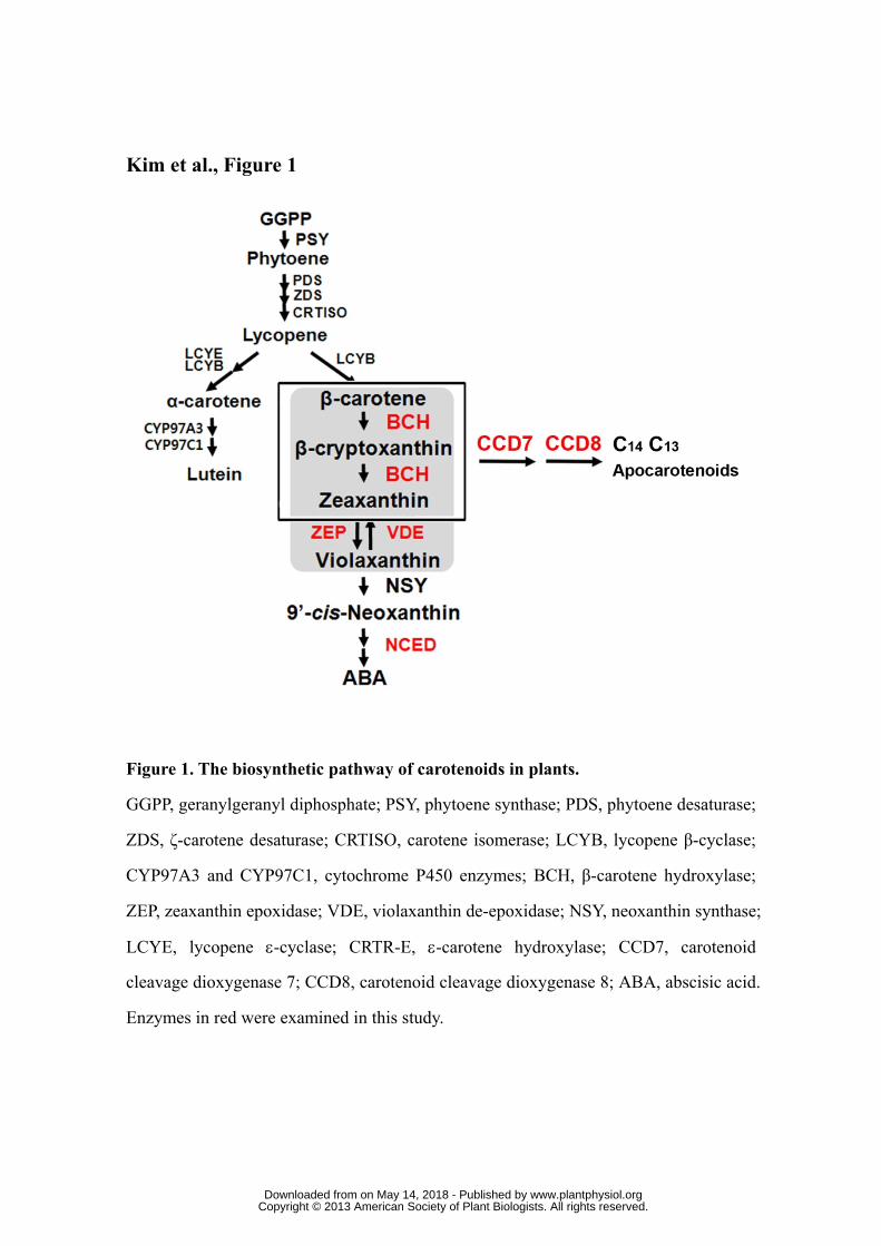
Kim et al., Figure 1
Figure 1. The biosynthetic pathway of carotenoids in plants.
GGPP, geranylgeranyl diphosphate; PSY, phytoene synthase; PDS, phytoene desaturase;
ZDS, ζ-carotene desaturase; CRTISO, carotene isomerase; LCYB, lycopene β-cyclase;
CYP97A3 and CYP97C1, cytochrome P450 enzymes; BCH, β-carotene hydroxylase;
ZEP, zeaxanthin epoxidase; VDE, violaxanthin de-epoxidase; NSY, neoxanthin synthase;
LCYE, lycopene ε-cyclase; CRTR-E, ε-carotene hydroxylase; CCD7, carotenoid
cleavage dioxygenase 7; CCD8, carotenoid cleavage dioxygenase 8; ABA, abscisic acid.
Enzymes in red were examined in this study.
www.plantphysiol.orgon May 14, 2018 - Published by Downloaded from Copyright © 2013 American Society of Plant Biologists. All rights reserved.
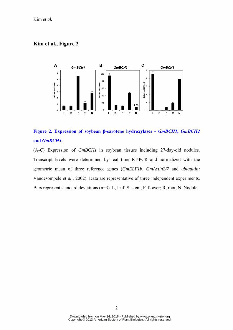
Kim et al.
Kim et al., Figure 2
Figure 2. Expression of soybean β-carotene hydroxylases - GmBCH1, GmBCH2
and GmBCH3.
(A-C) Expression of GmBCHs in soybean tissues including 27-day-old nodules.
Transcript levels were determined by real time RT-PCR and normalized with the
geometric mean of three reference genes (GmELF1b, GmActin2/7 and ubiquitin;
Vandesompele et al., 2002). Data are representative of three independent experiments.
Bars represent standard deviations (n=3). L, leaf; S, stem; F, flower; R, root, N, Nodule.
2
www.plantphysiol.orgon May 14, 2018 - Published by Downloaded from Copyright © 2013 American Society of Plant Biologists. All rights reserved.
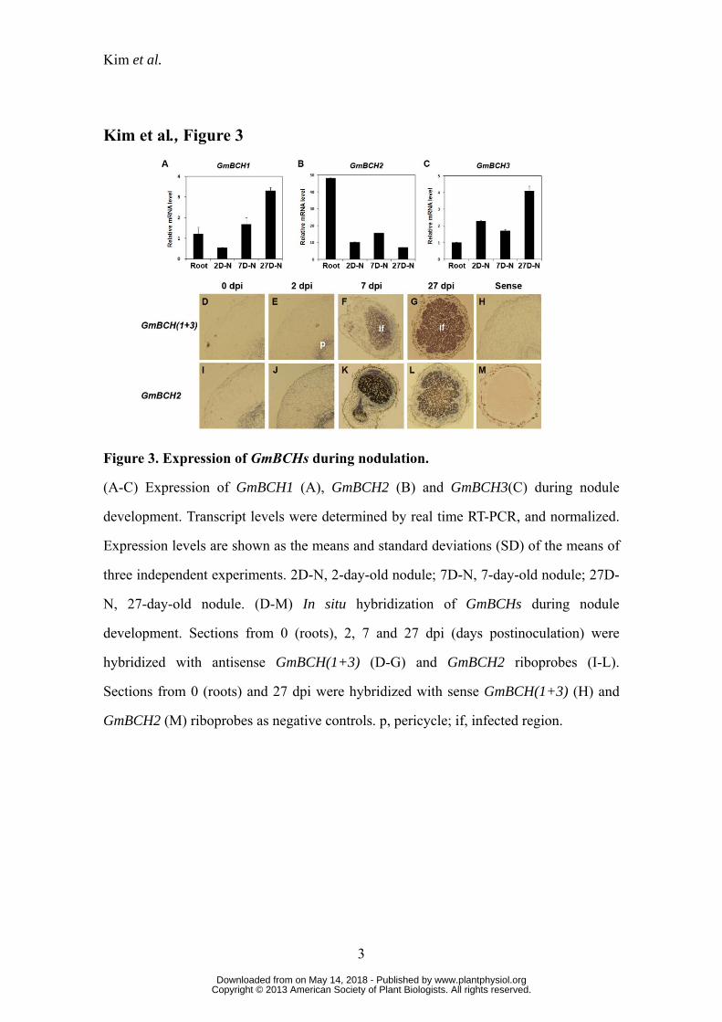
Kim et al.
Kim et al., Figure 3
Figure 3. Expression of GmBCHs during nodulation.
(A-C) Expression of GmBCH1 (A), GmBCH2 (B) and GmBCH3(C) during nodule
development. Transcript levels were determined by real time RT-PCR, and normalized.
Expression levels are shown as the means and standard deviations (SD) of the means of
three independent experiments. 2D-N, 2-day-old nodule; 7D-N, 7-day-old nodule; 27D-
N, 27-day-old nodule. (D-M) In situ hybridization of GmBCHs during nodule
development. Sections from 0 (roots), 2, 7 and 27 dpi (days postinoculation) were
hybridized with antisense GmBCH(1+3) (D-G) and GmBCH2 riboprobes (I-L).
Sections from 0 (roots) and 27 dpi were hybridized with sense GmBCH(1+3) (H) and
GmBCH2 (M) riboprobes as negative controls. p, pericycle; if, infected region.
3
www.plantphysiol.orgon May 14, 2018 - Published by Downloaded from Copyright © 2013 American Society of Plant Biologists. All rights reserved.
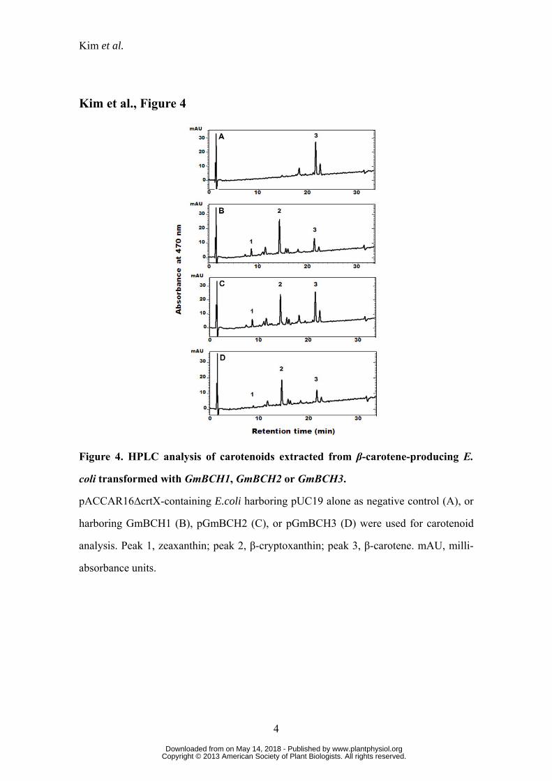
Kim et al.
Kim et al., Figure 4
Figure 4. HPLC analysis of carotenoids extracted from β-carotene-producing E.
coli transformed with GmBCH1, GmBCH2 or GmBCH3.
pACCAR16∆crtX-containing E.coli harboring pUC19 alone as negative control (A), or
harboring GmBCH1 (B), pGmBCH2 (C), or pGmBCH3 (D) were used for carotenoid
analysis. Peak 1, zeaxanthin; peak 2, β-cryptoxanthin; peak 3, β-carotene. mAU, milli-
absorbance units.
4
www.plantphysiol.orgon May 14, 2018 - Published by Downloaded from Copyright © 2013 American Society of Plant Biologists. All rights reserved.

Kim et al.
Kim et al., Figure 5
Figure 5. Subcellular localization of GmBCHs in Arabidopsis protoplasts and
soybean nodules.
Arabidopsis protoplasts were transfected with control vector (35S-GFP) (A), GmBCH1-
GFP (B), GmBCH2-GFP (C) or GmBCH3-GFP (D). Scale bar = 10 μm. (E) GmBCHs
were localized by EM immunogold labeling with an anti-GFP antiserum in soybean
nodules transformed with empty vector (pCAMBIA3301) (i), GmBCH1-GFP (ii),
GmBCH2-GFP (iii) or GmBCH3-GFP (iv). Gold particles (10-nm) are indicated by
5
www.plantphysiol.orgon May 14, 2018 - Published by Downloaded from Copyright © 2013 American Society of Plant Biologists. All rights reserved.

Kim et al.
arrowheads. B, bacteroids; Pl, plastid; M, mitochondria; C, cytoplasm; CW, cell wall.
cale bar = 100 nm.
S
6
www.plantphysiol.orgon May 14, 2018 - Published by Downloaded from Copyright © 2013 American Society of Plant Biologists. All rights reserved.
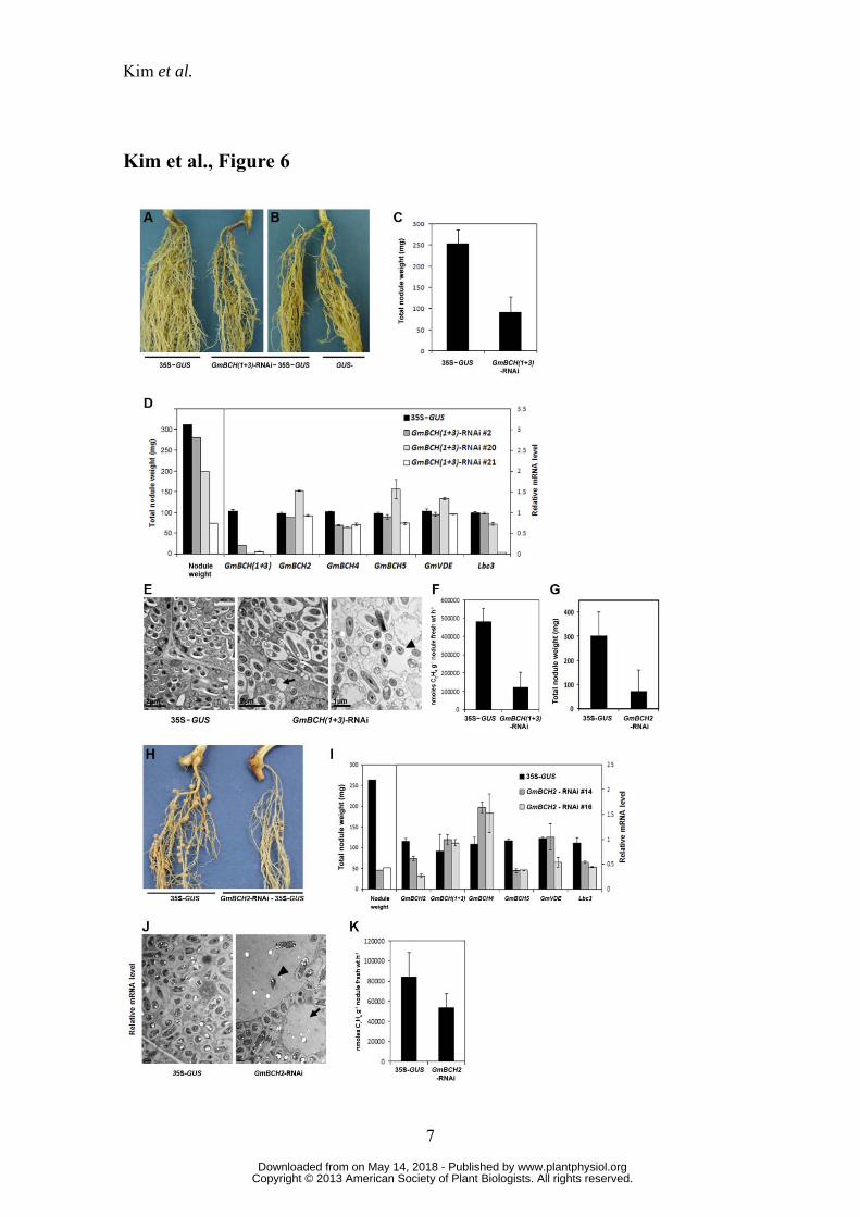
Kim et al.
Kim et al., Figure 6
7
www.plantphysiol.orgon May 14, 2018 - Published by Downloaded from Copyright © 2013 American Society of Plant Biologists. All rights reserved.
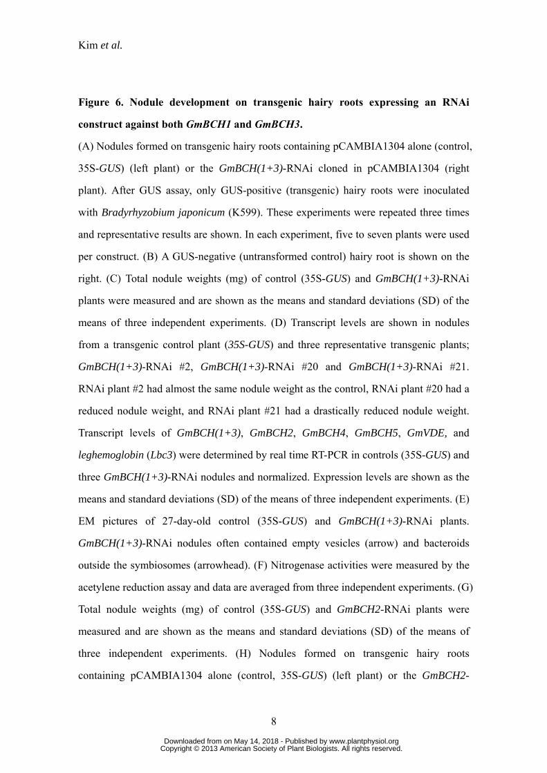
Kim et al.
Figure 6. Nodule development on transgenic hairy roots expressing an RNAi
construct against both GmBCH1 and GmBCH3.
(A) Nodules formed on transgenic hairy roots containing pCAMBIA1304 alone (control,
35S-GUS) (left plant) or the GmBCH(1+3)-RNAi cloned in pCAMBIA1304 (right
plant). After GUS assay, only GUS-positive (transgenic) hairy roots were inoculated
with Bradyrhyzobium japonicum (K599). These experiments were repeated three times
and representative results are shown. In each experiment, five to seven plants were used
per construct. (B) A GUS-negative (untransformed control) hairy root is shown on the
right. (C) Total nodule weights (mg) of control (35S-GUS) and GmBCH(1+3)-RNAi
plants were measured and are shown as the means and standard deviations (SD) of the
means of three independent experiments. (D) Transcript levels are shown in nodules
from a transgenic control plant (35S-GUS) and three representative transgenic plants;
GmBCH(1+3)-RNAi #2, GmBCH(1+3)-RNAi #20 and GmBCH(1+3)-RNAi #21.
RNAi plant #2 had almost the same nodule weight as the control, RNAi plant #20 had a
reduced nodule weight, and RNAi plant #21 had a drastically reduced nodule weight.
Transcript levels of GmBCH(1+3), GmBCH2, GmBCH4, GmBCH5, GmVDE, and
leghemoglobin (Lbc3) were determined by real time RT-PCR in controls (35S-GUS) and
three GmBCH(1+3)-RNAi nodules and normalized. Expression levels are shown as the
means and standard deviations (SD) of the means of three independent experiments. (E)
EM pictures of 27-day-old control (35S-GUS) and GmBCH(1+3)-RNAi plants.
GmBCH(1+3)-RNAi nodules often contained empty vesicles (arrow) and bacteroids
outside the symbiosomes (arrowhead). (F) Nitrogenase activities were measured by the
acetylene reduction assay and data are averaged from three independent experiments. (G)
Total nodule weights (mg) of control (35S-GUS) and GmBCH2-RNAi plants were
measured and are shown as the means and standard deviations (SD) of the means of
three independent experiments. (H) Nodules formed on transgenic hairy roots
containing pCAMBIA1304 alone (control, 35S-GUS) (left plant) or the GmBCH2-
8
www.plantphysiol.orgon May 14, 2018 - Published by Downloaded from Copyright © 2013 American Society of Plant Biologists. All rights reserved.
Kim et al.
RNAi cloned in pCAMBIA1304 (right plant). After GUS assay, nodules were formed as
in Panel A. (I) Transcript levels in nodules from one transgenic control plant (35S-GUS)
and two differentially repressed transgenic plants (GmBCH2-RNAi #14 and GmBCH2-
RNAi #16) are shown. Transcript levels of GmBCH2, GmBCH(1+3), GmBCH4,
GmBCH5, GmVDE, and leghemoglobin (Lbc3) were determined by real time RT-PCR
and normalized. Expression levels are shown as the means and standard deviations (SD)
of the means of three independent experiments. (J) EM pictures of 27-day-old control
(35S-GUS) and GmBCH2-RNAi nodules. GmBCH2-RNAi nodules often contained
empty vesicles (arrow) and bacteroids outside the symbiosomes (arrowhead). (K)
Nitrogenase activities were measured by the acetylene reduction assay as in Panel F.
9
www.plantphysiol.orgon May 14, 2018 - Published by Downloaded from Copyright © 2013 American Society of Plant Biologists. All rights reserved.
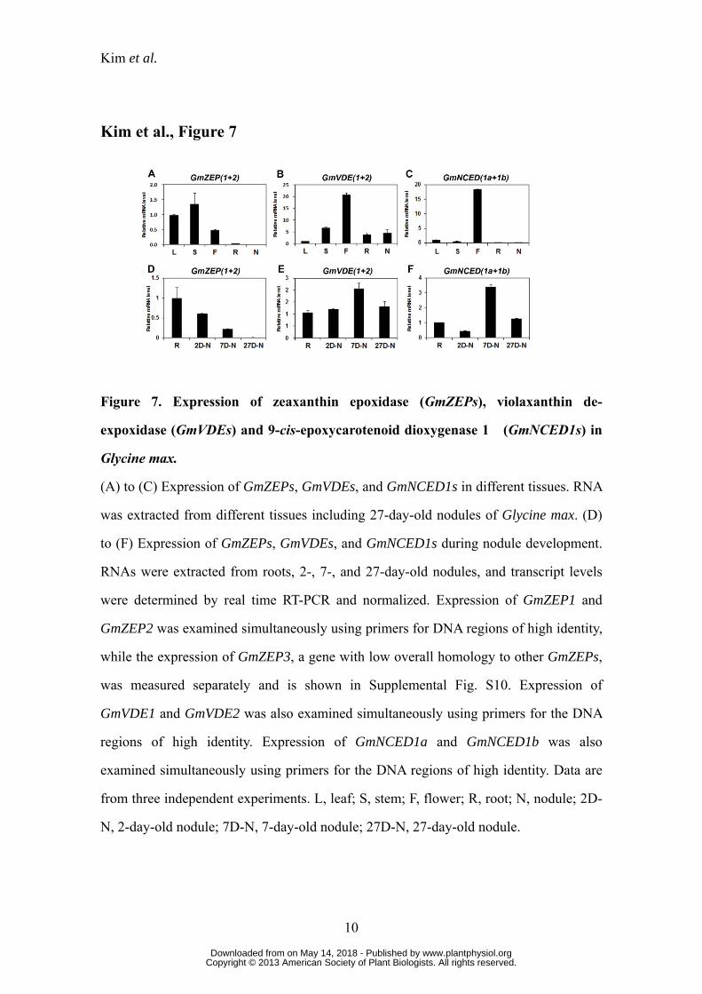
Kim et al.
Kim et al., Figure 7
Figure 7. Expression of zeaxanthin epoxidase (GmZEPs), violaxanthin de-
expoxidase (GmVDEs) and 9-cis-epoxycarotenoid dioxygenase 1 (GmNCED1s) in
Glycine max.
(A) to (C) Expression of GmZEPs, GmVDEs, and GmNCED1s in different tissues. RNA
was extracted from different tissues including 27-day-old nodules of Glycine max. (D)
to (F) Expression of GmZEPs, GmVDEs, and GmNCED1s during nodule development.
RNAs were extracted from roots, 2-, 7-, and 27-day-old nodules, and transcript levels
were determined by real time RT-PCR and normalized. Expression of GmZEP1 and
GmZEP2 was examined simultaneously using primers for DNA regions of high identity,
while the expression of GmZEP3, a gene with low overall homology to other GmZEPs,
was measured separately and is shown in Supplemental Fig. S10. Expression of
GmVDE1 and GmVDE2 was also examined simultaneously using primers for the DNA
regions of high identity. Expression of GmNCED1a and GmNCED1b was also
examined simultaneously using primers for the DNA regions of high identity. Data are
from three independent experiments. L, leaf; S, stem; F, flower; R, root; N, nodule; 2D-
N, 2-day-old nodule; 7D-N, 7-day-old nodule; 27D-N, 27-day-old nodule.
10
www.plantphysiol.orgon May 14, 2018 - Published by Downloaded from Copyright © 2013 American Society of Plant Biologists. All rights reserved.

Kim et al.
Kim et al., Figure 8
Figure 8. Expression of GmCCDs in Glycine max.
(A) to (C) Expression of GmCCD1s, GmCCD7s and GmCCD8s in different tissues.
RNA was extracted from various tissues including 27-day-old nodules of Glycine max.
(D) to (F) Expression of GmCCD1s, GmCCD7s and GmCCD8s during nodulation.
RNAs were extracted from roots, 2-, 7-, and 27-day-old nodules, and transcript levels
were determined by real time RT-PCR, and normalized. Expression levels are shown as
means of three independent experiments. L, leaf; S, stem; F, flower; R, root; N, nodule;
2D-N, 2-day-old nodule; 7D-N, 7-day-old nodule; 27D-N, 27-day-old nodule.
11
www.plantphysiol.orgon May 14, 2018 - Published by Downloaded from Copyright © 2013 American Society of Plant Biologists. All rights reserved.
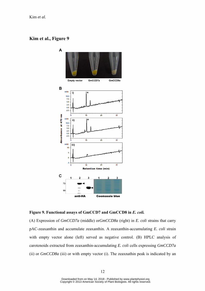
Kim et al.
Kim et al., Figure 9
Figure 9. Functional assays of GmCCD7 and GmCCD8 in E. coli.
(A) Expression of GmCCD7a (middle) orGmCCD8a (right) in E. coli strains that carry
pAC-zeaxanthin and accumulate zeaxanthin. A zeaxanthin-accumulating E. coli strain
with empty vector alone (left) served as negative control. (B) HPLC analysis of
carotenoids extracted from zeaxanthin-accumulating E. coli cells expressing GmCCD7a
(ii) or GmCCD8a (iii) or with empty vector (i). The zeaxnathin peak is indicated by an
12
www.plantphysiol.orgon May 14, 2018 - Published by Downloaded from Copyright © 2013 American Society of Plant Biologists. All rights reserved.
Kim et al.
13
asterisk (*). mAU, milli-absorbance units. (C) Proteins from zeaxanthin-accumulating E.
coli cells expressing HA-GmCCD7 (lane 2) and HA-GmCCD8 (lane 3) or with empty
vector (lane 1) were isolated (right panel) and immunoblotted with HA antibody (left
panel). Proteins of the expected sizes corresponding to GmCCD7 (70 kDa) and
GmCCD8 (60 kDa) were detected. Immunodetected bands are indicated by arrowheads.
www.plantphysiol.orgon May 14, 2018 - Published by Downloaded from Copyright © 2013 American Society of Plant Biologists. All rights reserved.