Embed Size (px)
Citation preview

1
Running title: Oxidation of plastid lipids
Mailing address:
Martin J. Mueller
Julius-von-Sachs-Institute of Biosciences, Biocenter, Pharmaceutical Biology,
University of Wuerzburg, Julius-von-Sachs-Platz 2, D-97082 Wuerzburg, Germany
Tel.: +49 931 3186160, Fax.: +49 931 3186182,
e-mail: [email protected]
Journal research area: Environmental stress and adaptation
Keywords: lipid oxidation, singlet oxygen, free radicals, lipoxygenase 2, azelaic acid,
pimelic acid
E-mail addresses of all authors: [email protected],
[email protected], [email protected],
[email protected], [email protected],
[email protected], [email protected]
Plant Physiology Preview. Published on July 22, 2012, as DOI:10.1104/pp.112.202846
Copyright 2012 by the American Society of Plant Biologists
www.plantphysiol.orgon July 19, 2018 - Published by Downloaded from Copyright © 2012 American Society of Plant Biologists. All rights reserved.

2
Lipid profiling of the Arabidopsis hypersensitive response reveals specific lipid
peroxidation and fragmentation processes: biogenesis of pimelic and azelaic acid1
Maria Zoeller, Nadja Stingl, Markus Krischke, Agnes Fekete, Frank Waller, Susanne
Berger, Martin J. Mueller*
Julius-von-Sachs-Institute of Biosciences, Biocenter, Pharmaceutical Biology,
University of Wuerzburg, Julius-von-Sachs-Platz 2, D-97082 Wuerzburg, Germany
www.plantphysiol.orgon July 19, 2018 - Published by Downloaded from Copyright © 2012 American Society of Plant Biologists. All rights reserved.
3
Footnotes
1This work was supported by the Deutsche Forschungsgemeinschaft (GK1342 and
SFB567).
*Corresponding author: [email protected]
The author responsible for distribution of materials integral to findings presented
in the article in accordance with the policy described in the instruction for authors
(http://www.plantphysiol.org) is: Martin J. Mueller
www.plantphysiol.orgon July 19, 2018 - Published by Downloaded from Copyright © 2012 American Society of Plant Biologists. All rights reserved.
4
ABSTRACT
Lipid peroxidation (LPO) is induced by a variety of abiotic and biotic stresses.
Although LPO is involved in diverse signaling processes, little is known about the
oxidation mechanisms and major lipid targets. A systematic lipidomics analysis of LPO
in the interaction of Arabidopsis thaliana with Pseudomonas syringae revealed that
LPO is predominantly confined to plastid lipids comprising galactolipid and
triacylglyceride species and precedes programmed cell death. Singlet oxygen was
identified as the major cause of lipid oxidation under basal conditions while a 13-
lipoxygenase (LOX2) and free radical-catalyzed lipid oxidation substantially contribute
to the increase upon pathogen infection. Analysis of lox2 mutants revealed that LOX2 is
essential for enzymatic membrane peroxidation but not for the pathogen-induced free
jasmonates production. Despite massive oxidative modification of plastid lipids, levels
of non-oxidized lipids dramatically increased after infection. Pathogen infection also
induced an accumulation of fragmented lipids. Analysis of mutants defective in 9-
lipoxygenases and LOX2 showed that galactolipid fragmentation is independent of
LOXs. We provide strong in vivo evidence for a free radical-catalyzed galactolipid
fragmentation mechanism responsible for the formation of the essential biotin precursor
pimelic acid as well as of azelaic acid, which was previously postulated to prime the
immune response of A. thaliana. Our results suggest that azelaic acid is a general
marker for LPO rather than a general immune signal. The proposed fragmentation
mechanism rationalizes the pathogen-induced radical amplification and formation of
electrophile signals such as phytoprostanes, malondialdehyde and hexenal in plastids.
www.plantphysiol.orgon July 19, 2018 - Published by Downloaded from Copyright © 2012 American Society of Plant Biologists. All rights reserved.
5
INTRODUCTION
Lipid peroxidation triggered by lipoxygenases and reactive oxygen species (ROS) is a
hallmark of plant pathogen responses, both in signal transduction processes and during
the execution of programmed cell death. Typically, lipoxygenases (LOX) oxidize free
fatty acids in the cytosol or chloroplasts thereby initiating several oxylipin pathways
including the jasmonate and hydroperoxide lyase pathway (Mosblech et al., 2009).
Among the ROS typically produced in plant stress responses, only singlet oxygen (1O2)
and free radicals are sufficiently reactive to oxidize polyunsaturated fatty acids directly
(Mueller et al., 2006). These short-lived ROS produced in different cellular
compartments including plasma membrane, plastids, mitochondria, peroxisomes, ER
and cytosol are thought to oxidize predominantly glycerolipids close to the site of ROS
production. In a recent study, 1O2 has been shown to be a major ROS species involved
in photooxidative lipid oxidation and damage in Arabidopsis thaliana leaves
(Triantaphylides et al., 2008). However, the major sites and molecular targets of lipid
oxidation as well as the relative contribution of different ROS species and LOXs to lipid
peroxidation and fragmentation have not been clarified.
LOXs and ROS have also been implicated in the formation of fragmented fatty acids in
plants and animals. Pathways for enzymatic and non-enzymatic fatty acid peroxide
cleavage yielding two aldehyde fragments have been described in plants and animals
(Fig. 1). The enzymatic fragmentation pathway (Fig. 1A), that has been described in
plants only, involves lipoxygenases (LOX) and hydroperoxide lyases (HPL) acting on
free fatty acids (Matsui et al., 2006). One aldehyde fragment harbors the fatty acid
carboxylate group (oxo fatty acid) while the other aldehyde fragment containing the
fatty acid methyl end is released as a volatile compound. Finally, oxo fatty acids can be
oxidized by aldehyde dehydrogenase (ADH) (Kirch et al., 2005; Mukhtarova et al.,
2011) yielding dicarboxylic acids. A. thaliana ecotype Col-0, however, lacks 9-HPL
genes and the 13-HPL gene has shown to be mutated and non-functional (Matsui et al.,
2006; Chehab et al., 2008). Since A. thaliana Col-0 produces fatty acid fragments such
as azelaic acid, another yet unknown fragmentation pathway must be operative (Jung et
al., 2009).
A non-enzymatic fatty acid fragmentation pathway has been described in vitro and in
animals in vivo (Fig. 1B). In animals, fatty acid peroxide fragmentation is catalyzed by
www.plantphysiol.orgon July 19, 2018 - Published by Downloaded from Copyright © 2012 American Society of Plant Biologists. All rights reserved.
6
free radicals and has been shown to take place in phospholipids and cardiolipins in vivo
under oxidative stress conditions (Hazen and Chisolm, 2002; Hazen, 2008). The non-
enzymatic mechanism produces a variety of aldehyde fragments from different fatty
acid peroxides including those aldehydes produced by plant HPL enzymes. After
radical-induced fragmentation of mammalian glycerolipids, however, the oxo acid
fragment remains esterified in the membrane while the other fragment is released from
the membrane. Oxidatively fragmented phospholipids were shown to serve as
endogenous pattern recognition ligands that activate the innate immune system of
animals in trace amounts (Hazen, 2008). However, such fragmented glycerolipids have
not yet been described in plants.
The non-enzymatic fragmentation mechanism has been investigated in detail (Schneider
et al., 2008) and shown to proceed through lipid peroxide dimers that spontaneously
undergo fragmentation (Fig. 1B). The main fragmented fatty acids found esterified in
mammalian glycerolipids are the C9-oxo acid fragments 9-oxononanoic acid (ONA) and
azelaic acid (AZA) and the C7-fragments 7-oxoheptanoic acid (OHA) and pimelic acid
(PIM). We hypothesized that C9- and C7-fragments could be generated in planta by free
radical catalyzed cleavage of esterified polyunsaturated C18- and C16-fatty acids,
respectively (Fig. 1C).
Interest in the fragmentation pathway stems from the fact that several short chain
aldehyde fragments (i.e. malondialdehyde, hydroxynonenal and hexenal) are potent
electrophiles that function as stress signals (Farmer and Davoine, 2007) in plants.
Important functions for dicarboxylic acids have also been reported. For instance, PIM is
an essential precursor of biotin. PIM biosynthesis has only recently been clarified in
bacteria (Lin et al., 2010 ) and there is yet no evidence that an enzymatic pathway is
operative in Arabidopsis. Another dicarboxylic acid, AZA, has recently been identified
as a pathogen-induced metabolite in Arabidopsis vascular sap that has been reported to
confer local and systemic resistance against the pathogen Pseudomonas syringae (Jung
et al., 2009; Chaturvedi, et al., 2012). AZA was also proposed to prime plants to
accumulate salicylic acid, a defense signal involved in systemic acquired resistance
(SAR) upon infection. Since AZA can be transported in the vascular sap it was
suggested to be a long-distance systemic signal in plants (Shah, 2009). Mutation of the
AZELAIC ACID INDUCED 1 (AZI1) gene, which was induced by AZA, results in the
www.plantphysiol.orgon July 19, 2018 - Published by Downloaded from Copyright © 2012 American Society of Plant Biologists. All rights reserved.
7
loss of systemic immunity triggered by pathogens or AZA (Jung et al., 2009). However,
direct genetic proof for a function of AZA in defense signaling is still missing. In case
of an enzymatic AZA biosynthesis, analysis of mutants deficient in AZA biosynthesis
would be one approach to test the function of AZA.
In this study, a systematic analysis of pathogenesis associated lipid peroxidation in
plants identified plastidic monogalactosyl- and digalactosyldiacylglycerols (MGDG and
DGDG) as well as triacylglycerols (TG) as major oxidized glycerolipids in A. thaliana
leaves. Detailed analysis of lipid oxidation products in wild type (WT) and mutants
related to lipid metabolism allowed to clarify different oxidation mechanisms involved
in lipid peroxidation and fragmentation in vivo, thereby identifying the biogenesis of
oxidized lipids in the plant response to abiotic and biotic stress. We also present a
model explaining the formation of reactive electrophile species previously shown to be
involved in important plant signaling processes.
www.plantphysiol.orgon July 19, 2018 - Published by Downloaded from Copyright © 2012 American Society of Plant Biologists. All rights reserved.

8
RESULTS
Oxidized fatty acids and fragmented fatty acids increase in A. thaliana leaves after
infection with avirulent P. syringae bacteria
In order to investigate the kinetics of lipid oxidation, leaves of 6-week old A .thaliana
plants were infiltrated with an avirulent or a virulent strain of P. syringae DC3000 (Pst
or PstavrRpm1, 108 colony forming units (cfu/ml)) and accumulation of enzymatically
and non-enzymatically formed oxidized lipids was monitored at different time points.
As shown in Figure 2, levels of several oxidized fatty acids including AZA and PIM
were elevated 5 to 10 h after infection with the avirulent strain and reached highest
levels at 24 h when the leaves showed severe visible damage. Levels of the established
marker (Mueller et al., 2006; Grun et al., 2007) of non-enzymatic 18:3 oxidation, 16-
hydroxyoctadecatrienoic acid (16-HO-18:3), increased with AZA and PIM as early as 5
to 10 h and reached 5 to 10-fold elevated levels after 24 h. In parallel, levels of jasmonic
acid (JA) synthesized via the 13-LOX pathway were also and even more dramatically
up-regulated (over 75-fold) within 5 to 10 h post infection. In contrast, infection with
the virulent P. syringae strain (108 cfu ml-1) induced only a low accumulation of all
tested oxidized fatty acids and visible damages were barely detectable after 24 h.
Infection experiments with lower bacterial densities of the avirulent Pst strain (106 and
107 cfu/ml) revealed that levels of oxidized lipids and visible leaf damages were lower,
likely due to the delayed cell death response (Supplemental Fig. S1, A). All lipids
analyzed were from plant origin and not detectable in both Pseudomonas strains. Both
early non-enzymatic and enzymatic oxylipin biosynthesis preceded visible leaf damage
and increased until cell death occurs (Fig. 2 and Supplemental Fig. S1, B). However, we
could not detect ONA or OHA, the putative precursors of AZA and PIM (Fig. 1),
respectively.
9-Lipoxygenases, trienoic fatty acids and AZELAIC ACID INDUCED 1 are not
essential for AZA and PIM biogenesis
It has been proposed, that the 9-lipoxygenase pathway (Fig. 1, A) is essential for the
formation of AZA in planta. Arabidopsis expresses two 9-LOX proteins (LOX1 and
LOX5). However, single and double T-DNA insertion lines (lox1, lox5, lox1/lox5) of
www.plantphysiol.orgon July 19, 2018 - Published by Downloaded from Copyright © 2012 American Society of Plant Biologists. All rights reserved.
9
both genes in the A. thaliana ecotype Wassilewskija that are completely deficient in the
respective 9-LOX activities (Vellosillo et al., 2007; Lopez et al., 2011) accumulated
wild type levels of AZA and PIM after infection with the avirulent Pst (Fig. 3, A).
Hence, enzymatic formation of AZA and PIM through the 9-LOX pathway can be ruled
out. In addition, LOX2, the most abundant 13-LOX, is also not essential for lipid
fragmentation (Fig. 3, B).
In addition to 9-LOX, the plastidic FATTY ACID DESATURASE 7 (FAD7) and
AZELAIC ACID INDUCED 1 (AZI1) have been suggested to be involved in the local
production or transport of AZA and/or the SAR signal, respectively (Chaturvedi et al.,
2008; Jung et al., 2009). We therefore tested mutants deficient in these genes for their
capacity to accumulate AZA and PIM in infected leaves. As shown in Fig. 3B, local
production of AZA and PIM was not compromised in the fatty acid desaturase triple
mutant fad3-2 fad7-2 fad8 (fad3/7/8) and azi1. In the fad3/7/8 mutant, trienoic fatty
acids are quantitatively replaced by dienoic fatty acids (McConn and Browse, 1996) that
may, however, also serve as precursors for fragmented fatty acids.
Previously, it has been suggested that AZA is transported to non-infected systemic
leaves after Pst infection (Jung et al., 2009). However, AZA and PIM levels in systemic
leaves 24 h post infection were not elevated and comparable to levels in leaves of non-
infected wild type and mutant plants (Fig. 3 and Supplemental Fig. S1, B). Moreover,
we observed that the AZI1 gene, which was reported to be induced by AZA and required
for the SAR response (Jung et al., 2009) was not induced by AZA. Although a weak and
transient induction of AZI1 after spraying of AZA (in MES buffer) could be measured
as reported by Jung et al. (2009), this induction was also seen after spraying of buffer
alone (Supplemental Fig. S2, A). More importantly, however, AZA pretreatment of
Arabidopsis leaves (as described in Jung et al., 2009) did not inhibit growth of spray
inoculated virulent Pst under our experimental conditions (Supplemental Fig. S2, B).
AZA, PIM and their precursors occur esterified in oxidized MGDG and DGDG in
vivo and accumulate after Pseudomonas infection
Alternatively to the enzymatic formation of AZA and PIM (Fig. 1, A), free radical-
catalyzed fragmentation of oxidized glycerolipids could take place as it has been
observed in animals (Fig. 1, B). In vitro, free radical oxidation experiments with
www.plantphysiol.orgon July 19, 2018 - Published by Downloaded from Copyright © 2012 American Society of Plant Biologists. All rights reserved.
10
unsaturated fatty acids revealed that 18:3 and 18:2, but not 18:1, yielded ONA and AZA
(Supplemental Fig. S3). In agreement with the proposed chemical fragmentation
mechanism (Schneider et al., 2008), the kinetics of the 18:3 and 18:2 oxidation products
suggested that at least three oxidation events are required to produce AZA from 18:3 or
18:2: first, a fatty acid (or acyl) hydroperoxide is formed that, under radical-catalysis,
fragments to yield ONA. Finally, ONA is oxidized in a radical-catalyzed process to
AZA (Supplemental Fig. S3). We also performed in vitro autoxidation experiments with
either 9- or 13-HOO-18:3 and observed that both peroxides non-enzymatically fragment
to produce 1 – 2.5 mol% ONA and 0.3 – 0.8 mol% AZA (Supplemental Fig. S3).
The free radical-catalyzed AZA biogenesis hypothesis (Fig. 1, B) predicts that 18:3
hydroperoxides, ONA and AZA are formed by oxidation of esterified 18:3 in
glycerolipids. In addition, 16:3 hydroperoxides, OHA and PIM would be expected to be
formed by oxidation of esterified 16:3 in glycerolipids. We therefore performed a
systematic analysis of glycerolipids by ultra performance liquid chromatography
(UPLC) coupled to a quadrupol-time of flight mass spectrometer (Q-TOF MS). After
reduction of peroxides to the corresponding hydroxides, parent lipid molecules were
determined that released fragments indicative for the presence of HO-18:3, HO-16:3,
ONA, OHA, AZA and PIM upon collision induced fragmentation. ONA, OHA, AZA
and PIM (along with the predicted HO(O)-18:3/16:3 precursor fatty acids) could be
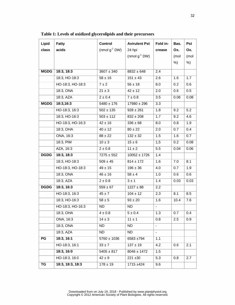
identified only esterified in MGDG and DGDG (Table 1). In order to determine the
basal and pathogen-induced levels of these oxidized glycerolipids, a targeted analysis of
the most abundant galactolipid species was performed by UPLC-MS/MS in the multiple
reaction monitoring (MRM) mode (see materials and methods for details). The complex
data set is shown in Table 1. Under control conditions (mock infiltration), oxidized
(18:3, 18:3)MGDG and DGDG contained 0.6 mol% ONA and about 0.05 mol% AZA
relative to the non-oxidized precursor. Oxidized (18:3, 16:3)MGDG and DGDG
contained, in addition to 18:3 derived ONA (1.6-2.5 mol%) and AZA (0.04 mol%), also
16:3 derived OHA (0,7 mol%) and PIM (0.2 mol%).
After pathogen infection, levels of both non-oxidized as well as of peroxidized or
fragmented galactolipids increased in average about twofold. Therefore, the degree of
oxidation of different galactolipid species expressed in mol% relative to the non-
oxidized precursor galactolipid did not change dramatically (Table 1). Under control
www.plantphysiol.orgon July 19, 2018 - Published by Downloaded from Copyright © 2012 American Society of Plant Biologists. All rights reserved.

11
conditions, total levels of ONA, OHA, AZA and PIM in the four major galactolipid
species per g of dry weight (DW) were 169 nmol, 44 nmol, 6 nmol and 10 nmol,
respectively. The presence of esterified fragmented fatty acids was also confirmed by
analysis of total lipids extracts before and after alkaline hydrolysis. This analysis
confirmed that ONA and OHA are only present in esterified but not in free form while
AZA and PIM are present in Arabidopsis leaves both in esterified and free form
(Supplemental Fig. S4).
These findings (Table 1) are in agreement with the free radical hypothesis of
galactolipid fragmentation in plastids in situ (Fig. 1, B). Alternatively, fatty acids are
fragmented first, followed by esterification into galactolipids.
Evidence for acyl chain fragmentation in galactolipids
In order to investigate the possibility of incorporation of ONA or AZA into
galactolipids, feeding experiments using [8,8-D2]ONA and unlabeled AZA were
performed. [8,8-D2]ONA (100 µM) was applied to the medium of A. thaliana seedlings
(10 d) grown in liquid medium. After different time points, seedlings were washed and
lipids were extracted and analyzed by UPLC-MS/MS. In seedling extracts, labeled
ONA could not be detected at any time indicating that ONA does not accumulate in free
form (Fig. 4, A). Instead, 1.5 h after [8,8-D2]ONA application, [2,2-D2)AZA and [2,2-
D2]PIM accumulated in the seedlings and reached level of 130 and 55 nmol g-1 DW,
respectively. Thereafter, levels of [2,2-D2]AZA decreased while levels of [2,2-D2]PIM
further increased and reached levels of 4 and 232 nmol g-1 DW, respectively, after 24 h
(Fig. 4, A). Labeled ONA, AZA or PIM were not incorporated into galactolipids. These
results indicate that after uptake, ONA is instantaneously metabolized to AZA which is
in turn degraded by ß-oxidation to PIM. When unlabeled AZA (100 µM) was fed to the
seedlings, transient accumulation of AZA followed by ß-oxidation to PIM was
observed. AZA was not found to be incorporated into MGDG (Fig. 4B). We could not
detect 9-hydroxynonanoic acid or ONA in any of the application experiments indicating
that reduction of the aldehyde or AZA is not a significant metabolic pathway in A.
thaliana. Results suggest that biogenesis of AZA and PIM starts in plastids where ONA
and OHA esterified in galactolipids are generated through free radical-catalyzed
oxidative fragmentation of polyunsaturated C18 and C16 fatty acids. Further free
www.plantphysiol.orgon July 19, 2018 - Published by Downloaded from Copyright © 2012 American Society of Plant Biologists. All rights reserved.
12
radical-catalyzed oxidation of esterified ONA and OHA leads to an accumulation of
esterified AZA and PIM in galactolipids. Hydrolytic release of fragmented fatty acids
by lipases may then result in the accumulation of free AZA and PIM. During
galactolipid hydrolysis, ONA and OHA may also be liberated. However, these oxoacids
do not accumulate in free form and are rapidly converted into AZA and PIM (Fig. 4),
most likely through aldehyde dehydrogenases.
Lipid peroxidation mechanisms: Singlet oxygen, free radical- and lipoxygenase-
catalyzed membrane peroxidation is triggered by P. syringae
We next addressed the question which mechanisms of lipid peroxidation are involved in
the formation of glycerolipid peroxides in vivo. Accumulation of glycerolipid peroxides
is essential for and precedes free radical-catalyzed lipid fragmentation (Supplemental
Fig. S3). The contribution of singlet oxygen-, free radical- and lipoxygenase mediated
peroxidation to total lipid peroxidation was assessed by fatty acid peroxide
fingerprinting, i.e. by determining the free and esterified HO(O)-18:3 fatty acid isomer
composition in A. thaliana Col-0 leaves (Mueller et al., 2006; Triantaphylides et al,
2008). Leaves from 6-week old plants were mock infiltrated (control) or infected with
avirulent Pst (108 cfu ml-1), harvested 24 h after treatment, immediately shock-frozen
and extracted under peroxide reducing conditions thereby converting lipid peroxides
into the corresponding stable hydroxides. Reducing conditions proved to be essential to
prevent degradation and artifact formation thereby yielding higher recovery of oxidized
lipids as compared to previous studies (Ibrahim et al., 2011; Vu et al., 2012). HO-18:3
isomers were determined in lipid extracts in free form as well as in esterified form (after
release from complex lipids by alkaline hydrolysis) by ultra performance liquid-
chromatography tandem mass spectrometry (UPLC-MS/MS). Levels of free HO-18:3
were 1% or less of the levels of esterified HO-18:3 under control conditions and after
infection (Fig. 5, A), indicating that glycerolipids are the major targets for fatty acid
peroxidation. From the HO-18:3 isomer pattern (Supplemental data, Supplemental Fig.
S4 and S5), levels of HO(O)-18:3 formed through 1O2, free radical oxidation and
lipoxygenases (Fig. 5, A) could be calculated (Triantaphylides et al., 2008). Under
control conditions, the observed isomer pattern of esterified HO-18:3 is indicative for a
prevalent 1O2-dependent oxidation mechanism (>90 mol%). HO-18:3 derived from free
www.plantphysiol.orgon July 19, 2018 - Published by Downloaded from Copyright © 2012 American Society of Plant Biologists. All rights reserved.
13
radical-catalyzed oxidation and 13-LOX comprised 3 and 7 mol% of all esterified HO-
18:3, respectively (Fig. 5, A). After P. syringae infection, levels of esterified HO-18:3
oxidized by 1O2, free radicals and 13-LOX increased by 2.9-, 43- and 56– fold and the
relative contribution of 1O2, free radicals and 13-LOX to overall HO-18:3 formation
changed to 40%, 18% and 40%, respectively.
LOX2 is central for LOX-mediated glycerolipid peroxidation and modulates the
levels of free hydroxy fatty acids after pathogen infection
LOX2 has been shown to be involved in the formation of arabidopsides from MGDG
and DGDG in plastids (Glauser et al., 2009; Seltmann et al., 2010). Analysis of the total
13-HO(O)-18:3 pattern in the lox2 mutant revealed that LOX2 is responsible for
virtually all the lipoxygenase-mediated synthesis of esterified 13-HO(O)-18:3 under
control (100%) and pathogen-induced (93%) conditions (Fig. 5B). This result suggests
that LOX2 may directly oxidize galactolipids.
To this end, pathogen-triggered oxidation of MGDG and DGDG was analyzed in WT
and lox2 mutant plants by UPLC-MS/MS after reduction of lipid peroxides to the
corresponding hydroxides. In the wild type, levels of galactolipids in which both acyl
chains were peroxidized (dihydroperoxides) dramatically increased in a LOX2
dependent manner while galactolipids with one peroxidized acyl chain
(monohydroperoxides) showed only a minor increase that was not dependent on LOX2
(Fig. 6, B). However, dihydroperoxy galactolipids were not completely lacking in the
lox2 mutant (Fig. 6, B). This finding is compatible with the hypothesis that galactolipid
monohydroperoxides are formed through random 1O2 –mediated oxidation of acyl
chains at the sn1 or sn2 position close to the production site of short lived 1O2 while
dihydroperoxides are predominantly generated by direct double oxygenation of
galactolipids by LOX2 (Fig. 6 B). The later galactolipid dihydroperoxides may serve as
direct precursors of arabidopsides that comprise two cyclooxylipin acyl chains (OPDA
or dinor OPDA). In fact, a massive synthesis of arabidopsides was only observed in the
wild type but not the lox2 mutant after Pseudomonas infection (Fig 6, C).
Arabidopsides A, B, G and E accumulated to almost 50% of the initially present non-
oxidized MGDG precursors which should be accompanied by a dramatic loss of non-
oxidized MGDG precursors. In contrast, a strong accumulation of the non-oxidized
www.plantphysiol.orgon July 19, 2018 - Published by Downloaded from Copyright © 2012 American Society of Plant Biologists. All rights reserved.
14
MGDGs (2.4- to 3.3-fold increase) and DGDGs (1.4- to 2.3-fold increase) was observed
(Table 1 and Fig. 6, A). This increase was detected both in the WT and the lox2 mutant.
Hence, pathogen-triggered stimulation of lipid accumulation appears not to be a
compensatory mechanism for LOX2-mediated lipid consumption.
Unexpectedly, however, LOX2 appears to contribute but not to be strictly required for
the accumulation of free 13-HO(O)-18:3, free OPDA and JA after pathogen infection
(Fig. 6, D). Since arabidopsides are virtually absent in the lox2 mutant, arabidopsides
are therefore no essential precursors for production of jasmonate signals after infection.
Although LOX2 increases dihydroperoxide galactolipid levels after infection (Fig. 6,
B), no accumulation of galactolipids with two fragmented acyl chains could be detected.
In addition, LOX2 appears to contribute but not to be strictly required for the formation
of fragmented fatty acids (Fig. 3B).
Compared to the WT, surprisingly, the lox2 mutation caused an excessive pathogen-
induced 13- and 9-lipoxygenase activation resulting in a 2- and 80-fold higher
accumulation of free 13- and 9-HO(O)-18:3 (Fig. 5, B, and 6, B). In addition, also a
dramatic pathogen-induced over-accumulation of non-enzymatically oxidized HO(O)-
18:3 was observed in the lox2 mutant (Fig. 5B). Notably, levels of free HO(O)-18:3
oxidized by singlet oxygen and free radicals were both about 10-fold higher in the lox2
mutant compared to the WT (Fig. 5) after infection. Hence, membrane oxidation by
LOX2 reduces via an unknown regulatory mechanism the accumulation of non-
enzymatically and enzymatically oxidized free fatty acids. Moreover, LOX2 and
arabidopsides are not essential for the production of free jasmonates, at least in the late
phase (24 h after infection) of the Arabidopsis-Pseudomonas interaction.
Galactolipids and triacylglycerols are major targets for peroxidative fatty acid
modification
To identify the major targets of non-enzymatic lipid peroxidation, lipid extracts were
analyzed (after reduction of peroxides to the corresponding hydroxides) for the presence
of complex lipids that comprised esterified HO-18:3 by UPLC-Q-TOF. Five classes of
glycerolipids could be identified that comprised esterified HO-18:3: MGDG, DGDG,
phosphatidylglycerols (PG), triacylglycerols (TG) and phosphatidylinositols (PI) while
esterified HO-18:3 could not be detected in phosphatidylcholines (PC) and
www.plantphysiol.orgon July 19, 2018 - Published by Downloaded from Copyright © 2012 American Society of Plant Biologists. All rights reserved.

15
phosphatidylethanolamines (PE). All oxidized glycerolipids were found to be derived
from the most highly abundant polyunsaturated glycerolipids within these lipid classes.
A targeted analysis of non-oxidized glycerolipids and their corresponding HO-18:3
comprising species was performed by UPLC-MS/MS in the multiple reaction
monitoring (MRM) mode (see materials and methods for details).
Analysis of mock-infiltrated Arabidopsis leaves revealed that the by far most highly
peroxidized glycerolipid species are (18:3, 16:3)MGDG, (18:3, 16:3)DGDG and (18:3,
18:3)DGDG (Table 1). Notably, total levels of oxidized versions of these species
(comprising one or two HO(O) acyl chains) relative to the level of the corresponding
non-oxidized galactolipid species were 19, 18 and 7 mol%, respectively. In contrast,
basal levels of oxidized (18:3, 18:3)MGDG, PG, PI and TG species were between 0.5
and 2.1 mol% (Table 1).
After Pseudomonas infection, levels of all peroxidized lipid species of MGDG, DGDG,
PG and PI increased about 2- to 8-fold. Notably, a 100- to 455-fold increase of HO(O)-
18:3 comprising TG species was observed. Under basal conditions, the degree of lipid
oxidation of polyunsaturated TG species was between 0.03 – 2.1 mol% (relative to the
non-oxidized precursor species). However, several TG species became highly oxidized
after pathogen infection and the degree of oxidation increased to 11 – 37 mol%.
Accumulation of oxidized lipids did not reduce the pool size of their non-oxidized
precursor lipids. In contrast, Pseudomonas infection induced a massive accumulation of
polyunsaturated MGDG, DGDG, PG, PE, PI and TG species while PC species became
less abundant. The most dramatic accumulation of lipids was observed within the TG
lipid pool with a 7- to 10-fold mass increase (Table 1). This increase was observed only
in infected leaves but not in non-infected leaves of the same plant (Supplemental Fig.
S7). It is not clear if this accumulation in infected leaves (displaying severe leaf damage
after 24 h) results from disturbed lipid catabolism or increased synthesis. All lipid
analyzed are from plant origin since Pseudomonas, as most bacteria (except for
cyanobacteria), cannot synthesize glycerolipids comprising trienoic fatty acids and
galactolipids.
www.plantphysiol.orgon July 19, 2018 - Published by Downloaded from Copyright © 2012 American Society of Plant Biologists. All rights reserved.
16
DISCUSSION
Lipid peroxidation in the Arabidopsis-Pseudomonas interaction
Oxidative stress and lipid peroxidation (LPO) are well known consequences of a variety
of abiotic and biotic stresses. However, the predominant ROS species and
lipoxygenases involved in membrane LPO as well as the major site(s) of LPO in plant
pathogen interactions remained largely unknown for decades. On the one hand,
enzymatic and non-enzymatic formation of low levels of ROS and LPO products in the
early stages of the HR have been implicated in processes such as defense signaling
(Torres et al., 2006; Mueller et al., 2008), plant stress adaptation (Mueller, 2004) and
SAR (Jung et al., 2009). The HR reaction is characterized by an oxidative burst initiated
by NADPH oxidases that produces superoxide anion radicals at the cell membrane
(Torres et al., 2005). NADPH oxidases have been regarded as the major source of ROS
species in plant-pathogen interactions involved in both signaling and LPO (Torres,
2010). However, NADPH oxidase produced superoxide anion radicals (and their
dismutation product H2O2) are not sufficiently reactive to directly oxidize lipids
(Frankel, 2005). Moreover, early NADPH oxidase activation within minutes at the cell
membrane (Torres, 2010) does not coincide with LPO observed in plastid lipids (Table
1) several hours after infection (Fig. 2). On the other hand, ROS-mediated excessive
LPO is associated with membrane damage and, hence, may contribute to the execution
of the cell death program (5 – 24 h after infection).
We show that 1O2 is a major ROS involved in basal LPO and pathogen-induced LPO
(Fig. 5). The remarkable high basal (8 to 19 mol%) and specific peroxidation of three
major plastid galactolipids located close to the major site of 1O2 production - i.e.
photosystem II - might be due to the short half-life and high reactivity of 1O2
(Triantaphylides et al., 2008). After initiation of the HR process, photosynthetic activity
is reduced (Berger et al., 2007) and progressive disorganization of the photosynthetic
apparatus leads to increased 1O2 formation and 1O2 -mediated LPO (Fig. 5).
To the most part, increase of LPO is due to LOX2-mediated double oxygenation of
plastid galactolipids (dihydroperoxides) that are rapidly converted into arabidopsides
(Fig. 6). It has been proposed that arabidopsides serve as OPDA storage molecules from
which OPDA can be rapidly mobilized to generate jasmonate signals (Kourtchenko et
al, 2007). However, LOX2 and arabidopsides were not found to be major sources of
www.plantphysiol.orgon July 19, 2018 - Published by Downloaded from Copyright © 2012 American Society of Plant Biologists. All rights reserved.
17
free jasmonates during the HR after Pseudomonas infection (Fig. 6) suggesting that one
or more of LOX 3, 4, and 6 are responsible for jasmonate accumulation under these
conditions. In contrast, LOX2 appears to produce the majority of free jasmonates after
wounding (Glauser et al., 2009) and during natural or dark induced senescence
(Seltmann et al., 2010). Despite the high LOX2-dependent jasmonate and arabidopside
accumulation after wounding, LOX2 was not found to be required for JA signaling
(Glauser et al., 2009) and appears to serve other functions.
Surprisingly, we found that non-enzymatic lipid oxidation as well as the enzymatic 9-
LOX pathway (Fig. 5B) was dramatically increased in the lox2 mutant and higher LPO
appeared to be associated with slightly increased visible leave damage after infection
with avirulent bacteria (Supplemental Fig. S1). Pretreatment of Arabidopsis leaves with
9-lipoxygenase products has been shown to protect the leaves against infection with
virulent Pst DC3000 bacteria but not with avirulent Pst DC3000 avrRpm1 bacteria
(Vincente et al., 2011). Hence, activation of the 9-LOX pathway in lox2 mutant plants
may increase the local resistance against virulent bacteria, however, this hypothesis
remains to be tested. The mechanism of how LOX2-mediated membrane oxidation
modulates total LPO levels also remains to be elucidated.
In addition to 1O2 and LOX2, free radicals contribute significantly to LPO during
Pseudomonas infection (Fig. 5). Radical-catalyzed LPO is low under basal conditions
but becomes a major ROS-mediated LPO process during the HR (Fig. 5). Free radical-
catalyzed LPO appears to take place predominantly in plastid lipids since we could
detect marker lipids for free radical-catalyzed oxidation (fragmented fatty acids) in
galactolipids but not in other glycerolipids (Tab. 1). Hence, we determined that
membrane LPO by all three LPO mechanisms (LOX2, 1O2 and free radicals) is
predominantly confined to plastid membranes. This result is in line with the light-
dependency of the HR in plant-pathogen interactions (e.g. Zeier et al., 2004, Montillet
et al., 2005).
Biogenesis of azelaic and pimelic acid by free radical-catalyzed fragmentation of
fatty acid hydroperoxides in A. thaliana
Lipid peroxides and H2O2 readily generate hydroxyl radicals in the presence of trace
amounts of free transition metals (such as Cu2+ or Fe2+) that are thought to be released
www.plantphysiol.orgon July 19, 2018 - Published by Downloaded from Copyright © 2012 American Society of Plant Biologists. All rights reserved.
18
from damaged metalloproteins (Spiteller, 2002). Another de novo source of hydroxyl
radicals appears to be the lipid fragmentation process itself that is amplified during HR.
The fragmentation mechanism of fatty acid hydroperoxides has recently been described
by Schneider, Porter, Brash and coworkers in vitro and in animals in vivo (Liu et al.
2011; Schneider et al., 2008). In animals, fatty acid hydroperoxides and fragmented
fatty acids accumulate in PCs in the cellular membrane and in cardiolipins in
mitochondrial membranes. Among the oxidized glycerolipids, oxidized PCs containing
ONA and AZA, are the most abundant molecules (Podrez et al., 2002; Chen et al.,
2008). The proposed fragmentation mechanism (Liu et al., 2011 ; Schneider et al.,
2008) rationalizes the key findings of this study in Arabidopsis (Fig. 7): A precondition
for the fragmentation process is the continuous formation and maintenance of a pool of
galactolipid hydroperoxides through 1O2-mediated LPO (Fig. 7, A). For the second step
to proceed, catalytic amounts of radicals are required to initiate peroxide dimer
formation within the pool of peroxidized galactolipids. The third step is the spontaneous
fragmentation of the dimer and production of four products: core aldehydes
(galactolipids containing an oxo acid fragment such as ONA or OHA), short chain
aldehydes (such as hydroperoxynonadienal or hexenal), an oxidized (non-fragmented)
glycerolipid radical and hydroxyl radicals (the detailed fragmentation mechanism is
shown in Suppl. Fig. S8). Hence, the fragmentation process itself dramatically amplifies
de novo radical production. These radicals further oxidize oxo acids in core aldehydes to
core dicarboxylic acids (such as AZA and PIM) and catalyze the formation of a great
variety of oxidized galactolipids. In fact, more than 50 species of oxidized galactolipids
have been identified in Arabidopsis leaves thus far (Ibrahim et al., 2011, Vu et al.,
2012). Oxidized and oxidatively fragmented galactolipids could represent a molecular
memory of stress conditions and may serve as storage molecules for preformed
oxylipins.
Our model also takes into account that oxidatively fragmented or polar acyl chains
(such as ONA or AZA) protrude into the aqueous phase while non-oxygenated and
mono-oxygenated fatty acids (such as in 13-HO-18:2) remain in the lipid phase, as
determined by NMR studies (Li et al., 2007). It has been suggested that membranes thus
“grow whiskers” according to the “Lipid Whisker Model” (Greenberg et al., 2008). This
conformational change of structural lipids during oxidation may contribute to the
www.plantphysiol.orgon July 19, 2018 - Published by Downloaded from Copyright © 2012 American Society of Plant Biologists. All rights reserved.
19
disruption of the membrane barrier and cell death (Fig. 7). In mammals, oxidized acyl
residues protruding out of membranes will be selectively removed by specific repair
lipases (Marques et al, 2002). In plants, specific repair lipases are not known, however,
oxidized and fragmented fatty acids are ultimately released from galactolipids (Fig. 2 ).
Under basal conditions, total amounts of esterified ONA, OHA, AZA and PIM are in
the range of 229 nmol g-1 DW (Table 1 and Fig. S4). Continuous formation, release and
rapid metabolism of these fragments to PIM (Fig. 4) may maintain a pool of free PIM
(2-4 nmol g-1 DW; Fig. 2) that is essential for the biosynthesis of biotin (< 1 nmol g-1
DW, as calculated from Shellhammer and Meinke (1990) in Arabidopsis leaves. Hence,
fragmentation of galactolipids could be a general plant specific and sufficient pathway
to provide PIM to feed the biotin biosynthesis pathway.
Function of lipid peroxidation and fatty acid fragmentation
It has been proposed that non-enzymatic LPO protects plants from oxidative stress by
scavenging ROS (Mene-Saffrane et al., 2009). Polyunsaturated galactolipids indeed
scavenge large quantities of 1O2 (Fig. 5, Table 1) that escaped the carotenoid quenching
mechanism (Ramel et al., 2012a), thereby preventing 1O2-mediated protein damage.
After pathogen infection, hydroxy fatty acids derived from 1O2-mediated LPO (Fig. 5;
Grun et al., 2007) increase and have been shown to induce a strong accumulation of
callose in Arabidopsis leaves which is a frequent response of cells to pathogen assault
(Vellosillo et al., 2010). Moreover, hydroxy fatty acids derived from either 1O2-,
radical- or LOX-mediated LPO have been shown to up-regulate defense genes that are
also up-regulated after Pst infection suggesting a function of hydroxy fatty acids in
plant pathogen defense responses (Vellosillo et al., 2007).
However, excessive or uncontrolled 1O2-mediated LPO increases the vulnerability of
lipids to over-oxidation, lipid fragmentation and de novo free radical production. It
should be emphasized, that hydroxyl radicals cannot be scavenged by polyunsaturated
lipids. In contrast, a radical chain reaction propagates through membranes and
ultimately leads to galactolipid and carotenoid fragmentation as well as radical
amplification. Fragmentation of carotenoids leads to the formation of β-cyclocitral that
induces defense genes protecting against oxidative stress (Ramel et al., 2012b). Hence,
LPO produces plastid lipids signals in the early stages of oxidative stress associated
www.plantphysiol.orgon July 19, 2018 - Published by Downloaded from Copyright © 2012 American Society of Plant Biologists. All rights reserved.
20
with high light and pathogen stress. However, massive LPO, exceeding a certain
threshold level of oxidized lipids, may contribute to the execution of cell death
(Triantaphylides et al., 2008).
In animals, pathogen-induced lipid oxidation and fragmentation takes place in the cell
membrane enabling physical contact between pattern recognition receptors of the Toll-
like family and oxidized glycerolipids (Hazen, 2008). These receptors recognize in the
first place pathogen surface lipids but also bind to endogenous oxidized surface lipids.
Hence, in animals, oxidative stress activates the innate immune system by utilizing
defense mechanisms evolved to fight pathogens (Binder et al., 2002). In plants, an
immune function of oxidized glycerolipids has not been identified while the biological
activity of several free oxidized lipids has been well recognized.
In general, for free oxylipins direct antimicrobial activity as well as signaling functions
have been suggested, reviewed in (Matsui et al., 2006)). Notably, oxidized and
fragmented fatty acids are released from membranes in two waves (Fig. 7). During the
first wave, fragmentation of oxidized lipids immediately generates reactive electrophile
species (RES-oxylipins) comprising short chain aldehydes such as MDA and hexenal
(Supplemental Fig. S8) which stimulate the expression of cell protection and rescue
genes as well as many other genes commonly up-regulated in environmental stress and
pathogenesis (Weber et al., 2004; Farmer and Davoine, 2007; Kishimoto et al., 2008).
In addition, oxidation and fragmentation of galactolipids yields a pool of preformed
esterified oxylipins. Activation of lipases, hence, results in the liberation of a second
wave of (in part electrophilic) oxylipin signals and other oxidized lipids.
Among these free oxylipins with reported or proposed signaling functions are AZA
(Jung et al., 2009; Shah, 2009; Chaturvedi et al., 2012 ), hydroxy fatty acids (Vellosillo
et al., 2007), OPDA and phytoprostanes (Mueller at al., 2008). All of these oxidized
lipids may contribute to the genetic reprogramming of metabolism and serve as damage
associated signals which induce detoxification and defense processes (Fig. 7). However,
genetic evidence that these lipids play an essential role in the systemic immune response
is scarce.
With respect to AZA, Jung et al. (2009) have proposed a function of free AZA as a
phloem mobile signal in priming the local and systemic immune response in
Arabidopsis. Although radiolabelled AZA infiltrated into leaves was shown to be
www.plantphysiol.orgon July 19, 2018 - Published by Downloaded from Copyright © 2012 American Society of Plant Biologists. All rights reserved.

21
transported through the phloem (Jung et al., 2009), we did not observe a systemic
accumulation of AZA after infection with an avirulent Pseudomonas strain (Fig. 3,
Supplemental Fig. S1). Moreover, we could not detect a specific induction of the AZI1
gene by AZA treatment and plant pretreatment with AZA did also not inhibit the growth
of P. syringae DC3000 bacteria (Supplemental Fig. S2). Although we used similar
experimental approaches as Jung et al. (2009), we could not confirm a general role of
AZA in enhancing plant defense responses under our experimental conditions. In line
with our results, Vincente et al. (2011) showed that AZA pretreatment displayed a
barely detectable inhibition of the growth of P. syringae DC3000 bacteria in both
pretreated and non-treated systemic leaves of the same plant. In addition, it was reported
that lower bacterial densities resulted in better induction of SAR than high amounts and
that the extent of tissue damage did not determine the extent of SAR (Mishina and
Zeier, 2007). The fact that lower densities of bacteria led to lower levels of AZA also
argues against a role of AZA in SAR (Suppl. Fig. S1). Therefore, we rather suggest that
AZA is a marker for free radical-induced lipid fragmentation associated with oxidative
membrane damage and cell death (Fig. 2).
Several RES-oxylipins such as OPDA and phytoprostanes induce the expression of
genes related to detoxification and defense. A large proportion of gene regulation by
OPDA and phytoprostanes is mediated by the TGA transcription factors TGA2, TGA5
and TGA6 (Mueller et al., 2008). A mutant defective in these three TGA factors is not
able to display SAR (Zhang et al., 2003). However, this defect is more likely caused by
the disturbance of SA signaling rather than by disturbance of oxylipin signaling. Growth
of this tga2/5/6 mutant is more sensitive to toxic xenobiotics (Fode et al., 2008)
suggesting that oxylipin signaling plays a role in detoxification metabolism.
Clarification of the biogenesis of oxidized lipids as well as the identification of
signaling factors specifically mediating the effects of oxidized lipids will enable a
deeper understanding of the function of these compounds in defense and signal
transduction processes.
www.plantphysiol.orgon July 19, 2018 - Published by Downloaded from Copyright © 2012 American Society of Plant Biologists. All rights reserved.

22
MATERIALS AND METHODS
Plant material, bacteria and growth conditions
Arabidopsis (Arabidopsis thaliana) wild-type ecotypes Columbia (Col-0) and
Wassilewskija (Ws) were grown in a growth chamber under a 9 h/15 h short day cycle
at 22/20 °C (65% humidity, 120 µmol m-2 s-1) in soil for 6 weeks. Mutant lines lox2-1
and (lox1, lox5) lox1/5 were kindly provided by E. Farmer (Glauser et al., 2009); fad3-
2,fad7-2,fad8-1 was kindly provided by J. Browse (McConn and Browse, 1996); azi1
was obtained from the Nottingham Arabidopsis Stock Centre (www.arabidopsis.org).
For pathogen infection virulent and avirulent Pseudomonas syringae pv. tomato
DC3000 strains with/without avrRPM1were used. Bacteria were cultured in King`s B
medium (40 g/l proteose peptone 3; 20 g/l glycerol; 10 ml/l MgSO4 (10% m/v); 10 ml/l
K2HPO4 (10% m/v)) at 28 °C with appropriate antibiotics. Bacterial suspension cultures
were grown over night, cells were collected by centrifugation, washed and resuspended
in 10 mM MgSO4.
For feeding experiments with fragmented fatty acids, Arabidopsis plants were grown in
Murashige and Skoog liquid medium as described (Mueller et al., 2008). Experiments
were performed with 10 d old seedlings.
Chemicals and plant treatments
Chemicals and solvents were from Sigma (Taufkirchen, Germany), VWR (Darmstadt,
Germany) AppliChem (Darmstadt, Germany) or Carl Roth (Karlsruhe, Germany) if not
stated otherwise, and of highest grade available. (18:0, 18:0)MGDG/DGDG were
purchased from Matreya LLC (Pleasant Gap, USA), triacylglycerols from Larodan
(Malmoe, Sweden) and phospholipids from Avanti Polar Lipids (Alabaster, USA).
Infiltration with bacteria or control treatment (10 mM MgSO4) was conducted by
syringe infiltration into the abaxial side of leaves. A bacterial suspension of OD600 = 0.2
(108 colony forming units per ml suspension) was used.
Analysis of oxidized fatty acids and complex lipids
Leaves from 6-week old plants were harvested, immediately shock-frozen and extracted
in the presence of the radical scavenger butylated hydroxytoluene (BHT) and the
peroxide reducing reagent triphenylphosphine (TPP). In complex plant matrix,
www.plantphysiol.orgon July 19, 2018 - Published by Downloaded from Copyright © 2012 American Society of Plant Biologists. All rights reserved.
23
peroxidized lipids proofed to be highly unstable compounds that are readily degraded
during extraction and analysis. Therefore, addition of both BHT and TPP proved to be
essential to prevent degradation and artifact formation. For lipid profiling, fresh leave
material (300 mg, shock frozen in liquid nitrogen) was extracted with 2-propanol (1
mL) containing TPP (5 mg) and BHT (1.5 mg). The following internal standards were
added: (18:0, 18:0)MGDG and (18:0, 18:0)DGDG (5 µg each), (10:0, 10:0, 10:0)TG
(100 ng), (17:0, 17:0)PG (1 µg), (16:0, 16:0)PI (1 µg), (17:0, 17:0)PS (1 µg), (18:0,
18:0)PE (1 µg), (17:0, 17:0)PC (2 µg), dihydro jasmonic acid (100 ng) and 15-hydroxy-
11,13(Z,E)-eicosadienoic acid (15-HEDE, 300 ng), sebacic acid (100 ng). The sample
was incubated for 15 min, sonicated for 5 min and centrifuged. The supernatant was
recovered and the residue was further extracted with 1.5 mL of chloroform:2-propanol
(1:2, v/v) followed by 1.5 mL of methanol:chloroform (1:2, v/v). The combined extracts
were dried under a stream of nitrogen at 40 °C and reconstituted in 100 µL of methanol
containing 1 mM ammonium acetate for UPLC-MS/MS analysis.
UPLC-MS/MS analyses were performed on a Waters Micromass Quattro Premier triple
quadrupole mass spectrometer with an electrospray interface coupled to an Acquity
UPLC system (Waters). Galactolipids, jasmonates, free and esterified hydroxy fatty acid
analysis were performed as described (Triantaphylides et al., 2008; Seltmann et al.,
2010). Phospholipid separation was carried out on a Waters UPLC BEH C18 Column
(2.1 x 50 mm, 1.7 µm with a 2.1 x 5 mm guard column) and eluted using a linear
gradient (0.3 mL min-1 at 40 °C) starting with 1mM ammonium acetate in
water/methanol (25:75, v/v) at 0 min to 0:100 (v/v) at 10 min. Free fragmented fatty
acids were eluted using a linear gradient (0.3 mL min-1 at 40 °C) starting with 0.1%
acetic acid in water/methanol (95:5, v/v) at 0 min to 0:100 (v/v) at 10 min. For
triacylglyceride analysis, a Waters UPLC BEH C8 Column (2.1 x 50 mm, 1.7 µm with a
2.1 x 5 mm guard column) was eluted using a linear gradient (0.3 mL min-1 at 40 °C)
starting with 1mM ammonium acetate in water/methanol (10:90, v/v) at 0 min to 0:100
(v/v) at 10 min. Lipids were analyzed in the positive (TG, PE) or negative (all other
lipids) electrospray ionization mode (see Supplementary Materials and Methods and
Table S1 for details).
www.plantphysiol.orgon July 19, 2018 - Published by Downloaded from Copyright © 2012 American Society of Plant Biologists. All rights reserved.
24
Acknowledgements
We thank D. Pezzetta for contributing AZI1 gene expression and systemic resistance
tests.
www.plantphysiol.orgon July 19, 2018 - Published by Downloaded from Copyright © 2012 American Society of Plant Biologists. All rights reserved.
25
SUPPLEMENTAL DATA
Supplemental Material and Methods
Supplementary Table S1: Mass transitions and conditions for electrospray ionization
HPLC-MS/MS analysis.
Supplementary Figure S1. Visible leave damage and increased levels of oxidized
lipids in local but not in systemic leaves after infection with avirulent Pst.
Supplementary Figure S2. Lack of effect of AZA treatment on local defense responses
against P. syringae DC3000 and expression of AZI1.
Supplementary Figure S3. Kinetics of singlet oxygen- and free radical-mediated fatty
acid oxidation in vitro.
Supplementary Figure S4. Total levels of free and esterified fragmented fatty acids in
Arabidopsis WT and lox2 mutant plants.
Supplementary Figure S5. Singlet oxygen- and free radical-mediated lipid oxidation in
vitro.
Supplementary Figure S6. Isomer patterns of free and esterified HO(O)-18:3 in A.
thaliana wild type and lox2 leaves after infection with avirulent Pst.
Supplementary Figure S7. Increase of triglyceride levels in local but not in systemic
leaves after infection with avirulent Pst.
Supplementary Figure S8. Non-enzymatic fragmentation mechanism of 9- and 13-
HOO-18:3
www.plantphysiol.orgon July 19, 2018 - Published by Downloaded from Copyright © 2012 American Society of Plant Biologists. All rights reserved.
26
REFERENCES
Berger S, Benediktyova Z, Matous K, Bonfig K, Mueller MJ, Nedbal L, Roitsch T
(2007) Visualization of dynamics of plant-pathogen interaction by novel
combination of chlorophyll fluorescence imaging and statistical analysis:
differential effects of virulent and avirulent strains of P. syringae and of
oxylipins on A. thaliana. J Exp Bot 58:797-806
Binder CJ, Chang MK, Shaw PX, Miller YI, Hartvigsen K, Dewan A, Witztum JL
(2002) Innate and acquired immunity in atherogenesis. Nat Med 8: 1218-1226
Chaturvedi R, Krothapalli K, Makandar R, Nandi A, Sparks AA, Roth MR, Welti
R, Shah J (2008) Plastid omega3-fatty acid desaturase-dependent accumulation
of a systemic acquired resistance inducing activity in petiole exudates of
Arabidopsis thaliana is independent of jasmonic acid. Plant J 54: 106-117
Chaturvedi R, Venables, B, Petros RA, Nalam V, Li V, Wang X, Takemoto LJ,
Shah J (2012) An abietane diterpenoid is a potent activator of systemic acquired
resistance. Plant J 71: 161-172
Chehab EW, Kaspi R, Savchenko T, Rowe H, Negre-Zakharov F, Kliebenstein D,
Dehesh K (2008) Distinct roles of jasmonates and aldehydes in plant-defense
responses. PLoS ONE 3: e1904
Chen X, Zhang W, Laird J, Hazen SL, Salomon RG (2008) Polyunsaturated
phospholipids promote the oxidation and fragmentation of gamma-
hydroxyalkenals: formation and reactions of oxidatively truncated ether
phospholipids. J Lipid Res 49: 832-846
Farmer EE, Davoine C (2007) Reactive electrophile species. Curr Opin Plant Biol 10:
380-386.
Fode B, Siemsen T, Thurow C, Weigel R, Gatz C (2008) The Arabidopsis GRAS
protein SCL14 interacts with class II TGA transcription factors and is essential
for the activation of stress-inducible promoters. Plant Cell 20: 3122-3135
Frankel EN (2005) Lipid Oxidation, Second Edition, Volume 18 in The Oily Press
Lipid Library, PJ Barnes & Associates, Bridgwater, UK.
www.plantphysiol.orgon July 19, 2018 - Published by Downloaded from Copyright © 2012 American Society of Plant Biologists. All rights reserved.
27
Glauser G, Dubugnon L, Mousavi SA, Rudaz S, Wolfender JL, Farmer EE (2009)
Velocity estimates for signal propagation leading to systemic jasmonic acid
accumulation in wounded Arabidopsis. J Biol Chem 284: 34506-34513
Greenberg ME, Li XM, Gugiu BG, Gu X, Qin J, Salomon RG, Hazen SL (2008)
The lipid whisker model of the structure of oxidized cell membranes. J Biol Chem
283: 2385-2396
Grun C, Berger S, Matthes D, Mueller MJ (2007) Early accumulation of non-
enzymatically synthesized oxylipins in Arabidopsis after infection with
Pseudomonas syringae. Func Plant Biol 34: 1-7
Hazen SL (2008) Oxidized phospholipids as endogenous pattern recognition ligands in
innate immunity. J Biol Chem 283: 15527-15531
Hazen SL, Chisolm GM (2002) Oxidized phosphatidylcholines: pattern recognition for
multiple pathways of the innate immune response. Proc Natl Acad Sci USA 99:
12515-12517
Ibrahim A, Schütz A-L, Galano J-M, Herrfurth C, Feussner K, Durand T,
Brodhun F, Feussner I (2011) The alphabet of galactolipids in Arabidopsis
thaliana. Front Plant Sci 2: 1-24
Jung HW, Tschaplinski TJ, Wang L, Glazebrook J, Greenberg JT (2009) Priming
in systemic plant immunity. Science 324: 89-91
Kirch HH, Schlingensiepen S, Kotchoni S, Sunkar R, Bartels D (2005) Detailed
expression analysis of selected genes of the aldehyde dehydrogenase (ALDH)
gene superfamily in Arabidopsis thaliana. Plant Mol Biol 57: 315-332
Kishimoto K, Matsui K, Ozawa R, Takabayashi J (2008) Direct fungicidal activities
of C6-aldehydes are important constituents for defense responses in Arabidopsis
against Botrytis cinerea. Phytochemistry 69: 2127-2132
Kourtchenko O, Andersson MX, Hamberg M, Brunnstrom A, Gobel C, McPhail
KL, Gerwick WH, Feussner I, Ellerstrom M (2007) Oxo-phytodienoic acid-
containing galactolipids in Arabidopsis: jasmonate signaling dependence. Plant
Physiol 145: 1658-1669
www.plantphysiol.orgon July 19, 2018 - Published by Downloaded from Copyright © 2012 American Society of Plant Biologists. All rights reserved.
28
Li XM, Salomon RG, Qin J, Hazen SL (2007) Conformation of an endogenous ligand
in a membrane bilayer for the macrophage scavenger receptor CD36.
Biochemistry 46: 5009-5017
Lin S, Hanson RE, Cronan JE (2010) Biotin synthesis begins by hijacking the fatty
acid synthetic pathway. Nat Chem Biol 6: 682-688
Liu W, Porter NA, Schneider C, Brash AR, Yin H (2011) Formation of 4-
hydroxynonenal from cardiolipin oxidation: Intramolecular peroxyl radical
addition and decomposition. Free Radic Biol Med 50: 166-178
Lopez MA, Vicente J, Kulasekaran S, Vellosillo T, Martinez M, Irigoyen ML,
Cascon T, Bannenberg G, Hamberg M, Castresana C (2011) Antagonistic
role of 9-lipoxygenase-derived oxylipins and ethylene in the control of oxidative
stress, lipid peroxidation and plant defence. Plant Journal 67: 447-458
Mene-Saffrane L, Dubugnon L, Chetelat A, Stolz S, Gouhier-Darimont C, Farmer
EE (2009) Nonenzymatic oxidation of trienoic fatty acids contributes to reactive
oxygen species management in Arabidopsis. J Biol Chem 284: 1702-1708
Marques M, Pei Y, Southall MD, Johnston JM, Arai H, Aoki J, Inoue T, Seltmann
H, Zouboulis CC, Travers JB (2002) Identification of platelet-activating factor
acetylhydrolase II in human skin. J Invest Dermatol 119: 913-919
Matsui K (2006) Green leaf volatiles: hydroperoxide lyase pathway of oxylipin
metabolism. Curr Opin Plant Biol 9: 274-280
Matsui K, Minami A, Hornung E, Shibata H, Kishimoto K, Ahnert V, Kindl H,
Kajiwara T, Feussner I (2006) Biosynthesis of fatty acid derived aldehydes is
induced upon mechanical wounding and its products show fungicidal activities
in cucumber. Phytochemistry 67: 649-657
McConn M, Browse J (1996) The critical requirement for linolenic acid in pollen
developement, not photosynthesis, in an Arabidopsis mutant. Plant Cell 8: 403-
416
Mishina TE, Zeier J (2007) Pathogen-associated molecular pattern recognition rather
than development of tissue necrosis contributes to bacterial induction of
systemic acquired resistance in Arabidopsis. Plant Journal 50: 500-513
Montillet JL, Chamnongpol S, Rusterucci C, Dat J, van de Cotte B, Agnel JP,
Battesti C, Inze D, Van Breusegem F, Triantaphylides C (2005) Fatty acid
www.plantphysiol.orgon July 19, 2018 - Published by Downloaded from Copyright © 2012 American Society of Plant Biologists. All rights reserved.
29
hydroperoxides and H2O2 in the execution of hypersensitive cell death in
tobacco leaves. Plant Physiology 138: 1516-1526
Mosblech A, Feussner I, Heilmann I (2009) Oxylipins: Structurally diverse
metabolites from fatty acid oxidation. Plant Physiol Biochem 47:511-517
Mueller M, Laurent M-S, Grun C, Farmer E (2006) Oxylipin analysis methods.
Plant J. 45: 472-489
Mueller MJ (2004) Archetype signals in plants: the phytoprostanes. Curr. Opin. Plant
Biol. 7: 441-448
Mueller S, Hilbert B, Dueckershoff K, Roitsch T, Krischke M, Mueller MJ, Berger
S (2008) General detoxification and stress responses are mediated by oxidized
lipids through TGA transcription factors in Arabidopsis. Plant Cell 20: 768-785
Mukhtarova LS, Mukhitova FK, Gogolev YV, Grechkin AN (2011) Hydroperoxide
lyase cascade in pea seedlings: Non-volatile oxylipins and their age and stress
dependent alterations. Phytochemistry 72: 356-364
Podrez EA, Poliakov E, Shen Z, Zhang R, Deng Y, Sun M, Finton PJ, Shan L,
Febbraio M, Hajjar DP, Silverstein RL, Hoff HF, Salomon RG, Hazen SL
(2002) A novel family of atherogenic oxidized phospholipids promotes
macrophage foam cell formation via the scavenger receptor CD36 and is
enriched in atherosclerotic lesions. J Biol Chem 277: 38517-38523
Ramel F, Birtic S, Cuine S, Triantaphylides C, Ravanat JL, Havaux M (2012a)
Chemical quenching of singlet oxygen by carotenoids in plants. Plant
Physiology 158: 1267-1278
Ramel F, Birtic S, Ginies C, Soubigou-Taconnat L, Triantaphylides C, Havaux M
(2012b) Carotenoid oxidation products are stress signals that mediate gene
responses to singlet oxygen in plants. Proceedings of the National Academy of
Sciences of the United States of America 109: 5535-5540
Schneider C, Porter NA, Brash AR (2008) Routes to 4-hydroxynonenal: fundamental
issues in the mechanisms of lipid peroxidation. J Biol Chem 283: 15539-15543
Seltmann MA, Stingl NE, Lautenschlaeger JK, Krischke M, Mueller MJ, Berger S
(2010) Differential impact of lipoxygenase 2 and jasmonates on natural and
stress-induced senescence in Arabidopsis. Plant Physiol 152: 1940-1950
www.plantphysiol.orgon July 19, 2018 - Published by Downloaded from Copyright © 2012 American Society of Plant Biologists. All rights reserved.
30
Shah J (2009) Plants under attack: systemic signals in defence. Curr Opin Plant Biol
12: 459-464
Shellhammer J, Meinke D (1990) Arrested embryos from the bio1 auxotroph of
Arabidopsis thaliana contain reduced levels of biotin. Plant Physiol 93: 1162-
1167
Spiteller G (2002) Do changes in the cell membrane structure induce the generation of
lipid peroxidation products which serve as first signalling molecules in cell to
cell communication? Prostaglan. Leukot. Essent. Fatty Acids 67: 151-162.
Torres MA, Jones JD, Dangl JL (2005) Pathogen-induced, NADPH oxidase-derived
reactive oxygen intermediates suppress spread of cell death in Arabidopsis
thaliana. Nature Genetics 37:1130-1134.
Torres MA, Jones JD, Dangl JL (2006) Reactive oxygen species signaling in response
to pathogens. Plant Physiol 141: 373-378
Torres MA (2010) ROS in biotic interactions. Physiol Plant 138: 414-429
Triantaphylides C, Krischke M, Hoeberichts FA, Ksas B, Gresser G, Havaux M,
Van Breusegem F, Mueller MJ (2008) Singlet oxygen is the major reactive
oxygen species involved in photooxidative damage to plants. Plant Physiol 148:
960-968
Vu HS, Tamura P, Galeva NA, Chaturvedi R, Roth MR, Williams TD, Wang X,
Shah J, Welti R (2012) Direct infusion mass spectrometry of oxylipin-
containing Arabidopsis membrane lipids reveals varied patterns in different
stress responses. Plant Physiol 158: 324-339
Vicente J, Cascon T, Vicedo B, Garcia-Agustin P, Hamberg M, Castresana C
(2011) Role of 9-Lipoxygenase and alpha-Dioxygenase Oxylipin Pathways as
Modulators of Local and Systemic Defense. Mol Plant [Epub ahead of print,
DOI 10.1093/mp/ssr105]
Vellosillo T, Martinez M, Lopez MA, Vicente J, Cascon T, Dolan L, Hamberg M,
Castresana C (2007) Oxylipins produced by the 9-lipoxygenase pathway in
Arabidopsis regulate lateral root development and defense responses through a
specific signaling cascade. Plant Cell 19: 831-846
www.plantphysiol.orgon July 19, 2018 - Published by Downloaded from Copyright © 2012 American Society of Plant Biologists. All rights reserved.
31
Vellosillo T, Vicente J, Kulasekaran S, Hamberg M, Castresana C (2010) Emerging
complexity in reactive oxygen species production and signaling during the
response of plants to pathogens. Plant Physiology 154: 444-448
Weber H, Chetelat A, Reymond P, Farmer EE (2004) Selective and powerful stress
gene expression in Arabidopsis in response to malondialdehyde. Plant J 37: 877-
888
Zhang Y, Tessaro MJ, Lassner M, Li X (2003) Knockout analysis of Arabidopsis
transcription factors TGA2, TGA5, and TGA6 reveals their redundant and
essential roles in systemic acquired resistance. Plant Cell 15: 2647-2653
Zeier J, Pink B, Mueller MJ, Berger S (2004) Light conditions influence specific
defense responses in incompatible plant-pathogen interactions: uncoupling
systemic resistance from salicylic acid and PR-1 accumulation. Planta: 219:
673-683
www.plantphysiol.orgon July 19, 2018 - Published by Downloaded from Copyright © 2012 American Society of Plant Biologists. All rights reserved.
32
Table 1: Levels of oxidized glycerolipids and their precursors
Lipid
class
Fatty
acids
Control
(nmol g-1 DW)
Avirulent Pst
24 hpi
(nmol g-1 DW)
Fold in-
crease
Bas.
Ox.
(mol
%)
Pst
Ox.
(mol
%)
MGDG 18:3, 18:3 3607 ± 340 8832 ± 648 2.4
18:3, HO-18:3 58 ± 16 151 ± 43 2.6 1.6 1.7
HO-18:3, HO-18:3 7 ± 2 56 ± 18 8.0 0.2 0.6
18:3, ONA 21 ± 3 42 ± 12 2.0 0.6 0.5
18:3, AZA 2 ± 0.4 7 ± 0.8 3.5 0.06 0.08
MGDG 18:3,16:3 5480 ± 176 17980 ± 296 3.3
HO-18:3, 16:3 502 ± 135 928 ± 261 1.8 9.2 5.2
18:3, HO-16:3 503 ± 112 832 ± 208 1.7 9.2 4.6
HO-18:3, HO-16:3 42 ± 16 336 ± 68 8.0 0.8 1.9
18:3, OHA 40 ± 12 80 ± 22 2.0 0.7 0.4
ONA, 16:3 88 ± 22 132 ± 32 1.5 1.6 0.7
18:3, PIM 10 ± 3 15 ± 6 1,5 0.2 0.08
AZA, 16:3 2 ± 0.8 11 ± 2 5.5 0.04 0.06
DGDG 18:3, 18:3 7275 ± 552 10052 ± 1726 1.4
18:3, HO-18:3 509 ± 45 814 ± 172 1.6 7.0 8.1
HO-18:3, HO-18:3 49 ± 15 196 ± 36 4.0 0.7 1.9
18:3, ONA 46 ± 16 58 ± 4 1.0 0.6 0.6
18:3, AZA 2 ± 0.8 3 ± 1 1.4 0.03 0.03
DGDG 18:3, 16:3 559 ± 67 1227 ± 88 2.2
HO-18:3, 16:3 45 ± 7 104 ± 12 2.3 8.1 8.5
18:3, HO-16:3 58 ± 5 93 ± 20 1.6 10.4 7.6
HO-18:3, HO-16:3 ND ND -
18:3, OHA 4 ± 0.8 5 ± 0.4 1.3 0.7 0.4
ONA, 16:3 14 ± 3 11 ± 1 0.8 2.5 0.9
18:3, ONA ND ND -
18:3, AZA ND ND -
PG 18:3, 16:1 5760 ± 1036 6583 ±794 1.1
HO-18:3, 16:1 33 ± 7 137 ± 19 4.2 0.6 2.1
18:3, 16:0 5405 ± 817 8046 ± 1472 1.5
HO-18:3, 16:0 42 ± 9 221 ±30 5.3 0.8 2.7
TG 18:3, 18:3, 18:3 178 ± 19 1715 ±424 9.6
www.plantphysiol.orgon July 19, 2018 - Published by Downloaded from Copyright © 2012 American Society of Plant Biologists. All rights reserved.
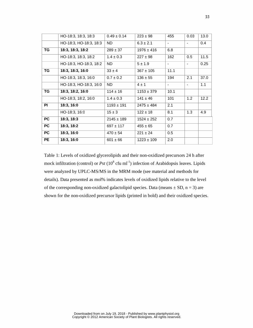
33
HO-18:3, 18:3, 18:3 0.49 ± 0.14 223 ± 98 455 0.03 13.0
HO-18:3, HO-18:3, 18:3 ND 6.3 ± 2.1 - - 0.4
TG 18:3, 18:3, 18:2 289 ± 37 1976 ± 416 6.8
HO-18:3, 18:3, 18:2 1.4 ± 0.3 227 ± 98 162 0.5 11.5
HO-18:3, HO-18:3, 18:2 ND 5 ± 1.9 - - 0.25
TG 18:3, 18:3, 16:0 33 ± 4 367 ± 105 11.1
HO-18:3, 18:3, 16:0 0.7 ± 0.2 136 ± 55 194 2.1 37.0
HO-18:3, HO-18:3, 16:0 ND 4 ± 1 - - 1.1
TG 18:3, 18:2, 16:0 114 ± 16 1153 ± 379 10.1
HO-18:3, 18:2, 16:0 1.4 ± 0.3 141 ± 46 101 1.2 12.2
PI 18:3, 16:0 1193 ± 191 2475 ± 484 2.1
HO-18:3, 16:0 15 ± 3 122 ± 18 8.1 1.3 4.9
PC 18:3, 18:3 2145 ± 189 1524 ± 252 0.7
PC 18:3, 18:2 697 ± 117 455 ± 65 0.7
PC 18:3, 16:0 470 ± 54 221 ± 24 0.5
PE 18:3, 16:0 601 ± 66 1223 ± 109 2.0
Table 1: Levels of oxidized glycerolipids and their non-oxidized precursors 24 h after
mock infiltration (control) or Pst (108 cfu ml-1) infection of Arabidopsis leaves. Lipids
were analyzed by UPLC-MS/MS in the MRM mode (see material and methods for
details). Data presented as mol% indicates levels of oxidized lipids relative to the level
of the corresponding non-oxidized galactolipid species. Data (means ± SD, n = 3) are
shown for the non-oxidized precursor lipids (printed in bold) and their oxidized species.
www.plantphysiol.orgon July 19, 2018 - Published by Downloaded from Copyright © 2012 American Society of Plant Biologists. All rights reserved.
34
FGURE LEGENDS
Figure 1. Fatty acid fragmentation pathways.
A, enzymatic oxidative fragmentation of 18:3 via 9-lipoxygenase (9-LOX), 9-
hydroperoxide lyase (HPL) and aldehyde dehydrogenase (ADH) yielding 9-oxononeate
(ONA), nonadienal (NDE) and azeleate (AZA) (Matsui, 2006). Grey bars represent the
carbohydrogen backbone of fatty acids. B, radical-catalyzed oxidative fragmentation of
esterified 18:2 in glycerolipids via the dimer pathway yielding oxidized glycerolipids
(GL), ONA, AZA and hydroperoxynonenal (HPNE) in animals (Schneider et al., 2008).
C, structures of predicted carboxylic acidic fragments generated through the dimer
pathway from different plant polyunsaturated fatty acids: ONA, AZA, 7-oxoheptanoate
(OHA), PIM.
Figure 2. Pathogen induced accumulation of oxidized free fatty acids in A. thaliana
leaves.
Levels of AZA and PIM together with levels of markers of non-enzymatic lipid
oxidation (16-hydroxyoctadecatrienoic acid, 16-HO-18:3) and enzymatic lipid oxidation
(jasmonic acid, JA) are shown after infection with avirulent (�) and virulent (�) Pst
(108 cfu ml-1) or mock infiltration (�). Shown are means ± SD, n = 3.
Figure 3. Pathogen-induced increase of AZA and PIM in A. thaliana.
Levels of AZA and PIM were determined 24 h after infection of A. thaliana leaves with
avirulent Pst (108 cfu ml-1) or mock infiltration (C). A, the single (lox1 and lox5) and
double (lox1/5) 9-LOX insertion lines were compared with the corresponding WT
(accession Wassilewskija, Ws). B, the mutant lines lox2, the fatty acid desaturase triple
fad3 fad7 fad8 (fad3/7/8) and azi1 were compared with the respective WT ecotype
Columbia (Col-0). Shown are means ± SD, n = 3.
Figure 4. Metabolism of ONA and AZA.
A. thaliana seedlings were grown in liquid medium for 10 d. A, after exogenous
application of [8,8-D2]ONA, endogenous levels of free ONA, OHA, AZA and PIM
were determined. B, after exogenous application of AZA, levels of free ONA, OHA,
www.plantphysiol.orgon July 19, 2018 - Published by Downloaded from Copyright © 2012 American Society of Plant Biologists. All rights reserved.
35
AZA and PIM as well as levels of oxidized (18:3,18:3)MGDG containing esterified
ONA and AZA were measured (mean ± SD, n =3).
Figure 5. Mechanisms of 18:3 oxidation in A. thaliana leaves after infection with
avirulent Pst.
Levels of esterified and free HO(O)-18:3 generated by 1O2 -oxidation, free radical
catalysis, and 9-LOX or 13-LOX catalysis are shown for A, WT, and B, lox2 mutant
plants, under control conditions (mock infiltration) and 24 h after infection with
avirulent Pst (108 cfu ml-1). Note different scale of the vertical axis as indicated. Shown
are means ± SD, n = 3.
Figure 6. LOX2-dependent lipid accumulation and oxidation after infection with
avirulent Pst.
Arabidopsis leaves of wild type (black bars) and lox2 (white bars) plants were infected
with avirulent Pst (108 cfu ml-1) (+) or mock infiltrated (-). Lipids were extracted 24 h
after infection and analyzed by UPLC-MS/MS. Arrows indicate putative precursor-
product relationships. Means ± SD of three independent experiments are shown.
A, levels of the most abundant non-oxidized MGDG and DGDG species increased after
infection in both genotypes.
B, after infection, levels of monooxygenated (at the acyl chain in sn1 or sn2 position)
MGDG and DGDG species increased in a LOX2 independent manner while the
dioxygenated (at the acyl chains in sn1 and sn2 position) species accumulated in a
LOX2-dependent fashion.
C, arabidopsides (Ara A – G) were only detected in wild type plants. Levels of
arabidopsides strongly increased after infection.
D, levels of non-esterified 18:3 oxidation products. A strong over-accumulation of the
9-LOX product HO(O)-18:3 was observed in lox2 mutant leaves while levels of 13-
LOX products (13-HO(O)-18:3, OPDA and JA) increased in a LOX2-independent
manner.
Figure 7. Lipid peroxidation and fragmentation in plastids (model).
www.plantphysiol.orgon July 19, 2018 - Published by Downloaded from Copyright © 2012 American Society of Plant Biologists. All rights reserved.

36
A, in the light continuous singlet oxygen mediated oxidation of MGDG close to PSII
generates a pool of oxidation sensitive MGDG hydroperoxides.
B, Pathogen-stress induced inhibition of PSII in parallel with LOX2 activation increases
MGDG peroxidation mediated by 1O2 (MGDG monohydroperoxides) and LOX2
(MGDG dihydroperoxides), respectively. MGDG dihydroperoxides are rapidly
converted into arabidopsides. MGDG monoperoxides are sensitive to radical-catalyzed
formation of peroxide dimers that spontaneously break down thereby generating
oxidized MGDG and MGDG core aldehydes and acids that remain in the membrane as
well as reactive electrophiles (RES) and radicals that are released instantaneously from
the membrane. Mild membrane damage may stimulate repair and protection
mechanisms through reactive lipid and oxygen species. However, excessive membrane
damage caused by amplification of lipid peroxidation contributes to cell death. During
membrane turnover and repair oxidized MGDG, core aldehydes and core acids are
hydrolyzed thereby releasing preformed biologically active oxylipins, oxoacids and
diacids. Finally, oxidized lipids are replaced by de novo synthesis of lipids.
www.plantphysiol.orgon July 19, 2018 - Published by Downloaded from Copyright © 2012 American Society of Plant Biologists. All rights reserved.
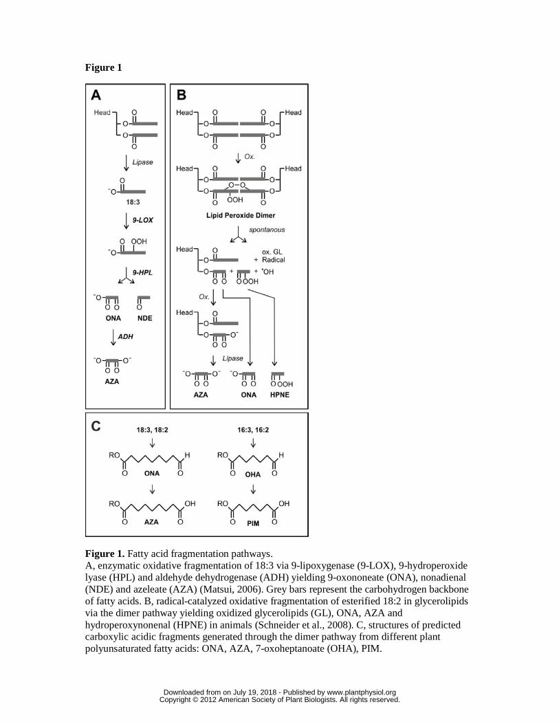
Figure 1
Figure 1. Fatty acid fragmentation pathways. A, enzymatic oxidative fragmentation of 18:3 via 9-lipoxygenase (9-LOX), 9-hydroperoxide lyase (HPL) and aldehyde dehydrogenase (ADH) yielding 9-oxononeate (ONA), nonadienal (NDE) and azeleate (AZA) (Matsui, 2006). Grey bars represent the carbohydrogen backbone of fatty acids. B, radical-catalyzed oxidative fragmentation of esterified 18:2 in glycerolipids via the dimer pathway yielding oxidized glycerolipids (GL), ONA, AZA and hydroperoxynonenal (HPNE) in animals (Schneider et al., 2008). C, structures of predicted carboxylic acidic fragments generated through the dimer pathway from different plant polyunsaturated fatty acids: ONA, AZA, 7-oxoheptanoate (OHA), PIM.
www.plantphysiol.orgon July 19, 2018 - Published by Downloaded from Copyright © 2012 American Society of Plant Biologists. All rights reserved.

Figure 2
Figure 2. Pathogen induced accumulation of oxidized free fatty acids in A. thaliana leaves. Levels of AZA and PIM together with levels of markers of non-enzymatic lipid oxidation (16-hydroxyoctadecatrienoic acid, 16-HO-18:3) and enzymatic lipid oxidation (jasmonic acid, JA) are shown after infection with avirulent (�) and virulent (�) Pst (108 cfu ml-1) or mock infiltration (�). Shown are means ± SD, n = 3.
www.plantphysiol.orgon July 19, 2018 - Published by Downloaded from Copyright © 2012 American Society of Plant Biologists. All rights reserved.
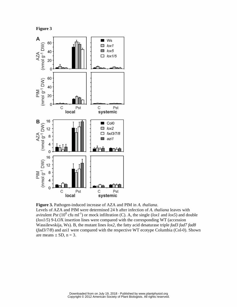
Figure 3
Figure 3. Pathogen-induced increase of AZA and PIM in A. thaliana. Levels of AZA and PIM were determined 24 h after infection of A. thaliana leaves with avirulent Pst (108 cfu ml-1) or mock infiltration (C). A, the single (lox1 and lox5) and double (lox1/5) 9-LOX insertion lines were compared with the corresponding WT (accession Wassilewskija, Ws). B, the mutant lines lox2, the fatty acid desaturase triple fad3 fad7 fad8 (fad3/7/8) and azi1 were compared with the respective WT ecotype Columbia (Col-0). Shown are means ± SD, n = 3.
www.plantphysiol.orgon July 19, 2018 - Published by Downloaded from Copyright © 2012 American Society of Plant Biologists. All rights reserved.
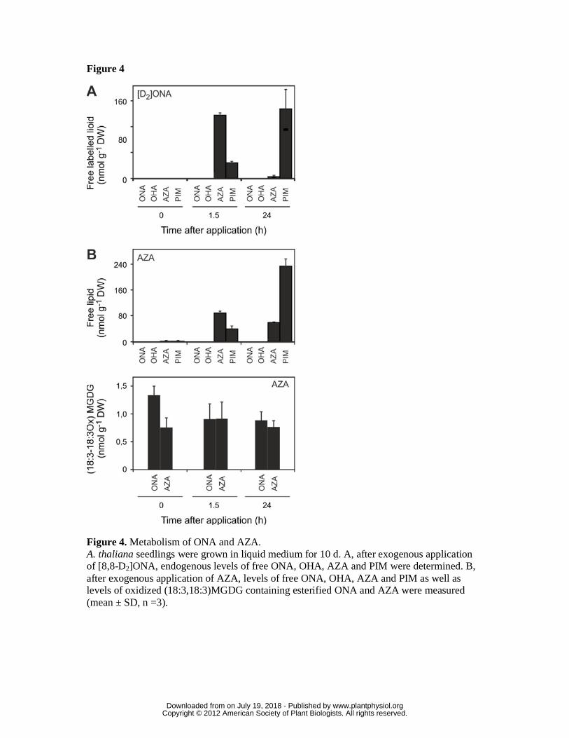
Figure 4
Figure 4. Metabolism of ONA and AZA. A. thaliana seedlings were grown in liquid medium for 10 d. A, after exogenous application of [8,8-D2]ONA, endogenous levels of free ONA, OHA, AZA and PIM were determined. B, after exogenous application of AZA, levels of free ONA, OHA, AZA and PIM as well as levels of oxidized (18:3,18:3)MGDG containing esterified ONA and AZA were measured (mean ± SD, n =3).
www.plantphysiol.orgon July 19, 2018 - Published by Downloaded from Copyright © 2012 American Society of Plant Biologists. All rights reserved.
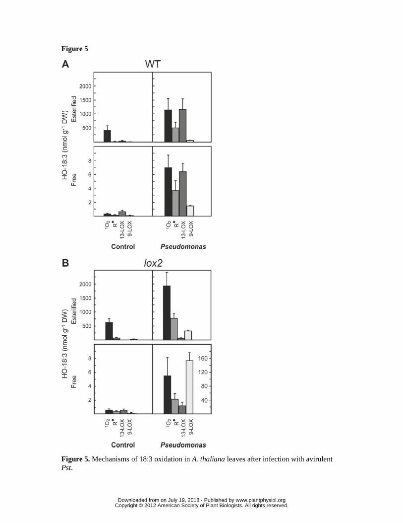
Figure 5
Figure 5. Mechanisms of 18:3 oxidation in A. thaliana leaves after infection with avirulent Pst.
www.plantphysiol.orgon July 19, 2018 - Published by Downloaded from Copyright © 2012 American Society of Plant Biologists. All rights reserved.
Levels of esterified and free HO(O)-18:3 generated by 1O2 -oxidation, free radical catalysis, and 9-LOX or 13-LOX catalysis are shown for A, WT, and B, lox2 mutant plants, under control conditions (mock infiltration) and 24 h after infection with avirulent Pst (108 cfu ml-1). Note different scale of the vertical axis as indicated. Shown are means ± SD, n = 3.
www.plantphysiol.orgon July 19, 2018 - Published by Downloaded from Copyright © 2012 American Society of Plant Biologists. All rights reserved.
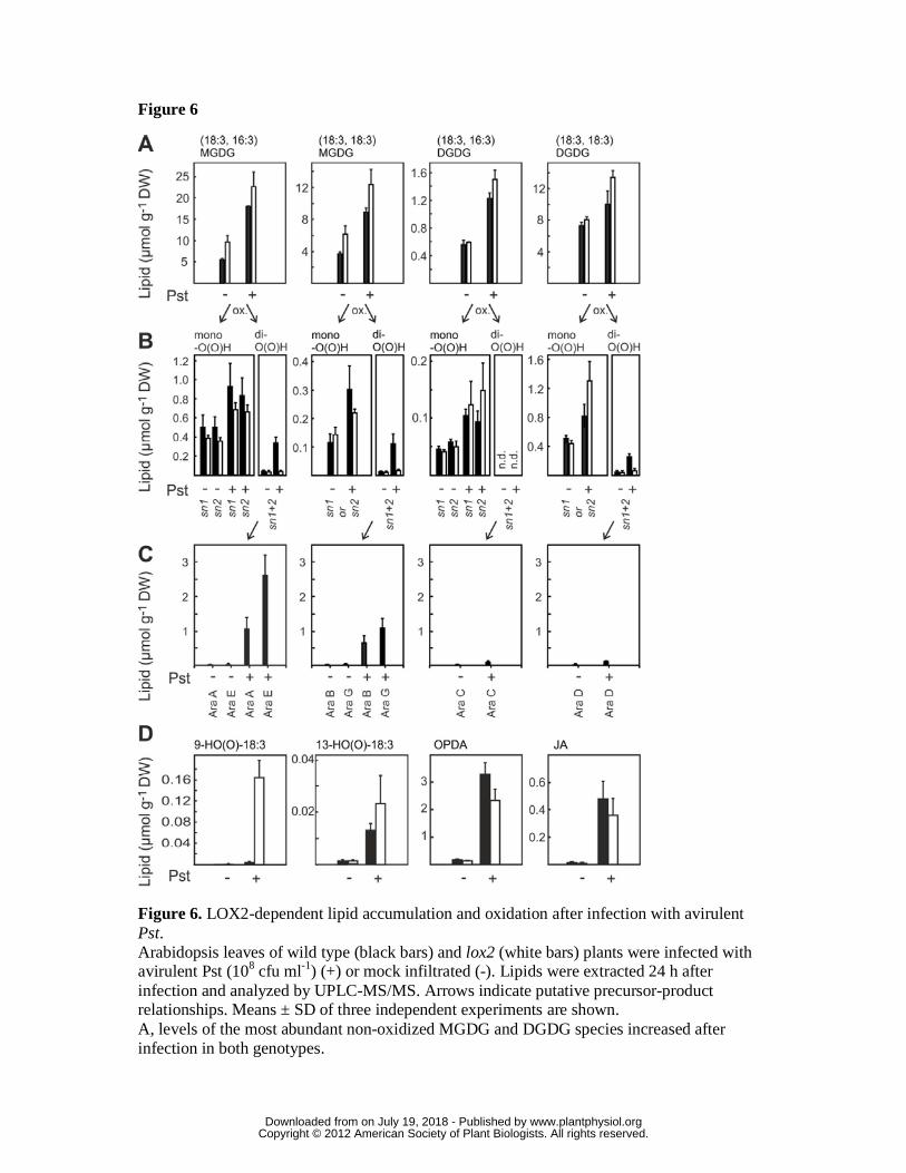
Figure 6
Figure 6. LOX2-dependent lipid accumulation and oxidation after infection with avirulent Pst. Arabidopsis leaves of wild type (black bars) and lox2 (white bars) plants were infected with avirulent Pst (108 cfu ml-1) (+) or mock infiltrated (-). Lipids were extracted 24 h after infection and analyzed by UPLC-MS/MS. Arrows indicate putative precursor-product relationships. Means ± SD of three independent experiments are shown. A, levels of the most abundant non-oxidized MGDG and DGDG species increased after infection in both genotypes.
www.plantphysiol.orgon July 19, 2018 - Published by Downloaded from Copyright © 2012 American Society of Plant Biologists. All rights reserved.
B, after infection, levels of monooxygenated (at the acyl chain in sn1 or sn2 position) MGDG and DGDG species increased in a LOX2 independent manner while the dioxygenated (at the acyl chains in sn1 and sn2 position) species accumulated in a LOX2-dependent fashion. C, arabidopsides (Ara A – G) were only detected in wild type plants. Levels of arabidopsides strongly increased after infection. D, levels of non-esterified 18:3 oxidation products. A strong over-accumulation of the 9-LOX product HO(O)-18:3 was observed in lox2 mutant leaves while levels of 13-LOX products (13-HO(O)-18:3, OPDA and JA) increased in a LOX2-independent manner.
www.plantphysiol.orgon July 19, 2018 - Published by Downloaded from Copyright © 2012 American Society of Plant Biologists. All rights reserved.
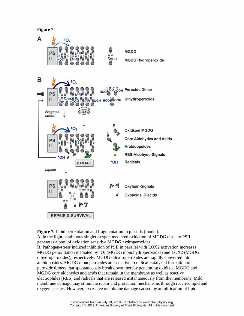
Figure 7
Figure 7. Lipid peroxidation and fragmentation in plastids (model). A, in the light continuous singlet oxygen mediated oxidation of MGDG close to PSII generates a pool of oxidation sensitive MGDG hydroperoxides. B, Pathogen-stress induced inhibition of PSII in parallel with LOX2 activation increases MGDG peroxidation mediated by 1O2 (MGDG monohydroperoxides) and LOX2 (MGDG dihydroperoxides), respectively. MGDG dihydroperoxides are rapidly converted into arabidopsides. MGDG monoperoxides are sensitive to radical-catalyzed formation of peroxide dimers that spontaneously break down thereby generating oxidized MGDG and MGDG core aldehydes and acids that remain in the membrane as well as reactive electrophiles (RES) and radicals that are released instantaneously from the membrane. Mild membrane damage may stimulate repair and protection mechanisms through reactive lipid and oxygen species. However, excessive membrane damage caused by amplification of lipid
www.plantphysiol.orgon July 19, 2018 - Published by Downloaded from Copyright © 2012 American Society of Plant Biologists. All rights reserved.
peroxidation contributes to cell death. During membrane turnover and repair oxidized MGDG, core aldehydes and core acids are hydrolyzed thereby releasing preformed biologically active oxylipins, oxoacids and diacids. Finally, oxidized lipids are replaced by de novo synthesis of lipids.
www.plantphysiol.orgon July 19, 2018 - Published by Downloaded from Copyright © 2012 American Society of Plant Biologists. All rights reserved.