Embed Size (px)
Citation preview
RESEARCH Open Access
Safety and efficacy of VisuMax® circlepatterns for flap creation and enhancementfollowing small incision lenticule extractionEkktet Chansue1*, Morakot Tanehsakdi1, Sukanda Swasdibutra1 and Colm McAlinden2,3
Abstract
Background: The purpose of this case series is to evaluate the safety and efficacy of VisuMax® Circle patterns ineyes that have undergone small incision lenticule extraction, thus creating a flap to perform an enhancementprocedure or residual lenticule extraction.
Methods: This prospective, single center, case study series evaluated the use of a VisuMax® Circle pattern to createa corneal flap following small incision lenticule extraction. Patients were treated and followed at TRSC InternationalLASIK Center (Bangkok, Thailand) for 3 months to assess the efficacy and safety of the procedure. Efficacy wasdetermined by the surgeon’s ability to lift the created corneal flap.
Results: The study enrolled 28 eyes. Twenty-seven underwent the VisuMax® Circle pattern procedure for refractiveenhancement, and one for residual lenticule extraction. In 100 % of cases (28 eyes) the lifting of the flap was possible,as planned. In all cases of refractive enhancement (27 eyes) by laser in situ keratomileusis (LASIK), the exposure of thestromal bed was sufficient for the necessary excimer laser ablation. No eyes lost two or more Snellen lines of correcteddistance visual acuity (CDVA) and no procedure or flap-related complications or serious adverse events occurred.
Conclusions: This initial case series demonstrates that VisuMax® Circle pattern is efficacious and a suitable method tocreate a corneal flap for enhancement, following small incision lenticule extraction.
Keywords: Small incision lenticule extraction, SMILE, Lenticule, Residual lenticule extraction, Flap creation, Circlepattern, Refractive enhancement, Femtosecond laser, Refractive surgery
BackgroundNew technologies such as femtosecond lasers are creating aparadigm shift in the surgeon’s ability to perform refractivecorrection with improved results [1–5]. The developmentof small incision lenticule extraction (SMILE) as a new,flapless procedure has been a major innovation in cornealrefractive surgery. This technique, performed usingRefractive Lenticule Extraction (ReLEx®) on the VisuMax®(Carl Zeiss Meditec, Jena, Germany) platform, allows forrefractive correction without the need to create a cornealflap. Rather, a small side cut incision, less than 4 mm insize is created at an approximate depth of 80 to 160 μm inthe cornea for lenticule extraction. This gives access to theintrastromal pocket created by the preceding lenticule
cuts. By creating an intrastromal pocket rather than thetraditional flap, surgeons can eliminate associated compli-cations including incomplete and irregular flap cuts, thinflaps, buttonholes, and free caps.In the past, enhancement following ReLEx® SMILE was a
challenge and a number of possible approaches have beenconsidered. Surgeons may contemplate surface ablation ifsuch ablation does not reach the pocket interface. An intra-ocular lens could be inserted to correct a larger refractiveerror if the residual stroma is inadequate for photoablation.Other options for the surgeon can either be laser in situkeratomileusis (LASIK), anterior to the SMILE pocket oran additional SMILE procedure, anterior (or posterior,depending on how deep the SMILE pocket is situated) tothe initial one. Carl Zeiss Meditec has recently provided anadditional option with the development of a series of fourcircle patterns, programmed within the VisuMax® platform,
* Correspondence: [email protected] International LASIK Center, 6th Floor, U Chu Liang Boulevard, 968Rama 4 Road, Bangkok, ThailandFull list of author information is available at the end of the article
© 2015 Chansue et al. Open Access This article is distributed under the terms of the Creative Commons Attribution 4.0International License (http://creativecommons.org/licenses/by/4.0/), which permits unrestricted use, distribution, andreproduction in any medium, provided you give appropriate credit to the original author(s) and the source, provide a link tothe Creative Commons license, and indicate if changes were made. The Creative Commons Public Domain Dedication waiver(http://creativecommons.org/publicdomain/zero/1.0/) applies to the data made available in this article, unless otherwise stated.
Chansue et al. Eye and Vision (2015) 2:21 DOI 10.1186/s40662-015-0031-5
which can be utilized to create a corneal flap after previousrefractive correction with ReLEx® SMILE. This strategyallows for the original SMILE incision pocket to be con-verted into a LASIK-like flap that can be easily lifted to allowfor stromal ablation of the residual refractive error with anexcimer laser. Intrastromal incisions include the creation of alamellar ring, anterior, posterior or adjacent to the previousSMILE pocket cut; a side cut with a hinge and a junction cutfrom the inner edge of the lamellar ring to junction depths.In 2013, Riau et al. investigated the use of four differ-
ent VisuMax® circle patterns when they performed −6.00D spherical correction using ReLEx® SMILE on six NewZealand white rabbits (12 eyes), and then 28 days later,one of the four circle patterns was used on each of the 12eyes and evaluated for ease of flap lift [6]. In that animalmodel, it was determined that pattern D, a lamellar ringadjacent to the cap cut, was the most optimal pattern forflap creation, and ultimately, SMILE re-treatment [8].Here, we describe the four different VisuMax® circle pat-
terns, which have been programmed to create a cornealflap. We also present the efficacy and safety results for 28patients upon whom circle pattern D was utilized to createa flap for residual lenticule extraction or refractive en-hancement by stromal excimer laser ablation after ReLEx®SMILE. We also discuss the other applications for whichthe circle patterns can be utilized. To our knowledge, thisis the first report of the circle pattern use in human eyes.
MethodsPatientsThis prospective clinical case study consisted of partici-pants recruited from TRSC International LASIK Center.Each was provided written informed consent that ex-plained the details of the procedure and study protocol inaccordance with the principles of the Declaration ofHelsinki. According to the Food and Drug Administration(FDA) Thailand, this study was considered clinical qualitycontrol and evaluation under CE mark status and there-fore, ethical approval was not necessary. In order to beincluded, patients had to be a minimum of 18 years of age,had previously undergone ReLEx® SMILE and now had aresidual refractive error from undercorrection, overcorrec-tion or regression, including residual and/or consecutiveastigmatism, that necessitated planned circle pattern treat-ment and LASIK touch-up. Additionally, a patient withresidual lenticule following ReLEx® SMILE, which requiredextraction was also included. For patients with a residualrefractive error, the current refraction had to be stable forat least 3 months. The determination if the previousSMILE incision could be opened, depended on whetherone of the two interfaces above or below the lenticulecould successfully be separated. Patients had to have theability to attend postoperative assessment appointments at1 day, 1 week, 1 month, and 3 months. Patients were
excluded if they had ocular conditions, other than residualmyopia, consecutive hyperopia with or without residual orconsecutive astigmatism following the original ReLExSMILE procedure.
AssessmentsPreoperatively, all post-ReLEx SMILE patients underwenta complete eye examination, which included objective andmanifest refractions, visual acuity (ETDRS LogMAR chart),scotopic pupil size evaluation (Colvard Light-AmplificationPupillometer, Oasis Medical, San Dimas, CA), comput-erized corneal topography (Orbscan, Bausch & Lomb,Bridgewater, NJ), pachymetry (DGH Technology, Inc.,Exton, PA), wavefront analysis (Wasca Analyzer, Wave-front Sciences, Albuquerque, NM), keratometric mea-surements (Sim-K values from Orbscan), and slit-lampexamination.The first day after the circle enhancement procedure,
patients had their uncorrected visual acuity assessed, a slit-lamp evaluation performed, and any complications oradverse events examined. At each of the remaining postop-erative appointments (1 week, 1 month, 3 months) patientswere assessed for corrected distance visual acuity, uncor-rected visual acuity, objective and manifest refractions,computerized corneal topography, wavefront analysis,keratometric measurements, and slit-lamp examination.
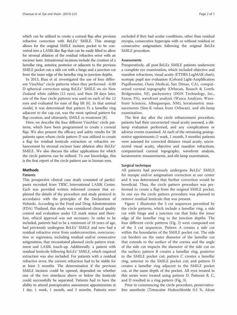
Surgical techniqueAll patients had previously undergone ReLEx® SMILEfor myopic and/or astigmatism correction at our centerand it was determined that further correction would bebeneficial. Thus, the circle pattern procedure was per-formed to create a flap from the original SMILE pocket.In one eye the circle pattern procedure was planned toremove residual lenticule that was present.Figure 1 illustrates the 3 cut sequences permitted by
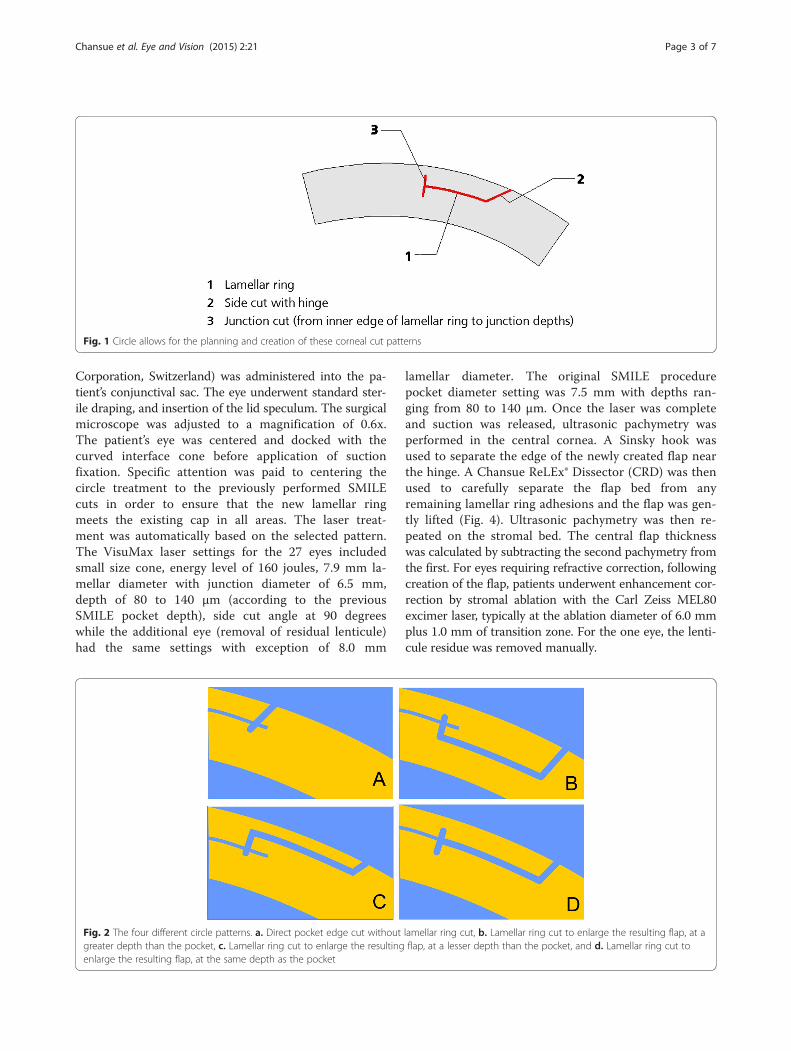
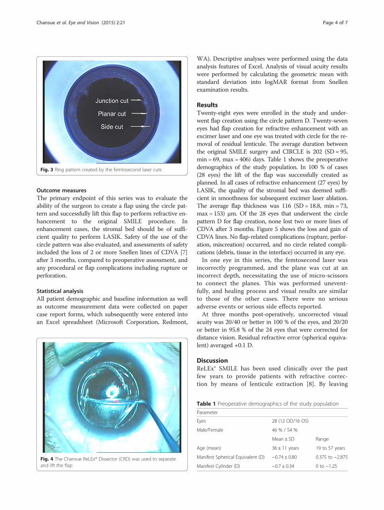
the circle patterns, which include a lamellar ring, a sidecut with hinge and a junction cut that links the inneredge of the lamellar ring to the junction depths. Thefour different circle patterns (Fig. 2) were composed outof the 3 cut sequences. Pattern A creates a side cutwithin the boundaries of the SMILE pocket cut. The sidecut borders on the outer diameter of the lamellar cutthat extends to the surface of the cornea and the angleof the side cut impacts the diameter of the side cut onthe surface; pattern B creates a lamellar ring, posteriorto the SMILE pocket cut; pattern C creates a lamellarring, anterior to the SMILE pocket cut; and pattern Dcreates a lamellar ring adjacent to the SMILE pocketcut, at the same depth of the pocket. All eyes treated inthis series were treated using pattern D. Patterns B, C,and D resulted in a ring pattern (Fig. 3).Prior to commencing the circle procedure, preservative-
free anesthetic (Tetracaine Hydrochloride 0.5 %, Alcon
Chansue et al. Eye and Vision (2015) 2:21 Page 2 of 7
Corporation, Switzerland) was administered into the pa-tient’s conjunctival sac. The eye underwent standard ster-ile draping, and insertion of the lid speculum. The surgicalmicroscope was adjusted to a magnification of 0.6x.The patient’s eye was centered and docked with thecurved interface cone before application of suctionfixation. Specific attention was paid to centering thecircle treatment to the previously performed SMILEcuts in order to ensure that the new lamellar ringmeets the existing cap in all areas. The laser treat-ment was automatically based on the selected pattern.The VisuMax laser settings for the 27 eyes includedsmall size cone, energy level of 160 joules, 7.9 mm la-mellar diameter with junction diameter of 6.5 mm,depth of 80 to 140 μm (according to the previousSMILE pocket depth), side cut angle at 90 degreeswhile the additional eye (removal of residual lenticule)had the same settings with exception of 8.0 mm
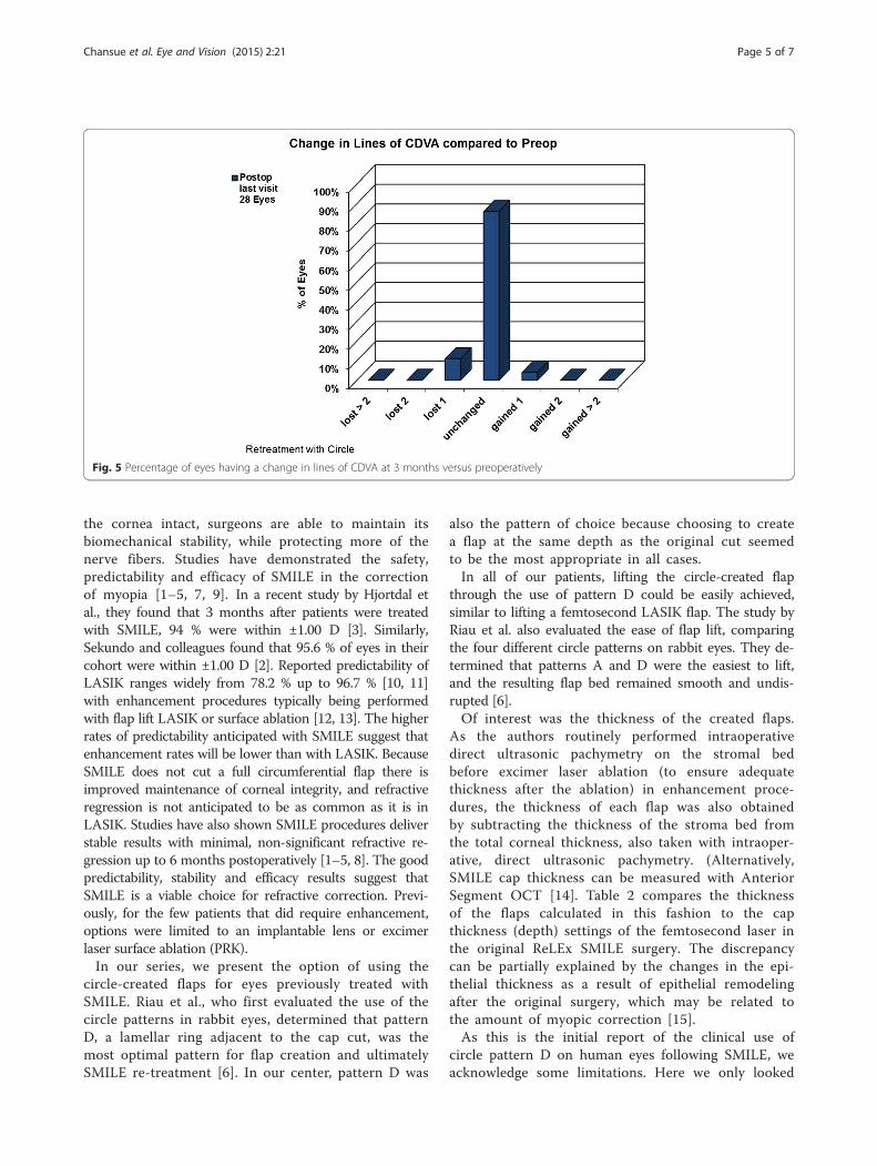
lamellar diameter. The original SMILE procedurepocket diameter setting was 7.5 mm with depths ran-ging from 80 to 140 μm. Once the laser was completeand suction was released, ultrasonic pachymetry wasperformed in the central cornea. A Sinsky hook wasused to separate the edge of the newly created flap nearthe hinge. A Chansue ReLEx® Dissector (CRD) was thenused to carefully separate the flap bed from anyremaining lamellar ring adhesions and the flap was gen-tly lifted (Fig. 4). Ultrasonic pachymetry was then re-peated on the stromal bed. The central flap thicknesswas calculated by subtracting the second pachymetry fromthe first. For eyes requiring refractive correction, followingcreation of the flap, patients underwent enhancement cor-rection by stromal ablation with the Carl Zeiss MEL80excimer laser, typically at the ablation diameter of 6.0 mmplus 1.0 mm of transition zone. For the one eye, the lenti-cule residue was removed manually.
Fig. 1 Circle allows for the planning and creation of these corneal cut patterns
Fig. 2 The four different circle patterns. a. Direct pocket edge cut without lamellar ring cut, b. Lamellar ring cut to enlarge the resulting flap, at agreater depth than the pocket, c. Lamellar ring cut to enlarge the resulting flap, at a lesser depth than the pocket, and d. Lamellar ring cut toenlarge the resulting flap, at the same depth as the pocket
Chansue et al. Eye and Vision (2015) 2:21 Page 3 of 7
Outcome measuresThe primary endpoint of this series was to evaluate theability of the surgeon to create a flap using the circle pat-tern and successfully lift this flap to perform refractive en-hancement to the original SMILE procedure. Inenhancement cases, the stromal bed should be of suffi-cient quality to perform LASIK. Safety of the use of thecircle pattern was also evaluated, and assessments of safetyincluded the loss of 2 or more Snellen lines of CDVA [7]after 3 months, compared to preoperative assessment, andany procedural or flap complications including rupture orperforation.
Statistical analysisAll patient demographic and baseline information as wellas outcome measurement data were collected on papercase report forms, which subsequently were entered intoan Excel spreadsheet (Microsoft Corporation, Redmont,
WA). Descriptive analyses were performed using the dataanalysis features of Excel. Analysis of visual acuity resultswere performed by calculating the geometric mean withstandard deviation into logMAR format from Snellenexamination results.
ResultsTwenty-eight eyes were enrolled in the study and under-went flap creation using the circle pattern D. Twenty-seveneyes had flap creation for refractive enhancement with anexcimer laser and one eye was treated with circle for the re-moval of residual lenticule. The average duration betweenthe original SMILE surgery and CIRCLE is 202 (SD = 95,min = 69, max = 406) days. Table 1 shows the preoperativedemographics of the study population. In 100 % of cases(28 eyes) the lift of the flap was successfully created asplanned. In all cases of refractive enhancement (27 eyes) byLASIK, the quality of the stromal bed was deemed suffi-cient in smoothness for subsequent excimer laser ablation.The average flap thickness was 116 (SD = 18.8, min = 73,max = 153) μm. Of the 28 eyes that underwent the circlepattern D for flap creation, none lost two or more lines ofCDVA after 3 months. Figure 5 shows the loss and gain ofCDVA lines. No flap-related complications (rupture, perfor-ation, miscreation) occurred, and no circle related compli-cations (debris, tissue in the interface) occurred in any eye.In one eye in this series, the femtosecond laser was
incorrectly programmed, and the plane was cut at anincorrect depth, necessitating the use of micro-scissorsto connect the planes. This was performed unevent-fully, and healing process and visual results are similarto those of the other cases. There were no seriousadverse events or serious side effects reported.At three months post-operatively, uncorrected visual
acuity was 20/40 or better in 100 % of the eyes, and 20/20or better in 95.8 % of the 24 eyes that were corrected fordistance vision. Residual refractive error (spherical equiva-lent) averaged +0.1 D.
DiscussionReLEx® SMILE has been used clinically over the pastfew years to provide patients with refractive correc-tion by means of lenticule extraction [8]. By leaving
Fig. 4 The Chansue ReLEx® Dissector (CRD) was used to separateand lift the flap
Table 1 Preoperative demographics of the study population
Parameter
Eyes 28 (12 OD/16 OS)
Male/Female 46 % / 54 %
Mean ± SD Range
Age (mean) 36 ± 11 years 19 to 57 years
Manifest Spherical Equivalent (D) −0.74 ± 0.80 0.375 to −2.875
Manifest Cylinder (D) −0.7 ± 0.34 0 to −1.25
Fig. 3 Ring pattern created by the femtosecond laser cuts
Chansue et al. Eye and Vision (2015) 2:21 Page 4 of 7
the cornea intact, surgeons are able to maintain itsbiomechanical stability, while protecting more of thenerve fibers. Studies have demonstrated the safety,predictability and efficacy of SMILE in the correctionof myopia [1–5, 7, 9]. In a recent study by Hjortdal etal., they found that 3 months after patients were treatedwith SMILE, 94 % were within ±1.00 D [3]. Similarly,Sekundo and colleagues found that 95.6 % of eyes in theircohort were within ±1.00 D [2]. Reported predictability ofLASIK ranges widely from 78.2 % up to 96.7 % [10, 11]with enhancement procedures typically being performedwith flap lift LASIK or surface ablation [12, 13]. The higherrates of predictability anticipated with SMILE suggest thatenhancement rates will be lower than with LASIK. BecauseSMILE does not cut a full circumferential flap there isimproved maintenance of corneal integrity, and refractiveregression is not anticipated to be as common as it is inLASIK. Studies have also shown SMILE procedures deliverstable results with minimal, non-significant refractive re-gression up to 6 months postoperatively [1–5, 8]. The goodpredictability, stability and efficacy results suggest thatSMILE is a viable choice for refractive correction. Previ-ously, for the few patients that did require enhancement,options were limited to an implantable lens or excimerlaser surface ablation (PRK).In our series, we present the option of using the
circle-created flaps for eyes previously treated withSMILE. Riau et al., who first evaluated the use of thecircle patterns in rabbit eyes, determined that patternD, a lamellar ring adjacent to the cap cut, was themost optimal pattern for flap creation and ultimatelySMILE re-treatment [6]. In our center, pattern D was
also the pattern of choice because choosing to createa flap at the same depth as the original cut seemedto be the most appropriate in all cases.In all of our patients, lifting the circle-created flap
through the use of pattern D could be easily achieved,similar to lifting a femtosecond LASIK flap. The study byRiau et al. also evaluated the ease of flap lift, comparingthe four different circle patterns on rabbit eyes. They de-termined that patterns A and D were the easiest to lift,and the resulting flap bed remained smooth and undis-rupted [6].Of interest was the thickness of the created flaps.
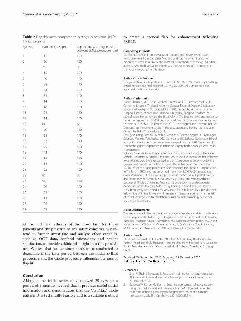
As the authors routinely performed intraoperativedirect ultrasonic pachymetry on the stromal bedbefore excimer laser ablation (to ensure adequatethickness after the ablation) in enhancement proce-dures, the thickness of each flap was also obtainedby subtracting the thickness of the stroma bed fromthe total corneal thickness, also taken with intraoper-ative, direct ultrasonic pachymetry. (Alternatively,SMILE cap thickness can be measured with AnteriorSegment OCT [14]. Table 2 compares the thicknessof the flaps calculated in this fashion to the capthickness (depth) settings of the femtosecond laser inthe original ReLEx SMILE surgery. The discrepancycan be partially explained by the changes in the epi-thelial thickness as a result of epithelial remodelingafter the original surgery, which may be related tothe amount of myopic correction [15].As this is the initial report of the clinical use of
circle pattern D on human eyes following SMILE, weacknowledge some limitations. Here we only looked
Fig. 5 Percentage of eyes having a change in lines of CDVA at 3 months versus preoperatively
Chansue et al. Eye and Vision (2015) 2:21 Page 5 of 7
at the technical efficacy of the procedure for thesepatients and the presence of any safety concerns. We in-tend to further investigate and analyze other variables,such as OCT data, confocal microscopy and patientsatisfaction, to provide additional insight into this proced-ure. We feel that further study needs to be conducted todetermine if the time period between the initial SMILEprocedure and the Circle procedure influences the ease offlap lift.
ConclusionAlthough this initial series only followed 28 eyes for aperiod of 3 months, we feel that it provides useful initialinformation and demonstrates that the VisuMax® circlepattern D is technically feasible and is a suitable method
to create a corneal flap for enhancement followingSMILE.
Competing interestsDr. Ekktet Chansue is an investigator (unpaid) and has received travelreimbursement from Carl Zeiss Meditec, and has no other financial orproprietary interest in any of the material or methods mentioned. All otherauthors have no financial or proprietary interest in any of the material ormethods mentioned in this study.
Authors’ contributionsDesign, analysis or interpretation of data (EC, MT, SS, CMA). Manuscript drafting,critical revision and final approval (EC, MT, SS, CMA). All authors read andapproved the final manuscript.
Authors’ informationEkktet Chansue, M.D. is the Medical Director of TRSC International LASIKCenter in Bangkok, Thailand. After his Cornea, External Disease & RefractiveSurgery fellowship in St. Louis, MO, in 1993, he taught at the RamathibodiHospital Faculty of Medicine, Mahidol University, Bangkok, Thailand, forseveral years. He performed the first LASIK in Thailand in 1994, and has sinceperformed more than 30,000 LASIK procedures. Dr. Chansue also performedthe first ReLEx® SMILE in Thailand in 2010. He designed the Chansue ReLEx®Dissector, an instrument to aid in the separation and freeing the lenticuleduring the ReLEx® procedure (NFI).After graduating from UCLA with a Bachelor of Science degree in PhysiologicalSciences, Morakot Tanehsakdi, O.D., went on to UC Berkeley Optometry Schoolfor doctor of optometry degree, where she graduated in 2004. Since then, Dr.Tanehsakdi gained experience in refractive surgery both clinically as well as inmanagement.Sukanda Swasdibutra, M.D. graduated from Siriraj Hospital Faculty of Medicine,Mahidol University in Bangkok, Thailand, where she also completed her residencyin ophthalmology. She is recognized as the first surgeon to perform LASIK in agovernment hospital in Thailand. Dr. Swasdibutra has performed more than10,000 refractive surgery procedures. She pioneered the Phakic IOL implantationin Thailand in 2006, and has performed more than 1,000 ReLEx® procedures.Colm McAlinden, PhD is a visiting professor at the School of Ophthalmologyand Optometry, Wenzhou Medical University, China, and Visiting AdjunctLecturer at Flinders University, Australia. He undertook his undergraduatedegree at Cardiff University followed by training in Moorfields Eye Hospital.He subsequently completed a Masters and a Ph.D., followed by a postdoctoralfellowship at Flinders University. His research interests are primarily in the fieldof refractive surgery, instrumentation evaluation, ophthalmology outcomesresearch, and statistics.
AcknowledgementsThe authors would like to thank and acknowledge the valuable contributionsto this paper of the following colleagues at TRSC International LASIK Center,Bangkok, Thailand: Pavika Thammano, MD, Sabong Srivannaboon, MD, PichitNaripthaphan, MD, Suchai Woraamornchot, MD, Varintorn Chuckpaiwong,MD, Chareenun Chirapapaisan, MD, and Pimsiri Chamanan, MD.
Author details1TRSC International LASIK Center, 6th Floor, U Chu Liang Boulevard, 968Rama 4 Road, Bangkok, Thailand. 2Flinders University, Bedford Park, Adelaide,South Australia, Australia. 3Wenzhou Medical College, Wenzhou, Zhejiang,China.
Received: 24 September 2015 Accepted: 11 December 2015
References1. Shah R, Shah S, Sengupta S. Results of small incision lenticule extraction:
All-in-one femtosecond laser refractive surgery. J Cataract Refract Surg.2011;37(1):127–37.
2. Sekundo W, Kunert KS, Blum M. Small incision corneal refractive surgeryusing the small incision lenticule extraction (SMILE) procedure for thecorrection of myopia and myopic astigmatism: results of a 6 monthprospective study. Br J Ophthalmol. 2011;95(3):335–9.
Table 2 Flap thickness compared to settings in previous ReLExSMILE surgeries
Eye No. Flap thickness (μm) Cap thickness setting in theprevious SMILE procedure (μm)
1 111 100
2 126 120
3 73 80
4 115 100
5 146 140
6 138 140
7 104 100
8 153 140
9 114 100
10 116 100
11 123 120
12 114 100
13 84 80
14 120 120
15 135 140
16 132 140
17 125 100
18 143 140
19 118 120
20 113 100
21 122 120
22 86 80
23 90 100
24 106 100
25 109 100
26 113 100
27 106 100
28 125 120
Chansue et al. Eye and Vision (2015) 2:21 Page 6 of 7
3. Vestergaard A, Ivarsen AR, Asp S, Hjortdal JØ. Small-incision lenticuleextraction for moderate to high myopia: Predictability, safety, and patientsatisfaction. J Cataract Refract Surg. 2012;38(11):2003–10.
4. Blum M, Kunert K, Schreoder M, Sekundo W. Femtosecond lenticuleextraction for the correction of myopia: Preliminary 6-month results. GraefesArch Clin Exp Ophthalmol. 2010;248(7):1019–27.
5. Chansue E, Tanehsakdi M, Swasdibutra S, McAlinden C. Efficacy, predictabilityand safety of small incision lenticule extraction (SMILE). Eye Vis (Lond). 2015;2:14.
6. Riau AK, Ang HP, Lwin NC, Chaurasia SS, Tan DT, Mehta JS. Comparison offour different VisuMax circle patterns for flap creation after small incisionlenticule extraction. J Refract Surg. 2013;29(4):236–44.
7. Waring GO, Reinstein DZ, Dupps WJ, Kohnen T, Mamalis N, Rosen ES, et al.Standardized graphs and terms for refractive surgery results. J Refract Surg.2011;27(1):7–9.
8. Reinstein DZ, Archer TJ, Gobbe M. Small incision lenticule extraction (SMILE)history, fundamentals of a new refractive surgery technique and clinicaloutcomes. Eye Vis (Lond). 2014;1:3.
9. Hjortdal JØ, Vestergaard AH, Ivarsen A, Ragunathan S, Asp S. Predictors forthe outcome of small incision lenticule extraction for Myopia. J Refract Surg.2012;28(12):865–71.
10. Dada T, Sudan R, Sinha R, Ray M, Sethi H, Vajpayee RB. Results of laser insitu keratomileusis for myopia of −10 to −19 diopters with a Technolas 217laser. J Refract Surg. 2003;19(1):44–7.
11. Sugar A, Rapuano CJ, Culbertson WW, Huang D, Varley GA, Agapitos PJ, etal. Laser in situ keratomileusis for myopia and astigmatism: safety andefficacy: a report by the American Academy of Ophthalmology.Ophthalmology. 2002;109(1):175–87.
12. McAlinden C, Moore JE. Retreatment of residual refractive errors with flaplift laser in situ keratomileusis. Eur J Ophthalmol. 2011;21(1):5–11.
13. McAlinden C, Moore J. Laser-assisted subepithelial keratectomy retreatmentsurgery. J Cataract Refract Surg. 2011;37(2):358–63.
14. Kanellopoulos AJ, Georgiadou S, Asimellis G. Objective Evaluation ofPlanned Versus Achieved Stromal Thickness Reduction in MyopicFemtosecond Laser-assisted LASIK. Refract Surg. 2015;31(9):628–32.
15. Kanellopoulos AJ, Asimellis G. Longitudinal postoperative lasik epithelialthickness profile changes in correlation with degree of myopia correction.J Refract Surg. 2014;30(3):166–71.
• We accept pre-submission inquiries
• Our selector tool helps you to find the most relevant journal
• We provide round the clock customer support
• Convenient online submission
• Thorough peer review
• Inclusion in PubMed and all major indexing services
• Maximum visibility for your research
Submit your manuscript atwww.biomedcentral.com/submit
Submit your next manuscript to BioMed Central and we will help you at every step:
Chansue et al. Eye and Vision (2015) 2:21 Page 7 of 7