Embed Size (px)
Citation preview
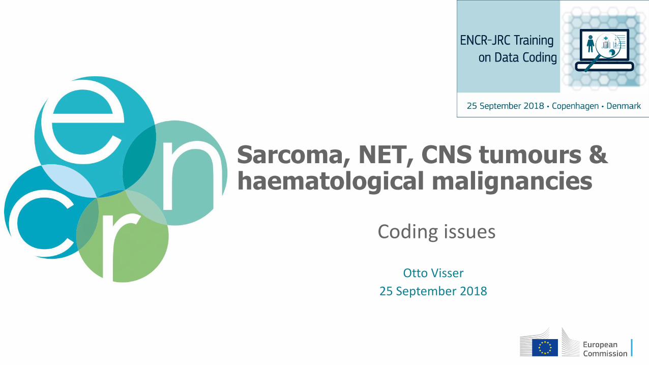
Sarcoma, NET, CNS tumours & haematological malignancies
Otto Visser
25 September 2018
Coding issues

Introduction
• For most (solid) cancers, the primary site of the most important factor for the prognosis and the choice of treatment
• For other cancers, especially haematological malignancies, but also for an increasing number of solid cancers, the morphological classification is the most important factor
How is a cancer diagnosis made?
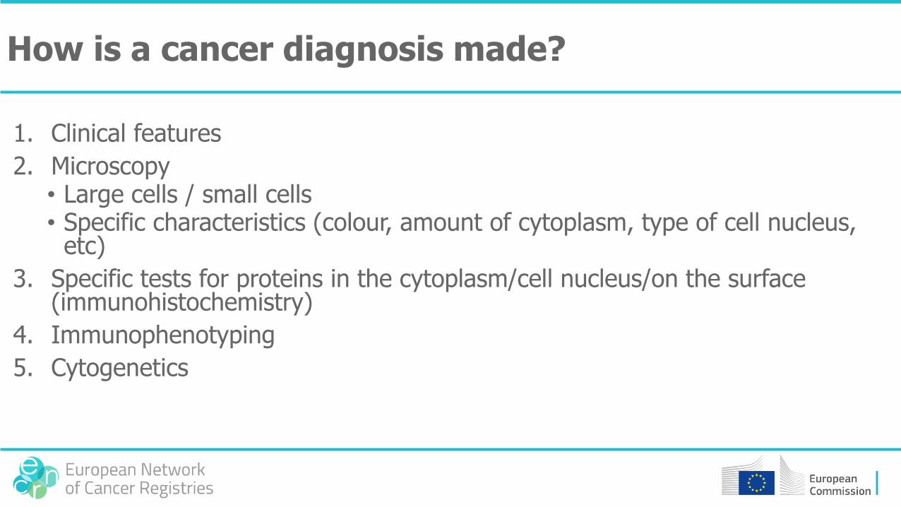
1. Clinical features
2. Microscopy• Large cells / small cells• Specific characteristics (colour, amount of cytoplasm, type of cell nucleus,
etc)
3. Specific tests for proteins in the cytoplasm/cell nucleus/on the surface (immunohistochemistry)
4. Immunophenotyping
5. Cytogenetics
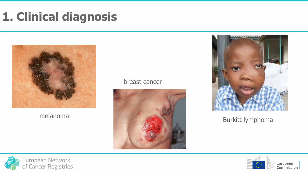
1. Clinical diagnosis
melanoma
breast cancer
Burkitt lymphoma
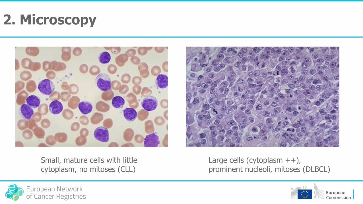
2. Microscopy
Small, mature cells with little cytoplasm, no mitoses (CLL)
Large cells (cytoplasm ++), prominent nucleoli, mitoses (DLBCL)
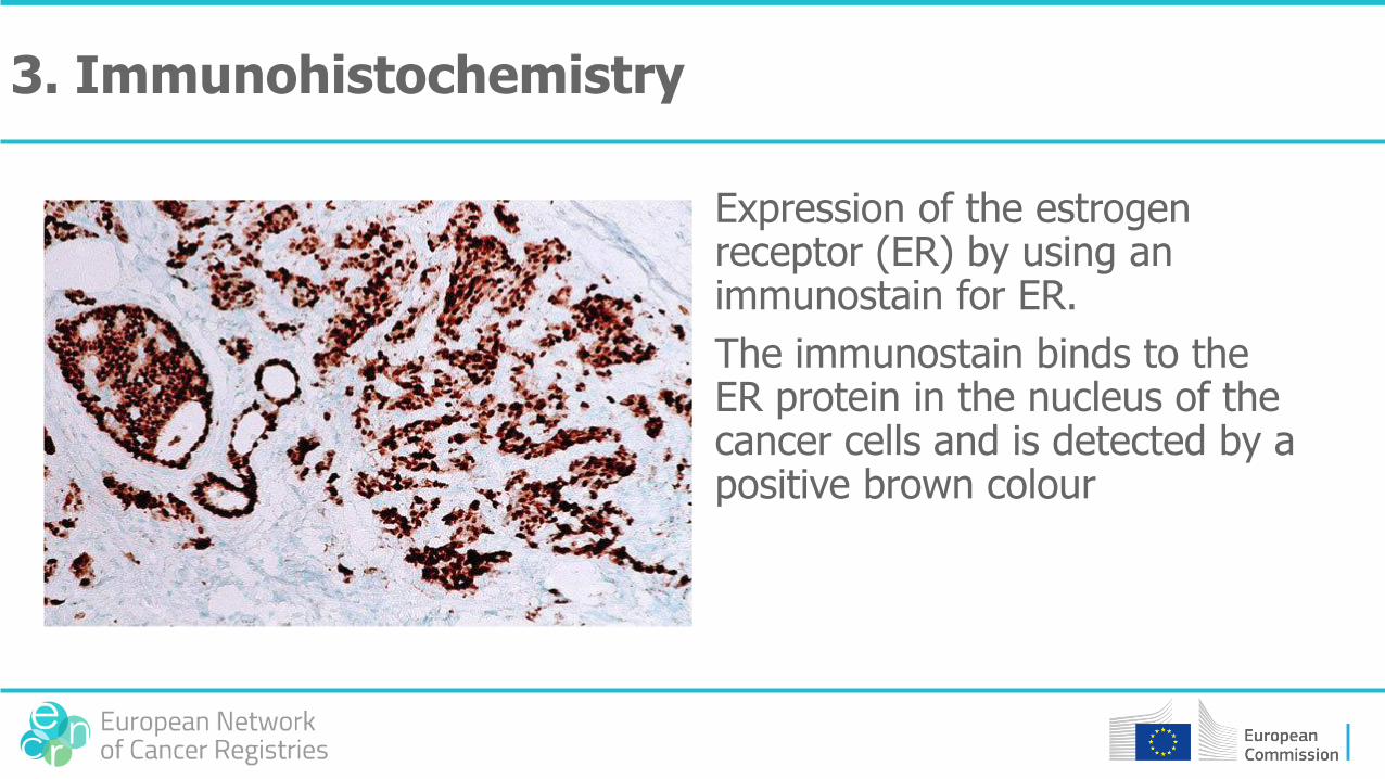
Expression of the estrogen receptor (ER) by using an immunostain for ER.
The immunostain binds to the ER protein in the nucleus of the cancer cells and is detected by a positive brown colour
3. Immunohistochemistry
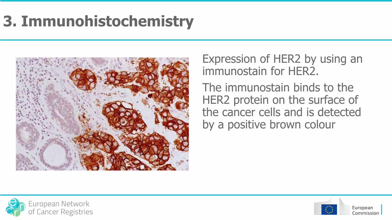
Expression of HER2 by using an immunostain for HER2.
The immunostain binds to the HER2 protein on the surface of the cancer cells and is detected by a positive brown colour
3. Immunohistochemistry
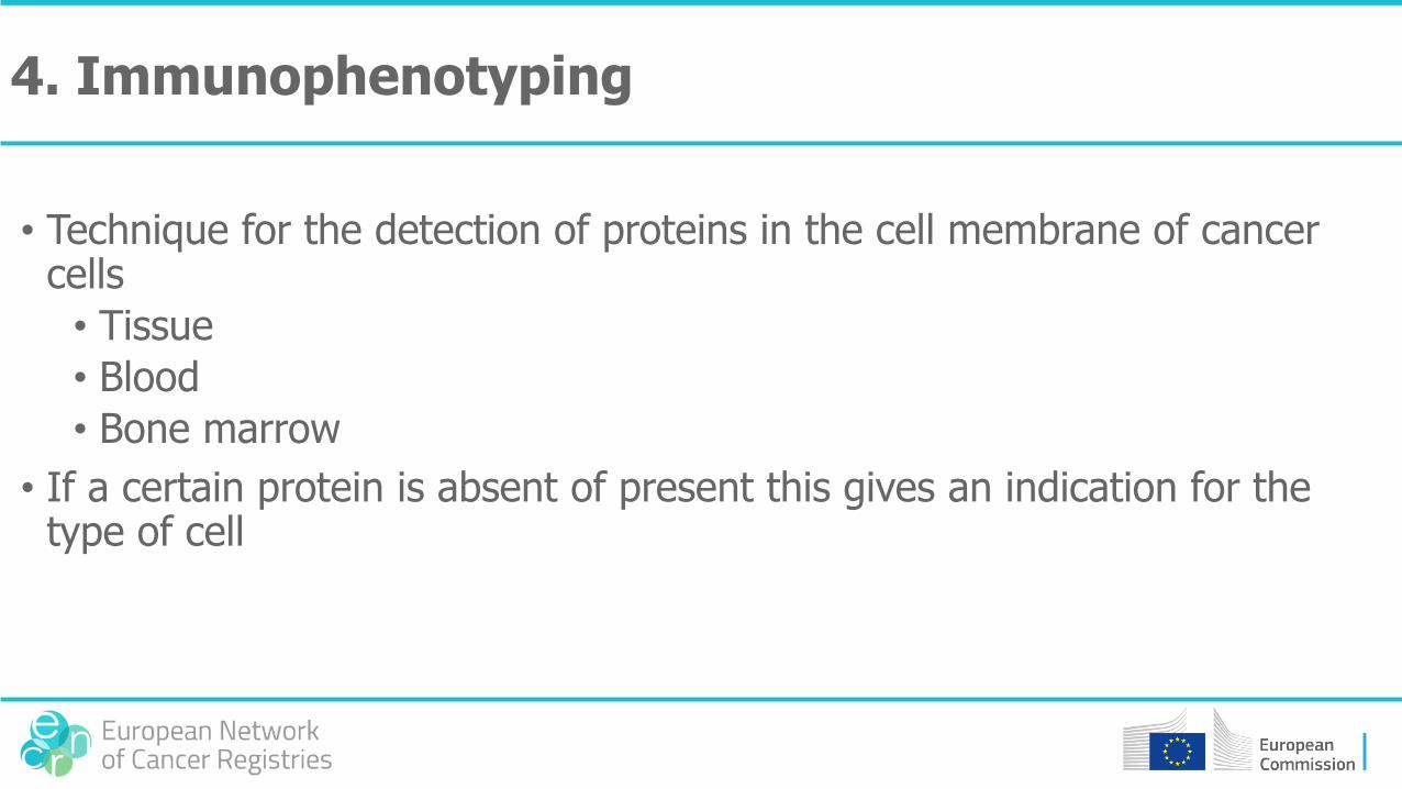
4. Immunophenotyping
• Technique for the detection of proteins in the cell membrane of cancer cells
• Tissue
• Blood
• Bone marrow
• If a certain protein is absent of present this gives an indication for the type of cell

4. Immunophenotyping
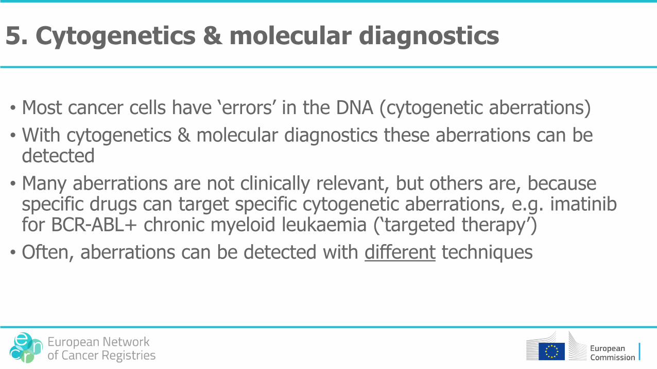
5. Cytogenetics & molecular diagnostics
• Most cancer cells have ‘errors’ in the DNA (cytogenetic aberrations)
• With cytogenetics & molecular diagnostics these aberrations can be detected
• Many aberrations are not clinically relevant, but others are, because specific drugs can target specific cytogenetic aberrations, e.g. imatinib for BCR-ABL+ chronic myeloid leukaemia (‘targeted therapy’)
• Often, aberrations can be detected with different techniques
5. Cytogenetics & molecular diagnostics
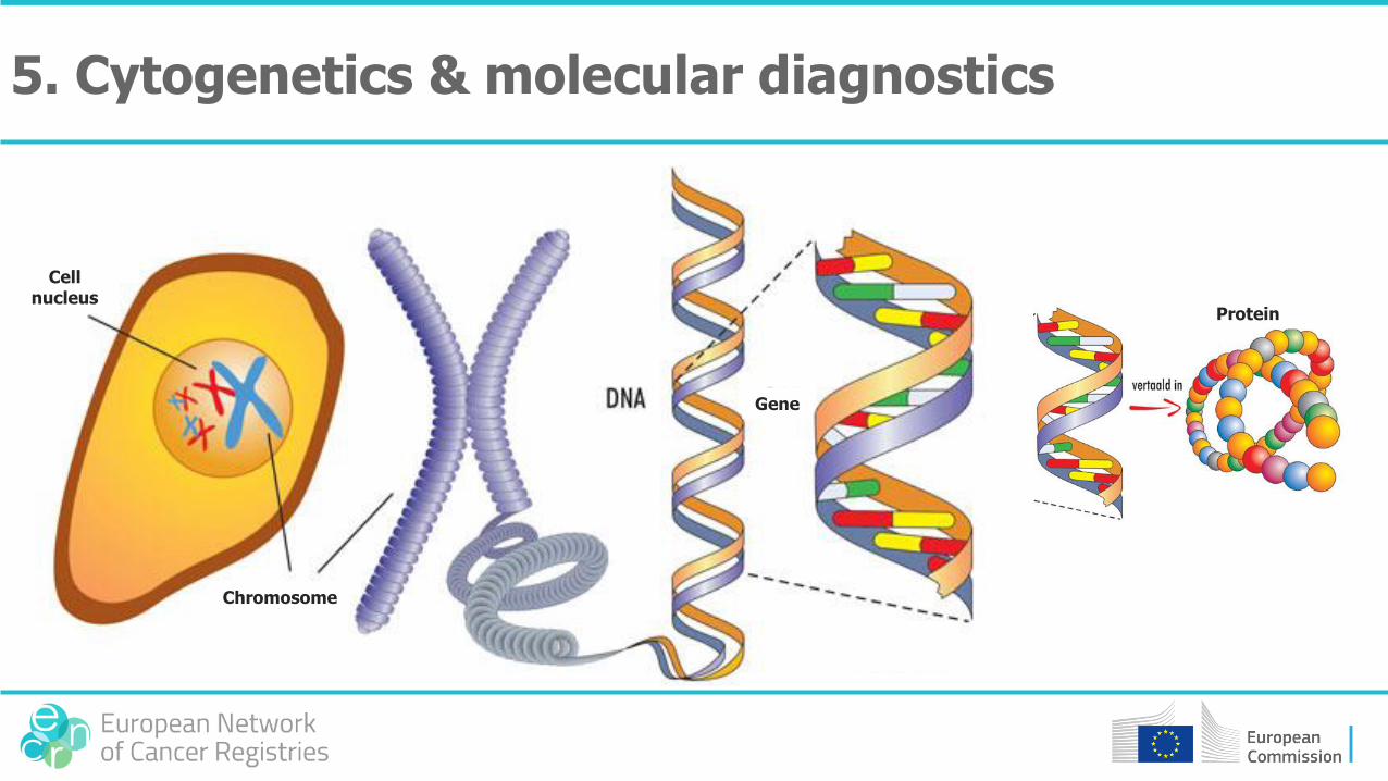
Cell nucleus
Chromosome
Gene
Protein
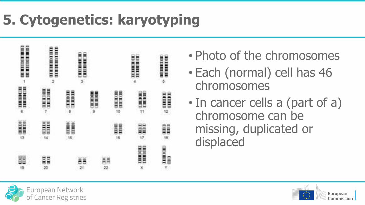
• Photo of the chromosomes
• Each (normal) cell has 46 chromosomes
• In cancer cells a (part of a) chromosome can be missing, duplicated or displaced
5. Cytogenetics: karyotyping
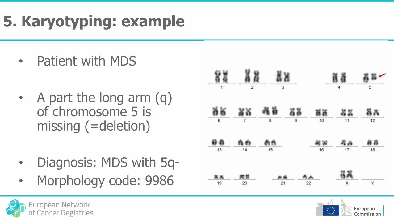
• Patient with MDS
• A part the long arm (q) of chromosome 5 is missing (=deletion)
• Diagnosis: MDS with 5q-
• Morphology code: 9986
5. Karyotyping: example
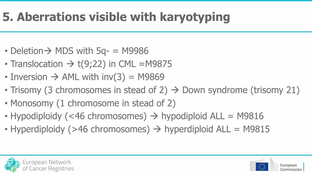
5. Aberrations visible with karyotyping
• Deletion MDS with 5q- = M9986
• Translocation t(9;22) in CML =M9875
• Inversion AML with inv(3) = M9869
• Trisomy (3 chromosomes in stead of 2) Down syndrome (trisomy 21)
• Monosomy (1 chromosome in stead of 2)
• Hypodiploidy (<46 chromosomes) hypodiploid ALL = M9816
• Hyperdiploidy (>46 chromosomes) hyperdiploid ALL = M9815
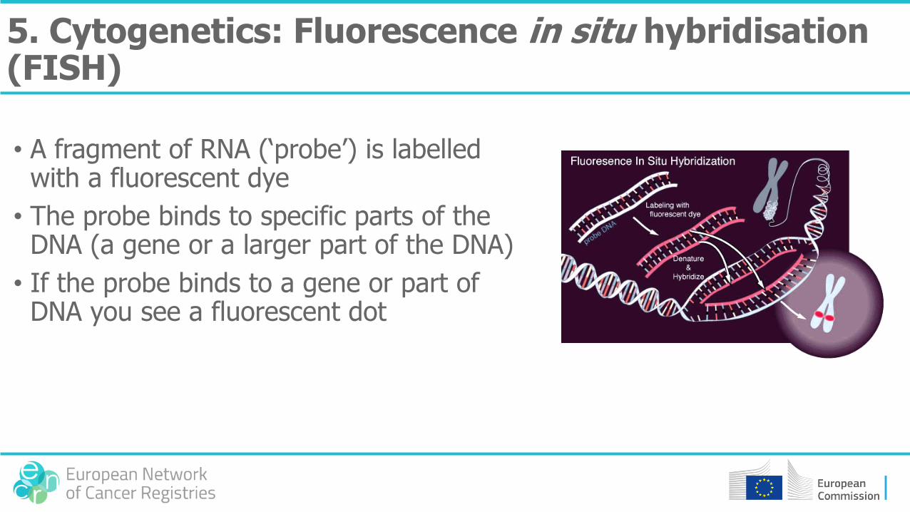
5. Cytogenetics: Fluorescence in situ hybridisation (FISH)
• A fragment of RNA (‘probe’) is labelled with a fluorescent dye
• The probe binds to specific parts of the DNA (a gene or a larger part of the DNA)
• If the probe binds to a gene or part of DNA you see a fluorescent dot
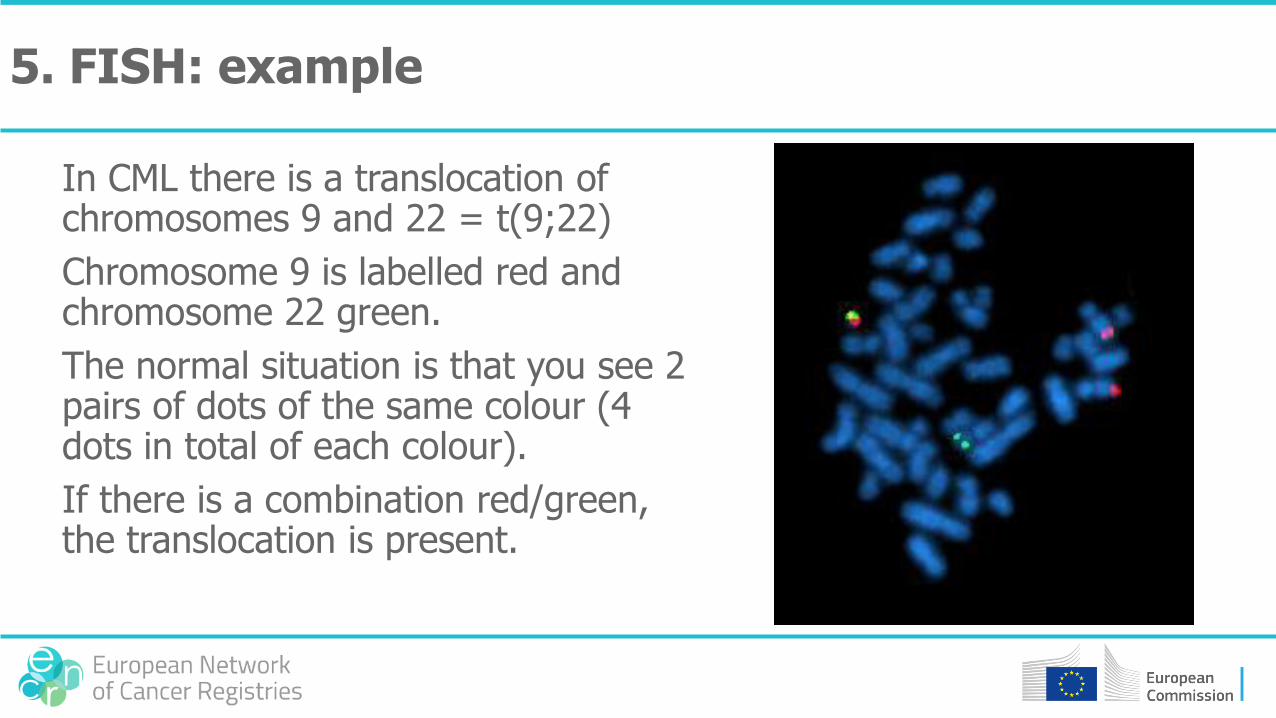
In CML there is a translocation of chromosomes 9 and 22 = t(9;22)
Chromosome 9 is labelled red and chromosome 22 green.
The normal situation is that you see 2 pairs of dots of the same colour (4 dots in total of each colour).
If there is a combination red/green, the translocation is present.
5. FISH: example

If a gene (or combination of genes = ‘fusion genes’) codes for a specific protein, you can also use molecular diagnostics to detect the protein.
The fusion gene in CML produces the protein BCR-ABL, which can be detected in blood.
5. Molecular diagnostics
Sarcoma
What is a sarcoma?
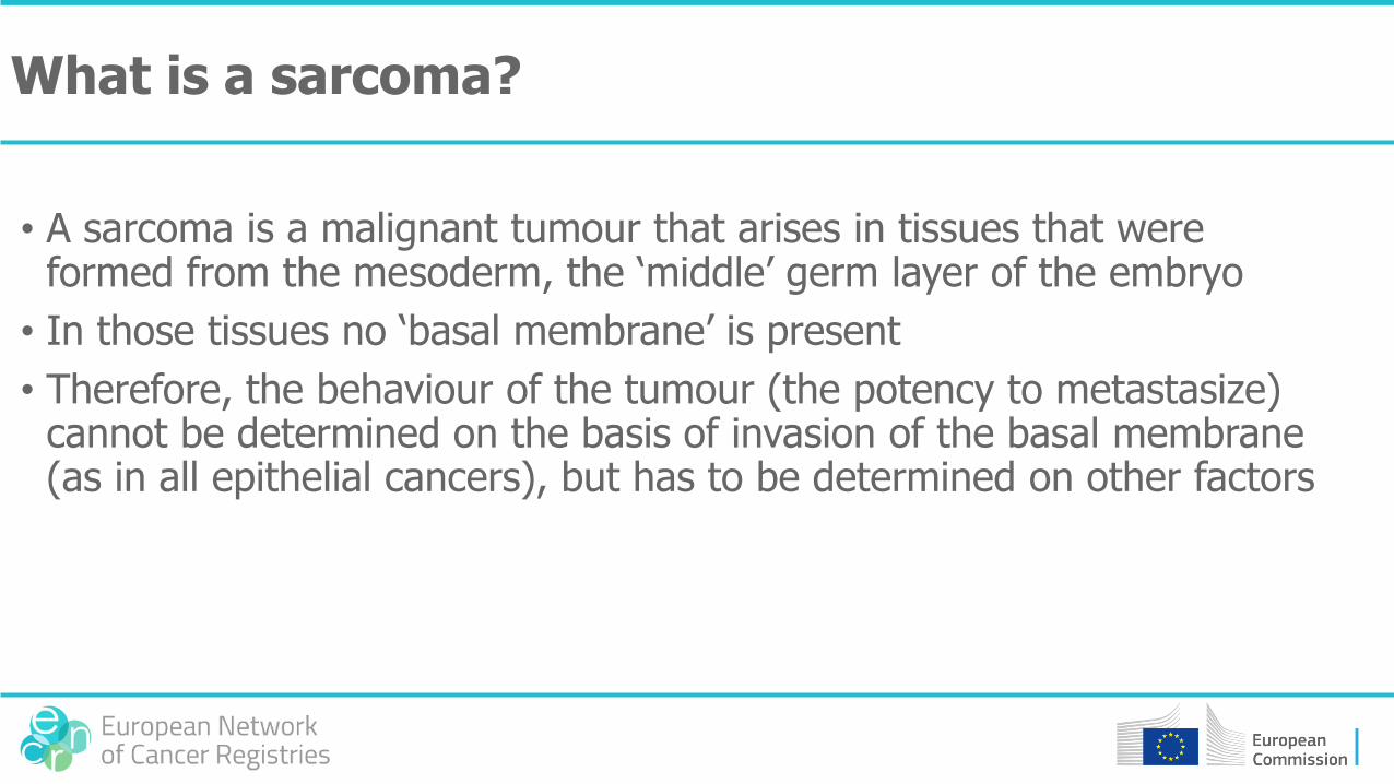
• A sarcoma is a malignant tumour that arises in tissues that were formed from the mesoderm, the ‘middle’ germ layer of the embryo
• In those tissues no ‘basal membrane’ is present
• Therefore, the behaviour of the tumour (the potency to metastasize) cannot be determined on the basis of invasion of the basal membrane (as in all epithelial cancers), but has to be determined on other factors
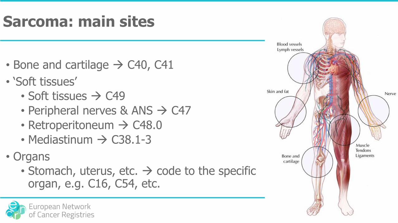
Sarcoma: main sites
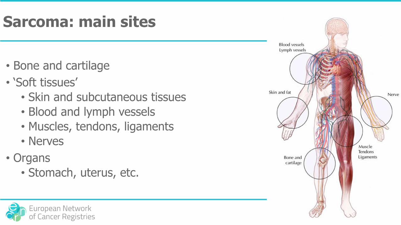
• Bone and cartilage
• ‘Soft tissues’
• Skin and subcutaneous tissues
• Blood and lymph vessels
• Muscles, tendons, ligaments
• Nerves
• Organs
• Stomach, uterus, etc.
Sarcoma: main sites
• Bone and cartilage C40, C41
• ‘Soft tissues’
• Soft tissues C49
• Peripheral nerves & ANS C47
• Retroperitoneum C48.0
• Mediastinum C38.1-3
• Organs
• Stomach, uterus, etc. code to the specific organ, e.g. C16, C54, etc.
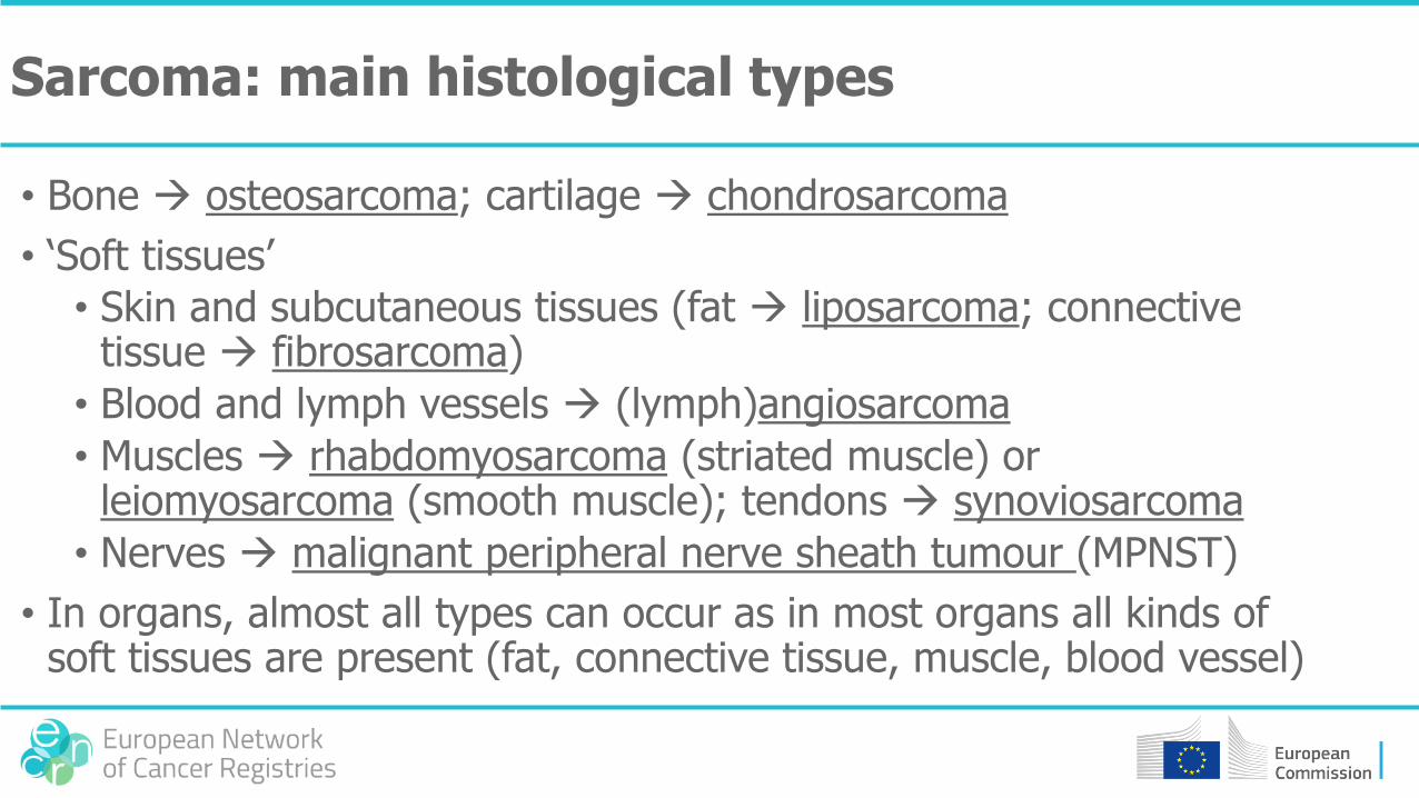
Sarcoma: main histological types
• Bone osteosarcoma; cartilage chondrosarcoma
• ‘Soft tissues’
• Skin and subcutaneous tissues (fat liposarcoma; connective tissue fibrosarcoma)
• Blood and lymph vessels (lymph)angiosarcoma
• Muscles rhabdomyosarcoma (striated muscle) or leiomyosarcoma (smooth muscle); tendons synoviosarcoma
• Nerves malignant peripheral nerve sheath tumour (MPNST)
• In organs, almost all types can occur as in most organs all kinds of soft tissues are present (fat, connective tissue, muscle, blood vessel)
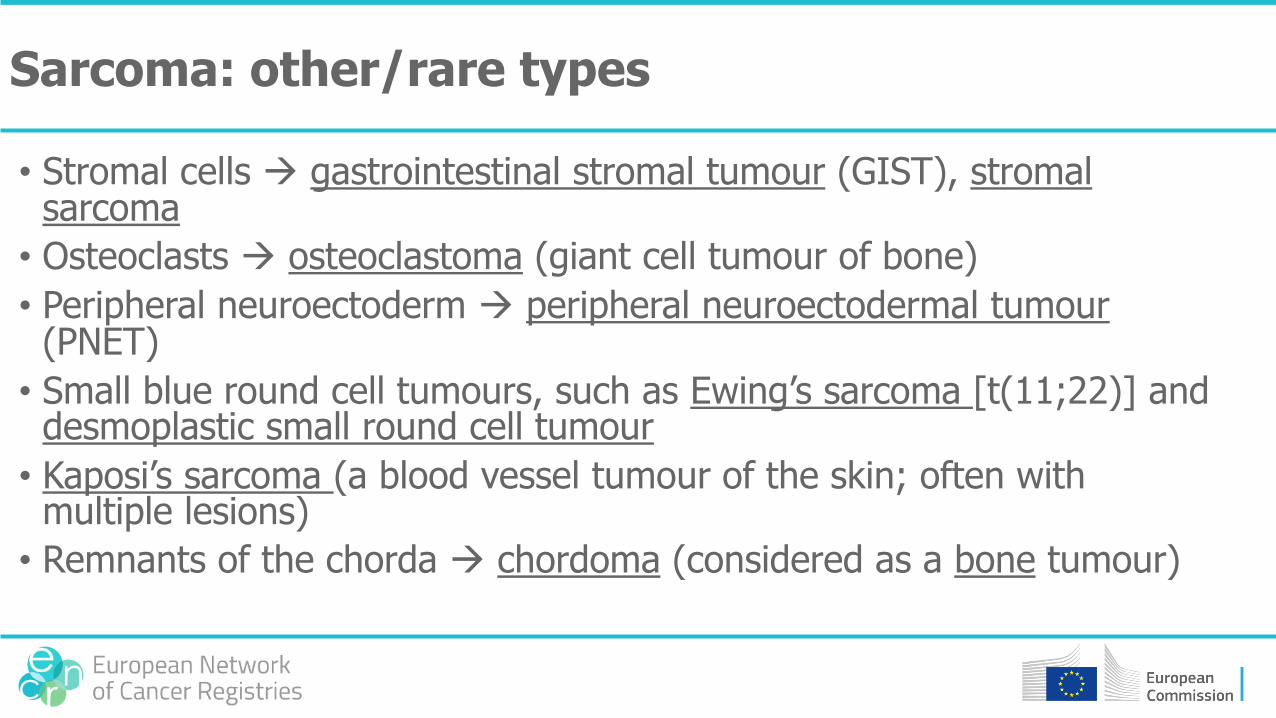
Sarcoma: other/rare types
• Stromal cells gastrointestinal stromal tumour (GIST), stromal sarcoma
• Osteoclasts osteoclastoma (giant cell tumour of bone)
• Peripheral neuroectoderm peripheral neuroectodermal tumour (PNET)
• Small blue round cell tumours, such as Ewing’s sarcoma [t(11;22)] and desmoplastic small round cell tumour
• Kaposi’s sarcoma (a blood vessel tumour of the skin; often with multiple lesions)
• Remnants of the chorda chordoma (considered as a bone tumour)
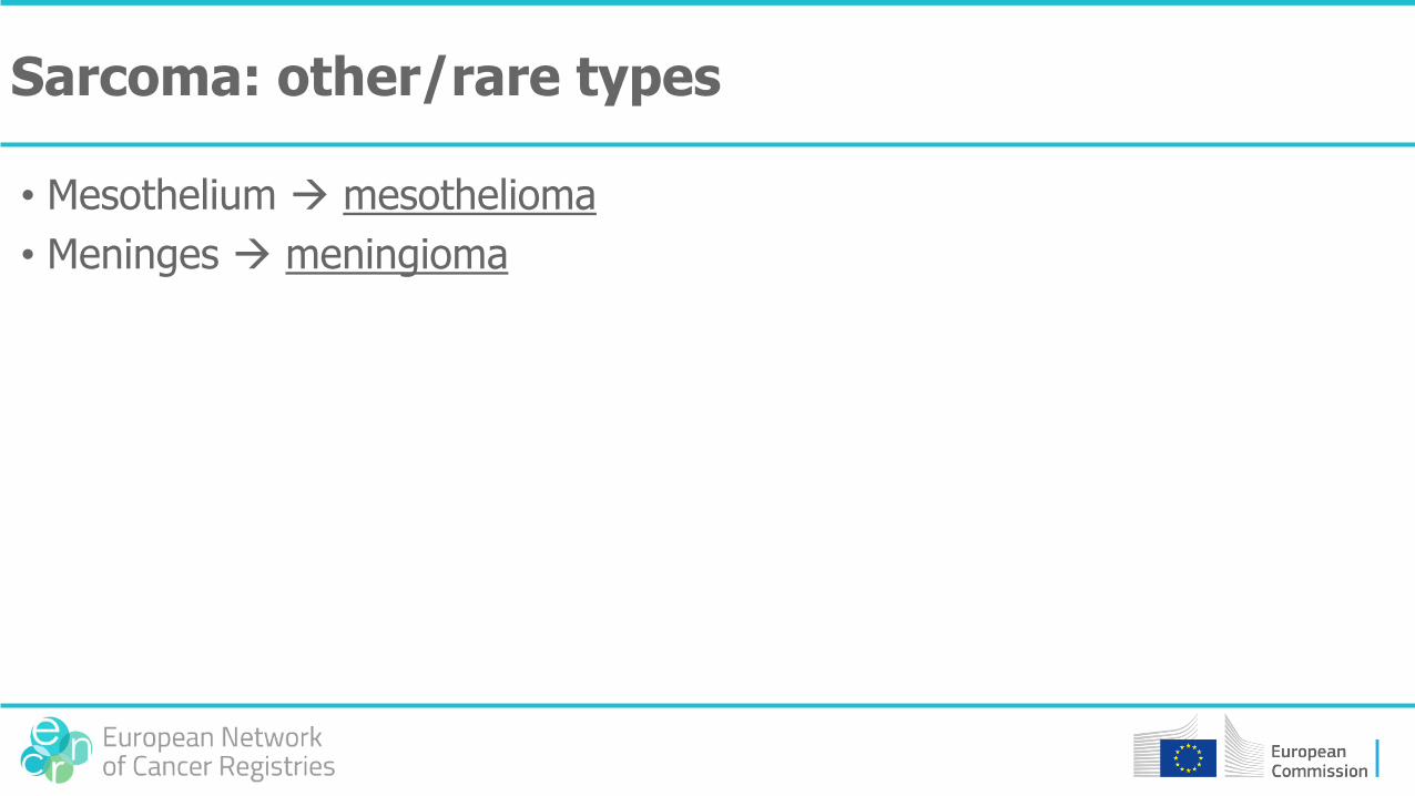
Sarcoma: other/rare types
• Mesothelium mesothelioma
• Meninges meningioma
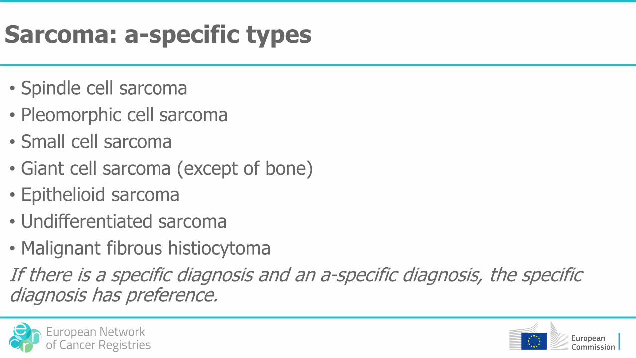
Sarcoma: a-specific types
• Spindle cell sarcoma
• Pleomorphic cell sarcoma
• Small cell sarcoma
• Giant cell sarcoma (except of bone)
• Epithelioid sarcoma
• Undifferentiated sarcoma
• Malignant fibrous histiocytoma
If there is a specific diagnosis and an a-specific diagnosis, the specific diagnosis has preference.
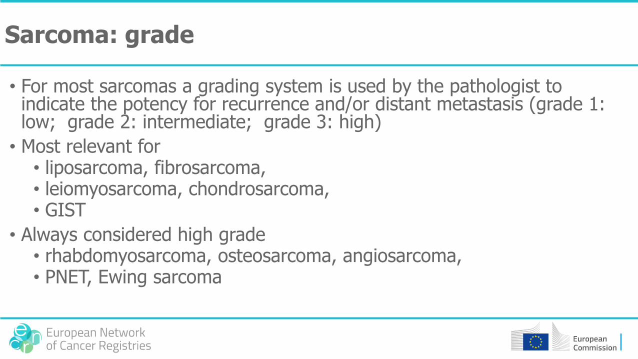
Sarcoma: grade
• For most sarcomas a grading system is used by the pathologist to indicate the potency for recurrence and/or distant metastasis (grade 1: low; grade 2: intermediate; grade 3: high)
• Most relevant for• liposarcoma, fibrosarcoma,• leiomyosarcoma, chondrosarcoma,• GIST
• Always considered high grade• rhabdomyosarcoma, osteosarcoma, angiosarcoma,• PNET, Ewing sarcoma
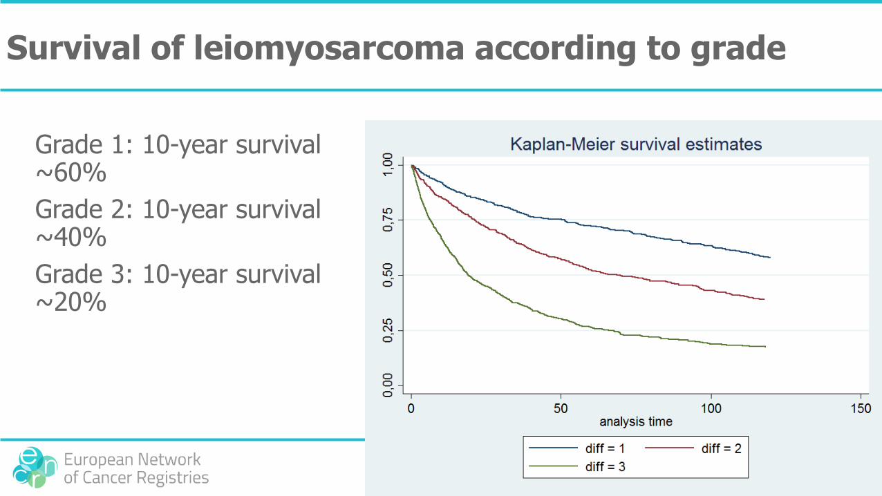
Survival of leiomyosarcoma according to grade
Grade 1: 10-year survival ~60%
Grade 2: 10-year survival ~40%
Grade 3: 10-year survival ~20%

Grade 1 liposarcoma and chondrosarcoma
According to the latest version of the WHO classification of sarcomas grade 1 liposarcoma (8850/31 or 8851/31) and grade 1 chondrosarcoma (9220/31) are no longer considered malignant and should be classified with behaviour code 1 (8850/11 and 9222/11, respectively).
For international comparison the behaviour code is of less relevance as long as the correct grade is coded.

GIST
The potency for recurrence or metastases of GIST is dependent of
• size of the tumour
• number of mitoses
Small tumours without mitosis are considered benign (/0), large tumours or tumours with many mitoses are considered malignant (/3); the intermediate group is considered borderline malignant (/1).
Rules for the exact classification will be drawn up later, as not all pathologist use the same rules.
It is recommended to register the tumour size and the number of mitosis
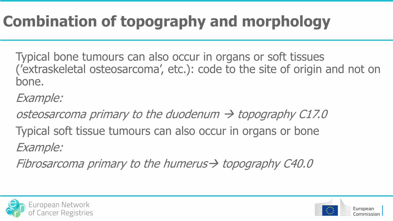
Combination of topography and morphology
Typical bone tumours can also occur in organs or soft tissues (’extraskeletal osteosarcoma’, etc.): code to the site of origin and not on bone.
Example:
osteosarcoma primary to the duodenum topography C17.0
Typical soft tissue tumours can also occur in organs or bone
Example:
Fibrosarcoma primary to the humerus topography C40.0
Combination of topography and morphology
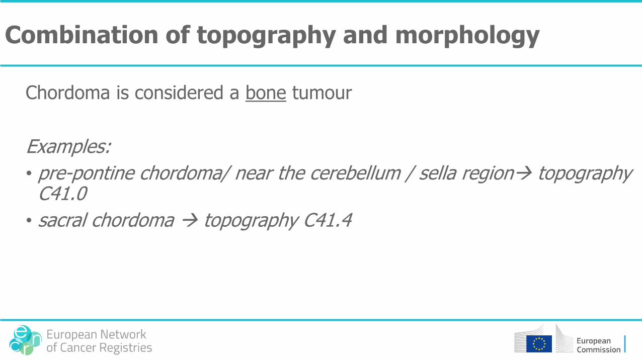
Chordoma is considered a bone tumour
Examples:
• pre-pontine chordoma/ near the cerebellum / sella region topography C41.0
• sacral chordoma topography C41.4
Combination of topography and morphology
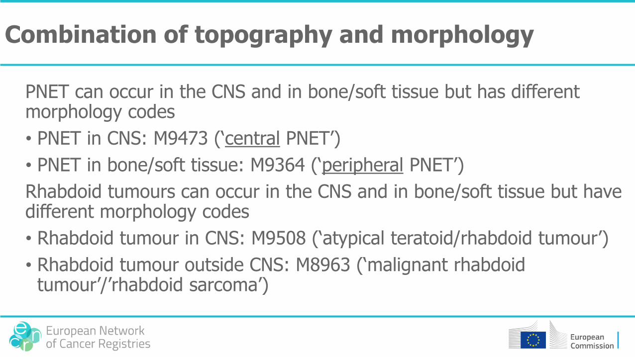
PNET can occur in the CNS and in bone/soft tissue but has different morphology codes
• PNET in CNS: M9473 (‘central PNET’)
• PNET in bone/soft tissue: M9364 (‘peripheral PNET’)
Rhabdoid tumours can occur in the CNS and in bone/soft tissue but have different morphology codes
• Rhabdoid tumour in CNS: M9508 (‘atypical teratoid/rhabdoid tumour’)
• Rhabdoid tumour outside CNS: M8963 (‘malignant rhabdoid tumour’/’rhabdoid sarcoma’)
NET & NEC (neuroendocrine tumour & carcinoma)
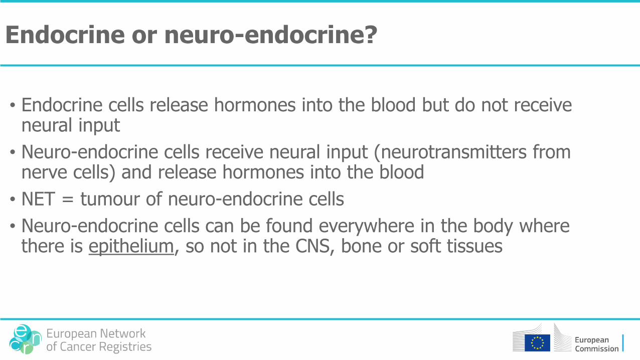
Endocrine or neuro-endocrine?
• Endocrine cells release hormones into the blood but do not receive neural input
• Neuro-endocrine cells receive neural input (neurotransmitters from nerve cells) and release hormones into the blood
• NET = tumour of neuro-endocrine cells
• Neuro-endocrine cells can be found everywhere in the body where there is epithelium, so not in the CNS, bone or soft tissues
Hormone production
• As neuro-endocrine tumours can produce (a large variety of) hormones, patients can have all kinds of symptoms related to the hormone production
• ‘carcinoid syndrome’ (serotonin production)
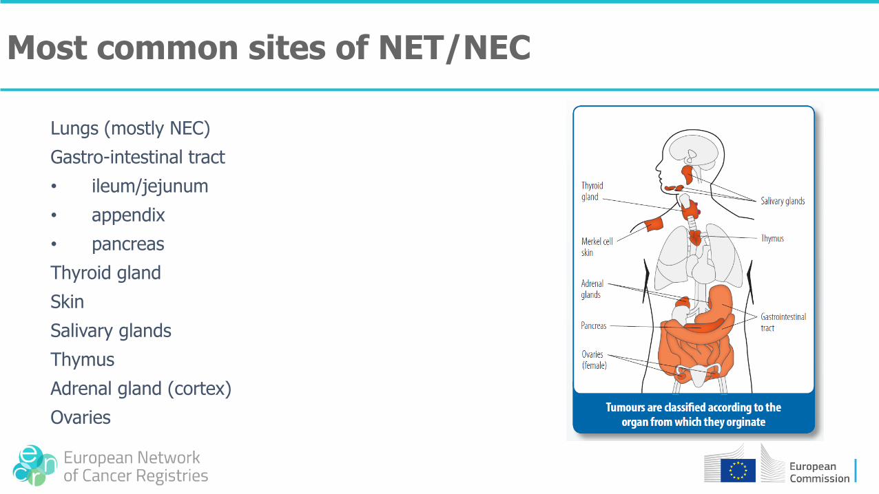
Lungs (mostly NEC)
Gastro-intestinal tract
• ileum/jejunum
• appendix
• pancreas
Thyroid gland
Skin
Salivary glands
Thymus
Adrenal gland (cortex)
Ovaries
Most common sites of NET/NEC
G1NET
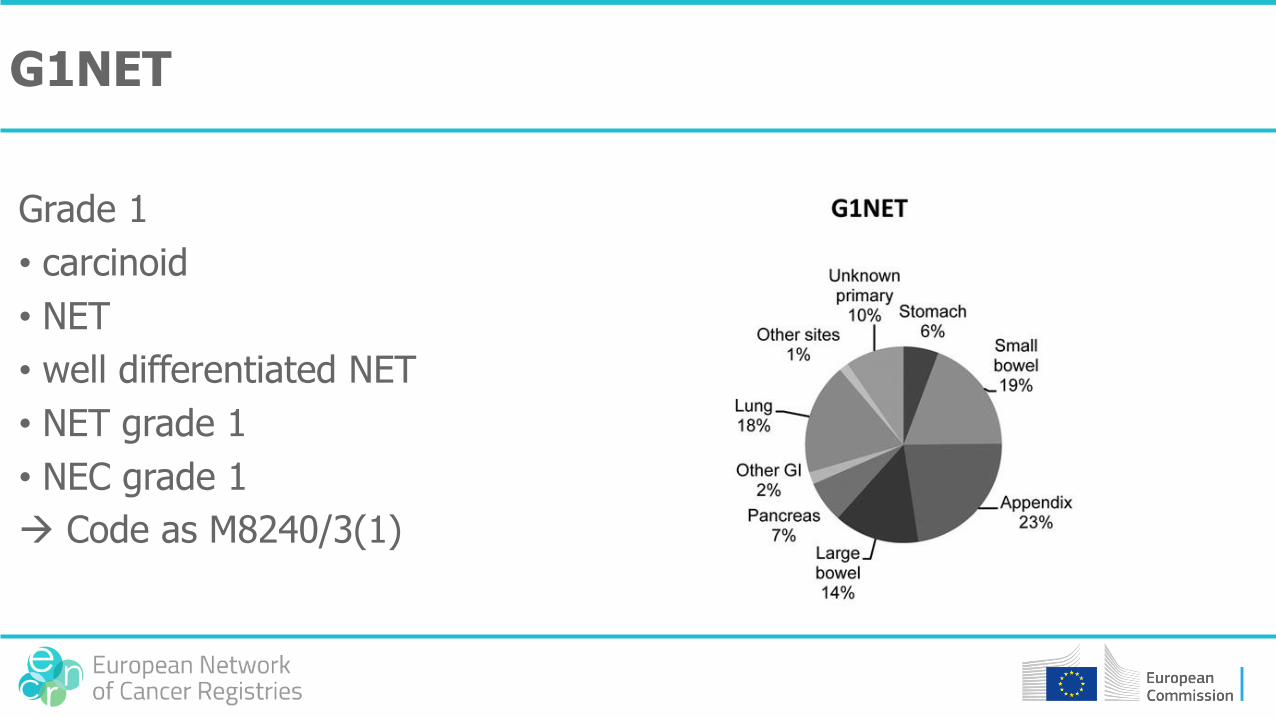
Grade 1
• carcinoid
• NET
• well differentiated NET
• NET grade 1
• NEC grade 1
Code as M8240/3(1)

G2NET
Grade 2
• atypical carcinoid
• moderately differentiated NET
• NET grade 2
• NEC grade 2
Code as M8249/3(2)
G3-LCNEC
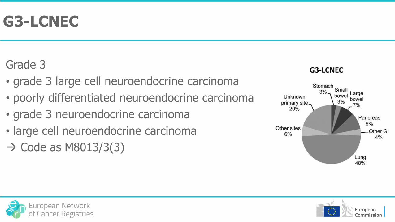
Grade 3
• grade 3 large cell neuroendocrine carcinoma
• poorly differentiated neuroendocrine carcinoma
• grade 3 neuroendocrine carcinoma
• large cell neuroendocrine carcinoma
Code as M8013/3(3)
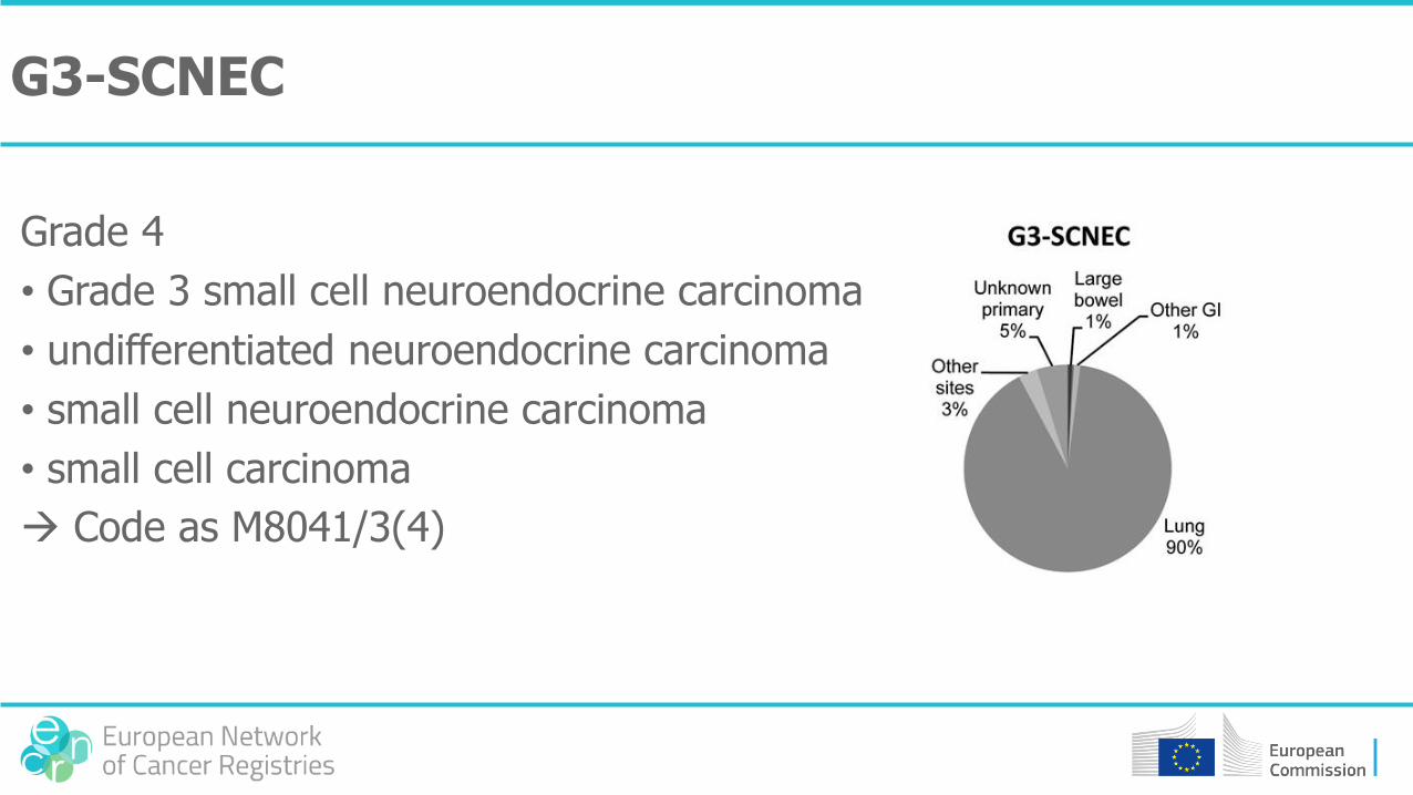
G3-SCNEC
Grade 4
• Grade 3 small cell neuroendocrine carcinoma
• undifferentiated neuroendocrine carcinoma
• small cell neuroendocrine carcinoma
• small cell carcinoma
Code as M8041/3(4)
Grade unknown
Grade unknown
• neuroendocrine carcinoma
Code as M8246/3
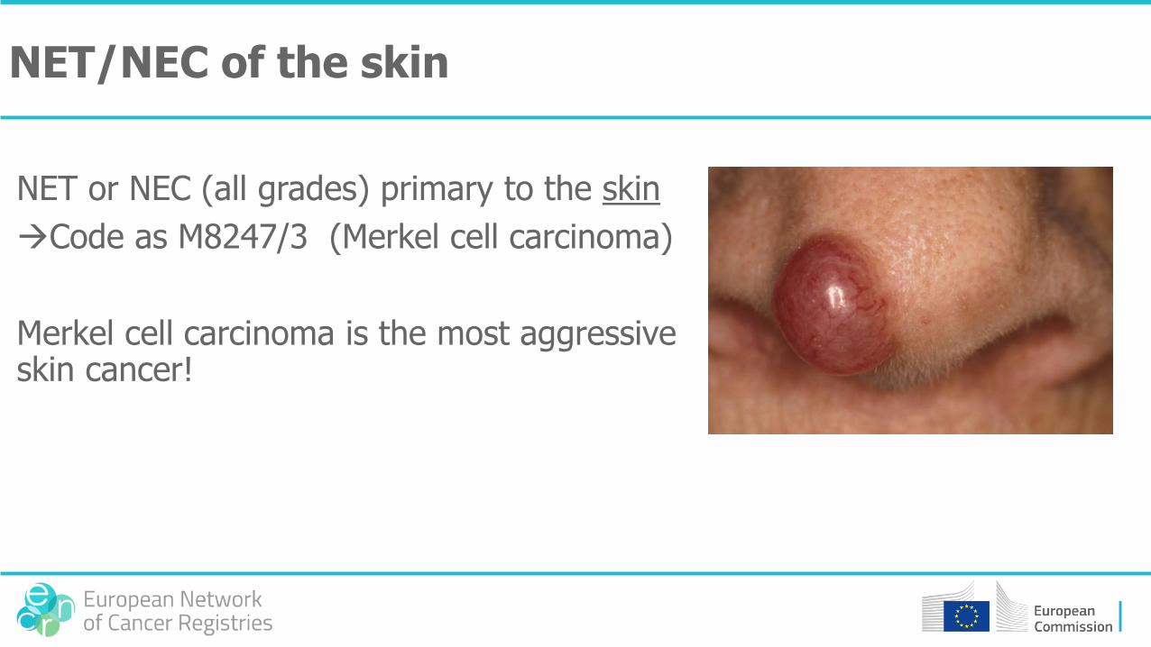
NET/NEC of the skin
NET or NEC (all grades) primary to the skin
Code as M8247/3 (Merkel cell carcinoma)
Merkel cell carcinoma is the most aggressive skin cancer!
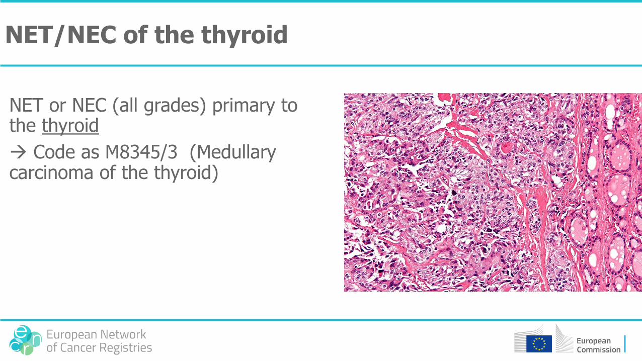
NET/NEC of the thyroid
NET or NEC (all grades) primary to the thyroid
Code as M8345/3 (Medullary carcinoma of the thyroid)
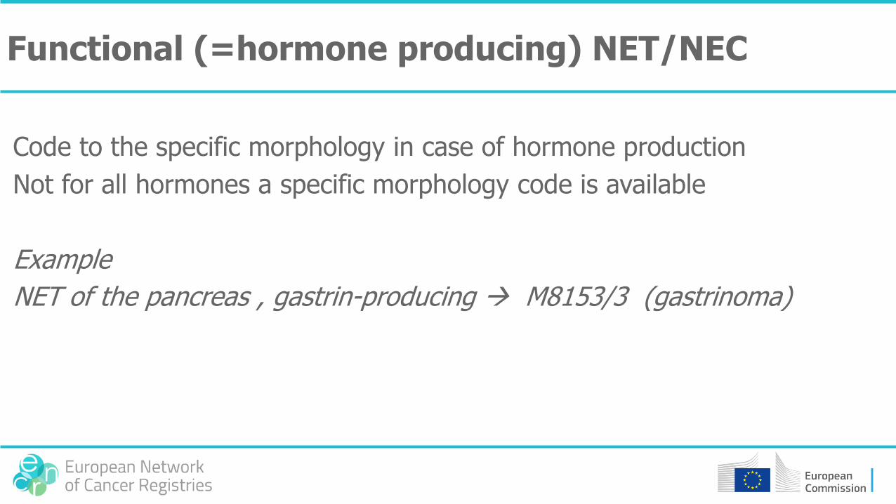
Functional (=hormone producing) NET/NEC
Code to the specific morphology in case of hormone production
Not for all hormones a specific morphology code is available
Example
NET of the pancreas , gastrin-producing M8153/3 (gastrinoma)

Tumours of the central nervous system (CNS)

Cranial nerves
• Olfactory nerve (I) C72.2
• Optic nerve (II) C72.3
• Acoustic nerve (VIII) C72.4
• Other cranial nerves C72.5
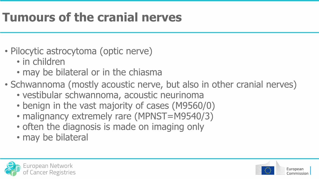
Tumours of the cranial nerves
• Pilocytic astrocytoma (optic nerve)• in children• may be bilateral or in the chiasma
• Schwannoma (mostly acoustic nerve, but also in other cranial nerves)• vestibular schwannoma, acoustic neurinoma• benign in the vast majority of cases (M9560/0)• malignancy extremely rare (MPNST=M9540/3)• often the diagnosis is made on imaging only• may be bilateral
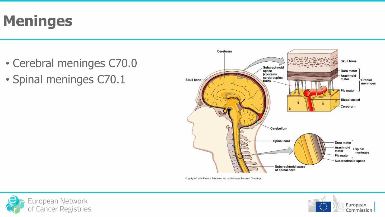
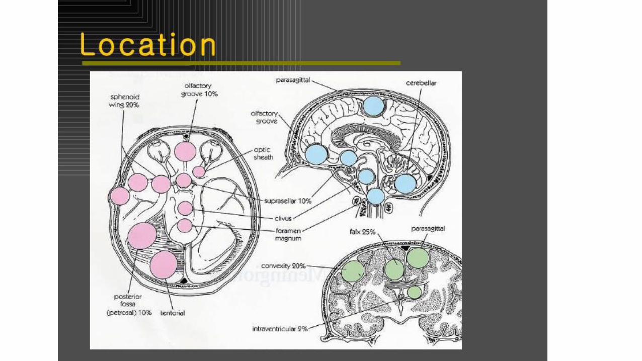
Meninges
• Cerebral meninges C70.0
• Spinal meninges C70.1
Tumours of the meninges
• Meningioma• Mostly benign (9530/0 – 9537/0)• Atypical (9538/1)• Rarely malignant (malignant meningioma = 9530/3 or meningial
sarcomatosis = 9539/3)
• Hemangiopericytoma• vestibular schwannoma, acoustic neurinoma
• Melanoma• Solitary (8720/3) or diffuse (8728/0, 8728/1, 8728/3)
Tumours of the meninges
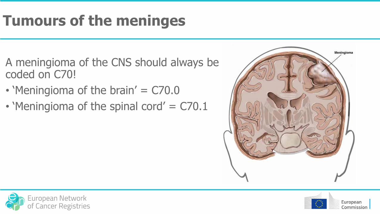
A meningioma of the CNS should always be coded on C70!
• ‘Meningioma of the brain’ = C70.0
• ‘Meningioma of the spinal cord’ = C70.1
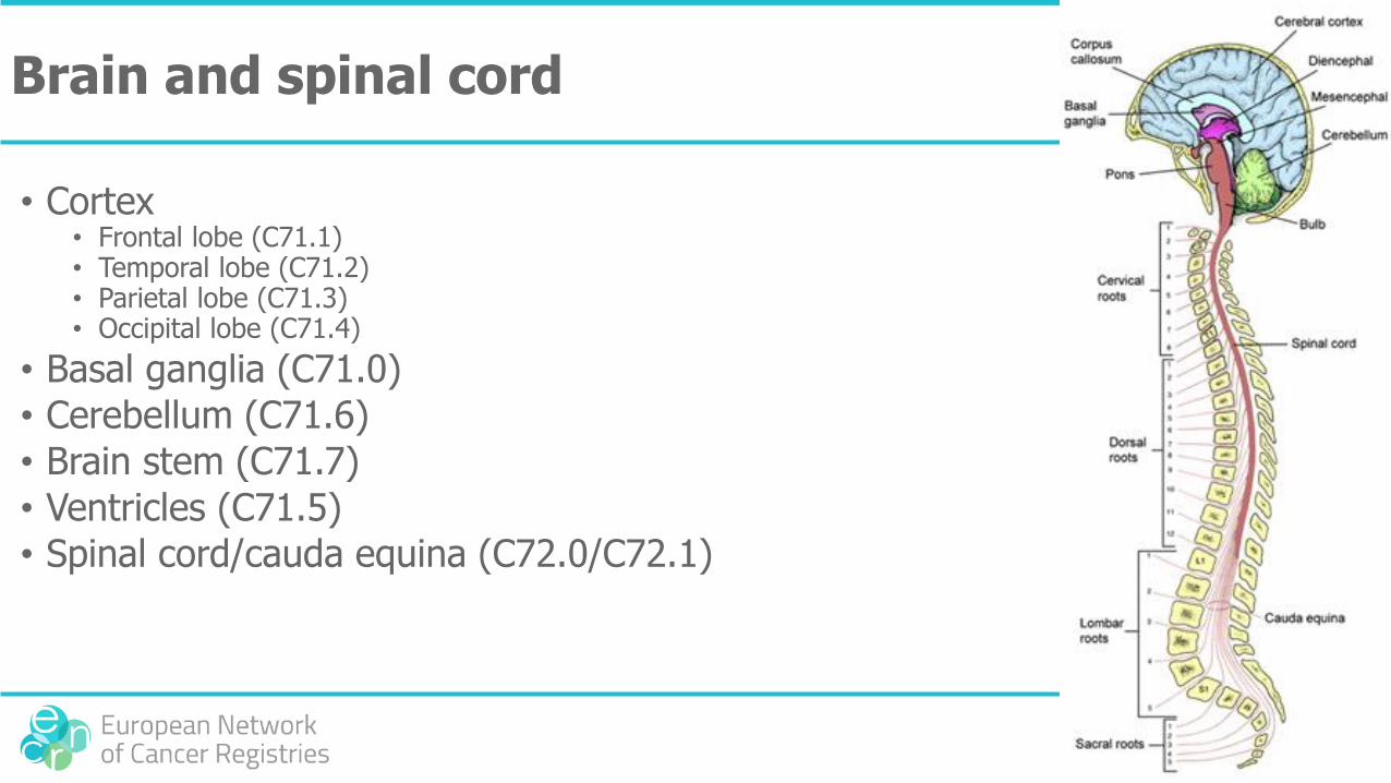
Brain and spinal cord
• Cortex• Frontal lobe (C71.1)• Temporal lobe (C71.2)• Parietal lobe (C71.3)• Occipital lobe (C71.4)
• Basal ganglia (C71.0)
• Cerebellum (C71.6)
• Brain stem (C71.7)
• Ventricles (C71.5)
• Spinal cord/cauda equina (C72.0/C72.1)

The ventricles of the brain
• The ventricles are filled with fluid; the only anatomical structure in the ventricles is the choroid plexus
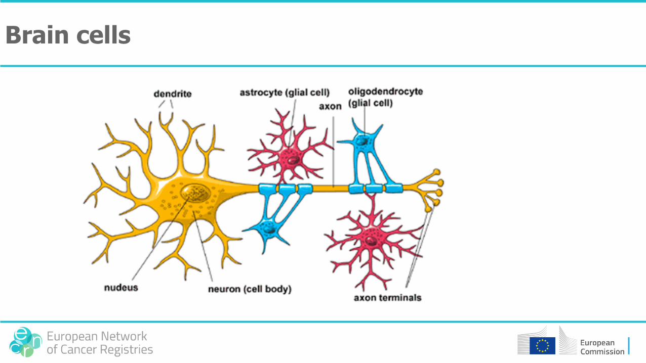
Brain cells
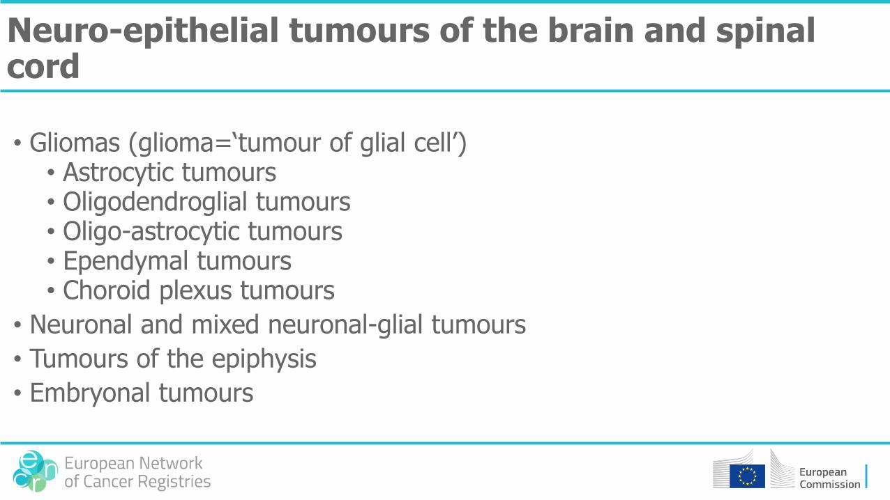
Neuro-epithelial tumours of the brain and spinal cord
• Gliomas (glioma=‘tumour of glial cell’)• Astrocytic tumours• Oligodendroglial tumours• Oligo-astrocytic tumours• Ependymal tumours• Choroid plexus tumours
• Neuronal and mixed neuronal-glial tumours
• Tumours of the epiphysis
• Embryonal tumours
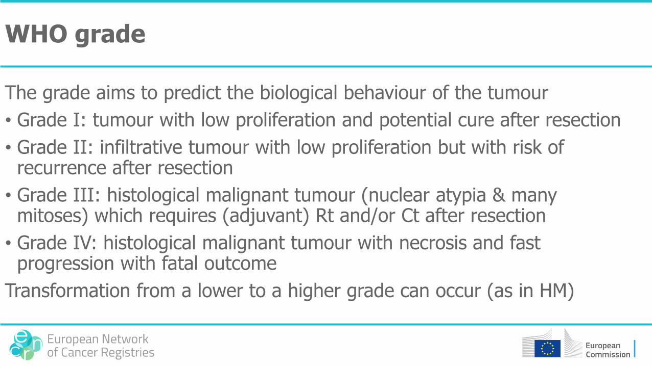
WHO grade
The grade aims to predict the biological behaviour of the tumour
• Grade I: tumour with low proliferation and potential cure after resection
• Grade II: infiltrative tumour with low proliferation but with risk of recurrence after resection
• Grade III: histological malignant tumour (nuclear atypia & many mitoses) which requires (adjuvant) Rt and/or Ct after resection
• Grade IV: histological malignant tumour with necrosis and fast progression with fatal outcome
Transformation from a lower to a higher grade can occur (as in HM)
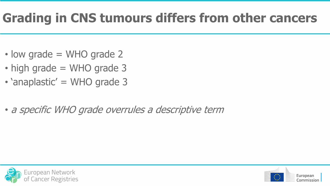
Grading in CNS tumours differs from other cancers
• low grade = WHO grade 2
• high grade = WHO grade 3
• ‘anaplastic’ = WHO grade 3
• a specific WHO grade overrules a descriptive term
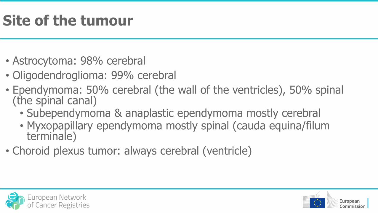
Site of the tumour
• Astrocytoma: 98% cerebral
• Oligodendroglioma: 99% cerebral
• Ependymoma: 50% cerebral (the wall of the ventricles), 50% spinal (the spinal canal)• Subependymoma & anaplastic ependymoma mostly cerebral• Myxopapillary ependymoma mostly spinal (cauda equina/filum
terminale)
• Choroid plexus tumor: always cerebral (ventricle)
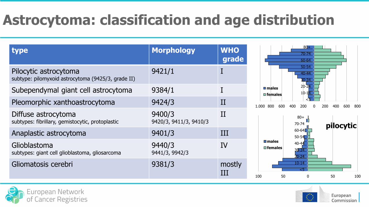
type Morphology WHOgrade
Pilocytic astrocytomasubtype: pilomyxoid astrocytoma (9425/3, grade II)
9421/1 I
Subependymal giant cell astrocytoma 9384/1 I
Pleomorphic xanthoastrocytoma 9424/3 II
Diffuse astrocytomasubtypes: fibrillary, gemistocytic, protoplastic
9400/39420/3, 9411/3, 9410/3
II
Anaplastic astrocytoma 9401/3 III
Glioblastomasubtypes: giant cell glioblastoma, gliosarcoma
9440/39441/3, 9942/3
IV
Gliomatosis cerebri 9381/3 mostlyIII
Astrocytoma: classification and age distribution
1.000 800 600 400 200 0 200 400 600 800
<5
10-14
20-24
30-34
40-44
50-54
60-64
70-74
80+
males
females
100 50 0 50 100
<5
10-14
20-24
30-34
40-44
50-54
60-64
70-74
80+
males
females
pilocytic
0,0
00,2
50,5
00,7
51,0
0
0 20 40 60analysis time
m = 502 piloc. astrocytoom m = 503 subep. reuscelastrocytoom
m = 504 pleom. xanthoastrocytoom m = 505 diff. astrocytoom
m = 506 anapl. astrocytoom m = 507 glioblastoom
m = 508 gliomatosis cerebri
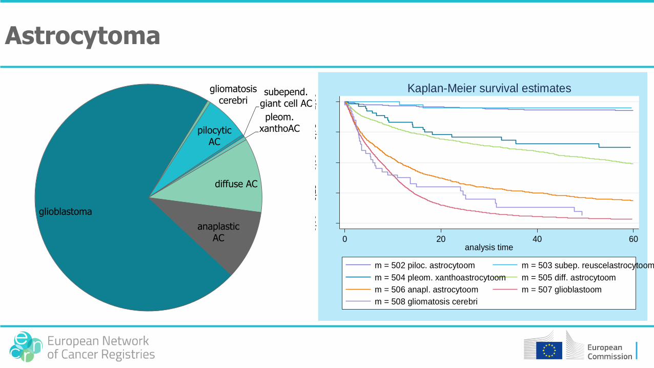
Kaplan-Meier survival estimates
pilocytic AC
subepend. giant cell AC
pleom. xanthoAC
diffuse AC
anaplastic AC
glioblastoma
gliomatosis cerebri
Astrocytoma
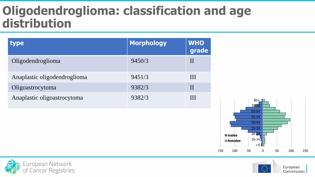
type Morphology WHOgrade
Oligodendroglioma 9450/3 II
Anaplastic oligodendroglioma 9451/3 III
Oligoastrocytoma 9382/3 II
Anaplastic oligoastrocytoma 9382/3 III
Oligodendroglioma: classification and age distribution
150 100 50 0 50 100 150
<5
10-14
20-24
30-34
40-44
50-54
60-64
70-74
80+
males
females
OD
anaplastic OD
OA
anaplastic OA
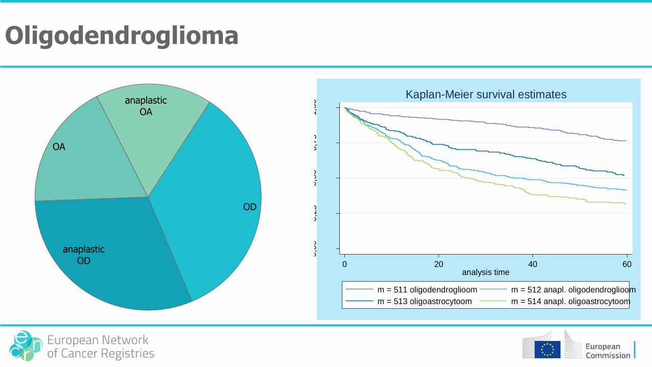
Oligodendroglioma
0,0
00,2
50,5
00,7
51,0
0
0 20 40 60analysis time
m = 511 oligodendroglioom m = 512 anapl. oligodendroglioom
m = 513 oligoastrocytoom m = 514 anapl. oligoastrocytoom
Kaplan-Meier survival estimates
type Morphology WHOgrade
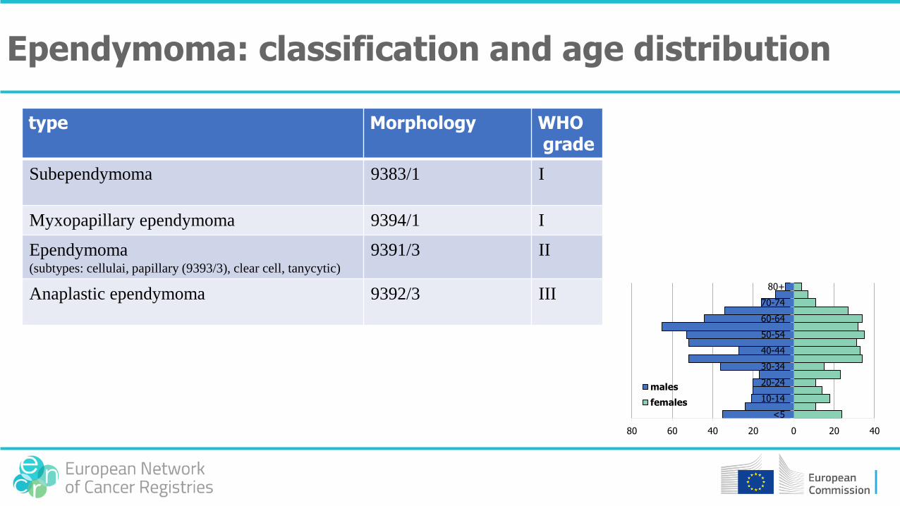
Subependymoma 9383/1 I
Myxopapillary ependymoma 9394/1 I
Ependymoma(subtypes: cellulai, papillary (9393/3), clear cell, tanycytic)
9391/3 II
Anaplastic ependymoma 9392/3 III
Ependymoma: classification and age distribution
80 60 40 20 0 20 40
<5
10-14
20-24
30-34
40-44
50-54
60-64
70-74
80+
males
females
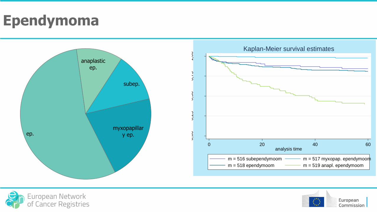
subep.
myxopapillary ep.ep.
anaplastic ep.
Ependymoma
0,0
00,2
50,5
00,7
51,0
0
0 20 40 60analysis time
m = 516 subependymoom m = 517 myxopap. ependymoom
m = 518 ependymoom m = 519 anapl. ependymoom
Kaplan-Meier survival estimates
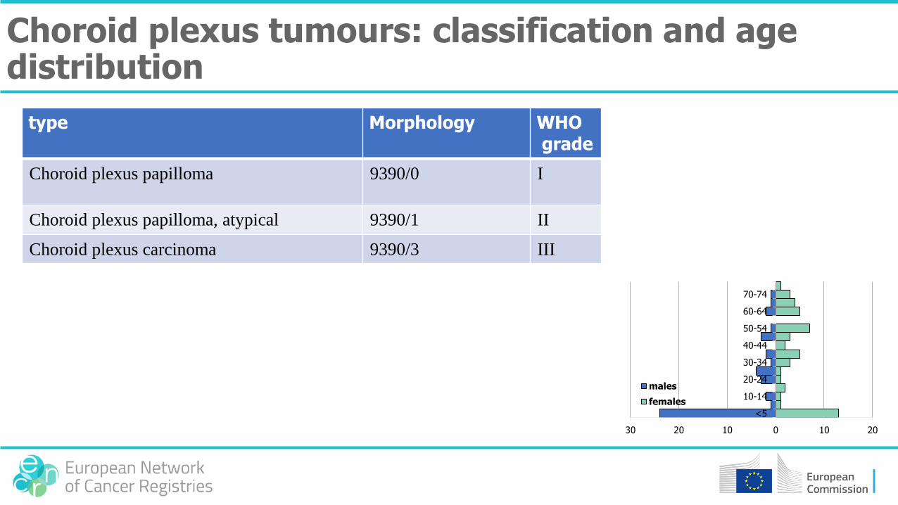
type Morphology WHOgrade
Choroid plexus papilloma 9390/0 I
Choroid plexus papilloma, atypical 9390/1 II
Choroid plexus carcinoma 9390/3 III
Choroid plexus tumours: classification and age distribution
30 20 10 0 10 20
<5
10-14
20-24
30-34
40-44
50-54
60-64
70-74
males
females

papilloma
atypical papilloma
carcinoma
Choroid plexus tumours
0,0
00,2
50,5
00,7
51,0
0
0 10 20 30 40 50analysis time
m = 521 choroïdplexuspapilloom m = 522 atyp. choroïdplexuspapilloom
m = 523 choroïdplexuscarcinoom
Kaplan-Meier survival estimates
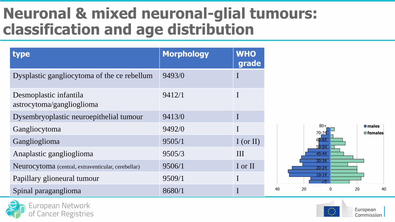
type Morphology WHOgrade
Dysplastic gangliocytoma of the ce rebellum 9493/0 I
Desmoplastic infantila
astrocytoma/ganglioglioma
9412/1 I
Dysembryoplastic neuroepithelial tumour 9413/0 I
Gangliocytoma 9492/0 I
Ganglioglioma 9505/1 I (or II)
Anaplastic ganglioglioma 9505/3 III
Neurocytoma (central, extraventicular, cerebellar) 9506/1 I or II
Papillary glioneural tumour 9509/1 I
Spinal paraganglioma 8680/1 I
Neuronal & mixed neuronal-glial tumours: classification and age distribution
40 20 0 20 40
<5
10-14
20-24
30-34
40-44
50-54
60-64
70-74
80+ males
females

type Morphology WHOgrade
Pineocytoma 9361/1 I (was
II)
Pineal parenchymal tumour of intermediate
differentiation
9362/3 II of III
(was III
of IV)
Pineoblastoma 9362/3 IV
Papillary tumour van de pinealis region 9395/3 II of III
Tumours of the pineal region: classification and age distribution
6 4 2 0 2 4 6
<5
10-14
20-24
30-34
40-44
50-54
60-64
70-74
80+ males
females

pineocytoma
pineal tumour of
int diff
pineoblastoma
pap. pineal tumor
Tumours of the pineal region
0,0
00,2
50,5
00,7
51,0
0
0 20 40 60analysis time
m = 536 pineocytoom m = 537 pineale tumor van ID
m = 538 pineoblastoom m = 539 pap. pinealistumor
Kaplan-Meier survival estimates
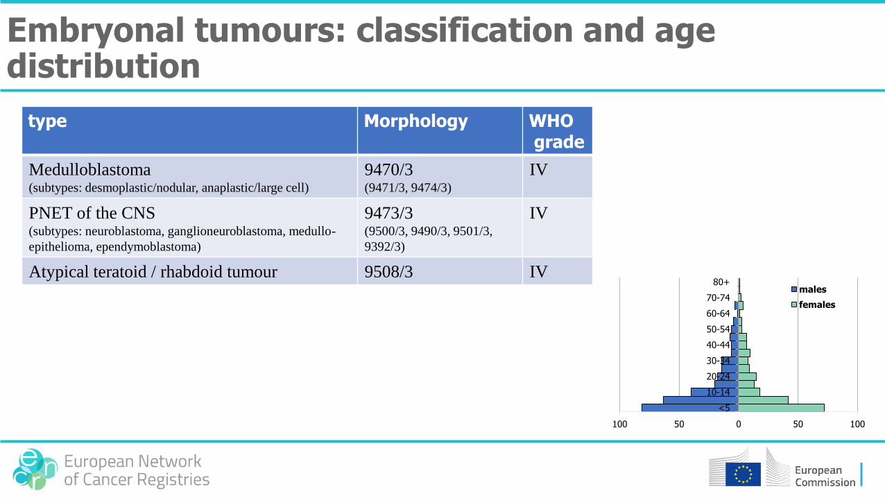
type Morphology WHOgrade
Medulloblastoma(subtypes: desmoplastic/nodular, anaplastic/large cell)
9470/3(9471/3, 9474/3)
IV
PNET of the CNS(subtypes: neuroblastoma, ganglioneuroblastoma, medullo-
epithelioma, ependymoblastoma)
9473/3(9500/3, 9490/3, 9501/3,
9392/3)
IV
Atypical teratoid / rhabdoid tumour 9508/3 IV
Embryonal tumours: classification and age distribution
100 50 0 50 100
<5
10-14
20-24
30-34
40-44
50-54
60-64
70-74
80+males
females

medullobl.
ATRT
PNET, NOS
epdendymobl.(ganglio)neu
robl.
medulloepithelioma
PNET
Embryonal tumours
0,0
00,2
50,5
00,7
51,0
0
0 20 40 60analysis time
m = 541 medulloblastoom m = 542 PNET
m = 543 ATRT
Kaplan-Meier survival estimates
Haematological malignancies

Haematopoiesis (overview)

Aim:
• To determine the cell type and ‘the normal counterpart’
• To determine subtypes which are relevant for the prognosis and/or the treatment
Classification of haematological malignancies

Haematological malignancy Normal counterpart
Multiple myeloma plasma cell
Follicular lymphoma germinal centre B-cell
B-ALL haematopoietic stem cell or a B-cell progenitor cell
Mantle cell lymphoma peripheral B-cell of the inner mantle zone (of a lymph node)
Examples

B-lymphocyte development with the malignant counterpart
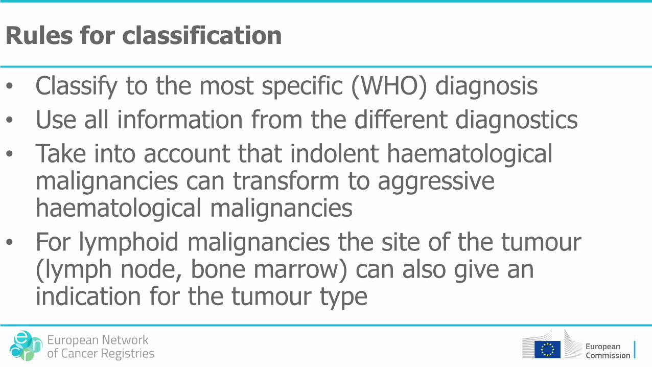
• Classify to the most specific (WHO) diagnosis
• Use all information from the different diagnostics
• Take into account that indolent haematological malignancies can transform to aggressive haematological malignancies
• For lymphoid malignancies the site of the tumour (lymph node, bone marrow) can also give an indication for the tumour type
Rules for classification
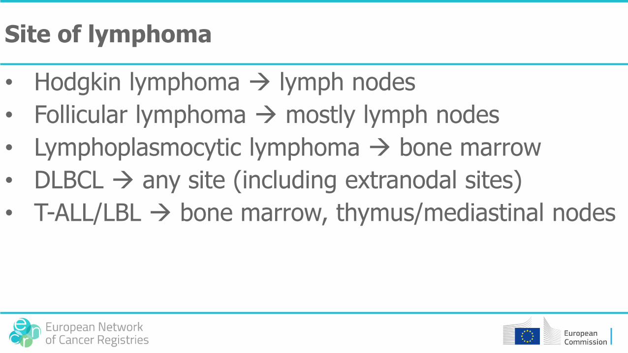
• Hodgkin lymphoma lymph nodes
• Follicular lymphoma mostly lymph nodes
• Lymphoplasmocytic lymphoma bone marrow
• DLBCL any site (including extranodal sites)
• T-ALL/LBL bone marrow, thymus/mediastinal nodes
Site of lymphoma
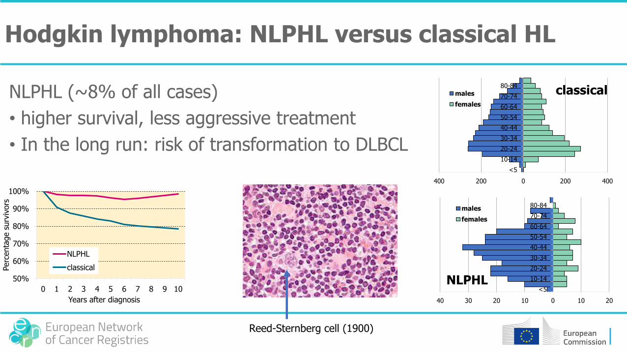
Hodgkin lymphoma: NLPHL versus classical HL
NLPHL (~8% of all cases)
• higher survival, less aggressive treatment
• In the long run: risk of transformation to DLBCL
50%
60%
70%
80%
90%
100%
0 1 2 3 4 5 6 7 8 9 10
Perc
enta
ge s
urv
ivors
Years after diagnosis
NLPHL
classical
400 200 0 200 400
<5
10-14
20-24
30-34
40-44
50-54
60-64
70-74
80-84males
females
40 30 20 10 0 10 20
<5
10-14
20-24
30-34
40-44
50-54
60-64
70-74
80-84males
females
NLPHL
classical
Reed-Sternberg cell (1900)
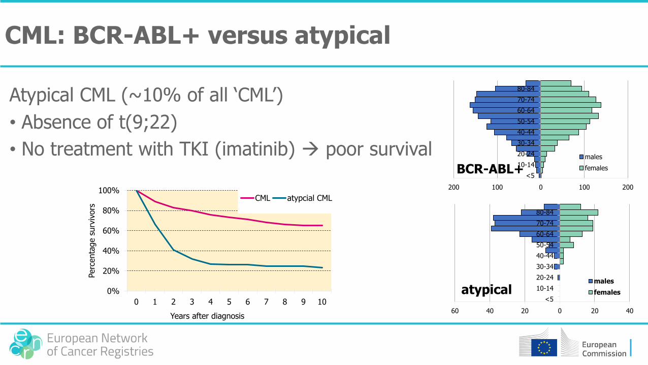
CML: BCR-ABL+ versus atypical
Atypical CML (~10% of all ‘CML’)
• Absence of t(9;22)
• No treatment with TKI (imatinib) poor survival
0%
20%
40%
60%
80%
100%
0 1 2 3 4 5 6 7 8 9 10
Perc
enta
ge s
urv
ivors
Years after diagnosis
CML atypcial CML
200 100 0 100 200
<5
10-14
20-24
30-34
40-44
50-54
60-64
70-74
80-84
males
females
60 40 20 0 20 40
<5
10-14
20-24
30-34
40-44
50-54
60-64
70-74
80-84
males
femalesatypical
BCR-ABL+
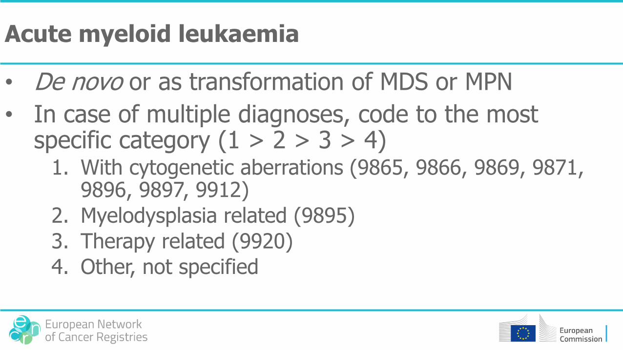
• De novo or as transformation of MDS or MPN
• In case of multiple diagnoses, code to the most specific category (1 > 2 > 3 > 4)
1. With cytogenetic aberrations (9865, 9866, 9869, 9871, 9896, 9897, 9912)
2. Myelodysplasia related (9895)
3. Therapy related (9920)
4. Other, not specified
Acute myeloid leukaemia
Examples
• Acute megakaryoblastic leukaemia (9910), therapy related (9920) 9920
• Acute myeloid leukaemia, t(8;21) (9896), therapy related (9920) 9896
• Acute myelomonocytic leukaemia (9867), t(8;21) (9896) 9896
Acute myeloid leukaemia

www.encr.eu