Embed Size (px)
Citation preview
1
Scavenger Receptor-BI is a Receptor for Lipoprotein(a)
Xiao-Ping Yang 1, Marcelo J. Amar2, Boris Vaisman2, Alexander V. Bocharov3, Tatyana G.
Vishnyakova3, Lita A. Freeman2, Roger J. Kurlander4, Amy P. Patterson 3, Lewis C. Becker 1
and Alan T. Remaley2
1 Cardiology Division, Department of Medicine, Johns Hopkins Medical Institutions. Baltimore,
Maryland.
2Lipoprotein Metabolism Section, Cardiopulmonary Branch, NHLBI, National Institutes of
Health, Bethesda, Maryland.
3Division of Diabetes, Endocrinology and Metabolic Diseases, NIDDK, National Institutes of
Health, Bethesda, Maryland.
4Department of Laboratory Medicine, NIH Clinical Center, National Institutes of Health,
Bethesda, Maryland.
Correspondence to Alan T. Remaley, M.D., Ph.D. National Institutes of Health,
Bldg.10, Rm. 2C-433, Bethesda, MD 20892-1508, USA Tel: 301-402-9796. Fax: 301-402-1885.
Email: [email protected]
Running title: Lp(a) and SR-BI
by guest, on January 3, 2019w
ww
.jlr.orgD
ownloaded from
2
ABSTRACT
Scavenger receptor class B type I (SR-BI) is a multi-ligand receptor that binds a variety
of lipoproteins, including high density lipoprotein (HDL) and low density lipoprotein (LDL), but
lipoprotein(a) (Lp(a)) has not been investigated as a possible ligand. Stable cell lines (HEK293
and HeLa) expressing human SR-BI were incubated with protein- or lipid-labeled Lp(a) to
investigate SR-BI-dependent Lp(a) cell association. SR-BI expression enhanced the association
of both 125I- and Alexa-Fluor-labeled protein from Lp(a). By confocal microscopy, SR-BI was
also found to promote the internalization of fluorescent lipids (BODIPY-CE- and DiI-labeled)
from Lp(a), and by immunocytochemistry the cellular internalization of apolipoprotein (a) and
apolipoprotein B. When dual-labeled (3H-Cholesteryl ether,125I-protein) Lp(a) was added to cells
expressing SR-BI, there was a greater relative increase in lipid uptake over protein, indicating
that SR-BI mediates selective lipid uptake from Lp(a). Compared to C57BL/6 control mice,
transgenic mice overexpressing human SR-BI in liver were found to have increased plasma
clearance of 3H-cholesteryl ester-Lp(a), whereas Sr-b1-KO mice had decreased plasma clearance
(FCR: 0.63+0.08/d, 1.64+0.62/d , and 4.64+0.40/d, for Sr-b1-KO, C57Bl/6, and hSR-BI-Tg
mice, respectively). We conclude that Lp(a) is a novel ligand for SR-BI and that SR-BI mediates
selective uptake of Lp(a)-associated lipids.
Supplementary key words: Scavenger Receptor-BI, Lipoprotein (a), Lipoprotein Receptors,
Atherosclerosis
by guest, on January 3, 2019w
ww
.jlr.orgD
ownloaded from
3
List of abbreviations: apo(a), apolipoprotein (a); apoB, apolipoprotein B; CE, cholesteryl ester;
CEth, cholesteryl ether; CETP, cholesteryl ester transfer protein; HDL, high-density lipoprotein;
hSR-BI-Tg, human SR-BI transgenic; HUVEC, human umbilical vein endothelial cell; LDL,
low-density lipoprotein; Lp(a), lipoprotein (a); LPDS, lipoprotein deficient serum; SR-BI,
human scavenger receptor-BI; Sr-b1, mouse scavenger receptor-BI, Sr-b1-KO, mouse Sr-b1
knockout; VLDL, very low-density lipoprotein.
by guest, on January 3, 2019w
ww
.jlr.orgD
ownloaded from
4
Scavenger receptor class B type I (SR-BI) is a multi-ligand receptor. It binds a variety of
oxidized as well as native lipoproteins, including high density lipoprotein (HDL) and low density
lipoprotein (LDL) (1-4). SR-BI preferentially mediates the cellular uptake of neutral lipids over
protein from lipoproteins, by a process termed selective uptake. In the liver, the selective uptake
of cholesteryl esters from HDL potentiates the reverse cholesterol transport pathway, by
increasing the hepatic excretion of cholesterol (5).
Lp(a) is a pro-atherogenic lipoprotein particle that contains one copy of apolipoprotein(a)
[apo(a)] covalently linked to apoB-100 by a disulfide bond. It is found in some primates but is
not present in most other mammalian species (6). Variation in the plasma level of Lp(a) is
largely under genetic control, with polymorphisms in the kringle 4 repeat of apo(a) accounting
for 40% of the variation. In general, apo(a) size is inversely associated with plasma Lp(a) level,
but the relationship between apo(a) size and Lp(a) concentration varies among ethnic groups (7-
9). In addition, a pentanucleotide repeat polymorphism (PNRP) in the 5’control region of apo(a)
accounts for 10-14% of the variation in Lp(a) concentrations in Caucasians (10).
Human tracer studies have shown that plasma concentrations of Lp(a) were not only
positively correlated with the secretion rates of Lp(a) protein, but also negatively correlated with
the fractional catabolic rates of Lp(a) proteins, both apo(a) and apoB-100, indicating that plasma
Lp(a) levels are also regulated by its rate of catabolism (10). However, the receptors that bind
and mediate the catabolism of Lp(a) are not well characterized. Although Lp(a) is structurally
and compositionally similar to LDL, it is known to be a relatively poor ligand for the LDL-
receptor (11), as well as for the lipoprotein receptor-related protein (12). Lp(a) can bind to the
VLDL-receptor, but mice defective in this receptor showed only a modest delay in the
by guest, on January 3, 2019w
ww
.jlr.orgD
ownloaded from
5
catabolism of heterologous Lp(a) (13), suggesting the presence of other receptor(s). Because of
the close structural similarity of Lp(a) with LDL, a known ligand for SR-BI (1-4), we
investigated in this study whether SR-BI can also serve as a receptor for Lp(a).
MATERIALS AND METHODS
Generation of stable cell lines
Human SR-BI cDNA, generated by RT-PCR from total RNA of human monocytes, was
inserted into a pcDNA3 vector (Invitrogen, Carlsbad, CA) and sequence-confirmed. HEK 293
and HeLa cells were purchased from American Type Culture Collection (ATCC). Cells were
cultured in DMEM growth medium (DMEM, 4 mM glutamine, 10% fetal bovine serum, 100
u/mL penicillin, 100 µg/mL streptomycin). SR-BI cDNA was transfected into 80% confluent
HEK 293 and HeLa cells (14), using Effectene transfection reagent (Qiagen, Valencia, CA).
Cell clones were selected in DMEM growth medium containing 2 mg/mL G418 for 2 weeks and
maintained with 0.25 mg/mL G418. To verify SR-BI expression, cells before or after transfection
were lysed and analyzed by Western blotting with rabbit anti-human polyclonal SR-BI or rabbit
anti-human polyclonal VLDLr antibodies (Abcam, Cambridge, UK). Western blotting was
performed as previously described (15).
Preparation of radiolabeled and fluorescently labeled Lp(a)
Lp(a) was purchased from Meridian Life Science, Inc. (Saco, MA). Each lot of Lp(a) was
prepared from a single donor with documented high Lp(a) concentration to enhance yield and
purity. Beginning with plasmapheresis, plasma was drawn directly into a Trasylol (aprotinin)-
containing receiver, giving 100KIU aprotinin/mL plasma. Immediately thereafter, sodium azide
(0.01%) and EDTA (0.001%) were added and the Lp(a) was isolated by ultracentrifugation at
by guest, on January 3, 2019w
ww
.jlr.orgD
ownloaded from
6
4oC followed by Lysine Sepharose chromatography (16), with elution by aminocaproic acid to
complete the purification. The purified Lp(a) was analyzed by SDS-PAGE without reducing
agents for the appearance of a single band, indicating the absence of other proteins and
lipoproteins. Lp(a) was sent the day of preparation, stored in 15 mM NaCl and 1 mM EDTA, pH
7.4, and used within 10 days to minimize oxidation and aggregation. The purity of Lp(a)
preparations was confirmed by fast protein liquid chromatography in our laboratory. The protein
molecular weight of the Lp(a) used in this study is approximately 1000 kDa (apo(a)
predominantly 550 kDa and apoB 500kDa), and the protein concentration was 0.6 mg/ml.
The Chloramine-T method was used for 125I iodination of Lp(a) protein. A labeling kit,
including carrier-free 125I, was purchased from MP Biomedicals LLC (Solon, OH). Labeled
lipoproteins were purified by a desalting column (PD-10 column, GE Healthcare,
Buckinghamshire, UK). The integrity and purity of all labeled lipoproteins was confirmed by
fast protein liquid chromatography. The 125I-labeled protein specific activities ranged between
400-456 cpm/ng protein. To fluorescently label Lp(a) proteins, an Alexa Fluor® 488 protein
labeling kit (Invitrogen) was used according to manufacturer’s instructions.
[3H]-Cholesteryl ether (3H-CEth) is a nonhydrolyzable analog of cholesteryl ester (17). (3H-
CEth)-labeled Lp(a), used in cell culture studies, was prepared as previously described (16).
Briefly, 25 mL human donor serum was adjusted to a density of 1.215 g/mL with potassium
bromide (Sigma Aldrich, St. Louis, MO), and ultracentrifuged at 40,000 rpm (Beckman L8-55
ultracentrifuge) at 80C for 48 h to obtain a lipoprotein-free serum fraction containing cholesteryl
ester transfer protein (CETP) (1.215 g/mL bottom fraction). 200-250 µCi of cholesteryl
hexadecyl ether [Cholesteryl-1,2-3H(N)-] (Perkin Elmer, Waltham, MA) was mixed with 22
nmol butylated hydroxytoluene (Sigma), and evaporated under nitrogen in a glass tube. After
by guest, on January 3, 2019w
ww
.jlr.orgD
ownloaded from
7
addition of 1 mL TSE buffer (50 mM Tris–HCl pH 7.4, 150 mM NaCl, 2 mM EDTA) and 80 µl
egg L-phosphatidylcholine (100 mg/mL, Sigma), the 3H-lipid mixture was sonicated. 600 µg of
Lp(a) was added to 3 mL of the d>1.215 g/mL bottom fraction. 1 mL of the 3H-lipid mixture was
added, and then incubated at 370C overnight under nitrogen. The labeled Lp(a) was then
dialyzed in TSE buffer, adjusted to a density of 1.050 g/mL and centrifuged (Beckman Optima
TL ultra-centrifuge) at 85,000 rpm at 40C for 12 h to separate any unincorporated lipid tracer.
The bottom fraction was dialyzed and re-adjusted to a density of 1.125 g/mL and centrifuged for
24 h. The upper fraction, which contained the final 3H-Lp(a) preparation, was dialyzed with PBS
buffer. The specific activities of the radiolabeled Lp(a) preparations were approximately 200-265
dpm/ng Lp(a) protein. For dual-labeled Lp(a), Lp(a) was first labeled with [3H]-CEth followed
by 125I labeling .
[3H]-Cholesteryl ester (3H-CE)-labeled Lp(a), used for kinetic studies in mice (Fig. 7),
was generated as previously described (18). 250 μCi of 3H-cholesteryl oleate (cholesteryl-
[1,2,6,7- 3H (N)] oleate) (60 Ci/mmol; American Radiolabeled Chemicals, Inc, St. Louis, MO)
was dried under nitrogen, resuspended in 10 μl of absolute ethanol, and added dropwise over 3
min to the lipoprotein solution (0.6 mg/ml protein concentration) with shaking and vortexing as
previously described (18). [3H]-CE-Lp(a) was then reisolated by ultracentrifugation, dialyzed
overnight against 1 × PBS, 0.01% EDTA, and finally analyzed by agarose gel electrophoresis to
ensure integrity of the particle.
HDL (d=1.063 to 1.21) and LDL (d=1.019 to 1.063) were isolated by density gradient
ultracentrifugation, as previously described (19) and used within 10 days of preparation.
Cell Association Studies
by guest, on January 3, 2019w
ww
.jlr.orgD
ownloaded from
8
To quantitate cellular Lp(a) cell association using [125I]-Lp(a), HEK 293 cells grown to
95% confluence in 24-well plates were incubated with the indicated concentration of [125I]-Lp(a)
for 1.5 hours either in the absence of unlableled Lp(a) (for calculating total cell association) or in
the presence of a 40-fold excess of unlabeled Lp(a) (for calculating nonspecific cell association)
at 370C. Cells were washed 4 times with PBS, and harvested with 0.01 N NaOH and 0.1% SDS
solution. Specific cell association was calculated by subtracting nonspecific cell association
from total cell association value and expressed as ng Lp(a)/mg cellular protein.
To observe cellular Lp(a) cell association by flow cytometry, HEK 293 cells were
incubated with 10 µg/mL (10 nM) Alexa Fluor® 488 labeled Lp(a) in DMEM containing 2
mg/mL BSA and 20 mM HEPES at 370C for 1.5 hrs. Cells were washed with PBS, detached
with a cell dissociation solution (Mediatech, Herndon, VA), fixed with 4% paraformaldehyde in
PBS, and analyzed by a fluorescence-activated cell sorter (FACS, model A; Hitachi).
To observe cellular Lp(a) cell association by confocal microscopy, HeLa cells on
coverslips were incubated with 5 µg/mL Alexa Fluor® 488 labeled Lp(a) in DMEM containing 2
mg/mL BSA and 20 mM HEPES, at 370C for 1 h, followed by a chase without labeled Lp(a) for
1 hour, then fixed and examined by confocal microscopy.
For the competition assay, unlabeled HDL and LDL were prepared by density gradient
ultra-centrifugation, using fresh human donor plasma and used within 10 days of preparation. To
convert the protein mass of lipoproteins to moles, protein concentrations (in mg/ml) of purified
LDL, Lp(a), and HDL were first determined by the BCA assay (BioRad), using BSA as the
standard. Molar concentrations were determined using estimated average protein molecular
weights of approximately 500 kDa for LDL (one 500 kDa apoB/particle), approximately 1000
by guest, on January 3, 2019w
ww
.jlr.orgD
ownloaded from
9
kDa for Lp(a) (one 500 kDa apoB + one 550 kDa apo(a)/particle), and 200 kDa for HDL (400
kDa average MW, 50% average protein content (20)), respectively.
For fluorescent competition studies, HEK 293 cells expressing SR-BI were incubated
with 10 nM AlexFluor-488 labeled Lp(a) in the presence or absence of 40-fold excess unlabeled
competitors (Lp(a), HDL or LDL) for 1.5 hours, then followed by the flow cytometry assay.
For 3H-Lp(a) competition studies, 3H-CEth-Lp(a) plus competitors (Lp(a), HDL or LDL)
at the indicated concentrations were added to the cell culture medium (DMEM with 5%
lipoprotein deficient serum (LPDS) (Sigma)) and incubated with the cells for 1.5 hours. Cells
were washed 5 times with PBS, and cell lysates, as prepared above, were used to measure protein
and lipid uptake.
Fluorescent lipid labeling and uptake studies
Fluorescent cholesteryl ester labeled Lp(a) [BODIPY-CE-Lp(a)] was prepared as previously
described (21). Briefly, 0.4 mg of BODIPY- Cholesteryl FLC12 (Invitrogen) was mixed with
0.2 mg of phosphatidylcholine (100 mg/ml, Sigma) and 22 nmol butylated hydroxytoluene, and
dried under nitrogen. After sonication in 1 mL of TBS, the mixture of fluorescent lipids was
added to 200 µg of Lp(a) in 2 ml of 1.215 g/mL bottom fraction with Ampicillin (final volume 4
ml), and gently shaken for 12 hours at 370C. The labeled BODIPY-CE-Lp(a) fraction (density
1.050 - 1.125 g/mL) was collected by sequential ultra-centrifugation of the sample at 85000 rpm
for 12 and 24 hours respectively, and dialyzed against PBS buffer.
DiI labeled lipoprotein was prepared as previously described (22). Briefly, 200 µg of Lp(a)
was mixed with 0.3 mg DiI [DiIC18(3), Invitrogen] (final concentration 100 µg/ml), 2 ml of
lipoprotein deficient serum (LPDS) (Sigma), and 100 µg/ml ampicillin, and gently shaken at 37
0C overnight. The labeled Lp(a) was adjusted to a density of 1.125 g/mL with potassium
by guest, on January 3, 2019w
ww
.jlr.orgD
ownloaded from
10
bromide (Sigma), and centrifuged at 85000 rpm (Beckman Optima TL ultra-centrifuge) for 24
hours to remove unincorporated dye. The supernatant was collected and dialyzed against PBS
buffer.
To observe cellular uptake of fluorescent lipids, HeLa cells transfected with vector (Mock) or
SR-BI cDNA were seeded onto glass coverslips coated with 0.1 mg/mL poly-L-lysine in 6-well
plates. After culture in DMEM with 5% LPDS for 24 hours, the cells were replaced with fresh
medium with 10 µg/ml fluorescent labeled Lp(a) at 370C for 1 h or 3 h. The medium was
discarded, and the coverslips were washed five times with PBS buffer and fixed with 4%
paraformaldehyde. After mounting and sealing, the coverslips were examined under a Zeiss 510
confocal microscope.
Immunohistochemistry
Sheep anti-human polyclonal apo(a) antibody was purchased from Meridian Life Science Inc,
and goat anti-human apoB polyclonal antibody from R&D Systems (Minneapolis, MN). The
antibodies for immunostaining were labeled with either the Alexa Fluor 488 or 546 Monoclonal
Antibody Labeling Kits (Molecular Probes, Eugene, OR). For direct immunostaining, HeLa
cells were cultured in DMEM medium with 5% LPDS serum for 24 hours; the cells were then
replaced with fresh DMEM (containing 2 mg/mL BSA and 20 mM HEPES) with Lp(a) (final
concentration 15 µg/mL), at 370C for 2 h. After washing 6 times, the cells were fixed with 4%
paraformaldehyde for 10 min, and permeabilized with methanol at -200C for 10 min.
Immunostaining was then performed with the fluorescent labeled antibodies. For indirect
immunostaining, the cells were incubated with 15 µg/mL Lp(a) at 40C for 1 hour, washed, and
then incubated at 370C for 2 hours. The cells were fixed, permeabilized, blocked, and incubated
by guest, on January 3, 2019w
ww
.jlr.orgD
ownloaded from
11
with primary antibodies for 2 hours, followed by detection with Northern LightsTM donkey anti-
sheep IgG-NL637 and anti-goat IgG-NL493 (R&D Systems).
Protein degradation studies
Protein degradation was monitored by determining the release of the 125I-labeled protein
degradation product (125I-monoiodotyrosine containing peptides) into the conditioned cell culture
media, as previously described (23). Briefly, 0.25 ml of 50% trichloracetic acid was added to 1
ml of conditioned media in a glass tube; the mixture was incubated at 40C for 30 minutes and
then centrifuged to precipitate undegraded 125I-lipoprotein. 0.5 ml of supernatant was then
mixed with 5 µl of 40% potassium iodide, followed by addition of 20 µl of 30% H2O2. 1 ml
chloroform was added and vortexed to extract free 125I-iodine. The upper aqueous layer was
removed and 0.2 ml was gamma-counted to measure the release of 125I-labeled peptides. Protein
degradation in cell lysates was similarly measured.
Generation of hSR-BI transgenic mice
The liver-specific expression vector pLIV.11 (24) was used to develop mice expressing
human SR-BI (hSR-BI). The full-length (1.7-kb) human SR-BI cDNA (GenBank: BC112037.1)
was flanked by Not I linkers and inserted into the unique Not I site of pLIV.11 and correctly
oriented clones were identified after digestion with Sph I and Aat II. The plasmid, pLIV11-hSR-
BI, was then digested with Sal I and Spe I, and a 11.6-kb DNA fragment, containing the
complete hSR-BI expression cassette, was isolated from a 0.8% agarose gel and purified by
CsCl density gradient ultracentrifugation (Beckman TL-100 tabletop ultracentrifuge, 95000
RPM, 24 hrs, 20ºC). After dialysis against 10 mM Tris-HCl, pH 7.4, 0.1 mM EDTA, the DNA
fragment was microinjected into pronuclei of fertilized eggs from C57Bl/6J females (Jackson
Laboratory, Bar Harbor, ME). Genotyping and expression analysis of the transgenic mice was
by guest, on January 3, 2019w
ww
.jlr.orgD
ownloaded from
12
performed as previously described (25). A founder line of mice containing approximately 25
copies of the human SR-BI gene per heterozygous genome was used to establish a colony of
human SR-BI transgenic (hSR-BI-Tg) mice. Similar to previous reports (4), the hSR-BI
transgenic mice had decreased plasma levels of HDL-C compared to control mice (HDL-C: hSR-
BI-Tg <3.0 mg/dL vs. C57Bl/6 50.2+1.5 mg/dL, n = 21, p<0.001).
In vivo tracer kinetics of 3H-Cholesteryl Ester-Lp(a)
[3H]-CE-Lp(a) (total of 1x10⁶ CPM) was injected into the retro-orbital sinus of C57Bl/6
mice (n=3), Sr-b1-KO (n=3), and hSR-BI-Tg mice (n=3). Approximately 50 microliters of
blood was collected at the indicated time points and plasma (25 microliters) was then separated
and counted for 5 min in a Tri-Carb 2500 TR liquid scintillation counter (Packard Instrument
Co., Downers Grove, IL). Plasma decay curves for [3H]-CE-Lp(a) were generated by dividing
the plasma radioactivity at each time point by the plasma radioactivity at the initial 1-min time
point. This initial time point did not differ among the study groups (P>0.4). The fractional
catabolic rate was determined from the area under the plasma radioactivity curves, using a multi-
exponential curve fitting technique on the SAAM II program.
Statistical Analysis
Statistical analyses were performed using GraphPad Prism Software (GraphPad Software,
Inc. La Jolla, CA). Unless otherwise indicated all results are expressed as the mean ± 1 SD of at
least triplicates, and the results shown represent two or more independent experiments. A 2-
tailed Student T-test was used for statistical analysis, and P values <0.05 were considered
significant.
RESULTS
by guest, on January 3, 2019w
ww
.jlr.orgD
ownloaded from
13
Stable cell lines expressing SR-BI
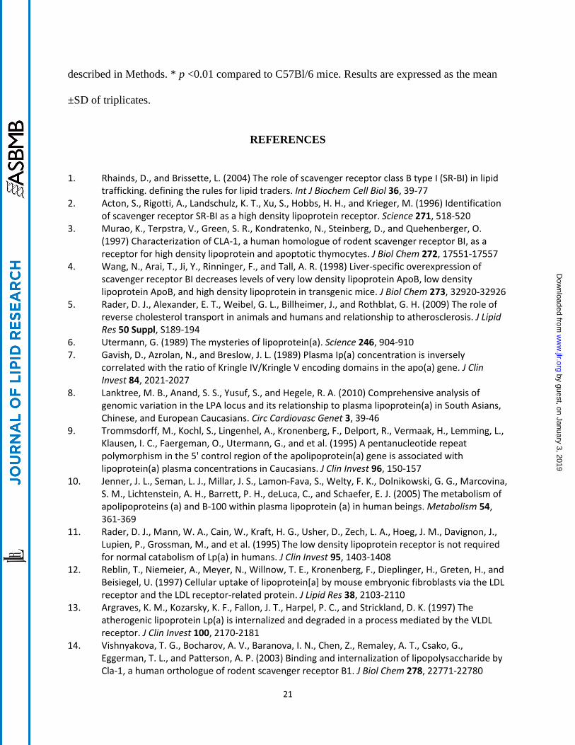
By Western blotting, HEK 293 and HeLa cells do not express significant amounts of
endogenous SR-BI (Fig. 1A) or VLDL-receptor (Fig. 1B), as previously reported (26). After
transfection with human SR-BI cDNA, both cell lines expressed significantly increased amounts
of SR-BI protein (Fig. 1A). Similarly, SR-BI was not detected by confocal microscopy in mock-
transfected HEK 293 or HeLa cells (Fig. 1C, D), after staining with a rabbit anti-human SR-BI
polyclonal antibody. In contrast, stably transfected HEK 293 and HeLa cells showed abundant
staining for SR-BI, which was primarily present on the cell surface, but also in intracellular
vesicles, particularly in the case of HeLa cells.
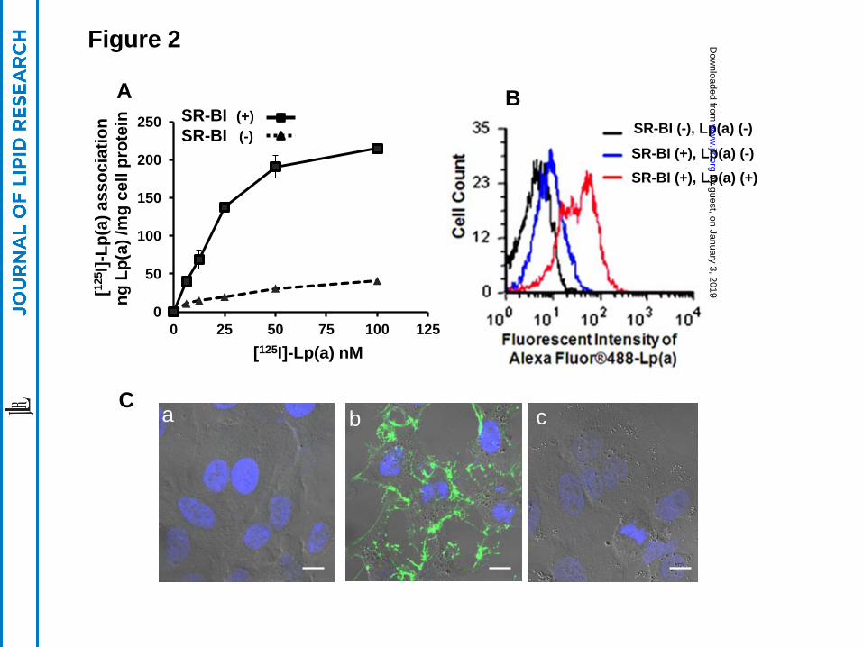
SR-BI mediates the association of Lp(a) with cells
Compared to control HEK 293 cells, a HEK 293 cell clone stably expressing human SR-
BI showed a 3-4-fold increase in its association with Lp(a), radiolabeled in its protein moiety by
125I (Fig. 2A). Similarly, by flow cytometry, stably transfected HEK 293 cells showed an
approximate 5-fold increased association with Alexa-fluorescent labeled Lp(a) protein compared
to control cells (Fig. 2B).
Stably transfected HeLa cells expressing SR-BI, which were better than HEK 293 cells
for visualization by confocal microscopy, also associated with increased amounts of Lp(a) that
was fluorescently labeled in its protein moiety by AlexaFluor-488 (Fig. 2C). Cell-bound Lp(a)
(green) could be detected in SR-BI transfected HeLa cells (Fig 2C, panel b) but not in mock-
transfected HeLa cells (Fig 2C, panel a) or in HeLa cells transfected with the human LDL-
receptor (Fig 2C, panel c).
by guest, on January 3, 2019w
ww
.jlr.orgD
ownloaded from
14
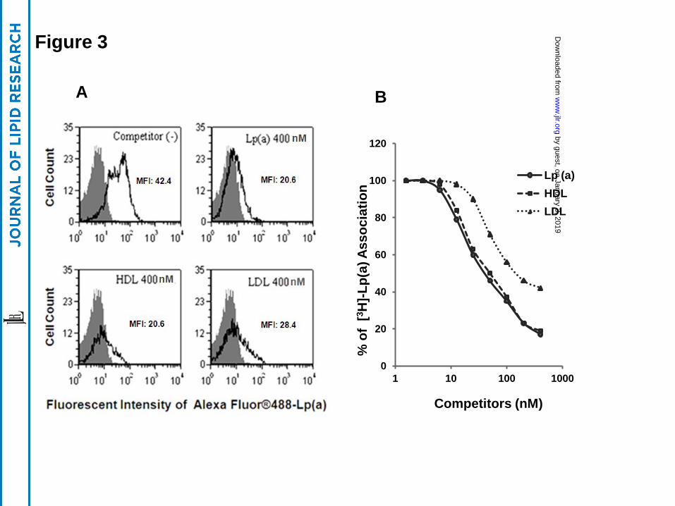
A 40-fold molar excess of unlabeled Lp(a), HDL, or LDL almost completely inhibited
the association of fluorescent labeled Lp(a) protein with HEK 293 cells transfected with SR-BI
(Fig. 3A). Similar results were obtained when unlabeled Lp(a), HDL or LDL were used to
compete for association of Lp(a) labeled with 3H-CEth, a non-hydrolyzable analogue of
cholesteryl ester (Fig. 3B), although LDL was found to be slightly less effective than the other
lipoproteins tested.
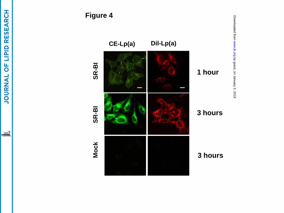
SR-BI mediates the uptake of lipid and protein components of Lp(a)
The uptake of the various lipid and protein components of Lp(a) by SR-BI-expressing
cells was further investigated by confocal microscopy. After incubating SR-BI transfected HeLa
cells with Lp(a) labeled with either BODIPY- Cholesteryl ester (green) (a marker for a neutral
core lipid) or DiI [DiIC18(3)] (red) (a marker for a surface amphipathic lipid), a time dependent
uptake of both fluorescent lipids was observed (Fig. 4, top panels). After one to three hours of
incubation, fluorescent lipids were primarily observed in intracellular vesicles around the Golgi
body and nucleus (Fig. 4, top and middle panels). In contrast, no significant lipid uptake was
observed in control cells after 3 hours (Fig. 4, lower panels).
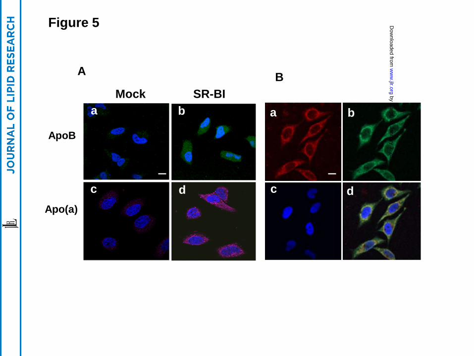
After incubation with Lp(a), HeLa cells were indirectly stained with either anti-apoB
(green) or anti-apo(a) (pink) antibodies (Fig. 5A). Readily observable apoB (green) and apo(a)
(pink) staining was seen only in HeLa cells transfected with SR-BI and not in control cells (Fig.
5A). When cells were stained directly with both fluorescent labeled antibodies (apoB-green:
apo(a)-red) significant intracellular co-localization (yellow) of the two proteins was seen,
particularly in the perinuclear region (Fig. 5B).
SR-BI mediates selective lipid uptake from Lp(a)
by guest, on January 3, 2019w
ww
.jlr.orgD
ownloaded from
15
HEK 293 cells were incubated with 10 µg/ml of dual-labeled (3H-CEth, [125I]-protein
Lp(a)) (Fig. 6). Compared to mock-transfected cells (lower panel), HEK 293 cells expressing
SR-BI (upper panel) showed a marked increase in the association of 3H-CEth labeled Lp(a) (Fig.
6). Although SR-BI transfected cells also showed an increase in the association of 125I-labeled
Lp(a) protein, the increase in protein binding relative to control cells was not as large (Fig. 6).
The level of 125I-protein counts associated with the SR-BI (+) cells also reached a plateau at 2
hours, whereas the cellular uptake of 3H-Cholesteryl ether from Lp(a) continued to increase over
the 8 hour incubation period. Negligible amounts of degraded Lp(a) protein was observed in the
conditioned cell media from either cell line.
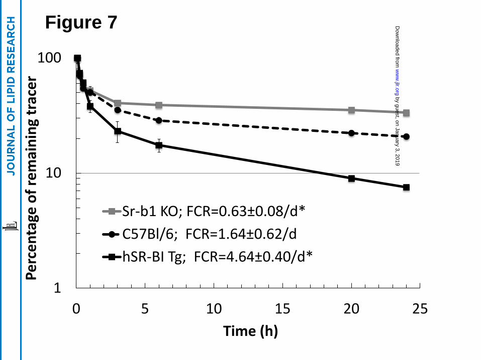
SR-BI mediates the plasma clearance of Lp(a)
In order to investigate the ability of SR-BI to interact with Lp(a) in vivo, 3H-CE-Lp(a)
was injected into three lines of mice with various levels of SR-BI, namely control C57BL/6
mice, Sr-b1-KO mice and hSR-BI-Tg mice, which overexpress the human SR-BI transgene in
the liver of C57Bl/6 mice. An approximate 3-fold increase in the plasma clearance of 3H-CE-
Lp(a) was observed in the hSR-BI-Tg mice compared to the control mice (Fig. 7). In contrast, a
marked delay in the plasma clearance of 3H-CE-Lp(a) was evident in Sr-b1-KO mice compared
to the control C57BL/6 mice.
DISCUSSION
A major finding from this study is that Lp(a) is a ligand for the SR-BI receptor. This was
shown to occur in two different cell lines, namely HEK 293 cells and HeLa cells. Both cell lines
when transfected with SR-BI showed increased association of Lp(a) (Figs. 2, 3). This was true
for both the lipid and protein components of Lp(a). Based on the competition studies (Fig. 3), all
by guest, on January 3, 2019w
ww
.jlr.orgD
ownloaded from
16
of the lipoproteins competed with Lp(a) for SR-BI binding. This was not unexpected, because
SR-BI is a scavenger receptor and is already known to bind to LDL and HDL. Interestingly,
LDL was slightly less effective than the unlabeled Lp(a) as a competitor, which suggests that
there may be a possible interaction of the apo(a) component of Lp(a) with SR-BI, which is
lacking in LDL.
Another major finding from this study is that SR-BI can promote the selective lipid
uptake of cholesteryl esters from Lp(a), as it does from other lipoproteins (27, 28). Although SR-
BI was found to mediate the cellular uptake of all the major protein and lipid components of
Lp(a) (Figs. 4, 5), it appears to preferentially promote the uptake of the cholesteryl esters, a
neutral core lipid (Fig. 4). When SR-BI expressing cells were incubated with dual labeled Lp(a),
a greater relative increase in the uptake of labeled cholesteryl ether was observed over the
protein label (Fig. 6). Furthermore, although some apoB and apo(a) protein label could be
observed in SR-BI expressing cells compared to control cells (Fig. 5), negligible amounts of
Lp(a) protein degradation was detected (Fig. 6).
The cell culture experiments showing that SR-BI can serve as a receptor for Lp(a) was
further confirmed in plasma turnover studies in mice (Fig. 7). hSR-BI-Tg mice, with increased
expression of hepatic SR-BI, showed increased clearance of plasma 3H-CE-Lp(a) compared to
C57BL/6 mice, whereas Sr-b1-KO mice had decreased clearance. Mice do not normally express
Lp(a), but the modulation of hepatic levels of SR-BI in mice resulted in changes in the
catabolism of exogenously added Lp(a) in a manner predicted based on the cell culture binding
studies. It is important to note, however, that although there was a delay in Lp(a) catabolism in
Sr-b1-KO mice, approximately 65% of the 3H-CE-Lp(a) was still removed from plasma of Sr-
by guest, on January 3, 2019w
ww
.jlr.orgD
ownloaded from
17
b1-KO mice 24 h after injection (Fig. 7). This must indicate that there are other receptors and/or
pathways for Lp(a) catabolism from the circulation.
Compared to LDL, Lp(a) is typically present in the plasma at a much lower
concentration (6); nevertheless, Lp(a) has been shown on a per particle basis to be more pro-
atherogenic (6). Various modifications of LDL in the vessel wall, such as oxidation and
aggregation, make it a good ligand for several different types of scavenger receptors on
macrophages (29), and perhaps Lp(a) can also undergo some of these same modifications.
Interestingly, Lp(a) and not LDL has also been shown to be the principal carrier of negatively
charged oxidized lipids in plasma (30). Enrichment of anionic charged lipids has previously been
shown to enhance the uptake of lipoproteins by SR-BI (31); thus, negatively charged oxidized
lipids could potentially explain the affinity of Lp(a) for SR-BI. This is an important area of
future investigation not only for possibly explaining the uptake of Lp(a) by SR-BI but also for
explaining why Lp(a) may be more pro-atherogenic than LDL (30). The delivery of oxidized
lipids to cells is believed to be pro-inflammatory (32, 33). Also of interest, cholesteryl ester
hydroperoxides from oxidized lipoproteins have been shown to be better substrates than
cholesteryl esters for selective lipid uptake by SR-BI (31). Given the myriad effects of oxidized
cholesterol on gene regulation and inflammation, the delivery of oxidized lipids from Lp(a) to
cells by SR-BI could be a contributing factor to the atherogenicity of Lp(a). The atherogenic risk
from Lp(a) is in part related to its plasma level, which is known to be inversely related to the size
of its apo(a) isoform (34). Whether apo(a) contributes to the binding of Lp(a) to SR-BI and
whether different size isoforms of apo(a) can also affect the binding of Lp(a) to SR-BI is not
known but would also be an important area of future investigation.
by guest, on January 3, 2019w
ww
.jlr.orgD
ownloaded from
18
In summary, SR-BI was found to bind and promote the cellular uptake of Lp(a). In
particular, SR-BI was found to favor the selective lipid uptake of cholesteryl esters from Lp(a).
These results indicate a possible new physiologic role for SR-BI as a receptor for Lp(a) and
suggests several novel mechanisms for the atherogenic properties of Lp(a).
ACKNOWLEDGMENTS
This work was supported by grants HL72518 (LCB), HL59684 (LCB), and HL65608
(LCB) from the NHLBI and intramural research funding of NHLBI (ATR). We thank the NHLBI
Light Microscopy Core for the use of their confocal microscopes.
FIGURE LEGENDS
Figure 1. Expression of SR-BI in transfected HEK 293 cells and HeLa cells. (Panel A) HEK
293 or HeLa cells before (mock) or after transfection (SR-BI) with SR-BI were analyzed by
immunoblots for SR-BI or GAPDH. (Panel B) Lysates from HUVEC, HEK 293 and HeLa cells
were analyzed by immunoblots for VLDL-receptor or GAPDH. 20 µg protein were loaded per
lane in all immunoblots. (Panels C and D) HEK 293 cells (Panel C) or HeLa cells (Panel D)
before (mock) or after transfection (SR-BI) were stained with a primary human SR-BI antibody
and analyzed by confocal microscopy (upper panel 20X image, lower panel 63X image; scale
bars are 50 um for 20X and 10 um for 63X). Cells were counter stained with DAPI.
Figure 2. Enhanced Lp(a) association with SR-BI-transfected cells. (Panel A) HEK 293 cells
stably transfected with (solid line) or without (dashed line) SR-BI were incubated with
increasing concentrations of [125I]-Lp(a) at 370C for 1.5 hours. Specific cell association of [125I]-
Lp(a) (shown) is expressed as ng Lp(a)/mg cellular protein. Specific cell association was
determined by subtracting nonspecific cell association (residual [125I]-Lp(a) cell association in
by guest, on January 3, 2019w
ww
.jlr.orgD
ownloaded from
19
the presence of a 40-fold excess of unlabeled Lp(a)) from total cell association. (Panel B) HEK
293 cells stably transfected with SR-BI were incubated with (SR-BI (+), Lp(a) (+)) or without
(SR-BI (+), Lp(a) (-)) 10 ug/ml Alexa Fluor-488 labeled Lp(a) for 1.5 hours and analyzed by
flow cytometry. SR-BI (-) cells without Lp(a), shown as (SR-BI (-), Lp(a) (-)), was included as a
control to define background staining. (Panel C) HeLa cells transfected with empty vector (a),
SR-BI (b) or LDL receptor (c) were incubated with 5 µg/mL Alexa Fluor-488-Lp(a) at 370C for
1 hour followed by Lp(a)-free chase for 1 hour, fixed and examined by microscopy. Confocal
photographs with differential interference contrast (DIC) are shown (63X). Scale bars are 10 um
for 63X magnification. Cells were counterstained with DAPI.
Figure 3. Unlabeled Lp(a), HDL and LDL compete with fluorescent- or 3H-CEth-labeled
Lp(a) cell association. (Panel A) HEK 293 cells expressing SR-BI were incubated with 10 nM
AlexFluor-488 labeled Lp(a) in the presence or absence of 40-fold excess unlabeled competitors
for 1.5 hours, and analyzed by flow cytometry. Black area shows cells incubated in the absence
of fluorescent labeled Lp(a). MFI: Mean Fluorescent Intensity. (Panel B) HEK 293 cells
transfected with SR-BI were incubated with 10 µg/ml (= 10 nM) [3H]CEth-Lp(a) at 370C in the
presence of the indicated concentration (nM) of competitors for 1.5 hours, and cell lysates were
counted for radioactivity.
Figure 4. SR-BI promotes uptake of fluorescent lipds from Lp(a)
HeLa cells stably transfected with SR-BI (top and middle panels) or without (bottom panels)
were incubated with BODIPY- Cholesteryl ester (green) or Dil [DiIC18(3)] (red) labeled Lp(a)
for 1 hour or 3 hours. After fixation, the cells were examined under confocal microscopy (63X).
Scale bars are 10 um for 63X magnification. Cells were counterstained with DAPI.
by guest, on January 3, 2019w
ww
.jlr.orgD
ownloaded from
20
Figure 5. Detection of cellular internalization of apo(a) and apoB . (Panel A) HeLa cells
stably transfected with SR-BI (b, d) or without (a, c) were incubated with 15 µg/mL Lp(a) at 40C
for 1 hour. After washing, the cells were incubated at 370C for 2 hours, then fixed and exposed
to goat anti-apoB antibody (a, b) or sheep anti-apo(a) antibody (c, d), and detected with
fluorescent labeled donkey anti-goat (green) or donkey anti-sheep (pink) antibodies and analyzed
by confocal microscopy. (Panel B) HeLa cells stably transfected with SR-BI were cultured in the
presence of 15 µg/ml of Lp(a) for 2 hours and washed, then stained with fluorescent labeled anti-
apoB (green) and anti-apo(a) (red) antibodies and analyzed by confocal microscopy (63X). Scale
bars are 10 um for 63X magnification. Merged image (yellow) is shown in panel d. Cells were
counterstained with DAPI. (a) apo(a) staining, (b) apoB staining, (c) DAPI staining, (d) Merge.
Figure 6. SR-BI mediates selective uptake of cholesteryl ester from Lp(a). 293 cells stably
transfected with SR-BI (Top Panel) or without (Bottom Panel) were incubated with 10 µg/ml (10
nM) 3H-CEth,125I-protein dual labeled Lp(a) and harvested at the indicated time points. Cell-
associated radioactivity (3H, solid line; 125I, dashed line) was expressed as ng Lp(a)/mg cell
protein. Conditioned media were also collected and the 125I-Lp(a) degraded protein (dotted line)
was also determined and expressed as ng Lp(a)/mg cell protein. Results are expressed as the
mean ±SD of triplicates.
Figure 7. SR-BI mediates plasma clearance of Lp(a). [3H]-CE-Lp(a) was injected into the
plasma of Sr-b1-KO (n=3), hSR-BI-Tg (n=3) and C57Bl/6 (n=3) mice and remaining counts of
the tracer in the plasma compartment were monitored at the indicated time points. Fifty
microliters of blood was collected in each time point, plasma (25 ul) was separated and counted
for 5 min. Plasma decay curves and fractional catabolic rate (FCR) were determined as
by guest, on January 3, 2019w
ww
.jlr.orgD
ownloaded from
21
described in Methods. * p <0.01 compared to C57Bl/6 mice. Results are expressed as the mean
±SD of triplicates.
REFERENCES
1. Rhainds, D., and Brissette, L. (2004) The role of scavenger receptor class B type I (SR-BI) in lipid trafficking. defining the rules for lipid traders. Int J Biochem Cell Biol 36, 39-77
2. Acton, S., Rigotti, A., Landschulz, K. T., Xu, S., Hobbs, H. H., and Krieger, M. (1996) Identification of scavenger receptor SR-BI as a high density lipoprotein receptor. Science 271, 518-520
3. Murao, K., Terpstra, V., Green, S. R., Kondratenko, N., Steinberg, D., and Quehenberger, O. (1997) Characterization of CLA-1, a human homologue of rodent scavenger receptor BI, as a receptor for high density lipoprotein and apoptotic thymocytes. J Biol Chem 272, 17551-17557
4. Wang, N., Arai, T., Ji, Y., Rinninger, F., and Tall, A. R. (1998) Liver-specific overexpression of scavenger receptor BI decreases levels of very low density lipoprotein ApoB, low density lipoprotein ApoB, and high density lipoprotein in transgenic mice. J Biol Chem 273, 32920-32926
5. Rader, D. J., Alexander, E. T., Weibel, G. L., Billheimer, J., and Rothblat, G. H. (2009) The role of reverse cholesterol transport in animals and humans and relationship to atherosclerosis. J Lipid Res 50 Suppl, S189-194
6. Utermann, G. (1989) The mysteries of lipoprotein(a). Science 246, 904-910 7. Gavish, D., Azrolan, N., and Breslow, J. L. (1989) Plasma Ip(a) concentration is inversely
correlated with the ratio of Kringle IV/Kringle V encoding domains in the apo(a) gene. J Clin Invest 84, 2021-2027
8. Lanktree, M. B., Anand, S. S., Yusuf, S., and Hegele, R. A. (2010) Comprehensive analysis of genomic variation in the LPA locus and its relationship to plasma lipoprotein(a) in South Asians, Chinese, and European Caucasians. Circ Cardiovasc Genet 3, 39-46
9. Trommsdorff, M., Kochl, S., Lingenhel, A., Kronenberg, F., Delport, R., Vermaak, H., Lemming, L., Klausen, I. C., Faergeman, O., Utermann, G., and et al. (1995) A pentanucleotide repeat polymorphism in the 5' control region of the apolipoprotein(a) gene is associated with lipoprotein(a) plasma concentrations in Caucasians. J Clin Invest 96, 150-157
10. Jenner, J. L., Seman, L. J., Millar, J. S., Lamon-Fava, S., Welty, F. K., Dolnikowski, G. G., Marcovina, S. M., Lichtenstein, A. H., Barrett, P. H., deLuca, C., and Schaefer, E. J. (2005) The metabolism of apolipoproteins (a) and B-100 within plasma lipoprotein (a) in human beings. Metabolism 54, 361-369
11. Rader, D. J., Mann, W. A., Cain, W., Kraft, H. G., Usher, D., Zech, L. A., Hoeg, J. M., Davignon, J., Lupien, P., Grossman, M., and et al. (1995) The low density lipoprotein receptor is not required for normal catabolism of Lp(a) in humans. J Clin Invest 95, 1403-1408
12. Reblin, T., Niemeier, A., Meyer, N., Willnow, T. E., Kronenberg, F., Dieplinger, H., Greten, H., and Beisiegel, U. (1997) Cellular uptake of lipoprotein[a] by mouse embryonic fibroblasts via the LDL receptor and the LDL receptor-related protein. J Lipid Res 38, 2103-2110
13. Argraves, K. M., Kozarsky, K. F., Fallon, J. T., Harpel, P. C., and Strickland, D. K. (1997) The atherogenic lipoprotein Lp(a) is internalized and degraded in a process mediated by the VLDL receptor. J Clin Invest 100, 2170-2181
14. Vishnyakova, T. G., Bocharov, A. V., Baranova, I. N., Chen, Z., Remaley, A. T., Csako, G., Eggerman, T. L., and Patterson, A. P. (2003) Binding and internalization of lipopolysaccharide by Cla-1, a human orthologue of rodent scavenger receptor B1. J Biol Chem 278, 22771-22780
by guest, on January 3, 2019w
ww
.jlr.orgD
ownloaded from
22
15. Yang, X. P., Mattagajasingh, S., Su, S., Chen, G., Cai, Z., Fox-Talbot, K., Irani, K., and Becker, L. C. (2007) Fractalkine upregulates intercellular adhesion molecule-1 in endothelial cells through CX3CR1 and the Jak Stat5 pathway. Circ Res 101, 1001-1008
16. Fless, G. M., and Snyder, M. L. (1996) Quantitation of lipoprotein (a) after lysine-sepharose chromatography and density gradient centrifugation. Methods Enzymol 263, 238-251
17. Stein, Y., Dabach, Y., Hollander, G., Halperin, G., and Stein, O. (1983) Metabolism of HDL-cholesteryl ester in the rat, studied with a nonhydrolyzable analog, cholesteryl linoleyl ether. Biochim Biophys Acta 752, 98-105
18. Shamburek, R. D., Pentchev, P. G., Zech, L. A., Blanchette-Mackie, J., Carstea, E. D., VandenBroek, J. M., Cooper, P. S., Neufeld, E. B., Phair, R. D., Brewer, H. B., Jr., Brady, R. O., and Schwartz, C. C. (1997) Intracellular trafficking of the free cholesterol derived from LDL cholesteryl ester is defective in vivo in Niemann-Pick C disease: insights on normal metabolism of HDL and LDL gained from the NP-C mutation. J Lipid Res 38, 2422-2435
19. Redgrave, T. G., Roberts, D. C., and West, C. E. (1975) Separation of plasma lipoproteins by density-gradient ultracentrifugation. Anal Biochem 65, 42-49
20. Gotto, A. M., and Pownall, H. J. (2003) Manual of Lipid Disorders: Reducing the Risk for Coronary Disease Lippincott, Williams and Wilkins, Philadelphia
21. Reaven, E., Tsai, L., and Azhar, S. (1996) Intracellular events in the "selective" transport of lipoprotein-derived cholesteryl esters. J Biol Chem 271, 16208-16217
22. Pitas, R. E., Innerarity, T. L., Weinstein, J. N., and Mahley, R. W. (1981) Acetoacetylated lipoproteins used to distinguish fibroblasts from macrophages in vitro by fluorescence microscopy. Arteriosclerosis 1, 177-185
23. Goldstein, J. L., Basu, S. K., and Brown, M. S. (1983) Receptor-mediated endocytosis of low-density lipoprotein in cultured cells. Methods Enzymol 98, 241-260
24. Fan, J., Wang, J., Bensadoun, A., Lauer, S. J., Dang, Q., Mahley, R. W., and Taylor, J. M. (1994) Overexpression of hepatic lipase in transgenic rabbits leads to a marked reduction of plasma high density lipoproteins and intermediate density lipoproteins. Proc Natl Acad Sci U S A 91, 8724-8728
25. Vaisman, B. L., Demosky, S. J., Stonik, J. A., Ghias, M., Knapper, C. L., Sampson, M. L., Dai, C., Levine, S. J., and Remaley, A. T. (2012) Endothelial expression of human ABCA1 in mice increases plasma HDL cholesterol and reduces diet-induced atherosclerosis. J Lipid Res 53, 158-167
26. Stockinger, W., Hengstschlager-Ottnad, E., Novak, S., Matus, A., Huttinger, M., Bauer, J., Lassmann, H., Schneider, W. J., and Nimpf, J. (1998) The low density lipoprotein receptor gene family. Differential expression of two alpha2-macroglobulin receptors in the brain. J Biol Chem 273, 32213-32221
27. Stangl, H., Cao, G., Wyne, K. L., and Hobbs, H. H. (1998) Scavenger receptor, class B, type I-dependent stimulation of cholesterol esterification by high density lipoproteins, low density lipoproteins, and nonlipoprotein cholesterol. J Biol Chem 273, 31002-31008
28. Stangl, H., Hyatt, M., and Hobbs, H. H. (1999) Transport of lipids from high and low density lipoproteins via scavenger receptor-BI. J Biol Chem 274, 32692-32698
29. Tabas, I., Williams, K. J., and Boren, J. (2007) Subendothelial lipoprotein retention as the initiating process in atherosclerosis: update and therapeutic implications. Circulation 116, 1832-1844
30. Tsimikas, S., and Witztum, J. L. (2008) The role of oxidized phospholipids in mediating lipoprotein(a) atherogenicity. Curr Opin Lipidol 19, 369-377
31. Fluiter, K., Sattler, W., De Beer, M. C., Connell, P. M., van der Westhuyzen, D. R., and van Berkel, T. J. (1999) Scavenger receptor BI mediates the selective uptake of oxidized cholesterol esters by rat liver. J Biol Chem 274, 8893-8899
by guest, on January 3, 2019w
ww
.jlr.orgD
ownloaded from
23
32. Jamieson, D. G., Usher, D. C., Rader, D. J., and Lavi, E. (1995) Apolipoprotein(a) deposition in atherosclerotic plaques of cerebral vessels. A potential role for endothelial cells in lesion formation. Am J Pathol 147, 1567-1574
33. Leonarduzzi, G., Gamba, P., Gargiulo, S., Biasi, F., and Poli, G. (2012) Inflammation-related gene expression by lipid oxidation-derived products in the progression of atherosclerosis. Free Radic Biol Med 52, 19-34
34. Kamstrup, P. R., Tybjaerg-Hansen, A., and Nordestgaard, B. G. (2013) Extreme Lipoprotein(a) Levels and Improved Cardiovascular Risk Prediction. J Am Coll Cardiol 61, 1146-1156
by guest, on January 3, 2019w
ww
.jlr.orgD
ownloaded from
A
Figure 1
Mock SR-B1 Mock SR-B1
20X
63X
C D
GAPDH
SR-B1
Transfection
HEK293 HeLa
B
GAPDH
VLDLr 110 kDa 70 kDa
38 kDa
98 kDa
38 kDa
by guest, on January 3, 2019w
ww
.jlr.orgD
ownloaded from
a b c d
Figure 2
C a b c
A B SR-BI (-), Lp(a) (-)
SR-BI (+), Lp(a) (-) SR-BI (+), Lp(a) (+)
0
50
100
150
200
250
0 25 50 75 100 125 [125I]-Lp(a) nM
[1
25I]-
Lp(a
) ass
ocia
tion
n
g Lp
(a) /
mg
cell
prot
ein SR-BI (+)
SR-BI (-) by guest, on January 3, 2019
ww
w.jlr.org
Dow
nloaded from
0
20
40
60
80
100
120
1 10 100 1000
Lp (a) HDL LDL
% o
f [3
H]-L
p(a)
Ass
ocia
tion
Competitors (nM)
Figure 3
A B by guest, on January 3, 2019
ww
w.jlr.org
Dow
nloaded from
CE-Lp(a) Dil-Lp(a)
SR
-BI
Moc
k S
R-B
I
Figure 4
1 hour
3 hours
3 hours
by guest, on January 3, 2019w
ww
.jlr.orgD
ownloaded from
A e
SR-BI Mock
ApoB
Apo(a)
Figure 5
b a
c d
a b
c d
B by guest, on January 3, 2019
ww
w.jlr.org
Dow
nloaded from
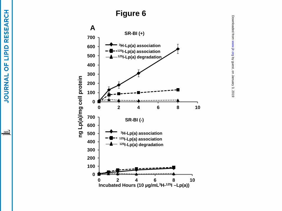
Figure 6
A
0
100
200
300
400
500
600
700
0 2 4 6 8 10
0
100
200
300
400
500
600
700
0 2 4 6 8 10 Incubated Hours (10 µg/mL3H-125I –Lp(a))
ng L
p(a)
/mg
cell
prot
ein
3H-Lp(a) association 125I-Lp(a) association 125I-Lp(a) degradation
3H-Lp(a) association 125I-Lp(a) association 125I-Lp(a) degradation
SR-BI (+)
SR-BI (-)
by guest, on January 3, 2019w
ww
.jlr.orgD
ownloaded from
1
10
100
0 5 10 15 20 25
Perc
enta
ge o
f rem
aini
ng tr
acer
Time (h)
Sr-b1 KO; FCR=0.63±0.08/d* C57Bl/6; FCR=1.64±0.62/d hSR-BI Tg; FCR=4.64±0.40/d*
Figure 7
by guest, on January 3, 2019w
ww
.jlr.orgD
ownloaded from


![Scavenger receptor class B type I regulates cellular ... · binding to the HDL receptor, the scavenger receptor class B, type I (SR-BI) and subsequent lipid transfer to the cell [25-27]](https://img.pdfslide.net/doc/110x75/60676de32012bd272f647095/scavenger-receptor-class-b-type-i-regulates-cellular-binding-to-the-hdl-receptor.jpg)