Embed Size (px)
Citation preview

184
is diagnostic of thrombocythaemic erythromelalgia. In untreatedcases, thrombocythaemic erythromelalgia frequently progresses topainful ischaemic acrocyanosis and frank gangrene. Low-doseaspirin can improve the peripheral ischaemic circulation,3 butRaynaud’s-like symptoms may continue because of the persistenceof acrocyanotic microvascular ischaemia.The incurable variant-burning distress in the absence of any
detectable underlying disorder-is rare. I have designated thisentity "idiopathic or primary erythermalgia and postulated sixcriteria for the condition: (1) attacks of local red vasodilatation withincreased local heat and burning pain constitute the basic
disturbances; (2) bilateral distribution; (3) production and
aggravation by exercise and heat; (4) relief always provided by cold,rest, and elevation of the affected extremities; (5) no primary orassociated disease; and (6) no response to treatment. In contrastwith the asymmetric or unilateral localisation of thrombocythaemicerythromelalgia in toes and fingers occurring in adulthood or oldage 2,3 primary erythermalgia arises in childhood or puberty asbilateral, more or less symmetrical, burning distress in the feet,ankles, and lower legs.4 There is relative sparing of the toes and noprogression to peripheral ischaemia and gangrene.
Secondary erythermalgia, without platelet involvement, has beendescribed in association with inflammatory and circulatorydisturbances such as gout, systemic lupus erythematosus,rheumatoid arthritis, cryoglobulinaemia, and endarteritis
obliterans, for example, and also with diabetes, neurologicalconditions, and treatment with vasoactive drugs. These secondaryforms of erythermalgia are not identical to the specific condition ofthrombocythaemic erythromelalgia, because prompt and lastingrelief of pain by aspirin is lacking.
Department of Haematology,Dijkzigt Univeristy Hospital,Erasmus University,3015 Rotterdam, Netherlands JAN J. MICHIELS
1. Michiels JJ, Ten Kate FWJ, Vuzevski VD, Abels J Histopathology of erythomelalgiam thrombocythaemia. Histopathology 1984; 8: 669-78.
2. Michiels JJ, Abels J, Steketee J, Van Vliet HHDM, Vuzevski VD. Erythromelalgiacaused by platelet mediated artenolar inflammation and thrombosis m
thrombocythemia. Ann Intern Med 1985; 102: 466-71.3. Michiels JJ, Van Joost Th. Erythromelalgia and thrombocythemia: a causal relation.
J Am Acad Dermatol 1990; 22: 107-11.4. Michiels JJ, Van Joost Th, Vuzevski VD. Idiopathic erythermalgia: a congenital
disorder. J Am Acad Dermatol 1989; 21: 1128-30.
Graves’ ophthalmopathy and smokingSIR,-In a case-control study the purpose of the comparison(control) group is to identify factors that occur more (or less) often inthe cases and which may increase or reduce the risk of the conditionunder investigation.1 The control group, difficult though it may beto identify, must therefore consist of an unbiased sample of the basepopulation from which the cases arise. They must not be biasedwith respect to the exposure of interest.
Unfortunately Dr Shine and colleagues (May 26, p 1261) did notclearly state the hypothesis that they intended to test, and oneconsequence was the selection of three small control groups. The
validity of each is debatable and the rationale for amalgamatingthem for comparison with the case group is not discussed. Thecontrol groups may have been seriously biased with respect tosmoking because the information was derived from questionnaires,to which there was a poor response. Thus the individuals whocontributed smoking information were self-selected responders andthus unlikely to have been representative of the groups from whichthey were derived.2 In one group the response rate was only 32%and the reported prevalence of current smoking was only 13 %. Thisgroup almost certainly provided a biased underestimate of smoking,and the apparently significant association of Graves’ disease withsmoking may have arisen simply as a consequence of control groupselection bias.
We agree with your May 12 editorial that criticism of
epidemiological studies should not be based on methodologicalpedantry. However, the fundamental problems relating to thecontrol groups used by Shine et al mean that their findings must be
viewed with considerable circumspection. The apparent associationbetween smoking and Graves’ ophthalmopathy does merit furtherexamination but this would be best achieved in a study large enoughto test a single hypothesis and with attention to complete datacollection. The effects of potential confounders such as age and sexmight also be taken into account.
Department of Public Health Medicine,Leicestershire Health Authority,Leicester LE1 6TP, UK R. SHUKLA
Department of Community Health,University of Leicester J. J. KURINCZUK
1. Breslow NE, Day NE. Statistical methods in cancer research: vol I-the analysis ofcase-control studies. Lyon: International Agency for Research on Cancer, 1980.
2. Seltzer CC, Bosse R, Garvey AJ. Mail response by smoking status. Am J Epidemiol1974; 100: 453-57
***This letter has been shown to Dr Shine and Dr Weetman, whosereply follows.-ED. L.
SIR,- We thank Dr Shukla and Dr Kurinczuk for their comments.Our hypotheses were that there is an association between Graves’ophthalmopathy and smoking, and that within the group of patientswith Graves’ ophthalmopathy there is an association between thedegree of ophthahnopathy and amount of tobacco consumption.Our control groups were all of a similar size to the patient group,
with much the same responses in three of the four groups. Althoughself-selection may lead to bias, the direction and magnitude of thebias might be expected to be similar in all groups. It is often onlypossible to detect bias after the event, by appropriate statisticalanalysis. We detected no significant differences between the threecontrol groups either with respect to prevalence (chi-squared 47,4 DF excluding the Graves’ ophthalmopathy group), or to
pack-year history (chi-squared 8-4, 4 DF excluding the Graves’ophthalmopathy group). We are thus happy with the homogeneityof the control groups, and that the first hypothesis is most probablycorrect. This hypothesis is further supported by our retrospectiveanalysis of data from case records.’ The second hypothesis is lentsupport by the association of smoking history with degree ofophthalmopathy (our fig 2). Here, there is no need for a specificcontrol group, since we are making a within-group comparison.The low prevalence of smoking seen in both the Moorfields and
Cambridge control groups, might be expected from the high overallages of our subjects. Our survey was conducted three years after thelast general survey reported by the Office of Population Censusesand Surveys. If the trend of the previous fifteen years has continued,we would expect a smaller proportion of the general population tobe smokers than in that series. There may, in addition, be regionaldifferences, for which our comparison groups should havecontrolled.
Institute of Ophthalmology,University of London,London EC1V 9AT, UK
BRIAN SHINETONY WEETMAN
1. Shme B, Edwards OM, Weetman AP. Graves’ ophthalmopathy and smoking ActaEndocrinologica 1989 121 (suppl 2): 182-84
2. Office of Population Censuses and Surveys Cigarette smoking 1972 to 1986. OPCSMonitor. London: Government Statistical Service, 1988; Feb 9: 1-10.
Schizophrenia and encephalitis lethargicaSIR,-My spirits were lifted by Dr Boyle’s letter (April 7, p 853) onthe prevalence of the sequelae of encephalitis lethargica in thepopulation studied by Kraepelin and Bleuler in their identificationof dementia praecox and schizophrenia. During my earlyapprenticeship in 1949, when I was charged with the care of 400 or500 chronic patients, there was in every ward a sprinkling of caseslabelled "encephalitis lethargica, effects of’. These were
distinguishable to me because of parkinsonism, choreoathetosis, ornight calls to deal with oculogyric crises. Although I had read one ortwo good books before starting my apprenticeship, I was not able todistinguish these patients from those labelled "chronic paranoidschizophrenia", except by observing their disorders of movement. I
185
put this down to my ignorance at that time, which was a few yearsbefore the introduction of psychotropic drugs.One of my patients today had a viral encephalitis in 1965. His
psychiatric symptoms, unusually, started with the acute illnesswithout remission since. After 25 years, he is just beginning to showa choreiform disorder of movement. Nevertheless, despite a clearhistory, he is still labelled chronic paranoid schizophrenia bypsychiatrist and neurologist alike because he is deluded and
hallucinatory. His disorder of movement is attributed to pastmedication for schizophrenia. The lesson of the encephalitislethargica epidemic 1916-27 is forgotten. I can assure Dr Boyle thatthese cases still appear but they are now labelled chronic
schizophrenia and tardive dyskinesia, whatever the extrapyramidalsigns may be.
Encephalitis is clearly recognisable in necropsy material whereasschizophrenia is not. This is not much solace to the encephalitic whohas been treated for schizophrenia. It is forgotten that the episode ofacute encephalitis may only be remembered as an attack of influenzaand the interval between the encephalitis and the neuropsychiatricsequelae can be 30 years or more, although it is more commonly halfthis period. I agree with your correspondent that the post-encephalitic cases with extrapyramidal symptoms are more rarelyseen-and when seen are never believed.
Box 1000,Ponoka, Alberta T0C 2H0, Canada R. H. BOARDMAN
Efficacy of pyrimethamine/sulfadoxine inuncomplicated severe falciparum malaria in
KenyaSIR,—The management of severe Plasmodiumfalciparum malaria ineastern Africa is based on parenteral quinine. However, the toxicity,frequency of administration, and cost of this drug restrict its use andavailability. Pyrimethamine/sulfadoxine (’Fansidar’, PSD) is a
single dose, long acting, antifolate combination which is much
cheaper than quinine and readily available. Both pyrimethamineand sulfadoxine are regarded as slow acting schizonticides, anddespite their demonstrated synergism when combined at a doseratio of 20 sulfadoxine to 1 pyrimethamine,’ are recommended onlycombined with quinine for the management of severe falciparummalaria This recommendation seems to be based on experience inSouth-East Asia where malaria patients are mainly non-immuneand where parasite clearance and symptomatic improvement afterPSD treatment is slower than with quinine, chloroquine,mefloquine, or artemether.3,4 However, this is not the case in
Nigeria where response rates are similar to those obtained withchloroquine,s or in Pakistan where parasite clearance times
averaged 1-25 days. Nor is it so in Kenya, where PSD treatment ofnon-severe, low parasitaemia (1000 to 50 000 asexual parasites perpl blood) falciparum malaria results in a mean parasite clearancetime of about 2-5 days7 which is similar to that for quinine (2-4 days)(Schwartz IK, personal communication). We have therefore
prospectively compared PSD and quinine in symptomatic,uncomplicated, high parasitaemia falciparum malaria.
22 outpatients at Magadi Hospital, Rift Valley Province (an areaof continuous malaria transmission with pronounced seasonalvariation), were recruited if they had slide positive falciparummalaria with parasitaemia of 50000IJll or over, but less than250 000/µl; non-severe clinical presentation; normal blood glucoseand creatinine levels; haemoglobin 7 g/dl or over; no history ofhypersensitivity to sulphonamides; and if they gave writteninformed consent to participate. After examination, patients wererandomly assigned to receive either oral quinine sulphate, 10 mgsalt/kg, every 8 hours for 15 doses (11 patients), or oral
PATIENT CHARACTERISTICS
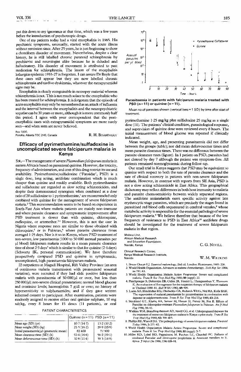
Parasitaemia in patients with falciparum malaria treated withPSD (n=11) or quinine (n=11).
Mean no of parasites shown (vertical bars= 1 SD) by time after start oftreatment.
pyrimethamine 1.25 mg/kg plus sulfadoxine 25 mg/kg as a singledose (11). The patients’ clinical condition, parasitological response,and supervision of quinine dose were reviewed every 8 hours. Theinitial measurement of blood glucose was repeated if clinicallyindicated.Mean weight, age, and presenting parasitaemia did not differ
between the groups (table); nor did mean defervescence times andmean parasite clearance times. There was no difference between theparasite clearance rates (figure). In 1 patient on PSD, parasites hadnot cleared by day 7 although the patient was symptom-free. Allpatients remained normoglycaemic during follow-up.Our small trial in Kenya suggests that PSD may be equivalent to
quinine with respect to both the rate of parasite clearance and therate of clinical recovery in patients with non-severe falciparummalaria. However, in contrast with reports from SE Asia, PSD isnot a slow acting schizonticide in East Africa. This geographicaldichotomy may reflect differences in both host immunity to malariaand parasite chemosensitivity between widely separated regions.The antifolate antimalarials exert specific activity against late
erythrocytic stage parasites, which are precisely the stages found inparasitised red blood cells sequestered in cerebral venules,8 whosemetabolic activity is responsible for the essential pathology of severefalciparum malaria.’’ We believe therefore that because of the lowfrequency of resistance to PSD in East Mrica10 antifolate drugsshould be investigated for the treatment of severe falciparummalaria in that region.Malaria Unit,African Medical Researchand Education Foundation,
Nairobi, Kenya C. G. NEVILL
Clinical Research Centre,Kenya Medical Research Institute,Nairobi W. M. WATKINS
1. Bruce-Chwatt LJ. Essential malariology, 2nd ed. London: Heinemann, 1985: 228.2. World Health Organisation. Advances in malaria chemotherapy. Tech Rep Ser 1984;
no 711: 63.3. World Health Organisation Malaria Action Programme. Severe and complicated
malaria. Trans R Soc Trop Med Hyg 1986; 80 (suppl): 22.4. Pinichpongse S, Doberstyn EB, Cullen JR, Yisunri L, Thongsombun Y, Thimsarn
K. An evaluation of five regimens for the outpatient therapy of falciparum malariam Thailand 1980-81. Bull WHO 1982; 60: 909.
5. Lucas AO, Hendrickse RG, Okubadejo OA, Richards WHG, Neil RA, Kofie BAK.The suppression of malarial parasitaemia by pynmethamine in combination withdapsone or sulphormethoxine. Trans R Soc Trop Med Hyg 1969; 63: 216.
6. Strickland GT, Khaliq AA, Sarwar M, Hassan H, Pervez M, Fox E. Effects ofFansidar on chloroquine-resistant Plasmodium falciparum in Pakistan. Am J MedHyg 1986; 35: 61.
7. Watkins WM, Brandling-Bennett AD, Nevill CG, et al. Chlorproguanil/dapsone forthe treatment of nonsevere falciparum malaria in Kenya: a pilot study. Trans R SocTrop Med Hyg 1988; 82: 398-403.
8. Phillips RE, Warrell DA. The pathophysiology of severe falciparum malaria. ParasitolToday 1986; 2: 271-82.
9. World Health Organisation Malaria Action Programme. Severe and complicatedmalaria. Trans R Soc Trop Med Hyg 1986; 80 (suppl): 5-7.
10. Miller KD, Lobel HO, Pappaionou M, Patchen LC, Churchill FC. Failures ofcombined Fansidar and chloroquine prophylaxis in Amencan travellers to E.Africa J Infect Dis 1986; 154: 689-91.