Embed Size (px)
Citation preview
ARBETE OCH HÄLSAVETENSKAPLIG SKRIFTSERIE
Nr 2014;48(3)
Scientific Basis for SwedishOccupational Standards XXXIII
ISBN 978-91-85971-52-7 ISSN 0346-7821
N-Methyl-2-pyrrolidoneCrystalline Silica, QuartzEpichlorohydrin
Swedish Criteria Group for Occupational StandardsEd. Johan Montelius
Swedish Work Environment AuthorityS-112 79 Stockholm, Sweden
Translation:John Kennedy, Space 360 and Johan Montelius,
the Swedish Work Environment Authority.The consensus reports in this volume are translated from Swedish. If there is any doubt as
to the understanding or interpretation of the English version, the Swedish version shall prevail.
Swedish Work Environment Authority
Arbete och Hälsa (Work and Health) is a scientific report series published by Occupational and Environmental Medicine at Sahlgrenska Academy, University of Gothenburg. The series publishes scientific original work, review articles, criteria documents and dissertations. All articles are peer-reviewed.
Arbete och Hälsa has a broad target group and welcomes articles in different areas. Instructions and templates for manuscript editing are available at http://www.amm.se/aoh
Summaries in Swedish and English as well as the complete original texts from 1997 are also available online.
Arbete och Hälsa 2014;48(3)Editor-in-chief: Kjell Torén, Gothenburg
Co-editors: Maria Albin, Lund Lotta Dellve, Stockholm Henrik Kolstad, Aarhus Roger Persson, Lund Tornqvist, Marianne Tör Kristin Svendsen , Trondheim Allan Toomingas, Stockholm Marianne Törner, Gothenburg
Managing editor: Cecilia Andreasson, Gothenburg
Editorial Board: Tor Aasen, Bergen Gunnar Ahlborg, Gothenburg Kristina Alexanderson, Stockholm Berit Bakke, Oslo Lars Barregård, Gothenburg Jens Peter Bonde, Kopenhagen Jörgen Eklund, Linkoping Mats Eklöf, Göteborg Mats Hagberg, Gothenburg
Kari Heldal, Oslo Kristina Jakobsson, Lund Malin Josephson, Uppsala Bengt Järvholm, Umea Anette Kærgaard, Herning Ann Kryger, Kopenhagen Carola Lidén, Stockholm Svend Erik Mathiassen, Gavle Gunnar D. Nielsen, Kopenhagen Catarina Nordander, Lund Torben Sigsgaard, Aarhus Staffan Skerfving, Lund Gerd Sällsten, Gothenburg Ewa Wikström, Gothenburg Eva Vingård, Uppsala
© University of Gothenburg & authors 2013 University of Gothenburg, SE-405 30 Gothenburg http://www.amm.se/aoh
ISBN 978-91-85971-52-7 ISSN 0346–7821
Printed at Kompendiet Gothenburg
Arbete och Hälsa
Preface
These documents have been produced by the Swedish Criteria Group for Occu-
pational Standards, the members of which are presented on the next page. The
Criteria Group is responsible for assessing the available data that might be used
as a scientific basis for the occupational exposure limits set by the Swedish Work
Environment Authority. It is not the mandate of the Criteria Group to propose
exposure limits, but to provide the best possible assessments of dose-effect and
dose-response relationships and to determine the critical effect of occupational
exposure.
The work of the Criteria Group is documented in consensus reports, which are
brief critical summaries of scientific studies on chemically defined substances or
complex mixtures. The consensus reports are often based on more comprehensive
criteria documents (see below), and usually concentrate on studies judged to be
of particular relevance to determining occupational exposure limits. More com-
prehensive critical reviews of the scientific literature are available in other docu-
ments.
Literature searches are made in various databases, including KemI-Riskline,
PubMed and Toxline. Information is also drawn from existing criteria documents,
such as those from the Nordic Expert Group (NEG), WHO, EU, NIOSH in the
U.S., and DECOS in the Netherlands. In some cases the Criteria Group produces
its own criteria document with a comprehensive review of the literature on a
particular substance.
As a rule, the consensus reports make reference only to studies published in
scientific journals with a peer review system. This rule may be set aside in ex-
ceptional cases, provided the original data is available and fully reported. Exce-
ptions may also be made for chemical-physical data and information on occur-
rence and exposure levels, and for information from handbooks or documents
such as reports from NIOSH and the Environmental Protection Agency (EPA)
in the U.S.
A draft of the consensus report is written in the secretariat of the Criteria Group
or by scientists appointed by the secretariat (the authors of the drafts are listed in
the Table of Contents). After the draft has been reviewed at the Criteria Group
meetings and accepted by the group, the consensus report is published in Swedish
and English as the Criteria Group’s scientific basis for Swedish occupational
standards.
This publication is the 33rd in the series, and contains consensus reports ap-
proved by the Criteria Group from June, 2012 through October, 2013. The con-
sensus reports in this and previous publications in the series are listed in the
Appendix (page 96).
Johan Högberg Johan Montelius
Chairman Secretary

The Criteria Group has the following membership (as of October, 2013)
Maria Albin Dept. Environ. Occup. Medicine,
University Hospital, Lund
Cecilia Andersson observer Confederation of Swedish Enterprise
Anders Boman Inst. Environmental Medicine,
Karolinska Institutet
Jonas Brisman Occup. and Environ. Medicine,
Göteborg
Per Eriksson Dept. Environmental Toxicology,
Uppsala University
Sten Gellerstedt observer Swedish Trade Union
Confederation
Märit Hammarström observer Confederation of Swedish Enterprise Johan Högberg chairman Inst. Environmental Medicine,
Karolinska Institutet
Anders Iregren observer Swedish Work Environment
Authority
Gunnar Johanson v. chairman Inst. Environmental Medicine,
Karolinska Institutet
Bengt Järvholm Occupational Medicine,
University Hospital, Umeå
Bert-Ove Lund Swedish Chemicals Agency
Mihalyn Matura Inst. Environmental Medicine,
Karolinska Institutet
Johan Montelius secretary Swedish Work Environment
Authority
Lena Palmberg Inst. Environmental Medicine,
Karolinska Institutet
Per-Åke Persson observer SEKO
Agneta Rannug Inst. Environmental Medicine,
Karolinska Institutet
Bengt Sjögren Inst. Environmental Medicine,
Karolinska Institutet
Ulla Stenius Inst. Environmental Medicine,
Karolinska Institutet
Marianne Walding observer Swedish Work Environment
Authority
Håkan Westberg Dept. Environ. Occup. Medicine,
University Hospital, Örebro
Contents
Consensus report for:
N-Methyl-2-pyrrolidone 1 1
Crystalline Silica, Quartz 2 25
Epichlorohydrin 3 77
Summary 95
Sammanfattning (in Swedish) 95
Appendix: Consensus reports in this and previous volumes 97
1 Drafted by Kristian Dreij, Inst. Environmental Medicine, Karolinska Institutet, Stockholm, Sweden.
2 Drafted by Ilona Silins, Inst. Environmental Medicine, Karolinska Institutet, Stockholm, Sweden. 3 Drafted by Birgitta Lindell, Swedish Work Environment Authority, Sweden.

1
Consensus Report for N-Methyl-2-pyrrolidone
December 5, 2012
This consensus report is based in part on a criteria document from the Nordic
Expert Group (61), as well as risk assessments carried out within the World
Health Organization (WHO), International Program on Chemical Safety (IPCS)
(22), EU Scientific Committee on Occupational Exposure Limits (SCOEL) (48),
EU Scientific Committee on Consumer Safety (SCCS) (47) and US Environmen-
tal Protection Agency (EPA) (17). Complementary literature searches were carried
out in November 2011 and in March and September 2012 on PubMed, Toxline
and Web of Science. The Swedish Criteria Group has previously published a
consensus report on N-methyl-2-pyrrolidone 1987 (33).
Chemical-physical data
CAS no 872-50-4
Synonyms NMP, 1-methyl-2-pyrrolidone,
N-methylpyrrolidone, N-methyl-α-
butyrolactam, N-methyl-2-ketopyrrolidine
Empirical formula C5H9NO
Structural formula
N
O
Molecular weight 99.13 g/mol
Density 1.028 kg/dm3 (25 °C)
Boiling point 202 °C
Melting point 24 °C
Vapour pressure 0.045 kPa (0.33 mm Hg) (25 °C)
Saturation concentration 446 ppm
Partition coefficient (log Ko/w) -0.38
Conversion factors 1 ppm = 4.12 mg/m3; 1 mg/m3 = 0.24 ppm
N-Methyl-2-pyrrolidone (NMP) is a colourless-to-yellowish liquid with an
unpleasant amine-like odour. The substance comprises a basic, polar molecule
which is relatively stable, though some degradation (oxidation) occurs with
2
exposure to light and air. NMP is hygroscopic, fully miscible with water and
soluble in a number of organic solvents, such as ethanol, diethyl ether, ethyl
acetate, chloroform and benzene.
Use, occurrence, exposure
The substance is mainly used as a solvent in a range of different processes in the
petroleum industry, in reaction media for the chemical production of polymers
(e.g., polyvinyl chloride) and in stripping and cleaning procedures in the manu-
facture of microelectronic components. NMP is used as a substitute for dichloro-
methane in the removal of paint and graffiti (61). NMP is also used as a pharma-
ceutical vehicle and as a penetration enhancer for pharmaceuticals applied to the
skin (24). The substance is also used as a binder in cosmetics, as a solvent in the
preparation of pigments, dyes and inks, and in various pesticide products (61).
NMP is not produced in Sweden but in 2008 ca 164 tonnes were imported, most
of which was used as a raw material in the production of paints and paint remo-
vers (28).
With regard to levels in the workplace environment, some studies have mea-
sured concentrations during graffiti removal in Sweden (3, 4, 29). NMP levels of
up to 25 mg/m3 have been measured in the atmosphere during short term exposure
and up to 5 mg/m3 on average during an eight-hour working day (8-h TWA, 8-
hours time-weighted average). In one study of occupational exposure in the micro-
electronics industry up to 6 mg NMP/m3 were measured (7). The levels increased
to 280 mg/m3 (8-h TWA) when heated (80 °C) NMP was used. Measurements of
NMP in a German adhesives plant showed levels of up to 85 mg/m3 during short
term exposure and up to 15.5 mg/m3 over a working day (8-h TWA) (5). Measure-
ments taken in two different industries in Japan showed levels that were lower
than 3 mg/m3 (8-h TWA) (39, 58).
NMP has been isolated from a marine fungus, Clathria frondifera, which shows
that the substance can be produced naturally (42).
Uptake, biotransformation and excretion
Both human and animal studies show that NMP is absorbed rapidly when inhaled
and when administered orally (6, 26, 43, 63, 64, 65). Several studies, in both ani-
mals and humans, show that NMP in liquid form is readily absorbed through the
skin (23). The study regarded as the most suitable for assessing dermal absorption
in humans (23) is that by Dick et al. (15) which measured dermal absorption of
NMP in human skin in vitro. The rate of absorption of undiluted NMP was calcu-
lated as 10 mg/cm2/hour. Extrapolation of data to 2000 cm2 skin (equivalent to
both hands and forearms) and 1 hour of exposure gave a dermal absorption of
20,000 mg. This uptake was estimated to be ca 10 times greater than the uptake
with 8 hours of inhalation at the Swedish occupational exposure limit (200
mg/m3). Other studies have shown a percutaneous absorption of ca 70% of the
applied dose in both rats (200 mg, 10 cm2 after 2 hours) (40) and humans (300
3
mg, 20 cm2 after 6 hours) (32). Dermal exposure to NMP in liquid form can
therefore result in significant absorption.
A significant dermal absorption has also been demonstrated in humans exposed
to NMP in vapour form (6). Research volunteers (n = 16) were exposed to 80 mg
NMP/m3 for 8 hours, either via the skin and respiratory system or via the skin
alone. The absorption of NMP was analyzed by measuring NMP and NMP meta-
bolites in the urine. The dermal uptake of NMP vapour at rest was calculated as
ca 42% of the total absorption (absorption by skin plus respiratory system). With
moderate work (75 W, 6 x 10 minutes) total absorption increased by 41% and
dermal absorption as a percentage of total absorption was somewhat lower, at
33%.
Animal studies have shown that NMP crosses the placenta in rats and that levels
in foetal blood are the same as levels in the mother's blood after 6 hours (43).
Absorbed NMP is rapidly metabolized in both humans and rats. NMP is first
metabolized to 5-hydroxy-NMP (5-HNMP) which in turn is metabolized further
to N-methylsuccinimide (MSI) and finally to 2-hydroxy-N-methylsuccinimide
(2-HMSI) (26, 64, 65). The same metabolic pathways are observed with both oral
and dermal administration. In addition, Carnerup and co-workers have identified
a further metabolite, 2-pyrrolidone, which is formed in small quantities (12, 13).
A human study showed a significant relationship between CYP2E1 mRNA
levels and levels of 5-HNMP and 2-HMSI in urine. The same relationship could
be demonstrated in rats by pretreating these animals with the CYP2E1 inhibitor
diethylthiocarbamate. (32).
The half-lives of NMP, 5-HNMP, MSI and 2-HMSI in plasma from male
participants in inhalation studies exposed to 10-80 mg NMP/m3 for 8 hours were
ca 4, 6, 8 and 28 hours, respectively, (6, 65). With dermal application of NMP
(300 mg for 6 hours) in humans the plasma concentration of NMP reached a
maximum 3 hours after exposure (62), which indicates that dermal absorption
was delayed relative to inhalation absorption. There was a further delay of ca 4
hours when NMP was applied as an aqueous solution (5-50%) (1, 6, 27, 62).
NMP is mainly excreted in the urine, with only a small part (<4%) being
eliminated through biliary excretion and exhalation (55). Inhalation studies with
male research subjects exposed to 10-80 mg NMP/m3 for 8 hours showed the
following relative levels of NMP and its metabolites in urine: NMP 1-2%, 5-
HNMP 60-68%, MSI 0.1% and 2-HMSI 31-37% (percentage of the total amount
of excreted NMP and metabolites) (6, 65). A similar relative proportion of meta-
bolites in urine was measured after oral administration of 100 mg NMP (63). 5-
HNMP has also been identified as the main metabolite in rat urine (32 , 40, 56).
In a number of studies both NMP and its various metabolites have been pro-
posed as biomarkers for NMP exposure. To sum up, the biggest disadvantages for
NMP as a biomarker are its short half-life and low concentrations in urine. The
metabolites 5-HNMP and 2-HSMI have been identified as the most promising
biomarkers for work-related exposure (inhaled and dermal) because of their long
half-lives and high concentrations in urine (6, 12, 13, 14, 26).
4
Toxic effects
Human data
In one study involving repeated exposure to NMP for 8 hours per day over 2 days,
10 out of 12 workers, most of whom were women, showed skin irritation and
contact dermatitis (31). The work task consisted of dipping one end of a plastic
casing in NMP (10 seconds) and then dipping the opposite end in NMP (10
seconds). After each dipping the surplus NMP was wiped off with a paper towel
which was replaced after every tenth plastic casing or 20 dippings. Each worker
treated ca 110 casings per hour over an 8-hour working day. There was no infor-
mation on exposure levels. Protective equipment consisted of latex gloves but it
was reported that these were not used regularly. The authors of the study point out
that after the workers began wearing cotton gloves (which were changed every
hour) under the latex gloves to avoid dampness, no further skin problems were
reported (31). Another study showed irritative contact dermatitis in three workers
who were exposed to NMP for the first time. The effects were explained by the
hygroscopic properties of NMP (25). There are no studies which show that occu-
pational exposure to NMP leads to sensitization.
Workers exposed to up to 280 mg/m3 NMP (8-h TWA) in the manufacture
of microelectronic components involving the use of hot (80 °C) NMP, reported
severe eye irritation and headaches (7). The exposure levels were on average
between 0.1 and 6 mg/m3 (8-h TWA) with processes in which NMP was not
heated, but even exposure to relatively low levels (ca 3 mg/m3) was reported to
cause headaches and chronic eye irritation. No conclusive data has been presented
on the frequency of these effects. Because of methodological shortcomings in
the study it has not been possible to establish any dose-response relationship.
In a study of 38 graffiti cleaners who worked an 8-hour shift in Stockholm's
underground rail system and were exposed to a mixture of solvents, including
NMP, an increased occurrence was observed of fatigue, headaches and symptoms
related to effects on the respiratory system, eyes and skin, when compared with
controls. As well as NMP the cleaning agent also contained glycol ethers (e.g.,
dipropylene- and propylene glycol methyl ether), toluene, xylene, 1,2,4-trimethyl-
benzene, limonene, nonane, octane, etc. Levels of NMP in the atmosphere mea-
sured over 15 minutes with various work tasks were on average 4.71 mg/mg3 (SD
± 6.17, range 0.01-24.6). The Swedish occupational exposure limits for short term
exposure (300 mg/m3, 15 min) or for 8-hours exposure (200 mg/m3) were not ex-
ceeded. Only trimethylbenzene levels exceeded the Swedish occupational expo-
sure limit (170 mg/m3) with short term exposure (maximum measured levels were
ca 280 mg/m3). Because of the mixed exposure it was not possible to establish a
specific relationship between exposure to NMP and the symptoms exhibited (29).
Workers at a German adhesives plant who cleaned containers with the help of
NMP reported irritation of the upper respiratory tract and eyes as well as head-
aches when exposed to 15.5 mg/m3 (8-h TWA) with a 5-minute maximum expo-
sure of 85 mg/m3 NMP (5). A Japanese study of 15 workers who cleaned compo-
nents with an NMP solution (>90%), examined clinical effects (urine and blood
5
status) and effects on motor and cognitive functions. Exposure, which was mainly
via inhalation, was measured as ca 0.6-1 mg/m3 (8-h TWA, 5 days). The authors
reported no effects when compared with the control group (39).
When six research volunteers were exposed to 10, 25, and 50 mg/m3 NMP over
8 hours, no acute nasal changes were observed in the form of swelling of mucous
membranes (measured using acoustic rhinometry), nor any effects on FEV1, vital
capacity and forced vital capacity measured with a spirometer. Responses to a
questionnaire about subjective symptoms related to smell and irritation of the
nose, eyes or respiratory tract revealed no feelings of discomfort or irritative
effects. Two research subjects reported a smell of acetone at 50 mg/m3 but this
was not described as unpleasant (65).
Van Thriel and co-workers (54) carried out a comprehensive exposure study of
16 healthy young male volunteers in order to examine any chemosensory effects
of NMP under conditions similar to occupational exposure. The research subjects
were exposed to 10, 40, and 80 mg/m3 for 8 hours, once a week, every second
week, for 8 weeks. To maximize any chemosensory effects a peak exposure sce-
nario was also included, with a basal exposure of 25 mg/m3 and 4 × 15-minute
peaks of 160 mg/m3 (the time-weighted average value measured was 72 mg/m3).
All exposure was carried out with or without physical activity, which consisted
of 6 × 10-minute sessions on a bicycle ergometer (75 watt) during the 8 hours of
exposure. The results showed that the research subjects were able to detect the
smell of NMP which was reported to be somewhat unpleasant. No other symp-
toms associated with chemosensory effects were observed and no dose-response
relationship (frequency of blinking, nasal airflow, breathing rate, mental concen-
tration, etc.) was demonstrated. The authors concluded that NMP is a foul-smel-
ling substance with no irritative properties, even at exposures of up to 160 mg/m3
(54).
Animal- and in vitro-data
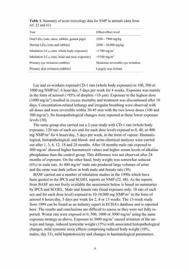
NMP shows weak acute toxicity in animals exposed to NMP orally, dermally or
via inhalation, see Table 1. Studies have failed to show sensitization with dermal
exposure of guinea pigs to NMP (E.I. du Pont de Nemours and Company 1976,
referred to in (22)). The potential irritant effects of NMP on the skin and eyes have
been tested in rabbits and the results have indicated low dermal irritation (0.5 ml
undiluted NMP) and a moderate and brief irritant effect on the eyes (0.1 ml undi-
luted NMP) (2).
A number of inhalation studies with repeated exposure to NMP (in the form of
aerosol or vapour) have been carried out but according to SCOEL (48) and IPCS
(22) the studies by Lee and co-workers (30) and the studies by BASF (1992-1995)
are regarded as the most reliable because they recorded sufficient details on me-
thods and results to allow them to be used as a basis for risk assessment. This
summary therefore focuses on these. The other studies are unpublished industrial
reports that are not publicly available.
6
Table 1. Summary of acute toxicology data for NMP in animals (data from
ref. 22 and 61)
Test Effect/effect level
Oral LD50 (rats, mice, rabbits, guinea pigs) 3500 - 7900 mg/kg
Dermal LD50 (rats and rabbits) 2500 - 10,000 mg/kg
Inhalation LC50 (rats; whole body exposure) ≈1700 mg/m3
Inhalation LC50 (rats; head and nose exposure) >5100 mg/m3
Primary eye irritation (rabbits) Moderate reversible eye irritation
Primary skin irritation (rabbits) Largely non-irritant
Lee and co-workers exposed CD-1 rats (whole body exposure) to 100, 500 or
1000 mg NMP/m3, 6 hour/day, 5 days per week for 4 weeks. Exposure was mainly
in the form of aerosol (>95% of droplets <10 µm). Exposure to the highest dose
(1000 mg/m3) resulted in excess mortality and treatment was discontinued after 10
days. Concentration-related lethargy and irregular breathing were observed with
all doses and were reversible within 30-45 min with the two lower doses (100 and
500 mg/m3). No histopathological changes were reported at these lower exposure
levels (30).
The same group also carried out a 2-year study with CD-1 rats (whole body
exposure, 120 rats of each sex and for each dose level) exposed to 0, 40, or 400
mg NMP/m3 for 6 hours/day, 5 days per week, in the form of vapour. Haemato-
logical, histopathological, and blood- and urine-chemical analyses were carried
out after 1, 3, 6, 12, 18 and 24 months. After 18 months male rats exposed to
400 mg/m3 showed higher haematocrit values and higher serum levels of alkaline
phosphatase than the control group. This difference was not observed after 24
months of exposure. On the other hand, body weight was somewhat reduced
(6%) in male rats. At 400 mg/m3 male rats produced large volumes of urine
and the urine was dark yellow in both male and female rats (30).
BASF carried out a number of inhalation studies in the 1990s which have
been quoted in the IPCS and SCOEL reports on NMP (22, 48). As the reports
from BASF are not freely available the assessment below is based on summaries
by IPCS and SCOEL. Male and female rats (head exposure only, 10 rats of each
sex and for each dose level) exposed to 10-10,000 mg NMP/m3 in the form of
aerosol 6 hours/day, 5 days per week for 2, 4 or 13 weeks. The 13-week study
from 1994 can be found as an industry report in ECHA's database and is reported
here. The results and conclusions are difficult to assess as they were not fully re-
ported. Wistar rats were exposed to 0, 500, 1000 or 3000 mg/m3 using the same
exposure strategy as above. Exposure to 3000 mg/m3 caused irritation of the air-
ways and lungs, reduced testicular weight (15%) with associated histopathological
changes, mild systemic toxic effects comprising reduced body weight (10%,
males, day 33), mild hepatotoxicity and changes in haematological parameters.
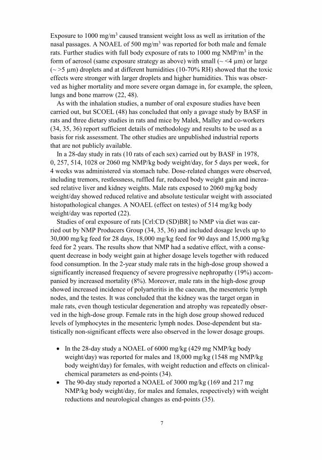
7
Exposure to 1000 mg/m3 caused transient weight loss as well as irritation of the
nasal passages. A NOAEL of 500 mg/m3 was reported for both male and female
rats. Further studies with full body exposure of rats to 1000 mg NMP/m3 in the
form of aerosol (same exposure strategy as above) with small (~ <4 µm) or large
(~ >5 µm) droplets and at different humidities (10-70% RH) showed that the toxic
effects were stronger with larger droplets and higher humidities. This was obser-
ved as higher mortality and more severe organ damage in, for example, the spleen,
lungs and bone marrow (22, 48).
As with the inhalation studies, a number of oral exposure studies have been
carried out, but SCOEL (48) has concluded that only a gavage study by BASF in
rats and three dietary studies in rats and mice by Malek, Malley and co-workers
(34, 35, 36) report sufficient details of methodology and results to be used as a
basis for risk assessment. The other studies are unpublished industrial reports
that are not publicly available.
In a 28-day study in rats (10 rats of each sex) carried out by BASF in 1978,
0, 257, 514, 1028 or 2060 mg NMP/kg body weight/day, for 5 days per week, for
4 weeks was administered via stomach tube. Dose-related changes were observed,
including tremors, restlessness, ruffled fur, reduced body weight gain and increa-
sed relative liver and kidney weights. Male rats exposed to 2060 mg/kg body
weight/day showed reduced relative and absolute testicular weight with associated
histopathological changes. A NOAEL (effect on testes) of 514 mg/kg body
weight/day was reported (22).
Studies of oral exposure of rats [Crl:CD (SD)BR] to NMP via diet was car-
ried out by NMP Producers Group (34, 35, 36) and included dosage levels up to
30,000 mg/kg feed for 28 days, 18,000 mg/kg feed for 90 days and 15,000 mg/kg
feed for 2 years. The results show that NMP had a sedative effect, with a conse-
quent decrease in body weight gain at higher dosage levels together with reduced
food consumption. In the 2-year study male rats in the high-dose group showed a
significantly increased frequency of severe progressive nephropathy (19%) accom-
panied by increased mortality (8%). Moreover, male rats in the high-dose group
showed increased incidence of polyarteritis in the caecum, the mesenteric lymph
nodes, and the testes. It was concluded that the kidney was the target organ in
male rats, even though testicular degeneration and atrophy was repeatedly obser-
ved in the high-dose group. Female rats in the high dose group showed reduced
levels of lymphocytes in the mesenteric lymph nodes. Dose-dependent but sta-
tistically non-significant effects were also observed in the lower dosage groups.
In the 28-day study a NOAEL of 6000 mg/kg (429 mg NMP/kg bodyweight/day) was reported for males and 18,000 mg/kg (1548 mg NMP/kgbody weight/day) for females, with weight reduction and effects on clinical-chemical parameters as end-points (34).
The 90-day study reported a NOAEL of 3000 mg/kg (169 and 217 mgNMP/kg body weight/day, for males and females, respectively) with weightreductions and neurological changes as end-points (35).
8
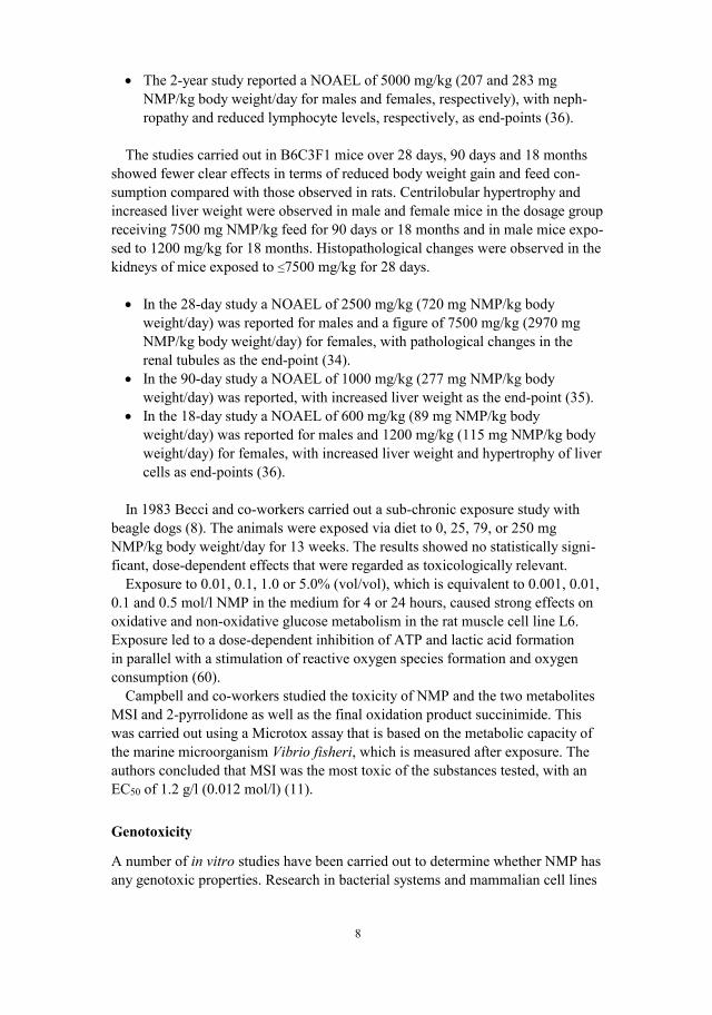
The 2-year study reported a NOAEL of 5000 mg/kg (207 and 283 mg NMP/kg body weight/day for males and females, respectively), with neph-ropathy and reduced lymphocyte levels, respectively, as end-points (36).
The studies carried out in B6C3F1 mice over 28 days, 90 days and 18 months
showed fewer clear effects in terms of reduced body weight gain and feed con-
sumption compared with those observed in rats. Centrilobular hypertrophy and
increased liver weight were observed in male and female mice in the dosage group
receiving 7500 mg NMP/kg feed for 90 days or 18 months and in male mice expo-
sed to 1200 mg/kg for 18 months. Histopathological changes were observed in the
kidneys of mice exposed to ≥7500 mg/kg for 28 days.
In the 28-day study a NOAEL of 2500 mg/kg (720 mg NMP/kg body weight/day) was reported for males and a figure of 7500 mg/kg (2970 mg NMP/kg body weight/day) for females, with pathological changes in the renal tubules as the end-point (34).
In the 90-day study a NOAEL of 1000 mg/kg (277 mg NMP/kg body weight/day) was reported, with increased liver weight as the end-point (35).
In the 18-day study a NOAEL of 600 mg/kg (89 mg NMP/kg body weight/day) was reported for males and 1200 mg/kg (115 mg NMP/kg body weight/day) for females, with increased liver weight and hypertrophy of liver cells as end-points (36).
In 1983 Becci and co-workers carried out a sub-chronic exposure study with
beagle dogs (8). The animals were exposed via diet to 0, 25, 79, or 250 mg
NMP/kg body weight/day for 13 weeks. The results showed no statistically signi-
ficant, dose-dependent effects that were regarded as toxicologically relevant.
Exposure to 0.01, 0.1, 1.0 or 5.0% (vol/vol), which is equivalent to 0.001, 0.01,
0.1 and 0.5 mol/l NMP in the medium for 4 or 24 hours, caused strong effects on
oxidative and non-oxidative glucose metabolism in the rat muscle cell line L6.
Exposure led to a dose-dependent inhibition of ATP and lactic acid formation
in parallel with a stimulation of reactive oxygen species formation and oxygen
consumption (60).
Campbell and co-workers studied the toxicity of NMP and the two metabolites
MSI and 2-pyrrolidone as well as the final oxidation product succinimide. This
was carried out using a Microtox assay that is based on the metabolic capacity of
the marine microorganism Vibrio fisheri, which is measured after exposure. The
authors concluded that MSI was the most toxic of the substances tested, with an
EC50 of 1.2 g/l (0.012 mol/l) (11).
Genotoxicity
A number of in vitro studies have been carried out to determine whether NMP has
any genotoxic properties. Research in bacterial systems and mammalian cell lines
9
shows only negative results with respect to mutagenicity. Cytotoxic effects in
Salmonella typhimurium exposed to high doses of NMP were reported by Wells
and co-workers (57). However, it has been shown that NMP can induce aneup-
loidy (an uneven number of chromosomes) in Saccharomyces cerevisiae at high
doses (7.6-23 g/l, equivalent to 0.08-0.23 mol NMP/l) (37, 38, 59).
Two in vivo studies carried out with NMRI mice and hamsters exposed orally
to doses of NMP up to 3800 mg/kg body weight, showed no mutagenic effects
(micronuclei or chromosome aberrations) (16).
Carcinogenicity
A 2-year inhalation study in CD-1 rats exposed to 0, 40, or 400 mg/m3 NMP for
6 hours/day, 5 days/week, in the form of vapour showed no carcinogenic effects
(30). Histopathological studies were carried out on kidneys, bone marrow, lymph
nodes, spleen and lungs. A 2-year study in rats [Crl:CD (SD)BR] exposed to 0,
1600, 5000 and 15,000 mg NMP/kg feed (equivalent to 0, 66.4, 207 and 678 mg
NMP/kg body weight/day for males and 0, 87.8, 283 och 939 mg NMP/kg body
weight/day for females) with histopathological studies (44 tissues), showed no
dose-dependent carcinogenic effects (36). A 2-year study in B6C3F1 mice expo-
sed to 0, 600, 1200 or 7200 mg NMP/kg feed (equivalent to 8, 89, 173 and 1089
mg NMP/kg body weight/day for males and 0, 115, 221 and 1399 mg NMP/kg
body weight/day for females) with histopathological studies (44 tissues), reported
a significant increase in the incidence of hepatocellular adenoma and carcinoma in
males in the dose group with 7200 mg NMP/kg feed (12 out of 50 animals and 13
out of 50 animals, respectively, compared with 5 out of 50 and 4 out of 50, respec-
tively, in the control group) in parallel with an increase in the number of hepato-
cellular foci. The same effect was observed in females in the same dose group (7
out of 50 animals and 3 out of 50 animals, respectively, compared with 2 out of 50
and 0 out of 50, respectively, in the control group) but the increase was regarded
as not exceeding the historical background variation. The authors suggested that
the tumours were not formed via a genotoxic mechanism but as a result of in-
creased cell proliferation in the liver (36). This was based in part on data on
centrilobular hypertrophy in males in the high-dose group.
No epidemiological studies of carcinogenicity have been found in the literature.
Effects on reproduction
Human data
A 23-year-old laboratory technician was exposed to NMP through her work du-
ring the first 20 weeks of her pregnancy, in particular in week 16 of her pregnancy
when she cleaned up spilled NMP During the 4 days following this exposure she
experienced a feeling of discomfort, headache and nausea, and in week 25 signs
of delayed foetal development were noticed. She gave birth to a stillborn baby in
week 31. There was no information on how much NMP the mother was exposed
10
to, and even though it is uncommon to have a miscarriage at this stage it was con-
cluded that it could not be ascertained whether NMP was the causative factor (10,
52).
Animal data
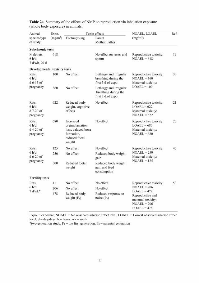
The effects of NMP exposure on reproduction have been investigated in several
studies in which exposure was mediated via inhalation or by oral or dermal ad-
ministration. The majority of studies were carried out in rats and the results from
these studies, found in publicly accessible databases (e.g., PubMed), have been
summarized together with established NOAEL- and LOAEL-values in Table 2a,
b and c.
The studies of repeated exposure carried out by Malley and co-workers showed
that exposure to high levels of NMP (15,000 mg/kg feed) led to reduced testicular
weight and histopathological changes, including degeneration and atrophy (36).
Toxicokinetic studies in rats with radioactively labeled NMP detected high levels
in the testes and at other sites (49, 55). A fertility study by Fries and co-workers
showed no effect on testicular morphology nor on sperm count in Mol:Wist rats
exposed to 618 mg/m3 NMP for 90 days (19). In another study male Imp:WIST
rats were exposed orally to 0, 100, 300 or 1000 mg NMP/kg body weight/day for
10 weeks and mated with non-exposed females to examine the effects on fertility
and foetal development. The results showed that 1000 mg/kg/day made the male
rats infertile. Histopathological studies showed severe necrotic changes in the epi-
thelial cells in the vas deferens. A 300 mg/kg/day dose did not affect fertility in
male rats but did reduce the viability of their offspring (50).
NMP has been shown to pass through the placenta in pregnant rats, with com-
parable levels in the blood of foetuses and mothers (43). It has been reported that
NMP does not cause reproductive toxicity or maternal toxicity in rats (Charles
River CD) exposed via inhalation to up to 360 mg/m3, 6 h/day on days 6-15 of
pregnancy (30). Mol:Wist rats exposed to 680 mg/m3 NMP vapour for 6 h/day
on days 4-20 of pregnancy showed increased preimplantation loss compared with
the control group. Delayed bone formation was observed in the foetuses, in the
cranium, cervical vertebrae, sternum, etc.(20). The same research group exposed
Mol:Wist rats to NMP (622 mg/m3, 6 h/day, days 7-20 of pregnancy) and obser-
ved effects on cognitive development in offspring, including increased latency
in the Morris water maze and operant delayed spatial alternation (Skinner boxes)
during weaning. No maternal toxicity was observed (21). Crl:CD (SD)BR rats
exposed to up to 478 mg NMP/m3 via inhalation showed no effects on the number
of pregnancies, nor on the size or viability of the litter. A somewhat lower foetal
weight was observed in the high dose group compared with the control group (up
to 12%) (53). In the same study some reduction was seen in response to noise by
mothers in the highest dose group.
11
Table 2a. Summary of the effects of NMP on reproduction via inhalation exposure
(whole body exposure) in animals.
Animal
species/type
of study
Expo.
(mg/m3)
Toxic effects NOAEL, LOAEL
(mg/m3)
Ref.
Foetus/young Parent
Mother/Father
Subchronic tests
Male rats,
6 h/d,
7 d/wk, 90 d
618
No effect on testes and
sperm
Reproductive toxicity:
NOAEL = 618
19
Developmental toxicity tests
Rats,
6 h/d,
d 6-15 of
pregnancy
100 No effect
Lethargy and irregular
breathing during the
first 3 d of expo.
Reproductive toxicity:
NOAEL = 360
Maternal toxicity:
LOAEL = 100
30
360 No effect
Lethargy and irregular
breathing during the
first 3 d of expo.
Rats,
6 h/d,
d 7-20 of
pregnancy
622 Reduced body
weight, cognitive
effects
No effect Reproductive toxicity:
LOAEL = 622
Maternal toxicity:
NOAEL = 622
21
Rats,
6 h/d,
d 4-20 of
pregnancy
680 Increased
preimplantation
loss, delayed bone
formation,
reduced foetal
weight
No effect Reproductive toxicity:
LOAEL = 680
Maternal toxicity:
NOAEL = 680
20
Rats,
6 h/d,
d 6-20 of
pregnancy
125 No effect No effect Reproductive toxicity:
NOAEL = 250
Maternal toxicity:
NOAEL = 125
45
250 No effect Reduced body weight
gain
500 Reduced foetal
weight
Reduced body weight
gain and feed
consumption
Fertility tests
Rats,
6 h/d,
7 d/wk*
41 No effect No effect Reproductive toxicity:
NOAEL = 206
LOAEL = 478
Reproductive and
maternal toxicity:
NOAEL = 206
LOAEL = 478
53
206 No effect No effect
478 Reduced body
weight (F1)
Reduced response to
noise (P0)
Expo. = exposure, NOAEL = No observed adverse effect level, LOAEL = Lowest observed adverse effect
level, d = day/days, h = hours, wk = week
*two-generation study, F1 = the first generation, P0 = parental generation
12
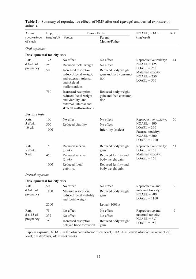
Table 2b. Summary of reproductive effects of NMP after oral (gavage) and dermal exposure of
animals.
Animal
species/type
of study
Expo.
(mg/kg/d)
Toxic effects NOAEL, LOAEL
(mg/kg/d)
Ref.
Foetus Parent
Mother/Father
Oral exposure
Developmental toxicity tests
Rats,
d 6-20 of
pregnancy
125 No effect No effect Reproductive toxicity:
NOAEL = 125
LOAEL = 250
Maternal toxicity:
NOAEL = 250
LOAEL = 500
44
250 Reduced foetal weight No effect
500 Increased resorption,
reduced foetal weight,
and external, internal
and skeletal
malformations
Reduced body weight
gain and feed consump-
tion
750 Increased resorption,
reduced foetal weight
and viability, and
external, internal and
skeletal malformations
Reduced body weight
gain and feed consump-
tion
Fertility tests
Rats,
5 d/wk,
10 wk
100 No effect No effect Reproductive toxicity:
NOAEL = 100
LOAEL = 300
Paternal toxicity:
NOAEL = 300
LOAEL = 1000
50
300 Reduced viability No effect
1000 - Infertility (males)
Rats,
5 d/wk,
9 wk
150 Reduced survival
(3 wk)
Reduced body weight
gain
Reproductive toxicity:
LOAEL = 150
Maternal toxicity:
LOAEL = 150
51
450 Reduced survival
(3 wk)
Reduced fertility and
body weight gain
1000 Reduced foetal
viability.
Reduced fertility and
body weight gain
Dermal exposure
Developmental toxicity tests
Rats,
d 6-15 of
pregnancy
500 No effect No effect Reproductive and
maternal toxicity;
NOAEL = 500
LOAEL = 1100
9
1100 Massive resorption,
reduced foetal viability
and foetal weight
Reduced body weight
gain
2500 - Lethal (100%)
Rats,
d 6-15 of
pregnancy
75 No effect No effect Reproductive and
maternal toxicity:
NOAEL = 237
LOAEL = 750
9
237 No effect No effect 750 Increased resorption,
delayed bone formation
Reduced body weight gain
Expo. = exposure, NOAEL = No observed adverse effect level, LOAEL = Lowest observed adverse effect
level, d = day/days, wk = week/weeks
13
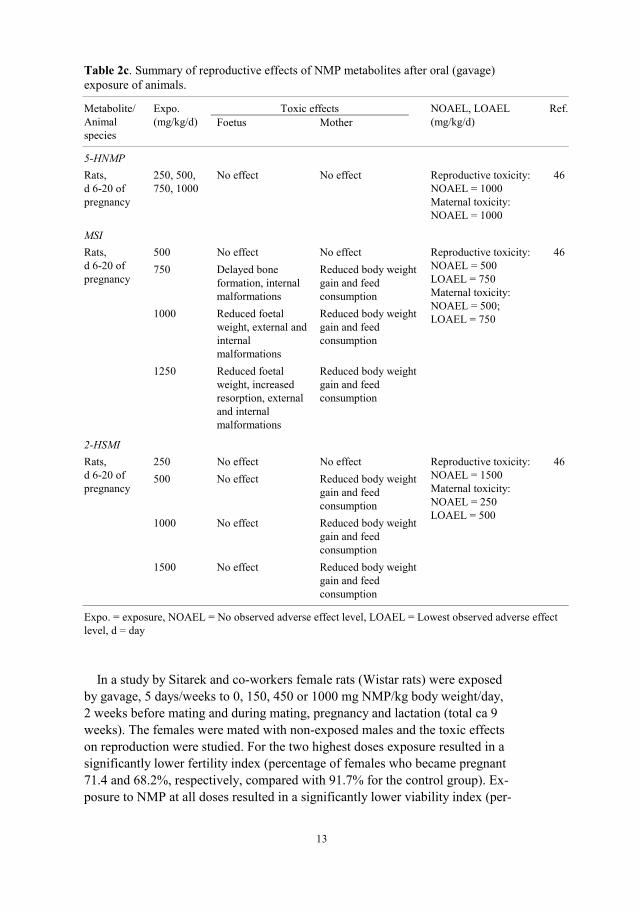
Table 2c. Summary of reproductive effects of NMP metabolites after oral (gavage)
exposure of animals.
Metabolite/
Animal
species
Expo.
(mg/kg/d)
Toxic effects NOAEL, LOAEL
(mg/kg/d)
Ref.
Foetus Mother
5-HNMP
Rats,
d 6-20 of
pregnancy
250, 500,
750, 1000
No effect
No effect Reproductive toxicity:
NOAEL = 1000
Maternal toxicity:
NOAEL = 1000
46
MSI
Rats,
d 6-20 of
pregnancy
500 No effect No effect Reproductive toxicity:
NOAEL = 500
LOAEL = 750
Maternal toxicity:
NOAEL = 500;
LOAEL = 750
46
750 Delayed bone
formation, internal
malformations
Reduced body weight
gain and feed
consumption
1000 Reduced foetal
weight, external and
internal
malformations
Reduced body weight
gain and feed
consumption
1250 Reduced foetal
weight, increased
resorption, external
and internal
malformations
Reduced body weight
gain and feed
consumption
2-HSMI
Rats,
d 6-20 of
pregnancy
250 No effect No effect Reproductive toxicity: NOAEL = 1500 Maternal toxicity: NOAEL = 250 LOAEL = 500
46
500 No effect Reduced body weight
gain and feed
consumption
1000 No effect Reduced body weight
gain and feed
consumption
1500 No effect Reduced body weight
gain and feed
consumption
Expo. = exposure, NOAEL = No observed adverse effect level, LOAEL = Lowest observed adverse effect
level, d = day
In a study by Sitarek and co-workers female rats (Wistar rats) were exposed
by gavage, 5 days/weeks to 0, 150, 450 or 1000 mg NMP/kg body weight/day,
2 weeks before mating and during mating, pregnancy and lactation (total ca 9
weeks). The females were mated with non-exposed males and the toxic effects
on reproduction were studied. For the two highest doses exposure resulted in a
significantly lower fertility index (percentage of females who became pregnant
71.4 and 68.2%, respectively, compared with 91.7% for the control group). Ex-
posure to NMP at all doses resulted in a significantly lower viability index (per-
14
centage survival of young after 4 days, were 86.4, 71.6, and 0%, respectively,
compared with 94% for the control group), reduced survival of newborn rats
during the first 3 weeks after birth (78.2, 43.4 and 0%, respectively, compared
with 96.1% for the control group) and lower body weight gain during days 0-20.
The reduction in body weight gain for mothers showed a dose-response relation-
ship, with 87.7, 75.6 and 40.7% relative body weight gain for the three doses,
compared with 100% for the control group (51).
Saillenfait and co-workers have carried out three studies in which they exa-
mined the effects on development of NMP and the three metabolites 5-HNMP,
MSI and 2-HMSI in Sprague-Dawley rats exposed via inhalation (45) or oral
administration (44, 46), see Tables 2a, b and c. In the inhalation study pregnant
females were exposed (whole body) to 0, 125, 250 or 500 mg NMP/m3, 6 h/day on
days 6-20 of pregnancy. Reduced foetal weight (5%) was observed at 500 mg/m3
and mothers showed reduced body weight gain (23%, day 6-13) which was asso-
ciated with reduced feed consumption (8%, day 6-21) at the same but not at lower
doses. The study with oral administration (gavage, 0, 125, 250, 500 or 750 mg
NMP/kg body weight/day on days 6-20) showed reduced body weight gain (25
and 50%, respectively) and reduced feed consumption (8 and 15%, respectively)
in pregnant rats given 500 and 750 mg/kg/day, respectively (44). Effects on the
foetus were observed with the 250 mg/kg/day dose and higher and consisted of
increased resorption, reduced foetal weight, and external, internal and skeletal
malformations. Exposure to the three NMP metabolites at up to 1500 mg/kg/day
on days 6-20 of pregnancy showed that 5-HNMP was toxic neither to the foetus
nor the mother and that 2-HSMI was only toxic to the mother (reduced body
weight gain and feed consumption) at the doses tested (46). The metabolite MSI
caused a dose-dependent increase in delayed bone formation, external and internal
malformations, and increased resorption at the 750 mg/kg/day dose and higher.
Effects on the mother were observed at this same dose and consisted in reduced
body weight gain and feed consumption. The authors concluded that none of the
three metabolites was a more potent teratogen than NMP.
A dermal exposure study carried out by Becci and co-workers in the 1980s ob-
served increased foetal resorption at the dose 750 mg NMP/kg body weight/day
(exposed on days 6-15) in pregnant Sprague-Dawley rats. The same dose resulted
in reduced body weight gain in mothers (9).
Flick and co-workers studied the reproductive toxicity of NMP and its three
metabolites (5-HNMP, MSI and 2-HMSI) in an in vitro culture system comprising
whole rat embryos. The embryos were exposed in vitro to up to 0.06% NMP
(vol/vol), equivalent to 0.006 mol/l in the medium, and up to 0.44% (vol/vol) of
5-HNMP, MSI or 2-HSMI on days 9.5-11.5 of pregnancy. The results showed that
exposure to NMP (≥0.03%/0.003 mol/l) and 5-HNMP (≥0.10%) caused foetal
injuries comprising abnormalities of the cranium, abnormal development of the
second visceral arch and delayed anterior neuropore closure. On this basis the
authors concluded that NMP and 5-HNMP can be classified as weak teratogens
and that 2-HMSI and MSI have no teratogenic properties (18).
15
Poet and co-workers used PBPK- and benchmark dose-modelling to calculate
the point of departure (POD) as the area under the blood concentration-time curve
(AUC) for NMP in the blood (41). The calculations were mainly based on two
experimental inhalation studies in rats (45, 53). Reduced weight of foetuses and
newborn was regarded as the critical effect. It was calculated that the POD value
was equivalent to inhalation exposure to 1977.6 mg/m3 (8 h/day, 5 days/week)
(41). It should be pointed out that this calculation of equivalence includes the
questionable assumption that rats and humans have similar sensitivity to NMP
at the same internal concentrations, i.e., they differ only in terms of toxicokinetics.
It can be noted that Sallenfait et al. (45) observed reduced foetal weight in rats at
500 mg/m3 (NOAEL 250 mg/m3).
The two companies BASF and GAF, which also produce NMP, have carried out
several further studies which examined the toxic effects of NMP on animal repro-
duction. As these reports are difficult to access, this review of them is based on
information and results presented as industrial reports in ECHA's database for
NMP. The results and conclusions are difficult to assess as they were not fully
reported.
In 1993 BASF studied the effects of NMP on reproduction in rabbits and in-
cluded an inhalation study and a dermal exposure study. In the inhalation study
pregnant rabbits were exposed (nose/head) to up to 1000 mg/m3 NMP (mix of
vapour/aerosol) 6 h/day on days 7-19 after insemination. The highest dose pro-
duced no maternal toxicity or effects on fertility but there was an increased inci-
dence of skeletal abnormalities (extra 13th rib) in foetuses. A NOAEL of 500
mg/m3 was established for foetal toxicity and 1000 mg/m3 for maternal toxicity.
In the dermal application study, in which pregnant rabbits were exposed to up to
1000 mg NMP/kg body weight/day as a 40% aqueous solution (6 h/day for days
7-19 after insemination), no toxic effects were observed in the female rabbits.
Teratogenic effects were only observed at 1000 mg/kg body weight/day and, in
this study too, included the development of a 13th rib. A NOAEL of 300 mg/kg
body weight/day was established for foetal toxicity and 1000 mg/kg body
weight/day for maternal toxicity.
In 1991 GAF carried out studies in which it dosed pregnant rabbits perorally
with 0, 55, 175 or 540 mg/kg body weight/day on days 6-18 after insemination.
Dosing with 175 or 540 mg/kg/day caused reduced body weight gain in the
mothers. Teratogenic effects were only observed at the highest dose and included
postimplantation loss, altered foetal morphology and increased incidence of car-
diovascular and cranial malformations. A NOAEL of 175 mg/kg body weight/day
was established for foetal toxicity and 55 mg/kg body weight/day for maternal
toxicity.
Dose-effect-/dose-response-relationships
The acute toxicity of NMP is relatively low, see Table 1. Dose-response relation-
ships for toxic effects after inhalation exposure in humans are summarized in
16
Table 3, and after inhalation exposure and oral exposure in animals in Tables 4
and 5, respectively.
Studies with human research subjects indicate that NMP is weakly irritant or
non-irritant with inhalation exposure (up to 160 mg/m3) (54, 65). Studies of occu-
pational exposure indicate that NMP can cause varying degrees of irritation, es-
pecially with exposure to high doses of NMP in vapour form (340 mg/m3) (5, 7)
or with skin exposure (25, 31). Workers experienced skin problems after 2 or
several days of exposure, while research subjects were exposed for one day (65)
or had one-day exposures with a 2-week gap between each (54). In these studies
the research subjects were exclusively men whereas the group of workers studied
included women. Another possibility that might be significant is that a damp
workplace environment, or alternatively a closed environment, might contribute
to irritant contact dermatitis. Such conditions might make studies carried out in
exposure chambers irrelevant in establishing exposure limits.
No studies reporting that NMP causes sensitization have been found in the
literature.
Inhalation studies in rats show that exposure to 100 mg NMP/m3 results in
transient lethargy and irregular breathing. Exposure of female rats at the same
level for 6 h/day on days 6-15 of pregnancy had no effect on the foetuses (30).
Reduced body weight in newborn animals was observed in a two-generation study
in rats exposed to 478 mg NMP/m3. The dose induced maternal toxicity in the
form of reduced response to noise (53). Reduced body weight and effects on
cognitive functions were observed in the offspring of rats exposed to 622 mg
NMP/m3, on days 7-20 of pregnancy. No maternal toxicity was seen at this dose
(21).
A reduced survival rate for rat offspring was observed 3 weeks after birth, in
parallel with reduced body weight gain (12%) for mothers exposed orally to 150
mg NMP/kg body weight/day (51). Exposure of rats to 250 mg NMP/kg body
weight/day, on days 6-20 of pregnancy resulted in reduced foetal weight. At the
500 mg NMP/kg body weight/day dose an increased number of resorptions and
skeletal, external and internal malformations was also observed. (44). At the
higher dose maternal toxicity was seen in the form of reduced body weight gain
(25%) and feed consumption (8).
The authors of two studies involving the oral exposure of rats and mice to NMP
commented that males appeared to be more sensitive to NMP than females. In
both studies this was possibly related to gender differences in the metabolism of
NMP (34,36).
Conclusions
There is insufficient data to establish a critical effect for NMP in occupational
exposure. On the basis of animal research the critical effect of NMP is transient
CNS effects (irregular breathing, drowsiness). This has been observed with
inhalation of 100 mg NMP/m3.
17
Irritant contact dermatitis has been reported with occupational exposure to NMP
in liquid form.
NMP is toxic to reproduction in animals. Reduced survival of newborn rats was
observed with an oral dose of 150 mg/kg body weight/day. With inhalation expo-
sure reduced body weight of offspring as well as some maternal effects were ob-
served at 478 mg/m3. Malformations and cognitive effects have been observed at
somewhat higher exposure levels.
NMP is efficiently absorbed both via the respiratory tract and the skin (also in
vapour form) and skin uptake can be substantial.
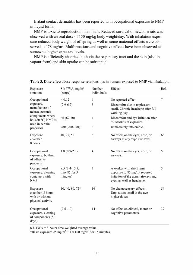
Table 3. Dose-effect-/dose-response-relationships in humans exposed to NMP via inhalation.
Exposure
situation
8-h TWA, mg/m3
(range)
Number
individuals
Effects Ref.
Occupational
exposure,
manufacture of
microelectronic
components where
hot (80 °C) NMP is
used in certain
processes
< 0.12 6 No reported effect. 7
(2.9-6.2) 5 Discomfort due to unpleasant
smell. Chronic headache after full
working day.
66 (62-70) 4 Discomfort and eye irritation after
30 seconds of exposure.
280 (200-340)
3 Immediately intolerable.
Exposure chamber, 8 hours
10, 25, 50 6 No effect on the eyes, nose, or airways at any exposure level.
63
Occupational exposure, bottling of adhesive products
1.8 (0.9-2.8) 4 No effect on the eyes, nose, or airways.
5
Occupational exposure, cleaning containers with NMP
8.5 (3.4-15.5; max 85 for 5 minutes)
3 A worker with short term
exposure to 85 mg/m3 reported
irritation of the upper airways and
eyes, as well as headache.
5
Exposure chamber, 8 hours with or without physical activity
10, 40, 80, 72* 16 No chemosensory effects. Unpleasant smell at the two higher doses.
54
Occupational exposure, cleaning of components (5 days).
(0.6-1.0)
14 No effect on clinical, motor or cognitive parameters.
39
8-h TWA = 8-hours time-weighted average value *Basic exposure 25 mg/m3 + 4 x 160 mg/m3 for 15 minutes.
18
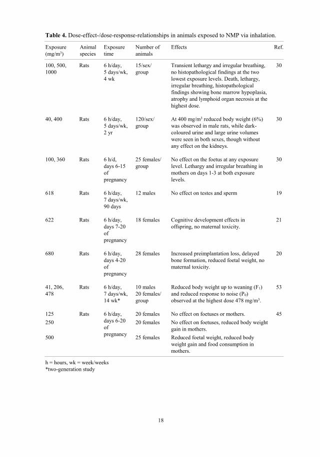
Table 4. Dose-effect-/dose-response-relationships in animals exposed to NMP via inhalation.
Exposure
(mg/m3)
Animal
species
Exposure
time
Number of
animals
Effects Ref.
100, 500,
1000
Rats 6 h/day,
5 days/wk,
4 wk
15/sex/
group
Transient lethargy and irregular breathing,
no histopathological findings at the two
lowest exposure levels. Death, lethargy,
irregular breathing, histopathological
findings showing bone marrow hypoplasia,
atrophy and lymphoid organ necrosis at the
highest dose.
30
40, 400 Rats 6 h/day,
5 days/wk,
2 yr
120/sex/
group
At 400 mg/m3 reduced body weight (6%)
was observed in male rats, while dark-
coloured urine and large urine volumes
were seen in both sexes, though without
any effect on the kidneys.
30
100, 360 Rats 6 h/d,
days 6-15
of
pregnancy
25 females/
group
No effect on the foetus at any exposure
level. Lethargy and irregular breathing in
mothers on days 1-3 at both exposure
levels.
30
618 Rats 6 h/day,
7 days/wk,
90 days
12 males No effect on testes and sperm 19
622 Rats 6 h/day,
days 7-20
of
pregnancy
18 females Cognitive development effects in
offspring, no maternal toxicity.
21
680 Rats 6 h/day,
days 4-20
of
pregnancy
28 females Increased preimplantation loss, delayed
bone formation, reduced foetal weight, no
maternal toxicity.
20
41, 206,
478
Rats 6 h/day,
7 days/wk,
14 wk*
10 males
20 females/
group
Reduced body weight up to weaning (F1)
and reduced response to noise (P0)
observed at the highest dose 478 mg/m3.
53
125 Rats 6 h/day,
days 6-20
of
pregnancy
20 females No effect on foetuses or mothers. 45
250 20 females No effect on foetuses, reduced body weight
gain in mothers.
500 25 females Reduced foetal weight, reduced body
weight gain and food consumption in
mothers.
h = hours, wk = week/weeks
*two-generation study
19
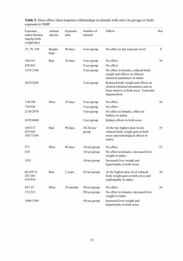
Table 5. Dose-effect-/dose-response relationships in animals with oral (via gavage or feed)
exposure to NMP.
Exposure,
males/females
(mg/kg body
weight/day)
Animal
species
Exposure
time
Number of
animals
Effects Ref.
25, 79, 250 Beagle
dogs
90 days 6/sex/group No effect at any exposure level. 8
149/161 Rats 28 days 5/sex/group No effect. 34
429/493 5/sex/group No effect.
1234/1548 5/sex/group No effect in females, reduced body
weight and effects on clinical-
chemical parameters in males.
2019/2268 5/sex/group Reduced body weight and effects on
clinical-chemical parameters and on
bone marrow in both sexes. Testicular
degeneration.
130/180 Mice 28 days 5/sex/group No effect. 34
720/920 5/sex/group No effect.
2130/2970 5/sex/group No effect in females, effect on
kidneys in males.
2670/4060 5/sex/group
Kidney effects in both sexes.
169/217
433/565
1057/1344
Rats 90 days 20-26/sex/
group
At the two highest dose levels
reduced body weight gain in both
sexes and neurological effects in
males.
35
277 Mice 90 days 10/sex/group No effect. 35
619 10/sex/group No effect in females, increased liver
weight in males.
1931 10/sex/group Increased liver weight and
hypertrophy in both sexes.
66.4/87.8
207/283
678/939
Rats 2 years 62/sex/group At the highest dose level reduced
body weight gain in both sexes and
nephropathy in males.
36
89/115 Mice 18 months 50/sex/group No effect. 36
173/221 50/sex/group No effect in females, increased liver
weight in males.
1089/1399 50/sex/group Increased liver weight and
hypertrophy in both sexes.
20
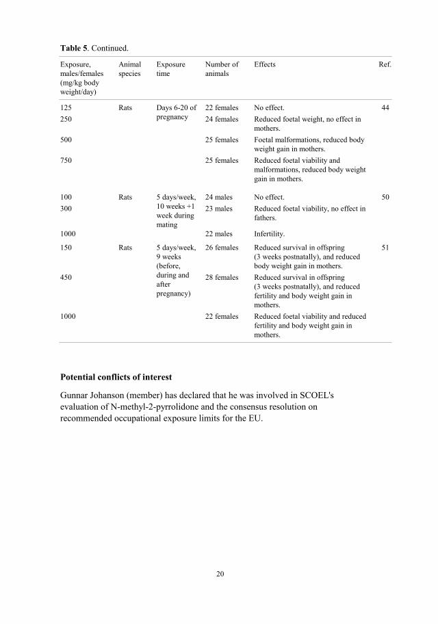
Table 5. Continued.
Exposure,
males/females
(mg/kg body
weight/day)
Animal
species
Exposure
time
Number of
animals
Effects Ref.
125 Rats Days 6-20 of
pregnancy
22 females No effect. 44
250 24 females Reduced foetal weight, no effect in
mothers.
500 25 females Foetal malformations, reduced body
weight gain in mothers.
750 25 females Reduced foetal viability and
malformations, reduced body weight
gain in mothers.
100 Rats 5 days/week,
10 weeks +1
week during
mating
24 males No effect. 50
300 23 males Reduced foetal viability, no effect in
fathers.
1000 22 males Infertility.
150 Rats 5 days/week,
9 weeks
(before,
during and
after
pregnancy)
26 females Reduced survival in offspring
(3 weeks postnatally), and reduced
body weight gain in mothers.
51
450 28 females Reduced survival in offspring
(3 weeks postnatally), and reduced
fertility and body weight gain in
mothers.
1000 22 females Reduced foetal viability and reduced
fertility and body weight gain in
mothers.
Potential conflicts of interest
Gunnar Johanson (member) has declared that he was involved in SCOEL's
evaluation of N-methyl-2-pyrrolidone and the consensus resolution on
recommended occupational exposure limits for the EU.
21
References
1. Akrill P, Cocker J, Dixon S. Dermal exposure to aqueous solutions of N-methyl pyrrolidone.
Toxicol Lett 2002;134:265-269.
2. Ansell JM, Fowler JA. The acute oral toxicity and primary ocular and dermal irritation of
selected N-alkyl-2-pyrrolidones. Food Chem Toxicol 1988;26:475-479.
3. Anundi H, Langworth S, Johanson G, Lind ML, Åkesson B, Friis L, Itkes N, Söderman E,
Jonsson BA, Edling C. Air and biological monitoring of solvent exposure during graffiti
removal. Int Arch Occup Environ Health 2000;73:561-569.
4. Anundi H, Lind ML, Friis L, Itkes N, Langworth S, Edling C. High exposures to organic
solvents among graffiti removers. Int Arch Occup Environ Health 1993;65:247-251.
5. Bader M, Rosenberger W, Rebe T, Keener SA, Brock TH, Hemmerling HJ, Wrbitzky R.
Ambient monitoring and biomonitoring of workers exposed to N-methyl-2-pyrrolidone in an
industrial facility. Int Arch Occup Environ Health 2006;79:357-364.
6. Bader M, Wrbitzky R, Blaszkewicz M, Schaper M, van Thriel C. Human volunteer study on
the inhalational and dermal absorption of N-methyl-2-pyrrolidone (NMP) from the vapour
phase. Arch Toxicol 2008;82:13-20.
7. Beaulieu HJ, Schmerber R. M-Pyrol (NMP) use in the microelectronics industry. Appl Occup
Environ Hyg 1991;6:874-880.
8. Becci PJ, Gephart LA, Koschier FJ, Johnson WD, Burnette LW. Subchronic feeding study in
beagle dogs of N-methylpyrrolidone. J Appl Toxicol 1983;3:83-86.
9. Becci PJ, Knickerbocker MJ, Reagan EL, Parent RA, Burnette LW. Teratogenicity study of
N-methylpyrrolidone after dermal application to Sprague-Dawley rats. Fundam Appl Toxicol
1982;2:73-76.
10. Bower DB. Stillbirth after occupational exposure to N-methyl-2 pyrrolidone. J Occup
Environ Med 1997;39:393-394.
11. Campbell HL, Striebig BA. Evaluation of N-methylpyrrolidone and its oxidative products
toxicity utilizing the microtox assay. Environ Sci Technol 1999;33:1926-1930.
12. Carnerup MA, Saillenfait AM, Jönsson BA. Concentrations of N-methyl-2-pyrrolidone
(NMP) and its metabolites in plasma and urine following oral administration of NMP to rats.
Food Chem Toxicol 2005;43:1441-1447.
13. Carnerup MA, Spanne M, Jönsson BA. Levels of N-methyl-2-pyrrolidone (NMP) and its
metabolites in plasma and urine from volunteers after experimental exposure to NMP in dry
and humid air. Toxicol Lett 2006;162:139-145.
14. Carnerup MA, Åkesson B, Jönsson BA. Determination of 5-hydroxy-N-methyl-2-pyrrolidone
and 2-hydroxy-N-methylsuccinimide in human plasma and urine using liquid
chromatography-electrospray tandem mass spectrometry. J Chromatogr B Biomed Sci Appl
2001;761:107-113.
15. Dick IP, Bounds SVJ, Parod RJ, Banton MI, Griffiths JC. In vitro dermal absorption of N-
methyl-2-pyrrolidone (NMP) through human and rat skin. Society of Toxicology 40th Annual
Meeting San Fransisco, USA 2001.
16. Engelhardt G, Fleig H. 1-Methyl-2-pyrrolidinone (NMP) does not induce structural and
numerical chromosomal aberrations in vivo. Mutat Res 1993;298:149-155.
17. EPA. Science assessment for N-methylpyrrolidone. United States Environmental Protection
Agency, Washington, D.C. 20460, 2006: 25 pp.
18. Flick B, Talsness CE, Jackh R, Buesen R, Klug S. Embryotoxic potential of N-methyl-
pyrrolidone (NMP) and three of its metabolites using the rat whole embryo culture system.
Toxicol Appl Pharmacol 2009;237:154-167.
22
19. Fries AS, Hass U, Jakobsen BM, Jelnes JE, Lund SP, Simonsen J. Toxic effects of N-
methylpyrrolidone on foetal development, the central nervous system, testes and semen in
rats. Arbeijdsmiljøfondet 1992;Report 790037. Cited in IPCS (22).
20. Hass U, Jakobsen BM, Lund SP. Developmental toxicity of inhaled N-methylpyrrolidone in
the rat. Pharmacol Toxicol 1995;76:406-409.
21. Hass U, Lund SP, Elsner J. Effects of prenatal exposure to N-methylpyrrolidone on postnatal
development and behavior in rats. Neurotoxicol Teratol 1994;16:241-249.
22. IPCS. Concise International Chemical Assessment Document No. 35. N-methyl-2-
pyrrolidone. Inter-Organization Progam for the Sound Managment of Chemicals (IOMC),
World Health Organization, Geneva, 2001.
23. Johanson G, Rauma M. Basis for skin notation. Part 1. Dermal penetration data for
substances on the Swedish OEL list. Arbete och Hälsa 2008;42(2). University of Gothenburg,
Sweden.
24. Jouyban A, Fakhree MA, Shayanfar A. Review of pharmaceutical applications of N-methyl-2-
pyrrolidone. J Pharm Pharmaceut Sci 2010;13:524-535.
25. Jungbauer FH, Coenraads PJ, Kardaun SH. Toxic hygroscopic contact reaction to N-methyl-
2-pyrrolidone. Contact Dermatitis 2001;45:303-304.
26. Jönsson BA, Åkesson B. Human experimental exposure to N-methyl-2-pyrrolidone (NMP):
toxicokinetics of NMP, 5-hydroxy- N-methyl-2-pyrrolidone, N-methylsuccinimide and 2-
hydroxy-N-methylsuccinimide (2-HMSI), and biological monitoring using 2-HMSI as a
biomarker. Int Arch Occup Environ Health 2003;76:267-274.
27. Keener SA, Wrbitzky R, Bader M. Human volunteer study on the influence of exposure
duration and dilution of dermally applied N-methyl-2-pyrrolidone (NMP) on the urinary
elimination of NMP metabolites. Int Arch Occup Environ Health 2007;80:327-334.
28. Kemikalieinspektionen (Swedish Chemicals Agency, oktober 2013):
http://www.kemi.se/Documents/Forfattningar/Reach/Amnen_pa_kandidatforteckningen_
konsoliderad.pdf
29. Langworth S, Anundi H, Friis L, Johanson G, Lind ML, Söderman E, Åkesson BA. Acute
health effects common during graffiti removal. Int Arch Occup Environ Health 2001;74:213-
218.
30. Lee KP, Chromey NC, Culik R, Barnes JR, Schneider PW. Toxicity of N-methyl-2-
pyrrolidone (NMP): teratogenic, subchronic, and two-year inhalation studies. Fundam Appl
Toxicol 1987;9:222-235.
31. Leira HL, Tiltnes A, Svendsen K, Vetlesen L. Irritant cutaneous reactions to N-methyl-2-
pyrrolidone (NMP). Contact Dermatitis 1992;27:148-150.
32. Ligocka D, Lison D, Haufroid V. Contribution of CYP2E1 to N-methyl-2-pyrrolidone
metabolism. Arch Toxicol 2003;77:261-266.
33. Lundberg P (ed). N-Methyl-2-pyrrolidon. Scientific Basis for Swedish Occupational
Standards. VIII. Swedish Criteria Group for Occupational Standards. Arbete och Hälsa
1987;39:173-177. National Institute of Occupational Health, Solna, Sweden.
34. Malek DE, Malley LA, Slone TW, Elliott GS, Kennedy GL, Mellert W, Deckardt K,
Gembardt C, Hildebrand B, Murphy SR, Bower DB, Wright GA. Repeated dose toxicity
study (28 days) in rats and mice with N-methylpyrrolidone (NMP). Drug Chem Toxicol
1997;20:63-77.
35. Malley LA, Kennedy GL, Elliott GS, Slone TW, Mellert W, Deckardt K, Gembardt C,
Hildebrand B, Parod RJ, McCarthy TJ, Griffiths JC. 90-day subchronic toxicity study in rats
and mice fed N-methylpyrrolidone (NMP) including neurotoxicity evaluation in rats. Drug
Chem Toxicol 1999;22:455-480.
36. Malley LA, Kennedy GL, Elliott GS, Slone TW, Mellert W, Deckardt K, Kuttler K,
Hildebrand B, Banton MI, Parod RJ, Griffiths JC. Chronic toxicity and oncogenicity of N-
23
methylpyrrolidone (NMP) in rats and mice by dietary administration. Drug Chem Toxicol
2001;24:315-338.
37. Mayer VW, Goin CJ. Investigations of aneuploidy-inducing chemical combinations in
Saccharomyces cerevisiae. Mutat Res 1988;201:413-421.
38. Mayer VW, Goin CJ, Taylor-Mayer RE. Aneuploidy induction in Saccharomyces cerevisiae
by two solvent compounds, 1-methyl-2-pyrrolidinone and 2-pyrrolidinone. Environ Mol
Mutagen 1988;11:31-40.
39. Nishimura S, Yasui H, Miyauchi H, Kikuchi Y, Kondo N, Takebayashi T, Tanaka S,
Mikoshiba Y, Omae K, Nomiyama T. A cross-sectional observation of effect of exposure to
N-methyl-2-pyrrolidone (NMP) on workers' health. Ind Health 2009;47:355-362.
40. Payan JP, Beydon D, Fabry JP, Boudry I, Cossec B, Ferrari E. Toxicokinetics and
metabolism of N-[14C]methylpyrrolidone in male Sprague-Dawley rats. A saturable NMP
elimination process. Drug Metab Dispos 2002;30:1418-1424.
41. Poet TS, Kirman CR, Bader M, van Thriel C, Gargas ML, Hinderliter PM. Quantitative risk
analysis for N-methyl pyrrolidone using physiologically based pharmacokinetic and
benchmark dose modeling. Toxicol Sci 2010;113:468-482.
42. Radhika G, Venkatesan R, Kathiroli S. N-methylpyrrolidone: Isolation and characterization of
the compound from the marine sponge Clathria frondifera (class: Demospongiae). Indian J
Mar Sci 2007;36:235-238.
43. Ravn-Jonsen A, Edelflors S, Hass U, Lund SP. The kinetics of N-methyl-2-pyrrolidone in
pregant rats and their foetuses compared with non-pregnant rats. Toxicol Lett 1992;Suppl
136:Abstract P5/P8.
44. Saillenfait AM, Gallissot F, Langonne I, Sabate JP. Developmental toxicity of N-methyl-2-
pyrrolidone administered orally to rats. Food Chem Toxicol 2002;40:1705-1712.
45. Saillenfait AM, Gallissot F, Morel G. Developmental toxicity of N-methyl-2-pyrrolidone in
rats following inhalation exposure. Food Chem Toxicol 2003;41:583-588.
46. Saillenfait AM, Sabate JP, Gallissot F. Comparative developmental toxicities of the three
major metabolites of N-methyl-2-pyrrolidone after oral administration in rats. J Appl Toxicol
2007;27:571-581.
47. SCCS (Scientific Committee on Consumer Safety), Opinion on N-Methyl-2-pyrrlidone
(NMP), 22 March 2001.
48. SCOEL (the Scientific Committee on Occupational Exposure Limits). Recommendation from
the Scientific Committee on Occupational Exposure Limits for N-methyl-2-Pyrrolidone.
SCOEL/SUM/119, 2007.
49. Sitarek K, Kilanowicz A. Tissue distribution and excretion of N-methyl-2-pyrrolidone in male
and female rats. Int J Occup Med Environ Health 2006;19:142-148.
50. Sitarek K, Stetkiewicz J. Assessment of reproductive toxicity and gonadotoxic potential of N-
methyl-2-pyrrolidone in male rats. Int J Occup Med Environ Health 2008;21:73-80.
51. Sitarek K, Stetkiewicz J, Wasowicz W. Evaluation of reproductive disorders in female rats
exposed to N-methyl-2-pyrrolidone. Birth Defects Res B Dev Reprod Toxicol 2012;95:195-
201.
52. Solomon GM, Morse EP, Garbo MJ, Milton DK. Stillbirth after occupational exposure to
N-methyl-2-pyrrolidone. A case report and review of the literature. J Occup Environ Med
1996;38:705-713.
53. Solomon HM, Burgess BA, Kennedy GL, Jr., Staples RE. 1-Methyl-2-pyrrolidone (NMP):
reproductive and developmental toxicity study by inhalation in the rat. Drug Chem Toxicol
1995;18:271-293.
54. van Thriel C, Blaszkewicz M, Schaper M, Juran SA, Kleinbeck S, Kiesswetter E, Wrbitzky
R, Stache J, Golka K, Bader M. Chemosensory effects during acute exposure to N-methyl-2-
pyrrolidone (NMP). Toxicol Lett 2007;175:44-56.
24
55. Wells DA, Digenis GA. Disposition and metabolism of double-labeled [3H and 14C] N-
methyl-2-pyrrolidinone in the rat. Drug Metab Dispos 1988;16:243-249.
56. Wells DA, Hawi AA, Digenis GA. Isolation and identification of the major urinary metabolite
of N-methylpyrrolidinone in the rat. Drug Metab Dispos 1992;20:124-126.
57. Wells DA, Thomas HF, Digenis GA. Mutagenicity and cytotoxicity of N-methyl-2-
pyrrolidinone and 4-(methylamino)butanoic acid in the Salmonella/microsome assay. J Appl
Toxicol 1988;8:135-139.
58. Xiaofei E, Wada Y, Nozaki J, Miyauchi H, Tanaka S, Seki Y, Koizumi A. A linear
pharmacokinetic model predicts usefulness of N-methyl-2-pyrrolidone (NMP) in plasma or
urine as a biomarker for biological monitoring for NMP exposure. J Occup Health
2000;42:321-327.
59. Zimmermann FK, Holzwarth UL, Scheel I, Resnick MA. Aprotic polar solvents that affect
porcine brain tubulin aggregation in vitro induce aneuploidy in yeast cells growing at low
temperatures. Mutat Res 1988;201:431-442.
60. Zimmermann S, Zarse K, Schulz TJ, Siems K, Muller-Kuhrt L, Birringer M, Ristow M. A
cell-based high-throughput assay system reveals modulation of oxidative and nonoxidative
glucose metabolism due to commonly used organic solvents. Horm Metab Res 2008;40:29-
37.
61. Åkesson B. The Nordic Expert Group for Criteria Documentation of Health Risks from
Chemicals. 115. N-methyl-2-pyrrolidone (NMP). Arbete och Hälsa 1994;40:1-23. National
Institute of Occupational Health, Solna, Sweden.
62. Åkesson B, Carnerup MA, Jönsson BA. Evaluation of exposure biomarkers from
percutaneous absorption of N-methyl-2-pyrrolidone. Scand J Work Environ Health
2004;30:306-312.
63. Åkesson B, Jönsson BA. Major metabolic pathway for N-methyl-2-pyrrolidone in humans.
Drug Metab Dispos 1997;25:267-269.
64. Åkesson B, Jönsson BA. Biological monitoring of N-methyl-2-pyrrolidone using 5-hydroxy-
N-methyl-2-pyrrolidone in plasma and urine as the biomarker. Scand J Work Environ Health
2000;26:213-218.
65. Åkesson B, Paulsson K. Experimental exposure of male volunteers to N-methyl-2-pyrrolidone
(NMP): acute effects and pharmacokinetics of NMP in plasma and urine. Occup Environ Med
1997;54:236-240.
25
Consensus Report for Crystalline Silica, Quartz
December 31, 2012
This consensus report is based on a document on crystalline silica published by
WHO in 2000 (39) and on a monograph on silica from IARC (The International
Agency for Research on Cancer) (72). Certain parts of the report are based on an
overview of current knowledge issued by the Swedish Work Environment Autho-
rity 2011 (11). There is a substantial volume of scientific literature on crystalline
silica and its health risks. In this report the literature concerning cancer and crys-
talline silica focuses on articles published after IARC's 1997 cancer classification
and on articles of special interest (e.g., Swedish studies).
The search term "silicosis" yielded 7456 articles from PubMed (November
2011), with the earliest published studies dating back to the 1920s. The consensus
report includes a description of those silicosis studies that are quoted as critical
studies in CICAD's document from 2000 (39), as well as a number of subsequent
epidemiological studies. The search term ”tridymite” retrieved 42,831 articles
from PubMed and ”cristobalite” 42,913 articles (November 2011), the majority
of which were studies of materials. Relevant studies of health effects have been
included.
The report does not cover quartz-based nanomaterials.
Chemical-physical data
Quartz, CAS no: 14808-60-7
Cristobalite, CAS no: 14464-46-1
Tridymite, CAS no: 15468-32-3
Chemical formula: SiO2
Synonyms: crystalline silicon dioxide
Mol. weight: 60.09 g/mol
Density: 2.6 g/ml (α-quartz)
2.3 g/ml (cristobalite)
2.3 g/ml (tridymite)
Melting point: 1610 °C (α-quartz)
Boiling point: 2230 °C (α-quartz)
Solubility: Insoluble in water
Colour: White or colourless
26
Occurrence, use
Silica (silicon dioxide) is a compound of silicon and oxygen. Crystalline silica in
the form of quartz is a common mineral in the earth's crust and occurs, for exam-
ple, in rock forms such as granite and gneiss. Quartz can be transformed into other
crystalline forms, such as cristobalite and tridymite. These forms occur naturally
at low levels but can be produced during industrial processes at high temperatures
and high pressures, for example, when diatomaceous earth (diatomite, kieselguhr)
is heated, in the manufacture of ceramics and silicon carbide, in foundry proces-
ses, or in other processes involving high temperatures. Silica is also found in an
amorphous form, a more disordered structure which occurs naturally, for example,
in opal, biogenic silica (in living matter), and kieselguhr. Amorphous silica is also
produced synthetically and is used in products such as fillers or anti-caking agents
(Swedish Chemicals Agency, Information on substances 2013: http://apps.kemi.se
/flodessok/floden/kemamne_eng/kiseldioxid_eng.htm, (39)).
Diatomaceous earth (kieselguhr) consists of amorphous silica (65-90%). When
kieselguhr (diatomite) is heated it is gradually converted to the crystalline forms
tridymite, cristobalite, and quartz; the proportion of cristobalite can be relatively
high.
The three crystalline structural types, quartz, cristobalite, and tridymite, occur
in both alpha and beta forms. The high temperatures involved in their formation
mean that the minerals initially exist in the beta form. When the minerals cool
they are converted to the alpha form. The alpha form is therefore the one that
occurs under normal conditions and the form that is commonly considered (39).
In scientific publications quartz dust is normally referred to as ”silica dust” and
the type of crystalline silica is not usually mentioned.
Sampling and analysis of α-quartz and cristobalite
The sampling of respirable dust is carried out by pumped sampling with use of
a pre-separator for respirable dust (68); separation of the respirable fraction is
carried out in accordance with international standards (49). The lowest quanti-
fiable concentration in the measurement of respirable dust can be 0.08 mg/m3
during sampling over a full 8-hour shift and at a flow rate of 2.5 l/min; the equi-
valent when sampling for 4 hours is 0.2 mg/m3 (16).
The respirable fraction of quartz is sampled on a membrane filter. The sample is
ashed in a low temperature oven, suspended in ethanol, and transferred to a silver
filter. α-Quartz is determined by x-ray diffractometry (70, 99, 103). A typical limit
of quantification could be 2 μg/sample, with a measurement uncertainty of up to
±25% at 2.0 – 19 μg and ±15% at 20 – 400 μg (17). For sampled air volumes of
600 and 1200 litres, respectively, from 4 and 8 hours sampling, respectively, at
a flow rate of 2.5 l/min, the quantifiable concentrations are 0.0033 and 0.0017
mg/m3, respectively. For cristobalite in air a quantifiable amount per sample could
27
be 5 μg (18), and therefore the lowest quantifiable concentrations in air for 4 and
8 hours sampling, respectively, would be 0.008 and 0.004 mg/m3, respectively.
Exposure
Exposure to quartz is one of the most common forms of occupational exposure.
In an international appraisal of occupational exposure, quartz was listed as the
substances to which the greatest proportion of workers were exposed (48). It has
been estimated that more than 3 million workers in Europe are exposed to quartz
during their work, of whom around 85,000 are based in Sweden (76). Exposure
to forms of crystalline silica other than quartz, e.g., cristobalite and tridymite, can
occur in certain areas of occupation. Studies carried out in American foundries
have demonstrated, for example, high levels of cristobalite in dust samples (74).
Exposure to quartz occurs during work involving soil and land and in the hand-
ling and use of products that contain quartz. Examples include agriculture, mining,
stonemasonry, steel industry work, glass- and abrasive products-manufacturing,
porcelain and ceramics work, building work (demolition, bricklaying, concrete-
and casting-work) and sandblasting, see Table 1 (39, 72, 86). Exposure to quartz
often involves dust with variable quartz content.
In the 1930s silicosis, pneumoconiosis, and lung disease caused by long term
exposure to quartz dust were recognized as occupational diseases in Sweden
(135). Stricter control of air levels of quartz was introduced and major efforts
were made to reduce quartz levels in the workplace; the incidence of silicosis
subsequently fell in Sweden (1).
A thesis on silicosis in Swedish industry was published in Sweden in the 1940s.
A quarter of iron ore mineworkers, sand blasters, brick layers in steelworks, and
workers in steel foundries had silicosis. The disease was most common in those
who worked in the mining or crushing of quartz (25). The silicosis project ”Sili-
cosis in Sweden”, which was started in the 1960s, tried to identify those occupa-
tional areas with an especially high risk of silicosis, e.g., where silicosis appeared
after a particularly short period of time or where the disease reached an advanced
stage during the period examined. Those occupations in which the highest risks
were identified were the manufacture of scouring powders, rock and tunnel blas-
ting, mining, sand blasting, iron foundry work, porcelain factory work, stonema-
sonry work, certain jobs in steel works and smelting plants, and the manufacture
of silica gel (56). See Table 2 for dust levels in various industries, data from 1974.
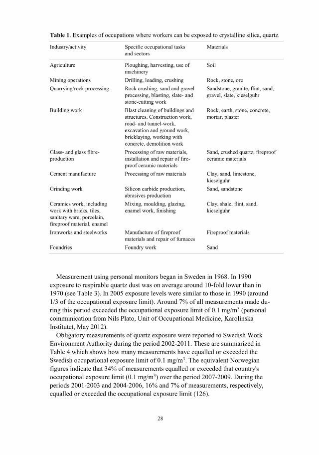
28
Table 1. Examples of occupations where workers can be exposed to crystalline silica, quartz.
Industry/activity Specific occupational tasks
and sectors
Materials
Agriculture Ploughing, harvesting, use of
machinery
Soil
Mining operations Drilling, loading, crushing Rock, stone, ore
Quarrying/rock processing Rock crushing, sand and gravel
processing, blasting, slate- and
stone-cutting work
Sandstone, granite, flint, sand,
gravel, slate, kieselguhr
Building work Blast cleaning of buildings and
structures. Construction work,
road- and tunnel-work,
excavation and ground work,
bricklaying, working with
concrete, demolition work
Rock, earth, stone, concrete,
mortar, plaster
Glass- and glass fibre-
production
Processing of raw materials,
installation and repair of fire-
proof ceramic materials
Sand, crushed quartz, fireproof
ceramic materials
Cement manufacture Processing of raw materials Clay, sand, limestone,
kieselguhr
Grinding work Silicon carbide production,
abrasives production
Sand, sandstone
Ceramics work, including
work with bricks, tiles,
sanitary ware, porcelain,
fireproof material, enamel
Mixing, moulding, glazing,
enamel work, finishing
Clay, shale, flint, sand,
kieselguhr
Ironworks and steelworks Manufacture of fireproof
materials and repair of furnaces
Fireproof materials
Foundries Foundry work Sand
Measurement using personal monitors began in Sweden in 1968. In 1990
exposure to respirable quartz dust was on average around 10-fold lower than in
1970 (see Table 3). In 2005 exposure levels were similar to those in 1990 (around
1/3 of the occupational exposure limit). Around 7% of all measurements made du-
ring this period exceeded the occupational exposure limit of 0.1 mg/m3 (personal
communication from Nils Plato, Unit of Occupational Medicine, Karolinska
Institutet, May 2012).
Obligatory measurements of quartz exposure were reported to Swedish Work
Environment Authority during the period 2002-2011. These are summarized in
Table 4 which shows how many measurements have equalled or exceeded the
Swedish occupational exposure limit of 0.1 mg/m3. The equivalent Norwegian
figures indicate that 34% of measurements equalled or exceeded that country's
occupational exposure limit (0.1 mg/m3) over the period 2007-2009. During the
periods 2001-2003 and 2004-2006, 16% and 7% of measurements, respectively,
equalled or exceeded the occupational exposure limit (126).
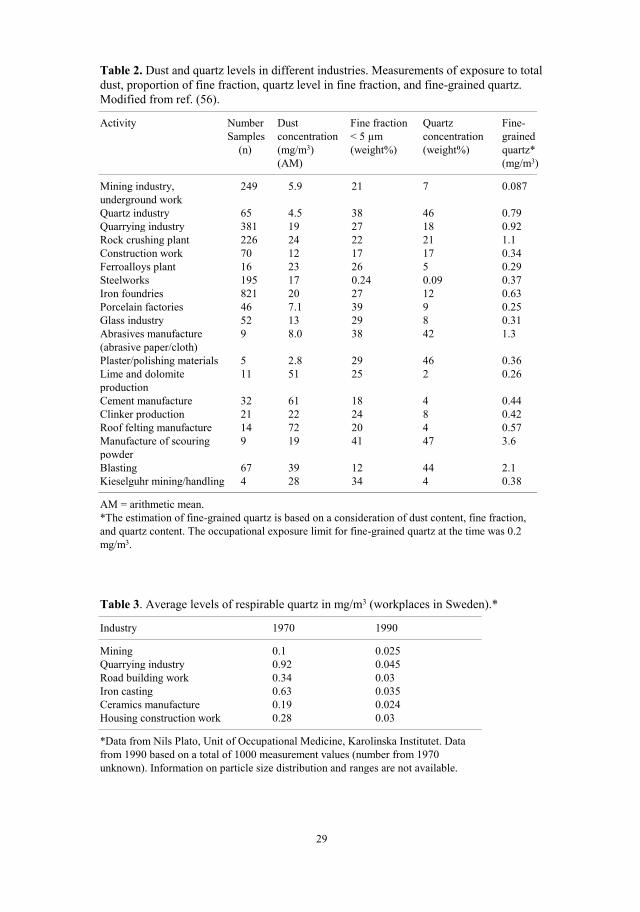
29
Table 2. Dust and quartz levels in different industries. Measurements of exposure to total
dust, proportion of fine fraction, quartz level in fine fraction, and fine-grained quartz.
Modified from ref. (56).
Activity Number
Samples
(n)
Dust
concentration
(mg/m3)
(AM)
Fine fraction
< 5 µm
(weight%)
Quartz
concentration
(weight%)
Fine-
grained
quartz*
(mg/m3)
Mining industry,
underground work
249 5.9 21 7 0.087
Quartz industry 65 4.5 38 46 0.79
Quarrying industry 381 19 27 18 0.92
Rock crushing plant 226 24 22 21 1.1
Construction work 70 12 17 17 0.34
Ferroalloys plant 16 23 26 5 0.29
Steelworks 195 17 0.24 0.09 0.37
Iron foundries 821 20 27 12 0.63
Porcelain factories 46 7.1 39 9 0.25
Glass industry 52 13 29 8 0.31
Abrasives manufacture
(abrasive paper/cloth)
9 8.0 38 42 1.3
Plaster/polishing materials 5 2.8 29 46 0.36
Lime and dolomite
production
11 51 25 2 0.26
Cement manufacture 32 61 18 4 0.44
Clinker production 21 22 24 8 0.42
Roof felting manufacture 14 72 20 4 0.57
Manufacture of scouring
powder
9 19 41 47 3.6
Blasting 67 39 12 44 2.1
Kieselguhr mining/handling 4 28 34 4 0.38
AM = arithmetic mean.
*The estimation of fine-grained quartz is based on a consideration of dust content, fine fraction,
and quartz content. The occupational exposure limit for fine-grained quartz at the time was 0.2
mg/m3.
Table 3. Average levels of respirable quartz in mg/m3 (workplaces in Sweden).*
Industry 1970 1990
Mining 0.1 0.025
Quarrying industry 0.92 0.045
Road building work 0.34 0.03
Iron casting 0.63 0.035
Ceramics manufacture 0.19 0.024
Housing construction work 0.28 0.03
*Data from Nils Plato, Unit of Occupational Medicine, Karolinska Institutet. Data
from 1990 based on a total of 1000 measurement values (number from 1970
unknown). Information on particle size distribution and ranges are not available.
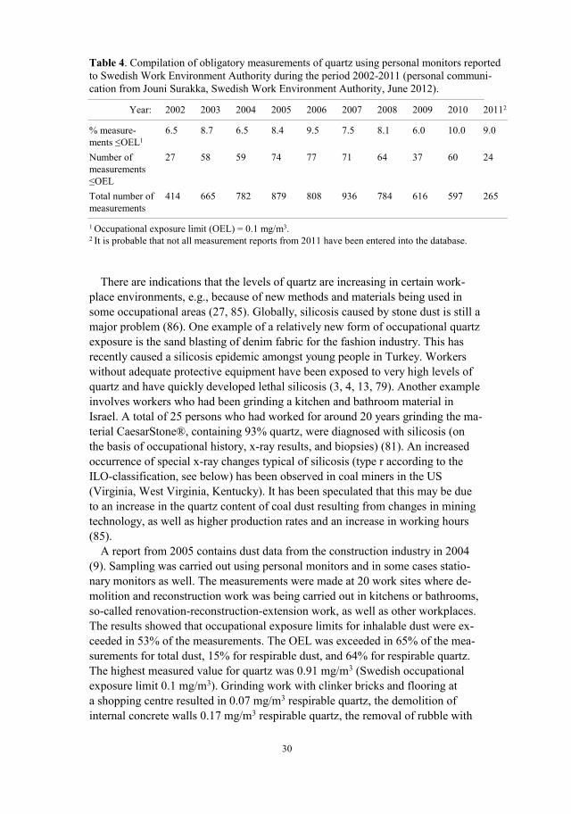
30
Table 4. Compilation of obligatory measurements of quartz using personal monitors reported
to Swedish Work Environment Authority during the period 2002-2011 (personal communi-
cation from Jouni Surakka, Swedish Work Environment Authority, June 2012).
Year: 2002 2003 2004 2005 2006 2007 2008 2009 2010 20112
% measure-
ments ≥OEL1
6.5 8.7 6.5 8.4 9.5 7.5 8.1 6.0 10.0 9.0
Number of
measurements
≥OEL
27 58 59 74 77 71 64 37 60 24
Total number of
measurements
414 665 782 879 808 936 784 616 597 265
1 Occupational exposure limit (OEL) = 0.1 mg/m3. 2 It is probable that not all measurement reports from 2011 have been entered into the database.
There are indications that the levels of quartz are increasing in certain work-
place environments, e.g., because of new methods and materials being used in
some occupational areas (27, 85). Globally, silicosis caused by stone dust is still a
major problem (86). One example of a relatively new form of occupational quartz
exposure is the sand blasting of denim fabric for the fashion industry. This has
recently caused a silicosis epidemic amongst young people in Turkey. Workers
without adequate protective equipment have been exposed to very high levels of
quartz and have quickly developed lethal silicosis (3, 4, 13, 79). Another example
involves workers who had been grinding a kitchen and bathroom material in
Israel. A total of 25 persons who had worked for around 20 years grinding the ma-
terial CaesarStone®, containing 93% quartz, were diagnosed with silicosis (on
the basis of occupational history, x-ray results, and biopsies) (81). An increased
occurrence of special x-ray changes typical of silicosis (type r according to the
ILO-classification, see below) has been observed in coal miners in the US
(Virginia, West Virginia, Kentucky). It has been speculated that this may be due
to an increase in the quartz content of coal dust resulting from changes in mining
technology, as well as higher production rates and an increase in working hours
(85).
A report from 2005 contains dust data from the construction industry in 2004
(9). Sampling was carried out using personal monitors and in some cases statio-
nary monitors as well. The measurements were made at 20 work sites where de-
molition and reconstruction work was being carried out in kitchens or bathrooms,
so-called renovation-reconstruction-extension work, as well as other workplaces.
The results showed that occupational exposure limits for inhalable dust were ex-
ceeded in 53% of the measurements. The OEL was exceeded in 65% of the mea-
surements for total dust, 15% for respirable dust, and 64% for respirable quartz.
The highest measured value for quartz was 0.91 mg/m3 (Swedish occupational
exposure limit 0.1 mg/m3). Grinding work with clinker bricks and flooring at
a shopping centre resulted in 0.07 mg/m3 respirable quartz, the demolition of
internal concrete walls 0.17 mg/m3 respirable quartz, the removal of rubble with
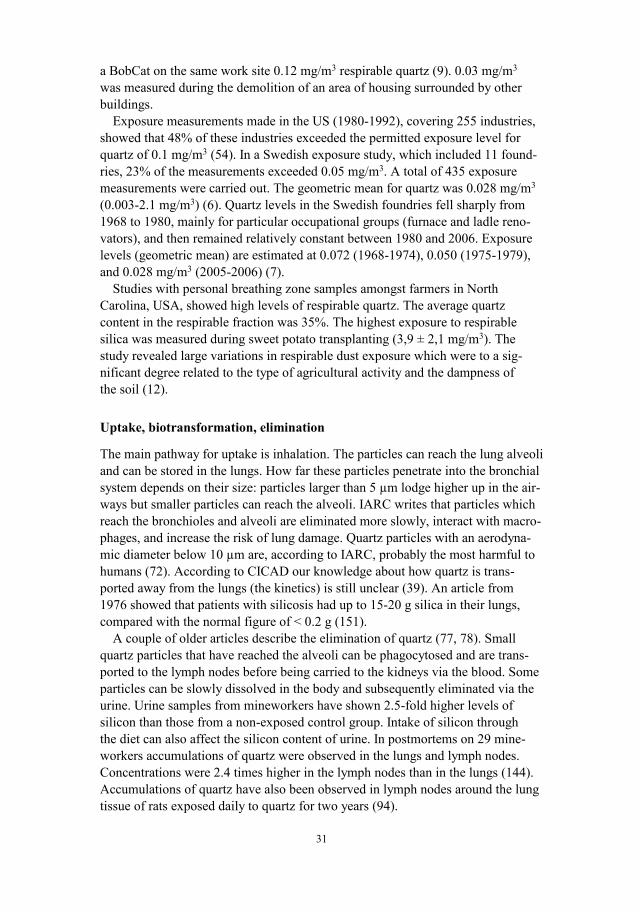
31
a BobCat on the same work site 0.12 mg/m3 respirable quartz (9). 0.03 mg/m3
was measured during the demolition of an area of housing surrounded by other
buildings.
Exposure measurements made in the US (1980-1992), covering 255 industries,
showed that 48% of these industries exceeded the permitted exposure level for
quartz of 0.1 mg/m3 (54). In a Swedish exposure study, which included 11 found-
ries, 23% of the measurements exceeded 0.05 mg/m3. A total of 435 exposure
measurements were carried out. The geometric mean for quartz was 0.028 mg/m3
(0.003-2.1 mg/m3) (6). Quartz levels in the Swedish foundries fell sharply from
1968 to 1980, mainly for particular occupational groups (furnace and ladle reno-
vators), and then remained relatively constant between 1980 and 2006. Exposure
levels (geometric mean) are estimated at 0.072 (1968-1974), 0.050 (1975-1979),
and 0.028 mg/m3 (2005-2006) (7).
Studies with personal breathing zone samples amongst farmers in North
Carolina, USA, showed high levels of respirable quartz. The average quartz
content in the respirable fraction was 35%. The highest exposure to respirable
silica was measured during sweet potato transplanting (3,9 ± 2,1 mg/m3). The
study revealed large variations in respirable dust exposure which were to a sig-
nificant degree related to the type of agricultural activity and the dampness of
the soil (12).
Uptake, biotransformation, elimination
The main pathway for uptake is inhalation. The particles can reach the lung alveoli
and can be stored in the lungs. How far these particles penetrate into the bronchial
system depends on their size: particles larger than 5 µm lodge higher up in the air-
ways but smaller particles can reach the alveoli. IARC writes that particles which
reach the bronchioles and alveoli are eliminated more slowly, interact with macro-
phages, and increase the risk of lung damage. Quartz particles with an aerodyna-
mic diameter below 10 µm are, according to IARC, probably the most harmful to
humans (72). According to CICAD our knowledge about how quartz is trans-
ported away from the lungs (the kinetics) is still unclear (39). An article from
1976 showed that patients with silicosis had up to 15-20 g silica in their lungs,
compared with the normal figure of < 0.2 g (151).
A couple of older articles describe the elimination of quartz (77, 78). Small
quartz particles that have reached the alveoli can be phagocytosed and are trans-
ported to the lymph nodes before being carried to the kidneys via the blood. Some
particles can be slowly dissolved in the body and subsequently eliminated via the
urine. Urine samples from mineworkers have shown 2.5-fold higher levels of
silicon than those from a non-exposed control group. Intake of silicon through
the diet can also affect the silicon content of urine. In postmortems on 29 mine-
workers accumulations of quartz were observed in the lungs and lymph nodes.
Concentrations were 2.4 times higher in the lymph nodes than in the lungs (144).
Accumulations of quartz have also been observed in lymph nodes around the lung
tissue of rats exposed daily to quartz for two years (94).
32
Biological exposure monitoring
A review article from 2006 examined the literature on biological exposure moni-
toring. Quartz particles or silicon in BAL (bronchoalveolar lavage), serum, or
urine indicate exposure. The effects of quartz particles in phagocytic cells can
reflect early effects and inflammation before fibrosis appears. A number of bio-
markers are proposed in this article: CC16 (in serum), TNF-alpha (from mono-
cytes), IL-8 (from monocytes), reactive oxygen species (ROS, from neutrophils),
8-isoprostanes (in serum), antioxidant levels and glutathione, glutathione peroxi-
dase, glutathione S-transferase, and PDGF (in serum). The authors believe that
these biomarkers should be able to identify those individuals who are particularly
sensitive (e.g., individuals with TNF-alpha polymorphism) (58). However, later
studies show that the Nalp3 inflammasome and the proinflammatory cytokine IL-
1β play central roles in quartz-induced inflammation (28, 47, 67).
In a study from 2010 silicosis patients were compared with healthy research
subjects who were exposed to quartz (62 silicosis patients, 24 research subjects
exposed to quartz, 19 who were not exposed). Serum levels of the cytokine TNF-
alpha were significantly higher in healthy individuals exposed to quartz and in
silicosis patients than in those who were not exposed (123).
The formation of collagen fibres is part of the process that leads to silicosis.
Hydroxyproline is an important component of collagen and its concentration has
been measured in the lungs of 29 deceased mineworkers. Hydroxyproline con-
centration showed good correlation with the radiological assessment but not with
the pathological assessment (144). Significantly higher hydroxyproline concen-
trations have also been observed in the urine of individuals with silicosis than
in the urine of those not exposed to quartz. On the other hand, individuals with
suspected silicosis (ILO 1980, category 0/1) did not have significantly higher
concentrations than controls. The authors concluded that the concentration of
hydroxyproline in the urine cannot be used for early detection of silicosis but
might possibly be used to evaluate the progression of silicosis that has already
been detected (97).
A study of individuals diagnosed with silicosis after exposure to quartz
during the sandblasting of denim fabric found a significant positive correlation
between the serum concentration of lactate dehydrogenase (LDH) and the degree
of x-ray changes in the lungs as well as with impairment of lung function (44).
The authors do not recommend LDH as a diagnostic test for silicosis but do
propose that LDH can be a biomarker which indicates the extent of lung paren-
chymal changes.
Toxic effects
Mechanisms of toxicity and tumour development
When quartz is processed fresh surfaces are created and silicon radicals can be
generated on the fracture surfaces. Minerals and inorganic compounds can reduce
(e.g., aluminium salts) or increase (e.g., dust from coal mines) the biological
33
activity of quartz and affect the toxicity in a complex manner. This can be difficult
to predict (46, 118). The highest risk of adverse health effects, e.g., lung cancer,
appears to be associated with exposure to very small and dry, newly generated
particles (39, 72, 93). An appraisal of experimental and epidemiological literature
has revealed that the risk is lower for exposure to particles that are "aged" or are
contaminated with, for example, an aluminium-containing mineral (69).
There are several studies which have investigated how quartz can cause cyto-
toxicity. Mechanistic studies indicate that a number of cellular processes can lead
to toxicity, inflammation, and immunological changes (59). These include effects
on the production of pulmonary surfactant, receptor-mediated binding and toxicity
in alveolar macrophages, increased production of free radicals, and lysosomal
damage. None of these pathways exclude other mechanisms and they can act
together (83).
Surface reactivity, cytotoxicity, and the morphological transformation of
kieselguhr were studied in SHE cells. The samples that were examined were un-
treated kieselguhr (amorphous silicon dioxide), kieselguhr heated up to 900 °C
and 1200 °C, and a commercial product (Chd) containing cristobalite (heated
during the manufacture of the product), as well as Chd-f, a fraction of finer mate-
rial (<10 µm) separated from Chd. X-ray diffraction showed that kieselguhr heated
to 900 °C was mainly in the amorphous form whereas heat treatment at 1200 °C
converted the material to the crystalline form cristobalite. A study of the ability to
release free radicals showed that the finer fraction (<10 µm) released the most
radicals, followed by kieselguhr heated to 1200 °C. The authors concluded that the
ability of kieselguhr to transform cells in vitro can be linked to heat treatment and
the formation of oxygen radicals. The finer fraction, <10 µm, seems to be the most
toxic (50).
Studies have shown that the inflammatory response and the development of
pulmonary fibrosis after quartz exposure is dependent on the receptor Nalp3 (28,
47). Quartz is recognized by the Nalp3 receptor and this leads to the release of
interleukin-1 beta from macrophages. The Nalp3 receptor is thought to play an
important initial role in the inflammation process which can lead to the develop-
ment of, for example, silicosis. Genetically modified mice lacking the Nalp3
receptor developed less inflammation. Quartz was administered to the animals via
intranasal instillation (20 mg/ml, 50 µl, on days 0 and 14) and inflammation and
fibrosis in the lung were studied from 3 months after the first treatment. Wild type
mice that had undergone the same level of exposure developed major inflamma-
tory changes whereas mice lacking the Nalp3 receptor experienced significantly
less inflammation (inflammation score) and granuloma formation as well as less
collagen deposition (28).
Diseases such as silicosis and other pneumoconioses have been linked to in-
creased levels of antibodies, immune complexes, and overproduction of immuno-
globulins, particularly IgG. Chronic exposure to quartz and the development of
silicosis can lead to a chronic stimulation of the immune system through quartz-
induced damage to macrophages in the lungs. This leads to an elevation of in-
34
flammatory cytokinin levels, e.g., interleukin-1 and TNF-alpha, which in turn
stimulate other cells and increase the inflammatory response. In addition, increa-
sed levels of oxygen radicals can contribute to immune system activation (107).
In its latest evaluation of human carcinogens IARC writes that impaired particle
clearance, leading to macrophage activation and inflammation, is an established
carcinogenic mechanism for quartz dust (134).
MARCO receptors (macrophage receptor with collagenous structure) on macro-
phages in the lungs bind quartz particles and mediate their uptake in vitro. When
exposed to quartz, genetically modified mice lacking this receptor developed more
marked lung inflammation and silicosis when compared with wild type mice.
Macrophages from MARCO-/-mice also showed a reduced uptake of quartz parti-
cles, indicating impaired clearance of quartz. In addition, MARCO-/-mice had
higher levels of cytokinins and inflammatory cells than wild type mice. The
authors conclude that the MARCO receptor is an important factor in the clearance of quartz (137).
As discussed below (Mutagenicity, genotoxicity) the genotoxicity of quartz has
been investigated in a number of studies. A review article from 2011 differentiates
between indirect (secondary) and direct (primary) genotoxicity and discusses their
role in cancer development. It is maintained that indirect genotoxicity, which is
assumed to be caused by inflammation, is the one that is important for the deve-
lopment of cancer, as it can occur at moderate exposure levels. In direct geno-
toxicity quartz has a direct effect on DNA but in experimental studies very high
levels of exposure are required and the authors believe this mechanism is im-
probable. According to the authors inflammation is the driving force behind the
genotoxicity observed in vivo, which leads to cancer (22).
Human data
Silicosis
Silicosis is one of the oldest known occupational diseases. It was described as
early as the 1700s by Carl von Linné who gave the disease the Swedish name:
“Orsasjukan”. In English-speaking countries the disease has been known by names
such as such as miners' phthisis and grinder's asthma. One of the early epidemio-
logical studies of silicosis, published in 1900, concerned 30 deaths from silicosis
amongst quartz workers in Nevada, USA (14). Silicosis is a potentially fatal di-
sease from which thousands of people die each year throughout the world. Sili-
cosis occurs in a variety of forms and stages, ranging from mild and symptom-free
to rapidly progressing with breathing difficulties and high mortality. The disease is
an inflammatory process which leads to fibrosis in the lungs. Unlike other forms
of lung fibrosis it begins in the upper parts of the lungs and involves the develop-
ment of nodules with a characteristic appearance under the microscope. Acute or
fulminant silicosis is a variant that can develop after very high-level exposure and
involves rapid disease progression (13, 106, 113). In acute silicosis the alveoli
become filled with a fluid which contains lipid-rich protein residues from cells in
the respiratory tract (so called alveolar lipoproteinosis, which can be seen under
35
a microscope) and reactive oxygen species can be formed (57). Two large clusters
of acute silicosis in the USA have been described. One is from the beginning of
the 1930s when, in order to supply water to a power plant at Hawk´s Nest in West
Virginia, Union Carbide had a tunnel built through a mountain with a very high
quartz content (≥90%) in 1930-1932. A total of 3000 persons worked in the
tunnel; about half of them only worked there occasionally. No measures were
taken to reduce dust levels and the workers had no respiratory protection. The data
on how many died is very unclear. A conservative (”necessarily speculative but
consistently conservative”) estimate is that more than 700 men died from acute
silicosis while working on the project and during a five-year period after the
tunnel work was completed. In addition, approximately 2000 more workers were
exposed to high levels of quartz but were not followed up (36). In the second
cluster (from the start of the 1990s) 100 workers developed acute silicosis from
sand blasting in Texas (141). However, silicosis normally develops gradually over
many years and can be first clinically diagnosed only after many years of expo-
sure. The progression of the disease can cease when exposure stops but can also
progress even in the absence of further exposure. It is generally thought that the
cumulative (gradually accumulating) dose is the important factor in the develop-
ment of the disease (86).
Many studies have examined exposure to quartz and the rates of illness or death
from silicosis in various occupational cohorts. More recent relevant studies are
summarized below along with the critical studies in CICAD's report (39). The
Criteria Group has chosen to describe studies with radiologically based diagnosis
of silicosis separately from studies based on death from silicosis since the out-
come is different: the risk of contracting the disease and the risk of dying from
the disease.
The International Labour Organization (ILO) has a standardized classification
system for the changes in chest radiographs that are used, for example, in dia-
gnosing silicosis. The guidelines were updated in 2011 (73). The system uses,
for example, four categories (0, 1, 2 and 3) that are based on profusion of small
opacities, where 0 indicates an absence of opacities or opacities less than for cate-
gory 1. There are 12 subcategories1. Examples of categorization are 0/1, 1/0, 1/1,
etc., where the first figure denotes the category which best matches the x-ray
image and the second figure indicates the subcategory that has been considered as
an alternative; for example, 0/1 signifies that there are no or very small opacities
and that category 1 has been considered as a possible alternative. Category 1/0
denotes that the x-ray image has been classed as category 1 after carefully con-
sidering category 0; category 1/1 means that the x-ray-image has been classed as
category 1 and that no other category has been considered (73). The classification
also includes designations of the shape and size of the opacities. Studies have
1 ILO-categories:
Categories 0 1 2 3
Subcategories 0/- 0/0 0/1 1/0 1/1 1/2 2/1 2/2 2/3 3/2 3/3 3/+
36
shown that sensitivity can be low in radiological examination. Postmortems on
granite workers in Vermont, USA, showed that a large proportion of those wor-
kers without silicosis symptoms and showing no positive radiological results
nevertheless had silicosis (141). Radiological studies combined with postmortem
results showed that amongst South African gold mine workers only 20-40% of
silicosis cases were diagnosed by x-ray examinations (ILO-classification 1/1).
This was particularly noticeable when the workers' exposure to quartz dust was
low (63). Nevertheless, studies have shown that ILO-classification of silicosis is
more reliable and more sensitive than routine radiological assessment (5). Another
reason why radiological diagnosis of silicosis is unreliable is that different con-
clusions can be reached when an x-ray image is assessed by different people or by
the same person on different occasions. In addition, changes in lung x-ray results
corresponding to the ILO-classification ≥0/1 are observed in the general popu-
lation which is considered not to have been exposed to dust in the workplace
environment. In a literature review (eight studies) by Meyer et al. the occurrence
of opacities (ILO ≥1/0) was found to vary between 0.2% and 12% in control
groups/the general population. The total pooled prevalence was 5.3% (95% CI
2.9–7.7%). The authors believe there is a background level of such x-ray changes
in the general population. The reason for the changes is unclear. The authors point
out that in order to avoid the large variation in classification between studies it is
important to have well matched control groups, so as to establish a base level for
opacities, and for x-ray images to be interpreted blind (92). To sum up, the spe-
cificity of radiological diagnosis using ILO-classification is normally high (around
95%, i.e., 5% false positives) (92) but even though the sensitivity is improved
when this classification is compared with the routine assessment, two studies con-
clude that it is still limited (63, 141). In the references below the terms used by the
authors, e.g., "silicosis" and "signs of silicosis", have been reproduced wherever
possible.
The Swedish silicosis register was established in 1953 but had been collecting
notifications of silicosis since 1931. In 1979 the silicosis register was replaced by
ISA (Informationssystemet om arbetsskador; The Swedish Information System on
Occupational Accidents and Work-related Diseases) (149). Statistics from the
Swedish Cause of Death Register (Dödsorsaksregistret) show that the number
of deaths from silicosis in Sweden was highest in the 1970s but fell in the 1980s.
In 2000 nine men and four women died from silicosis; all were aged over 65
(www.kemiguiden.se, Dödsorsaksregistret). A report from The Swedish Work
Environment Authority (Arbetsmiljöverket) (10) mentions that of the Swedish
deaths from pneumoconiosis in 2007, four cases were related to quartz exposure
(all were men aged over 85).
37
Studies that mainly describe silicosis diagnosed on the basis of radiological
changes
A cohort study looked at 2235 South African gold mine workers who began
employment before 1938. Measurements showed that dust in the mine contained
30% quartz. A total of 313 (14%) workers developed silicosis (ILO-category ≥1/1)
during the follow-up period 1968-1971 to 1991. For the majority (57%) of the
silicosis cases the disease developed after the end of employment and at an ave-
rage age of 59 (64).
An article from 1978 describes 6 Swedish cases of silicosis caused by kiesel-
guhr after a relatively short exposure time (2-20 years, median 4 years); diagnosis
was mainly by x-radiography. According to the authors it was tridymite and cristo-
balite exposure in particular that caused silicosis from working with kieselguhr.
By 1978 there had been no cases of kieselguhr-silicosis registered since 1958 (19).
A study from 2005 examined Dutch building workers for signs of silicosis. A
group of 1335 workers were examined for silicosis in 1998 using mobile x-radio-
graphy; 0.8% showed signs of silicosis (ILO-classification ≥ 1/1). Four years later,
in 2002, 96 of the workers were studied using a newer data tomography technique
(high-resolution computed tomography) which revealed signs of silicosis in 9% of
the workers; most of these had not been found in the previous study. The cumu-
lative average exposure to quartz was estimated at 5.7 mg/m3 x years. The authors
state that, at similar exposure levels, the risk of contracting silicosis is lower for
building workers than for other workers exposed to quartz. According to the
authors this could be attributed to the effect of other particles in the building work
dust, e.g., iron and aluminium, substances that can alter the toxicity of quartz. The
authors estimated a lifetime silicosis risk of > 5% for Dutch building workers with
high levels of quartz exposure. Exposure was measured for 34 building workers
who worked with concrete and mortar or were involved in demolition or building
site clearance. In 55% of samples the Dutch exposure limit for respirable quartz
(0.075 mg/m3) was exceeded. Materials in the dust which gave the highest expo-
sure levels were limestone/sandstone, bricks, mortar, and concrete (138).
A study from 2004 examined the incidence of silicosis amongst 520 workers
(aged 40 years or older) in a South African gold mine. 101 individuals in the
group were found to have tuberculosis and nine workers were taking tuberculosis
medication. The aim was to study the dose-response relationship with quartz dust.
The average exposure level for respirable dust was 0.37 mg/m3 and for quartz
0.053 mg/m3. The incidence of silicosis (ILO-classification ≥ 1/1) was 18-20%.
Those exposed to quartz were divided into five exposure categories: 0-0.8, 0.8-
0.99, 0.99-1.24, 1.24-1.48, and 1.48-3.08 mg/m3 x years. The prevalence of sili-
cosis in these groups was 12, 9, 22, 28, and 47%, respectively (taken from figure 2
in the article). Using logistic regression analysis a significant relationship was
observed between silicosis and the length of employment, average exposure, and
cumulative exposure. The authors of the study believe that the workers developed
silicosis despite being exposed to quartz concentrations below the exposure limit
of 0.1 mg/m3 and are of the opinion that this exposure limit does not protect
38
against silicosis. South African law requires that workers with both silicosis
and tuberculosis should be transferred and so this cross sectional study probably
underestimated the incidence of silicosis amongst these gold mine workers (38).
The EPA in the US estimated (using benchmark-dose methodology and data
from Hnizdo and Sluis-Cremer 1993 (64), ILO-classification ≥ 1/1, see Table 8a)
that the risk of developing silicosis at an environmental exposure level of 0.008
mg quartz/m3 (based on high levels in heavily polluted city air in the USA) over
70 years (lifetime exposure) was 2.4% for healthy individuals (141). However,
the authors of the report believe that city air differs from the air in mines both as
regards particle size (larger particles in city air) and particle surfaces (more newly
generated particles in mine air). There are reports of silicosis cases in groups that
have not been occupationally exposed, e.g., people exposed to dust storms in the
Himalayas and Bedouins in the Negev desert in Israel. In these studies signs of
silicosis were observed in both postmortems (e.g., quartz particles <3 μm in
nodules in the lungs) and in x-radiographic examinations. A total of 395 indivi-
duals in northern China exposed to dust storms (average dust concentration: 8.3–22 mg/m3) were compared with a control group of 88 individuals (average dust
concentration: 1.1–2.3 mg/m3). Twenty eight individuals (7.1%) exposed to dust
storms had silicosis, compared with none in the control group. No data on quartz
concentrations was found (121).
A study of the incidence of silicosis at the Kiruna mine in 1931-1977 was
published in 1986. During this period 144 cases of silicosis were identified. The
concentrations of respirable quartz were estimated at 0.8 mg/m3 (total dust 35-45
mg/m3) in the 1960s, falling to less than 0.05 mg/m3 by the end of the 1970s. Sili-
cosis occurred only in underground workers. The cumulative incidence was 18%
in 1921-1930 but fell to 2% in 1951-1960. In 34 cases (24%) the disease had pro-
gressed. Seven (5%) of the employees with silicosis had confirmed tuberculosis
and 17 (12%) had suspected tuberculosis (x-ray observation). Tuberculosis was
more common amongst those with progressive silicosis. Mortality was higher in
those with stage II and III silicosis and in all stages (I-III) in those with tubercu-
losis. The authors conclude that, with an unchanged dose-response relationship for
those who began working in the mine in the 1980s, there should be a cumulative
incidence rate of 0.2% (1 case of silicosis per 500 workers). Since the 1960s only
one case of stage I silicosis has been diagnosed (75). The classification I, II, III is
in accordance with a system which was approved at a conference in Johannesburg
in 1930 and which has been used in Sweden (for stage I x-ray images show a
spread of spot-like opacities up to a pinhead in size; stage II is characterized by
opacities up to the size of a pea and stage III by opacities that have merged to
form large continuous blocks (2)).
A total of 134 male residents (aged 40 or over) in a mining town (the mined
metals were molybdenum, lead, zinc, and gold) in Colorado, USA, were studied.
One hundred were mine workers exposed to crystalline silica and 34 were non-
exposed controls. Crystalline silica comprised 12% of the total dust. 32% of the
exposed workers had silicosis (ILO-classification ≥ 1/0). Men with lower exposure
39
levels tended to be slightly older, to smoke slightly more, and to have a shorter
exposure time when compared with those with the higher exposure levels. Those
with an average exposure ≤0.05 mg/m3 quartz, had a 10% prevalence (”silicosis
rate”) for silicosis, those with an average exposure > 0.05-0.1 mg/m3 had a pre-
valence of 23%, and those with an average exposure > 0.1 mg/m3 had a preva-
lence of 49% (82). Amongst mine workers with a cumulative quartz exposure of
2 mg/m3 x years 20% had silicosis and amongst those with an exposure > 2 mg/m3
x years 63% had silicosis. On the basis of cumulative quartz exposure 94 of the
100 quartz-exposed mine workers were allocated to four subgroups: >0-1 mg/m3 x
years (n = 32), >1-2 mg/m3 x years (n = 38), >2-3 mg/m3 x years (n = 18), and >3
mg/m3 x years (n = 6). The prevalence of silicosis (ILO-classification ≥ 1/0) in
these subgroups was 12.5, 26.3, 55.6, and 83.3%, respectively. The study esti-
mated the dose-response relationship for silicosis using three exposure indices.
The number of years of exposure was calculated from occupational history and
the cumulative dust exposure index was calculated using gravimetric dust expo-
sure data. Cumulative dust and quartz exposure was also assessed on the basis of
occupational history. The dust index was converted to a quartz estimate in order
to compare with analyses based on the quartz index. The prevalence of silicosis
amongst the 100 workers was 15% for those with < 20 years exposure, 30%
amongst those with 20-29 years exposure, and 47% amongst those with > 30 years
exposure. Using a logistic regression model the silicosis risk for an average expo-
sure of 0.05 mg/m3 was calculated as 9% after 25 years work (with a 20-year
follow-up the risk increased to 36%) and as 50% after 45 years work (82).
Checkoway and colleagues have studied the relationship between respirable
crystalline silica and non-malignant lung diseases and lung cancer mortality in
the kieselguhr industry in several articles (32, 33, 71, 105). Dust from kieselguhr
contains cristobalite. In the study from 2002 (105) 2342 workers employed for at
least 1 year (1942-1994) at a plant in California were assessed. The average time
of exposure was 7.4 years; a few workers were followed up after the end of their
employment (4%). The level of crystalline silica varied between 1 and 25%. A
total of 70 workers (3%) had a silicosis classification (ILO-classification ≥ 1/0). 67
workers had died from a lung disease other than cancer. The authors found a dose-
response relationship between the risk of death from lung disease and exposure to
crystalline silica. It was estimated that at a level of 0.05 mg/m3 there was an extra
risk of 54/1000 workers (5%) for 45 years of exposure and at 0.1 mg/m3 the risk
was 100/1000 workers. Using a regression model it was calculated that for 45
years of exposure there was an increase in the risk of contracting silicosis (ILO-
classification ≥ 1/0) to 16/1000 workers (1.6%) at an average exposure of 0.01
mg/m3, 7.5% at 0.05 mg/m3, and 14% at 0.1 mg/m3 (105).
Rosenman and colleagues studied 1072 workers in the iron foundry industry
in the USA. Average exposure time was 19.2 years and for the overwhelming
majority of participants (90%) the follow-up time from the start of employment
was less than 30 years. There were 28 cases of silicosis (2.9%, ILO-classification
≥ 1/0) more than half of which were pensioned off workers. Amongst workers
40
with an average exposure of <0.05 mg/m3, 0.8% had silicosis, while the pre-
valence was 0.3% for workers with an average exposure of 0.05-0.15 mg/m3,
4.9% for workers exposed to >0.15–0.45 mg/m3, and 6.3% for exposure to > 0.45
mg/m3. The risk of silicosis increased with employment time, cumulative quartz
exposure, type of work task, and smoking. A quartz exposure of 0.05 mg/m3 over
20 years should mean that around 1% of those exposed would contract silicosis
and over 40 years around 2% should be affected. If quartz exposure was 0.1
mg/m3 around 2% should contract silicosis after 20 years exposure and around
3% after 40 years (114).
Quartz exposure and respiratory disease were studied in 440 granite workers
in Spain (111). A total of 77 workers (18%) had diagnosed silicosis (ILO-classi-
fication ≥ 1/1). 73 of these had been exposed to more than 3.5 mg/m3 x years (the
occupational exposure limit for cumulative exposure recommended by OSHA, i.e.
0.1 mg/m3 over 35 working years). A significant dose-response relationship was
observed between quartz exposure and the odds ratio for contracting silicosis. For
average exposures to quartz the odds ratio was 1.0 for 1.2 mg/m3 x years, 2.8
(0.78–10) for 4.9 mg/m3 x years, 10 (3.3–31) for 8.8 mg/m3 x years, and 31 (10–96) for 19 mg/m3 x years. Tuberculosis and asthma were also reported amongst
the workers. Lung function tests revealed 18 cases (4.3%) with FEV1/FVC < 70%.
A study from 1995 investigated the relationship between quartz exposure and
silicosis. The aim was to try to identify a NOAEL- and LOAEL-level (the highest
dose level tested that produced no adverse effect and the lowest dose tested that
produced an adverse effect, respectively) for silicosis based on epidemiological
studies (112). NOAEL varied from 0.007 to 0.1 mg/m3 and LOAEL from 0.008 to
0.25 mg/m3. The authors concluded that the large variation could be due to many
factors, such as the surface properties and particle size of quartz from various
mines, the definition and x-radiographic classification of silicosis cases, methods
for estimating exposure and risks, background concentrations of airborne crys-
talline silica, etc. The study included several epidemiological studies which con-
tained quantitative information on quartz exposure and the risk of silicosis (see
Table 5). The values were not statistically based but were estimated on the basis of
demonstrating that the risk of silicosis was higher than that caused by background
exposure. For studies where the variations in exposure were given, the value at
the mid-point was used. Six studies were included, two of which used statistical
models to determine the relationship between exposure and risk.
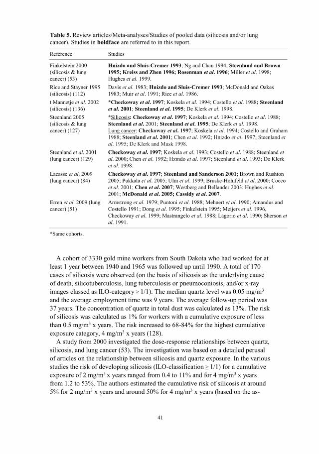
41
Table 5. Review articles/Meta-analyses/Studies of pooled data (silicosis and/or lung
cancer). Studies in boldface are referred to in this report.
Reference Studies
Finkelstein 2000
(silicosis & lung
cancer) (53)
Hnizdo and Sluis-Cremer 1993; Ng and Chan 1994; Steenland and Brown
1995; Kreiss and Zhen 1996; Rosenman et al. 1996; Miller et al. 1998;
Hughes et al. 1999.
Rice and Stayner 1995
(silicosis) (112)
Davis et al. 1983; Hnizdo and Sluis-Cremer 1993; McDonald and Oakes
1983; Muir et al. 1991; Rice et al. 1986.
t Mannetje et al. 2002
(silicosis) (136)
*Checkoway et al. 1997; Koskela et al. 1994; Costello et al. 1988; Steenland
et al. 2001; Steenland et al. 1995; De Klerk et al. 1998.
Steenland 2005
(silicosis & lung
cancer) (127)
*Silicosis: Checkoway et al. 1997; Koskela et al. 1994; Costello et al. 1988;
Steenland et al. 2001; Steenland et al. 1995; De Klerk et al. 1998.
Lung cancer: Checkoway et al. 1997; Koskela et al. 1994; Costello and Graham
1988; Steenland et al. 2001; Chen et al. 1992; Hnizdo et al. 1997; Steenland et
al. 1995; De Klerk and Musk 1998.
Steenland et al. 2001
(lung cancer) (129)
Checkoway et al. 1997; Koskela et al. 1993; Costello et al. 1988; Steenland et
al. 2000; Chen et al. 1992; Hzindo et al. 1997; Steenland et al. 1993; De Klerk
et al. 1998.
Lacasse et al. 2009
(lung cancer) (84)
Checkoway et al. 1997; Steenland and Sanderson 2001; Brown and Rushton
2005; Pukkala et al. 2005; Ulm et al. 1999; Bruske-Hohlfeld et al. 2000; Cocco
et al. 2001; Chen et al. 2007; Westberg and Bellander 2003; Hughes et al.
2001; McDonald et al. 2005; Cassidy et al. 2007.
Erren et al. 2009 (lung
cancer) (51)
Armstrong et al. 1979; Puntoni et al. 1988; Mehnert et al. 1990; Amandus and
Costello 1991; Dong et al. 1995; Finkelstein 1995; Meijers et al. 1996,
Checkoway et al. 1999; Mastrangelo et al. 1988; Lagorio et al. 1990; Sherson et
al. 1991.
*Same cohorts.
A cohort of 3330 gold mine workers from South Dakota who had worked for at
least 1 year between 1940 and 1965 was followed up until 1990. A total of 170
cases of silicosis were observed (on the basis of silicosis as the underlying cause
of death, silicotuberculosis, lung tuberculosis or pneumoconiosis, and/or x-ray
images classed as ILO-category ≥ 1/1). The median quartz level was 0.05 mg/m3
and the average employment time was 9 years. The average follow-up period was
37 years. The concentration of quartz in total dust was calculated as 13%. The risk
of silicosis was calculated as 1% for workers with a cumulative exposure of less
than 0.5 mg/m3 x years. The risk increased to 68-84% for the highest cumulative
exposure category, 4 mg/m3 x years (128).
A study from 2000 investigated the dose-response relationships between quartz,
silicosis, and lung cancer (53). The investigation was based on a detailed perusal
of articles on the relationship between silicosis and quartz exposure. In the various
studies the risk of developing silicosis (ILO-classification ≥ 1/1) for a cumulative
exposure of 2 mg/m3 x years ranged from 0.4 to 11% and for 4 mg/m3 x years
from 1.2 to 53%. The authors estimated the cumulative risk of silicosis at around
5% for 2 mg/m3 x years and around 50% for 4 mg/m3 x years (based on the as-
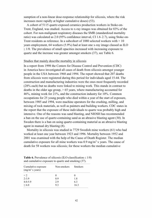
42
sumption of a non-linear dose-response relationship for silicosis, where the risk
increases more rapidly at higher cumulative doses) (53).
A cohort of 5115 quartz-exposed ceramics production workers in Stoke-on-
Trent, England, was studied. Access to x-ray images was obtained for 83% of the
cohort. For non-malignant respiratory diseases the SMR (standardized mortality
ratio) was calculated as 2.0 (95% confidence interval, CI 1.6–2.7), using Stoke-on-
Trent residents as reference. In a subcohort of 1080 selected workers with > 10
years employment, 64 workers (5.9%) had at least one x-ray image classed as ILO
≥ 1/0. The prevalence of small opacities increased with increasing exposure to
quartz and the increase was greater amongst smokers (37), see Table 6.
Studies that mainly describe mortality in silicosis
In a report from 1998 the Centers for Disease Control and Prevention (CDC)
in America have investigated all cases of death from silicosis amongst younger
people in the USA between 1968 and 1994. The report showed that 207 deaths
from silicosis were registered during this period for individuals aged 15-44. The
construction and manufacturing industries were the ones most frequently recorded
(28% each) but no deaths were linked to mining work. This stands in contrast to
deaths in the older age group, > 65 years, where manufacturing accounted for
46%, mining work for 21%, and the construction industry for 10%. Common
occupations for 25 young people who died within a year of the start of exposure,
between 1985 and 1994, were machine operators for the crushing, milling, and
mixing of rock materials, as well as painters and building workers. CDC states in
the report that the exposure of these individuals to quartz was probably high and
intensive. One of the reasons was sand blasting, and NIOSH has recommended
a ban on the use of quartz-containing sand as an abrasive blasting agent (30). In
Sweden there is a ban on using quartz-containing material as an abrasive blasting
agent in manual dry blasting (8).
Mortality in silicosis was studied in 7729 Swedish mine workers (61) who had
worked at least one year between 1923 and 1996. Mortality between 1952 and
2001 was examined with the help of the Cause of Death Register. The median
cumulative exposure for all mine workers was 0.9 mg/m3 x years. The cause of
death for 58 workers was silicosis; for these workers the median cumulative
Table 6. Prevalence of silicosis (ILO-classification ≥ 1/0)
and cumulative exposure to quartz and smoking (37).
Cumulative exposure
(mg/m3 x years)
Non-smokers Smokers
< 2 0 0
2-3.9 0.9 1.8
4.0-5.9 2.9 6.3
≥ 6.0 9.3 16.3
43
exposure was 4.8 mg/m3 x years. A dose-response trend was observed for cu-
mulative exposure. For a cumulative dose of 0-0.9 mg/m3 x years the silicosis
mortality rate per 100,000 person years was 19; for 1-2.9 mg/m3 x years, it was 33;
for 3-4.9 mg/m3 x years, 117; for 5-6.9 mg/m3 x years, 129; for > 7 mg/m3 x years,
140, after adjustment for age and year of birth. The authors concluded that an
increased risk of fatal silicosis seemed to appear at exposure levels of around
3 mg/m3 x years.
The relationship between quartz exposure and the silicosis mortality rate was
studied using pooled data from six cohorts in a case-control study (136). 18,364
workers were involved in the study, 170 of whom had died from silicosis. Death
from silicosis was defined as silicosis or unspecified pneumoconiosis being the
underlying cause of death (150 and 20 deaths, respectively). The authors believe
that the definition is specific but not sensitive as death from silicosis may have
been attributed to other causes of death, such as tuberculosis or COPD (chronic
obstructive pulmonary disease). The authors therefore believe that the definition
probably leads to an underestimation of the number of deaths caused by silicosis.
The average age of death from silicosis was 69 years (range 32-91 years). Sixteen
individuals (9%) had died within one year of leaving their employment but 15 of
these had long exposure time which, according to the authors, points to chronic
silicosis rather than acute silicosis. The average exposure time was 28 years and
the median cumulative exposure was 7.2 mg/m3 x years (compared with 10 years
and 0.62 mg/m3 x years for the cohort as a whole). The median exposure was 0.26
mg/m3 for those who had died, compared with 0.07 mg/m3 for the cohort as a
whole. The risk of death from silicosis at 0-0.99 mg/m3 x years was calculated
as 4.7/100,000 person years and 234/100,000 person years at > 28 mg/m3 x years.
The risk of death from silicosis at 0.1 mg/m3 (45 years lifetime exposure) was
calculated as 1.3% and at 0.05 mg/m3 as 0.6%. The authors believe that an occu-
pational exposure limit of around 0.1 mg/m3 neither protects against contracting
silicosis nor against death from silicosis (136).
A review article from 2005 examined available dose-response data for silicosis
and lung cancer with quartz exposure. The increased risk of death or of contrac-
ting these diseases by the age of 75 was compared for exposure to 0.1 mg/m3 res-
pirable crystalline silica for 45 years (lifetime exposure). The risk of contracting
silicosis (ILO-class 1/1 or higher) was estimated as 47-77% from three studies and
the risk of death from silicosis as 1.9% (0.8-2.9%) from six studies. The risk of
death from lung cancer was estimated as 1.7% (0.2-3.6%) from ten studies (127).
Lung cancer
In 1997 IARC classified quartz as a Group 1 carcinogen (carcinogenic to humans).
Quartz had previously been classified by IARC as a Group 2A carcinogen (pro-
bably carcinogenic to humans) (72). The new classification was based on results
from epidemiological studies where simultaneous exposure to other carcinogenic
agents was low. IARC concluded: ”There is sufficient evidence in humans for the
carcinogenicity of inhaled crystalline silica in the form of quartz or cristobalite
44
from occupational sources” (72). NIOSH (National Institute for Occupational
Safety and Health, USA) and NTP (National Toxicology Program, USA) have
also classified quartz as carcinogenic. However, IARC noted that quartz was not
carcinogenic in certain studies and that this may have been related to particular
properties of quartz or external factors which affected its biological activity. In
2009 IARC confirmed its earlier conclusion that quartz is a Group 1 carcinogen
(134).
As mentioned above it has been shown in many studies that the biological effect
of quartz can be explained by its surface activity, principally its ability to form
reactive oxygen species and cause oxidative stress, but also by the activation of
Nalp3 och IL-1β by quartz particles (86). The surface activity can be modified by
a number of different substances, e.g., other minerals, which should be able to
affect the risk of developing lung cancer in various workplace environments. Lung
cancer caused by crystalline silica via the mechanism of inflammation is discussed
in a review article from 2011 by Borm et al. (22). This mechanism is driven by
inflammatory cells (macrophages and neutrophils) which are recruited to the
inflammatory areas around the quartz particles. The chronic inflammation that
arises can lead to oxidative stress and DNA-damage/genotoxicity. At the same
time cell proliferation can be stimulated, contributing to the development of
cancer (22).
The risk of lung cancer linked to quartz exposure is difficult to study because
the occurrence of simultaneous exposure to other agents that can cause lung can-
cer and affect lung cancer frequency. The situation varies, depending on the work-
place environment, and can also be influenced by smoking (so called confoun-
ding).
Cancer studies mainly published after IARC's evaluation of 1997
A study from 1997 examined mortality from lung cancer in Swedish silicosis
patients (admitted to hospital for silicosis between 1965 and 1983). The analysis
included 1052 men with silicosis. The risk of cancer/mortality was calculated
using the standardized incidence ratio (SIR) and the standardized mortality ratio
(SMR), defined as the relationship between the observed number and the expected
number of cancer cases or deaths for all silicosis patients combined. The total can-
cer risk was increased, SIR=1.5 (confidence interval, 95% CI 1.3–1.7). The risk of
respiratory cancer (cancer of the respiratory organs) was increased, SIR=2.8 (95%
CI 2.0–3.8), as was the risk of lung cancer SIR=3.1 (95% CI 2.1–4.2). Mortality
from lung cancer was also increased, SMR=2.9 (95% CI 2.1–3.9) (23). The
authors considered the results to be in line with earlier studies which showed
an increased risk of lung cancer in silicosis patients.
Lung cancer and exposure to quartz were studied in Swedish workers at iron ore
mines in Kiruna and Malmberget. A total of 8804 men employed between 1923
and 1998 (working both above and below ground) were included in the cohort.
The measurements showed that the average concentration of quartz in the dust to
which the workers were exposed was 2.5%. An increase in the relative risk of lung
45
cancer was observed in workers at the Kiruna mine (compared with the general
population), standardized incidence ratio (SIR)=1.5 (95% CI 1.2–1.8). Under-
ground work was associated with a relative risk of 1.4 (SIR=1.6, 95% CI 1.3–2.0).
An increased lung cancer risk was also observed at exposures < 2 mg x years/m3
(15). A statistically significant relationship between cumulative quartz exposure
and lung cancer was not observed. The authors concluded that mine workers at
the Kiruna mine had an elevated risk of lung cancer due to quartz exposure. The
authors pointed out the difficulty in assessing the quality of certain measurement
data and that information on smoking habits was lacking. According to the authors
this should not affect the results as the mine workers and the general population
had similar smoking habits. The elevated risk of lung cancer in workers in the
Malmberget mine was thought to be due to both quartz and radon exposure (the
radon levels at Malmberget were much higher than at the Kiruna mine) (15).
A major European collaboration aimed at investigating the role of occupational
exposure in lung cancer development found a relationship between quartz expo-
sure and the risk of lung cancer. The study made adjustments for possible con-
founding factors, including smoking (29). 2852 cases of lung cancer were inclu-
ded in the study, as well as 3104 controls. The proportion of smokers (including
former smokers) was higher amongst the cancer cases (90%) than the controls
(64%). The odds ratio for lung cancer and quartz exposure after adjustment for
smoking was 1.4 (1.1–1.7) (29). There was a trend towards increased risk of lung
cancer with increasing cumulative exposure and duration of exposure.
A study from 2007 looked at Chinese mine and ceramics workers. The aim was
to separate the effects of quartz from other risk factors. A total of 511 lung cancer
cases and 1879 controls were included in the study. A dose-response relationship
was observed for the carcinogenic effect of quartz when adjustments were made
for smoking. This relationship disappeared when the analysis was adjusted for
other risk factors (radon, arsenic, and PAH). The authors concluded that the study
did not support the hypothesis that exposure to quartz was causally linked to an
increased risk of lung cancer and that other risk factors, such as radon, arsenic,
PAH, asbestos, diesel exhaust gases, and smoking, influenced the risk of lung
cancer (35) The authors stated that: the job with the highest exposure to quartz
(median 4.8 mg/m3 x years, in tungsten mines) also involved high exposure to
radon; jobs with moderate quartz exposure (in tin mines and during ceramics work
the median exposure was 2.6 and the cumulative exposure was 2.1 mg/m3 x years)
also involved high exposure to PAH (ceramics work) and arsenic (tin mines). No
increase in lung cancer risk was observed for the job with the lowest exposure
(median 0.2 mg/m3 x years), regardless of whether adjustments were made (35).
One study analysed various histological types of lung tumours in lung cancer
patients at two French university hospitals (1997-2006). A significant relationship
was observed between adenocarcinoma in the lung and previous exposure to
quartz, amongst other substances, but this was not observed for other histological
types (104).
46
Birk et al. studied mortality in a German cohort from the porcelain industry
between 1985 and 2005. No increase in mortality from lung cancer was found,
SMR=0.7 (95% CI 0.56–0.89) (20). According to the authors this was one of the
largest cohorts of porcelain workers and included 8200 men and 9300 women.
The authors believed that exposure to quartz dust in this study (over the period
1985-1987) was more relevant to modern industrial conditions than many pre-
vious studies. The weaknesses in the study, as pointed out by the authors, were
that the cohort was relatively young and the follow-up period was short. The same
cohort of porcelain workers was investigated in a study from 2011 (95). The aim
was to quantify the risk of silicosis and lung cancer in porcelain workers who had
been exposed to respirable quartz. Exposure to >4 mg/m3 x years (cumulative) or
more than 0.15 mg/m3 (mean) was strongly related to an increased risk of silicosis
(ILO-classification ≥ 1/1), but no dose-response relationship was observed for in-
creased risk of lung cancer.
An article from 2011 presents a study of occupational exposure and lung cancer
amongst non-smoking Chinese individuals in Hong Kong. 132 cases of lung can-
cer were included in the study, along with 536 controls without cancer. A signifi-
cantly increased risk of lung cancer was observed for jobs involving exposure to
quartz dust. With quartz dust exposure the increase in lung cancer risk was asso-
ciated with the duration of employment. Separate analyses were made for the risk
of adenocarcinoma. A significantly increased odds ratio (2.9, 95% CI 1.1–7.7) was
observed for workers exposed to quartz dust. The authors thought the study sup-
ported an independent effect of quartz on lung cancer risk in non-smokers (139).
A study from 2011 examined cause-specific mortality in an Italian cohort inclu-
ding silicosis patients. The study involved workers who had received compensa-
tion for silicosis between 1943 and 1986 (criteria for compensation were: radio-
logically confirmed silicosis, verified occupational exposure to silica dust, and
a reduction in work capacity exceeding 10%, equivalent to a 35% reduction in
pulmonary function ). The cohort included 2034 men with silicosis. The follow-up
period was from January 1987 to December 2006. The study revealed a significant
increase in mortality from cancer of the trachea, bronchi, and lungs, tuberculosis,
and cirrhosis of the liver. An increased risk of lung cancer, tuberculosis, and liver
cirrhosis was observed for bricklayers/stone masons and an increased risk of non-
malignant respiratory diseases, COPD, and throat cancer was observed for mine
workers (117). The study did not have access to data on smoking habits.
A study from 2010 examined quartz exposure and mortality from lung cancer,
renal cancer, other renal diseases, silicosis, and diseases of the respiratory organs
in granite workers in Vermont, USA. A total of 7052 workers employed between
1947 and 1998 were included in the study. Mortality (SMR) from lung cancer was
1.4 (95% CI 1.2–1.5), from silicosis 59 (95% CI 45–77), from tuberculosis 22
(95% CI 18–26) and from other lung diseases 1.7 (95% CI 1.5–2.0). No increase in
mortality was observed for renal cancer and other renal diseases. A significant re-
lationship with cumulative quartz exposure was observed for silicosis as well as
for other diseases of the respiratory organs. Exposure to quartz was higher before
47
1940 and the study found a strong relationship between quartz exposure and mor-
tality from silicosis amongst men born before 1925. A dose-response analysis
found no substantial evidence of an association between quartz exposure and lung
cancer. The authors therefore thought that the increase in mortality from lung can-
cer was due instead to smoking or to some other exposure not related to work
(142).
Dose-response studies for lung cancer which were mainly published after IARC's
evaluation of 1997
Checkoway and co-workers reported a clear dose-response relationship between
exposure to crystalline silica (primarily cristobalite) and mortality from lung di-
seases and lung cancer (33). The cohort comprised 2342 workers from a kiesel-
guhr plant at Lompoc, California. Data from 5709 measurements made over the
period 1962-1988 were obtained from the company. Measurement data prior to
1962 were based on particles whereas analyses carried out after 1962 used gravi-
metric measurements of total dust or respirable dust. An increase was found in
mortality from non-malignant lung diseases (SMR 2.0, 95% CI 1.6–2.6) along
with a smaller increase in the risk of mortality from lung cancer (SMR 1.3, 95%
CI 1.0–1.6). The authors found a dose-response relationship between non-malig-
nant lung diseases (principally silicosis and its disease sequelae) and both respi-
rable dust and respirable quartz, after 15 years of exposure. For quartz concentra-
tions lower than 0.5 mg/m3 the relative risk was 1.0; for 0.5–<1.1 mg/m3 2.04; for
1.1–<2.1 mg/m3 1.96; for 2.1–<5.0 mg/m3 3.2; concentrations above 5.0 mg/m3
gave a relative risk of 5.4. A smaller but clear dose-response relationship was ob-
served between lung cancer and respirable quartz (for ≤0.5 mg/m3 1.0; for 0.5–<1.1 mg/m3 0.96; for 1.1–<2.1 mg/m3 0.77; for 2.1–<5.0 mg/m3 1.3; for ≥5.0
mg/m3 2.2) (33).
Steenland and Sanderson carried out a dose-response analysis with 4027 wor-
kers in the sand industry. Quantitative calculations of quartz exposure were made
for each employee with respect to duration and occupation via a job-exposure mat-
rix. Personal monitoring data for respirable quartz were available for the period
1974-1988. Exposure levels were calculated for four exposure categories and ten
occupational categories. Earlier levels of exposure were extrapolated back in time
from 1974. The follow up was carried out using a cause of death register. A more
detailed dose-response analysis was undertaken in a case control study within the
cohort. Although only a limited amount of data on smoking was found in the co-
hort, this was applied to the entire cohort. 99% of the cohort consisted of men,
24% of whom had died. The average follow-up period was 24 years. The average
period of employment was 9 years (though 30% were employed for more than 10
years). SMR (standardized mortality ratio) for tuberculosis was 3.4 (95% CI 1.1–7.9), for lung cancer 1.6 (95% CI 1.3–1.9), for silicosis 66 (95% CI 33-120), and
for heart disease 1.22 (95% CI 1.1–1.4). The dose-response analysis showed
SMRs for lung cancer of 1.4 (>0-0.10 mg/m3 x years), 1.3 (>0.1-0.51 mg/m3 x
years), 1.6 (>0.51–1.3 mg/m3 x years), and 2.4 (>1.28 mg/m3 x years) (lagged 5
48
years, the US population as reference) (130). Data on smoking showed that the
cohort smoked slightly more than the US population (based on smoking data from
1987), especially amongst younger workers, but the authors estimated that this
contributed to only 10-20% of the increase in lung cancer risk. Workers in the
sand industry are probably not exposed to any other materials. The study showed
an extra lung cancer risk of 60% compared with the US population (SMR=1.60,
95% CI 1.3–1.9).
A Swedish study from 2012 examined the relationship between quartz and lung
cancer in a cohort study. It involved a total of 3045 workers from ten Swedish
foundries who had worked for at least 1 year between 1913 and 2005. An analysis
was made of the incidence of lung cancer between 1958 and 2004. A significant
increase was observed in the risk of contracting lung cancer, SIR 1.6 (95% CI 1.2–2.1). No difference in risk was observed between the different exposure groups
(short- and long-term exposure) but after a latency period of 20 years a significant
increase in the risk of contracting lung cancer was seen in those groups exposed
for 10-19 years and >20 years (SIR 2.4, 95% CI 1.1–4.3 and SIR 1.7, 95% CI 1.1–2.6, respectively). The authors saw no dose-response relationships with cumula-
tive quartz exposure. The study lacked data on the smoking habits of the workers
(148).
An epidemiological study showed an increase in mortality from lung cancer
in American workers exposed to sand. The study involved eight sand producing
companies and 2670 men who had worked for the companies for at least three
years. A significant increase was found in mortality from tuberculosis, lung di-
seases, and lung cancer after more than 20 years exposure. Mortality from silicosis
was high in the group of workers employed before 1940. The higher lung cancer
mortality was due to mortality increases at four companies. For the remaining
companies there was no increase in lung cancer mortality whereas mortality from
silicosis was higher (90). Studying workers in the sand producing industry had
the advantage of few confounders [no arsenic, nickel, radon, or polyaromatic
hydrocarbons (PAH)] and a very high quartz content in the mineral (normally
over 98%). According to the authors the estimates of exposure were relatively
reliable and information on smoking habits was available. An updated study of
the American sand companies from 2005 showed that the odds ratio increased
with increases in cumulative exposure and average exposure to quartz. Mortality
from silicosis was strongly related to exposure and the odds ratio (lagged 15 years)
for cumulative exposure was (relative to the lowest exposure group, ≤ 0.7 mg/m3 x
years) 2.2 at 0.7-1.8 mg/m3 x years, 4.3 at 1.8–5.1 mg/m3 x years, and 5.5 at > 5.1
mg/m3 x years. Data for lung cancer, see Table 9a. Adjustments were made for
asbestos exposure and smoking. (91).
A study in Dutch workers found an increase in the relative risk of lung cancer
in the group with the longest duration of exposure and the highest cumulative
quartz exposure. The elevation in lung cancer risk was 1.7 (95% CI 1.1–2.4) for
26-51 years exposure, when compared with no exposure, and 1.5 (95% CI 0.93–2.3) for exposure levels ≥ 3 mg/m3 x years, when compared with levels < 3 mg/m3
49
x years. The authors concluded that, despite efforts to reduce levels of quartz in
the workplace environment, levels can still be high (especially in the construction
sector). The study included adjustments for smoking and the results did not appear
to have been influenced by asbestos exposure. The authors were of the opinion
that quartz should be regarded as a human carcinogen (109).
The relationship between lung cancer and quartz exposure was studied in 5115
ceramics workers in Stoke-on-Trent, England. The workers were born between
1916 and 1945 and 80% of them had started employment before 1970. A total of
88 workers (1.7%) had a diagnosis of lung cancer entered on their death certifi-
cates. Access to lung x-ray images was obtained for 83% of the cohort. An SMR
of 1.9 (95% CI 1.5–2.4) was calculated for lung cancer, with the population of
England as reference, and an SMR of 1.3 (1.0–1.6) with the population of Stoke-
on-Trent as reference (37). Fifty two cases of lung cancer were examined in a
case-control study and this revealed a dose-response relationship with intensity
of exposure but not with duration of employment (37).
Studies containing pooled data/meta-analyses and review articles which could in-
clude data from several of the above mentioned original articles. See also Table 5
A study from 2000 investigated the dose-response relationships between quartz,
silicosis, and lung cancer (53). The research was based on two studies published
in 1997 and gave a quantitative meta-analysis for lung cancer in relation to quartz
exposure. Cancer mortality was calculated as 1.4 (SMR) for a cumulative expo-
sure of 3 mg/m3 x years (12% increase in risk per mg/m3 x years). The authors
concluded that the risk of lung cancer could be increased 30% by a lifetime expo-
sure of 0.1 mg/m3 or more (53). Data for lung cancer, see Table 9b.
The relationship between quartz exposure and lung cancer was studied in a po-
oled cohort (including data from 10 previous studies) with a total of 44,160 mine
workers and 21,820 controls. The median cumulative exposure in the 10 studies
varied between 0.13 and 11 mg/m3 x years (gold mine workers in the USA and
gold mine workers in Australia, respectively). The median of average exposures
for respirable quartz varied between 0.05 and 0.59 mg/m3 (granite workers and
gold mine workers in the USA and granite workers in Finland, respectively). An
analysis of mortality from silicosis gave odds ratios of 1.0, 3.1 (2.5–4.0), 4.6 (3.6–5.9), 4.5 (3.5–5.8), and 4.8 (3.7–6.2) for the quintiles of cumulative exposure for
the cases. No trend was observed for silicosis mortality and duration of exposure;
odds ratios 1.0, 1.3 (1.0–1.6), 1.8 (1.4–2.3), 1.4 (1.1–1.7, and 1.2 (0.89–1.5). The
authors concluded that the data was heterogeneous and that the highest value for
South African gold mine workers could be influenced by confounding from radon.
The authors found a positive monotonic dose-response trend for lung cancer with
50
Table 7. Epidemiological studies with SMR (standardized
mortality ratio) for lung cancer which were used in the pooled
cohort by Steenland et al. (129).
Study SMR
Kieselguhr workers, USA 1.3 (1.0-1.6)
Granite workers, Finland 1.4 (1.0-2.0)
Granite workers, USA 1.2 (1.0-1.3)
Sand workers, USA 1.6 (1.2-1.9)
Ceramics workers, China 1.1 (0.84-1.4)
Tin workers, China 2.1 (1.7-2.6)
Tungsten workers, China 0.63 (0.53-0.75)
Gold mine workers, South Africa -
Gold mine workers, USA 1.2 (1.0-1.4)
Gold mine workers, Australia 1.8 (1.5-2.1)
All (excluding gold mine workers, South Africa) 1.2 (1.1-1.3)
cumulative exposure: (categorical) odds ratio 1.0 at <0.4 mg/m3 x years; 1.0
(0.85–1.3) at 0.4-2.0 mg/m3 x years; 1.3 (1.1–1.7) at 2.0–5.4 mg/m3 x years; 1.5
(1.2–1.9) at 5.4–13 mg/m3 x years; 1.6 (1.3–2.1) at >13 mg/m3 x years, (pooled
results). There were no data for smoking (129). The SMRs for lung cancer in the
different studies are shown in Table 7.
In a meta-analysis of the dose-response relationship, Lacasse (2009) and co-
workers saw an increase in the relative risk of lung cancer with increased cumu-
lative exposure to quartz. The studies were adjusted for smoking. The authors
concluded that quartz is a lung carcinogen. A threshold value (exposure level
above which the risk of lung cancer increases) was calculated as >1.8 mg/m3 x
years (84). The meta-analysis included studies published in 1997 or later. Data
for lung cancer, see Table 9b.
Amongst the epidemiological studies that have been published, it has often been
observed that the risk of lung cancer is higher in workers with silicosis. Some
studies which show an increased risk of lung cancer with exposure to quartz,
indicate that this mainly involved those groups with the highest cumulative expo-
sure, interpreted by the authors as a threshold value (an exposure level below
which lung cancer does not develop) (24). Most studies show an increase in the
risk of lung cancer which in most cases is less than doubled for the group with
the highest exposure (>3-5 mg/m3 x years). According to many researchers this
indicates that quartz is a weak carcinogen (109, 129).
At least two risk assessments (HSE 2003 and SCOEL 2003) were based on the
assumption that exposure which does not lead to silicosis does not give rise to a
significant risk of developing lung cancer (60, 120). Several cohort studies have
reported a relative risk of lung cancer of 1.3–1.4 in workers exposed to quartz (84,
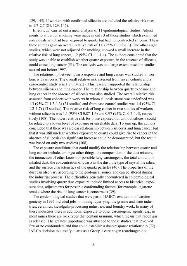
51
129, 145). If workers with confirmed silicosis are included the relative risk rises
to 1.7–2.7 (84, 129, 145).
Erren et al. carried out a meta-analysis of 11 epidemiological studies. Adjust-
ments to allow for smoking were made in only 3 of those studies which examined
individuals who had been exposed to quartz but had not contracted silicosis. These
three studies gave an overall relative risk of 1.0 (95% CI 0.8-1.3). The other eight
studies, which were not adjusted for smoking, showed a small increase in the
relative risk of lung cancer, 1.2 (95% CI 1.1–1.4). The authors considered that the
study was unable to establish whether quartz exposure, in the absence of silicosis,
could cause lung cancer (51). The analysis was to a large extent based on studies
carried out before 1997.
The relationship between quartz exposure and lung cancer was studied in wor-
kers with silicosis. The overall relative risk assessed from seven cohorts and a
case-control study was 1.7 (1.4–2.2). This research supported the relationship
between silicosis and lung cancer. The relationship between quartz exposure and
lung cancer in the absence of silicosis was also studied. The overall relative risk
assessed from cohorts with workers in whom silicosis status was undefined was
1.3 (95% CI 1.2–1.3) (24 studies) and from case control studies was 1.4 (95% CI
1.2–1.7) (13 studies). The relative risk of lung cancer in two studies of workers
without silicosis was 1.1 (95% CI 0.87–1.6) and 0.97 (95% CI 0.7–1.4), respec-
tively (108). The lower relative risk for those exposed but without silicosis could
be related to a lower level of exposure or unreliable data. To sum up, the authors
concluded that there was a clear relationship between silicosis and lung cancer but
that it was still unclear whether exposure to quartz could give rise to cancer in the
absence of silicosis (no significant increase could be demonstrated, but the result
was based on only two studies) (108).
The exposure conditions that could modify the relationship between quartz and
lung cancer include, amongst other things, the composition of the dust mixture,
the interaction of other known or possible lung carcinogens, the total amount of
inhaled dust, the concentration of quartz in the dust, the type of crystalline silica,
and the surface characteristics of the quartz particles (40). The properties of the
dust can also vary according to the geological source and can be altered during
the industrial process. The difficulties generally encountered in epidemiological
studies involving quartz dust exposure include limited access to historical expo-
sure data, adjustments for possible confounding factors (for example, cigarette
smoke where the risk of lung cancer is concerned) (39).
The epidemiological studies that were part of IARC's evaluation of carcino-
genicity in 1997 included jobs in mining, quarrying, the granite and slate indus-
tries, ceramics, kieselguhr-processing industries, and foundry work. In many of
these industries there is additional exposure to other carcinogenic agents; e.g., in
most mines there are rock types that contain uranium, which means that radon gas
is released. The greatest importance was attached to those studies that involved
few or no confounders and that could establish a dose-response relationship (72).
IARC's decision to classify quartz as a Group 1 carcinogen (carcinogenic to
52
humans) has in part been criticized. Some argue that the decision was based on
studies which failed to demonstrate dose-response relationships and that the in-
creased risk of lung cancer was so small that confounding or other lifestyle factors
could be involved (55, 125). In his article from 2011 (55) Gamble only used mate-
rial from IARC's review of 1997 and did not include studies carried out later. The
article therefore did not shed any light on the current state of knowledge and is
therefore of limited value. Several studies carried out after IARC's classification in
1997 have shown an increased risk of lung cancer and in 2009 IARC confirmed its
classification of quartz (134).
Summary of the relationship between quartz, silicosis, and lung cancer
There are many studies which show an increased risk of lung cancer with exposure
to quartz. The risk is still greater in workers with silicosis. It is still unclear
whether workers without silicosis have a higher risk of lung cancer, as the few
studies that have investigated this and that have made adjustments for smoking,
have shown no increase or a non-significant increase. This could be because few
studies with the relevant adjustment for smoking have examined this possibility or
because there is no increase. IARC's conclusion does not mention silicosis as an
important factor in the link with lung cancer but this is discussed in the main
document and it is stated that a relationship between silicosis and lung cancer has
been observed in several studies (72). Therefore there is no strong epidemiological
evidence that lung cancer occurs without pre-existing silicosis but the data does
not rule out a risk.
In an article from 2000 Checkoway and Franzblau discussed whether it is
even possible, on the basis of epidemiological studies, to answer the question
of whether silicosis is a necessary prerequisite for an increase in the risk of lung
cancer in workers exposed to quartz (31). The authors believed there was a lot of
uncertainty over diagnosis in which routine radiological examination could be
relatively insensitive in detecting small changes in the lung at an early stage of the
disease. They also thought that epidemiological studies aimed at investigating the
risk of lung cancer with low levels of exposure to quartz should include large
populations with a wide range of exposure groups (including low and high levels).
The authors concluded that it was too early to draw the inference that silicosis was
necessary for lung cancer to develop with exposure to quartz, and that silicosis and
lung cancer should be regarded separately as these effects are not necessarily rela-
ted (31). In a review article on quartz dust Steenland discussed whether, given that
silicosis can be considered as a marker for high levels of quartz exposure, it was
logical to think, if one assumes that quartz itself can cause lung cancer, that indi-
viduals with silicosis should have a higher risk of lung cancer than those without
silicosis, even if lung cancer is independent of silicosis (127).
Cancers other than lung cancer
Studies have demonstrated an association between exposure to quartz and cancer
of the oesophagus, stomach, skin, and skeletal system. The findings have been
53
inconsistent (few studies, both positive and negative results) and in most cases
there has been a combination of exposure with other risk factors (24). Several
studies show cancer of the stomach and oesophagus, see below.
A Swedish study from 2007 examined the risk of stomach cancer in Swedish
workers. Moderate or high levels of exposure to quartz dust were seen to be
associated with a significant increase in the risk of stomach cancer: incidence rate
ratio (IRR) 1.2 (95% CI 1.0–1.4) and 1.3 (95% CI 1.0–1.7), respectively (122).
A Japanese study from 2001 observed a non-significant increase in stomach
cancer and oesophageal cancer in individuals exposed to quartz and in silicosis
patients. Adjustments had been made to allow for smoking (140).
Mortality in the German porcelain industry between 1985 and 2005 was in-
vestigated in a cohort study of 17,644 workers (20). Increased mortality from
pancreatic cancer was observed amongst male workers, SMR 1.7 (95% CI 1.2–2.4). Increased mortality from liver cancer and silicosis was also observed, SMR
2.0 (95% CI 1.3–2.9) and 7.2 (95% CI 2.3–17), respectively. There was no in-
crease in mortality from renal cancer. For women the only increase in mortality
was from diabetes, SMR 1.7 (95% CI 1.1–2.7). The reference groups were the
German population and the population of the German federal state of Bayern.
A meta-analysis of occupational exposure and pancreatic cancer showed that
exposure to quartz dust resulted in a non-significant increase in risk (meta-risk
estimate) of 1.4 (95% CI 0.9-2.0) (102).
Autoimmune diseases
Occupational exposure to quartz dust was investigated as a possible risk factor for
a number of systemic autoimmune diseases; e.g., rheumatoid arthritis, scleroder-
ma, and SLE (systemic lupus erythematosus). Occupational groups in which these
disease are found include mine workers, granite workers, and stonemasons (107).
A Swedish study reported a strong link between quartz exposure and SLE (23).
Women diagnosed with SLE in Boston were studied in order to assess the risk
of SLE and their history of exposure to quartz dust (52). Data was gathered at in-
terviews to establish detailed work histories of all participants, including previous
exposures. 95 patients were included in the study along with 191 controls matched
for age and ethnic origin. A dose-response relationship was observed for duration
of exposure. Exposure to quartz for more than one year was associated with SLE
(odds ratio 4.3; 95% CI 1.7–11); with exposure for 1-5 years the odds ratio was
4.0 (95% CI 1.2–13); exposure for >5 years gave an odds ratio of 4.9 (95% CI 1.1–22). This effect was not influenced by smoking. In this study those occupations
most commonly associated with quartz exposure were the manufacture of dental
prosthetics (dental laboratory), sandblasting work, and the production of scouring
powder.
A meta-analysis of 16 studies from 2010 examined the relationship between
occupational exposure to quartz and scleroderma. The combined estimate for
relative risk (CERR) was 1.0 (95% CI 0.74–1.4) for women and 3.0 (95% CI 1.2–7.4) for men. For case-control studies (nine) the CERR was 2.2 (95% CI 1.7–3.3)
54
and for cohort studies (three) it was 15 (95% CI 4.5–53). The authors concluded
that exposure to quartz could be a significant risk factor for scleroderma, parti-
cularly in men (89).
Sluis-Cremer et al. investigated a possible association between silicosis and
rheumatoid arthritis in a case-control study amongst South African gold mine
workers. The authors calculated an odds ratio of 2.8 (95% CI 1.4–4.3) for rheuma-
toid arthritis amongst silicosis cases. No significant difference in cumulative dust
exposure or duration of exposure was observed and the mean intensity of dust
exposure was significantly lower for silicosis cases than for controls. The authors
concluded that mine workers with rheumatoid arthritis had an increased risk of
developing silicosis (124).
Two Swedish studies examined the relationship between quartz exposure and
rheumatoid arthritis (132, 133). A case-control study with 577 cases and 659
controls showed that individuals exposed to quartz had a higher risk of rheumatoid
arthritis (positive for ACPA, anti-citrullinated protein antibodies), odds ratio 1.7
(95% CI 1.1–2.5). Individuals who had worked in rock drilling had a further in-
crease in risk (odds ratio 2.3, 95% CI 1.2–4.7). An even higher risk was found
amongst smokers and the authors thought this indicated an interaction between
smoking and quartz exposure.
A study from 1993 looked at 50 workers who had jobs in a scouring powder
plant at Sevilla, Spain. The powder contained 90% crystalline silica. Thirty two
(64%) of the workers had some form of autoimmune disease, which was a much
higher percentage than for the general population (116).
Kidney diseases
Kidney diseases associated with exposure to quartz have also been reported. Ng
and co-workers studied urine samples from granite workers. Workers with high-
level, long-duration exposure (> 10 years) to quartz had higher levels of beta-2-
microglobulin, alpha-1-microglobulin and beta-N-acetylglucosaminidase when
compared with control individuals, which indicates renal toxicity. The authors
also studied granite workers with silicosis (ILO-classification ≥ 1/0) and found
that workers with silicosis had the highest levels of beta-2-microglobulin, alpha-1-
microglobulin and beta-N-acetylglucosaminidase when compared with the group
without silicosis (98).
A case-control study from 2011 reported that quartz exposure was associated
with a 40% increase in the risk of chronic kidney disease (odds ratio = 1.40, 95%
CI 1.04–1.89). The cases were patients with newly diagnosed chronic kidney
disease and the controls were selected randomly and matched for age and gender.
Both patients and controls were interviewed about any previous occupational
exposure. As well as facing general questions on sand- or quartz-exposure and
employment in industries where there is a potential for exposure, the workers were
also asked whether, for each job they mentioned, they had been exposed to sand or
quartz on at least 5 occasions. Quartz exposure was then individually assessed and
a qualitative dose estimate was calculated for each job. The study contained no
55
quantitative data. The study included the following diagnoses of chronic kidney
disease: diabetes with renal manifestations, hypertensive renal and heart disease,
chronic glomerulonephritis, nephritis and nephropathy, chronic renal failure, renal
sclerosis, chronic pyelonephritis. The average cumulative exposure to quartz was
significantly higher in exposed cases than in exposed controls (33 compared with
25 years). Compared with non-exposed controls, the odds ratio was 1.20 (95% CI
0.77–1.9) for individuals whose exposure time was below the average value and
1.8 (95% CI 1.1–2.7) for those with an above-average exposure time (146).
A review article from 2005 examined available dose-response data for quartz
and kidney disease. The increased risk of death from kidney disease or of con-
tracting kidney disease by the age of 75 was studied for exposure to 0.1 mg/m3
respirable crystalline silica for 45 years (lifetime exposure). The increased
(absolute/excess) risk of kidney disease was 0.8–9.7%, based on data from three
cohorts. Excess risk of kidney disease (end-stage) was estimated as 5.1% (2.2–7.3%, based on one cohort study) (127).
An epidemiological study of sand-exposed American workers, a total of 2670
men at eight sand-production companies, observed increased mortality from
kidney disease amongst workers employed for more than 10 years (90).
Kidney disease and arthritis were studied in a cohort of 4626 quartz-exposed
workers in the sand industry. The authors found an increase in mortality (SMR)
of 2.6 (95% CI 1.5–4.2) for acute kidney disease, 1.6 (95% CI 1.1–2.2) for chronic
kidney disease and 4.4 (95% CI 2.8–6.5) for arthritis. The authors found an in-
creased incidence (standardized incidence ratio) of 2.0 (95% CI 1.3–3.0) for end-
stage renal disease, with the incidence being highest for glomerulonephritis (renal
inflammation), SIR 3.9 (95% CI 1.6–7.9). They also found a significant dose-
response relationship between cumulative exposure and the development of
kidney disease (131).
Heart disease
A study from 2007 showed an increase in mortality from ischaemic heart disease
in Swedish mine workers and other workers exposed to quartz. However, the
authors concluded that it was not clear whether the increase was due entirely to
quartz exposure or whether other factors may have contributed (147). Lung in-
flammation was discussed as a possible cause; this was supported by the associa-
tion between heart disease and inflammation caused by inhaled particles (101).
Steenland and Sanderson carried out a dose-response analysis with 4027
workers in the sand industry. The SMR (standardized mortality ratio) for heart
disease was 1.2 (95% CI 1.1–1.4) (130). The study is referred to in more detail
under the heading ”Dose-response studies for lung cancer which were mainly
published after IARC's evaluation of 1997”. A total of 1130 men with silicosis
were identified in the Swedish hospital discharge register and mortality was
compared with that of the total population. An increase in mortality was observed
for ischaemic heart disease (SMR 1.5, 95% CI 1.4–1.8) (23), which was probably
not a secondary effect of silicosis as this disease usually (88%) occurs later.
56
Other respiratory system diseases
Studies have also shown a relationship between quartz exposure and emphysema,
COPD and tuberculosis (34, 65, 110, 115).
Silicosis increases the likelihood of contracting tuberculosis, but even occu-
pational exposure to quartz which has not resulted in silicosis increases the risk
of tuberculosis (86). According to Smittskyddsinstitutet (the Swedish Institute for
Infectious Disease Control) one third of the world's population carries the tubercu-
losis bacterium and every year 9 million people contract the disease and around 2
million people die from it. In Sweden an earlier decrease in the incidence of the
disease has levelled off in subsequent years (http://www.smittskyddsinstitutet.se/
sjukdomar/tuberkulos/, March 2012). An increase in the risk of pulmonary tuber-
culosis has been observed in patients with silicosis, even in regions where tuber-
culosis is uncommon. A study of gold mine workers in South Africa found that
15% of the cohort (178 out of 1153) developed tuberculosis between 1984 and
1991 (42). A total of 418 men had silicosis classified as main category 1 according
to ILO classification, 355 as category 2 and 45 as category 3 (total 818). Tubercu-
losis was diagnosed bacteriologically, radiologically, with the tuberculin skin test
and in some cases histologically. 23 of 335 workers without silicosis and 155 of
818 workers with silicosis developed tuberculosis, i.e., 7 and 19%, respectively.
The relative risk of developing tuberculosis for men with silicosis was 2.8 (95%
CI 1.9–4.1), compared with men without silicosis. Workers with silicosis classi-
fied in a higher category had an even higher risk of contracting tuberculosis (for
category 3 silicosis the relative risk was 6.3 and for category 1 silicosis the risk
was 2.2). The authors concluded that the study showed an unusually high inci-
dence of tuberculosis, possibly because the research subjects included no women
and were older on average than the general population. The most important factor
was thought to be the regular monitoring and reporting of tuberculosis amongst
workers compared with the general population (42).
A Swedish study from 1986 examined silicosis and the risk of lung cancer and
tuberculosis in silicosis cases in the National Swedish Pneumoconiosis Register
(1959-1977). The authors observed a higher risk of developing lung cancer in the
category ”mine workers, tunnel workers, quarry workers” (mortality rate 1.3, in-
cidence 2.1, compared with controls). An increased risk of tuberculosis was also
observed in silicosis cases (29 cases of tuberculosis amongst silicosis cases, com-
pared with 1 case amongst controls) (150).
An association between silicosis, HIV infection and tuberculosis has also been
demonstrated. A retrospective cohort study of 1374 HIV-positive and 2648 HIV-
negative quartz-exposed mine workers in South Africa showed tuberculosis in-
cidences of 4.9 and 1.1 per 1000 person years for HIV-positive and HIV-negative
workers, respectively. The authors concluded that the high prevalence of tubercu-
losis in HIV-positive mine workers could be attributed to a multiplicative effect
of HIV infection and silicosis (41).
An association between chronic obstructive pulmonary disease (COPD) and
quartz exposure has been reported. A review article from 2003 examined the
57
literature on COPD to determine whether exposure to low levels of quartz, which
does not result in silicosis, can cause COPD (66). Pulmonary fibrosis and tubercu-
losis can contribute to peripheral airway obstruction in workers exposed to quartz.
A number of epidemiological studies have reported a dose-response relationship
between reduced FEV1 and FEV1/FVC and cumulative quartz exposure in both
smokers and non-smokers as well as in workers without silicosis (average quartz
levels were between 0.1 and 0.2 mg/m3). Concomitant smoking was shown to
increase the negative effect of quartz dust. The values were adjusted for age,
height and smoking. The authors concluded that chronic exposure to low levels
of quartz dust (which does not cause debilitating silicosis) can lead to emphysema,
chronic bronchitis and airflow obstruction, even in the absence of radiological
indications of silicosis (66). Studies of South African gold mine workers who
began work between 1936 and 1943 showed that the combined effect of smoking
and quartz exposure resulted in a higher rate of death from COPD than in non-
smokers (all the deceased were smokers). This result indicated that the combined
exposure to smoking and quartz dust acted synergistically in causing COPD (62).
The authors concluded that workers who had been exposed to quartz dust and who
also smoked had a higher risk of dying from COPD than smokers who had not
been exposed to quartz dust.
Changes in lung function were studied in a group of Swedish granite workers.
Forty-five workers who had been studied in 1976 were followed up in 1988 and
were compared with control individuals who had not been exposed. A statistically
significant reduction was observed in the concentration of total dust, respirable
dust and respirable silica from the period before 1976 to the period 1976-1988
(the concentration of respirable silica was 0.21 mg/m3 before 1976 and fell to 0.16
mg/m3 in 1976-1988). Five of the workers had FEV1-values less than 80% of the
expected value (all were smokers or former smokers). One man had a severe re-
duction in lung function with a mixed restrictive and obstructive pattern and a
reduced transfer factor consistent with silicosis. Another showed chest radio-
graphic changes suggestive of silicosis as well as a reduction in static compliance.
Five workers exhibited an obstructive decrease in lung function but had normal
gas exchange. Compared with the control group, the granite workers had signi-
ficantly lower FEV1/VC (forced expiratory volume in the first second/vital
capacity), FEF50 (forced expiratory flow at 50% FVC) and a steeper phase III
slope with the nitrogen washout test. Over 12 years FEV1 had fallen by 4.6%,
FEV1/VC by 5.4%, MEF (maximum expiratory flow, PEF) by 8% and FEF50 by
13.7%, compared with the control group. However, the change in lung function
was not significantly correlated with inhaled respirable dust, nor with age and
smoking. The authors concluded that peripheral airway obstruction can arise
independently of and prior to silicosis (88).
58
Animal data
A large number of animal studies have been carried out to investigate the effects
of quartz. Long term studies with animals have shown pulmonary toxicity, an
effect on lung weight, pulmonary fibrosis, altered collagen content, cytotoxicity
and biochemical changes in the lung, as well as reduced phagocytosis in alveolar
macrophages (39). Long term exposure of rats to quartz resulted in significant
increases in adenocarcinoma and squamous cell carcinoma in the lungs in most
studies. Different animal species have been found to vary significantly in their
sensitivity: quartz is clearly carcinogenic to rats whereas mice and hamsters are
less sensitive (39, 45, 72).
Short term experiment summarized by CICAD: rats exposed to 12-50 mg quartz
(Min-U-Sil with a particle diameter < 5 µm) by intratracheal instillation over 20-
30 days. The exposure caused cell proliferation, increases in levels of water, pro-
tein, and phospholipase, and granuloma formation in the lung. An inhalation study
with quartz particles (aerodynamic diameter 3.7 µm) in rats showed pulmonary
inflammation and a deterioration in clearance function in alveolar macrophages.
Progressive lung changes were observed within a month following 3 days of expo-
sure to 100 mg/m3 quartz in aerosol form. The changes developed into pneumoni-
tis after 2 months. Apoptotic cells were observed in lavage from rats exposed to
quartz 2.5–22.5 mg (intratracheal instillation), 1-56 days after exposure (39).
A two-year study in rats looked at the effect of inhaled silica of the type
DQ-12 (87% alpha quartz). Fischer-344 rats (50 females and 50 males) inhaled
1 mg/m3 crystalline silica 6 hours/day, 5 days/week for 24 months. The animals
were killed six weeks after the end of exposure. The quartz-exposed animals had
higher levels of material in the lymph nodes around the lung tissue than did rats
exposed to TiO2-particles. According to the authors this indicates a progressive
and massive movement of the quartz particles from the lung to the lung-associated
lymph nodes. Cytological changes were observed in bronchoalveolar lavage and
lung clearance was inhibited. Inflammatory changes (leukocytes) and pulmonary
fibrosis were also observed in 70 and 92% of the quartz exposed animals, respect-
tively. Nearly all (95%) exposed rats had bronchoalveolar hyperplasia. Keratini-
zing squamous metaplasia were observed in 5 out of 50 males and 13 out of 50
females. An increase in lung tumours was observed, with a total of 20 primary
lung tumours in 19 animals. 15 of the 20 tumours were of the adenoid type (94).
CICAD has calculated the human equivalent concentration for LOAEL in the
study (1 mg/m3) as 0.18 mg/m3 (39). According to CICAD there are not enough
dose-response data for rats or other animals.
Lung toxicity and carcinogenicity caused by different dust particles were in-
vestigated in an animal study in which quartz was used as a positive control (80).
Quartz in the form of DQ12 (90% < 2.3 µm, 50% < 1.1 µm, 10% < 0.6 µm) was
dispersed in physiological saline solution and was administered to 59 animals via
intratracheal instillation (0.3 ml suspension, 3 mg) once daily for 29 months.
Nearly half (40%) the animals developed lung tumours, compared with 0% of
59
control animals. The most common tumour type was bronchioalveolar carcinoma
(26%), followed by squamous cell carcinoma (13%) and bronchioalveolar ade-
noma (11%). 43% of the animals had bronchioalveolar hyperplasia and 43% had
preneoplastic changes. Pulmonary inflammation and fibrosis were also observed.
In the authors' opinion these findings confirmed data from previous studies and
validated the use of DQ12 quartz as a positive control in instillation studies (80).
Mutagenicity, genotoxicity
Several studies have been carried out to investigate the genotoxic properties of
quartz in standardized mutagenicity tests on bacteria, but few have yielded posi-
tive results. CICAD has concluded that direct genotoxic effects of quartz have
neither been confirmed nor ruled out (39). A human study carried out in Turkey
has shown a dose-dependent increase in the frequency of micronuclei in nasal
epithelial cells and in lymphocytes from workers in the glass industry, and in sand
blasting and stone polishing work which involve exposure to quartz-containing
dust at levels that exceed the Turkish occupational exposure limit of 0.25 mg/m3
(43). The workers had some level of protective equipment, with 86% using
respirators and 74% using gloves. All workers were exposed to dust containing
70-100% quartz. For some carcinogens, micronuclei have been shown to be
predictive for the later onset of cancer (21). This has not been studied for quartz.
CICAD has summarized a number of studies of the genotoxicity of quartz.
DNA strand breaks and the occurrence of micronuclei have been demonstrated in
vitro, sometimes at relatively high concentrations (up to 30,000 µg/ml). Positive
results for cell transformation in vitro have also been observed. Studies have
shown DNA damage (with comet assay) in lung fibroblasts at between 17 and 103
µg/cm2 (39). HPRT mutations were detected in vivo in rats (alveolar epithelial
cells) after intratracheal instillation (5 and 100 mg/kg body weight) (39). Rats
exposed to quartz (2.5 mg/rat) had higher levels of 8-oxoguanine in the lungs 7,
21 and 90 days after exposure. Increased inflammation and cell proliferation were
also observed (96). One study of coal workers showed higher levels of oxidative
DNA damage (8-oxoguanine) in peripheral blood lymphocytes in coal miners than
in non-exposed controls (119).
A review article from 2011 summarizes in vitro results from several genoto-
xicity studies with crystalline silica. The results showed that the estimated dose
for a direct/primary genotoxic effect was 60-120 times higher than the dose
that would produce an inflammatory effect. According to the authors these data
show that inflammation is the driving force behind the genotoxicity observed in
vivo which leads to cancer. The primary genotoxic effect of quartz should only
play a role where the levels of quartz are very high (22). Genotoxicity caused by
inflammation was regarded as secondary and the dose-response curve in secondary
genotoxicity is considered to be non-linear and to have a threshold related to the
thresholds for activation of inflammation and for the subsequent exhaustion of
cellular defence mechanisms. Primary genotoxicity, on the other hand, is thought
to follow a linear dose-response curve (22).
60
The criteria group concluded that oxidative DNA damage is likely. This is cau-
sed by reactive oxygen species (ROS) formed in inflammatory processes.
A study from 2000 investigated mutation spectra in lung tumours from workers
with silicosis. The results showed high mutation frequency in the p53 gene, but
the authors pointed out that the mutation pattern was different from those most
commonly found in lung tumours (i.e., in tumours not caused by quartz) (87).
Reproductive effects
Data on the reproductive or developmental effects of quartz have not been found
in the literature.
Dose-effect/dose-response relationship
Silicosis
A large number of occupational environmental studies have examined the risk of
silicosis and dose-response relationships has been established, see Tables 8a and
8b. The studies were often based on radiological changes which probably gave
some degree of under-diagnosis, particularly with mild illness. The present under-
standing is that the risk of silicosis can be estimated from the cumulative expo-
sure. Acute silicosis develops when the concentration is very high (well over the
current Swedish exposure limit of 0.1 mg/m3). The dose-response relationship for
acute silicosis is not known.
A number of studies conclude that an exposure level of 0.05 mg quartz/m3 can
cause silicosis and in some cases silicosis can arise at even lower levels if expo-
sure has continued for many years. Thus Rosenman reports (114) that 0.8% of
workers exposed to <0.05 mg/m3 had silicotic changes in their lungs (ILO-classi-
fication ≥1/0). Park et al. (105) calculated a 1.6% increase in the risk of contrac-
ting silicosis (ILO-classification ≥ 1/0) with an average exposure level of 0.01
mg/m3 over 45 years. The risk increased by 7.5% with an average exposure level
of 0.05 mg/m3 and by 14% with 0.1 mg/m3. The workers were mainly exposed to
cristobalite. Kreiss and Zhen (82) reported a 10% prevalence of silicosis (ILO-
classification ≥1/0) amongst workers with an average exposure level of <0.05
mg/m3 and a 20% prevalence with a cumulative exposure of 2 mg/m3 x years
(which would mean 0.05 mg/m3 for 40 years of occupational exposure). A safe
lower limit for quartz exposure cannot be specified but it should be possible to
relate it to the level that can cause the inflammation which gives rise to silicosis.
A dose-response curve with breakpoints has been described where the increase
in risk was greater with increased exposure above the breakpoint. Figure 1 shows
the increase in the risk of radiological changes indicating silicosis (ILO-classi-
fication ≥1/0 or ≥1/1), based on data from four key studies (see Table 8a). The
breakpoint lies at around 1-2 mg/m3 x years (which should mean 0.025-0.05
mg/m3 for 40 years of occupational exposure). Figure 2 shows the silicosis
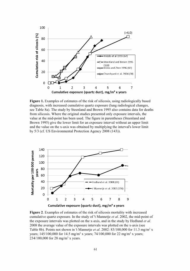
61
Figure 1. Examples of estimates of the risk of silicosis, using radiologically based
diagnosis, with increased cumulative quartz exposure (lung radiological changes,
see Table 8a). The study by Steenland and Brown 1995 also contains data for deaths
from silicosis. Where the original studies presented only exposure intervals, the
value at the mid-point has been used. The figure in parentheses (Steenland and
Brown 1995) give the lower limit for an exposure interval without an upper limit
and the value on the x-axis was obtained by multiplying the interval's lower limit
by 5/3 (cf. US Environmental Protection Agency 2008 (143)).
Figure 2. Examples of estimates of the risk of silicosis mortality with increased
cumulative quartz exposure. In the study of 't Mannetje et al. 2002, the mid-point of
the exposure intervals was plotted on the x-axis, and in the study by Hedlund et al.
2008 the average value of the exposure intervals was plotted on the x-axis (see
Table 8b). Points not shown in 't Mannetje et al. 2002: 83/100,000 for 11.3 mg/m3 x
years; 145/100,000 for 14.5 mg/m3 x years; 74/100,000 for 22 mg/m3 x years;
234/100,000 for 28 mg/m3 x years.
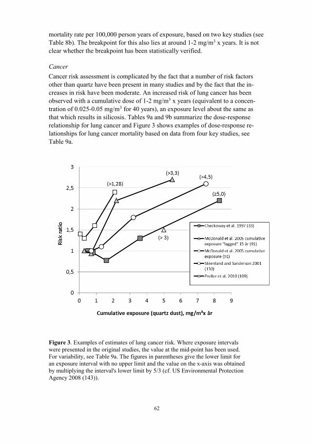
62
mortality rate per 100,000 person years of exposure, based on two key studies (see
Table 8b). The breakpoint for this also lies at around 1-2 mg/m3 x years. It is not
clear whether the breakpoint has been statistically verified.
Cancer
Cancer risk assessment is complicated by the fact that a number of risk factors
other than quartz have been present in many studies and by the fact that the in-
creases in risk have been moderate. An increased risk of lung cancer has been
observed with a cumulative dose of 1-2 mg/m3 x years (equivalent to a concen-
tration of 0.025-0.05 mg/m3 for 40 years), an exposure level about the same as
that which results in silicosis. Tables 9a and 9b summarize the dose-response
relationship for lung cancer and Figure 3 shows examples of dose-response re-
lationships for lung cancer mortality based on data from four key studies, see
Table 9a.
Figure 3. Examples of estimates of lung cancer risk. Where exposure intervals
were presented in the original studies, the value at the mid-point has been used.
For variability, see Table 9a. The figures in parentheses give the lower limit for
an exposure interval with no upper limit and the value on the x-axis was obtained
by multiplying the interval's lower limit by 5/3 (cf. US Environmental Protection
Agency 2008 (143)).
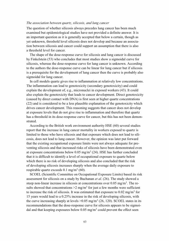
63
The association between quartz, silicosis, and lung cancer
The question of whether silicosis always precedes lung cancer has been much
examined but epidemiological studies have not provided a definite answer. It is
an important question as it is generally accepted that below a certain, though as
yet unknown, threshold level silicosis does not develop and because an associa-
tion between silicosis and cancer could support an assumption that there is also
a threshold level for cancer.
The shape of the dose-response curve for silicosis and lung cancer is discussed
by Finkelstein (53) who concludes that most studies show a sigmoidal curve for
silicosis, whereas the dose-response curve for lung cancer is unknown. According
to the authors the dose-response curve can be linear for lung cancer but if silicosis
is a prerequisite for the development of lung cancer then the curve is probably also
sigmoidal for lung cancer.
In cell models quartz gives rise to inflammation at relatively low concentrations.
The inflammation can lead to genotoxicity (secondary genotoxicity) and could
explain the development of, e.g., micronuclei in exposed workers (43). It could
also explain the genotoxicity that leads to cancer development. Direct genotoxicity
(caused by direct contact with DNA) is first seen at higher quartz concentrations
(22) and is considered to be a less plausible explanation of the genotoxicity which
drives cancer development. This reasoning suggests that cancer does not develop
at exposure levels that do not give rise to inflammation and therefore that quartz
has a threshold in its dose-response curve for cancer, but this has not been demon-
strated.
According to the British work environment authority HSE (60) several studies
report that the increase in lung cancer mortality in workers exposed to quartz is
limited to those who have silicosis and that exposure which does not lead to sili-
cosis, does not lead to lung cancer. However, the opinion was later put forward
that the existing occupational exposure limits were not always adequate for pre-
venting silicosis and that increased risks of silicosis have been demonstrated even
at exposure concentrations below 0.05 mg/m3 (24). HSE has further concluded
that it is difficult to identify a level of occupational exposure to quartz below
which there is no risk of developing silicosis and also concluded that the risk
of developing silicosis increases sharply when the average daily exposure to
respirable quartz exceeds 0.1 mg/m3 (60).
SCOEL (Scientific Committee on Occupational Exposure Limits) based its risk
assessment for silicosis on a study by Buchanan et al. (26). The study showed a
steep non-linear increase in silicosis at concentrations over 0.05 mg/m3. The re-
sults showed that concentrations >2 mg/m3 for just a few months were sufficient
to increase the risk of silicosis. It was estimated that exposure to 0.02 mg/m3 for
15 years would lead to a 0.25% increase in the risk of developing silicosis, with
the curve increasing sharply at levels >0.05 mg/m3 (26, 120). SCOEL states in its
recommendations that the dose-response curve for silicosis appears to be sigmoi-
dal and that keeping exposures below 0.05 mg/m3 could prevent the effect seen
64
in the steeper section of the curve where relatively small increases in exposure
can cause a significant increase in the risk of silicosis (120).
In a quantitative risk assessment by NIOSH (National Institute for Occupational
Safety and Health) it was calculated that 1.9% of those workers exposed to the
current US occupational exposure limit for respirable cristobalite dust (around
0.05 mg/m3) for 45 years were at risk of dying from lung cancer. The death rate
from lung diseases other than cancer was calculated at 5.4% and the death rate
from radiologically diagnosed silicosis caused by exposure was calculated at 7.5%
(100).
CICAD concluded that, on the basis of those studies of lung cancer and quartz
exposure with the least confounding, the risk appears to increase with cumulative
exposure (exposure time x exposure intensity), with the development of radiologi-
cally diagnosed silicosis, and with increased follow-up time after silicosis diagno-
sis (39).
Other effects
A review article by Hnizdo and Vallyathan in 2003 reported that rats developed
emphysema with lower doses of quartz than those which resulted in fibrosis and
silicotic nodules. It was also noted that several epidemiological studies which did
not find any radiological indications of silicosis nevertheless showed a significant
increase in airway resistance (reduced FEV1/FVC) with low quartz exposure (66).
However, the dose-response relationship is unclear as the contribution of quartz
to a decline in lung function in dusty environments is uncertain and because the
decline could be related to silicosis.
Kidney disease, autoimmune diseases and reduced resistance to tuberculosis
infection have been shown to be consequences of quartz exposure but the dose-
response relationship is unclear.
Conclusions
The critical effects of occupational exposure to quartz dust are silicosis and lung
cancer. Increased risk of silicosis (radiological changes corresponding to ILO-
classification ≥1/0 or ≥1/1), increased risk of death from silicosis, and increased
risk of lung cancer have been observed with a cumulative dose of around 1-2
mg/m3 x years (equivalent to a concentration of 0.025-0.05 mg/m3 for 40 years).
A non-effect level has not been identified.
It has not yet been determined whether silicosis is a prerequisite for the de-
velopment of lung cancer resulting from quartz exposure. However, genotoxico-
logical studies suggest that cancer development is driven by the inflammatory
response to quartz.
Occupational exposure to quartz has been shown to give rise to chronic obstruc-
tive pulmonary disease (COPD), kidney disease and autoimmune diseases (sclero-
derma, rheumatoid arthritis, systemic lupus erythematosus). It has not been
possible to establish effect levels but studies with rats and lung physiology data
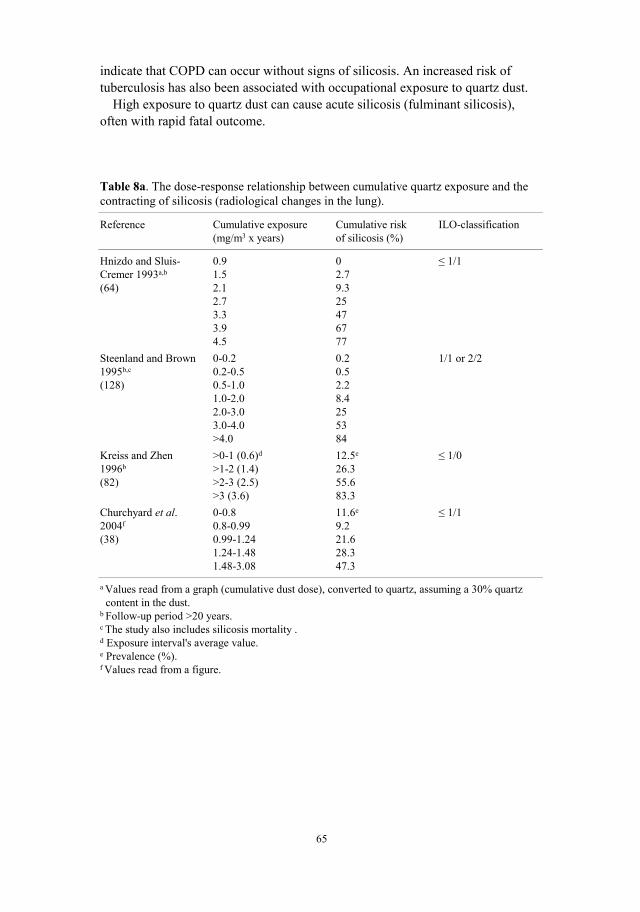
65
indicate that COPD can occur without signs of silicosis. An increased risk of
tuberculosis has also been associated with occupational exposure to quartz dust.
High exposure to quartz dust can cause acute silicosis (fulminant silicosis),
often with rapid fatal outcome.
Table 8a. The dose-response relationship between cumulative quartz exposure and the
contracting of silicosis (radiological changes in the lung).
Reference Cumulative exposure
(mg/m3 x years)
Cumulative risk
of silicosis (%)
ILO-classification
Hnizdo and Sluis-
Cremer 1993a,b
(64)
0.9
1.5
2.1
2.7
3.3
3.9
4.5
0
2.7
9.3
25
47
67
77
≥ 1/1
Steenland and Brown
1995b,c
(128)
0-0.2
0.2-0.5
0.5-1.0
1.0-2.0
2.0-3.0
3.0-4.0
>4.0
0.2
0.5
2.2
8.4
25
53
84
1/1 or 2/2
Kreiss and Zhen
1996b
(82)
>0-1 (0.6)d
>1-2 (1.4)
>2-3 (2.5)
>3 (3.6)
12.5e
26.3
55.6
83.3
≥ 1/0
Churchyard et al. 2004f (38)
0-0.8
0.8-0.99
0.99-1.24
1.24-1.48
1.48-3.08
11.6e
9.2
21.6
28.3
47.3
≥ 1/1
a Values read from a graph (cumulative dust dose), converted to quartz, assuming a 30% quartz
content in the dust. b Follow-up period >20 years. c The study also includes silicosis mortality . d Exposure interval's average value. e Prevalence (%). f Values read from a figure.
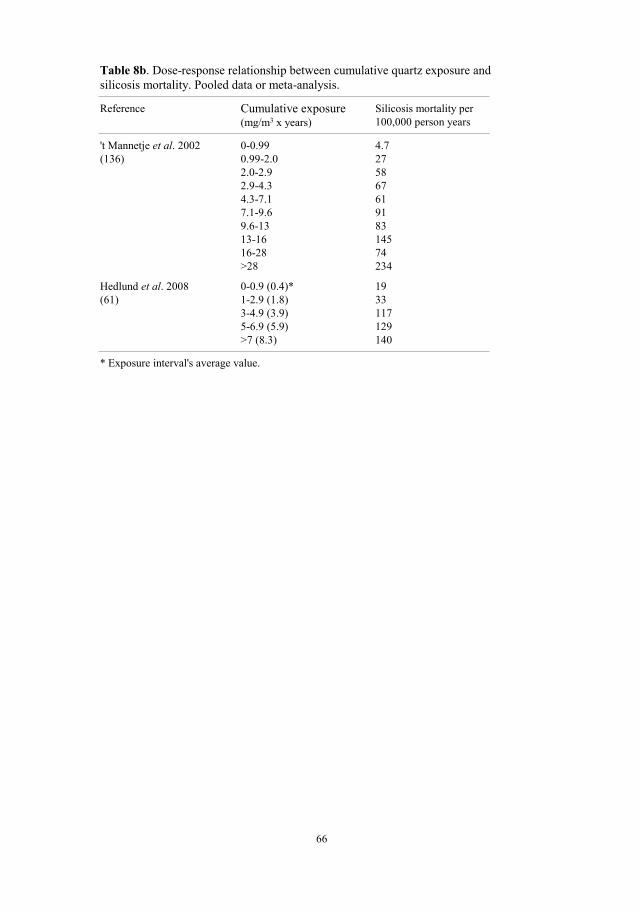
66
Table 8b. Dose-response relationship between cumulative quartz exposure and
silicosis mortality. Pooled data or meta-analysis.
Reference Cumulative exposure (mg/m3 x years)
Silicosis mortality per
100,000 person years
't Mannetje et al. 2002
(136)
0-0.99
0.99-2.0
2.0-2.9
2.9-4.3
4.3-7.1
7.1-9.6
9.6-13
13-16
16-28
>28
4.7
27
58
67
61
91
83
145
74
234
Hedlund et al. 2008
(61)
0-0.9 (0.4)*
1-2.9 (1.8)
3-4.9 (3.9)
5-6.9 (5.9)
>7 (8.3)
19
33
117
129
140
* Exposure interval's average value.
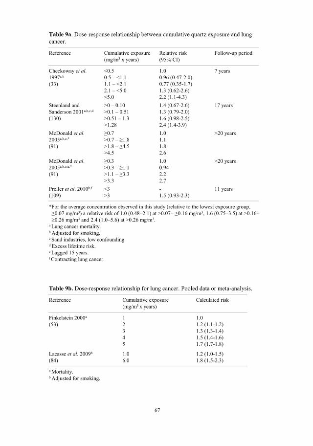
67
Table 9a. Dose-response relationship between cumulative quartz exposure and lung
cancer.
Reference Cumulative exposure
(mg/m3 x years)
Relative risk
(95% CI)
Follow-up period
Checkoway et al.
1997a,b
(33)
<0.5
0.5 – <1.1
1.1 – <2.1
2.1 – <5.0
≥5.0
1.0
0.96 (0.47-2.0)
0.77 (0.35-1.7)
1.3 (0.62-2.6)
2.2 (1.1-4.3)
7 years
Steenland and
Sanderson 2001a,b,c,d
(130)
>0 – 0.10
>0.1 – 0.51
>0.51 – 1.3
>1.28
1.4 (0.67-2.6)
1.3 (0.79-2.0)
1.6 (0.98-2.5)
2.4 (1.4-3.9)
17 years
McDonald et al.
2005a,b,c,*
(91)
≤0.7
>0.7 – ≤1.8
>1.8 – ≤4.5
>4.5
1.0
1.1
1.8
2.6
>20 years
McDonald et al.
2005a,b,c,e,*
(91)
≤0.3
>0.3 – ≤1.1
>1.1 – ≤3.3
>3.3
1.0
0.94
2.2
2.7
>20 years
Preller et al. 2010b,f
(109)
<3
>3
-
1.5 (0.93-2.3)
11 years
*For the average concentration observed in this study (relative to the lowest exposure group,
≤0.07 mg/m3) a relative risk of 1.0 (0.48–2.1) at >0.07– ≤0.16 mg/m3, 1.6 (0.75–3.5) at >0.16–
≤0.26 mg/m3 and 2.4 (1.0–5.6) at >0.26 mg/m3. a Lung cancer mortality. b Adjusted for smoking. c Sand industries, low confounding. d Excess lifetime risk. e Lagged 15 years. f Contracting lung cancer.
Table 9b. Dose-response relationship for lung cancer. Pooled data or meta-analysis.
Reference Cumulative exposure
(mg/m3 x years)
Calculated risk
Finkelstein 2000a
(53)
1
2
3
4
5
1.0
1.2 (1.1-1.2)
1.3 (1.3-1.4)
1.5 (1.4-1.6)
1.7 (1.7-1.8)
Lacasse et al. 2009b
(84)
1.0
6.0
1.2 (1.0-1.5)
1.8 (1.5-2.3)
a Mortality. b Adjusted for smoking.
68
Potential conflicts of interest
Johan Högberg (member), Ulla Stenius (member) and Ilona Silins (author of
draft), in a previous assignment from Swedish Work Environment Authority, have
expressed opinions concerning questions posed in the consensus report on quartz.
Johan Högberg (member) and Ulla Stenius (member) have received research
grants from AFA Insurance for a project on cancer risks from low-dose exposure
to quartz.
Maria Albin (member), in a text book issued by Prevent and in the Bulletin
from Arbets- och miljömedicin (Occupational and Environmental Medicine) in
Lund, has argued that current knowledge indicates that the existing occupational
exposure limit for quartz does not adequately protect those exposed from the risk
of silicosis/lung cancer.
Gunnar Johanson (member) has declared that he was involved in SCOEL's
evaluation of quartz and recommendations on health-based indicative occupa-
tional exposure limits for the EU.
Bengt Sjögren (member) and Gunnar Johanson (member), in a letter to Swedish
Work Environment Authority dated 2006-05-05, have drawn this authority's
attention to the view that the classification of quartz and cristobalite as carcino-
gens should be considered, on the basis of the published scientific evidence
(Lundberg 1996, SCOEL 2002, IARC 1997).
Håkan Westberg (member) has declared that he has received research grants
from AFA Insurance for a project ”Kvarts i svenska järngjuterier” (Quartz in
Swedish iron foundries) in collaboration with SWEREA - Svenska Gjuteri-
föreningen (Swedish Foundry Association).
69
References
1. Ahlmark A. Silikos i Sverige (Silicosis in Sweden). Redogörelse för Arbetsmedicinska
institutets och Arbetarskyddsstyrelsens utredning om silikos. Arbetsmedicinska institutet,
1967, 255 pp. (In Swedish)
2. Ahlmark A, Gerhardsson L. Silikosen i Sverige sedan 1930. Arbete och Hälsa 1981;15:1-55.
Arbetarskyddsverket (The Swedish National Board of Occupational Safety and Health),
Solna. (In Swedish, summary in English)
3. Akgun M, Araz O, Akkurt I, Eroglu A, Alper F, Saglam L, Mirici A, Gorguner M, Nemery B.
An epidemic of silicosis among former denim sandblasters. Eur Respir J 2008;32:1295-1303.
4. Akgun M, Mirici A, Ucar EY, Kantarci M, Araz O, Gorguner M. Silicosis in Turkish denim
sandblasters. Occup Med (Lond) 2006;56:554-558.
5. Albin M, Engholm G, Fröström K, Kheddache S, Larsson S, Swantesson L. Chest x ray films
from construction workers: International Labour Office (ILO 1980) classification compared
with routine readings. Br J Ind Med 1992;49:862-868.
6. Andersson L, Bryngelsson IL, Ohlson CG, Nayström P, Lilja BG, Westberg H. Quartz and
dust exposure in Swedish iron foundries. J Occup Environ Hyg 2009;6:9-18.
7. Andersson L, Burdorf A, Bryngelsson IL, Westberg H. Estimating trends in quartz exposure
in Swedish iron foundries--predicting past and present exposures. Ann Occup Hyg
2012;56:362-372.
8. Arbetarskyddsstyrelsen (The Swedish National Board of Occupational Safety and Health).
Arbetarskyddsstyrelsens kungörelse med föreskrifter om kvarts. Arbetarskyddsstyrelsens
författningssamling 1992;16. (In Swedish)
9. Arbetsmiljöverket (The Swedish Work Environment Authority). Byggdamm vid ROT-
arbeten. Mätprojekt Arbetsmiljöverket 2004. Rapport 2005:2, 17 pp. Arbetsmiljöverket,
Solna. (In Swedish)
10. Arbetsmiljöverket (The Swedish Work Environment Authority). Järvholm B, Bystedt J,
Reuterwall C. Kunskapsöversikt. Arbetsrelaterade dödsfall i Sverige - arbetsrelaterad
dödlighet i cancer, hjärt-kärlsjukdomar och lungsjukdomar i Sverige. Rapport 2010:3, 68 pp.
Arbetsmiljöverket, Stockholm. (In Swedish)
11. Arbetsmiljöverket (The Swedish Work Environment Authority). Högberg J, Silins I, Stenius
U. Kunskapsöversikt. Kvarts och dess cancerframkallande förmåga. Rapport 2011:5, 36 pp.
Arbetsmiljöverket, Stockholm. (In Swedish)
12. Archer JD, Cooper GS, Reist PC, Storm JF, Nylander-French LA. Exposure to respirable
crystalline silica in eastern North Carolina farm workers. AIHA J 2002;63:750-755.
13. Bakan ND, Ozkan G, Camsari G, Gur A, Bayram M, Acikmese B, Cetinkaya E. Silicosis in
denim sandblasters. Chest 2011;140:1300-1304.
14. Banks DE, Barkman HW, Jr. Silicosis epidemic reported in 1900. JAMA 1984;251:217.
15. Bergdahl IA, Jonsson H, Eriksson K, Damber L, Järvholm B. Lung cancer and exposure to
quartz and diesel exhaust in Swedish iron ore miners with concurrent exposure to radon.
Occup Environ Med 2010;67:513-518.
16. Bergström B. Damm i luft, respirabelt. Arbets- och miljömedicin, Analyslaboratoriet,
Universitetssjukhuset Örebro, 2010. (In Swedish)
17. Bergström B. Kvarts i luft. Arbets- och miljömedicinska kliniken, Analyslaboratoriet,
Universitetssjukhuset Örebro, 2011. (In Swedish)
18. Bergström B. Kristobalit i luft. Arbets- och miljömedicinska kliniken, Analyslaboratoriet,
Universitetssjukhuset Örebro, 2012. (In Swedish)
19. Beskow R. Silicosis in diatomaceous earth factory workers in Sweden. Scand J Respir Dis
1978;59:216-221.
70
20. Birk T, Mundt KA, Guldner K, Parsons W, Luippold RS. Mortality in the German porcelain
industry 1985-2005: first results of an epidemiological cohort study. J Occup Environ Med
2009;51:373-385.
21. Bonassi S, Znaor A, Ceppi M, Lando C, Chang WP, Holland N, Kirsch-Volders M, Zeiger E,
Ban S, Barale R, Bigatti MP, Bolognesi C, Cebulska-Wasilewska A, Fabianova E, Fucic A,
Hagmar L, Joksic G, Martelli A, Migliore L, Mirkova E, Scarfi MR, Zijno A, Norppa H,
Fenech M. An increased micronucleus frequency in peripheral blood lymphocytes predicts the
risk of cancer in humans. Carcinogenesis 2007;28:625-631.
22. Borm PJ, Tran L, Donaldson K. The carcinogenic action of crystalline silica: a review of the
evidence supporting secondary inflammation-driven genotoxicity as a principal mechanism.
Crit Rev Toxicol 2011;41:756-770.
23. Brown LM, Gridley G, Olsen JH, Mellemkjaer L, Linet MS, Fraumeni JF, Jr. Cancer risk and
mortality patterns among silicotic men in Sweden and Denmark. J Occup Environ Med
1997;39:633-638.
24. Brown T. Silica exposure, smoking, silicosis and lung cancer--complex interactions. Occup
Med (Lond) 2009;59:89-95.
25. Bruce T. Die Silikose als Berufskrankheit in Schweden (Silikosen som yrkessjukdom i
Sverige). Eine klinische und gewerbemedizinische Studie. Acta Med Scand 1942;110, Suppl
129. (In German)
26. Buchanan D, Miller BG, Soutar CA. Quantitative relations between exposure to respirable
quartz and risk of silicosis. Occup Environ Med 2003;60:159-164.
27. Cainelli F, Tanko MN, Vento S. Silica exposure and silicosis: action is needed now. South
Med J 2010;103:1078.
28. Cassel SL, Eisenbarth SC, Iyer SS, Sadler JJ, Colegio OR, Tephly LA, Carter AB, Rothman
PB, Flavell RA, Sutterwala FS. The Nalp3 inflammasome is essential for the development of
silicosis. Proc Natl Acad Sci 2008;105:9035-9040.
29. Cassidy A, t Mannetje A, van Tongeren M, Field JK, Zaridze D, Szeszenia-Dabrowska N,
Rudnai P, Lissowska J, Fabianova E, Mates D, Bencko V, Foretova L, Janout V, Fevotte J,
Fletcher T, Brennan P, Boffetta P. Occupational exposure to crystalline silica and risk of lung
cancer: a multicenter case-control study in Europe. Epidemiology 2007;18:36-43.
30. CDC. Silicosis deaths among young adults-United States, 1968-1994. Centers for Disease
Control and Prevention1998;47:331-335.
31. Checkoway H, Franzblau A. Is silicosis required for silica-associated lung cancer? Am J Ind
Med 2000;37:252-259.
32. Checkoway H, Heyer NJ, Demers PA, Breslow NE. Mortality among workers in the
diatomaceous earth industry. Br J Ind Med 1993;50:586-597.
33. Checkoway H, Heyer NJ, Seixas NS, Welp EA, Demers PA, Hughes JM, Weill H. Dose-
response associations of silica with nonmalignant respiratory disease and lung cancer
mortality in the diatomaceous earth industry. Am J Epidemiol 1997;145:680-688.
34. Chen GX, Burnett CA, Cameron LL, Alterman T, Lalich NR, Tanaka S, Althouse RB.
Tuberculosis Mortality and Silica Exposure: A Case-Control Study Based on a National
Mortality Database for the Years 1983-1992. Int J Occup Environ Health 1997;3:163-170.
35. Chen W, Bochmann F, Sun Y. Effects of work related confounders on the association
between silica exposure and lung cancer: a nested case-control study among Chinese miners
and pottery workers. Int Arch Occup Environ Health 2007;80:320-326.
36. Cherniack MG. The Hawk's Nest Incident. America's worst industrial disaster. New Haven
and London, Yale University Press 1986.
37. Cherry NM, Burgess GL, Turner S, McDonald JC. Crystalline silica and risk of lung cancer in
the potteries. Occup Environ Med 1998;55:779-785.
71
38. Churchyard GJ, Ehrlich R, teWaterNaude JM, Pemba L, Dekker K, Vermeijs M, White N,
Myers J. Silicosis prevalence and exposure-response relations in South African goldminers.
Occup Environ Med 2004;61:811-816.
39. CICAD. Concise International Chemical Assessment Document 24. Crystalline silica, quartz.
International Programme on Chemical Safety, World Health Organization, Geneva, 2000.
40. Cocco P, Rice CH, Chen JQ, McCawley MA, McLaughlin JK, Dosemeci M. Lung cancer
risk, silica exposure, and silicosis in Chinese mines and pottery factories: the modifying role
of other workplace lung carcinogens. Am J Ind Med 2001;40:674-682.
41. Corbett EL, Churchyard GJ, Clayton TC, Williams BG, Mulder D, Hayes RJ, De Cock KM.
HIV infection and silicosis: the impact of two potent risk factors on the incidence of
mycobacterial disease in South African miners. AIDS 2000;14:2759-2768.
42. Cowie RL. The epidemiology of tuberculosis in gold miners with silicosis. Am J Respir Crit
Care Med 1994;150:1460-1462.
43. Demircigil GC, Coskun E, Vidinli N, Erbay Y, Yilmaz M, Cimrin A, Schins RP, Borm PJ,
Burgaz S. Increased micronucleus frequencies in surrogate and target cells from workers
exposed to crystalline silica-containing dust. Mutagenesis 2010;25:163-169.
44. Deniz O, Gumus S, Ors F, Yaman H, Battal B, Karaman B, Acikel CH, Ucar E, Cakir E,
Tozkoparan E, Bilgic H. Serum lactate dehydrogenase levels significantly correlate with
radiological extent of disease and spirometric values in patients with silicosis due to denim
sandblasting. Clin Chem Lab Med 2012;50:483-488.
45. Donaldson K, Borm PJ. The quartz hazard: a variable entity. Ann Occup Hyg 1998;42:287-
294.
46. Donaldson K, Stone V, Duffin R, Clouter A, Schins R, Borm P. The quartz hazard: effects of
surface and matrix on inflammogenic activity. J Environ Pathol Toxicol Oncol 2001;20 Suppl
1:109-118.
47. Dostert C, Petrilli V, Van Bruggen R, Steele C, Mossman BT, Tschopp J. Innate immune
activation through Nalp3 inflammasome sensing of asbestos and silica. Science
2008;320:674-677.
48. Driscoll T, Nelson DI, Steenland K, Leigh J, Concha-Barrientos M, Fingerhut M, Pruss-Ustun
A. The global burden of disease due to occupational carcinogens. Am J Ind Med
2005;48:419-431.
49. EEC. Workplace atmospheres. Size fraction definitions of measurement of airborne particles
ISO/TR 7708, 88/642/EEC. EN 481:1993.
50. Elias Z, Poirot O, Fenoglio I, Ghiazza M, Daniere MC, Terzetti F, Darne C, Coulais C,
Matekovits I, Fubini B. Surface reactivity, cytotoxic, and morphological transforming effects
of diatomaceous Earth products in Syrian hamster embryo cells. Toxicol Sci 2006;91:510-
520.
51. Erren TC, Glende CB, Morfeld P, Piekarski C. Is exposure to silica associated with lung
cancer in the absence of silicosis? A meta-analytical approach to an important public health
question. Int Arch Occup Environ Health 2009;82:997-1004.
52. Finckh A, Cooper GS, Chibnik LB, Costenbader KH, Watts J, Pankey H, Fraser PA, Karlson
EW. Occupational silica and solvent exposures and risk of systemic lupus erythematosus in
urban women. Arthritis Rheum 2006;54:3648-3654.
53. Finkelstein MM. Silica, silicosis, and lung cancer: a risk assessment. Am J Ind Med
2000;38:8-18.
54. Freeman CS, Grossman EA. Silica exposures in workplaces in the United States between
1980 and 1992. Scand J Work Environ Health 1995;21 Suppl 2:47-49.
55. Gamble JF. Crystalline silica and Lung cancer: A critical review of the occupational
epidemiology literature of exposure-response studies testing this hypothesis. Crit Rev Toxicol
2011;41:404-465.
72
56. Gerhardsson G, Engman L, Andersson A, Isaksson G, Magnusson E, Sundquist S.
Silikosprojektets slutrapport. Del 2. Målsättning, omfattning och resultat.
Unersökningsrapport AMT 103/74-2. Arbetarskyddsstyrelsen (The Swedish National Board
of Occupational Safety and Health) 1974. (In Swedish)
57. Greenberg MI, Waksman J, Curtis J. Silicosis: a review. Dis Mon 2007;53:394-416.
58. Gulumian M, Borm PJ, Vallyathan V, Castranova V, Donaldson K, Nelson G, Murray J.
Mechanistically identified suitable biomarkers of exposure, effect, and susceptibility for
silicosis and coal-worker's pneumoconiosis: a comprehensive review. J Toxicol Environ
Health B Crit Rev 2006;9:357-395.
59. Hamilton RF, Jr., Thakur SA, Holian A. Silica binding and toxicity in alveolar macrophages.
Free Radic Biol Med 2008;44:1246-1258.
60. HSE. Respirable crystalline silica - Phase 2. Carcinogenicity. Health and Safety Executive
2003; EH75/5.
61. Hedlund U, Jonsson H, Eriksson K, Järvholm B. Exposure-response of silicosis mortality in
Swedish iron ore miners. Ann Occup Hyg 2008;52:3-7.
62. Hnizdo E. Combined effect of silica dust and tobacco smoking on mortality from chronic
obstructive lung disease in gold miners. Br J Ind Med 1990;47:656-664.
63. Hnizdo E, Murray J, Sluis-Cremer GK, Thomas RG. Correlation between radiological and
pathological diagnosis of silicosis: an autopsy population based study. Am J Ind Med
1993;24:427-445.
64. Hnizdo E, Sluis-Cremer GK. Risk of silicosis in a cohort of white South African gold miners.
Am J Ind Med 1993;24:447-457.
65. Hnizdo E, Sullivan PA, Bang KM, Wagner G. Association between chronic obstructive
pulmonary disease and employment by industry and occupation in the US population: a study
of data from the Third National Health and Nutrition Examination Survey. Am J Epidemiol
2002;156:738-746.
66. Hnizdo E, Vallyathan V. Chronic obstructive pulmonary disease due to occupational
exposure to silica dust: a review of epidemiological and pathological evidence. Occup
Environ Med 2003;60:237-243.
67. Hornung V, Bauernfeind F, Halle A, Samstad EO, Kono H, Rock KL, Fitzgerald KA, Latz E.
Silica crystals and aluminum salts activate the NALP3 inflammasome through phagosomal
destabilization. Nat Immunol 2008;9:847-856.
68. HSE. General methods for sampling and gravimetric analysis of inhalable and respirable dust.
In: Methods for the determination of hazardous substances. Health and Safety Executive
2000; MDHS 14/3.
69. HSE. Respirable crystalline silica - Phase 1. Variability in fibrogenic potency and exposure-
response relationships for silicosis. Health and Safety Executive 2002; EH75/4.
70. HSE. Crystalline silica in respirable airborne dusts. Direct-on-filter analysis by infrared
spectroscopy and X-ray diffraction. In: Methods for the determination of hazardous
substances. Health and Safety Executive 2005; MDHS 101.
71. Hughes JM, Weill H, Checkoway H, Jones RN, Henry MM, Heyer NJ, Seixas NS, Demers
PA. Radiographic evidence of silicosis risk in the diatomaceous earth industry. Am J Respir
Crit Care Med 1998;158:807-814.
72. IARC. Silica, some silicates, coal dust and para-aramid fibrils. IARC Monographs on the
Evaluation of Carcinogenic Risks to Humans. Volume 68. Lyon: International Agency for
Research on Cancer 1997;68:41-242.
73. ILO. Guidelines for the use of the ILO International Classification of Radiographs of
Pneumoconioses. Revised edition 2011. Geneva, International Labour Office, 2011.
74. Janko M, McCrae RE, O'Donnell JF, Austria RJ. Occupational exposure and analysis of
microcrystalline cristobalite in mullite operations. Am Ind Hyg Assoc J 1989;50:460-465.
73
75. Jörgensen HS. Silicosis in the iron-ore mine in Kiruna, Sweden, and the future need for
silicosis control. Int Arch Occup Environ Health 1986;58:251-257.
76. Kauppinen T, Toikkanen J, Pedersen D, Young R, Ahrens W, Boffetta P, Hansen J,
Kromhout H, Maqueda Blasco J, Mirabelli D, de la Orden-Rivera V, Pannett B, Plato N,
Savela A, Vincent R, Kogevinas M. Occupational exposure to carcinogens in the European
Union. Occup Environ Med 2000;57:10-18.
77. King EJ, Dolan M. Silicosis and the metabolism of silica. Canad Med Ass J 1934;31,21-26.
78. King EJ, McGeorge M. The Biochemistry of Silicic Acid - The Solution and Excretion of
Silica. Biochem J 1938;32:426-433.
79. Koksal N, Kahraman H. Acute silicosis in teflon-coated pan manufacturing due to metal
sandblasting. Int J Occup Environ Health 2011;17:210-213.
80. Kolling A, Ernst H, Rittinghausen S, Heinrich U. Relationship of pulmonary toxicity and
carcinogenicity of fine and ultrafine granular dusts in a rat bioassay. Inhal Toxicol
2011;23:544-554.
81. Kramer MR, Blanc PD, Fireman E, Amital A, Guber A, Rhahman NA, Shitrit D. Caesarstone
silicosis: disease resurgence among artificial stone workers. Chest 2012;142:419-424.
82. Kreiss K, Zhen B. Risk of silicosis in a Colorado mining community. Am J Ind Med
1996;30:529-539.
83. Kurihara N, Wada O. Silicosis and smoking strongly increase lung cancer risk in silica-
exposed workers. Ind Health 2004;42:303-314.
84. Lacasse Y, Martin S, Gagne D, Lakhal L. Dose-response meta-analysis of silica and lung
cancer. Cancer Causes Control 2009;20:925-933.
85. Laney AS, Petsonk EL, Attfield MD. Pneumoconiosis among underground bituminous coal
miners in the United States: is silicosis becoming more frequent? Occup Environ Med
2010;67:652-656.
86. Leung CC, Yu IT, Chen W. Silicosis. Lancet 2012;379:2008-2018.
87. Liu B, Guan R, Zhou P, Miao Q, Wang H, Fu D, You B. A distinct mutational spectrum of
p53 and K-ras genes in lung cancer of workers with silicosis. J Environ Pathol Toxicol Oncol
2000;19:1-7.
88. Malmberg P, Hedenström H, Sundblad BM. Changes in lung function of granite crushers
exposed to moderately high silica concentrations: a 12 year follow up. Br J Ind Med
1993;50:726-731.
89. McCormic ZD, Khuder SS, Aryal BK, Ames AL, Khuder SA. Occupational silica exposure as
a risk factor for scleroderma: a meta-analysis. Int Arch Occup Environ Health 2010;83:763-
769.
90. McDonald AD, McDonald JC, Rando RJ, Hughes JM, Weill H. Cohort mortality study of
North American industrial sand workers. I. Mortality from lung cancer, silicosis and other
causes. Ann Occup Hyg 2001;45:193-199.
91. McDonald JC, McDonald AD, Hughes JM, Rando RJ, Weill H. Mortality from lung and
kidney disease in a cohort of North American industrial sand workers: an update. Ann Occup
Hyg 2005;49:367-373.
92. Meyer JD, Islam SS, Ducatman AM, McCunney RJ. Prevalence of small lung opacities in
populations unexposed to dusts. A literature analysis. Chest 1997;111:404-410.
93. Mossman BT, Churg A. Mechanisms in the pathogenesis of asbestosis and silicosis. Am J
Respir Crit Care Med 1998;157:1666-1680.
94. Muhle H, Kittel B, Ernst H, Mohr U, Mermelstein R. Neoplastic lung lesions in rat after
chronic exposure to crystalline silica. Scand J Work Environ Health 1995;21 Suppl 2:27-29.
95. Mundt KA, Birk T, Parsons W, Borsch-Galetke E, Siegmund K, Heavner K, Guldner K.
Respirable crystalline silica exposure-response evaluation of silicosis morbidity and lung
cancer mortality in the German porcelain industry cohort. J Occup Environ Med
2011;53:282-289.
74
96. Nehls P, Seiler F, Rehn B, Greferath R, Bruch J. Formation and persistence of 8-oxoguanine
in rat lung cells as an important determinant for tumor formation following particle exposure.
Environ Health Perspect 1997;105 Suppl 5:1291-1296.
97. Ng TP, Jones MG, Pang CP, Chan SL. Fasting urinary hydroxyproline: creatinine ratios in
silicosis. Ann Acad Med Singapore 1989;18:692-695.
98. Ng TP, Lee HS, Phoon WH. Further evidence of human silica nephrotoxicity in
occupationally exposed workers. Br J Ind Med 1993;50:907-912.
99. NIOSH. Silica, crystalline, by XRD (respirable). Method 7500. NIOSH Manual of analytical
methods, 4th ed., 3rd Suppl. The National Institute for Occupational Safety and Health, 2003.
100. NIOSH. Silica, lung cancer, and respiratory disease quantitative risk. The National Institute
for Occupational Safety and Health 2011, NIOSH publication No 2011-120.
101. O'Connor RE, Bossaert L, Arntz HR, Brooks SC, Diercks D, Feitosa-Filho G, Nolan JP,
Vanden Hoek TL, Walters DL, Wong A, Welsford M, Woolfrey K. Part 9: acute coronary
syndromes: 2010 International Consensus on Cardiopulmonary Resuscitation and Emergency
Cardiovascular Care Science With Treatment Recommendations. Circulation 2010;122:S422-
465.
102. Ojajarvi IA, Partanen TJ, Ahlbom A, Boffetta P, Hakulinen T, Jourenkova N, Kauppinen TP,
Kogevinas M, Porta M, Vainio HU, Weiderpass E, Wesseling CH. Occupational exposures
and pancreatic cancer: a meta-analysis. Occup Environ Med 2000;57:316-324.
103. OSHA. Quarts and Cristobalite in workplace atmospheres. Occupational Safety and Health
Administration, Method ID-142, 1996.
104. Paris C, Clement-Duchene C, Vignaud JM, Gislard A, Stoufflet A, Bertrand O, Thiberville L,
Grosdidier G, Martinet Y, Benichou J, Hainaut P. Relationships between lung
adenocarcinoma and gender, age, smoking and occupational risk factors: A case-case study.
Lung Cancer 2010;68:146-153.
105. Park R, Rice F, Stayner L, Smith R, Gilbert S, Checkoway H. Exposure to crystalline silica,
silicosis, and lung disease other than cancer in diatomaceous earth industry workers: a
quantitative risk assessment. Occup Environ Med 2002;59:36-43.
106. Parkes RW, ed. Occupational Lung Disorders, 3rd ed. Oxford: Butterworth Heinemann,
1994.
107. Parks CG, Conrad K, Cooper GS. Occupational exposure to crystalline silica and autoimmune
disease. Environ Health Perspect 1999;107 Suppl 5:793-802.
108. Pelucchi C, Pira E, Piolatto G, Coggiola M, Carta P, La Vecchia C. Occupational silica
exposure and lung cancer risk: a review of epidemiological studies 1996-2005. Ann Oncol
2006;17:1039-1050.
109. Preller L, van den Bosch LM, van den Brandt PA, Kauppinen T, Goldbohm A. Occupational
exposure to silica and lung cancer risk in the Netherlands. Occup Environ Med 2010;67:657-
663.
110. Rees D, Murray J. Silica, silicosis and tuberculosis. Int J Tuberc Lung Dis 2007;11:474-484.
111. Rego G, Pichel A, Quero A, Dubois A, Martinez C, Isidro I, Gil M, Cuervo V, Gonzalez A.
High prevalence and advanced silicosis in active granite workers: a dose-response analysis
including FEV1. J Occup Environ Med 2008;50:827-833.
112. Rice FL, Stayner LT. Assessment of silicosis risk for occupational exposure to crystalline
silica. Scand J Work Environ Health 1995;21 Suppl 2:87-90.
113. Rimal B, Greenberg AK, Rom WN. Basic pathogenetic mechanisms in silicosis: current
understanding. Curr Opin Pulm Med 2005;11:169-173.
114. Rosenman KD, Reilly MJ, Rice C, Hertzberg V, Tseng CY, Anderson HA. Silicosis among
foundry workers. Implication for the need to revise the OSHA standard. Am J Epidemiol
1996;144:890-900.
115. Rushton L. Chronic obstructive pulmonary disease and occupational exposure to silica. Rev
Environ Health 2007;22:255-272.
75
116. Sanchez-Roman J, Wichmann I, Salaberri J, Varela JM, Nunez-Roldan A. Multiple clinical
and biological autoimmune manifestations in 50 workers after occupational exposure to silica.
Ann Rheum Dis 1993;52:534-538.
117. Scarselli A, Binazzi A, Forastiere F, Cavariani F, Marinaccio A. Industry and job-specific
mortality after occupational exposure to silica dust. Occup Med (Lond) 2011;61:422-429.
118. Schins RP, Duffin R, Hohr D, Knaapen AM, Shi T, Weishaupt C, Stone V, Donaldson K,
Borm PJ. Surface modification of quartz inhibits toxicity, particle uptake, and oxidative DNA
damage in human lung epithelial cells. Chem Res Toxicol 2002;15:1166-1173.
119. Schins RP, Schilderman PA, Borm PJ. Oxidative DNA damage in peripheral blood
lymphocytes of coal workers. Int Arch Occup Environ Health 1995;67:153-157.
120. SCOEL. Recommendation of the Scientific Committee on Occupational Exposure Limits for
Silica, Crystalline (respirable dust). SCOEL/SUM/94, 2003.
121. Selinus O, Alloway B, Centeno JA, Finkelman R, Fuge R, Smedley P, eds. Essentials of
Medical Geology: impacts of the natural environment on public health. Burlington, Elsevier
Academic Press, 2005:1-812.
122. Sjödahl K, Jansson C, Bergdahl IA, Adami J, Boffetta P, Lagergren J. Airborne exposures
and risk of gastric cancer: a prospective cohort study. Int J Cancer 2007;120:2013-2018.
123. Slavov E, Miteva L, Prakova G, Gidikova P, Stanilova S. Correlation between TNF-alpha and
IL-12p40-containing cytokines in silicosis. Toxicol Ind Health 2010;26:479-486.
124. Sluis-Cremer GK, Hessel PA, Hnizdo E, Churchill AR. Relationship between silicosis and
rheumatoid arthritis. Thorax 1986;41:596-601.
125. Soutar CA, Robertson A, Miller BG, Searl A, Bignon J. Epidemiological evidence on the
carcinogenicity of silica: factors in scientific judgement. Ann Occup Hyg 2000;44:3-14.
126. STAMI. Faktabok om arbeidsmiljø og helse 2011. Status og utviklingstrekk. Statens
Arbetsmiljöinstitutt (National Institute of Occupational Health), Oslo, 2011:1-279. (In
Norwegian, English summary)
127. Steenland K. One agent, many diseases: exposure-response data and comparative risks of
different outcomes following silica exposure. Am J Ind Med 2005;48:16-23.
128. Steenland K, Brown D. Silicosis among gold miners: exposure--response analyses and risk
assessment. Am J Public Health 1995;85:1372-1377.
129. Steenland K, Mannetje A, Boffetta P, Stayner L, Attfield M, Chen J, Dosemeci M, DeKlerk
N, Hnizdo E, Koskela R, Checkoway H. Pooled exposure-response analyses and risk
assessment for lung cancer in 10 cohorts of silica-exposed workers: an IARC multicentre
study. Cancer Causes Control 2001;12:773-784.
130. Steenland K, Sanderson W. Lung cancer among industrial sand workers exposed to crystalline
silica. Am J Epidemiol 2001;153:695-703.
131. Steenland K, Sanderson W, Calvert GM. Kidney disease and arthritis in a cohort study of
workers exposed to silica. Epidemiology 2001;12:405-412.
132. Stolt P, Källberg H, Lundberg I, Sjögren B, Klareskog L, Alfredsson L. Silica exposure is
associated with increased risk of developing rheumatoid arthritis: results from the Swedish
EIRA study. Ann Rheum Dis 2005;64:582-586.
133. Stolt P, Yahya A, Bengtsson C, Källberg H, Ronnelid J, Lundberg I, Klareskog L, Alfredsson
L. Silica exposure among male current smokers is associated with a high risk of developing
ACPA-positive rheumatoid arthritis. Ann Rheum Dis 2010;69:1072-1076.
134. Straif K, Benbrahim-Tallaa L, Baan R, Grosse Y, Secretan B, El Ghissassi F, Bouvard V,
Guha N, Freeman C, Galichet L, Cogliano V. A review of human carcinogens--part C: metals,
arsenic, dusts, and fibres. Lancet Oncol 2009;10:453-454.
135. Sundin J, Hogstedt C, Lindberg J, Moberg H, eds. Svenska folkets hälsa i historiskt
perspektiv. Statens Folkhälsoinstitut 2005:1-487.
76
136. t Mannetje A, Steenland K, Attfield M, Boffetta P, Checkoway H, DeKlerk N, Koskela RS.
Exposure-response analysis and risk assessment for silica and silicosis mortality in a pooled
analysis of six cohorts. Occup Environ Med 2002;59:723-728.
137. Thakur SA, Beamer CA, Migliaccio CT, Holian A. Critical role of MARCO in crystalline
silica-induced pulmonary inflammation. Toxicol Sci 2009;108:462-471.
138. Tjoe Nij E, Heederik D. Risk assessment of silicosis and lung cancer among construction
workers exposed to respirable quartz. Scand J Work Environ Health 2005;31 Suppl 2:49-56.
139. Tse LA, Yu IS, Au JS, Qiu H, Wang XR. Silica dust, diesel exhaust, and painting work are
the significant occupational risk factors for lung cancer in nonsmoking Chinese men. Br J
Cancer 2011;104:208-213.
140. Tsuda T, Mino Y, Babazono A, Shigemi J, Otsu T, Yamamoto E. A case-control study of the
relationships among silica exposure, gastric cancer, and esophageal cancer. Am J Ind Med
2001;39:52-57.
141. US-EPA. Ambient levels and noncancer health effects of inhaled crystalline and amorphous
silica. U.S. Environmental Protection Agency, Office of Research and Development, National
Center For Environmental Assessment, Research Triangle Park Office, Research Triangle
Park, NC, EPA/600/R-95/115, 1996.
142. Vacek PM, Verma DK, Graham WG, Callas PW, Gibbs GW. Mortality in Vermont granite
workers and its association with silica exposure. Occup Environ Med 2011;68:312-318.
143. van der Bij S, Koffijberg H, Lenters V, Portengen L, Moons KG, Heederik D, Vermeulen
RC. Lung cancer risk at low cumulative asbestos exposure: meta-regression of the exposure-
response relationship. Cancer Causes Control 2013;24:1-12.
144. Verma DK, Ritchie AC, Muir DC. Dust content of lungs and its relationships to pathology,
radiology and occupational exposure in Ontario hardrock miners. Am J Ind Med 2008;51:524-
531.
145. Vida S, Pintos J, Parent ME, Lavoue J, Siemiatycki J. Occupational exposure to silica and
lung cancer: pooled analysis of two case-control studies in Montreal, Canada. Cancer
Epidemiol Biomarkers Prev 2010;19:1602-1611.
146. Vupputuri S, Parks CG, Nylander-French LA, Owen-Smith A, Hogan SL, Sandler DP.
Occupational Silica Exposure and Chronic Kidney Disease. Ren Fail 2012;34:40-46.
147. Weiner J, Barlow L, Sjögren B. Ischemic heart disease mortality among miners and other
potentially silica-exposed workers. Am J Ind Med 2007;50:403-408.
148. Westberg H, Andersson L, Bryngelsson IL, Ngo Y, Ohlson CG. Cancer morbidity and quartz
exposure in Swedish iron foundries. Int Arch Occup Environ Health 2012;86:499-507.
149. Westerholm P. Silicosis. Observations on a case register. Scand J Work Environ Health
1980;6 Suppl 2:1-86.
150. Westerholm P, Ahlmark A, Maasing R, Segelberg I. Silicosis and risk of lung cancer or lung
tuberculosis: a cohort study. Environ Res 1986;41:339-350.
151. Ziskind M, Jones RN, Weill H. Silicosis. Am Rev Respir Dis 1976;113:643-665.
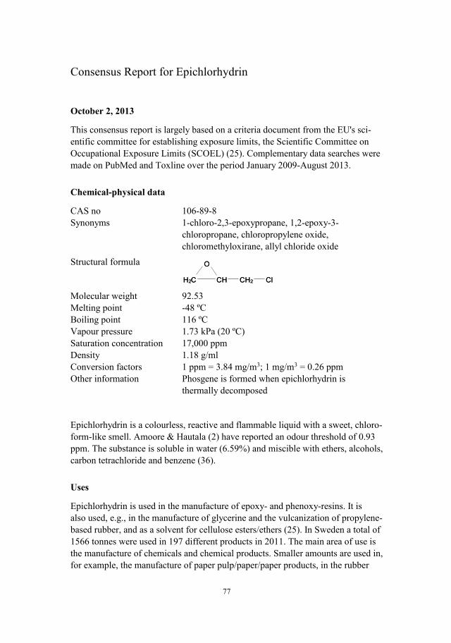
77
Consensus Report for Epichlorhydrin
October 2, 2013
This consensus report is largely based on a criteria document from the EU's sci-
entific committee for establishing exposure limits, the Scientific Committee on
Occupational Exposure Limits (SCOEL) (25). Complementary data searches were
made on PubMed and Toxline over the period January 2009-August 2013.
Chemical-physical data
CAS no 106-89-8
Synonyms 1-chloro-2,3-epoxypropane, 1,2-epoxy-3-
chloropropane, chloropropylene oxide,
chloromethyloxirane, allyl chloride oxide
Structural formula
CH2CH
O
ClH2C CH2CH
O
ClH2C
Molecular weight 92.53
Melting point -48 ºC
Boiling point 116 ºC
Vapour pressure 1.73 kPa (20 ºC)
Saturation concentration 17,000 ppm
Density 1.18 g/ml
Conversion factors 1 ppm = 3.84 mg/m3; 1 mg/m3 = 0.26 ppm
Other information Phosgene is formed when epichlorhydrin is
thermally decomposed
Epichlorhydrin is a colourless, reactive and flammable liquid with a sweet, chloro-
form-like smell. Amoore & Hautala (2) have reported an odour threshold of 0.93
ppm. The substance is soluble in water (6.59%) and miscible with ethers, alcohols,
carbon tetrachloride and benzene (36).
Uses
Epichlorhydrin is used in the manufacture of epoxy- and phenoxy-resins. It is
also used, e.g., in the manufacture of glycerine and the vulcanization of propylene-
based rubber, and as a solvent for cellulose esters/ethers (25). In Sweden a total of
1566 tonnes were used in 197 different products in 2011. The main area of use is
the manufacture of chemicals and chemical products. Smaller amounts are used in,
for example, the manufacture of paper pulp/paper/paper products, in the rubber
78
and plastics industries, in construction work and in the paint industry (SPIN data-
base 2013, www.kemi.se).
Uptake, biotransformation, excretion
Epichlorhydrin is absorbed and distributed rapidly and completely in laboratory
animals, with both inhalation and peroral administration. More than 90% is taken
up and distributed in rats within 2-4 hours. In studies in rats using radioactively
labeled epichlorhydrin, the highest levels of radioactivity were found in the nasal
mucosa after inhalation and in the stomach after peroral administration. The high-
est systemic concentrations were found in the kidneys, intestines, liver, lacrimal
glands, pancreas and spleen (25). No quantitative data on dermal absorption has
been found
Epichlorhydrin is a bifunctional alkylating substance which can bind covalently
to macromolecules. Haemoglobin adducts and DNA adducts have been demons-
trated in vivo (25). After an initial reaction with glutathione epichlorhydrin is me-
tabolized to the mercapturic acid derivative which has been detected in urine, pri-
marily N-acetyl-S-(3-chloro-2-hydroxypropyl)-L-cysteine [and to a small extent
also to S-(2,3-dihydroxypropyl)-L-cysteine and N-acetyl-S-(2,3-dihydroxypropyl)-
L-cysteine)]. In addition the metabolite 3-chloro-1,2-propanediol is formed via
hydrolysis (25). For a high peroral dose of epichlorhydrin (200 mg/kg; in water)
the half life in the blood of mice was 5.5 minutes (24). Epichlorhydrin (metabo-
lites) is mainly excreted in urine and via the lungs (25).
Toxic effects
Human data
A cross-sectional study (20) reported increased incidence of symptoms of irrita-
tion in the respiratory tract (questionnaire) and poorer results in lung function tests
in individuals exposed to epichlorhydrin. Average levels of epichlorhydrin in air
were measured (sampling for 30 minutes to 3 hours) and the workers (n=167)
were divided into three exposure groups (8-hour time-weighted average values):
”non-exposed” (<0.026 ppm) controls, low-level exposure group (0.026-<0.2 ppm;
average value 0.064 ppm) and high-level exposure group (≥0.2 ppm, range 0.2-5.9
ppm; average value 1.7 ppm). There was also exposure to dimethylformamide
(DMF) and toluene. The average values for the ”non-exposed”, low exposure and
high exposure groups were 11.5 ppm, 13.9 ppm and 3 ppm, respectively, for DMF
and 7.3 ppm, 1.1 ppm and 1.6 ppm, respectively, for toluene. Odds ratios (OR)
(95% CI, adjusted for smoking, gender, duration of employment, DMF) for low-
and high-epichlorhydrin exposure groups were 2.92 (1.07, 7.92) and 4.71 (1.36,
16.3), respectively, for cough (p trend=0.019), 2.36 (0.89, 6.3) and 3.74 (1.08,
12.9), respectively, for mucous (p trend=0.047), 2.41 (0.94, 6.2) and 3 (1.04,
8.67), respectively, for tightness in the chest (p trend=0.038) and 5.1 (1.01, 25.8)
and 4.86 (0.85, 27.7), respectively, for breathlessness (p trend= 0.078). Signi-
79
ficantly (p<0.05) lower average values were observed in the lung function test,
compared with controls (only 59 of 88 controls were tested) in both low exposure
(MMEF, FEV1) and high exposure (MMEF, FEV1, FEV1/FVC) groups. The pre-
valence of abnormal MMEF was 4/59 (6.8%) in controls, 11/38 (29%) in the low
exposure group and 13/41 (31.7%) in the high exposure group, and OR (95% CI,
adjusted for smoking, gender, duration of employment, DMF) were 6.82 (1.75,
26.6) and 7.46 (1.63, 34.1) (linear trend test p=0.007). The corresponding preva-
lences for FEV1/FVC were 1/59 (1.7%), 2/38 (5.3%) and 4/41 (9.8%) and the odds
ratios were 3.82 (0.28, 52.5) and 15.3 (0.66, 354) (linear trend test p=0.16). In
multivariate logistic regression analysis both low and high epichlorhydrin expo-
sure were significantly associated with abnormal MMEF values (p=0.005 and
p=0.0085, respectively) after adjustment for other factors. High epichlorhydrin
exposure also showed an association with abnormal FEV1/FVC-values (p=0.083)
that was of borderline significance. Neither gender, duration of employment, smo-
king status, nor DMF exposure was significantly associated with a decrease in
lung function. The authors concluded that exposure to epichlorhydrin may be
associated with respiratory tract irritation symptoms and small airway lung dys-
function and that such effects can occur at air levels <0.2 ppm. They also sugges-
ted that epichlorhydrin may cause obstructive lung abnormalities at higher concen-
trations (≥0.2 ppm). However, they emphasized that longitudinal studies are
needed to determine a causal association (20). The study has a number of weak-
nesses, such as large confidence intervals and imprecise adjustments for concomi-
tant exposure to DMF.
A subpopulation (total 68 workers) from the study by Luo et al. (20) was in-
vestigated in a subsequent study (21) with regard to the modifying effects of
glutathione S-transferase polymorphism. The study showed that individuals
lacking the GSTM1 gene were more sensitive to epichlorhydrin exposure (21).
The results strengthens the causal association between epichlorhydrin exposure
and the effects and present a reason for the variation in individual sensitivity.
Circa 50% of the population lacks the GSTM1 gene (36).
Epichlorhydrin in liquid form is highly irritant to the skin and eyes (1). It has
been reported that opacity and necrosis of the cornea occur with direct contact
(25). Many cases of burn injuries to the skin have been reported amongst workers
who have had skin contact with epichlorhydrin, though it is usually many minutes-
hours before signs of skin damage appear (1). It has been reported that epichlor-
hydrin can penetrate leather shoes and rubber gloves, thereby causing chemical
burn injuries (36). However, it has been reported that gloves made from butyl
rubber, polyvinyl alcohol with an outer laminated polyethylene film, or polyvi-
nyl alcohol, in themselves offer good protection against epichlorhydrin (Anders
Boman, personal communication 2013-02-14).
Patch testing with epichlorhydrin (1% in vaseline) has shown positive results in
individuals (around twenty) with allergic contact dermatitis who have been occu-
pationally exposed to, amongst other things, epichlorhydrin, mainly in factories
that manufacture epoxy resins (23, 34, 35).
80
Animal data
The LC50 for a 4-hour exposure of rats was 500 ppm (25). Laskin et al. (19)
reported an LC50-value of 360 ppm for a 6-hour exposure. The LD50 for peroral
administration to rodents was circa 220 mg/kg body weight (25). The LD50 for
dermal application in rabbits has been reported as 754 and 1038 mg/kg body
weight (25). ACGIH reported that 7 out of 10 mice died when their tails were
immersed in epichlorhydrin for 15-20 minutes (1).
A study in rats reported a clear reduction in respiratory rate within 15 minutes
at an air level of 363 ppm and an RD50 (the air level that gives a reduction of 50%
in respiratory rate) of 1342 ppm was reported (8, 25). Another study reported an
RD50 of 687 ppm for mice. This concentration was reported to cause injuries in
the nasal cavity and also in the lower respiratory tract (exposure 6 hours) (3). An
old study (7) reported laboured breathing in rats after 3 hours exposure to 120
ppm. Increased urinary excretion of protein and histopathological changes in the
lungs (including inflammation), kidneys (tubular atrophy) and liver were reported
following repeated exposure to 120 ppm (Table 2).
An inhalation study in rats and rabbits exposed to 5, 25 or 50 ppm for 10 weeks
showed dose-dependent irritation-related effects in the nose (mainly in the respi-
ratory epithelium in the nasal turbinates), including inflammation, focal burn in-
jury and metaplasia, at the two highest exposure levels. Such histopathological
changes in the nose were not observed 10 weeks after the end of exposure. In
addition, a significant reduction in body weight gain was reported for rats (both
sexes) and rabbits at 50 ppm. Furthermore, a significant increase in absolute and
relative kidney weights was observed in rats (both sexes), along with a small
accentuation of spontaneous renal changes at 50 ppm. However, the authors
reported that there were no indications of tubular degeneration or necrosis at any
of the exposure levels and that the values for kidney function were normal (11),
see Table 2. A cancer study in male rats with a lifetime exposure to 10 and 30
ppm revealed inflammatory changes in the lungs as well as squamous metaplasia
in the nasal mucosa (2% and 4% vs. 0 in controls). In addition a dose-dependent
increase was shown in the incidence and degree of renal changes, including de-
generative changes in the tubules. Similar renal changes were seen in control
animals. Renal changes (incidence and degree the same as at 30 ppm) were ob-
served with exposure to 100 ppm for 30 days, along with severe inflammatory
changes in the respiratory tract and squamous metaplasia in the nasal mucosa
(19), see Table 2.
In one study epichlorhydrin was given perorally (gastric intubation) to rats for
10 days (3, 7, 19, 46 mg/kg body weight/day) or 90 days (1, 5, 25 mg/kg body
weight/day) (Table 3). In the 10-day study a significant increase in relative kidney
weight was observed and, in males, a significant increase in relative liver weight
at doses ≥19 mg/kg/day. There was also a significant increase in relative liver
weight in females and a significant increase in relative testicular weight at the
highest dose. In the 90-day study a significant increase in relative liver weight was
reported in males at 5 mg/kg/day as well as significant increases in absolute and
81
relative kidney and liver weights in both sexes at 25 mg/kg/day. In the high dose
group haematological changes (reduction in red blood cell counts, haemoglobin,
haematocrit) were also observed, as well as an increase in urine proteins (males)
and other changes. An increased incidence of chronic inflammation in the kidneys
was observed in males (90-day study) and the authors therefore did not rule out
effects on the kidneys in males. (They also established that increased kidney
weight occurred in both sexes; see above). However, histopathological changes
that were clearly related to exposure were only seen in the stomach. Hyperplasia
and hyperkeratosis in the gastric mucosa were observed at 5 and 25 mg/kg/day
(significant increase in incidence and degree), but were not seen at 1 mg/kg/day.
In the 10-day study an increased incidence of hyperplasia and hyperkeratosis in
the stomach were seen at doses ≥3 mg/kg/day (females) and ≥7 mg/kg/day (males),
respectively. For both exposure periods the changes were minimal-to-mild except
at the highest doses (25 and 46 mg/kg/day) which caused more pronounced hyper-
plasia, especially in females. Low-grade gastric mucosal degeneration was obser-
ved at 46 mg/kg/day (both sexes). Dose-related gastric mucosal degeneration was
also observed in the 90-day study (at all doses in males), but no details were re-
ported. The authors established a LOAEL of 3 mg/kg body weight/day (10-day
study) and a NOAEL of 1 mg/kg body weight/day (90-day study) (5). Gastric
hyperplasia was observed in a cancer study in rats at doses of 2 and 10 mg/kg
body weight (Table 3).
It was reported that a 5% solution of epichlorhydrin in cottonseed oil did not
cause eye irritation in laboratory animals but undiluted or concentrated solutions
caused pronounced eye irritation and damaged the cornea (36). Over a 24-hour
period a 0.3% solution of epichlorhydrin in cottonseed oil (0.2 ml, occlusion)
did not cause irritation to the skin of rabbits whereas the 5% solution caused pro-
nounced skin irritation (36). Undiluted epichlorhydrin (0.5 ml, 24 hours) was
reported to be intensely irritant and necrotic on the shaved skin of rabbits (36).
In a laboratory study aimed at investigating skin sensitizing properties in the
Guinea pig maximization test (GPMT) a positive reaction was seen in 9/15 ani-
mals, with induction by 5% epichlorhydrin in ethanol and provocation by 1%
epichlorhydrin in ethanol. No reaction was observed in control animals (0/15).
Epichlorhydrin was classed as a moderate (Grade III) contact allergen (31).
Genotoxicity
Epichlorhydrin is a direct-acting mutagen which induces genotoxic effects in
most test systems in vitro without metabolic activation. In bacterial tests, gene
mutations and DNA damage were observed and in yeast also aneuploidy and
recombinations. In mammalian cells epichlorhydrin caused mutations, DNA
single- and double-strand breaks, and sister chromatid exchange (SCE). DNA
adducts and DNA crosslinks have also been reported after reaction with epi-
chlorhydrin in vitro (10, 15, 25). Induction of cell transformation has been de-
monstrated in mammalian cells in vitro but, at low-to-moderate concentrations
of epichlorhydrin, only in the presence of tumour promoters (14).
82
Genotoxic effects have been reported in several studies involving the cyto-
genetic investigation of workers exposed to epichlorhydrin (Table 1). In a pro-
spective study Czechoslovakian workers (31-35 individuals) were monitored at
an epichlorhydrin production plant. Blood samples were taken before the start of
production (controls) and after 1 and 2 years of exposure. A significant increase in
structural chromosome-, chromatid-aberrations in lymphocytes was reported and
the increase was greatest after 2 years. Exposures of 0.13-1.3 ppm (0.5-5.0 mg/m3)
were reported (17). Sram et al. (29) examined 23 of these workers (and 5 other
workers exposed to epichlorhydrin) after a further 2 years. There was a significant
increase in chromosomal aberrations when compared with 34 controls (matched
by age, gender, smoking habits etc.) and 21 individuals from the general popu-
lation. In the final 2 years air levels of epichlorhydrin were ca. 0.25 ppm (range
0.05-0.5 ppm; 0.94 mg/m3, range 0.20-1.85 mg/m3) (29, 30). A follow-up study
involving exposure for a further 4 years reported some increase (non-significant)
in chromosomal aberrations in lymphocytes in exposed workers (2.00% vs.
1.68%) compared with matched controls. In the final 6 months before sampling
the average level of epichlorhydrin in the atmosphere was 0.1 ppm (range 0.01-0.3
ppm) (0.38 mg/m3; range 0.05-1.11 mg/m3) and it was stated that peak exposure
rarely exceeded 0.26 ppm (1 mg/m3) (30).
A significant increase in lymphocyte SCE was reported in a Taiwanese study
in a group of 21 workers with "high" exposure to epichlorhydrin associated with
the manufacture of epoxy resin (results were significant for smokers but not for
non-smokers). The group with "high" exposure to epichlorhydrin was exposed
to average air levels of 1.1-3.9 ppm (time-weighted average, median value). No
increase in SCE frequency was observed in 35 workers exposed to low levels (0.1-
0.2 ppm epichlorhydrin; time-weighted average, median value). No relationship
between DMF exposure and SCE incidence was observed in the study (4). In
another study a significant increase in SCE in lymphocytes was observed in 15
workers exposed to epichlorhydrin in a German factory, when compared with a
German control group. An increase in micronucleus frequency was also seen,
though this was of borderline significance, and no effect on gene mutations was
observed. There was a significant increase in haemoglobin adducts for the exposed
group, though only when compared with a Swedish control group. The level of
haemoglobin adducts was higher in smokers than in non-smokers. Linear reg-
ression analysis revealed no significant correlation between individual data on
haemoglobin adducts and genetic effects. Air levels of epichlorhydrin levels
were 0.11-0.23 ppm for 45 hours/week and 0.2-2.6 ppm for 3 hours/week (18).
An increase in damaged cells (including cells with chromosome breakage) were
observed in cytogenetic examination of peripheral lymphocytes from 93 workers
who may have been exposed to high air levels of epichlorhydrin, when compared
with 75 controls (matched for gender and to some extent for age). No exposure
data was made available in the study but air levels of epichlorhydrin probably cor-
responded to current exposure limits (5 ppm, 8-hour time-weighted average) (22).
83
Results from laboratory animal studies are to some extent contradictory. An
older study reported that epichlorhydrin increased the proportion of abnormal cells
(e.g., with chromosome breakage) in the bone marrow of mice when injected into
the abdominal cavity (1-50 mg/kg body weight; 5 x 5-20 mg/kg body weight) or
administered perorally (5-100 mg/kg body weight; 5 x 20 mg/kg body weight)
(28), see Table 3. However, in another study in mice a single peroral dose of
epichlorhydrin (50 or 200 mg/kg body weight) did not cause any significant in-
crease in the frequency of chromosomal aberrations in bone marrow cells (24).
Covalent binding to DNA in various organs has been reported in research invol-
ving intra-abdominal injection in mice and rats (25). In a host-mediated assay in
mice (with Salmonella bacteria as indicator organisms) an increase in mutations
was observed after intramuscular (100 mg/kg body weight) and subcutaneous (50
and 100 mg/kg body weight) injections (28). Other host-mediated assay studies
gave negative results (10). Further negative results have been reported in a number
of studies involving dominant lethal tests and micronucleus tests in mice (25). Re-
cessive lethal mutations in banana flies were reported in two studies, whereas a
third study gave negative results. (25).
Carcinogenicity
No tumours were reported after exposure to 10 ppm in an inhalation study in
rats subjected to whole body exposure to epichlorhydrin for up to 2 years (10 or
30 ppm) or for 30 days (100 ppm) with a lifetime follow-up. After exposure to
30 ppm tumours were found in the nasal cavity of 2/100 animals. At 100 ppm
tumours were observed in the respiratory tract of 18/140 animals, with squamous
cell carcinomas in the nasal cavity of 15/140 animals (not observed in control
animals) (19), see Table 2. No tumours other than those found in the respiratory
tract were regarded by the authors as exposure-related (there were, for example,
4 pituitary adenomas and 4 malignant lymphomas at 100 ppm).
Localized tumours in the gastrointestinal tract of rats have been observed in
rats given epichlorhydrin perorally (Table 3). A dose-dependent increase in fore-
stomach tumours was reported in a long term study with doses of 2 and 10 mg
epichlorhydrin/kg body weight (gastric intubation 5 days/week, 2 years). No sig-
nificant increase in tumours was found in other organs (37). A dose-dependent
increase in forestomach tumours was seen in another study (375, 750, 1500 ppm
in drinking water, 81 weeks) at total doses ≥8.9 g/rat (750 ppm). Tumours (squa-
mous cell carcinomas) were also reported in the oral cavity in the high-dose group
(total dose 15.1 g/rat; 2/12 animals) (16). Localized sarcomas have been observed
in long term studies in mice subjected to repeated subcutaneous injections with
epichlorhydrin (25).
In 1998 IARC concluded that there was sufficient evidence in experimental
animals for the carcinogenicity of epichlorhydrin and that there was inadequate
evidence in humans. However, the overall evaluation was that epichlorhydrin is
probably carcinogenic to humans (Group 2A). In making the overall evaluation
the known chemical reactivity of epichlorhydrin and its direct activity in a wide
84
range of genetic tests was considered (10). SCOEL (25) categorized epichlor-
hydrin as a genotoxic carcinogen for which there was no threshold (group A)
because of the substance's clear, direct genotoxicity. A theoretical calculation
using linear extrapolation, based on data in the study of Laskin et al. (19) and the
incidence of localized tumours in the respiratory tract in rats at 30 ppm (2/100),
gave an extra lifetime cancer risk of 4 per 100,000 for 40 years of occupational
exposure to 0.19 mg epichlorhydrin/m3 (0.05 ppm) and 4 per 1000 for 40 years
of exposure to 19 mg/m3 (4.9 ppm) (6).
Effects on reproduction
Effects on fertility were investigated in rats and rabbits while they were subjected
to inhalation exposure to 5, 25 or 50 ppm epichlorhydrin for 10 weeks and during
a recovery period of 10 weeks (11), see Table 2. Male rats were mated with non-
exposed female rats (during and after the exposure period), females rats were
mated with non-exposed male rats (after the exposure period) and male rabbits
were mated with non-exposed female rabbits (during the exposure period). Sperm
samples were taken from rabbits on repeated occasions before, during and after
the exposure period. No effects on fertility were observed in male rats exposed
to 5 ppm. Exposure to 25 and 50 ppm had dose-dependent negative effects with
fewer fertile male rats (only significant at 50 ppm) and fewer implantations in
non-exposed female rats. At both air levels the fertility of male rats returned to
normal by 2 weeks after the end of exposure. The reproductive capacity of expo-
sed female rats was unaffected at all exposure levels. Studies with male rabbits
showed no effects on fertility or sperm (motility, viability, concentration, morpho-
logy) at any exposure level. Exposure to 50 ppm caused a significant reduction in
body weight gain in both rats (both sexes) and rabbits.
Effects on embryos, foetuses and reproduction were examined for inhalation
exposure to 2.5 or 25 ppm epichlorhydrin for a part of the pregnancy (12), see
Table 2. Parameters investigated included the numbers of corpora lutea, implan-
tations, resorptions and living foetuses, as well as foetus weight/length and mal-
formations/variations. It was reported that epichlorhydrin was neither embryotoxic
nor teratogenic at any exposure level in either rats or rabbits.
Sperm and reproductive organs were examined in male rats following peroral
administration of epichlorhydrin for 10 weeks (0, 3.3, 10, 30 mg/kg/day). Sperm
analysis revealed a dose-dependent reduction in the number of sperm (testicular
and epididymal) and sperm motility and a dose-dependent increase in abnormal
sperm. The high-dose group showed significant differences in these parameters
when compared with the control group. Also reported was an essentially dose-
dependent reduction in antioxidants/antioxidant enzymes (catalase, GST, SOD
and GSH) and a dose-dependent increase in MDA (indicator of oxidative stress) in
epididymides (at 3.3 mg/kg/day only the reduction in GSH was significant) (27).
In an older study epichlorhydrin was administered by gastric intubation to males
rats (12.5, 25, 50 mg/kg body weight/day) for 21 days and to female rats (25, 50,
100 mg/kg/day) from 2 weeks before mating (non-exposed males) up until the end
85
of pregnancy. No significant effects on reproduction were observed in exposed
females. Total infertility in males was observed at the highest dose (the only dose
where this was studied). A significant dose-dependent deterioration of various
parameters for epididymal sperm motility was observed at all doses. The number
of sperm from the epididymides was somewhat (significantly) reduced at the
highest dose but it was judged that this did not cause infertility (32). Various
parameters for sperm motility (epididymal sperm) and fertility were examined
further in a subsequent study in rats given 6.25, 12.5, or 25 mg/kg/day perorally
for 23 days and mated with untreated females. A significant reduction was ob-
served in the proportion of ova that were fertilized (84.1%, 28.1%, 1.8% vs.
97.4% for controls) at all doses, as well as a significant reduction in implantations
(93,2%, 42,6%, 0% vs. 85.8% for controls) at the two highest doses. The only
significant sperm analysis value for the low-dose group was the proportion of
sperm that were motile (50.6% vs. 56.3%). In the mid-dose group there were also
significant adverse effects on other sperm motility parameters and in the high-dose
group these adverse effects were substantial (33). According to the authors (32,
33) the effects on sperm that were observed were consistent with reported meta-
bolic changes in sperm exposed to the metabolite 3-chloro-1,2-propanediol. This
metabolite has been reported to affect, amongst other things, ATP levels and
sperm motility and to cause a deterioration in sperm function (25).
A substantial dose-dependent reduction in fertility (expressed as the frequency
of implantations/corpus lutea of pregnancy; 8% and 2%, respectively, vs. 62% for
controls) was observed in a study in rats in which male rats were injected intra-
abdominally with 3 or 6.25 mg epichlohydrin/kg body weight/day for 4 days,
before sperm from their epididymides were inserted into the uteri of female rats.
No significant effects were observed on parameters relating to sperm morphology
or sperm count but at the higher dose there was a significant reduction in the
percentage of sperm that were motile or progressively motile (13).
Dose-effect/dose-response relationships
Genotoxic effects have been reported in several studies involving the cytogenetic
investigation of workers exposed to epichlorhydrin (Table 1). Repeated studies
involving workers at an epichlorhydrin production plant revealed a significant
increase in the frequency of chromosomal aberrations in lymphocytes. Exposure
was 0.13-1.3 ppm in the initial years but later increased to ca. 0.25 ppm (range
0.05-0.5 ppm) (17, 29). A significant increase in SCE in lymphocytes was re-
ported in a group of workers (smokers) exposed to average air levels of 1.1-3.9
ppm epichlorhydrin but no increase in SCE was seen in workers exposed to 0.1-
0.2 ppm (4). In another study with reported air levels of 0.11-0.23 ppm and 0.2-2.6
ppm for short periods, a significant increase was observed in SCE in lymphocytes
as well as an increase in micronucleus frequency that was of borderline signifi-
cance. A significant increase in haemoglobin adducts was also observed (particu-
larly amongst smokers) but there was no correlation between individual data for
haemoglobin adducts and genetic effects (18). An increase in peripheral lympho-
86
cytes with chromosome breakage was seen in workers who may have been expo-
sed to high air levels of epichlorhydrin. There was a lack of exposure data but air
levels were probably <5 ppm (22).
A cross sectional study reported that, when compared with non-exposed con-
trols, epichlorhydrin at <0.2 ppm (8-hour time-weighted average) had a significant
effect on lung function, with several individuals experiencing respiratory tract
symptoms (20). There were a number of weaknesses in the study (large confidence
intervals, concomitant DMF exposure, etc.). It was shown that individuals in a
sub-population lacking the GSTM1 gene were more sensitive to epichlorhydrin
exposure (21). These data strengthens the causal relationship and present a reason
for the variation in individual sensitivity. However, the dose-response relationship
is uncertain.
An inhalation study in rats showed squamous metaplasia in nasal mucosa
(2/100 animals) and inflammatory changes in the lungs at 10 ppm. At 30 ppm
tumours were observed in the nasal cavity in 2/100 animals (1 squamous cell
carcinoma) and at 100 ppm squamous cell carcinomas were observed in the nasal
cavity in 15/140 animals (19). Hyperplasia and a dose-dependent increase in
gastric tumours were observed in a long term study in rats given 2 and 10 mg
epichlorhydrin/kg body weight perorally (37). Ginsberg et al. reported that epi-
chlorhydrin produced tumours when administered by the route that achieved the
highest concentration (9). They compared the target organ concentrations in three
studies using different methods of administration (16, 19, 37). The authors con-
cluded that the local dose rate was the most important factor in determining cancer
potency. A theoretical calculation with linear extrapolation, based on tumour in-
cidence in the nasal cavity (2/100) in rats at 30 ppm in the study by Laskin et al.
(19), gave an extra lifetime cancer risk of 4 per 100,000 for 40 years of occupa-
tional exposure to 0.19 mg epichlorhydrin/m3 (0.05 ppm) and 4 per 1000 for 40
years of exposure to 19 mg/m3 (4.9 ppm) (6).
A dose-dependent, reversible reduction in fertility was observed in male rats
exposed via inhalation to 25 and 50 ppm epichlorhydrin (11). Some reductions
were observed in the proportion of sperm that were motile and the proportion of
ova that were fertilized when male rats were given 6.25 mg/kg body weight/day.
At doses ≥12,5 mg/kg/day (male rats) a deterioration was observed in several
parameters for sperm motility as well as dose-dependent reductions in fertilized
ova and implantations (32, 33). Male rats injected intra-abdominally with 3 or
6.25 mg epichlohydrin/kg body weight/day for 4 days showed a substantial dose-
dependent reduction in fertility compared with controls. A lower percentage
of motile and progressively motile sperm was observed at the higher dose (13).
The study by Laskin et al. reported a dose-dependent increase in the incidence
and degree of degenerative changes in the renal tubules (19) in long term exposure
to 10 and 30 ppm. An increase in absolute and relative kidney weight was obser-
ved in rats (both sexes) at 50 ppm, but no effect on renal function was observed
(11). Increased relative liver weight as well as an increased incidence of chronic
kidney inflammation were reported for male rats given repeated peroral doses of
87
5 mg/kg body weight/day. Increased relative kidney weight was reported for both
sexes at 19 mg/kg/day. Increases in absolute and relative kidney and liver weights
were observed at 25 mg/kg/day (5), and in males also an increase in urine protein
and in the incidence of chronic kidney inflammation.
The effects on laboratory animals exposed via inhalation and perorally are pre-
sented in Tables 2 and 3.
Conclusions
Epichlorhydrin is a direct-acting mutagenic substance which gives rise to tu-
mours in laboratory animals and chromosomal aberrations in exposed workers
and should be regarded as carcinogenic to humans despite a lack of conclusive
epidemiological studies. An increased incidence of chromosomal aberrations in
lymphocytes has been registered in workers exposed to 0.05-0.5 ppm. Symptoms
of irritation and some deterioration in lung function have been reported in one
human study with mid-level exposure, <0.2 ppm, but the dose-response relation-
ship was uncertain and the critical effect could not be given.
A temporary decrease in fertility has been observed in male rats exposed to 25
ppm by inhalation.
Epichlorhydrin is a contact sensitizer and can cause allergic contact dermatitis.
Exposure to epiclorhydrin as a liquid can cause severe skin and eye damage, de-
pending on the substance's reactive properties. Epichlorhydrin is easily absorbed
through the skin.
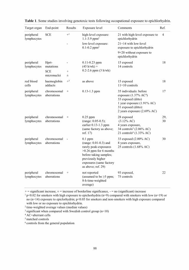
88
Table 1. Some studies involving genotoxic tests following occupational exposure to epichlorhydrin.
Target organ End-point Results Exposure level Comments Ref.
peripheral
lymphocytes
SCE +1
-
high-level exposure:
1.1-3.9 ppm2
low-level exposure:
0.1-0.2 ppm2
21 with high-level exposure to
epichlorhydrin
21+14 with low-level
exposure to epichlorhydrin
9+20 without exposure to
epichlorhydrin
4
peripheral
lymphocytes
Hprt-
mutations
SCE
micronuclei
-
+
±
0.11-0.23 ppm
(45 h/wk) +
0.2-2.6 ppm (3 h/wk)
15 exposed
14 controls
18
red blood
cells
haemoglobin
adducts
+3 as above 15 exposed
11+10 controls
18
peripheral
lymphocytes
chromosomal
aberrations
+ 0.13-1.3 ppm
35 individuals: before
exposure (1.37% AC4)
33 exposed (ditto)
1 year exposure (1.91% AC)
31 exposed (ditto):
2 years exposure (2.69% AC)
17
peripheral
lymphocytes
chromosomal
aberrations
+ 0.25 ppm
(range: 0.05-0.5);
earlier 0.13-1.3 ppm
(same factory as above;
ref. 17)
28 exposed
(3.12% AC)
4 years exposure,
34 controls5 (2.06% AC)
21 controls6 (1.33% AC)
29,
30
peripheral
lymphocytes
chromosomal
aberrations
- 0.1 ppm
(range: 0.01-0.3) and
rarely peak-exposures
>0.26 ppm for 6 months
before taking samples;
previously higher
exposures (same factory
as above; ref. 29)
33 exposed (2.00% AC)
8 years exposure,
25 controls (1.68% AC)
30
peripheral
lymphocytes
chromosomal
aberrations
+ not reported
(assumed to be ≤5 ppm;
8-h time-weighted
average)
93 exposed,
75 controls
22
+ = significant increase, ± = increase of borderline significance, - = no (significant) increase 1 p<0.02 for smokers with high exposure to epichorhydrin (n=9) compared with smokers with low (n=19) or
no (n=14) exposure to epiclorhydrin; p<0.05 for smokers and non-smokers with high exposure compared
with low or no exposure to epichlorhydrin. 2 time-weighted average values (median values) 3 significant when compared with Swedish control group (n=10) 4 AC=aberrant cells 5 matched controls 6 controls from the general population
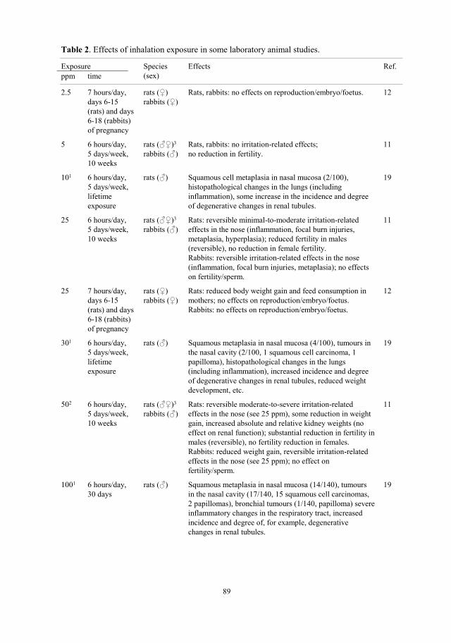
89
Table 2. Effects of inhalation exposure in some laboratory animal studies.
Exposure Species
(sex)
Effects Ref.
ppm time
2.5 7 hours/day,
days 6-15
(rats) and days
6-18 (rabbits)
of pregnancy
rats (♀)
rabbits (♀)
Rats, rabbits: no effects on reproduction/embryo/foetus. 12
5 6 hours/day,
5 days/week,
10 weeks
rats (♂♀)3
rabbits (♂)
Rats, rabbits: no irritation-related effects;
no reduction in fertility.
11
101 6 hours/day,
5 days/week,
lifetime
exposure
rats (♂) Squamous cell metaplasia in nasal mucosa (2/100),
histopathological changes in the lungs (including
inflammation), some increase in the incidence and degree
of degenerative changes in renal tubules.
19
25 6 hours/day,
5 days/week,
10 weeks
rats (♂♀)3
rabbits (♂)
Rats: reversible minimal-to-moderate irritation-related
effects in the nose (inflammation, focal burn injuries,
metaplasia, hyperplasia); reduced fertility in males
(reversible), no reduction in female fertility.
Rabbits: reversible irritation-related effects in the nose
(inflammation, focal burn injuries, metaplasia); no effects
on fertility/sperm.
11
25 7 hours/day,
days 6-15
(rats) and days
6-18 (rabbits)
of pregnancy
rats (♀)
rabbits (♀)
Rats: reduced body weight gain and feed consumption in
mothers; no effects on reproduction/embryo/foetus.
Rabbits: no effects on reproduction/embryo/foetus.
12
301 6 hours/day,
5 days/week,
lifetime
exposure
rats (♂) Squamous metaplasia in nasal mucosa (4/100), tumours in
the nasal cavity (2/100, 1 squamous cell carcinoma, 1
papilloma), histopathological changes in the lungs
(including inflammation), increased incidence and degree
of degenerative changes in renal tubules, reduced weight
development, etc.
19
502 6 hours/day,
5 days/week,
10 weeks
rats (♂♀)3
rabbits (♂)
Rats: reversible moderate-to-severe irritation-related
effects in the nose (see 25 ppm), some reduction in weight
gain, increased absolute and relative kidney weights (no
effect on renal function); substantial reduction in fertility in
males (reversible), no fertility reduction in females.
Rabbits: reduced weight gain, reversible irritation-related
effects in the nose (see 25 ppm); no effect on
fertility/sperm.
11
1001 6 hours/day,
30 days
rats (♂) Squamous metaplasia in nasal mucosa (14/140), tumours
in the nasal cavity (17/140, 15 squamous cell carcinomas,
2 papillomas), bronchial tumours (1/140, papilloma) severe
inflammatory changes in the respiratory tract, increased
incidence and degree of, for example, degenerative
changes in renal tubules.
19
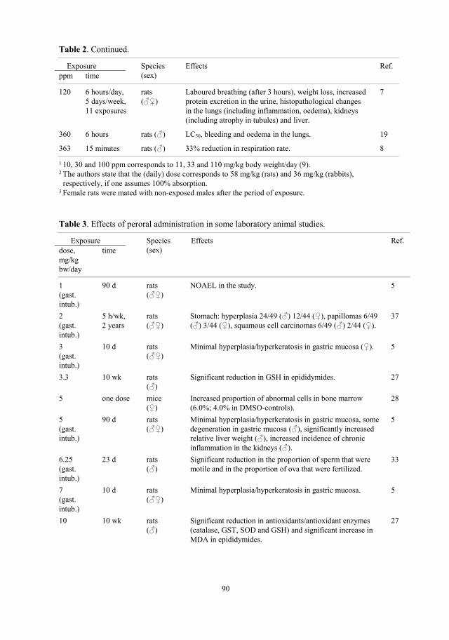
90
Table 2. Continued.
Exposure Species
(sex)
Effects Ref.
ppm time
120 6 hours/day,
5 days/week,
11 exposures
rats
(♂♀)
Laboured breathing (after 3 hours), weight loss, increased
protein excretion in the urine, histopathological changes
in the lungs (including inflammation, oedema), kidneys
(including atrophy in tubules) and liver.
7
360 6 hours rats (♂) LC50, bleeding and oedema in the lungs. 19
363 15 minutes rats (♂) 33% reduction in respiration rate. 8
1 10, 30 and 100 ppm corresponds to 11, 33 and 110 mg/kg body weight/day (9). 2 The authors state that the (daily) dose corresponds to 58 mg/kg (rats) and 36 mg/kg (rabbits),
respectively, if one assumes 100% absorption. 3 Female rats were mated with non-exposed males after the period of exposure.
Table 3. Effects of peroral administration in some laboratory animal studies.
Exposure Species
(sex)
Effects Ref.
dose,
mg/kg
bw/day
time
1
(gast.
intub.)
90 d rats
(♂♀)
NOAEL in the study. 5
2
(gast.
intub.)
5 h/wk,
2 years
rats
(♂♀)
Stomach: hyperplasia 24/49 (♂) 12/44 (♀), papillomas 6/49
(♂) 3/44 (♀), squamous cell carcinomas 6/49 (♂) 2/44 (♀).
37
3
(gast.
intub.)
10 d rats
(♂♀)
Minimal hyperplasia/hyperkeratosis in gastric mucosa (♀). 5
3.3 10 wk rats
(♂)
Significant reduction in GSH in epididymides. 27
5 one dose mice
(♀)
Increased proportion of abnormal cells in bone marrow
(6.0%; 4.0% in DMSO-controls).
28
5
(gast.
intub.)
90 d rats
(♂♀)
Minimal hyperplasia/hyperkeratosis in gastric mucosa, some
degeneration in gastric mucosa (♂), significantly increased
relative liver weight (♂), increased incidence of chronic
inflammation in the kidneys (♂).
5
6.25
(gast.
intub.)
23 d rats
(♂)
Significant reduction in the proportion of sperm that were
motile and in the proportion of ova that were fertilized.
33
7
(gast.
intub.)
10 d rats
(♂♀)
Minimal hyperplasia/hyperkeratosis in gastric mucosa. 5
10 10 wk rats
(♂)
Significant reduction in antioxidants/antioxidant enzymes
(catalase, GST, SOD and GSH) and significant increase in
MDA in epididymides.
27
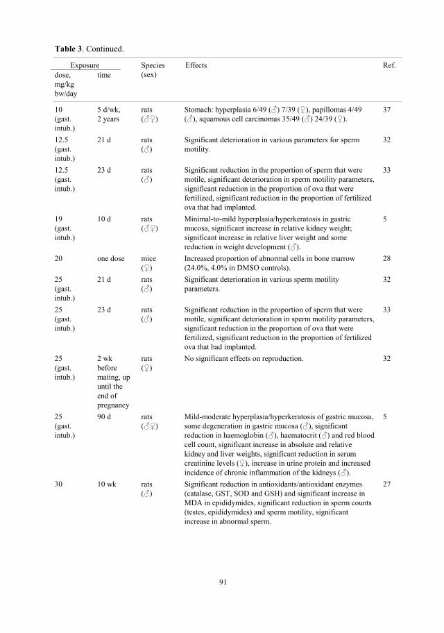
91
Table 3. Continued.
Exposure Species
(sex)
Effects Ref.
dose,
mg/kg
bw/day
time
10
(gast.
intub.)
5 d/wk,
2 years
rats
(♂♀)
Stomach: hyperplasia 6/49 (♂) 7/39 (♀), papillomas 4/49
(♂), squamous cell carcinomas 35/49 (♂) 24/39 (♀).
37
12.5
(gast.
intub.)
21 d rats
(♂)
Significant deterioration in various parameters for sperm
motility.
32
12.5
(gast.
intub.)
23 d rats
(♂)
Significant reduction in the proportion of sperm that were
motile, significant deterioration in sperm motility parameters,
significant reduction in the proportion of ova that were
fertilized, significant reduction in the proportion of fertilized
ova that had implanted.
33
19
(gast.
intub.)
10 d rats
(♂♀)
Minimal-to-mild hyperplasia/hyperkeratosis in gastric
mucosa, significant increase in relative kidney weight;
significant increase in relative liver weight and some
reduction in weight development (♂).
5
20 one dose mice
(♀)
Increased proportion of abnormal cells in bone marrow
(24.0%, 4.0% in DMSO controls).
28
25
(gast.
intub.)
21 d rats
(♂)
Significant deterioration in various sperm motility
parameters.
32
25
(gast.
intub.)
23 d rats
(♂)
Significant reduction in the proportion of sperm that were
motile, significant deterioration in sperm motility parameters,
significant reduction in the proportion of ova that were
fertilized, significant reduction in the proportion of fertilized
ova that had implanted.
33
25
(gast.
intub.)
2 wk
before
mating, up
until the
end of
pregnancy
rats
(♀)
No significant effects on reproduction. 32
25
(gast.
intub.)
90 d rats
(♂♀)
Mild-moderate hyperplasia/hyperkeratosis of gastric mucosa,
some degeneration in gastric mucosa (♂), significant
reduction in haemoglobin (♂), haematocrit (♂) and red blood
cell count, significant increase in absolute and relative
kidney and liver weights, significant reduction in serum
creatinine levels (♀), increase in urine protein and increased
incidence of chronic inflammation of the kidneys (♂).
5
30 10 wk rats
(♂)
Significant reduction in antioxidants/antioxidant enzymes
(catalase, GST, SOD and GSH) and significant increase in
MDA in epididymides, significant reduction in sperm counts
(testes, epididymides) and sperm motility, significant
increase in abnormal sperm.
27
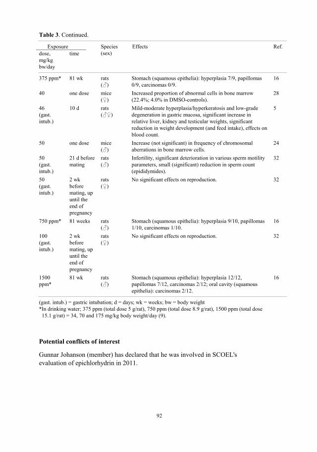
92
Table 3. Continued.
Exposure Species
(sex)
Effects Ref.
dose,
mg/kg
bw/day
time
375 ppm* 81 wk rats
(♂)
Stomach (squamous epithelia): hyperplasia 7/9, papillomas
0/9, carcinomas 0/9.
16
40 one dose mice
(♀)
Increased proportion of abnormal cells in bone marrow
(22.4%; 4.0% in DMSO-controls).
28
46
(gast.
intub.)
10 d rats
(♂♀)
Mild-moderate hyperplasia/hyperkeratosis and low-grade
degeneration in gastric mucosa, significant increase in
relative liver, kidney and testicular weights, significant
reduction in weight development (and feed intake), effects on
blood count.
5
50 one dose mice
(♂)
Increase (not significant) in frequency of chromosomal
aberrations in bone marrow cells.
24
50
(gast.
intub.)
21 d before
mating
rats
(♂)
Infertility, significant deterioration in various sperm motility
parameters, small (significant) reduction in sperm count
(epididymides).
32
50
(gast.
intub.)
2 wk
before
mating, up
until the
end of
pregnancy
rats
(♀)
No significant effects on reproduction. 32
750 ppm* 81 weeks rats
(♂)
Stomach (squamous epithelia): hyperplasia 9/10, papillomas
1/10, carcinomas 1/10.
16
100
(gast.
intub.)
2 wk
before
mating, up
until the
end of
pregnancy
rats
(♀)
No significant effects on reproduction. 32
1500
ppm*
81 wk rats
(♂)
Stomach (squamous epithelia): hyperplasia 12/12,
papillomas 7/12, carcinomas 2/12; oral cavity (squamous
epithelia): carcinomas 2/12.
16
(gast. intub.) = gastric intubation; d = days; wk = weeks; bw = body weight
*In drinking water; 375 ppm (total dose 5 g/rat), 750 ppm (total dose 8.9 g/rat), 1500 ppm (total dose
15.1 g/rat) = 34, 70 and 175 mg/kg body weight/day (9).
Potential conflicts of interest
Gunnar Johanson (member) has declared that he was involved in SCOEL's
evaluation of epichlorhydrin in 2011.
93
References
1. ACGIH. Epichlorohydrin. Documentation of the Threshold Limit Values and Biological
Exposure Indices. 7th ed. Cincinnati, Ohio: American Conference of Governmental Industrial
Hygienists, 2001:7 pp.
2. Amoore JE, Hautala E. Odor as an aid to chemical safety: odor thresholds compared with
threshold limit values and volatilities for 214 industrial chemicals in air and water dilution.
J Appl Toxicol 1983;3:272-290.
3. Buckley LA, Jiang XZ, James RA, Morgan KT, Barrow CS. Respiratory tract lesions induced
by sensory irritants at the RD50 concentration. Toxicol Appl Pharmacol 1984;74:417-429.
4. Cheng TJ, Hwang SJ, Kuo HW, Luo JC, Chang MJW. Exposure to epichlorohydrin and
dimethylformamide, glutathione S-transferases and sister chromatid exchange frequencies in
peripheral lymphocytes. Arch Toxicol 1999;73:282-287.
5. Daniel FB, Robinson M, Olson GR, Page NP. Toxicity studies of epichlorohydrin in Sprague-
Dawley rats. Drug Chem Toxicol 1996;19:41-58.
6. DECOS (Dutch Expert Committee on Occupational Standards). Epichlorohydrin. Health
based calculated occupational cancer risk values. The Hague: Health Council of the
Netherlands, 2000; publication no. 2000/10OSH.
7. Gage JC. The toxicity of epichlorohydrin vapour. Brit J Ind Med 1959;16:11-14.
8. Gardner RJ, Burgess BA, Kennedy GL. Sensory irritation potential of selected nasal
tumorigens in the rat. Food Chem Toxicol 1985;23:87-92.
9. Ginsberg GL, Pepelko WE, Goble RL, Hattis DB. Comparison of contact site cancer potency
across dose routes. Case study with epichlorohydrin. Risk Analysis 1996;16:667-681.
10. IARC. Re-evaluation of some organic chemicals, hydrazine and hydrogen peroxide. IARC
Monographs on the Evaluation of Carcinogenic Risks to Humans. Vol. 71. Lyon:
International Agency for Research on Cancer 1999;71:603-628.
11. John JA, Quast JF, Murray FJ, Calhoun LG, Staples RE. Inhalation toxicity of
epichlorohydrin: Effects on fertility in rats and rabbits. Toxicol Appl Pharmacol 1983;68:415-
423.
12. John JA, Gushow TS, Ayres JA, Hanley TR, Quast JF, Rao KS. Teratologic evaluation of
inhaled epichlorohydrin and allyl chloride in rats and rabbits. Fund Appl Toxicol 1983;3:437-
442.
13. Klinefelter GR, Laskey JW, Ferrell J, Suarez JD, Roberts NL. Discriminant analysis indicates
a single sperm protein (SP22) is predictive of fertility following exposure to epididymal
toxicants. J Androl 1997;18:139-150.
14. Kolman A, Dušinská M. Comparison of propylene oxide and epichlorohydrin effects in two
transformation tests (C3H/10T1/2 and SHE cells). Toxicol Lett 1995;81:213-221.
15. Kolman A, Chovanec M, Osterman-Golkar S. Genotoxic effects of ethylene oxide, propylene
oxide and epichlorohydrin in humans: update review (1990-2001). Mutat Res 2002;512:173-
194.
16. Konishi Y, Kawabata A, Denda A, Ikeda T, Katada H, Maruyama H, Higashiguchi R.
Forestomach tumors induced by orally administered epichlorohydrin in male Wistar rats.
Gann 1980;71:922-923.
17. Kučerová M, Zhurkov VS, Polívková Z, Ivanova JE. Mutagenic effects of epichlorohydrin.
Mutat Res 1977;48:355-360.
18. Landin Hindsø H, Grummt T, Laurent C, Tates A. Monitoring of occupational exposure to
epichlorohydrin by genetic effects and hemoglobin adducts. Mutat Res 1997;381:217-226.
19. Laskin S, Sellakumar AR, Kuschner M, Nelson N, La Mendola S, Rusch GM, Katz GV,
Dulak NC, Albert RE. Inhalation carcinogenicity of epichlorohydrin in noninbred Sprague-
Dawley rats. JNCI 1980;65:751-757.
94
20. Luo JC, Kuo HW, Cheng TJ, Chang MJW. Pulmonary function abnormality and respiratory
tract irritation symptoms in epichlorohydrin-exposed workers in Taiwan. Am J Ind Med
2003;43:440-446.
21 Luo JC, Cheng TJ, Kuo HW, Chang MJW. Decreased lung function associated with
occupational exposure to epichlorohydrin and the modification effects of glutathione S-
transferase polymorphisms. J Occup Environ Med 2004;46:280-286.
22. Picciano D. Cytogenetic investigation of occupational exposure to epichlorohydrin. Mutat Res
1979;66:169-173.
23. Prens EP, de Jong G, van Joost T. Sensitization to epichlorohydrin and epoxy system
components. Contact Dermatitis 1986;15:85-90.
24. Rossi AM, Migliore L, Lascialfari D, Sbrana I, Loprieno N, Tortoreto M, Bidoli F, Pantarotto
C. Genotoxicity, metabolism and blood kinetics of epichlorohydrin in mice. Mutat Res
1983;118:213-226.
25. SCOEL. Recommendation from the Scientific Committee on Occupational Exposure Limits
for epichlorohydrin. SCOEL/SUM/169, September 2011.
26. Seidegård J, Pero RW. The genetic variation and the expression of human glutathione
transferase µ. Klin Wochenschr 1988;66 Suppl 11:125-126.
27. Shin IS, Lim JH, Kim SH, Kim KH, Park NH, Bae CS, Kang SS, Moon C, Kim SH, Jun W,
Kim JC. Induction of oxidative stress in the epididymis of rats after subchronic exposure to
epichlorohydrin. Bull Environ Contam Toxicol 2010;84:667-671.
28. Šrám RJ, Černá M, Kučerová M. The genetic risk of epichlorohydrin as related to the
occupational exposure. Biol Zbl 1976;95:451-462.
29. Srám RJ, Zudová Z, Kuleshov NP. Cytogenetic analysis of peripheral lymphocytes in workers
occupationally exposed to epichlorohydrin. Mutat Res 1980;70:115-120.
30. Srám RJ, Landa L, Samková I. Effect of occupational exposure to epichlorohydrin on the
frequency of chromosome aberrations in peripheral lymphocytes. Mutat Res 1983;122:59-64.
31. Thorgeirsson A, Fregert S. Allergenicity of epoxy resins in the guinea pig. Acta Derm
Venereol 1977;57:253-256.
32. Toth GP, Zenick H, Smith MK. Effects of epichlorohydrin on male and female reproduction
in Long-Evans rats. Fund Appl Toxicol 1989;13:16-25.
33. Toth GP, Stober JA, Zenick H, Read EJ, Christ SA, Smith MK. Correlation of sperm motion
parameters with fertility in rats treated subchronically with epichlorohydrin. J Androl
1991;12:54-61.
34. Van Joost Th. Occupational sensitization to epichlorohydrin and epoxy resin. Contact
Dermatitis 1988;19:278-280.
35. Van Joost Th, Roesyanto ID, Satyawan I. Occupational sensitization to epichlorohydrin
(ECH) and bisphenol-A during the manufacture of epoxy resin. Contact Dermatitis
1990;22:125-126.
36. Waechter JM, Veenstra GE. Epoxy compounds - aromatic diglycidyl ethers, polyglycidyl
ethers, glycidyl esters, and miscellaneous epoxy compounds. In: Bingham E, Cohrssen B,
Powell CH, eds. Patty´s Toxicology, vol 6, 5th ed. New York: John Wiley & Sons inc,
2001:1087-1145.
37. Wester PW, van der Heijden CA, Bisschop A, van Esch GJ. Carcinogenicity study with
epichlorohydrin (CEP) by gavage in rats. Toxicology 1985;36:325-339.
95
Summary
Montelius J (ed). Swedish Criteria Group for Occupational Standards. Scientific Basis for Swedish Occupational Standards. XXXIII. Arbete och Hälsa 2014;48(3):1-103. University of Gothenburg, Sweden.
Critical review and evaluation of those scientific data which are relevant as a background for discussion of Swedish occupational exposure limits. This volume consists of the consensus reports given by the Criteria Group at the Swedish Work Environmental Authority from June, 2012 through October, 2013.
Key Words: Crystalline Silica, Consensus report, Epichlorohydrin, N-Methyl-2-
pyrrolidone, Occupational exposure limit (OEL), Quartz, Risk assessment, Scientific basis, Toxicology.
Sammanfattning
Montelius J (ed). Kriteriegruppen för hygieniska gränsvärden. Vetenskapligt underlag för hygieniska gränsvärden. XXXIII. Arbete och Hälsa 2014;48(3):1-103. Göteborgs Universitet.
Sammanställningar baserade på kritisk genomgång och värdering av de vetenskapliga fakta, vilka är relevanta som underlag för fastställande av hygieniskt gränsvärde. Volymen omfattar de underlag som avgivits från Kriteriegruppen för hygieniska gränsvärden under perioden juni 2012 – oktober 2013.
Nyckelord: Epiklorhydrin, Hygieniskt gränsvärde, Kristallin kiseldioxid, Kvarts, N-Metyl-2-pyrrolidon, Riskvärdering, Toxikologi, Vetenskapligt underlag.
En svensk version av dessa vetenskapliga underlag finns publicerad i Arbete och Hälsa 2013;47(8):1-101.

96
97
APPENDIX
Consensus reports in this and previous volumes
Substance Consensus date
Published in Arbete och Hälsa year;volume(No)
No. in series of Consensus Reports
Acetaldehyde February 17, 1987 1987;39 VIII Acetamide December 11, 1991 1992;47 XIII Acetic acid June15, 1988 1988;32 IX Acetone October 20, 1987 1988;32 IX Acetonitrile September 12, 1989 1991;8 XI Acrylamide April 17, 1991 1992;6 XII Acrylates December 9, 1984 1985;32 VI Acrylonitrile April 28, 1987 1987;39 VIII Aliphatic amines August 25, 1982 1983;36 IV Aliphatic hydrocarbons, C10-C15 June 1, 1983 1983;36 IV Aliphatic monoketons September 5, 1990 1992;6 XII Allyl alcohol September 9, 1986 1987;39 VIII Allylamine August 25, 1982 1983;36 IV Allyl chloride June 6, 1989 1989;32 X Aluminum April 21, 1982 1982;24 III revised September 14, 1994 1995;19 XVI Aluminum trifluoride September 15, 2004 2005;17 XXVI p-Aminoazobenzene February 29, 1980 1981;21 I Ammonia April 28, 1987 1987;39 VIII revised October 24, 2005 2006;11 XXVII Ammonium fluoride September 15, 2004 2005;17 XXVI Amylacetate March 23, 1983 1983;36 IV revised June 14, 2000 2000;22 XXI Aniline October 26, 1988 1989;32 X Anthraquinone November 26, 1987 1988;32 IX Antimony + compounds December 8, 1999 2000;22 XXI Arsenic, inorganic December 9, 1980 1982;9 II revised February 15, 1984 1984;44 V Arsine October 20, 1987 1988;32 IX Asbestos October 21, 1981 1982;24 III Asphalt fumes April 14, 2010 2011;45(6) XXXI
Barium June 16, 1987 1987;39 VIII revised January 26, 1994 1994;30 XV Benzene March 4, 1981 1982;9 II revised February 24, 1988 1988;32 IX Benzoyl peroxide February 13, 1985 1985;32 VI Beryllium April 25, 1984 1984;44 V Bitumen fumes April 14, 2010 2011;45(6) XXXI Borax October 6, 1982 1983;36 IV Boric acid October 6, 1982 1983;36 IV Boron Nitride January 27, 1993 1993;37 XIV Butadiene October 23, 1985 1986;35 VII
98
1-Butanol June 17, 1981 1982;24 III Butanols June 6, 1984 1984;44 V Butyl acetate June 6, 1984 1984;44 V Butyl acetates February 11, 1998 1998;25 XIX n-Butyl acrylate September 28, 2011 2013;47(6) XXXII Butylamine August 25, 1982 1983;36 IV Butyl glycol October 6, 1982 1983;36 IV γ-Butyrolactone June 2, 2004 2005;7 XXV
Cadmium January 18, 1980 1981;21 I revised February 15, 1984 1984;44 V revised May 13, 1992 1992;47 XIII revised February 5, 2003 2003;16 XXIV Calcium fluorid September 15, 2004 2005;17 XXVI Calcium hydroxide February 24, 1999 1999;26 XX Calcium nitride January 27, 1993 1993;37 XIV Calcium oxide February 24, 1999 1999;26 XX Caprolactam October 31, 1989 1991;8 XI Carbon dioxide June 15, 2011 2013;47(6) XXXII Carbon monoxide December 9, 1981 1982;24 III Cathecol September 4, 1991 1992;47 XIII Chlorine December 9, 1980 1982;9 II Chlorine dioxide December 9, 1980 1982;9 II Chlorobenzene September 16, 1992 1993;37 XIV revised April 2, 2003 2003;16 XXIV o-Chlorobenzylidene malononitrile June 1, 1994 1994;30 XV Chlorocresol December 12, 1990 1992;6 XII Chlorodifluoromethane June 2, 1982 1982; 24 III Chlorophenols September 4, 1985 1986;35 VII Chloroprene April 16, 1986 1986;35 VII Chromium December 14, 1979 1981;21 I revised May 26, 1993 1993;37 XIV revised May 24, 2000 2000;22 XXI Chromium trioxide May 24, 2000 2000;22 XXI Coal dust September 9, 1986 1987;39 VIII Cobalt October 27, 1982 1983;36 IV Cobalt and cobalt compounds October 22, 2003 2005;7 XXV Copper October 21, 1981 1982;24 III Cotton dust February14, 1986 1986;35 VII Creosote October 26, 1988 1989;32 X revised December 5, 2007 2009;43(4) XXIX Cresols February 11, 1998 1998;25 XIX Crystalline Silica December 31, 2012 2014;48(3) XXXIII Cumene June 2, 1982 1982;24 III Cyanamid September 30, 1998 1999;26 XX Cyanoacrylates March 5, 1997 1997;25 XVIII Cycloalkanes, C5-C15 April 25, 1984 1984;44 V Cyclohexanone March 10, 1982 1982;24 III revised February 24, 1999 1999;26 XX Cyclohexanone peroxide February 13, 1985 1985;32 VI Cyclohexylamine February 7, 1990 1991;8 XI
Desflurane May 27, 1998 1998;25 XIX
99
Diacetone alcohol December 14, 1988 1989;32 X Dichlorobenzenes February 11, 1998 1998;25 XIX 1,2-Dibromo-3-chloropropane May 30, 1979 1981;21 I Dichlorodifluoromethane June 2, 1982 1982;24 III 1,2-Dichloroethane February 29, 1980 1981;21 I Dichloromethane February 29, 1980 1981;21 I Dicumyl peroxide February 13, 1985 1985;32 VI Dicyclopentadiene March 23, 1994 1994;30 XV Diesel exhaust December 4, 2002 2003;16 XXIV Diethanolamine September 4, 1991 1992;47 XIII Diethylamine August 25, 1982 1983;36 IV revised February 16, 2011 2013;47(6) XXXII 2-Diethylaminoethanol January 25, 1995 1995;19 XVI Diethylene glycol September 16, 1992 1993;37 XIV Diethyleneglycol ethylether + acetate December 11, 1996 1997;25 XVIII Diethyleneglycol methylether + acetate March 13, 1996 1996;25 XVII Diethyleneglycol monobutylether January 25, 1995 1995;19 XVI Diethylenetriamine August 25, 1982 1983;36 IV revised January 25, 1995 1995;19 XVI Diisocyanates April 8, 1981 1982;9 II revised April 27, 1988 1988;32 IX revised May 30, 2001 2001;20 XXII Diisopropylamine February 7, 1990 1991;8 XI N,N-Dimethylacetamide March 23, 1994 1994;30 XV Dimethyl adipate December 9, 1998 1999;26 XX Dimethylamine December 10, 1997 1998;25 XIX N,N-Dimethylaniline December 12, 1989 1991;8 XI Dimethyldisulfide September 9, 1986 1987;39 VIII Dimethylether September 14, 1994 1995;19 XVI Dimethylethylamine June 12, 1991 1992;6 XII Dimethylformamide March 23, 1983 1983;36 IV Dimethyl glutarate December 9, 1998 1999;26 XX Dimethylhydrazine January 27, 1993 1993;37 XIV Dimethyl succinate December 9, 1998 1999;26 XX Dimethylsulfide September 9, 1986 1987;39 VIII Dimethylsulfoxide, DMSO December 11, 1991 1992;47 XIII Dioxane August 25, 1982 1983;36 IV revised March 4, 1992 1992;47 XIII Diphenylamine January 25, 1995 1995;19 XVI 4,4'-Diphenylmethanediisocyanate (MDI) April 8, 1981 1982;9 II revised May 30, 2001 2001;20 XXII Dipropylene glycol May 26, 1993 1993;37 XIV Dipropyleneglycol monomethylether December 12, 1990 1992;6 XII Disulfiram October 31, 1989 1991;8 XI
Epichlorohydrin October 2, 2013 2014;48(3) XXXIII Enzymes, industrial June 5, 1996 1996;25 XVII Ethanol May 30, 1990 1991;8 XI Ethanolamine September 4, 1991 1992;47 XIII revised May 30, 2012 2013;47(6) XXXII Ethylacetate March 28, 1990 1991;8 XI Ethylamine August 25, 1982 1983;36 IV revised February 16, 2011 2013;47(6) XXXII
100
Ethylamylketone September 5, 1990 1992;6 XII Ethylbenzene December 16, 1986 1987;39 VIII Ethylchloride December 11, 1991 1992;47 XIII Ethylene December 11, 1996 1997;25 XVIII Ethylene chloride February 29, 1980 1981;21 I Ethylene diamine August 25, 1982 1983;36 IV Ethylene glycol October 21, 1981 1982;24 III Ethylene glycol ethylether + acetate February 6 2009;43(4) XXIX Ethylene glycol methylether + acetate June 2, 1999 1999;26 XX Ethyleneglycol monoisopropylether November 16, 1994 1995;19 XVI Ethyleneglycol monopropylether + acetate September 15, 1993 1994;30 XV Ethylene oxide December 9, 1981 1982;24 III Ethylenethiourea September 27, 2000 2001;20 XXII Ethylether January 27, 1993 1993;37 XIV Ethylglycol October 6, 1982 1983;36 IV
Ferbam September 12, 1989 1991;8 XI Ferric dimethyldithiocarbamate September 12, 1989 1991;8 XI Flour dust December 10, 1997 1998;25 XIX Fluorides September 15, 2004 2005;17 XXVI Formaldehyde June 30, 1979 1981;21 I revised August 25, 1982 1983;36 IV revised June 9, 2010 2011;45(6) XXXI Formamide December 12, 1989 1991;8 XI Formic acid June 15, 1988 1988;32 IX Furfural April 25, 1984 1984;44 V Furfuryl alcohol February 13, 1985 1985;32 VI
Gallium + Gallium compounds January 25, 1995 1995;19 XVI Glutaraldehyde September 30, 1998 1999;26 XX Glycol ethers October 6, 1982 1983;36 IV Glyoxal September 13, 1996 1996;25 XVII Grain dust December 14, 1988 1989;32 X revised February 4, 2009 2010;44(5) XXX Graphite December 10, 1997 1998;25 XIX
Halothane April 25, 1985 1985;32 VI 2-Heptanone September 5, 1990 1992;6 XII 3-Heptanone September 5, 1990 1992;6 XII Hexachloroethane September 15, 1993 1994;30 XV Hexamethylenediisocyanate (HDI) April 8, 1981 1982;9 II revised May 30, 2001 2001;20 XXII Hexamethylenetetramine August 25, 1982 1983;36 IV n-Hexanal March 29, 2006 2006;11 XXVII n-Hexane January 27, 1982 1982;24 III 2-Hexanone September 5, 1990 1992;6 XII Hexyleneglycol November 17, 1993 1994;30 XV Hydrazine May 13, 1992 1992;47 XIII Hydrochloric acid June 3, 2009 2010;44(5) XXX Hydrogen bromide February 11, 1998 1998;25 XIX Hydrogen cyanide February 7, 2001 2001;20 XXII Hydrogen fluoride April 25, 1984 1984;44 V revised September 15, 2004 2005;17 XXVI
101
Hydrogen peroxide April 4, 1989 1989;32 X Hydrogen sulfide May 4, 1983 1983;36 IV Hydroquinone October 21, 1989 1991;8 XI
Indium March 23, 1994 1994;30 XV Industrial enzymes June 5, 1996 1996;25 XVII Isocyanic Acid (ICA) December 5, 2001 2002;19 XXIII Isophorone February 20, 1991 1992;6 XII Isopropanol December 9, 1981 1982;24 III Isopropylamine February 7, 1990 1991;8 XI Isopropylbenzene June 2, 1982 1982;24 III
Lactates March 29, 1995 1995;19 XVI Lactate esters June 2, 1999 1999;26 XX Laughing gas June 7, 2006 2006;11 XXVII Lead, inorganic February 29, 1980 1981;21 I revised September 5, 1990 1992;6 XII revised December 8, 2004 2005;17 XXVI Lithium and lithium compounds June 4, 2003 2003;16 XXIV Lithium boron nitride January 27, 1993 1993;37 XIV Lithium nitride January 27, 1993 1993;37 XIV
Maleic anhydride September 12, 1989 1991;8 XI Manganese February 15, 1983 1983;36 IV revised April 17, 1991 1992;6 XII revised June 4, 1997 1997;25 XVIII Man made mineral fibers March 4, 1981 1982;9 II revised December 1, 1987 1988;32 IX Mercury, inorganic April 25, 1984 1984;44 V Mesityl oxide May 4, 1983 1983;36 IV Metal stearates, some September 15, 1993 1994;30 XV Methacrylates September 12, 1984 1985;32 VI Methanol April 25, 1985 1985;32 VI Methyl acetate March 28, 1990 1991;8 XI Methylamine August 25, 1982 1983;36 IV Methylamyl alcohol March 17, 1993 1993;37 XIV Methyl bromide April 27, 1988 1988;32 IX Methyl chloride March 4, 1992 1992;47 XIII Methyl chloroform March 4, 1981 1982;9 II 4,4´-methylene-bis-(2-chloroaniline) February 4, 2004 2005;7 XXV Methylene chloride February 29, 1980 1981;21 I 4,4'-Methylene dianiline June 16, 1987 1987;39 VIII revised October 3, 2001 2002;19 XXIII Methyl ethyl ketone February 13, 1985 1985;32 VI Methyl ethyl ketone peroxide February 13, 1985 1985;32 VI Methyl formate December 12, 1989 1991;8 XI Methyl glycol October 6, 1982 1983;36 IV Methyl iodide June 30, 1979 1981;21 I Methylisoamylamine September 5, 1990 1992;6 XII Methylisoamylketone February 6, 2002 2002;19 XXIII Methylisocyanate (MIC) December 5, 2001 2002;19 XXIII Methyl mercaptane September 9, 1986 1987;39 VIII Methyl methacrylate March 17, 1993 1993;37 XIV
102
Methyl pyrrolidone June 16, 1987 1987;39 VIII revised December 5, 2012 2014;48(3) XXXIII α-Methylstyrene November 1, 2000 2001;20 XXII Methyl-t-butyl ether November 26, 1987 1988;32 IX revised September 30, 1998 1999;26 XX Mixed solvents, neurotoxicity April 25, 1985 1985;32 VI MOCA February 4, 2004 2005;7 XXV Molybdenum October 27, 1982 1983;36 IV revised Februari 4, 2009 2010;44(5) XXX Monochloroacetic acid February 20, 1991 1992;6 XII Monochlorobenzene September 16, 1993 1993;37 XIV Monomethylhydrazine March 4, 1992 1992;47 XIII Mononitrotoluene February 20, 1991 1992;6 XII Monoterpenes February 17, 1987 1987;39 VIII Morpholine December 8, 1982 1983;36 IV revised June 5, 1996 1996;25 XVII
Naphthalene May 27, 1998 1998;25 XIX Natural crystallinic fibers, except asbestos June 12, 1991 1992;6 XII Nickel April 21, 1982 1982;24 III Nicotine June 2, 2004 2005;7 XXV Nitric acid June 3, 2009 2010;44(5) XXX Nitric oxide December 11, 1985 1986;35 VII revised June 13, 2007 2008;42(6) XXVIII Nitroethane April 4, 1989 1989;32 X Nitrogen dioxide December 11, 1985 1986;35 VII revised September 12, 2007 2008;42(6) XXVIII Nitrogen oxides December 11, 1985 1986;35 VII Nitroglycerin February 13, 1985 1985;32 VI Nitroglycol February 13, 1985 1985;32 VI Nitromethane January 6, 1989 1989;32 X Nitropropane October 28, 1986 1987;39 VIII 2-Nitropropane March 29, 1995 1995;19 XVI Nitroso compounds December 12, 1990 1992;6 XII Nitrosomorpholine December 8, 1982 1983;36 IV Nitrotoluene February 20, 1991 1992;6 XII Nitrous oxide December 9, 1981 1982;24 III revised June 7, 2006 2006;11 XXVII
Oil mist April 8, 1981 1982;9 II Organic acid anhydrides, some September 12, 1989 1991;8 XI revised June 4, 2008 2009;43(4) XXIX revised September 29, 2010 2011;45(6) XXXI Oxalic acid February 24, 1988 1988;32 IX Ozone April 28, 1987 1987;39 VIII revised February 7, 2007 2008;42(6) XXVIII
Paper dust February 7, 1990 1991;8 XI Penicillins November 23, 2005 2006;11 XXVII Pentaerythritol November 16, 1994 1995;19 XVI 1,1,1,2,2-Pentafluoroethane February 24, 1999 1999;26 XX Pentyl acetate June 14, 2000 2000;22 XXI Peroxides, organic February 13, 1985 1985;32 VI
103
Phenol February 13, 1985 1985;32 VI Phosphoric acid June 3, 2009 2010;44(5) XXX Phosphorous chlorides September 30, 1998 1999;26 XX Phosphorous oxides February 11, 1998 1998;25 XIX Phthalates December 8, 1982 1983;36 IV Phthalic anhydride September 12, 1989 1991;8 XI Piperazine September 12, 1984 1985;32 VI Plastic dusts December 16, 1986 1987;39 VIII Platinum June 4, 1997 1997;25 XVIII Polyaromatic hydrocarbons February 15, 1984 1984;44 V Polyisocyanates April 27, 1988 1988;32 IX Potassium aluminium fluoride June 4, 1997 1997;25 XVIII Potassium cyanide February 7, 2001 2001;20 XXII Potassium dichromate May 24, 2000 2000;22 XXI Potassium Fluoride September 15, 2004 2005;17 XXVI Potassium hydroxide Marsh 15, 2000 2000;22 XXI 2-Propanol December 9, 1981 1982;24 III Propene September 13, 1996 1996;25 XVII Propionic acid November 26, 1987 1988;32 IX Propylacetate September 14, 1994 1995;19 XVI Propylene glycol June 6, 1984 1984;44 V Propylene glycol-1,2-dinitrate May 4, 1983 1983;36 IV Propylene glycol monomethylether October 28, 1986 1987;39 VIII Propylene oxide June 11, 1986 1986;35 VII Pyridine May 13, 1992 1992;47 XIII
Quartz March 13, 1996 1996;25 XVII revised December 31, 2012 2014;48(3) XXXIII Resorcinol September 4, 1991 1992;47 XIII
Selenium December 11, 1985 1986;35 VII revised February 22, 1993 1993;37 XIV Sevoflurane May 27, 1998 1998;25 XIX Silica March 13, 1996 1996;25 XVII Silver October 28, 1986 1987;39 VIII Sodium cyanide February 7, 2001 2001;20 XXII Sodium Fluoride September 15, 2004 2005;17 XXVI Sodium hydroxide August 24, 2000 2000;22 XXI Stearates, metallic, some September 15, 1993 1994;30 XV Stearates, non-metallic, some November 17, 1993 1994;30 XV Strontium January 26, 1994 1994;30 XV Styrene February 29, 1980 1981;21 I revised October 31, 1989 1991;8 XI revised April 1, 2009 2010;44(5) XXX Sulfur dioxide April 25, 1985 1985;32 VI Sulfur fluorides March 28, 1990 1991;8 XI Sulfuric acid June 3, 2009 2010;44(5) XXX Synthetic inorganic fibers March 4, 1981 1982;9 II revised December 1, 1987 1988;32 IX revised December 3, 2003 2005;7 XXV Synthetic organic and inorganic fibers May 30, 1990 1991;8 XI
Talc dust June 12, 1991 1992;6 XII
104
Terpenes, mono- February 17, 1987 1987;39 VIII Tetrabromoethane May 30, 1990 1991;8 XI Tetrachloroethane June 4, 1997 1997;25 XVIII Tetrachloroethylene February 29, 1980 1981;21 I 1,1,1,2-Tetrafluoroethane March 29, 1995 1995;19 XVI Tetrahydrofuran October 31, 1989 1991;8 XI Tetranitromethane April 4, 1989 1989;32 X Thioglycolic acid June 1, 1994 1994;30 XV Thiourea December 1, 1987 1988;32 IX revised June 2, 1999 1999;26 XX Thiram October 31, 1989 1991;8 XI Thiurams, some October 31, 1989 1991;8 XI Tin and inorganic tin compounds October 22, 2003 2005;7 XXV Titanium dioxide February 21, 1989 1989;32 X Toluene February 29, 1980 1981;21 I revised February 6, 2002 2002;19 XXIII Toluene-2,4-diamine November 1, 2000 2001;20 XXII Toluene-2,6-diamine November 1, 2000 2001;20 XXII Toluene-2,4-diisocyanate April 8, 1981 1982;9 II revised May 30, 2001 2001;20 XXII Toluene-2,6-diisocyanate April 8, 1981 1982;9 II revised May 30, 2001 2001;20 XXII 1,1,1-Trifluoroethane February 24, 1999 1999;26 XX Trichlorobenzene September 16, 1993 1993;37 XIV 1,1,1-Trichloroethane March 4, 1981 1982;9 II Trichloroethylene December 14, 1979 1981;21 I Trichlorofluoromethane June 2, 1982 1982;24 III 1,1,2-Trichloro-1,2,2-trifluoroethane June 2, 1982 1982;24 III Triethanolamine August 25, 1982 1983;36 IV revised October 23, 2002 2003;16 XXIV Triethylamine December 5, 1984 1985;32 VI Trimellitic anhydride September 12, 1989 1991;8 XI Trimethylolpropane November 16, 1994 1995;19 XVI Trinitrotoluene April 17, 1991 1992;6 XII
Vanadium March 15, 1983 1983;36 IV Vinyl acetate June 6, 1989 1989;32 X Vinyl toluene December 12, 1990 1992;6 XII
White spirit December 16, 1986 1987;39 VIII revised November 13, 2006 2008;42(6) XXVIII Wood dust June 17, 1981 1982;9 II revised June 25, 2000 2000;22 XXI
Xylene February 29, 1980 1981;21 I revised September 14, 2005 2005;17 XXVI
Zinc April 21, 1982 1982;24 III Zinc chromate May 24, 2000 2000;22 XXI Zinc dimethyl dithiocarbamate September 12, 1989 1991;8 XI Ziram September 12, 1989 1991;8 XI
Sent for publication November 2014

2010;44(7). G Aronsson, W Astvik och K Gustafsson. Arbetsvillkor, återhämtning och hälsa - en studie av förskola, hemtjänst och socialtjänst.
2010;44(8). K Torén, M Albin och B Järvholm (Ed.) Systematiska kunskapsöversikter; 1. Betydelsen av fukt och mögel i inomhusmiljö för astma hos vuxna.
2010;44(9). C Wulff, P Lindfors och M Sverke. Hur förhåller sig begåvning i skolåldern och psykosocial arbetsbelastning i vuxenlivet till olika aspekter av självrapporterad hälsa bland yrkesarbetande kvinnor och män?
2010;44(10). H Kantelius Inhyrningens logik Lång-tidsinhyrda arbetare och tjänstemäns utvecklings-möjligheter och upplevda anställningsbarhet.
2011;45(1). E Tengelin, A Kihlman, M Eklöf och L Dellve. Chefskap i sjukvårdsmiljö: Avgränsning och kommunikation av egen stress.
2011;45(2). A Grimby-Ekman. Epidemiological aspects of musculoskeletal pain in the upper body.
2011;45(3). J Montelius (Ed.) Vetenskapligt Underlag för Hygieniska Gränsvärden 31. Kriteriegruppen för hygieniska gränsvärden.
2011;45(4). The Nordic Expert Group for Criteria Documentation of Health Risks from Chemicals and the Dutch Expert Committee on Occupational Safety. 144. Endotoxins.
2011;45(5). M Albin, J Alkan-Olsson, M Bohgard, K Jakobsson, B Karlson, P Lundqvist, M Ottosson, F Rassner, M Svensson and H Tinnerberg. (Ed.) 55th Nordic Work Environment Meeting.The Work Environment – Impact of Technological, Social and Climate Change.
2011;45(6). J Montelius (Ed.) Scientific Basis for Swedish Occupational Standards XXXI. Swedish Criteria Group for Occupational Standards.
2011;45(7). The Nordic Expert Group for Criteria Documentation of Health Risks from Chemicals and the Dutch Expert Committee on Occupational Safety. 145. Aluminium and aluminium compounds.
2012;46(1). B Lindell. The Nordic Expert Group for Criteria Documentation of Health Risks from Chemicals. 146. Polychlorinated biphenyls. (PCBs)
2012:46(2). K Torén, M Albin och B Järvholm. Systematiska kunskapsöversikter; 2. Exponering för helkroppsvibrationer och uppkomst av länd-ryggssjuklighet.
2012:46(3). G Sjögren Lindquist och E Wadensjö. Kunskapsöversikt kring samhälls-ekonomiska kostnader för arbetsskador.
2012;46(4). C Mellner, G Aronsson och G Kecklund. Segmentering och integrering – om mäns och kvinnors gränssättningsstrategier i högkvalificerat arbete.
2012;46(5). T Muhonen. Stress, coping och hälsa under kvinnliga chefers och specialisters karriärer.
2012;46(6). J Montelius (Ed.) Vetenskapligt Underlag för Hygieniska Gränsvärden 32. Kriteriegruppen för hygieniska gränsvärden.
2012;46(7). H Stockmann-Juvala. The Nordic Expert Group for Criteria Documentation of Health Risks from Chemicals. 147. Carbon monoxide.
2013;47(1). I Lundberg, P Allebeck, Y Forsell och P Westerholm. Systematiska kunskapsöversikter; 3. Kan arbetsvillkor orsaka depressionstillstånd? En systematisk översikt över longitudinella studier i den vetenskapliga litteraturen 1998-2012.
2013;47(2). K Elgstrand and E Vingård (Ed.) Occupational Safety and Health in Mining. Anthology on the situation in 16 mining countries.
2013;47(3). A Knutsson och A Kempe. Systematiska kunskapsöversikter; 4. Diabetes och arbete.
2013;47(4). K Jakobsson och P Gustavsson. Systematiska kunskapsöversikter; 5. Arbetsmiljö-exponeringar och stroke – en kritisk granskning av evidens för samband mellan exponeringar i arbets-miljön och stroke.
2013;47(5). M Hedmer, M Kåredal, P Gustavsson and J Rissler. The Nordic Expert Group for Criteria Documentation of Health Risks from Chemicals. 148. Carbon nanotubes.
2013;47(6). J Montelius (Ed.) Scientific Basis for Swedish Occupational Standards XXXII. Swedish Criteria Group for Occupational Standards.
2013;47(7). C Håkansta. Between Science and Politics. Swedish work environment research in a historical perspective.
2013;47(8). J Montelius (Ed.) Vetenskapligt Underlag för Hygieniska Gränsvärden 33. Kriteriegruppen för hygieniska gränsvärden.
2014;48(1). L-G Gunnarsson och L Bodin. Systematiska kunskapsöversikter; 6. Epidemologiskt påvisade samband mellan Parkinsons sjukdom och faktorer i arbetsmiljön.
2014;48(2). L-G Gunnarsson och L Bodin. Systematiska kunskapsöversikter; 7. Epidemiologiskt påvisade samband mellan Amyotrofisk Lateral Skleros (ALS) och faktorer i arbetsmiljön.