Embed Size (px)
Citation preview
Searching for microbesPart XIII.
Parasitology
Ondřej ZahradníčekTo practical of VLLM0421c
For introduction… Picture by: Petr Ondrovčík
Are you sure, that your new clone of giant bed-bug is my favourite birthday
present?
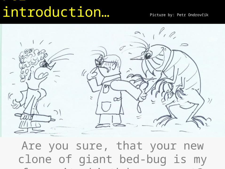
Survey of topics
Parasites – introduction
Parasites – clinical description
Parasites – sampling
Parasites – diagnostic methods
Parasites – pictures
Check-up questionsCheck-up questions
Parasites – introduction
Parasites classification
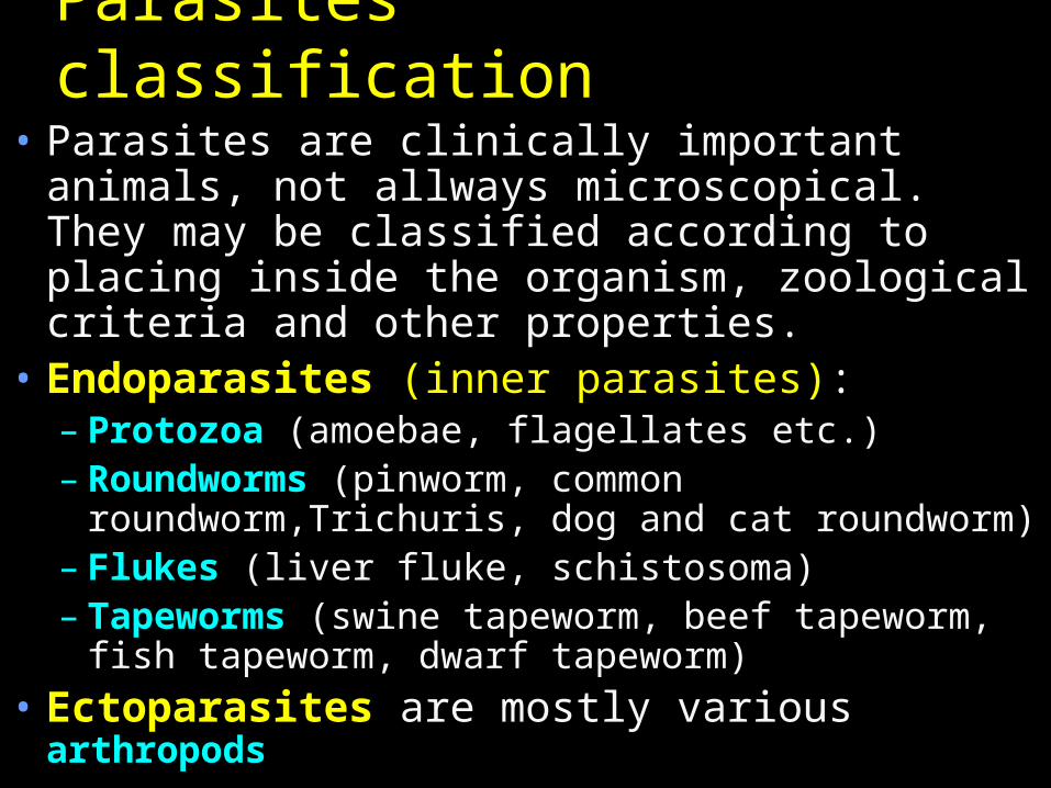
• Parasites are clinically important animals, not allways microscopical. They may be classified according to placing inside the organism, zoological criteria and other properties.
• Endoparasites (inner parasites):– Protozoa (amoebae, flagellates etc.)– Roundworms (pinworm, common
roundworm,Trichuris, dog and cat roundworm)– Flukes (liver fluke, schistosoma)– Tapeworms (swine tapeworm, beef tapeworm,
fish tapeworm, dwarf tapeworm)• Ectoparasites are mostly various
arthropods
Historical term „worms“
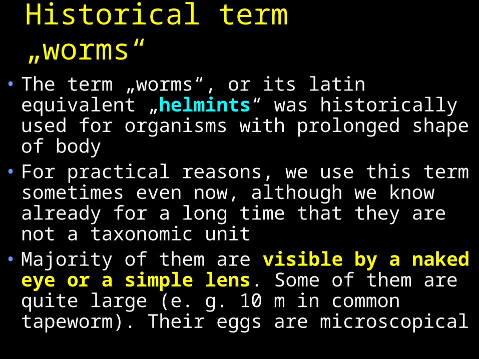
• The term „worms“, or its latin equivalent „helmints“ was historically used for organisms with prolonged shape of body
• For practical reasons, we use this term sometimes even now, although we know already for a long time that they are not a taxonomic unit
• Majority of them are visible by a naked eye or a simple lens. Some of them are quite large (e. g. 10 m in common tapeworm). Their eggs are microscopical
Flat worms and roundworms
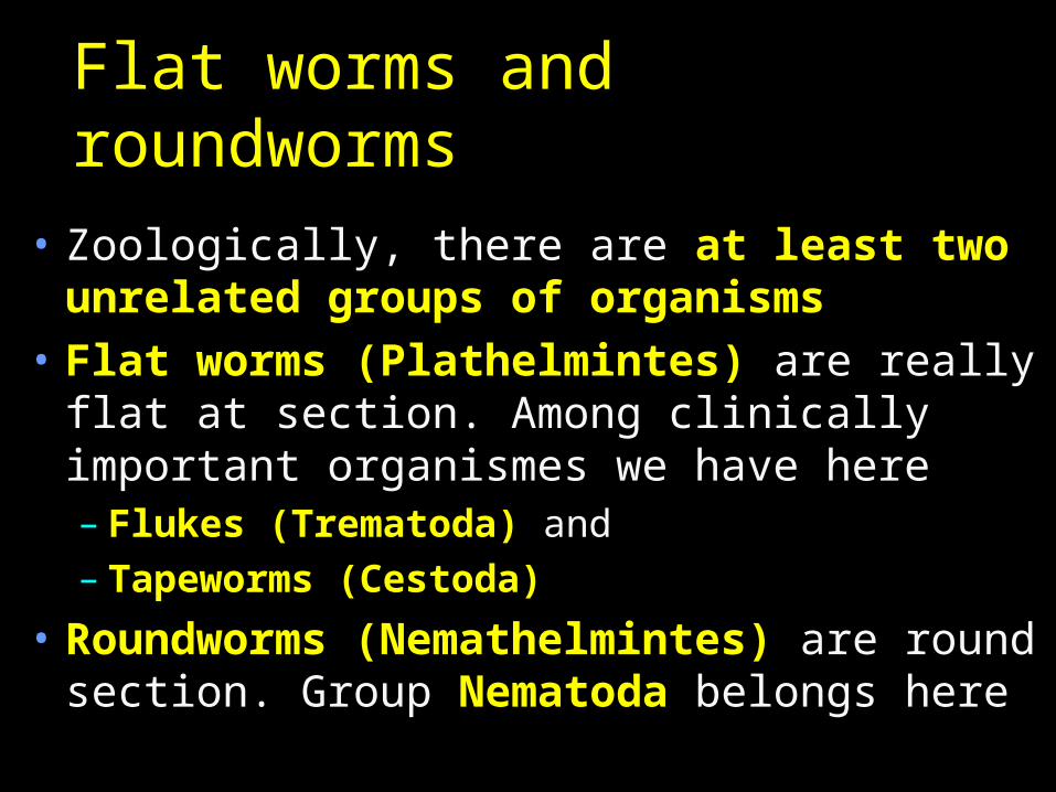
• Zoologically, there are at least two unrelated groups of organisms
• Flat worms (Plathelmintes) are really flat at section. Among clinically important organismes we have here– Flukes (Trematoda) and– Tapeworms (Cestoda)
• Roundworms (Nemathelmintes) are round section. Group Nematoda belongs here
Another parasites classification
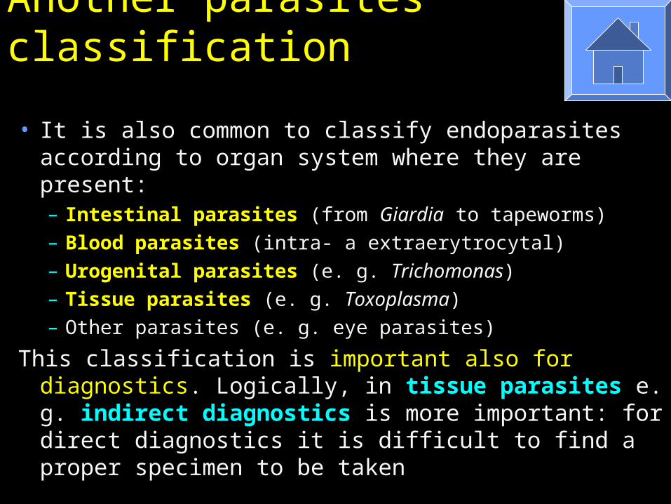
• It is also common to classify endoparasites according to organ system where they are present:– Intestinal parasites (from Giardia to tapeworms)– Blood parasites (intra- a extraerytrocytal)– Urogenital parasites (e. g. Trichomonas)– Tissue parasites (e. g. Toxoplasma)– Other parasites (e. g. eye parasites)
This classification is important also for diagnostics. Logically, in tissue parasites e. g. indirect diagnostics is more important: for direct diagnostics it is difficult to find a proper specimen to be taken
Parasites – clinical description

Story one• Little Nicol all the time scraped her
buttocks, and it was evident to parents and teachers that something is wrong. She was also inquiet and unconcentrated. So they placed a translucent scotch-tape on her anus and sent to the laboratory. And the result did not surprise anybody. Nicol started to use drugs and in a short time everything was in order again…
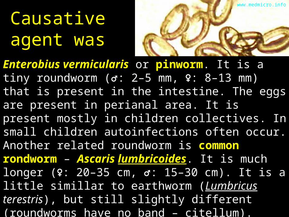
Causative agent was
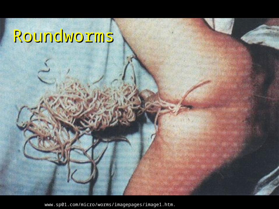
Enterobius vermicularis or pinworm. It is a tiny roundworm (♂: 2–5 mm, ♀: 8–13 mm) that is present in the intestine. The eggs are present in perianal area. It is present mostly in children collectives. In small children autoinfections often occur.Another related roundworm is common rondworm – Ascaris lumbricoides. It is much longer (♀: 20–35 cm, ♂: 15–30 cm). It is a little simillar to earthworm (Lumbricus terestris), but still slightly different (roundworms have no band – citellum). Roundwors may cause various problems, from allergic problems to mechanical obliteration of orifices of bile bladder and pancreas.
www.medmicro.info
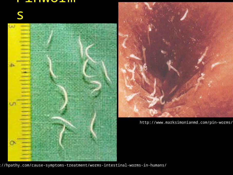
Pinworms
http://www.marksimonianmd.com/pin-worms/
http://hpathy.com/cause-symptoms-treatment/worms-intestinal-worms-in-humans/
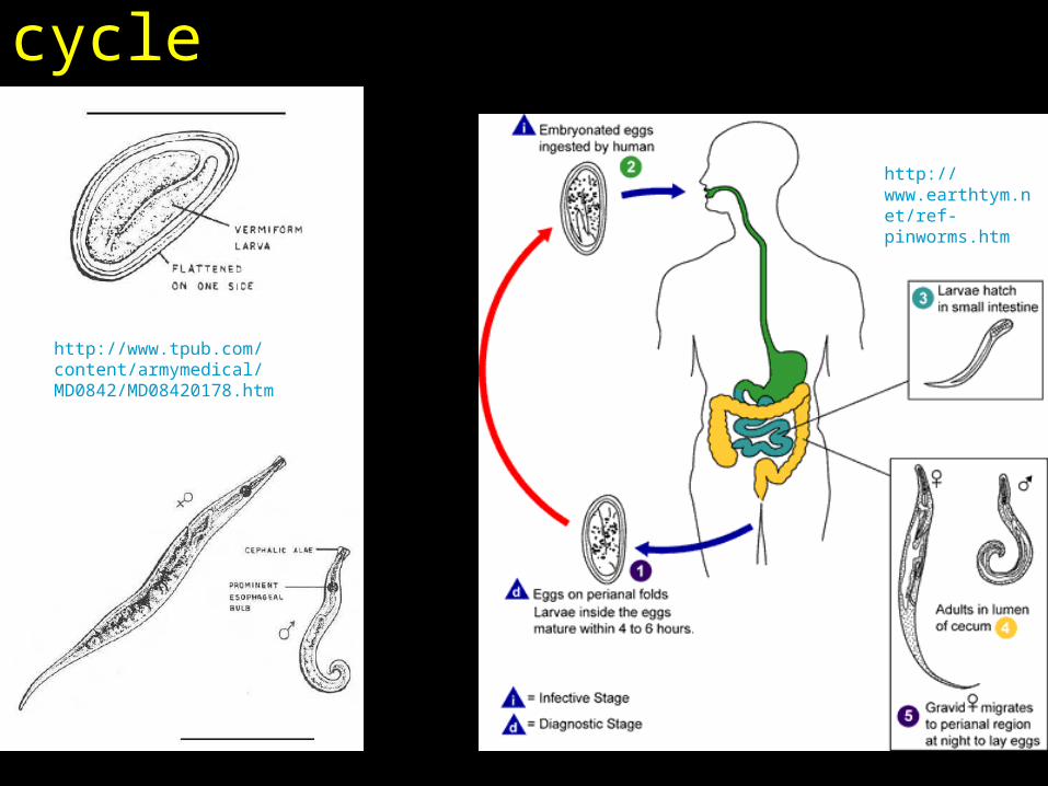
Pinworm: egg, worms, life cycle
http://www.earthtym.net/ref-pinworms.htm
http://www.tpub.com/content/armymedical/MD0842/MD08420178.htm
www.sp01.com/micro/worms/imagepages/image1.htm.
RoundwormsRoundworms
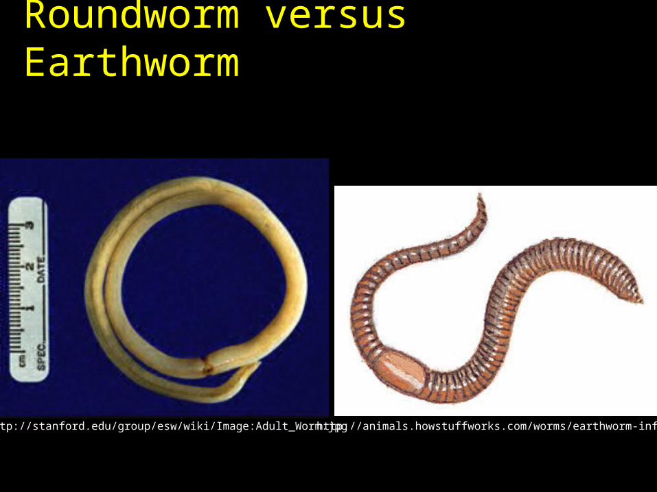
Roundworm versus Earthworm
http://animals.howstuffworks.com/worms/earthworm-info.htmhttp://stanford.edu/group/esw/wiki/Image:Adult_Worm.jpg

Roundworm life cycle and world distribution
http://stanford.edu/group/esw/wiki
http://stanford.edu/group/esw/wiki
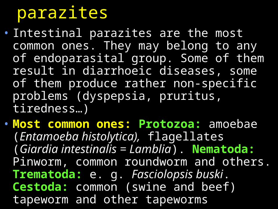
Intestinal parazites• Intestinal parazites are the most common
ones. They may belong to any of endoparasital group. Some of them result in diarrhoeic diseases, some of them produce rather non-specific problems (dyspepsia, pruritus, tiredness…)
• Most common ones: Protozoa: amoebae (Entamoeba histolytica), flagellates (Giardia intestinalis = Lamblia). Nematoda: Pinworm, common roundworm and others. Trematoda: e. g. Fasciolopsis buski. Cestoda: common (swine and beef) tapeworm and other tapeworms
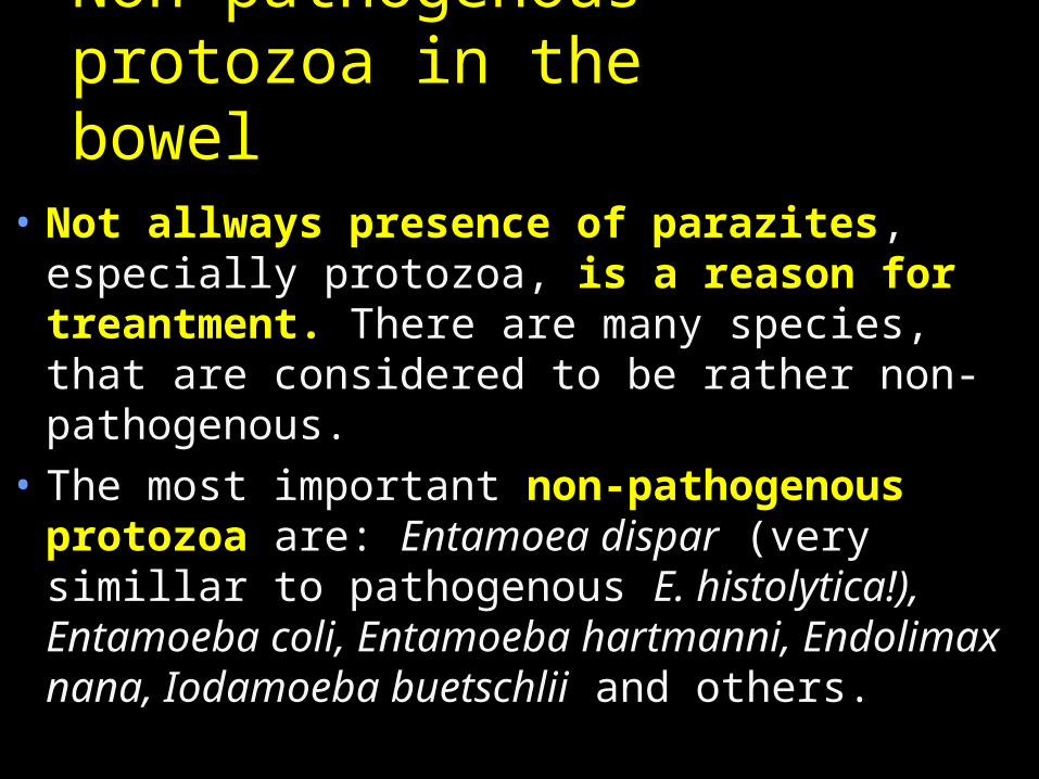
Non-pathogenous protozoa in the bowel
• Not allways presence of parazites, especially protozoa, is a reason for treantment. There are many species, that are considered to be rather non-pathogenous.
• The most important non-pathogenous protozoa are: Entamoea dispar (very simillar to pathogenous E. histolytica!), Entamoeba coli, Entamoeba hartmanni, Endolimax nana, Iodamoeba buetschlii and others.
Story two• Bianca, who took care of several cats, had
enlarged lymphonodes for some time, and long time it was a question, what is the reason. The throat swabs showed nothing, and also results ot other investigations were resultless.
• Bianca planned to have a child, and so she was affraid. And it was shown, that it is really so: the causative agent responsible for her lymphonode sydrome is really sometimes dangerous for pregnant women…
The causative agent was
• Toxoplasma gondii, a protozoon, that is transmitted by cats, although people say that people with dogs are in risk more than people with cats (they can wear the particles of cat faeces in their fur)
• In immunocompetent persons, majority of infections is asymptomatical, or only with mild symptomas (enlarged lymphonodes). Rarely, ocular form may occur
• In pregnant women toxoplasmosis may lead to abortion or embryonic organ defects
• Even asymptomatic cases may lead to carriership of cysts, that is also asymptomatic. Toxoplasma may only reactivate in case of immunity problems (HIV+), especially in decrease of CD4+ lymphocyte count
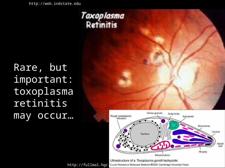
Rare, but important: toxoplasma retinitis may occur…
http://web.indstate.edu
http://fullmal.hgc.jp
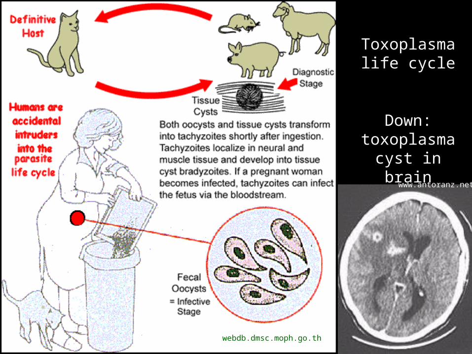
Toxoplasma life cycle
Down: toxoplasma cyst in brain
webdb.dmsc.moph.go.th
www.antoranz.net
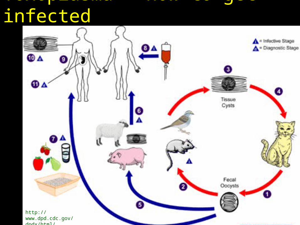
Toxoplasma – how to get infected
http://www.dpd.cdc.gov/dpdx/html/Toxoplasmosis.htm
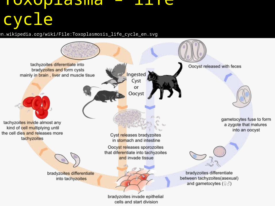
Toxoplasma – life cyclehttp://en.wikipedia.org/wiki/File:Toxoplasmosis_life_cycle_en.svg
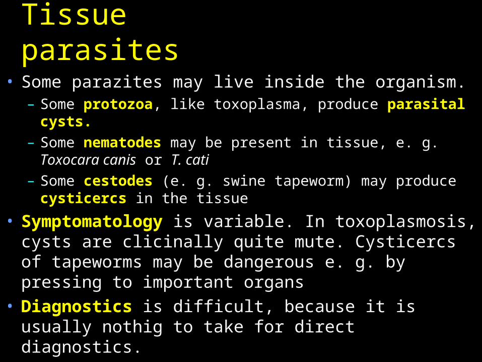
Tissue parasites• Some parazites may live inside the organism.
– Some protozoa, like toxoplasma, produce parasital cysts.
– Some nematodes may be present in tissue, e. g. Toxocara canis or T. cati
– Some cestodes (e. g. swine tapeworm) may produce cysticercs in the tissue
• Symptomatology is variable. In toxoplasmosis, cysts are clicinally quite mute. Cysticercs of tapeworms may be dangerous e. g. by pressing to important organs
• Diagnostics is difficult, because it is usually nothig to take for direct diagnostics.
Tropical and subtropical tissue parasites
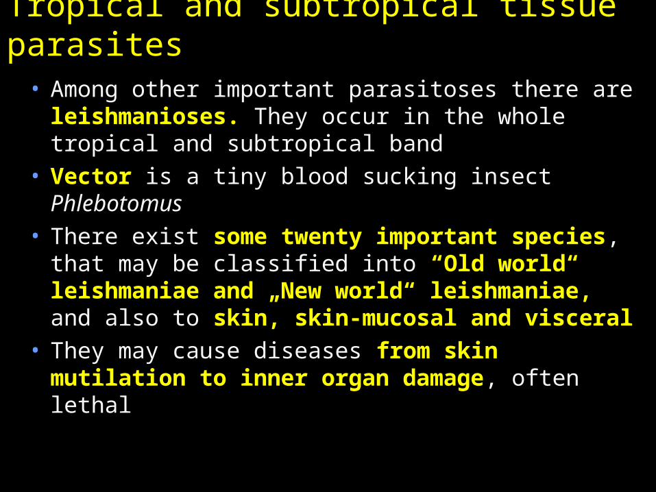
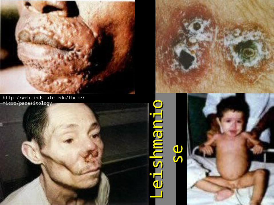
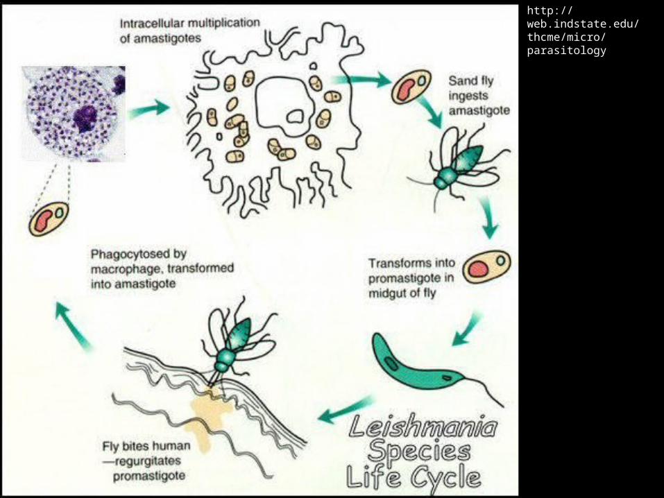
• Among other important parasitoses there are leishmanioses. They occur in the whole tropical and subtropical band
• Vector is a tiny blood sucking insect Phlebotomus
• There exist some twenty important species, that may be classified into “Old world“ leishmaniae and „New world“ leishmaniae, and also to skin, skin-mucosal and visceral
• They may cause diseases from skin mutilation to inner organ damage, often lethal
http://web.indstate.edu/thcme/micro/parasitology
Leis
hm
an
ios
Leis
hm
an
ios
e e
http://web.indstate.edu/thcme/micro/parasitology
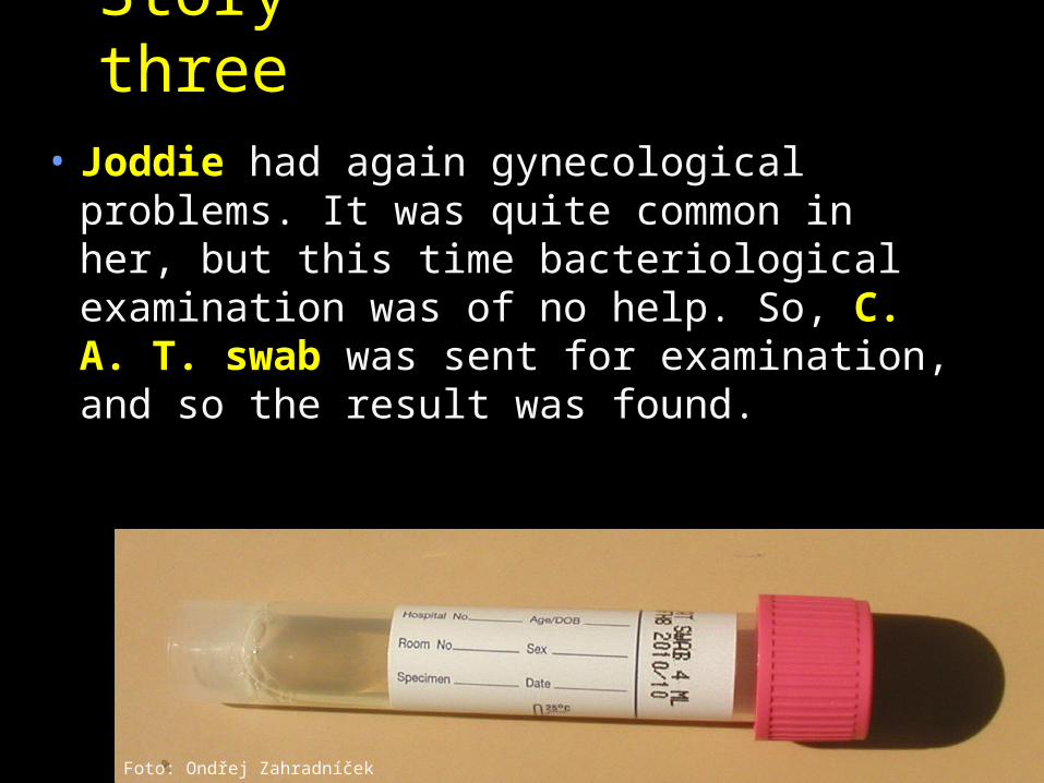
Story three
• Joddie had again gynecological problems. It was quite common in her, but this time bacteriological examination was of no help. So, C. A. T. swab was sent for examination, and so the result was found.
Foto: Ondřej Zahradníček
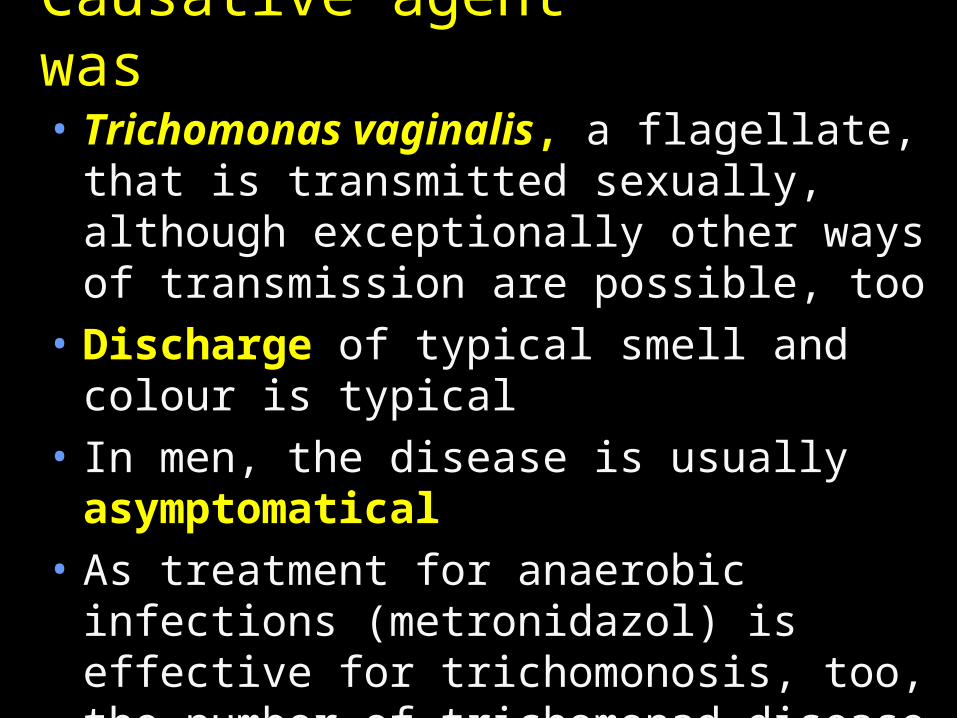
Causative agent was• Trichomonas vaginalis, a flagellate,
that is transmitted sexually, although exceptionally other ways of transmission are possible, too
• Discharge of typical smell and colour is typical
• In men, the disease is usually asymptomatical
• As treatment for anaerobic infections (metronidazol) is effective for trichomonosis, too, the number of trichomonad disease decreases in recent times.
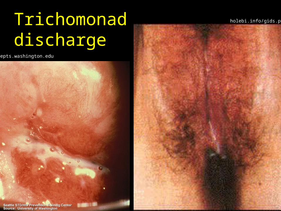
Trichomonad discharge
depts.washington.edu
holebi.info/gids.php
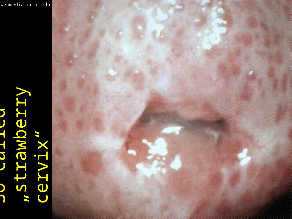
So c
alle
d
„str
aw
berr
y
cerv
ix“
webmedia.unmc.edu
Urogenital parasites• Among sexually transmitted
parasital diseases, trichomonosis is the only really important (if not discussing the pubic louse).
• Usually its diagnostic is done together with bacteriology examination, as the ethiology is rarely clear at the first moment
• In the urinary tract, most important parasites are some flukes (schistosomas), i. e. Trematodes. Diagnostic is partially microbiological, partially histological.

Story four• Mr. Hiker used to travel thoughout
the world. After coming back from the last trip he started to have some probles, he caugth fever, then it finished, but during two days it started again. The GP sent him to infection department. They took his blood and made a smear to two slides, using two various methods. And really the causative agent was…
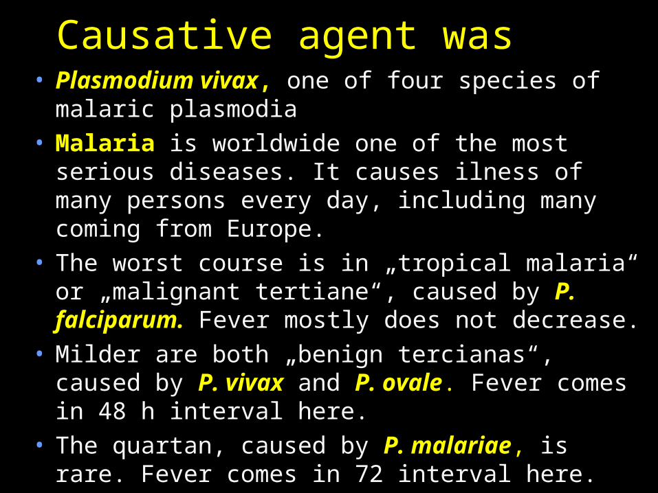
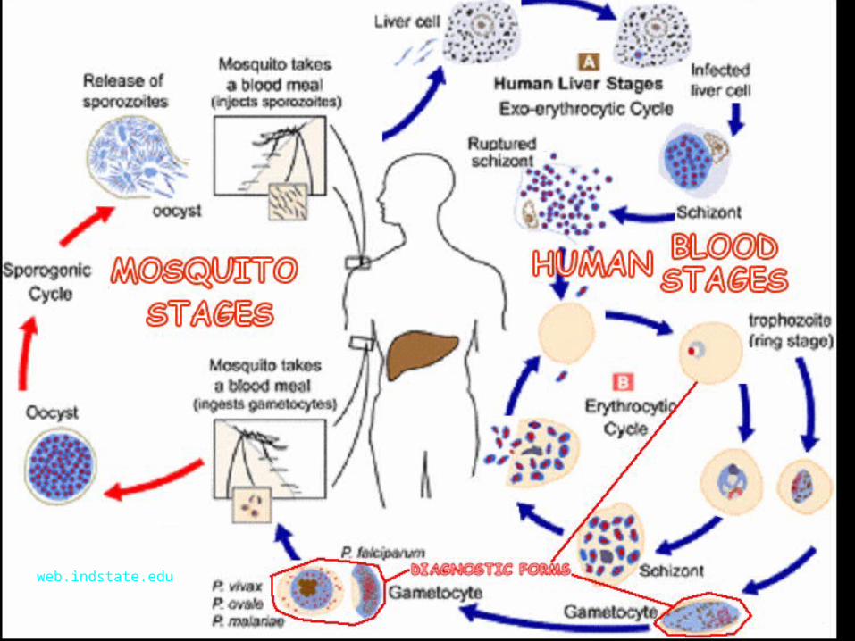
Causative agent was• Plasmodium vivax, one of four species of
malaric plasmodia• Malaria is worldwide one of the most serious
diseases. It causes ilness of many persons every day, including many coming from Europe.
• The worst course is in „tropical malaria“ or „malignant tertiane“, caused by P. falciparum. Fever mostly does not decrease.
• Milder are both „benign tercianas“, caused by P. vivax and P. ovale. Fever comes in 48 h interval here.
• The quartan, caused by P. malariae, is rare. Fever comes in 72 interval here.
web.indstate.edu
Bloostream parasites
• Among bloodstream parasites, of course, malarial plasmodia are the most important, but not the only ones.
• A flagellate, Trypanosoma brucei lives in bloodsteam, causing sleeping disease. Trypanosoma cruzi lives there too, producing Chagas disease
• Nematodes – filariae may live in bloodstream, too (in tropical countries)
• Symptomatology: in all of them fever appeares, other symptomas are related to the agent
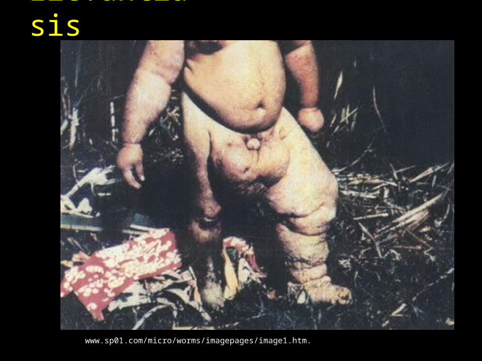
Elefantiasis
www.sp01.com/micro/worms/imagepages/image1.htm.
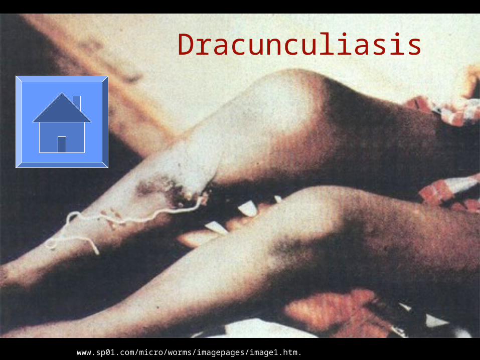
Dracunculiasis
www.sp01.com/micro/worms/imagepages/image1.htm.
Parasites – sampling

Sampling• For intestinal parasites rectal swab is not
sufficient, a bit of stool is needed (see more )
• For Trichomonas either a slide for Giemsa staining is sent (alone or in pair with another one for Gram staining), or a C. A. T. swab
• For Acantamoeba used contact lenses are sent in their own fluid, eventually corneal scraping might be performed
• For tissue parasites serum is sent usually• In other pararasites we sample ccording to
situation (urine, content of a cyst)
Sampling for intestinal parasites
• To send stool for parasitological sampling (usually using Kato and Faust methods), we need sample of stool sized like a hazel nut. A vessel for sampling need not be sterile. Unlike virological examination the sample does not need low transport temperature
• Specimen sized like a coconut (as sometimes some student say) is not recomended
Sampling for pinworms• For pinwoms, it is possible to send a stool
specimen as for other intestinal parasites, but it is not optimal, because a better method exists
• It is Graham method, where a tape is placed around anal orifice, then removed, placed on a slide and sent to the laboratory (and here examined microscopically
• For technical reasons, the method is not very useful for adult persons, here classical stool specimen is sent usually
Sampling for blood parasites
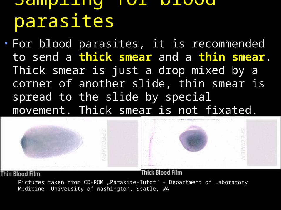
• For blood parasites, it is recommended to send a thick smear and a thin smear. Thick smear is just a drop mixed by a corner of another slide, thin smear is spread to the slide by special movement. Thick smear is not fixated.
Pictures taken from CD-ROM „Parasite-Tutor“ – Department of Laboratory Medicine, University of Washington, Seatle, WA
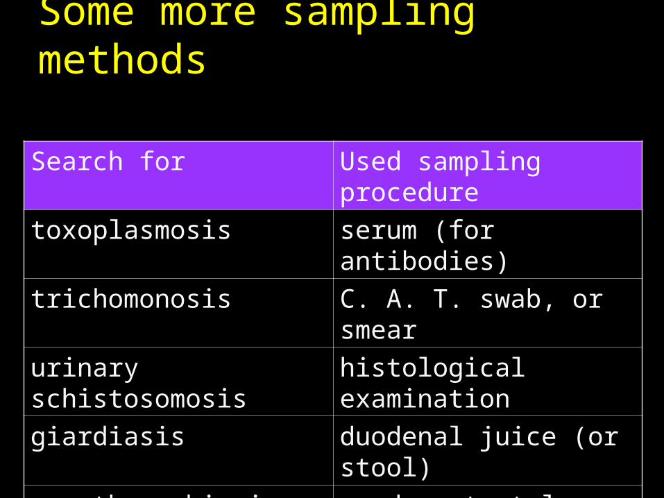
Some more sampling methods
Search for Used sampling procedure
toxoplasmosis serum (for antibodies)
trichomonosis C. A. T. swab, or smear
urinary schistosomosis histological examination
giardiasis duodenal juice (or stool)
acanthamoebiasis used contact lens
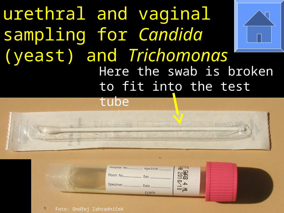
C. A. T. swab for urethral and vaginal sampling for Candida (yeast) and Trichomonas
Foto: Ondřej Zahradníček
Here the swab is broken to fit into the test tube
Parasites – diagnostic methods
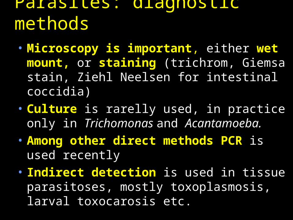
Parasites: diagnostic methods• Microscopy is important, either wet
mount, or staining (trichrom, Giemsa stain, Ziehl Neelsen for intestinal coccidia)
• Culture is rarelly used, in practice only in Trichomonas and Acantamoeba.
• Among other direct methods PCR is used recently
• Indirect detection is used in tissue parasitoses, mostly toxoplasmosis, larval toxocarosis etc.
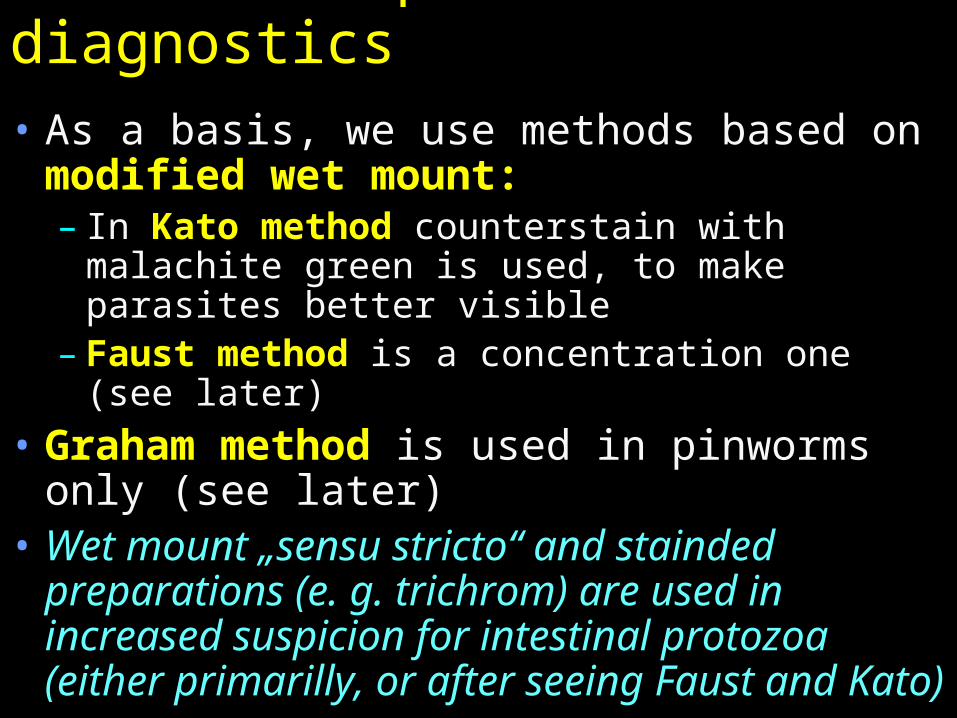
Intestinal parasites diagnostics• As a basis, we use methods based on
modified wet mount:– In Kato method counterstain with malachite
green is used, to make parasites better visible– Faust method is a concentration one (see
later)• Graham method is used in pinworms
only (see later)• Wet mount „sensu stricto“ and stainded
preparations (e. g. trichrom) are used in increased suspicion for intestinal protozoa (either primarilly, or after seeing Faust and Kato)
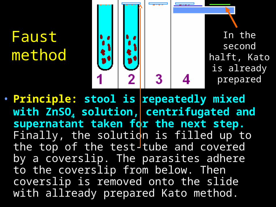
Faust method
In the second halft, Kato is
already prepared
• Principle: stool is repeatedly mixed with ZnSO4 solution, centrifugated and supernatant taken for the next step. Finally, the solution is filled up to the top of the test-tube and covered by a coverslip. The parasites adhere to the coverslip from below. Then coverslip is removed onto the slide with allready prepared Kato method.
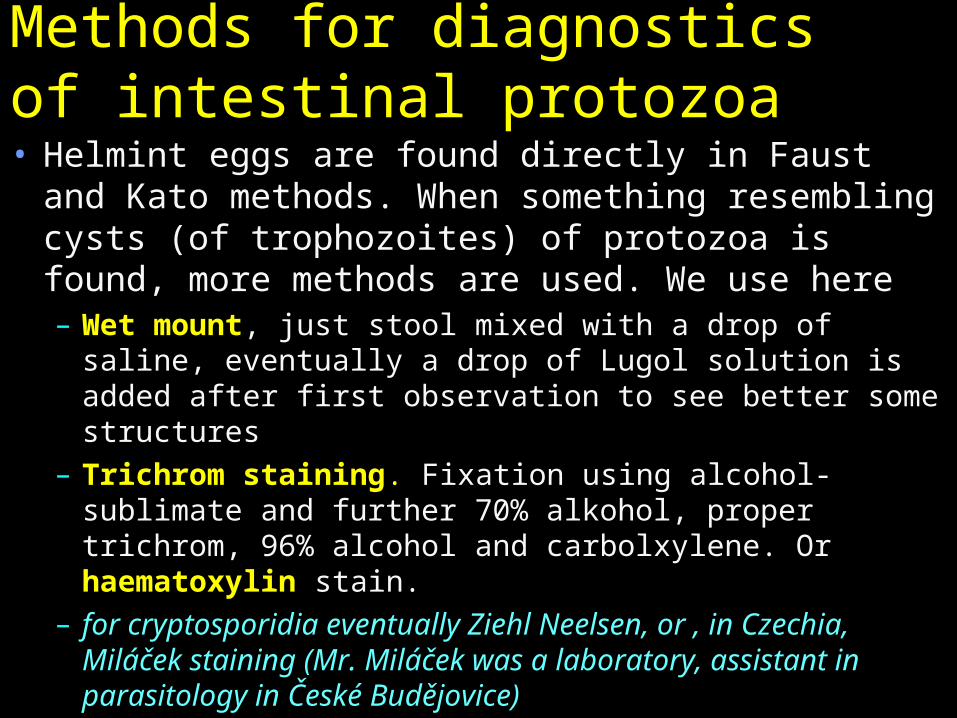
Methods for diagnostics of intestinal protozoa• Helmint eggs are found directly in Faust and
Kato methods. When something resembling cysts (of trophozoites) of protozoa is found, more methods are used. We use here– Wet mount, just stool mixed with a drop of saline,
eventually a drop of Lugol solution is added after first observation to see better some structures
– Trichrom staining. Fixation using alcohol-sublimate and further 70% alkohol, proper trichrom, 96% alcohol and carbolxylene. Or haematoxylin stain.
– for cryptosporidia eventually Ziehl Neelsen, or , in Czechia, Miláček staining (Mr. Miláček was a laboratory, assistant in parasitology in České Budějovice)
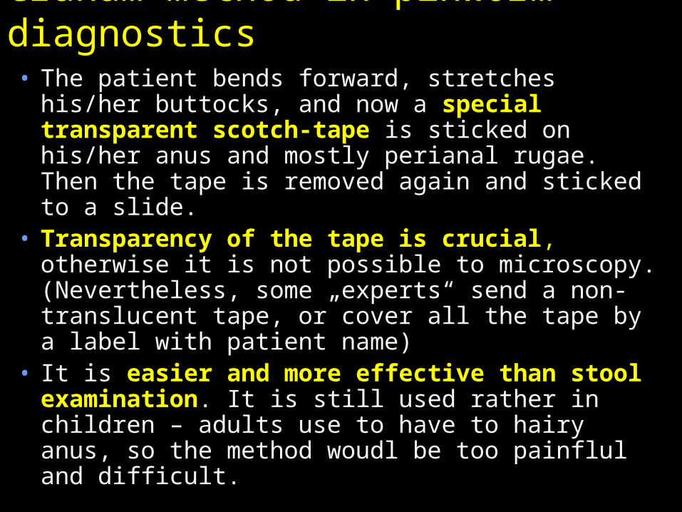
Graham method in pinworm diagnostics• The patient bends forward, stretches his/her
buttocks, and now a special transparent scotch-tape is sticked on his/her anus and mostly perianal rugae. Then the tape is removed again and sticked to a slide.
• Transparency of the tape is crucial, otherwise it is not possible to microscopy. (Nevertheless, some „experts“ send a non-translucent tape, or cover all the tape by a label with patient name)
• It is easier and more effective than stool examination. It is still used rather in children – adults use to have to hairy anus, so the method woudl be too painflul and difficult.
Practical microscopy in Faust, Kato and Graham methods
In all three methods we use no immersion, objectives 10×, 20×, 40×.
Faust and Kato methods are usually observed together, on the same slide
Graham method result is microscopied just in the form how it comes to the laboratory, no preparation is needed.
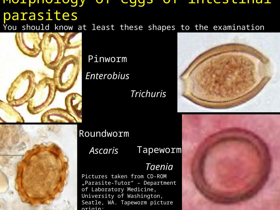
Morphology of eggs of intestinal parasitesYou should know at least these shapes to the examination
Pinworm
Enterobius
Roundworm
Ascaris
Trichuris
Tapeworm
TaeniaPictures taken from CD-ROM „Parasite-Tutor“ – Department of Laboratory Medicine, University of Washington, Seatle, WA. Tapeworm picture origin: http://www.biolib.cz/cz/taxonimage/id17433/?taxonid=43809

Evaluation of protozoan diagnostics• Gomori trichrom or Heidenheim
haematoxylin is used. Size of the protozoon, number of nuclei, in Entamoeba genus also central and peripherial chromatine
• Unlike Entamoeba coli, E. histolytica has maximally four nuclei. Usually you cannot see all nuclei together, they appear and disappear during focusing.
• Entamoeba histolytica/dispar cannot be differentiated microscopically, in reference laboratory PCR discrimination is performed

Diagnostics of blood parasites: thin smear and thick drop
• In diagnostics of blood parasites it is important to perform a smear using special methods of thin smear and thick drop.
• For both methods, fresh blood is used, of non-clotted blood, if the smear is not performed immediatelly. The thin smear is fixated, the thick drop is not. Both of them are Giemsa stained.
• Look at following pictures.
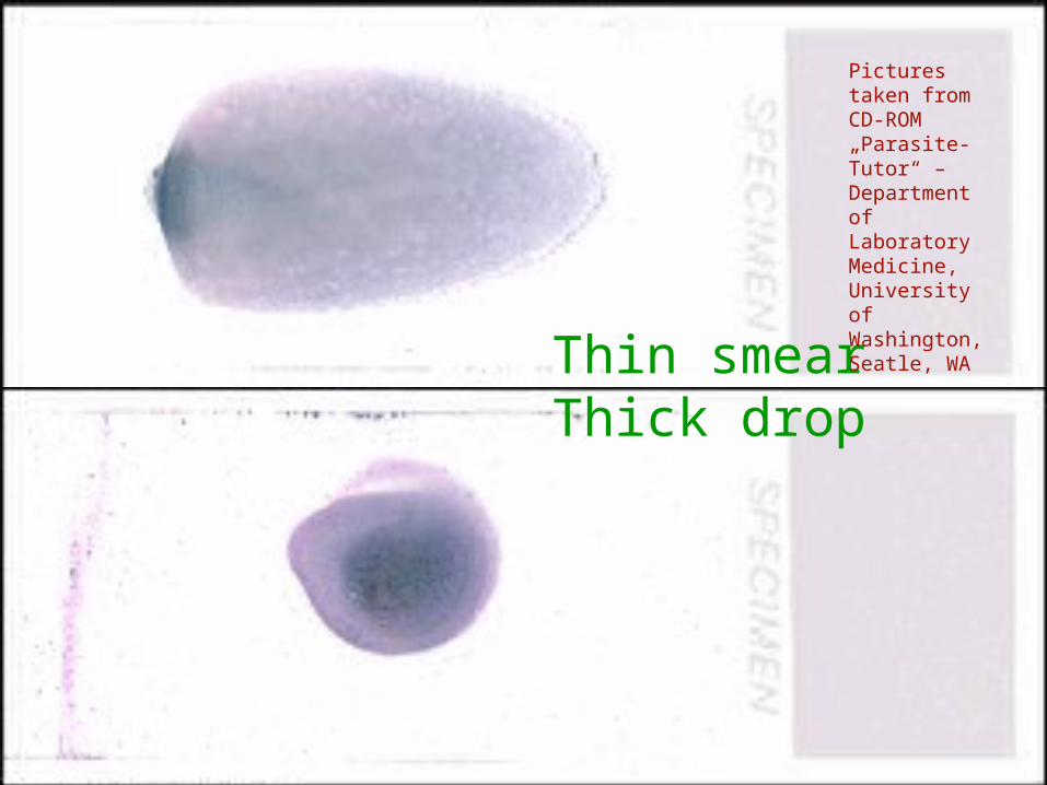
Thin smearThick drop
Pictures taken from CD-ROM „Parasite-Tutor“ – Department of Laboratory Medicine, University of Washington, Seatle, WA
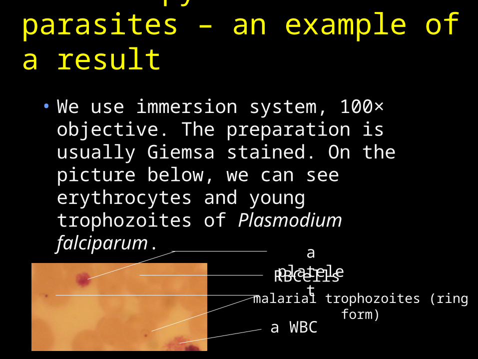
Microscopy of blood parasites – an example of a result
• We use immersion system, 100× objective. The preparation is usually Giemsa stained. On the picture below, we can see erythrocytes and young trophozoites of Plasmodium falciparum.
a plateletRBCells
malarial trophozoites (ring form)
a WBC
web.indstate.edu
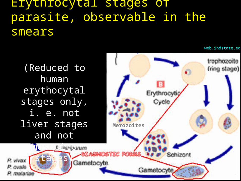
Erythrocytal stages of parasite, observable in the smears
(Reduced to human
erythocytal stages only, i. e. not liver stages
and not mosquito stages)
Merozoites

Erythrocytar stages of plasmodia
Pictures from CD-ROM „Parasite-Tutor“ – Department of Laboratory Medicine, University of Washington, Seatle, WA
merozoites
„ring-form“
older trophozoite
schizont
gametocyte
mosquito stages
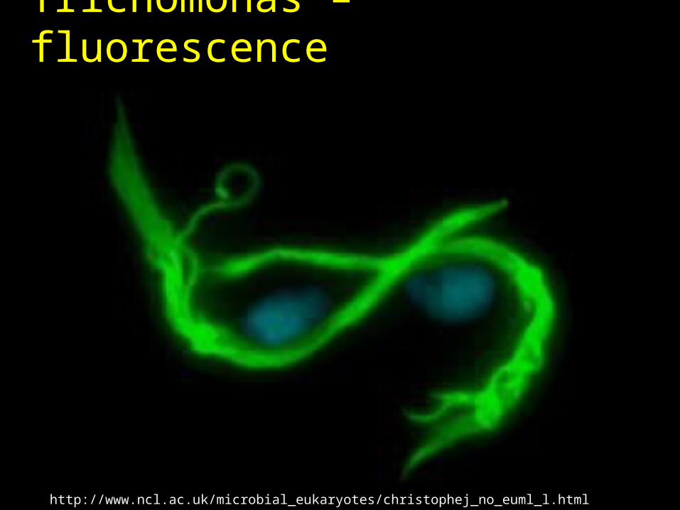
Trichomonas diagnostics• Trichomonads are recently diagnosed mostly
using culture-microscopical:– A C. A. T. swab is performed– The medium is cultured overnight– A drop of medium is microscopied as a wet
mount.
• The preparations cannot be preserved• Therefore in our practical we have the second
possible way of diagnostics – Giemsa stained smear on a slide. When it is a part of „Microscopical appearance of vaginal microflora“ (MAVM), it is described as MAVM V.
• Other ways (e. g. fluorescence staining, see picture) are used rarelly
Trichomonas – fluorescence
http://www.ncl.ac.uk/microbial_eukaryotes/christophej_no_euml_l.html
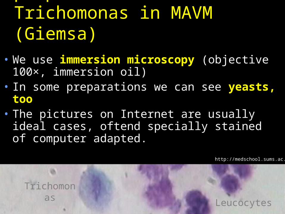
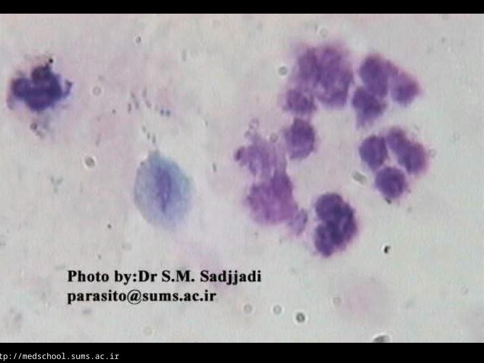
Microscopical preparations of Trichomonas in MAVM (Giemsa)
• We use immersion microscopy (objective 100×, immersion oil)
• In some preparations we can see yeasts, too
• The pictures on Internet are usually ideal cases, oftend specially stained of computer adapted.
http://medschool.sums.ac.ir
Leucocytes
Trichomonas
http://medschool.sums.ac.ir
Diagnostics of other parasital diseases
• In ectoparasites majority of diagnostics is non-microbiological (everything can be observed by a laik, eventually a dermatologist in case of Sacroptes scabiei)
• In tissue parasites serum for indirect diagnostics is sent usually (CFT, ELISA)
• In some cases, mostly tropical parasitioses, it is better to consult sampling technique with a laboratory
In some filarioses the sampling is recomended to perform during night only, or during day only.
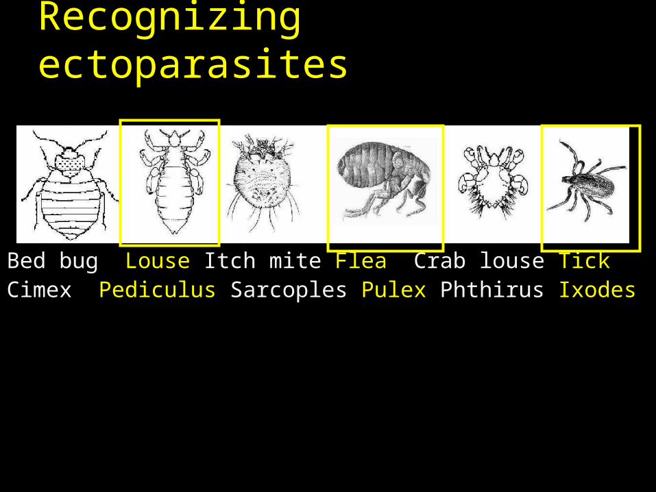
Recognizing ectoparasites
Bed bug Louse Itch mite Flea Crab louse TickCimex Pediculus Sarcoples Pulex Phthirus Ixodes
Diagnostics of Toxoplasma gondii using serological tests• In toxoplaxmosis, we often combine two
tests.• Complement fixing test (CFT). It is
performed like other complement fixing tests, see J08 practical
• ELISA, too, is like other ELISA reactions. One specific feature is that instead of IgM + IgG we mostly search for IgA + IgG antibodies. IgA antibodies are typical for recent infections, IgG for „status after“ an infection. Paradoxically, pregnant women with IgG + are in a better situation than IgG – (they are protected).
Parasites – pictures
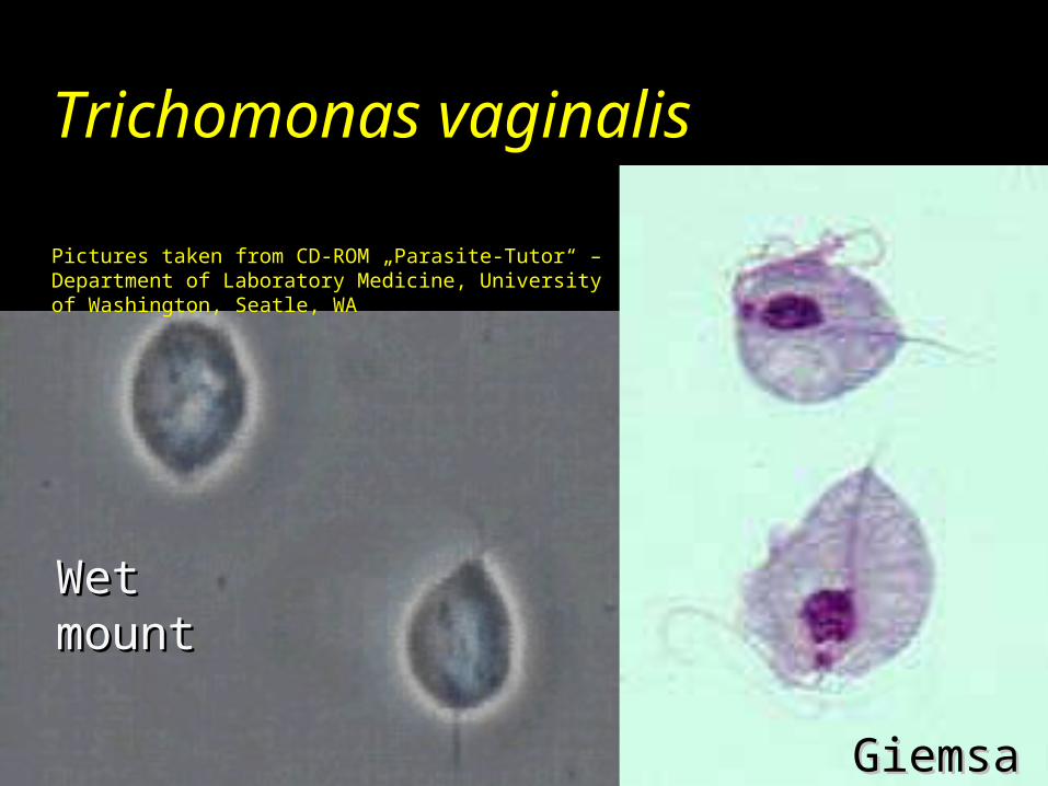
Trichomonas vaginalis
Pictures taken from CD-ROM „Parasite-Tutor“ – Department of Laboratory Medicine, University of Washington, Seatle, WA
GiemsaGiemsa
Wet Wet mountmount
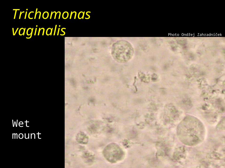
Trichomonas vaginalis
Wet mount
Photo Ondřej Zahradníček

Giardia intestinalis (Lamblia) (trophozoites)
Picture taken from CD-ROM „Parasite-Tutor“ – Department of Laboratory Medicine, University of Washington, Seatle, WA
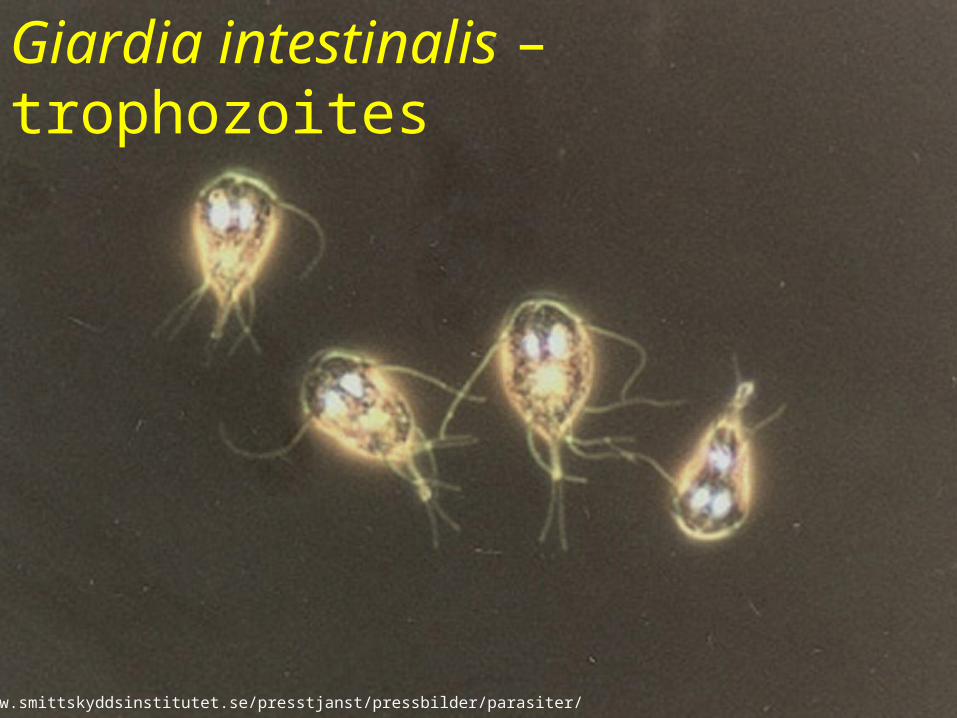
Giardia intestinalis – trophozoites
http://www.smittskyddsinstitutet.se/presstjanst/pressbilder/parasiter/
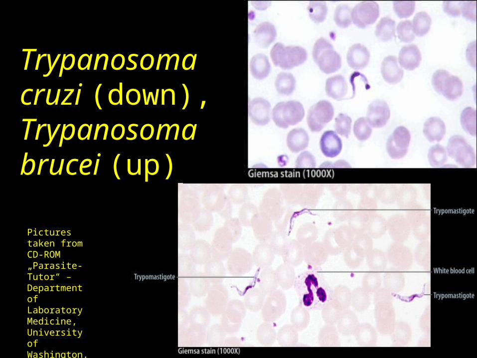
Trypanosoma cruzi (down), Trypanosoma brucei (up)
Pictures taken from CD-ROM „Parasite-Tutor“ – Department of Laboratory Medicine, University of Washington, Seatle, WA
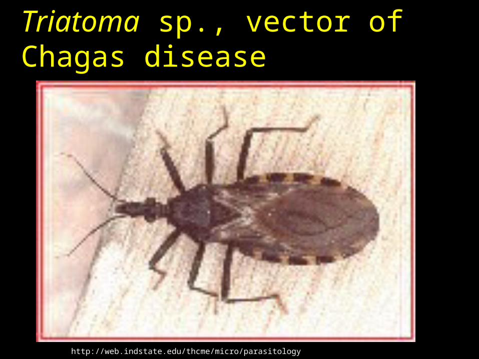
Triatoma sp., vector of Chagas disease
http://web.indstate.edu/thcme/micro/parasitology
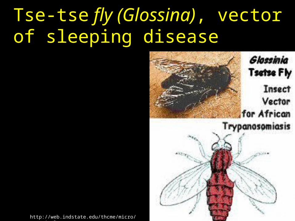
Tse-tse fly (Glossina), vector of sleeping disease
http://web.indstate.edu/thcme/micro/parasitology
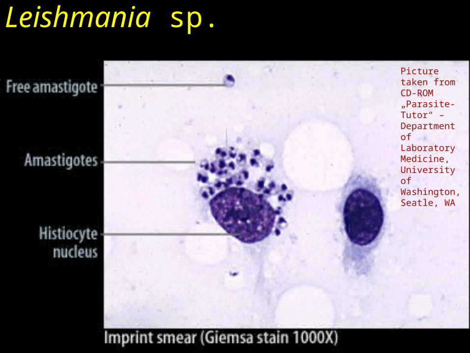
Leishmania sp.
Picture taken from CD-ROM „Parasite-Tutor“ – Department of Laboratory Medicine, University of Washington, Seatle, WA
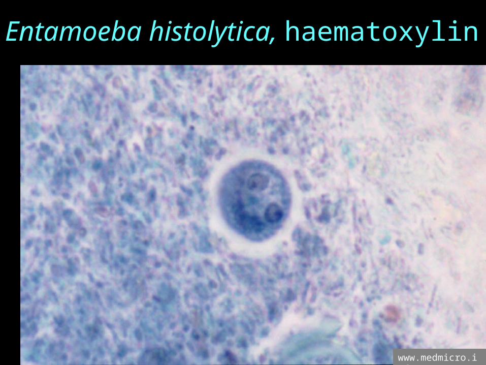
Entamoeba histolytica, haematoxylin
www.medmicro.info
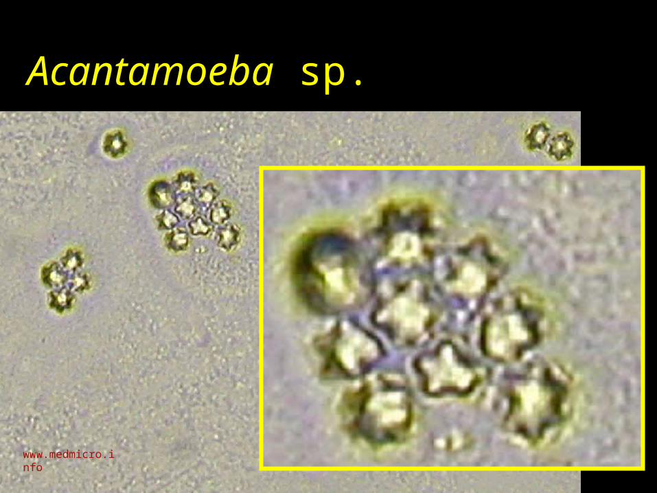
Acantamoeba sp.
www.medmicro.info

Toxoplasma gondiihttp://webdb.dmsc.moph.go.th/ifc_nih/applications/pics/Toxoplasma.jpg
http://www.smittskyddsinstitutet.se/upload/Analyser/ToxoplasmaSB.jpg
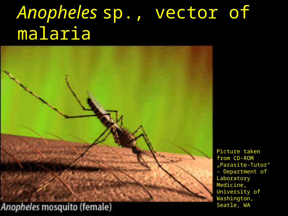
Anopheles sp., vector of malaria
Picture taken from CD-ROM „Parasite-Tutor“ – Department of Laboratory Medicine, University of Washington, Seatle, WA
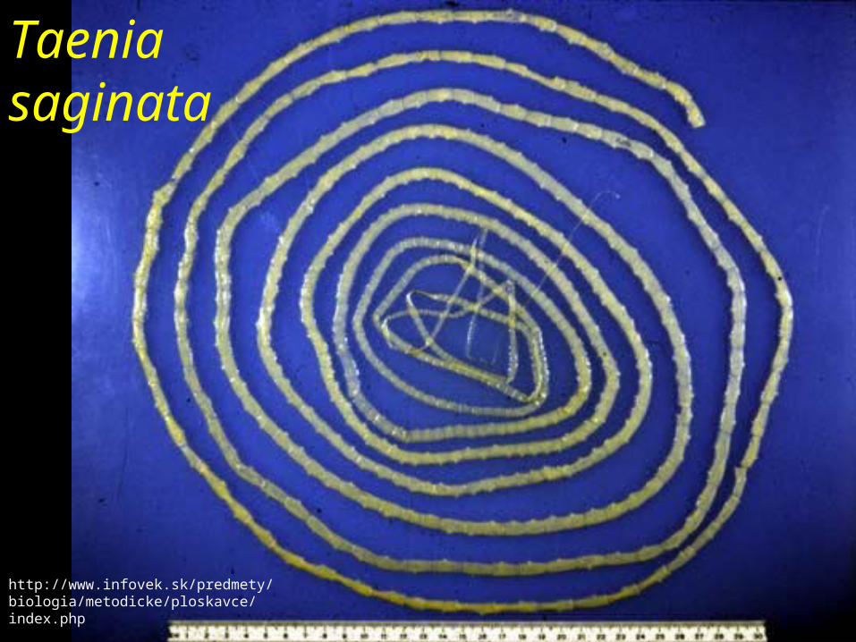
Taenia saginata
http://www.infovek.sk/predmety/biologia/metodicke/ploskavce/index.php
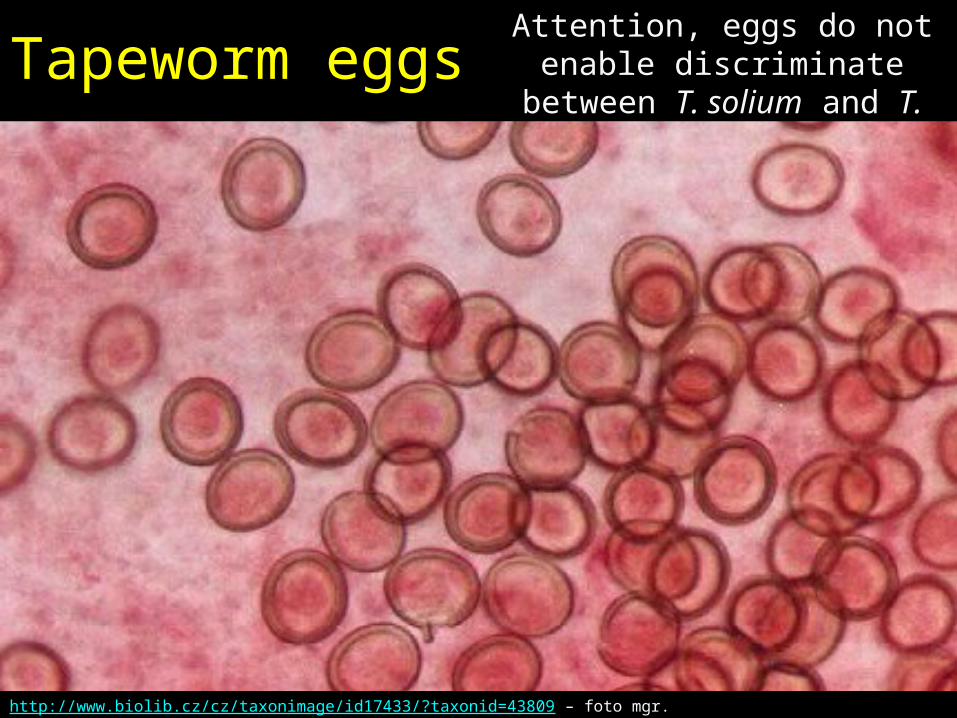
Tapeworm eggsAttention, eggs do not
enable discriminate between T. solium and T. saginata, we would need
proglotids for this!
http://www.biolib.cz/cz/taxonimage/id17433/?taxonid=43809 – foto mgr. Vladimír Bádr

Proglotid of a beef tapeworm(a swine tapeworm would have less branches)
Pictures taken from CD-ROM „Parasite-Tutor“ – Department of Laboratory Medicine, University of Washington, Seatle, WA
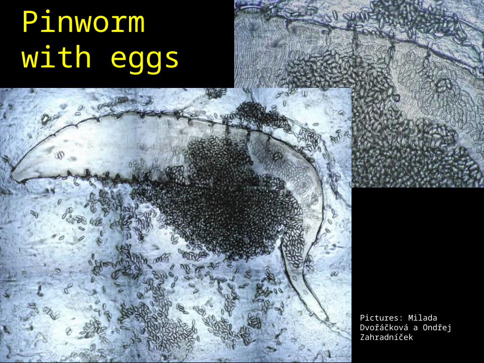
Pinworm with eggs
Pictures: Milada Dvořáčková a Ondřej Zahradníček
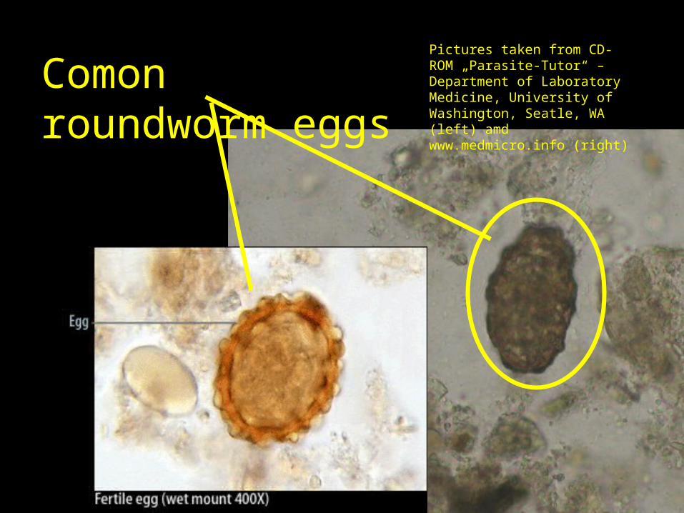
Comon roundworm eggs
Pictures taken from CD-ROM „Parasite-Tutor“ – Department of Laboratory Medicine, University of Washington, Seatle, WA (left) amd www.medmicro.info (right)
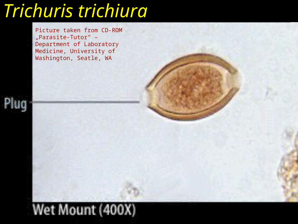
Trichuris trichiuraPicture taken from CD-ROM „Parasite-Tutor“ – Department of Laboratory Medicine, University of Washington, Seatle, WA
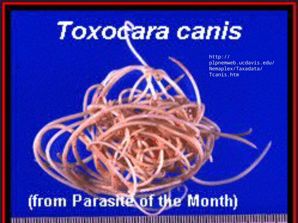
http://plpnemweb.ucdavis.edu/Nemaplex/Taxadata/Tcanis.htm
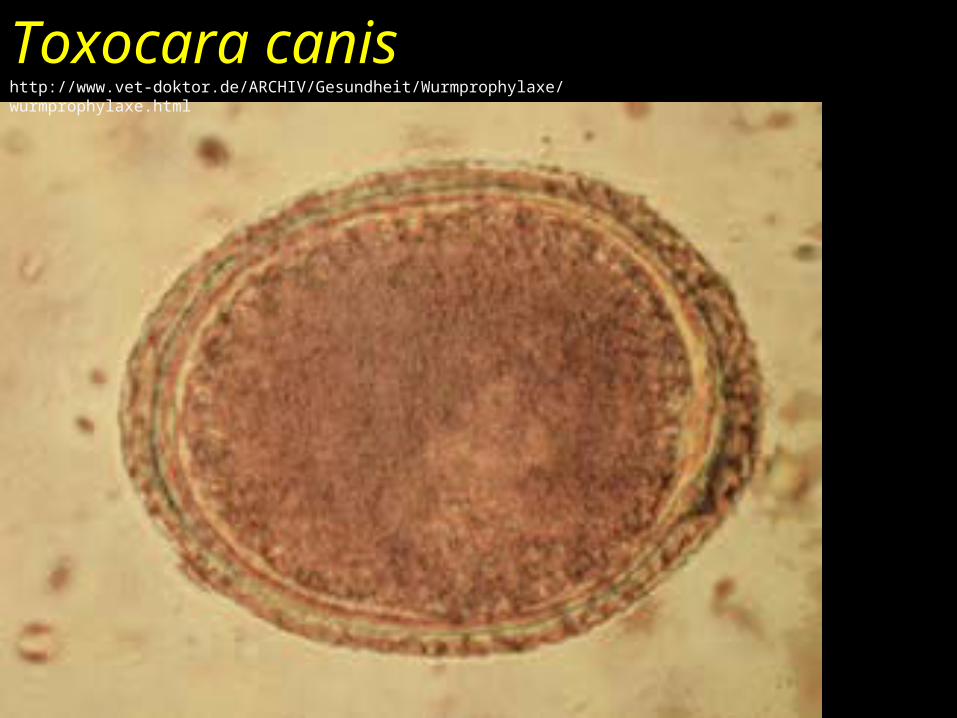
Toxocara canishttp://www.vet-doktor.de/ARCHIV/Gesundheit/Wurmprophylaxe/wurmprophylaxe.html
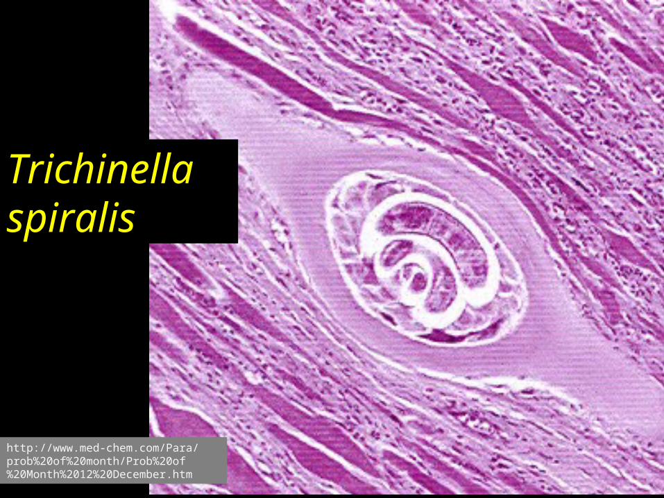
Trichinella spiralis
http://www.med-chem.com/Para/prob%20of%20month/Prob%20of%20Month%2012%20December.htm
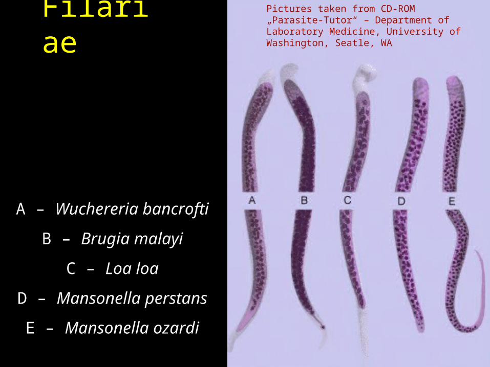
Filariae
A – Wuchereria bancrofti
B – Brugia malayi
C – Loa loa
D – Mansonella perstans
E – Mansonella ozardi
Pictures taken from CD-ROM „Parasite-Tutor“ – Department of Laboratory Medicine, University of Washington, Seatle, WA
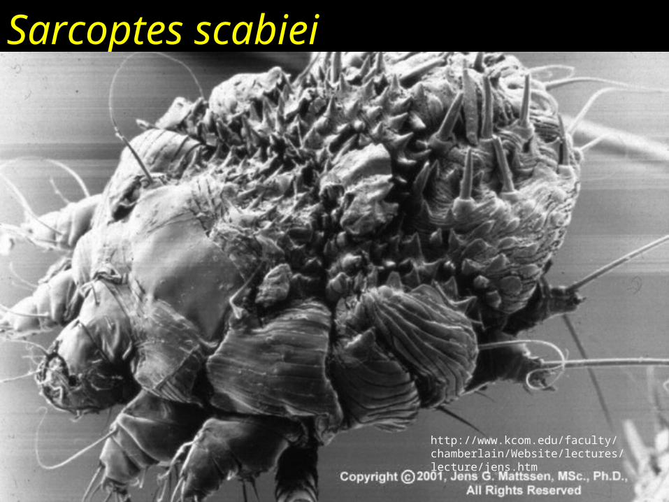
Sarcoptes scabiei
http://www.kcom.edu/faculty/chamberlain/Website/lectures/lecture/jens.htm
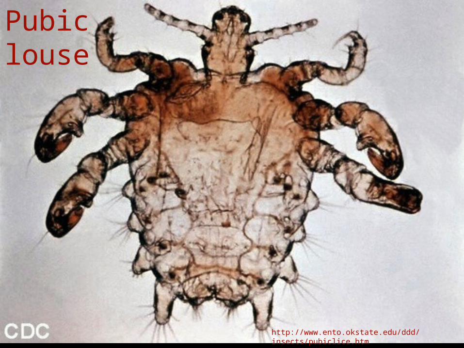
Pubic louse
http://www.ento.okstate.edu/ddd/insects/pubiclice.htm
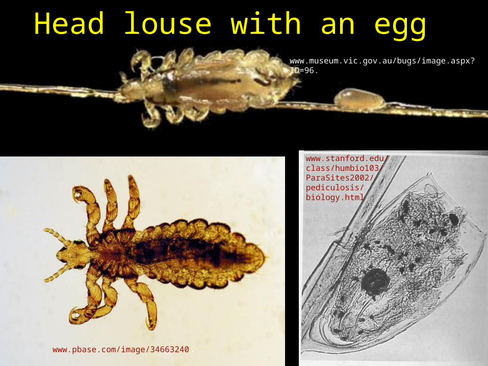
Head louse with an eggwww.museum.vic.gov.au/bugs/image.aspx?ID=96.
www.pbase.com/image/34663240.
www.stanford.edu/class/humbio103/ParaSites2002/pediculosis/biology.html

The End
Picture on this page – toxoplasmosis in artistic view
http://tn3-2.deviantart.com/300W/fs7.deviantart.com/i/2005/189/1/9/toxoplasma_by_chocko.jpg
Check-up questions• 1. What diseases are caused by protozoa of genus
Leishmania?• 2. What are bloodstream parasites other than malaric
plasmodia? • 3. What are staining methods for diagnostics of Giardia
and intestinal amoebae?• 4. What staining method can be used for diagnostics of
Cyclospora cayetanensis and Cryptosporidium parvum?• 5. Which non-pathogenic intestinal amoeba cannot be
distinguished microscopically from Entemoeba histolytica?• 6. What are diseases transmitted by Glossina fly,
Anopheles mosquito, Aëdes mosquito, Ixodes ricinus tick, Phlebotomus gnat
• 7. What is the name of the artificial (iatrogenous) myiase used for treatment? (It is a method, where larva of Lucillia serricata fly are administered to the wound to eat diseased, but not healthy tissue.)