Embed Size (px)
DESCRIPTION
Brachy-short Cryo-cold Crypto-hidden Duro-hard Eury-broad Hetero-different Holo-entire Idio-special Iso-equal Lept-thin Macro-large Mega-big Micro-small. Neo-new Ortho-straight Oxy-sharp Pachy-thick Pia-soft Platy-broad Proprio-one’s own Sclero-hard Scolio-crooked - PowerPoint PPT Presentation
Citation preview
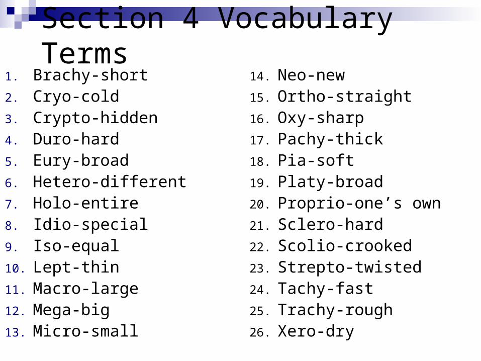
Section 4 Vocabulary Terms1. Brachy-short2. Cryo-cold3. Crypto-hidden4. Duro-hard5. Eury-broad6. Hetero-different7. Holo-entire8. Idio-special9. Iso-equal10. Lept-thin11. Macro-large12. Mega-big13. Micro-small
14. Neo-new15. Ortho-straight16. Oxy-sharp17. Pachy-thick18. Pia-soft19. Platy-broad20. Proprio-one’s own21. Sclero-hard22. Scolio-crooked23. Strepto-twisted24. Tachy-fast25. Trachy-rough26. Xero-dry

Section 5 vocabulary: Directions1. Ultra-beyond
2. Medio-middle
3. Intra- within
4. Gyro- Circular
5. Trans- across
6. Proximo- nearest
7. Per- through
8. Opistho- behind
9. En- in
10. Leve- left
11. Ex- out from
12. Endo- within
13. Ecto- on the outer side
14. Contra- against
15. Dia- through
16. Dextro- right
17. Dis- apart from
18. Cycl- circular
19. Amphi- on both sides
20. Ad- toward
21. Ab- away

10/5- Complete Bones Discussion, Start Axial Skeleton Lab
10/6- Axial lecture, Axial Lab10/7- Axial Skeleton Pop Quiz, Axial Lab due end of class (EOC)10/8- Vocab Quiz (4&5), Append. Skeleton- Upper & Lower
Limbs; Appendicular Lab10/9- Holiday
10/12- Holiday10/13- Appendicular Lab, Review for practical (3rd)10/14- PSAT, Review for practical, Finish Appen. Skel. Lab 10/15- Skeletal Practical10/16- Joints Lecture and Joints Lab
10/19- Finish Joints Notes & Lab Work10/20- Disease and Disorder Lecture – Food Day 10/21- Class case study, Children of Glass Video 10/22- Review for test10/23- Bones & Skeletal Exam

Bones
Chapter 6

Classification of Bones
206 named bones Axial Skeleton: bones of the skull,
vertebral column, and rib cageProtect, support or carry other body parts
Appendicular skeleton: girdles and bones of the upper and lower limbsLocomotion and manipulation

Functions of the Skeletal System
A. Support (framework)
B. Protection of enclosed structures
C. Movement with muscles
D. Storage of calcium
E. Blood cell formation-aka hematopoiesis

Classification of Bones Four kinds: Some Examples: Long bones Femur, Humerus,
Tibia, Phalanges Short bones Carpals, Tarsals Flat bones Scapula, Sternum,
Ribs, Skull Irregular bones Vertebra, Hip


Compact and Spongy Bone
Compact bone is the external dense outer layer
Spongy bone or cancellous bone is the internal honeycomb of small flat pieces called trabeculae.
The spaces between trabeculae will be filled with either red or yellow bone marrow

Typical Long Bone Structure
A. Diaphysis- thick & hollow shaft; compact bone
B.Medullary cavity- AKA marrow cavity; central part of diaphysis; in adults, contains yellow marrow
C.Epiphyses- bulbous endings; spongy; epiphyseal plate in development

Typical Long Bone Structure
D.Articular cartilage- hyaline; cushions joints
E.Periosteum- strong fibrous membrane covering long bone except at joint surfaces
F.Endosteum- epithelial inner lining of medullary cavity

Gross anatomy of bone (1)Epiphysis: compact outside
and spongy (cancellous) bone inside.
The joint surfaces are covered with articular cartilage which acts as a cushion.
Epiphyseal line is a remnant of the epiphyseal plate, disc of hyaline cartilage that grows during childhood.

Gross anatomy of bone (1)
Diaphysis: thick collar of compact bone, shaft, medullary cavity or marrow cavity. In adults contains fat and is then called the yellow bone cavity


Gross anatomy of bone (2)
Periosteum: double layer of an outer fibrous dense irregular connective tissue and an inner layer (osteogenic layer) that consists of the bone forming cells the osteoblasts and bone destroying cells the osteoclasts
DiaphysisEpiphysis
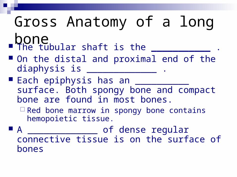
Gross Anatomy of a long bone
The tubular shaft is the ___________ . On the distal and proximal end of the
diaphysis is _____________ . Each epiphysis has an __________ surface. Both
spongy bone and compact bone are found in most bones. Red bone marrow in spongy bone contains
hemopoietic tissue. A _____________ of dense regular connective
tissue is on the surface of bones
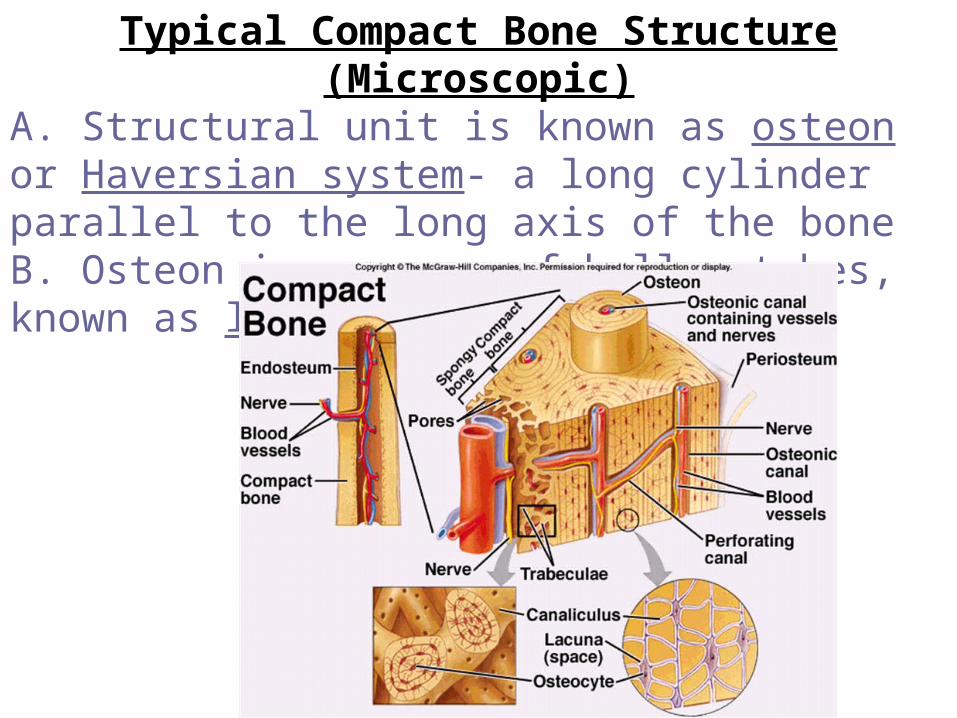
Typical Compact Bone Structure (Microscopic)A. Structural unit is known as osteon or Haversian system- a long cylinder parallel to the long axis of the boneB. Osteon is a group of hollow tubes, known as lamellae.

C. Running through core of osteon is a Haversian canal with blood vessels and nerve fibers
D. Perforating or Volkmann’s canals lie at right angles to the long axis of bone.

E. Spider-shaped osteocytes occupy small cavities aka lacunae.
F. Hairlike canals aka canaliculi connect the lacunae to each other and the central canal.

Bone Development1. Osteogenesis or ossification: bone formation
A. Intramembranous Ossification- formed from a fibrous membrane ex. flat bones
B. Endochondral Ossification- formed from cartilage ex. long bone

Process - cartilage bone collar spongy bone formation diaphysis elongates and medullary cavity forms epiphyses ossify Postnatal Bone
Growthgrowth in length at the
epiphyseal platesgrowth in thickness

Bone development
Intramembranous ossification e.g. skullOsteogenic cells switched on and lay down
bone in connective tissue “membrane”
Endochondral ossification e.g. femurOsteogenic cells switched on lay down bone
on cartilage framework


Types of Bone Cells
1.Osteoblasts- “bone-forming”; responsible for mineralized bone formation
2.Osteoclasts- “bone-breaking”, erosion of bone material In a 24 hour day, there is an alternation of osteoblast and osteoclast activity.
3.Osteocytes- mature, non-dividing osteoblasts; located in lacunae

The Skeleton (Ch. 7)
Consists of 206 separate bones/ 216 if you count individually-fused bones
Greek “dried up body” or “mummy” 20% of body mass

Axial Skeleton: 80 bones
Skull: Consists of 22 flat and irregular bones Cranium - 8 bones (frontal, 2 parietal, 2
temporal, occipital, sphenoid, and ethmoid) Facial Bones - 14 bones (2 maxilla, 2
zygomatic, 2 nasal, 1 mandible, 2 lacrimal, 2 palatine, 2 inferior nasal conchae, 1 vomer)