Embed Size (px)
Citation preview

DESCRIPTION OF A NEW SPECIES OF COLLEMBOLA AND ITS ANATOMY.
By DURGADAS MUKERJI, University Lecl-urer in Zoology, Calcutta University.
(Plates V-VI!.)
INTRODUCTION.
A number of species of Collembola has been recorded from India by Parona (1893), Ritter (1910-11), Imm,s (1912), Carpenter <1917, 1924) and Handschin (1929). Owing to the difficulty of making a thorough collection of these minute insects, our knowledge of the Indian species of the group is far from, complete. Moreover, the mouth-parts which are of considerable importance from the points of view of phylogeny and taxonomy, have received in the case of the Indian species, scantyattention, and a general account of the anatomy of the Indian species is lacking. Again, most -0£ the authors who deal with the structure and. function of the organs of those species of Collem.bola that occur outside India are at variance. For their various views, the works of the following authors, among others given in the bibliography at the end of this paper, may be referred to : Sommer (1884-85)) Oudemans (1888), Folsom (1899), Willem (1900), Imms (1906), Hoffmann (1904, 1905, 1908, 1911), Philiptschenko (1906, 1908) and Denis (1928).
In the present- paper a description of a new species of Collembola collected from Calcutta is given, together with an account of its anatomy. In dealing with the anatomy I have chiefly confined myself to descriptions of the digestive system and cephalic glands, as these offer consider-able structural peculiarities. . .
"I wish to acknowledge with thanks my indebtedness to Dr. G. W. Carpenter to whom the specimens were sent for confirmation of the identification, for his kind help and valuable suggestions. My best thanks are due to Lt.-Col. R. B. Seymour Sewell, Director of the Zoological Survey of India, Indian Museum and Dr. H. S. Pruthi of the Entomological Section, Indian Museum, for permitting me to examine the type specimens of Collembola preserved in the Indian Museum. It is a pleasure to record my thanks to Lt.-Col. Sewell for his ~eady help and advice.
Technique.
Live specimens of the species were observed under a binocular microscope and their habits were noted. For dissection of the mouth-parts, specimens were killed in hot water and treated with KOH solution. The dissected parts were either mounted in glycerine or stained in alcoholic eosin and mounted in canada balsam. For serial section cutting the following method, after trial, was found to be highly satisfactory. Fresh specimens were killed in hot 70 per cent alcohol containing 1 per cent acetic acid. They were then fixed in the following mixture.
[ U ] H

Records of the Indian Museum. [VOL. XXXIV,
Saturated solution of picric acid in 90 per cent alcohol 75 parts, formalin 25 parts, nitric acid 5 parts. Fixed material, after dehydration, was cleared in cedar wood oil, embedded in paraffin and serially cut. In a few cases the material after dehydration was cleared for a short time in clove oil and was infiltrated in the cold by celloidin dissolved in clove oil. The infiltrated specimens were then transferred to a thick drop of celloidin solution put on a cover slip previously coated with a thin layer of paraffin. This layer of paraffin, before the celloidin solution was dropped on it, was cross marked by a soft china glass pencil; the specimens were oriented under a binocular microscope. The celloidin mass containing the specimens was then solidified by dipping the cover slip into chloroform and later was re-embedded in paraffin. The pencil marking impressed on the celloidin mass could be seen through the thin coating of the paraffin block and indicated the position of the specimen. The double embedding in celloidin and paraffin was particularly advantageous for facilitating easy orientation of these minute insects during section cutting.
Sections were stained by Ehrlich's haematoxylin, and counter stained hy alcoholic eosin, Heidenhain's iron haematoxylin, or Mallory's mixture (Saurefuchsin, Orange G. and Wasserblau). Mallory's stain, after slight modification of the concentration of the stain and a suitable adjustment of timings, yielded the best results.
Camera lucida drawings and microphotographs of typical sections were taken.
Family PODURIDAE Lbk.-Bor.
Sub-family ACHORUTIN~ Bor.
Tribe ACHORUTINI Bor.
Genus Protanura Borner (1906).
ThiS genus includes those Achorutini in which the maxillae have toothed apices and are provided with lamellae and basal lobes. The allied genus Neanura, which along with Protanura was included in the Neanurinae Borner (1901) but subsequently was transferred to the Achorutinae by Borner himself, differs from Protanura in having a maxilla that is pointed and is without any tooth or lamella.!
The number of species of Protanura and Neanura previously recorded from India, Burma and Ceylon is one and seven respectively. But since the mouth-parts of t.he majority of these species are not known, it is doubtful, as has been pointed out by Carpenter (1917), which of these described species are really referrable to Protanura and which to Nea;n,ura.
1 It is worth mentioning in this oonneotion that Neanura, whioh Borner now calls Achorutes, was proposed by Maogillivray (1893) to replace the genus Anoura erected by Gervais (1842), as the latter generio name was preoccupied. Achorutes (Templ., 1835) and Anoura Gerv., (1842), however, as expounded by Tullberg (1871) in his monograph " SlJeriges Podurider," are two different genera. The substitution of Neanura Macgillv., bV Achorute8 Templ., Bor., in acoordance with Borner's olassifioation, is, therefore~ not withQut the danger of confusion and was oontested by Carpenter (1916).

1982.] DURGADAS MUKERJI: A new Oollembole and its Anatomy. 49
Protanura carpenteri, sp. nov_
-Length, 1-2 to 2·5 mm. Head broader than long. Pigmented ocelli, three in number, on each
side of the head; two of them close to each other, the third being placed towards the base of the head at a slight distance from them. Antennae, with antennal organ and sensory hairs, a little shorter than the head in length. The proportional lengths of the segments of the antenna are approximately 1·6: 2:.1: 1·8. The segments of the trunk are approximately related to one another as 6: 11: 12: 10: 8: 10: 11: 4: 3 ..
TEXT-FIG. I.-Dorsal view of Protanura carpenleri, sp., nov., X ca. 28.
Tubercles carrying three or four sensory bristles present on the dorsal and lateral sides of the body. An additional pair of small ventro-lateral tubercles on the m,eso- and meta-thoracic segments. The tubercles of the last two abdominal segments well developed and lobed. Legs short, femur and tibia of the third leg subequal; superior claw without a tooth; lower claw vestigial.
Colouration deep red, but white when preserved in spirit. The species is gregarious in habit and occurs in Calcutta in shady and Inoist places. Examples of the species were also collected from other parts of Bengal under decaying vegetable leaves and leaf-stems of plantain trees.
The mouth-parts consist of labrum, labium, hypopharynx or tongue, mandible and maxilla. The detailed structure of the mandibles and maxillae, which offer certain specific peculiarities and are of advanta~e in discussing the affinities of the species, is best seen in dissected materIal previously treated with a solution of potassium hydroxide. The rest of the mouth-parts and their topographical positions with reference t.o the head and buccal cavity will be taken up, for the sake of conveluence, when describing the digestive system.
H2

50 Records of the Indian Museum. [VOL. XXXIV,
The mandible (text-fig. 2 md.) is an elongated tubular chitinous structure, stout at the base and narrow at the apex. The apical region bears five teeth and m.ay be designated the dental portion. The dental portion is slightly bent, its outer surface being convex and the inner concave. Four of the teeth are inserted on the concave side, and one tooth on the dorsal side. The apices of all these teeth are directed inwards and in dissected specimens are found to oppose, in the region of the tongue, members of the opposite side. The first tooth is curved inward. The second tooth, which arises from the dorsal side of the dental portion, is pointed. The third tooth is stouter and slightly smaller than the preceding ones. The fourth and fifth teeth are minute in size and are so close to each other that it appears as if they were united at their bases. The basal portion of the mandible runs back obliquely as far as the posterior third of the head and reaches the angle subtended by the cardo and stipes. It is hollow for the insertion of the muscles.
The maxilla (text-fig. 2 mx) is a chitinous rod, a little longer than the mandible, and can be subdivided into two regions: an apical region carrying a toothed process, a comb-appendage and a lamella, and a basal region, called stipes, which constitutes the supporting stalk of the apical region. The toothed process is comparable to the galea described by Carpenter (1904, p. 303) in the case of Protanura citronella (Carpenter), and bears three teeth. The first tooth is sickle~shaped and pointed at its extremity. The second is conical and pointed. The third tooth, lying behind the second, is peculiar in being truncated and blunt. The
J:II~---- C TnX.
tm~ ___ ~~~ /Jt=----l~x.
th,. ___ ~~~_ t------slp.
tst.--____ ~Hm~--~
, __ --m3l!.
cc. ____ ...},. ~==-=-'-
TEXT-FIG. 2.-Diagram of the mouth-parts: X ca. 112. cmx., oomb.appendage of the maxilla; ce., oardo; lmx., lamella of the maxilla; lst., lingual stalk; md., m.andible; mx., maxilla; slp., superlingua; th., tongue; tmx., toothed process of the maxilla; tst., transverse bar joining the lingual stalk.
comb-appendage (text-fig. 2, cmx) is composed of a tapering median axis with two rows of bristles forming a bipinnate structure. Sometimes the two rows of bristles are directed to one side and give the appearance of a brush. The axis itself is gently sinuate. The comb-appendage is comparable to the lacinia of P. citronella (Carpenter, 1904).

1932.] DURGADAS MUKERJI : A new Collembole and its Anatomy. 51
The apices of the maxillae anteriorly converge towards each other, while the stipes towards the posterior end are divergent. The base of the stipes is connected by a ligamentous joint with the outer end of the cardo. The cardo (text-fig. 2, cc) is a straight chitinous rod, lying transversely to the main axis of the body near the posterior region of the head, and corresponds to the articulare of N eanU'la mUSCO'lUl1t
(Temp!.), depicted by Tullberg (1871). The outer end of the cardo nearly subtends an angle of 45° with the stipes with which it is joined. The inner end of the cardo is attached to the basal end of the lingual stalk (text-fig. 2, 1st) and lies nearly perpendicular to it. The lingual stalk is a straight chitinous rod ending anteriorly in a sharp point. The terminal end of the lingual stalk converges towards its fellow of the opposite side and at about the junction of the middle and distal thirds of their length they are connected with each other by means of a transverse thin chitinous bar (text-fig. 2, tst). The portions of the stalks lying anterior to the transverse bar, support the base of the tongue (text-fig 2, th) with which they are intimately connected. The lingual stalk cor.responds to the' fulcrum, hypopharyngis' of Neanura muscorum (Temp!.), described by Tullberg (l.c.), and is called the hypopharyngeal apodeme by Snodgrass (1928). It is interesting to note that Snodgrass (l.c.) holds that the hypopharyngeal apophyses of the Apterygota are the primary elements of the Pterygote tentorium, and the "Zungenapparatus" is homologous with this structure (Prell, 1913). It is also worth noting that the lingual stalks resemble the lingual lorae of crustacea, which according to Crampton (1921) are the precursors of portions of the· tentorium of insects.
The tentorium in Collembola has been reported as a distinct chitinous piece by Folsom (1899), Hoffmann (1905) and Denis (1928). The body of the tentorium in the present instance is best seen in a transverse section, lying between the oesophagus and the sub-oesophageal ganglia.
The mouth-parts of the present species, while agreeing in general with those of Neanura mU8corurn (Temp!'), as given by Tullberg (1871), diller markedly from the latter in the presence of the teeth and the combappendage of .the maxilla. They also differ in a similar way from those of Neanura sexoculata Carpenter (1916), which is recorded from Peradeniya (Ceylon) by Handschin (1928) under the name Achorutes sexoculatus (Carp.). N. sexoculata Carpenter possesses a maxilla with an acute apex and a single delicate process.
The present species can easily be distinguished from Protanura spinifera Carpenter (1917), collected from the North-East of Assam and Lower Burma, and also from Protanura kraepelini Borner (1906), obtained from Java, by the character of their mandibles and maxillae. In both P. spinijera and P. kraepelini, the mandible is provided with a delicate dorsal lamella beset with fine teeth, which is absent in the present case. The comb-appendage of the maxilla of P. 8pinifera has an inner row of bifid teeth, whereas in the present instance the comb-appendage is without any bifid teeth and has, instead, two rows of bristles forming a brush extended over one-third the length of the axis of the appendage. The maxilla of P. kraepelini, again, is without the lamella noted in the present species. Of all the species of Neanurinae, the present one, if

52 Reco1'ds of tlte Indian Museum. [VOL. XXXIV,
the structure of the maxilla be considered as the basis of comparison, comes nearer to Protanura citronella (Carpenter, 1904), reported from the Sandwich Islands. In both, the maxilla possesses a distinct toothed galea, a lacinia fringed with curved setae, and a lamtlla or basal lobe. The row of curved setae in the comb-appendage (lacinia) is, however, single in P. citronella, and is double in this species. The basal lobe in P. citronella is in the form of a conical palp, while in the present species it is bifid. So far as the mandible is concerned, the Hawaiian species P. citronella closely resembles P. spinijera and P. k'l'aepelini and differs from the species under report in the details of the structure of the mandible. If the structural details of the mouth-parts are taken into consideration, this species is, in many respects, distinct from those previously described. It is also easily distinguished from the remain.it;lg Indian species of N eanurinae, whose mouth-parts are not known. It is separated from N. dubiosa Ritter (1910-11), collected from Peradeniya "under stone ", by the pigmented eyes of the latter. It is also marked ofi from N. cO'rallina Imms (1912), which it resembles, by the number of eyes. N. corallina Im,ms has two eyes on either side and is reported from Peradeniya (Ceylon). It also differs from N. intermedia ImIns (1912) collected from Bhowali, N ainita} district, in the number of eyes Mid in the presence of dorso-Iateral protuberances of the body. N. intermedia Imms possesses two eyes on either side and lacks dorso-Iateral protuberances of the body. From the description of N. pudibunda Imms (1912), which was found in the Khayen Caves near Moulmein, Lower Burma, the species under report seems to be allied to it, but is distinguished by its dorso-ventrally flattened body and by the o.bsence of a tooth in the superior claw. N. pudibunda Imms, types of which I had the opportunity of examining, possesses a sub cylindrical body and is without any lateral protuberance in the thorax. The species also difielS from Achorutes indicus Handscrun (1928), obtained from Dodabetta (Nilgiris), by the number and character of eyes. In A. indicus Handschin, the eyes are two in number on each side and are devoid of pigment. The species also differs from Ackorutes kirtellus Borner (1906) reported by Handschin (1928) from Coonoor (Nilgiris) and Dodabetta (NilgirisJ, by the character of the eyes, the structure of bristles on the body and the nature of the claw. In..A. hirtellus Borner, the bristles are feathery, whereas in this case they are simple. It is important to note also that none of the known species of Protanura and N eanura hitherto recorded, have been from Bengal and the occurrence of the genus Protanura in Bengal is recorded here for the first time.
The Digestive System. In the following account of the digestive system I have given, for the
convenience of description, first, a brief outline of the head and its append .. ages, and then have passed on to the description of the alimentary canal. The topographic position of the mouth-parts, the structural peculiarity of which has already been referred to, is treated in connection with the buccal cavity which contains them. Glandular structures present in the head region have been dealt with under a separate heading, as their physiological activity in relation to the process of alimentation is controversial.

1982.] DURGADAS MUKERJI: .. 4 new Oollembole and its Anatomy. 53
The head, as in other Collembola, is bounded above by the labrum and the clypeus, and below by the labium, and its sides are called by Denis (1928) "pli oraL" The labrum is suspended from the lower edge of the clypeus and in sagittal section is seen to be drawn out anteriorly in the form of a beak (text-fig. 3). The labium is similarly drawn out beyond the head. By the meeting of the edges of these mouth-parts, projecting beyond and beneath the head, a kind of snoUt is formed_ The external surface of the labrum bears sensory hairs. A transverse suture shows the line of junction of the clypeus and jihe labrum. The clypeus slopes upwards and posteriorly meets the epicranium .0£ the head, its posterior limit being marked by a deep transverse suture formed by the infolding of the chitinous cuticle (text-fig. 3, ic.). The region in front of this transverse groove, according to Denis (1928), belongs to the gnathocephalon and may be called the frons or clypeo-frons. The ventral surface of the -head is constituted by the labium. The labium not only retains its paired condition but anteriorly the members of the pair are distinctly separated from each other by the presence of a na~row median slit-like gap (pl. V, fig. 2, 1no). The paired labial pieces carry setigerous protuberances, which are sensory in function. The setigerous portion corresponds to the" I\:alauenteil " of Hoffmann. Folsom (1899) considers those portions of the labium, which are tactile in function, to be palpi, and according to Prell (1913) they represent the palpi as well as the external lobes. In the posterior region the labial pieces ·are fused with each other in the median pla.ne and form the ventral wall of the head.
The alimentary canal is represented by a long cylindrical tube running more or less straight from mouth to anus, and giving off along the middle region of its course a few narrow and small diverticula. Three distinct regions can be recognised, namely the foregut, the midgut and the hindgut. The limits of these regions are easily made out by the differences in their widths, by the character of their epithelium and the presence of internal valves.
The foregut commences from the mouth and reaches the anterior region of the prothorax; the midgut extends from the prothorax to the third abdominal segment; and the hindgut commences from the posterior limit of the third abdominal segment and terminates at the anus. With regard to their relative lengths, the midgut is the longest, being about three times the length of the foregut, while the foregut and hindgut are nearly equal in length. The diameter of the digestive tube also varies in different regions; the midgut is the widest, while the hindgut is narrower than the foregut. The histological peculiarities of each of these regions of the alimentary canal are dealt with later.
The foregut, again, is, divisible from before backwar.ds, into the buccal cavity, pharynx and oesophagus, owing to structural distinctions between them. With regard to the limits of these portions it may be stated that the buccal cavity, which represents the anterior end of the foregut, dorsally extends from the tip of the snout to the level of the middle region of the clypeus (text-fig. 3), while ventrally it reaches back nearly to the level of the junction of the pharynx and the oesophagus. The pharynx (PI. V, fig. 1, ph), commencing from th'e pOf\terior limit of

54 Records oftltB Indian Museum. [VOL. XXXIV,
the epipharyngeal groove, slopes at first gently upwards, then curves .down and finally, passing through the circum-oesophageal nerve connectives, is continued posteriorly as the oesophagus. The oesophagus (PI. V, fig. 1, oe) comprises the portion of the foregut lying between the level of the fused supra-oesophageal ganglion and the midgut. The oesophagus widens as it passes backward to open into the midgut and at the point where it joins the midgut, forms a protrusion into the lumen of the latter, the rim of the projecting portion being thick and fringed with papillae. This projecting rim constitutes the oesophageal valve (text-fig. 3, voe), which guards the aperture of communication between
•
pdm.
606. ecr.
m&,
vo~. ic-. cl.dm.s.
oe,. dme. r.
v»t,s. l!~ C!P17V.
In.,.,." llrr.
8-i,lJ. t}"
vbc-.
tan" TV. l1f&.",. cv. t. m,x" l~
TEXT-FIG. 3.-A sagittal section of the head (diagrammatic): X ca. 274. cl., clypeus ; ev., clump of sensory cells; dme., dorsal dilator muscle of the epipharynx; dms., dorsal muscle of the pharynx; dpm., depressor muscle ; ecr., epicranium; ie., chitinous infolding of the cuticle; lbr., labrum; l~t., lingual stalk; la., labium; lmn., lingual nerve; lan., labial nerve; me., midgut; mn., mandibular nerve; mx., maxilla; •. , nerve joining the sensory cells with the suboesophageal ganglion; oe., oesophagus; pdm., postero-dorsal dilator muscle of the pharynx; r., ridge of the pharynx; ph., pha,rynx; sob., supraoesophageal ganglion; sib., suboesophageal ganglion; th., lingua; vbe., ventral chamber of the buccal cavity; voe., oesophageal valve; vms., ventral dilator musole of the pharynx.
the foregut and the midgut. The valve consists of cells whose Dody, as pointed out by Sommer (1884), is drawn out in the form of a flask with a long neck and a broad base. The base contains the nucleus and projects into the lumen of the midgut. A thin delicate membrane, which is continuous with the lining intima of the oesophagus, covers these valves.
Having given above the limits of the different regions of the foregut I now pass on to the composition of the mouth and the buccal cavity, and give in that connection histological peculiarities of the different sub-regions of the foregut indicated above.
The mouth, as in Neanurina, is produced, cone-like, beneath the head, giving rise to the formation of a short snout, reference to the composition of which' has already been made. The mouth leads into the buccal cavity.

1932.] DURGADAS MUKERJI: A new Oollembole and its Anatomy. 55
The buccal cavity (Pl. V, fig. 2) occupies a large part of the inside of the head and is separated from the haemocoelic cavity, dorsomedially by the epipharynx and dorso-Iaterally by ligaments that are hypodermal in origin. It is capacious and is divided by the tongue or hypopharynx into an upper or dorsal and a lower or ventral chamber (PI.. V, fig. 3). The dorsal chamber of the cavity is occupied by the epipharyngeal groove (PI. V, fig. 2, epr) and the ventral chamber contains the mandibles, maxillae and lingual stalks whose apices support the medial portion of the tongue (text-fig. 3). The upper and lower portions of the buccal cavity, though sharply marked off from each other in the region of the head, are confluent with each other at the tip of the snout. The upper portion of the buccal cavity, which can be identified with the epipharyngeal groove, leads into the pharynx, which is con ... tinued posteriorly as the oesophagus to open into the midgut. The nutriment taken into the mouth with the help of the mandibles and maxillae passes along the passage leading from the dorsal chamber of the buccal cavity to the midgut. The whole of this passage is situated above the ventral chamber of the buccal cavity and constitutes the food-channel (PI. V, fig. 1).
The epipharyngeal groove is bounded above by the epipharynx and below by the dorsal surface of the free portion of the tongue. The epipharynx (PI. V, fig. 2, eph) is a thin chitinous membrane continuous at the tip of the snout with the clypeus, that forms the roof of the epipharyngeal groove: in this species it is peculiar in not being provided with teeth in its anterior region. Posteriorly it reaches the level of the middle region of the clypeus, and there merges into the roof of the pharynx. The epipharyngeal groove is narrow at its anterior end and gradually widens posteriorly. In cross section the groove (PI. V, fig. 3) is dorso-ventrally compressed. The lining membrane of its lumen is chitinous in nature and when examined under a high power reveals fine striations. Anteriorly the epipharyngeal groove and the ventral chamber of the buccal cavity are continuous around the sides of the tongue and it thus receives the apices of the mandibles (PI. V, fig. 2). The epipharynx gradually merges into the roof of the pharynx, and it is thus difficult to draw a sharp line of demarcation between them; but the two regions can to a certain extent be distinguished by the character of their linings and by the arrangement of their musculature. In general it may be stated that the protoplasm of the lining membrane of the pharynx is granular and contains distinct nuclei, wher&as, in the case of the epipharynx, which is chitinous in nature, nuclei cou1d not be made out, specially in the anterior region.
Distinction can also be made in a similar way by the character of Hnings between the pharynx and the oesophagus which follows it. The walls of the pharynx and the oesophagus consist of three layers, as described by Imms (1906) in the Anurida. The chitinous intima forms the innermost layer, the middle layer is represented by an epithelium, and the outer by a muscular ring. Imms remarks, however, in this conneotion that it is uncertain whether the epithelial layer spoken of here is composed of true epithelium or not. The character of the epithelial layer of the pharynx and the oesophagus is as follows: in the
56 Records of tke Indian M use'Um. [VOL. XXXIV,
anterior region of the pharynx the cytoplasm of the epithelial layer is granular and completely fills up -the space between the inner chitinous intima and the outer ring musculature, the cell outlines being indistinci (text-fig. 4, ph). In the region of the pharynx lying immediately in front of the supra-oesophageal ganglion the cytoplasm of the epithelial layer, however, shrinks, so as to leave a space between the chitinous intima and the ring musculature-. The protoplasm is reticulated in appearanc61 and contains nuclei. In the region where the base of the tongue is intimately fused with the ventra] wall of the pharynx, the floor of the pharynx is peculiar in being thickened and raised in the form of a broad ridge tha~ projects backwards into the lumen of the pharynx (text-fig. 3, 't.). This protruding ridge has also been noted by HoHmann (1905) in the case of Tmnocerus. The ventral area of this region of the pharynx contains a number of flask-shaped cells having large· nuclei. I am not certain whether these are sensory or glandular cells. In the oesophagus the epithelial layer occupies the whole of the area between the chitinous intima and the ring musculature ; its cytoplasm is distinctly reticulated and the nuclei are prominent. The lumen of the oesoph3gus (PI. V, fig. 5, oe), w:hen cut transversely, is more or less circular, whereas that of the pharynx and the epipharynx is elliptio in contour.
With regard to the musculature it may be recalled that a layer of circular muscles surrounds the epithelial wall of the pharynx and oesophagus in the form of a ring. This ring-musculature is absent in the region of the epipharyngeal groove: but towards the posterior end of the groove, near its junction with the pharynx, two sectors of circular muscles are found above the lining membrane of the epipharyngeal groove (PI. V, fig. 3).
The disposition of the radial muscles serves also to distinguish the epipharyngeal groove from the pharynx. The epipharyngeal groove is supplied with a single set of radial muscles dorsal in position to the alimentary canal. The pharynx on the other hand is supplied with two sets of radial muscles; one set, dorsal, in'3erted into its roof and the other, ventral, attached to its :floor. The arrangement of these radial muscles is clearly brought out in the sagittal sections of the head region.
In text-fig. 3, a pair of radial muscles is seen inserted into the anterior end of.the epipharynx. This pair slopes backwards and is attaohed to the labIum at its junction with .the clypeus. This pair of muscles (text-fig. 3, dpm) is cal1ed by Hoffmann (1905) in Tomocerus plumbeus (L.), the depressor of the 1abrum, since by pulling on the 1abrum it brings about the closure of the lumen of the epipharyngeal groov~. Posterior to the point. of insertion of this muscle other longitudinal muscles occur. These are termed by Hoffmann (loc. cit.) dilators of the epipharynx (text-fig. 3, dme), as they help in widening the lumen of the epipharyngeal groove. According to the direction and point of attachment, these dilator muscles of the epipharynx can be grouped into two series. The first series includes the anterior muscles which are directed forward to be attached to the inner surface of the clypeus, while the second seri~s comprises the posterior muscles that run obliquely backward to join the chitinous infolding of the head at the posterior limit of the clypeus. The dorsal set of radial muscles of the pharynx (text-fig. 3, dms) can

1932.] DURGADAS MUKERJI: A new Oollembole and its Anatomy. 57
-similarly be grouped into two series: an anterior series composed of more than eight longitudinal muscles, arising from the dorsal surface of the pharynx and converging to be attached to the chitinous infolding of the roof of the head and a posterior series (text-fig. 3, pcZm) represented by one or t\VO longitudinal muscles that are inserted into the pharyn x close to the supra-oesophageal ganglion and pass up to be attached to the epicranium of the head. The ventral set of radial muscles of the pharynx (PI. V, fig. 1; text-fig. 3, vms), supplying its floor, pursues a slanting course more or less paral1el to the longitudinal axis of the oesophagus and is attaQhed to the tentorium. The ventral wall of the pharynx at the place of insertion of the ventral set of radial muscles shows considerable thiokening and contains nuclei of large dimensions, evidently of sensory cells: such a thickening dces not occur on the ventral side of the epipharyngeal groove or of the oesophagus. The radial mUEcles are distinctly striated transversely and are directly inserted into the chitinous intima lining the lumen of the foregut. The radial sets of muscles of the pharynx by their contraction widen the cavity of the pharynx and aTe known as dilators of the pharynx. It is interesting to note also that a dorsal set of radial mu~cles, such as is present in the pharynx, does not occur in the region of the oesophagus, which can thus easily be distinguished from the pharyngeal region, which is richly supplied with both dorsal and ventral dilator muscles.
The ventral chamber of the buccal cavity is more capacious than the dorsal chamber and lodges the mandibles, maxillae, hypopharynx and the lingual stalks (PI. V, fig. 3; PI. VI, fig. 1). The disposition of the ventral chamber with reference to the enclosed mouth-parts, however, varies considerably as -we pass from the tip of the snout to basal region of the head.
The tongue, or hypopharynx as Denis calls it (1929, p. 16), separates the dorsal chamber from the ventral one (PI. V, figs. 2, 3). It is composed of a median part known as lingua and two lateral lobes termed superlinguae (PI. V, fig. 2, slp). These lobes have been homologised by Crampton (1921) with the paragnaths of Crustacea and since the terms paraglossae and maxillulae are undesira,ble, the term superlinguae employed by Folsom '1899) has been applied to them, in accordance with the suggestion made by Denis (1928) and Imms (1925). The lingua (textfig. 3, th) in a sagittal section is seen to extend as a chitinous shelf within the buccal cavity and forms the floor of the epipharyngeal groove and part of the pharynx. The base of the lingua is fused with the ventral wall of the pharynx and its apical region is supported on the terminal parts of the lingual stalks. The posterior portion of the lingual stalk slopes down as it passes below the ventral wall of the pharynx (textfig.3; PI. V, fig. 1). The position of the lateral lobes of the tongue varies somewhat in different regions of the head. In PI. V, fig. 2 of a transverse section through the snout, the sides and partially the floor also of the buccal cavity are seen to be bounded by the labial pieces (la). The lateral lobes (slp) of the tongue are here seen together with the median lobe or lingua -(tit), separating the buccal cavity into the upper and lower chambers, as mentioned above. The median portion of the upper chamber represents the epipharyngeal groove (epr) which is in oommuni-

58 Records of the Indian Museum. [VOL. XXXIV,
cation with the lateral pouches containing the mandib!es (md). It is for this reason that the mandibles are seen in this figure to lie inside the
ems. . .
. /:.:1 epg .
..,,-. ....... ..;.;: ""~I-IfIio~r+.:-------+-p k. 7I:----..:~-mdp.
-~:___-----It~~~ ~-~ ~~~W-fi---~~- nv6.
~~ __ ~ ____ == mx~ llv.
l~-----....... lJm~. m,o. _________ _
TEXT-FIG. 4.-A transverse section of the head through the anterior region of the pharynx, (diagrammatio). (Upper half not shown in the figure): X ca. 225. aeg., acinose gland; ems., circula.r layer of muscle; epg., epipharyngeal ganglion; lh., hypodermal liga.ment; la., labium; lst., lingual stalk; md., mandible; mx., maxilla; mdp., mandibular pocket; mxp., maxillary pocket; mo., median gap in the labium; ph., pharynx; nvb., nerve band; slp., superlingua; vme., m.edian portion of the ventral chamber of the buccal cavity.
superlinguae (slp), which are bent inward in the form of a L. The cut portions of the lingual stalks (lst) lie immediately below the medial pOltion of the tongue, flanked on the sides by the inner ends of the Buperlinguae. rrhe maxillae (mx) ~re found to lie just below the horizontallimbs of the superlinguae and a little away from the lingual stalks. The configuration of the lateral lobes of the tongue changes, however, as we pass towards the posterior end of the head. In text-fig. 4 the lateral lobes of the tongue are seen as chitinous transverse bars imperfectly dividing the ventra] chamber of the buccal cavity into upper and lower halves .. In the section figured in PI. V, fig. 3 the epipharyngeal groove is shut off completely from -the lateral pouches containing the mandibles, as well as from the medial portion of the ventral chamber. The superlinguae appear as stumps flanking the medial portion of the ventral wall of the epipharynx. In dissected specimens (text-fig. 2, slp) the lateral lobes of t~e tongue are found as chitinous plates slightly rolled into an imperfect tube through which the maxillae are seen to pass: they closely resemble the maxillulae of Protanura spinijera, Carpenter (1907) and Protanura cit1'onella Carpenter (1904).
It is clear from the study of these transverse sections that the anterior region of the ventral chamber of the buccal cavity is somewhat different from its posterior portion. In PI. V, fig. 3, the anterior region of the mid -portion of the ventral chamber of the buccal oa vity lies below the epipharyngeal groove. 1n the posterior region, however, the ventral chamber (Pl. VI, fig. 1, vrnc) gives off two dorso-Iateral evaginations forming two pouches, one on either side of the food -channel. These pouches enclose the vasal portions of mandililes and maxillae. These

1932.] DURGADAS MUKERJI: A new Oollembole and its Anatomy. 59
pouches again are divided imperfeotly by menns of ligaments (text-fig. 4, lh) into upper and lower halves, the upper half oontaining the bases of mandibles and the lower half those of maxillae. The pouches containing mandibles and maxillae have been described by Denis (1928, p. 14) as " pouche mandibulare '? and" pouche maxillaire " respeotively.
The lateral pouches (PI. V, fig. 4, lbc) extend baok as far as the level of the anterior end of the supra-oesophageal ganglion and are separated from each other at their posterior limit by means of a broad partition composed of cells, which seem to be glandular in nature (PI. V, fig. 4, mgp).
The buccal cavity is comparable to the ' Miindhohle' of Tomocerus plumbeus (L.), desoribed by Hoffmann (1906, 1908) and resembles, to a certain extent, the atrial oavity defined by Denis (1928-29, p. 13). It corresponds also to the pre-oral cavity of a generalised insect as depicted by Snodgrass (1928, p. 48, fig. 19). Snodgrass' figure, however, refers to an inseot having mouth-parts of an ectognathous type, while in the present instance the mouth-parts are concealed within the head as in other species of Collembola. Folsom (1899) and Imms (1906) have called the anterior end of the foregut, that encloses the mouth-parts, the pharynx, aud the remaining portion of the foregut the oesophagus. Sommer (1884-85), however, divides the foregut of Macrotoma plumbea L., excluding, it seems to me, the portion called by me the buccal cavity, into the pharynx (Sohlund) and the oesophagus on account of the differences of musculature along the length of the foregut. It seem3, therefore,_ that the portion of the foregut called pharynx by Sommer really corresponds to the anterior portion of the part designated oesophagus by Folsom and Imms. In the present instance the buccal cavity enclosing the mouth-parts is quite distinct from the remainder of the stomodaeum which is distingujsha ble into two distinct regions, the pharynx and the oesophagus. The employment of the term pharynx to denote the buccal cavity, enclosing the mouth-parts, and in addition the anterior portion of the oesopb.agus, seems to be confusing. I have therefore adopted t"he term bucoal cavity, first employed by Fernald (1890). to indicate the mouth-cavity, enclosing the mouth-parts, and have, restrioted the term pharynx to the anterior portion of the foregut lying between the buccal cavity and the oesophagus. The histological difierenti~tion and the method of formation of the box-like oavity enclosing the mouth-parts by the fusion of the fundaments of labium and labrum with the lateral evaginations of the facial region in the embryo, as mentioned by Imms (1906), justify also the employment of the terms used here. Tullberg (1871) first described the epipharynx in Collembola. The term epipharyngeal groove has been adopted from Hoffmann (1905, pp. 643-646) who called it " epipharyngeal Rinne" It should be noted, h.owever, that the epipharyngeal groove described by Hoffmann contained chitinous hooks which in this instance are wholly absent.
The midgut is cylindrical in form and its limits have already been mentioned. In specimens mounted whole in canada balsam, the midgut becomes sufficiently transparent to allow the granular food matter contained in it to be seen. fie wall of the midgut consists of an epithelil:lID (PI. VII, fig. 3, mep) resting externally on a tunica propria and

60 Records of the Indian Museum. [VOl". XXXIV,
surrounded by a muscuJar coating, as in the case of Anurida described by Imms (1906). The external muscular coat is formed by a system of circular and longitudinal muscle fibres. The epithelial layer is composed of cubical cells with distinct nuclei. The boundaries of the epithelial cells are clear; but distinctness of these cells, as pointed out by Imms (1906), depends largely upon the particular phase of the physiological activity of the digestive system. In some of my preparations the pro toplasm of these cells is found aggregated in the form of a granular heap toward the lumen of the gut. In some the internal free edge exhibits fine striations recalling the "Harschensaum" or ciliated band which Fernald (1890, p. 439) believes to be produced as a result of poor fixation. The protoplasm either in the middle or peripherial zone of these cells is vacuolated. Embedded in the peripherial zone of the epithelial layer, are found small vesicles deeply stained like the· nuclei, which occupy a more central position. Imms (1906) m~ntions these vesicles as chromatic particles of unknown function. In my sections these granules closely resemble nuclei in structure with this difference, that they are smaller in size. The cytoplasm around them is also marked off from that around the nuclei. In view, however, of the fact that no selective cytological reagent was employed in order to test the nature of these vesicles, it is impossible to say whether they are to be regarded as chromatio particles or true nuclei. After examining a number of seotions differently stained, I am, however, inolined to believe that these particles are nuclear in nature and that the epithelium. surrounding the }um.en of the midgut is possibly, therefore, composed of two layers.
In the t.horacic region the midgut gives off laterally a number of shallow divert.icula (text-fig. 5). Similar thoracic divertioula have been recorded in the case of Neanura muscorum by Tullberg (1871) and Willem (1900). Since these diverticula are not known to occur in other subfamilies of Collembola it is possible that they represent a character peculiar to the Neanurinae. The wall of these diverticula is similar in composition to that of the midgut. In addition to these divertioula, a pair is given off that is directed forward and runs into tbe head (PI. VI, fig. 8, dve). Such a pair has not, so far, been recorded as occurring in the head region of Collembola, and from their peculiar position in the body I have termed them "cephalic diverticula" Their origin from the midgut is interesting. The anterior end of the midgut, on account of the inclination of the axis of the oesophagus to th.at of the midgut, presents on its dorsal side a median sac-like cavity projecting a little forward beyond the line of junction of the two regions: a similar cavity has also been noted by Sommer and also by Fernald (loc. C1:t.). The cephalic diverticula (PI. VI, fig. 2, a've) arise from this dorsal cavity, one on either side of the oesophageal valve, and extend nearly as far as the posterior limit of the supra-oesophageal ganglion. Their method of origin and their relation to the oesophageal valves are clearly seen in transverse sections through this region. Near their origin, these diverticula lie close to each other, so much so that their medial walls are confluent with each other: as they pass forward, they diverge and lie above the plane of the oesophagus,· one on each side (PI. V, fig. 5, dve). Their diameter is wider than that of the oeso:phagus but n~rrQwer than the

1932.] DURGADAS MUKERJI: A new Oollembole and its Anatomy. 61
lumen of the midgut. The wall of the cephalic diverticula is closely similar in composition to that of the midgut, being formed of a thick epithelium: the nuclei of the epithelial cel1s are distinct and the cytoplasm is reticulated; the inner zone of the epithelium is tliicker in comparison with that of the midgut proper and shows fine striations when examined under the high power. These cephalic diverticula bear some resemblance to the anterior caeca of Petrobiu8, described by Oudemann (1888), and it is possible that they are homologous organs: it is, however, equally possible that they may be an example of parallelism in evolution. It would be premature, therefore, to say whether the presence of these orgallB has any bearing on the question of the affinities of Thysanura and Collembola.
The hindgut extends, as already mentioned, from the third abdominal segment to the anus; it is separated from the midgut by means of valves projecting inwards (text-fig. 5). Immediately posterior to the
TEXT-FIG. 5.-A sagittal section through the body (diagrammatic): X ca. 38. kg. hindgut ;-me., midgut mtg., metathoracic ganglion; pi., tubular pocket of the hindgut; Bob., supra-oesophageal ganglion; vng., ventral nerve cord; tb., ventral tube.
valves and at the oommencement of the hindgut the lumen of the digestive tube suddenly ·widens out to form a small pocket-like cavity (textfig. 5, pi). The rest of the lumen of the hindgut is not uniform along its length. Along the whole of the length of the hindgut the epithelial layer surrounding the lumen is granular and is without distinct cell
TEXT-FIG. 6.-A transverse section through the middle region of t.he hindgut: X ca·. 466. ems., circular layer of muscles; epi., epithelium of the intestine.
boundaries. The chitinous intima lining its lumen is, comparatively speaking, well developed and iJl. the anterior region lies closely applied
62 Records of the Indian Museum. [ VOL. XXXIV,
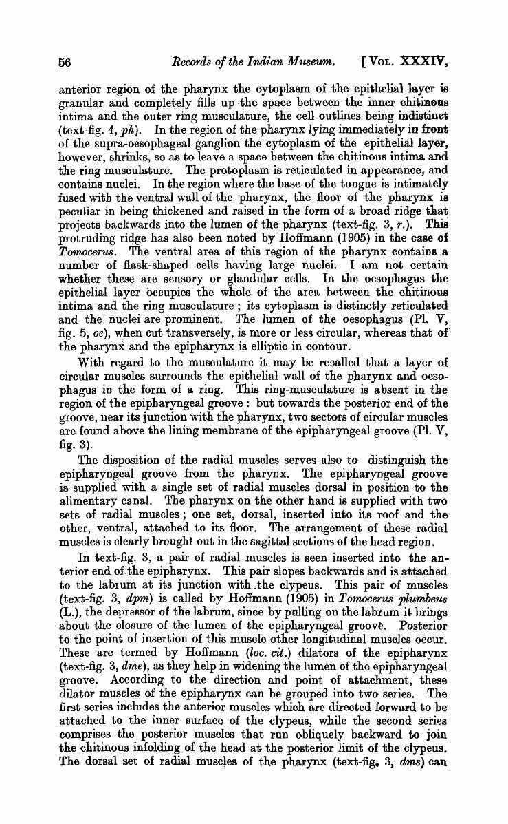
to the epithelial layer : in the anal region,' however, it liesd~tached from the epithelial lining, leaving a narrow spaoe between them. This detachment of the intima from the epithelial membrane may, however, have been caused· by fixation or be due to moulting. The muscular coa.t, external to the epithelial layer, V"ariea in thickness in different regions of the hindgut: in the anterior region the muscular ring is thin and is in olose contact with the epithelial layer ; but towards the posterior end, this ring musculature gains in thickness and in the middle portion of the hindgut forms a broad circular band (text-fig. 5, ems) that oompletely surrounds the gut; but a large clear space intervenes between the inner limits of the muscle ring and the basement membrane of the epithelial layer. The space thus enclosed communicates freely with the haemocoelic oavity of the body: Imms reports the presence of blood corpuscles in this intervening space. Sommer working on Maerotoma plumbea describes a similar cavity in the region of the rectum and hints at its respiratory function, as it is filled with blood. I have noted several granules in this space but I do not find any blood corpuscles in it. The physiological significance of this space is, therefore, obscure; it is possible, however, that it is a mechanical contrivance for the expansion of the hindgut when choked with contents. Towards the posterior end of the hindgut, in the region of the rectum, the muscles are arranged in six longitudinal bands that come into direct contact with the peripheral zone of the epithelial layer and in transverse sections through the anal region, form a series of six projections into the lumen. These projections are conical in shape and small in size (text-fig. 7, cmp). Radial muscles, called' dilatores recti' and' dilatores ani' occur in the rectum., as des .. cribed by Jmms (1906) in Anwrida.
.. -~epv.
TEXT-FIG. 7.-A transverse section through the posterior region of the hindgut: X ca. 533. cmp., musoular projeotions; epi., epithelium of the reotum.
The nature of the epithelial layer of the hindgut, varies somewhat along its length. In the anterior region the cell-boundaries of the epithelial layer are indistinct and the protoplasm of the cells is granular and vacuolated: the chitinous lining is very thin and the cytoplasm of the epithelial layer from its inner surface sends up irregular processes into the lumen. Near . the middle region, where tbe ring musculature is prominent, the epithelial layer and the chitinorF intima are thrown

1932.] DURGADAS MUKERJI: .A. new Oollembole and its Anatomy. 63
into folds and ridges (text-fig. 6, epi) and, as a result, the lumen of the hindgut in this region becomes irregular. In the region of the rectum the epithelial layer, as already mentioned, projects into the lumen of the gut in the form of conical processes, the protoplasm is granular and without cell-boundaries, and in my sections nuclei cannot be detected.
From the nature of the mouth-parts and the buccal cavity, the mouth can be regarded as a sucking apparatus. Willem, (1900) in the case of Neanura (Gerv.) considers the buccal cavity to be adapted for sucking and the tongue, which he terms the ligula, as playing the role of a piston within the buccal cavity. The mandibles and maxillae are considered by him to be perforating organs. As the mandibles in the present instance, are provided with teeth I am of the opinion that the mandibles cut the soft woody tissues and the maxillae help in seizing and bringing food into the mouth by means of the feathery appendage and toothed lamellae attached at their ends. It may be pointed out that in live specimens tihe maxillae are protruded out beyond the oral aperture, and their purpose may, therefore, be to draw food into the buccal cavity and reduce it to pulp, so as to help in the suction of the juice. The dilatation of the buccal cavity and the pharynx by means of muscles will further help in the suction of food. In order to ascertain the nature of their food I kept examples in the laboratory in large, open vessels filled with earth, dug frOIQ. the neighbourhood of a tank and periodically moistened with water. Individuals were kept alive for weeks together, although no food was added. Apparently they lived on the organic vegetable m~tter contained in the soil. According to Macnamara (1924) the Collembola fall into three -categories, viz., (i) vegetable feeders, (ii) carnivorous, and (iii) thosa that live on a liquid diet. Species depending on a vegetable diet possess mandibles provided with a molar surface. In the present instance the mandibles do not possess a molar surface and individuals have not been O'bserved in the field either feeding on animal matter or present in the neighbourhood of a carcase. Further, Macnamara (1924, p. 99) observes" the suctorial species seem to find an innocent nourishment in the fluids of rotting wood and fungi" I presume, therefore, that this species chiefly lives on vegetable nutriment, rich in juice.
The Nervous System.
The nervous system is built on the same plan as that of Podura aquatica (L.), described by Willem (1900) and also differs but little, except in details from that of other examples of the Collembola investigated by Folsom (1889), Willem (1900), Imms (1906), Hoffmann (1908), Hilton (1913) and Denis (1928). It consists of the supra-oesophageal ganglion, the suboesophageal ganglion and the nerve cord comprised of three thoracic ganglia (text-fig. 5). These ganglia usually occur in insects in pairs, but in this species the members of each pair are so intimately fused with each other that they appear as a single ganglion (PI. V, fig. 4, sob).
The supra-oesophageal ganglion (text-fig. 3, sob), formed by the fusion of proto-, deuto- and trito cere brallo bes, gives off paired nerves to the ocelli, antennae and labrum. The labral nerve trunk originates from the supra-oesophageal ganglion near the junction of the latter with the
l

64 Records of the Indian Museum. [VOL. XXXIV,
oesophageal connectives and after coursing forwards over the pharynx, divides into three branches which innervate the pharynx, epipharynx and labrum" respectively. The oesophageal connectives are stout and pass round the oesophagus to connect the supra-oesophageal ganglion with the anterior end of the suboesophageal ganglion. The suboesophageal ganglion (text-fig. 3, sib) is like the supra-oesophageal, a complex ganglion being formed, accorrung to Imms (1906), by the coalescence of four primitive ganglia. It is spindle-shaped and extends below the oesophagus. It gives ofi paired nerves to the mandible, maxilla and labium, and an unpaired nerve (text-fig. 3, lmn) to the lingua or tongue. The mandibular (text-fig. 3 mn) and the maxillary nerves, soon after their origin, divide into two branches: a branch passes to the base of the appendage concerned and terminates in a mass of filiform cells, while the other innervates the muscles supplying the appendage. The labial nerve (text-fig. 3, Zan) arises as a main trunk which divides and ultimately terminates in sensory cells in the labium. The median nerve sUflplying the lingua arises from the anterior end of the suboesophageal ganglion close to the points of attachment of the oesophageal connectives and, passing through the thick ventral wall of the anterior portion of the pharynx, breaks up there into fine fibrils.
A peculiarity worth noting is that in transverse sections through the anterior region of the pharynx or through the epiphaqngeal groove a large mass composed of :Bask-shaped cells (text-fig. 4, epg.; PI. VI, fig. 1) is noticed to lie on either side of the digestive tract. This mass is connected with its member of the opposite side by means of a nerve b~nd (text-fig. 4, nvb) that passes ventral to the digestive tube, a litt.le below the level of the points of insertion of the ventral dilator muscles of the pharynx. Similar masses of cells have been described by HoHmann (1908) in the case of Tomoce'J'us plumbeus (L.) under the name epipharyngeal ganglion. The cells composing the mass are evidently sensory nerve-cells and may be gustatory in function.
Again, in sagittal sections passing through the head a little lateral to the median vertical plane, a large clump of similar cells (text-fig. 3, cv) is noted lying in the ventral chamber of the buccal cavity close to the basal region of the maxilla. The clump (text-fig. 3, cv) is connected with the suboesophageal ganglion by means of a short nerve (text-fig. 3, n) that is swollen in the mid-region into a ganglionated knot. I believe this mass is distinct from the superlingual or maxillary ganglion and different from the cephalic glands of the ventral tube. It seems to me that the constituent cells represent sensory cells but I am uncertain of the exact homology of this organ.
The ventral nerve cord, as in other Collembola, is a concentrated one and is composed of three ganglia that are serially placed in the pro-, meso- and meta-thorax. The meta-thoracic ganglion (text-fig. 5, mtg) is considered to be formed by the fusion of the last thoracic and the first abdominal ganglion which has m.igrated forwards. All the three thoracic ganglia are joined to one another by distinct double connectives. A pair of stout and parallel nerves arise from the last ganglion of the nerve cord (PI. VII, fig. 3, mt) and pass backwards over the ventral tube. This, ltowever, does ~9t reach the posterior e~tremity of the body.

1932.] DURGADAS MUKERJI: A new Collembole and its Anatomy. 65
Oephalic Glands.
Diverse views ha ve been expressed regarding the disposition homology and function of the glands existing in the head region of Collembola. A review of these is given by Folsom (1899), Willem (1900, 1901), Hoffmann (1904), Tmms (1906) and recently by Denis (1928). I, therefore, refer here to the principal controversial points only.
Denis (1928) classifies the glandular systems belonging to the head region of Collembola as follows: (1) Cephalic glands of the ventral tube, composed of (a) tubular glands and (b) globose or acinose glands. Secretions of these glands, according to this author, flow out of the buccal cavity through the labial cleft to the ventral groove and finally reach the ventral tube, which, being thus smeared with the secretions, acts as an adhesive organ. .(2) Salivary glands composed of (a) an anterior pair situated on the external wall of the "pli oral" and opening into the atrial cavity~ and (b) a posterior pair located within the cephalic cavity above the level of the maxillary pouch. The ducts of these glands follow the mandibles closely and open into the buccal cavity. (3) Excretory glands-(Reins Cephaliq·ues)-closed glands without any duct and containing concretions of mates.
Attention may be -directed to the fact that, so far as the structure and function of- the cephalic glands of the ventral tube are concerned, Denis is in general agreement with the observations of Fernald (1890), Willem and Sabbe (1897), Willem (1900, 1901), Hoffmann (1904) and Imms (1906) ; but he differs from Folsom (1899), who held that the secretion of the cephalic glands could not pass to the ventral tube, since the 'vent/ral groove, which was thought by Fernald (1890) to be the connecting passage between the opening of the duct of these glands and the ventral tube, was itself interrupted in places. Folsom (loc. cit.) p.oted further, that the secretion from these glands which were called by him salivary glands, was conveyed through the trough like median cleft of the labium to the border of the mouth to moisten the food before it was taken in. The ducts of these glands were shown to open separately into the buccal cavity and thus they differed in their course from that indicated by Willem and his school.
The cephalic glands of the ventral tube have, on more than one occasion, been confused with th~ salivary glands. Fernald (1890), who first showed that a morphological connection between the cephalic glands and the ventral tube existed by means of the ventral groove, called these glands salivary glands. Willem and Sabbe (1897), to whom the credit belongs for explaining the functional correlation between the cephalic glands and the ventral tube alluded to, called these glands, since they supply adhesive substance to the ventral tube, the cephalic glands of the ventral tube. Willem (1901) clearly distinguished the cephalic glands of the ventral tube from the salivary glands, and pointed out that the salivary glands mentioned by Tullberg and Nassanow were tubular glands, and that Lubbock (1862, 1873) and OHler (1862) mistook adipose cells for these glands. According to him, the cephalic glands of the ventral tube are composed of tubular and globose glands; the ducts of the tubular glands anteriorly receive the ducts from the globose ~lands and e~en b:r a common duct into the buccal cavity close to the
(2

66 Records of tke Indian Museum. [VOL. XXXIV,
labial cleft on the ventral side of the head; further, in addition to these, a pair of salivary glands occurs in the head and their ducts are quite distinct from those of the tubular and globose glands, and open on the hypopharynx. Hoffmann (1904) studied the structuxal details of the cephalic glands of the ventral tube and agreed in most respects with Willem. Imms (1906) accepted the nomenclature of the glands enunciated by Willem and HofImann but pointed out that Fernald mistook the cephalic glands for the salivary glands. Somm,er (1884) and Folsom (1899) on the other hand advocated different views. Sommer (1884) reported that in place of the so-called salivary glands recorded by earlier authors, cells containing concretions of urate occur, which must have been mistaken by them for the salivary glands. Folsom (1899) maintained that the salivary glands, as meant by him, weI'e homologous to the salivary glands depicted by .Tullberg (1871) and N assanow (1887). Besides these Folsom described another pair of glands, situated close to the bases of the mandibles and maxillae and having chitinous ducts which coursed down between the mandible and the maxilla. It seems from the topography and structure of the glands that those mentioned by Folsom as lying near the mandible really correspond to the salivary glands mentioned by Willem and the salivary glands described by Folsom are comparable to the tubular glands indicated by Denis. Philiptschenko (1908) describes two pairs of salivary glands, an anterior and a posterior, and also a pair of tubular glaD ds. He regards the tubular glands as excretory in function.
The homology of these cephalic glands is by no means certain. Willem considers that the tubular glands belong to the segment corresponding to the first maxilla and that the salivary glands belong to the mandibular segment. Imms on the basis of Uzel's observation concludes that the salivary glands are homologous with the shell glands of Crustacea. Phlliptschenko assigns the tubular glands to the labial segment and -regards them as homologous with the antennary and maxillary glands of Crustacea and representing the head kidney. The anterior pair of salivary glands, according to him belongs to the mandibular segment and the posterior pair of salivary glands to the labial segment. The salivary glands, further, in his view are comparable to the cephalic glands of Lithobius and Scolopendra. Denis, howevel, is of the opinion that both the tubular and globose glands belong to the labial segment and the salivary glands to the mandibular segment. He points out also that the anterior pair of salivary glands, mentioned by Phlliptschenko, correspond to the salivary glands described by him, while the posterior ones correspond to the globose glands.
With regard to the excretory glands described by Denis it may be said, that they too are not beyond the range of controversy. HofImann (1904) has the credit of first reporting these glands as " Kopfnieren " : but Becker (1910) contests that they are sensory cells of the post-antennal organ. Nabert (1913) is opposed to Becker's view but Quiel (1915) supports the latter. Denis (1928) upholds Hoffmann's observation, and, further, is of the opinion that though these glands can be compared with fat cells containing a deposit of urate, any homology between them ~,:p.d the antennary and maxillary glands of Crustacea, a·s indicp,t.ed by

1932.] lJURGADAS MUKERJI: A new Ooilemhole and its Anatorny. 67
Philip tsc henko, is untenable. He considers these glands as belonging to the trito-cephalic metamere.
It is likely that the above difierences of opinion may to a certain extent have been caused by the structural similarity hinted ~t by some of the authors, between salivary, tubular and acinose glands. and also by the difficulty of tracing the ducts of these glands; but the possibility of variation in number, ~rrangement and disposition of these glands in different groups or species, cannot be ignored. Further observations on species other than those already investigated are needed before any definite conclusion can be reached.
The glandular organs located within the head region of the present species of Protanura, occur iu pairs and fall, according to their structure, into two distinct systems: (a) tubular glands, (b) acinose or globose glands. In dividing the cephalic glands into the above two categories only, I have excluded those structures which have not been established beyond doubt to be true glandular elements and which, on account of their morphology, have been included by me in the nervous system as sensory cells; and I have relied neither on the topographic position of the glandular organs in the body, which itself is variable, nor on the course of their ducts, which cannot be traced without some element of doubt, but have been pr~cipally guided by their form and histology.
I also refrain from homologising the above two types of glands or giving any definite names to them, as, so far, previous authors have not made out a clear histological difierentiation between the acinose gland proper and the Salivary gland or between the tubular gland and the salivary g~and.
(a) Tubular gland.
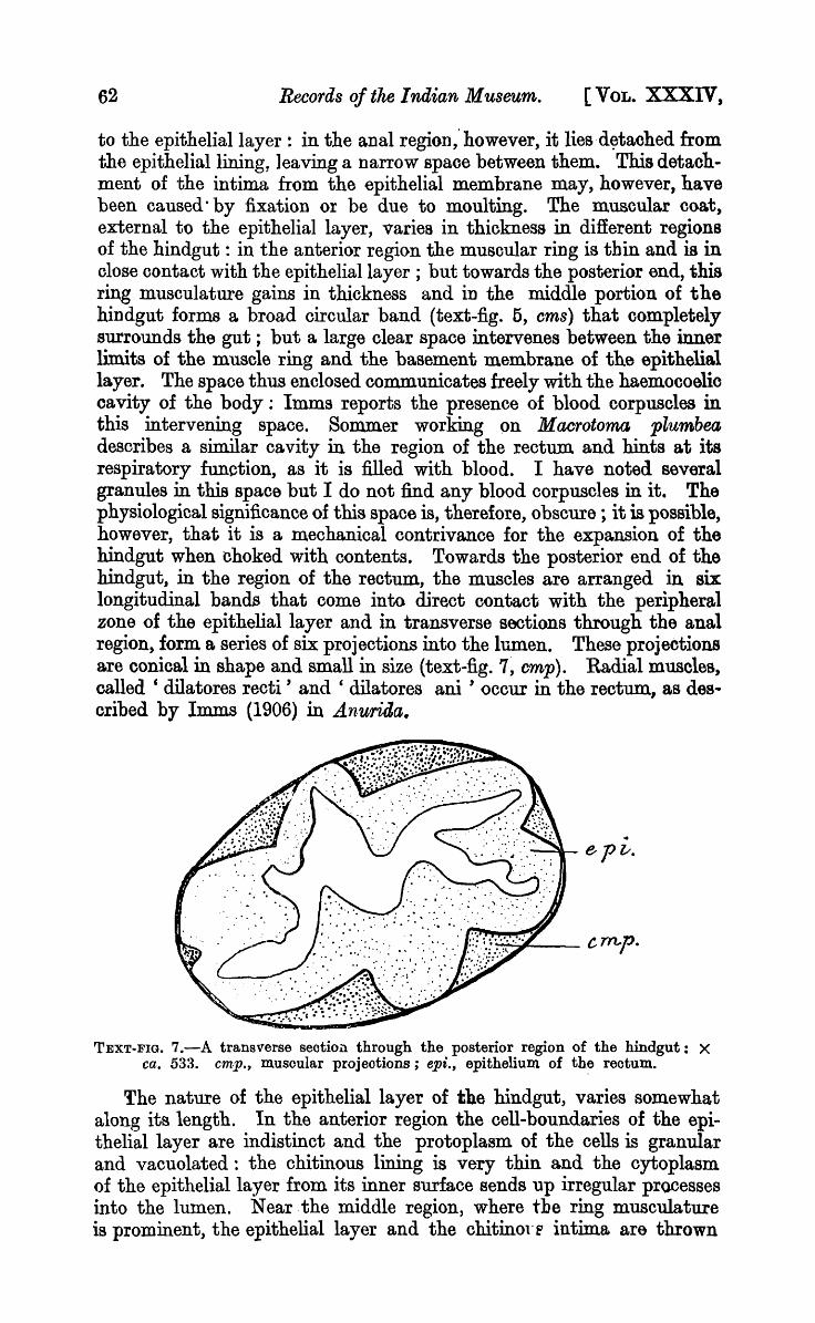
The tubular gland (PI. V I, fig. 3, tbg), in its essential principle, is built up of a long convoluted tube with a well defined lumen and a wall composed of a single layer· of gland cells, and extends from the middle region of the head as far as the metathorax, lying on the sides of the alimentary canal. It can be subdivided into the following three parb; : (a) a sac, (b) a labyrinth, and (c) a duct.
The sac (PI. "I,. fig. 2, sgl) is situated in the prothoracic region and encloses a large cavity and is compressed from side to side. The sac as flne traces it towards the posterior end of the body, becomes narrower and cylindrical, and on reaching the metathorax, bends down in the form of a U (PI. v II, fig. 1, tbg.). The lower limb of this is continued forwards as the labyrinth. The labyrinth, which has a narrow bore, passes anteriorly below the sac, following a somewhat winding course, and on reaching the prothoracic region ascends upwards and comes to lie above the level of the sac (text-fig. 8). In the anterior region of the pr.othorax, it forms loops. A dorsal loop (PI. VII, fig. 2, dl) is given off which passes into the head and comes very close to the anterior acinose glands of the head, and a ventral loop (PI. VII, fig. 2, vl) that continues forward as the duct of the tubular gland and, crossing the level of the suboesophageal ganglion, lies on the floor of the buccal cavity. The duct which is narrower than the labyrinth passes onward through the hypodermalla yer and joins its fellow of the opposite side to form a common
68 Records of the Indian Museum. [VOL. XXXIV,
chitinous duct which slopes down to open to the exterior on the ventral surface of the head immediately posterior to the median gap between the abial pieces.
dt.
TExrr.ll'IG. S.-Diagram of the tn bular gland. sgl., sac; lbg., labyrinth; dl., dorsa.lloop; dct., duct.
The end-sac and the labyrinth, which together constitute the greater portion of the tubular gland, represent the secretory part of the organ and the duct leads the secreted substance to the exterior. The tubular gland and its duct thxoughout their length are lined by a single layer of cells but the character of these lining cells varies in the three parts into which the tubular gland has been divided by me. In the end-sa.c the cells are rectangular, being longer than broad. Cell-boundaries are quite distinct and the nuclei are prominent. In a few sections, these eells, when examined under an oil-immersion lens, revealed the presence of very fine canals. Possibly the secretions of these cells pass through these canals into the lumen of the sac. The cavity of the saC enclosed by these cells is spacious in comparison to that of the labyrinth or the duct. The secretions evidently first collect in the lumen of this sac, and are then conducted to the exterior; the end-sac therefore may be looked upon as forming a reservoir, the wall of which is secretory. The glandular cells lining the labyrinth, though similar to those of the endsac, differ from them in shape and size and in the absence of fine canals. The walls of the duct are thicker than those of the preceding portions and differ from these in that cell-boundaries are here absent and the lining appears to form a syncytium.
The divisions of the tubular gland as made here correspond, to some extent, to those indicated by Philiptschenko (1908) with this difference that tho sac is not thin-walled.
The shape and the course of the tubular gland in this species, it may be noted, closely resemble that of the tubular gland of Orchesella described by Willero (1901), but with this difierence that the common duct in the present instance does not receive the ducts of the acinose glands situated in the anterior rogion of the head, as shown by Willem. The gland differs from the tubular gland described by Hofimann both as regards the character of the common duct and the convolutions of the tube.
(b) Acinose Glands.
The Acinose gland comprises a group of unicellular glands, each with a fine ductiole which opens into a common chitinous duct of narrow bore and without an epithelial wall of its own.
1932.] DURGADAS MUKERJI: ... 4 new Oollembole and its ... 4natomy~ 69
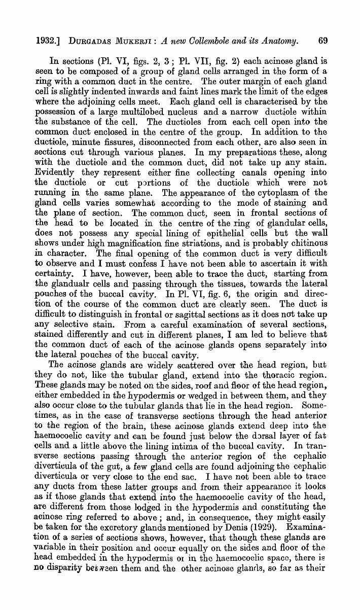
In sections (PI. VI, figs. 2, 3; PI. VII, fig. 2) each acinose gland is seen to be composed of a group of gland cells arranged in the form of a ring with a common duct in the centre. The outer margin of each gland cell is slightly indented inwards and faint lines mark the limit of the edges where the adjoining cells meet. Each gland cell is characterised by the possession of a large multilobed nucleus and a narrow ductiole within the substance of the cell. The ductioles from each cell open into the common duct enclosed in the centre of the group. In addition to the ductiole, minute fissures, disconnected from each other, are also seen in sections cut through various planes. In my preparations these, along with the ductiole and the common duct, did not take up any stain. Evidently they represent either fine collecting canals opening into the ductiole or cut pJrtions of the ductiole which were not running in the same plane. The appearance of the cytoplasm of the gland cells varies somewhat according to the mode of staining and the plane of section. The common duct, seen in frontal sections of the head to be located in the centre of the ring of glandular cells, does not possess any special lining of epithelial cells but the wall shows under high magnification fine striations, and is probably chitinous in character. The final opening of the common duct is very difficult to observe and I must confess I have not been able to ascertain it with certainty. I have, however, been able to trace the duct, starting from the glandualr cells and passing through the tissues, towards the lateral pouches of the buccal cavity_ In PI. VI, fig. 6, the origin and direction of the course of the common duct are clearly seen. The duct is difficult to distinguish in frontal or sagittal sections as it does not take up any selective stain. From a careful examination of several sections, stained differently and cut in different planes, I am led to believe that the common duct of each of the acinose glands opens separately into the lateral pouches of the buccal cavity. .
The acinose glands are widely scattered over the head region, but they do not, like the tubular gland, extend into the thoracic region. These glands may be noted on the sides, roof and floor of the head region, either embedded in the hypodermis or wedged in between them, and they also occur close to the tubular glands that lie in the head region. Sometimes, as in the case of transverse sections through the head anterior to the region of the brain, these acinose glands extend deep into the haemocoelic cavity and can be found just below the d:lrsallayer of fat cells and a little above the lining intima of the buccal cavity. In transverse sections passing through the anterior region of the cephalic diverticula of the gut, a few gland cells are found adjoining the cephalic diverticula or very close to the end sac. I have not been able to trace any ducts from these latter groups and from their appearance it looks as if those glands that extend into the haemocoelic cavity of the head, are different from those lodged in the hypodermis and constituting the acinose ring referred to above; and, in consequence, they might easily be taken for the excretory glands mentioned by Denis (1929). Examination of a series of sections shows, however, that though these glands are variable in their position and occur equally on the sides and floor of the head embedded in the hypodermis or in the haemocoelic spaco, there is no disparity bet N3en them and the other acinose glanrls, so far as their

70 Records of tlte 1 ndian Museum. [VOL. XXXIV,
structure is concerned. I consider them, therefore, irrespective of their topographical position and size, as component parts of the same gland system. The excretory glands described by Denis, it may be remembered, are without any duot and contain concretions of urates. The number of groups of aci~ose glands can roughly be computed from the frontal and sagittal sections of the head. There are four pairs in the head. fley are distributed as follows: a pair in the anterior region of the head, a pair in the basal region of the head and two pairs, one behind the other, intervening between the anterior and the posterior pairs. The anterior and the posterior pairs are situated near the dorsal wall of the head. The intervening pairs occupy a more lateral position in the ventro-la~eral portions of the he·ad. Each group possesses a duct of its own which opens into the lateral pouches of the buccal cavity.
Since all four pairs of acinose glands closely resemble each other in structure and in the position of the openings of their ducts they cannot be separated into salivary glands and acinose glands. The acinose glands proper, it may be remembered, have been pointed out by previous workers as opening into the anterior end of the common duct of the tubular gland and the salivary glands have been shown to have ducts of their own :opening independently into the buccal cavity. In this case no such difference could be made out and, therefore, the observations recorded here differ from those of previous workers. The whole series appears to belong to the group hitherto called salivary glands by Denis (1928).
So far as the function of the acinose glands are concerned, as these glands open into the buccal cavity and as no separate salivary glands have been found by me, it is possible that they act as salivary glands. That the secretions liberated by them have a softening effect on succulent vegetable tissues, the juices of which the animals probably suck, has been mentioned by Willem (1900) in the case of Nean1tra. I do not agree, therefore,· with Denis that both salivary and acinose glands occur as separate glands though -the posterior pairs of acinose glands, from their topographic position. at the base of the head, may corlespond to the so-called salivary glands.
Ventral Tube.
The ventral tube has been the subject of much discussion. The conflicting views of earliel' authors have been summarised by HofImann (1904) and Imms (1906), it is therefore, unnecessary, to state them again here beyond mentioning the chief controversial points. Previous authors ascribed particular functions to the ventral tube, such as those of support, reproduction, respiration, lubrioation, absorption of moisture from the surface of the body and adhesion. Opposing views are also expressed as to the presence or absence of secretory gland cells within it. The consensus of opinion at present is in favour of regarding the ventral tube as an organ of adhesion, enabling the Collembola to walk over smooth surfaces; but the way in which suoh adhesion is effected has not been settled beyond dispute. Again,' while its adhesive function is admitted, the possibility of a respiratory function is also emphasised (vide Willem, 1900; Hofinlann, 1904; Imms, 1907). Macnammara (1919), however,

.1932.] DURGADAS MUXERji: A new Oollembole and its Anatomy. 71
denies its respiratory function. The diverse views as to the manner, in which the adhesion of the ventral tube to a smooth surface is effected, may be grouped for sake of convenience under three different categories: (a) mechanical adhesion by suction, (b) adhesion helped by the secretion of adhesive substance elaborated by the glands of the ventral tube itself, (c) adhesion by reasons of the adhesive substance supplied to the ventral tube by the cephalic glands. Bonrelet, Oller, and Tullberg partly support the first view. The opinions of de Geer, Nicolet, Lubbock Maonammara and Tillyard come under the second category; while Willem, Hoffmann, Imms and Denis hold the last.
In view of the contradictory statements about the ventral tube the neoessity of re-investigation is olear. I have, therefore, studied the organ in the living state as well as by microscopical sections. It may also be Doted that the observations of previous authors were based upon specimens belonging to genera other than Protanura Borner and it is hoped, therefore, that a comparison of my observations with those of previous authors will be fruitful in clearing up the doubts about its function.
The ventral tube is a cylindrical organ, the terminal portion of which is divided into two equal ha1ves by a median furrow. It is situated on the first ventral abdominal segment. Comparatively speaking, the ventral tube is small in the present species and in the ordinary resting state appears as a small medially cleft knob, the apex of which is nearly flush with the ventral surface of the other abdominal segments but when extended the ventral tube is nearly perpendicular to the longitudinal axis of the body. The groove dividing the organ into two symmetrical halves runs, in the contracted conditio!!, in an anterio-posterior directIon parallel to the longitudinal axis of the body and nearly reaches to the base of the tube. In the extended condition two small flat lobes, one at the tip of each half, are simultaneously evaginated and pressed against the surface on which the animal moves. The portion of the ventral tube, within whioh the eversible lobes can be withdrawn, may be called the collar. The lobes are spoken of by Tillyard (1925) as eversible sacs. In the retracted condition the eversible lobes remain covered by the extension of the external wall of the tube up to its apex, and only in t,he extended state oan the ventral tube be differentiated, as sho\vn by Hoffmann (1904), int·o (a) a cylindrical tube, (b) a collar and (c) the eversible lobes. The ventral tube is hollow and highly contractile and the contraction is brought about by means of muscles that pass through the cylindrical tube into each of the eversible· sacs. Elongation of the ventral tube possibly takes place, as is held to be the case by most of t,he authors alluded to above, as the result of turgescence produced by the inflow of blood, controlled by the ventral muscles of the abdomen which when eontracted bend the body of the insect and force blood into the ventral tube.
Since the chief points of dispute centre' round ~he question whether the eversible sacs of the ventral tube exude a sticky substanoe, the olue to the solution lies in the presence or absenoe of glandular elements, in or· conneoted with the ventral tube. In PI. VII, fig. 4, a transverse section through the terminal region of the ventral tube is given. TIle

72 Records of the Indian 'Museum. [VOL. XXXIV;
plane of the section passes slightly oblique to the longitudinal axis of the ventral tube and in oonsequence the two halves are not symmetrically disposed. The deep median furrow is seen to divide the distal ena of the ventral tube into right and left lateral halves or lobes. The chitinous cuticle not only surrounds the ventral tube but also runs within the furrow separating the two lobes. A narrow cavity is s~n inside each lobe~ The inner wall of each lobe facing the median furrow contains a thick layer of large cells with distinct nuclei (ggl). These cells are the glandular elements in the ventral tube. The outer portion of the wall of each 10 be contains a thin peripheral layer of hypodermis (hy) and a layer of fat cells ~djoining it. In transverse sections of the body (text-fig. 9) passing
TEXT-FIG. 9.-A section of the ventral tube cut by a, plane passing tra.ns~erecly across the body: x ca. 146_ ggl., glandu18ir layer of cells of the ventral tube.
through the tip of the ventral tube and, therefore, through the eversible sacs, tIle cavity enolosed within' each lobe is seen to be lined on all sides by a layer of glandular cells (ggl) and the hypodermal layer and fat cells are found confined to the external wall of the ventral tube. Such a condition is due to the tucking in of the eversible sac within the tube, and the glandular cells lining the cavity of the each lobe represent, therefore, the external wall of the eversible sac whose glandular nature thereby is established. Sagittal sections through the ventral tube (PI. VII, fig. 3, vt) are also instructive inasmuch as they serve to show the extent of the gland cells along the length of the ventral tube. In such sections the glandular cells, containing granular cytoplasm and large nuclei, are found at the tip of the ventral tube. The hypodermal layer on the anterior and the posterior walls of the tube, it may be observed, is very thin and the constituent cells can be easily distinguished from the glandular cells by the size and character of their cytoplasm and nuclei. The cavity of the ventral tube, as may be seen, is largely encroached on by the inner layer of fat cells_
It may here be pointed out that glandular elements in the apioa.l region of the ventral tube were long ago shown to be present by Sommer (1884); but Hoffmann (1904) contested that the so-called glandular cells were really hypodermal cells. Since the same objection, in spite of the clear distinction between the hypodermal and the glandular cells made in this case, may crop up again, I have attempted t~ check the results derived from morphological investigation by direct observation on living specimens. Direct exudation of fluid from the tip of the ven .. tral tube is a convincing proof of the presence of glandular eleme~ts in the ventral tube. Before I pass on to record my' observations on live specimens a brief reference to the ventral groove must be made as the

1932.] DURGADAS MUKERJI: A new Oollembole ani/, its Anatomy. 73
latter morphologically appears to connect the external openings of the tubular glands and the ventral tube, as was first shown by Willem and Sabbe (1897).
The ventral groove (PI. V, fig. 5, vgr) runs in an anterio-posterior direction along the longitudinal a,xis of the body on the ventral surface. It starts from the posterior end of the labial cleft and reaches as far as the ventral tube. The anterior portion of the groove which is very small in comparison to the remaining portion, can be separated from the remainder by the difference in the character of the groove and by the presence of t~e external opening of the common duct of the tubular glands. The external opening of the common duct, which opens into the groove at the point of separation of the anterior and the posterior portions of the groove, is very difficult to see, but its position can easily be located by a reference to the configuration of the internal structures" The external opening of the duct and, consequently, the line of demaroation between the anterior and the posterior portions of the ventral groove lie in a plane that cuts the head transversely and passes through the extreme anterior limit of the supra-oesophageal ganglion. The anterior portion of the groove that serves as the external communicating passage between the ventral cleft of the labium and the external opening of the common duct of the tubula.r glands is formed by the infolding of the external chitinous cuticle investing the ventral side of the head. The groove in transverse section appears as a narrow-necked flask the broad inner end of .the flask protruding slightly into the haemocoelic ca vity. The ventral cleft of the labium, which posteriorly meets the anterior portion of this groove, extends as far as the oral aperture. From my 0 bservations on live specimens and from its structural peculiarity, it is inferred that the secretion from the tubular glands is conducted forward ah.ng the anterior portion of the groove to the mouth and not backward to the ventral tube.
The posterior portion of the ventral groove, reaching back as far as the ventral tube, is formed by the curling in of the edges of the chitinous cuticle of the body. ~he two bent limbs of the groove look like curved horns attached dorsally and directed downwards (PI. V, fig. 5, vgr). In spite of the difference between the anterior and posterior portions of the groove, the two portions are continuous with each other. The gro ove morphologically forms a communicating channel between the ventral cleft of the labium and the ventral tube and is not interrupted as mentioned by Folsom (1899). Folsom's contention that the ventral groove is ill adapted for the conveyance of a sticky fluid, secreted by the cephalic glands, from the base of the head to as far as the ventral tube seems justifiable for reasons other than that of the discontinuity of the groove. The passage of a sticky Huid over a compara.tively long distance through a narrow open chitinous channel, exposed below to dust and atmospheric air, seems untenable on physical grounds; but such objection cannot be raised with equal force if the fluid secreted passes anteriorly from the opening of the common duct of the tubular glands to the ventral cleft of the labium and thus to the mouth, as in such a case the substance extruded has only to traverse a very short distance through the anterior portion of the groove.

'74 Records of"tke Indian Museum. [VOL. XXXIV,
Live speoimens of different sizes with a small quantity of the debris ~()f earth, on which they are fOlmd in the natural state, were gently put iin a small glass vial with a narrow neck. As examples of the present lspecies do not survive in a dry atmosphere, a few drops of water were :introduced into the tube to keep the contents moist. The specimens ]lived for a week and were active in their habits. Observations of their Jrnovements were made from time to time under a binocular dissecting lmioroscope and the glass plate, to whioh they were transferred, was from -time to time turned over to facilitate observation of the organ from the 'Ventral aide. The animals progressed on vertical walls with the tips of -their legs resting on the surface and the ventral tube in tbe contI'ltcted Istate: the eversible lobes of the ventral tube were seen at short intervals to be closely applied against the surface of the glass and appeared to act as a sucker. The pair of lobes, when pressed against the glass, appeared to be soft, membraneous and moist. In a few cases the evaginated lobes, while adhering to the surface, were found to be drawn forward, sliding over the surface of the glass, to a distance equalling half the length of the animal. In no case, however, were the insects found to keep the end of the ventral tube continuously in touch with the sur:face. The terminal portion of the tube was attached to the surfaoce by means of the lobes whenever the legs appeared to slip against the glass and the lobes at this time secreted fine droplets of fluid. The animals behaved similarly when they ran upside down on the horizontal undersurface of the glass plate. The movements of these insects were watohed for hours but in no case was any viscous fluid noticed to be running along the ventral groove. On the contrary some individuals after walking f ()r an hour or so, stopped moving, pressed the end of the ventral tube against the surface of the glass and remained with their bodies hanging in the inverted position with the tips of their legs resting upon the lower surface of the glass: in this position, they bent their body: their fore legs were drawn near the mounh and a drop of viscous fluid was noticed to 'collect gradually at the mouth. It is possible, as mentioned before, that the fluid was derived f.rom the tubular glands and flowed from the external opening of the common duot of the gland to the mouth through the short anterior portion "of the ventral groove. The tips of the fore legs were then brought in contact with the fluid and thus became smeared with it. By the contraction of the body the middle legs were also drawn close to the mouth and the tips of these legs, later on, rubbed against each other. Thus·the fluid, collected neal' the mouth, was used to smear the tips of the legs. Aft'er this the animals again began to walk upon the tips of their legs·in the inverted position. All these operations could be clearly seen, and it seems obviQUS that the extruded secretion was sticky in nature and that the legs were smeared with the secreted fluid to enable the animals to move over smooth surfaces with agility.
It is interesting to Dote that though Reuter (1880) was wrong in his interpretation that the tarsal claws gathered water to the mouth, he must have noticed that these claws were brought to the mouth, as has been observed by me.
It is seen hom above that lny observations, based on this species, lend support to Folsom's view (1899) that secretions elaborated by the
1932.] DURGADAS MUKERJI: A new Oollembole and its Anatomy. 75
tubular glands, pass to the mouth and do not reach the ventral tube through the ventral groove as advooated by Will em , Hoffmann, Imms and Denis. I differ, however, from Folsom in my belief that the secretions of these glands are smeared over the olaws of the feet and thereby help the animal to walk over smooth surfaces irrespective of tl1e presence or absence of tenent hairs (cf. Packard, 1909, p. 99). It is also possible that these secretions may have a cleaning effect on structures which are smeared with them, and I do not deny the possibility that the seoretions of the tubular glands, conducted to the mouth, may become mixed up with the secretions of the acinose glands and have a softening effect on the food taken in by the mouth: but observations on live specimens do not lend support to this inference.
From the structure of the tubular glands we have seen that they resemble the tubular glands desoribed by Bruntz and Philiptsohenko, and may, therefore, be comparable to the excretory glands of Crustacea. Even if such a homology be held to be correct, the change in function, as indicated above, is not hard to reconcile, in view of the fact that the last nephridium of the land leeches exudes fluid adhesive in action (Moore, 1927).
With regard to the function of the ventral tube I wholly agree with FGlsom, Maonammara and Tillyard that the ventral tube exudes adhesive seoretions. Sommer, therefore, was right in indicating the glandular elements in the ventral tube, though he was led astray in attributing to it a function like the preening gland of a bird. A special respiratory function of the ventral tube, ascribed to it by Hoffmann and others, can not be entertained, for, in the absence of tracheae the surface of the body appears to be well adapted for cutel,ueous respiration and the ventral tube does not possess any special features adapted for carrying on gaseous exchange.
BIBLIOGRAPHY.
1. Absolon, K. (1901). Dber Neanura tenebrarum D. Bp. aus den Hohlen der mahrischen karstes; iiber die Gattlmg Tetrodontopkora Reuter und einige SinneS()rgane der Collembolen. ZooZ. Anz., Vol. XXIV, pp. 575-585.
2.1 Becker, E. (1903). Anatomic comparee des glandes cephaliques chez les Collemboles. Mem. Soc. Amis S. N Et'f. Moscow, Vol. XCVIII (en russe).
3. Becker, E. (1910). Bau des Post-antennal organs der Collembolen. Zeits. Wiss. Zool., Vol. XCIV, pp. 327-399.
4. Berlese, A. (1909). Gli Insetti. Milano, Vol. I, p. 530. 5. Borner, C. (1901). Vorlalifige Mittheilungen liber einige Aphoru ..
rinen und zur Systematik der Collembolen. Zool. Anz., Vol. XXIV, pp. 1-15.
6. Borner, C. (1906). Das System der Collembolen nebst Beschreibung neuer C.)llembolen des Hamburger naturhistorischen Museums. Mit. Nat. Mus. Hamburg, Vol. XXIII, pp. 147-188.
1 Not accessible to the author.

76 Records of tke Indian Museum. [VOL. XXXIV,
7. Borner, C. (1908). Apterygoten (1), Collembolen aU8 Siidafrika nebst einer studien iiber die erste Maxille der Collembolen, in Schultze Forschungreise in Westlichen und zentra.Ien Sudafrika. Jenaische Denkschr., Vol. XIII, pp. 53-68.
8. Borner, C. (1909). Neue Homologien zwischen Cru.stacean und Hexapod en. Die Beissmandibel der Insekten und ihre Phylogenetische Bedeutung. Zool. Anz., Vo1. XXXIV, pp. 100-125.
9. Borner, C. (1913). Die Familien der Collembolen. Zool. Anz., Vol. XLI, pp. 315-322.
10. Borner, C. (1914). Die Gliedmassen der Arthropoden. Lang Handbuch der Wirbellosen Tjere~ Jena, Vol. IV, pp. 649-694.
11. Bourlet, l'abbe, (1841-42). Memoires sur les Podurelles. Mem. Soc. Roy. Et. Centro l'Agric. Sci. et. Arts. Douai.
12. Bruntz, L. (1908a). Les reines labiaux· et les glandes cephaliques des ThysanoUl'es. .Arch. Zool. Exp. (4), Vol. IX, pp. 95-228.
13.1 Bruntz, L. (1908b). Note sur l'anatomie et la physiologiques des Thysanouree. C. R. Soc. Biol. Paris, Vol. LXIV, No.5, p. 231.
14. Carpenter, G. H. (1904). Collembola.. Fauna Hawaiiensis, Vol. Ill, Part 4, pp. 299-303.
15. Carpenter, G. H. (1903). On the relationships between the classes of the Arthropoda. Proc. Roy. Irish Acad., Vol. XXIV, Sec. B, pp. 320-353.
16. Carpenter, G. H. (1916). The Apterygota of the Seychelles. P'1oc. Roy. Irish. Acad., Vol. XXXIII, Sec. B, pp. 1-70.
17. Carpenter, G. H. (1917). Zoological results of the Abor Expedition (1911-12). Rec. Ind. Mus., Vol. VIII, pp. 561-568.
18. Carpenter, G. H. (1924). Collembola of the Siju Cave, A8~am. ReG. Ind. Mus., Vol. XXXVI, pp. 285-289.
19. Crampton, G. C. (1921 a). The origin and homologies of the so-called Super-linguae or Paraglossae (Paragnatlts) of insects and rel~ted Arthropods. Psyche, Vol. XXVIII, pp. 84-92.
20.1 Crampton, G. C. (1921 b). The phylogenetic origin of the mandibles of Insects and their arthropodan relatives. A ,contribution to the study of the evolution of Arthropoda. Journ. New YO'1k Ent. Soc., Vol. XXIX, pp. 63-100.
21. Crampton, G. C.- (1922). A comparison of the first maxillae 9f apterygotan insects and crustacea from the standpoint of Phylogeny. Proc. Ent. Soc. Washington, Vol. XXIV, pp. 65-82.
22. Collinge, W. E. (1910). Collembola as injurious insects. J.' Eerm. Ent., Vol. III, pp. 204-205.
23. Davenport, C. B. (1903). The Collembola of cold spring beach with special reference to the movements of the Poduridae. Cold Spring Harbour Monograph, Vol. II, p. 32.
24. Denis, J. R. (1928). Etudes sur l'anatomie de la Tete des quelques Collemboles. Arch. Zool. Exp. Gen. Vol. LXVIII, pp. 1-291.
25. Evans, A. M. (1921). On the structure and occurrence of MaxilluIae· in the orders of insects. J ourn. Linn. Soc. LondO'1l, Zool., Vol. XXXIV, pp. 429-456.
1 Not accessibI() to the autho~.

1932.] DURGADAS MUKERJI: ,A. new Oollembole and its Anatomy. 77
26. Fernald, H. T. (1890). The Relationships of Arthropods. Studies Biol. Lab. Johns Hopkins Univ., Vol. IV, No.7, pp. 431-513.
27 Folsom, J. W (1899). The Anatomy and Physiology of the mouthparts of the Collembolan Orchesella cincta L.; Bull. Mus. Compo Zool. Harvard, Vol. XXXV, No.2, pp. 7-39.
28. Folsom, J. W (1900). The Development of the mouth-parts of Anurida maritima Guer. Bull. Mus. Compo Zool. Harvard., Vol. XXXVI, No.5, pp. 87-157
29. Folsom, J. W (1916). North American Collembolous insects of the subfamilies Achorutinae, Neanurinae, and Poduridae. Proc. U. S. Nat. Mus., Vol. L, pp. 477-525.
30. Gervais, P. (1842). Une quinzaine d'especes d'insectes apteres. Ann. Soc. Ent. France, Vol. XI, Bull., p. XLVII.
31. Gervais, P. (1844). In Walckenaer Insp: Histoire naturelle des insectes apteres. Paris, VoL III, pp. 377-456.
32. de Geer, Ch. (1783). Abhandlungen zur Geschichte der Insekten. Vol. VII, Nurnberg.
33. Handschin, E. (1929). Beitrage zur Collembolen Fauna von Sud-Indian. Rev. Suisse. Zool., T. XXXVI, No. 16, pp. 229-262.
34. Haase, E. (1889). Die abdominalanhange der Insekten. Morph. J ahrbuch., Vol. XV, pp. 331-435.
35. Heymons, R. (1897). 'Ober die Bildung und den Bau des Darmkanals bei niederen Insekten. SB. Ges. N aturf. Berlin, p. 111.
36. Hilton, (1913). Nervous System of Collembola. J. Ent. Zool., Vol. V, pp. 37-42.
37. Hoffmann, R. W (1904). TIber den Ventraltubus von Tomocerus plumbeus L., und seine Beziehungen zu den gross en unteren Kopfdrtisen. Ein Beitrag zur Kenntnis der Collembolen I. Zool. Anz. Vol. XXVIII, N"o. 3, pp. 87-115.
38. Hoffmann, R. W. (1905). Dber die Morphologie und die Funktion der Kauwerkzeuge von Tomocerus plumbeus L. II. Beitrag zur Kenntnis der Collembolen: Zeits. Wiss. Zool., Vol. LXXXII, pp. 639-662.
39. Hoffmann, R. W (1908). Ober die Morphologie und die Funktion der Kauwerkzeuge und tiber das Kopfnerven system von Tomocerus plumbeus L. III. Beitrage zur Kenntnis der Collembolen. Zeits. Wiss. Zool., Vol. LXXXIX, pp. 598-689.
40. Hofimann, R. W (1911). Die Entwicklung der Mundwerkzeuge von Tomocerus plumbeus L. Zool. Anz., Vol. XXXVII, pp. 353-377.
41. Imms, A. D. (1906). Anurida. L. M. B. O. Memoirs, Vol. XXIII, pp. 1-99.
42. Imms, A. D. (1912). On some Collembola from India, Burma and Ceylon, with a catalogue of Oriental species of the Order. Pl'OC.
Zool. Soc. Land., Vol. I, pp. 80-125. 43. Imms, A. D. (1925). Text-book of Entomolgy. 44.1 Kolenati, F. A. (1858). Zwei neue Osterreichische Poduriden.
Wien. Sitzbr. d. Matk.-Nat. Ol. d. k. Akad d. Wiss., Vol. XXIX.
1 Not accessible to the aut!lOr.

78 Records oftke Indian Museum. [VOL. XXXIV,
45. Lubbock, J. (1862). Notes on the Thysanura. Trans. Linn. Soc., Vol. XXIII, pp. 569-601.
46. Lubbock, J. (1873). Monograph of the Collembola and Thysanura. Ray. Soc., London. _
47. MacGillivray, A. D. (1893). North American Thysanura. Cando Ent., Vol. XXV, pp. 313-318.
48. Macnammara, C. (1919). Popular and Practica1 Entomology. Canada Ent.~ Vol. LI, pp. 73-80.
49. Macnam,mara, C. (1924). Food of Collembola. Cand. Ent., Vol. LVI, pp. 99-105.
50. Moore, P. (1927). Leeches. Fauna Brit. Ind., pp. 246-247. 51. Nabert, A. (1913). Die Corpora allata der Insekten. Zeits.
Wiss. Zool., Vol. CIV, p. 181. 52.1 Nassonow, N. (1887). Zur Morphologische der neidersten Insekten
(Lepisma, Campodea und Podura). Russisch. Schrijten der Gesellschaft der Freunde der Naturw. Moscow, Vol. III, p. 15.
53. Nicolet, H. (1841-42). Recherches pour servir jj' l'histoire nature lIe des Podurelles. Nouv. Mem. Soc. Helv. S. O. Nat., Neuchatel, Vol. VI; pp. 1-88.
54. Oifers, E. V. (1862). Annotationes ad anatomiupl Podurarum. Inaug, Diss., Berlin.
55. Oudemans, J. T. (1888). Beitrage zur Kenntnis der Thysanura und Collembola. B~idr. tot Dierk., Aft., (Amsterdam), XVI, pp. 147-226.
56. Parker, R. N. (1921). Insects living in snow at 14,000 ft. in India. Journ. Bombay Nat. Hist. Soc., Vol. XXVII, pp. 639-640.
57.1 Packard, A. S. (1871). Bristle tails and spring tails. A mer. Nat., Vol. V, M. 91-107.,
58. Packard, A. S. (1909). Text-book of Entomology. 59. Philiptschenko, J. (1906). Anatomische Studien liber Collembola.
Zeits. Wiss. Zool., Vol. LXXXV, pp. 270-304. 60. Phi~iptschenko, J. (1908). Beitrage zur Kenntnis der Apterygoten.
Dber die Kopdriisen der Thysanuren. Zeits. Wiss. Zool., Vol. XCI, pp. 92-111.
61. Philiptschenko, J. (1912). Embryonalentwicklung von Isotoma. Zeits. Wiss. ZooZ., Vol. CIII, p. 579.
62. Parona, C. (1893), Di alcune Tisanuri e Collembole della Birmania. Atti. Soc. ltal. Sci. Nat., Vol. XXXIV, p. 123.
63. Prell, H. (1913). Das chitin skelett von Eosentomen. Zoologica, Vol. LXIV, pp. 1-55.
64. Prowazek, S. (1900). Bau und Entwicklung der Collembolen. Arbeit. Zool. lnst. Wien., Vol. XII, p. 335.
65. Quiel, G. (1915). Anatomische Untersuchungen an Collembolen. Zeits. Wiss. Zooz., Vol. CXIII, pp. 113-164.
66. Ritter, W. (1910). Neue Thysanuren und Collembolen aus Ceylon und Bombay, gesammelt von Dr. Uzel. Ann. K. K. Nat. Ho/mud., Vol. XXIV, p. 393.
67.1 Reuter, 0,. M. (1880). Sur la fonction du tube ventral des CoUemboles: Entom. Tidskr., Vol. I, p. 162.
1 Not accessib!e to the author.

1932.] DURGADAS MUKERJI: A new Oollembole and °its Anatomy. 79
68. Schoebotham, J. W. (1917). Notes on Collembola. The classification of the Collembola; with a, list of genera known to occur in the British Isles. Ann. Mag. Nat. Hist., Series 8, Vol. XIX, pp. 425-436.
69. Schroder, O. (1925). Handbuck d. Ent., Vol. III, pp. 409-413. 70. Sommer, A: (1884-85). tIber Macrotornn, plumbea. Beitrage zur
Anatomie der Poduriden. Zeits. WiS8. Zool., Vol. XLI, pp. 683-718.
71. Snodgrass, R. E. (1928). Morphology and Evolution of the insect head and its appendages. Smith. Misc. Goll., Vol. TJXXXT, No.3, p .. 48.
12. Stummer-Traunfels, R. vou. (1891). Vergleichende Untersuchungen iiber die Mundwerkzeuge der Thysanuren und Collembolen. SB. Akad. Berlin, Vol. 0, Aht. 1, Heft. 4, p. 216.
73. Strebel, O. (1928). Biologische Studien an einheimischen Collembolen. Zeits. wiss. Insekten Biologie, Vol. XXIII, pp. 135 .. 143.
74. Templeton, R. (1835-36); Thysanurae Hibrenicae or Descriptions of such species of spring tailed Insects (P oo,ura and Lepisma Linn.) as have been observed in Ireland. Trans. Ent. Soc. Lond., Vol. I, pp. 89-98.
75. Tillyard, J. (1925). Primitive wingless insects. N. Z. Jour. Sci. and Teck., Vol. VII, pp. 299-300.
76. Tullberg, T. (1871-72). Sveriges Podurider. Kangl. Sv. Vet ... Akad. HaniJ,i., Stockholm, Vol. X, No. 10, pp. 1-70.
77. Uzel, H. (1898). Studien iiber die Entwicklung der Apterygoten Insekten. Berlin.
78. Willem, V. and Sabbe, H. (1897). Le tube ventral et les glandes cephaliques des Sminthures; Ann. Soc. Ent. Belg., Vol. XLI, pp. 130-132.
79. Willem, V. (1900). Recherches sur les Collemboles et les Thysanoures. Mem. Gour. l'.A.cad. Sci. Belg., T. LVIII, pp. 1-144.
80. Willem, V (1901). Les glandes cephaliques des Orcheselles. Arch. de Biol., Vol. XVII, pp. 653-671.
K
MGIPO ";'M-Ill-8 22-19.3-32. __ 500