Embed Size (px)
Citation preview
Cerebral Cortex V 14 N 10 © Oxford University Press 2004; all rights reserved Cerebral Cortex October 2004;14:1110–1121; doi:10.1093/cercor/bhh071
Selective Amplification of Neocortical Neuronal Output by Fast Prepotentials In Vivo
S. Crochet, P. Fuentealba, I. Timofeev and M. Steriade
Laboratoire de Neurophysiologie, Faculté de Médecine, Université Laval, Québec, Canada G1K 7P4
Neocortical cells integrate inputs from thousands of presynapticneurons distributed along their dendritic arbors. Propagation of post-synaptic potentials to the soma is crucial in determining neuronaloutput. Using intracellular recordings in anesthetized and non-anesthetized, naturally awake and sleeping cats, we found evidencefor generation of fast, all-or-none events recorded at the soma inabout 20% of regular-spiking and intrinsically-bursting neurons.These events, termed fast prepotentials (FPPs), were suppressed byhyperpolarizing the neurons or by inhibiting synaptic transmissionwith perfusion of Ca2+-free artificial cerebrospinal fluid. FPPs couldbe evoked by activation of specific cortical inputs and allowedneurons to fire at more hyperpolarized levels of membrane potentials.Thus, FPPs represent a powerful mechanism to boost the output ofneocortical neurons in response to given inputs. We further foundevidence for modulation of FPPs generation across the waking–sleepcycle, indicating important changes in the integrative properties ofneocortical neurons in different states of vigilance. We suggest thatFPPs represent attenuated spikes generated in hot spots of thedendritic arbor and constitute a powerful mechanism to reinforce thefunctional connections between specific elements of the corticalnetworks.
Keywords: dendritic spikes, EPSP amplification, neocortex, synaptic integration.
IntroductionNeocortical neurons operate as integrators of presynapticneuronal activity. Each of them receives thousands of excita-tory and inhibitory inputs, the majority of which arise in localcircuits, which are mainly targeting their dendritic arbors(Szentágothai, 1965; Cragg, 1967; Gruner et al., 1974; DeFelipeand Farinas, 1992; Peters, 1994). Postsynaptic potentials (PSPs)from many dendritic branches propagate to, and are integratedat, the soma and axon hillock, where they are transduced intooutput spike-trains (Yuste and Tank, 1996; Magee, 2000;Reyes, 2001; Williams and Stuart, 2003). Pyramidal-shapedneurons possess branched dendrites that can span virtually alllayers, so that some presynaptic terminals are located close tothe soma whereas others are situated in remote locations. Animportant issue concerning synaptic integration is how distantPSPs propagate to, and impact on, the soma.
The propagation of PSPs along the dendrites of cortical cellshas been extensively studied during the last decade (Johnstonet al., 1996; Yuste and Tank, 1996; Magee, 2000; Reyes, 2001;Hausser and Mel, 2003; Williams and Stuart, 2003). Accordingto the passive cable theory, synaptic inputs would attenuate ina distance-dependent manner along the dendritic arbor, so thatproximal inputs have a stronger somatic impact than distalones (Rall, 1967, 1977; Spruston et al., 1994). This attenuation
is partially compensated by an increase in the local amplitudeof EPSPs, resulting from both an increase in local input resist-ance and a decrease in local capacitance with distance from thesoma (Jaffe and Carnevale, 1999; Magee, 2000; Magee andCook, 2000; Williams and Stuart, 2002). Although a relativeincrease in AMPA receptor density has been found in the distaldendrites of neocortical pyramidal cells (Dodt et al., 1998), thesomatic amplitude of unitary EPSPs decrease with the distanceof their generation site to the soma (Magee and Cook, 2000;Williams and Stuart, 2002). Thus, the synaptic currents under-lying EPSPs do not fully compensate for the filtering due to thepassive properties of the dendritic membrane, as is the case inhippocampal pyramidal cells (Magee and Cook, 2000; Smith et
al., 2003). On the other hand, the dendrites of pyramidalneurons are provided with voltage- and Ca2+-dependentconductances that actively participate in the propagation orattenuation of PSPs (Johnston et al., 1996; Reyes, 2001).
Regenerative potentials in the dendrites of neocorticalneurons have been mainly studied in pyramidal cells in vitro. Awide variety of dendritic potentials has been found to be initi-ated in the apical arbor, including fast Na+ spikes, slow Ca2+
spikes or complex Na+–Ca2+ potentials (Kim and Connors,1993; Schiller et al., 1997; Schwindt and Crill, 1997; Stuart et
al., 1997; Schwindt and Crill, 1998; Larkum et al., 1999b,2001). Simultaneous dendritic and somatic recordings haverevealed that dendritic spikes generally attenuate when theypropagate to the soma (Schiller et al., 1997; Stuart et al., 1997;Larkum et al., 2001). Recent studies have also revealed thegeneration of Na+ and Ca2+ spikes in apical dendrites of pyram-idal neocortical cells in vivo (Helmchen et al., 1999; Svobodaet al., 1999; Zhu and Connors, 1999; Larkum and Zhu, 2002).Both experimental data (Larkum and Zhu, 2002) and modelingstudies (Rhodes and Llinás, 2001; Rudolph and Destexhe,2003) suggest that the in vivo condition could be more favor-able for active propagation of dendritic regenerative poten-tials. Indeed, the active propagation of dendritic spikes in vivo
could be facilitated by a more depolarized level of membranepotential (Vm) and depolarizing fluctuations due to synapticbackground could occasionally boost the impact of dendriticspikes.
In the present study, we show that ∼20% of regular-spiking(RS) or intrinsically bursting (IB) neocortical neurons displayfast and high-amplitude events recorded at the soma, whichwere different from EPSPs. These sharp events, previouslydescribed as fast prepotentials (FPPs) in the hippocampal(Spencer and Kandel, 1961), neocortical (Deschênes, 1981)and thalamocortical (Steriade et al., 1991; Timofeev andSteriade, 1997) neurons presumably represent fast Na+
dendritic spikes that could be evoked by activation of specificcortical inputs. FPPs were able to enhance significantly the
Dow
nloaded from https://academ
ic.oup.com/cercor/article/14/10/1110/275721 by guest on 08 D
ecember 2021
Cerebral Cortex October 2004, V 14 N 10 1111
somatic impact of such inputs, thus resulting in an importantboosting of the neuronal output. We also show that, in chron-ically implanted animals, the generation of FPPs is activelymodulated across natural states of vigilance.
Materials and Methods
Animal PreparationExperiments were performed in adult cats under anesthesia as well asin chronically implanted, naturally waking and sleeping adult cats.The experimental protocols were approved by the Committee forAnimal Care and Protection of Laval University (permission no.2002–007) and also conformed to the policy of the American Physio-logical Society. Every effort was made to minimize animal suffering,and only the minimum number of animals necessary to obtain reliabledata was utilized.
For acute experiments, 34 adult cats (2.5–4 kg) were anesthetizedwith pentobarbital (Somnotol, 35 mg/kg, i.p.) (n = 29) or keta-mine–xylazine (10–15 mg/kg and 2–3 mg/kg i.m., respectively) (n =5). The animals were paralyzed with gallamine triethiodide after theelectroencephalogram (EEG) showed typical signs of deep generalanesthesia and supplementary doses of anesthetics were administeredat the slightest changes toward activated EEG patterns. The cats wereventilated artificially with the control of end-tidal CO2 at 3.5–3.7%.The body temperature was maintained at 37–38°C and the heart ratewas ∼90–100 beats/min. Stability of intracellular recordings wasensured by hip suspension, drainage of cisterna magna, bilateralpneumothorax and filling the hole made in the scull with a solution of4% agar.
Experiments on non-anesthetized animals were conducted on eightadult cats, chronically implanted as previously described (Steriade et
al., 2001; Timofeev et al., 2001). Briefly, surgical procedures forchronic implantation of recording and stimulating electrodes werecarried out under deep barbiturate anesthesia (Somnotol, 35 mg/kg,i.p.), followed by two to three administrations, every 12 h, ofbuprenorphine (0.03 mg/kg, i.m.) to prevent pain. Penicillin (500 000units i.m.) was injected during three consecutive days. Duringsurgery, the cats were implanted with electrodes for electro-oculo-gram (EOG), electromyogram (EMG) from neck muscles, and intracor-tical EEG recordings. In addition, one to three chambers allowing theintracellular penetrations of micropipettes were placed over variousneocortical areas. Acrylic dental cement was used to fix on the skullthe electrodes and recording chambers, and a previously describedsystem (Steriade and Glenn, 1982) allowed head fixation without painor pressure during recording sessions.
At the end of experiments, the cats were given a lethal dose ofpentobarbital.
Intracellular Recordings and StimulationIntracellular recordings were performed using glass micropipettesfilled with a solution of 3 M potassium acetate (KAc) and directcurrent (DC) resistances between 25 and 75 MΩ. A high-impedanceamplifier with active bridge circuitry was used to record the Vm andinject current into the neurons.
In acute experiments field potentials were recorded at 3–6 mmfrom the impaled neurons, using bipolar coaxial electrodes, with thering at the pial surface and the tip at the cortical depth, separated by0.8–1 mm. Stimulating electrodes (one or two, similar to those usedfor field potential recordings) were inserted in the vicinity of micro-pipettes and into related thalamic nuclei. Cortical neurons wererecorded from areas 5, 7 or 21 of intact cortex and in small isolatedcortical slab (7 × 12 mm) from suprasylvian association areas 5 and 7.The preparation of cortical slabs is described elsewhere (Timofeev et
al., 2000).Chronically implanted cats were trained to sleep in the stereotaxic
apparatus for 4–5 days after surgery. Intracellular recordings beganwhen the cat displayed normal sleep–waking cycles. After smallperforation of the dura, a glass micropipette was inserted in thecortex and the recording chamber was filled with warm sterile solu-tion of 4% agar. Two to three recording sessions, lasting for 1–3 h,
were performed daily; 7–10 days of recordings could be made in eachchamber.
MicrodialysisLocal change in extracellular Ca2+ concentration ([Ca2+]o) in thecortex was achieved using the reverse microdialysis method. Themembrane of the microdialysis probe (2 mm length, 0.22 mm diam-eter, from EICOM, Kyoto, Japan) was inserted in the cortex and therecording micropipettes were placed at 0.2–0.3 mm from themembrane. The microdialysis probe was perfused with the followingartificial cerebrospinal fluids (ACSF) (concentration in mM): control(NaCl 124, KCl, 2.5, NaHCO3 26, NaH2PO4 1.25, MgSO4 2, MgCl2 1,CaCl2 1); high Ca2+ (NaCl 124, KCl, 2.5, NaHCO3 26, NaH2PO4 1.25,MgSO4 2, MgCl2 0, CaCl2 5); and Ca2+ free (NaCl 125, KCl, 2.5,NaHCO3 26, NaH2PO4 0, MgSO4 2, MgCl2 1, CaCl2 0, MnCl2 1). Theactual [Ca2+]o at the recording site was measured with a Ca2+-sensitivemicroelectrodes (Diamond General, Ann Arbor, MI).
Data AnalysisSignals were recorded with a 16-channel Vision data acquisitionsystem (Nicolet, Madison, WI) at a sampling rate of 20 kHz. Analyseswere performed off-line on a personal computer using IgorPro soft-ware (WaveMetrics, Lake Oswego, OR). For the analysis of sponta-neous synaptic activity, intracellular recordings were filtered off-linebetween 0 and 1000 Hz to eliminate high-frequency electronic noise.The peak-to-peak amplitude of residual noise in the intracellularrecordings was measured during periods of absence of spontaneoussynaptic activity (i.e. during inter-spindle lulls under barbiturateanesthesia). The maximal noise amplitude ranged between 0.1 and0.2 mV. All spontaneous depolarizing events (presumably EPSPs) withamplitude >0.2 mV were then extracted automatically using a custom-written routine in IgorPro. The events were detected as positive peaksin the first derivative of the intracellular signal. The detection of non-monotony in the rising phase of each event allowed the discrimina-tion of compound events that appeared as single peaks in the intra-cellular signal. The depolarizing slope of each event was fitted with asigmoid function. The amplitude and maximum slope were deter-mined between the beginning and the end of the rising phase ofdetected events. The rise-time was measured between 10 and 90 % ofpeak amplitude. For determination of FPPs and action potentials (APs)threshold, we measured the membrane potential (Vm) for each indi-vidual FPP and AP at the initiation point, determined in the averagedtraces. For APs, the initiation point was determined in averaged APsthat were not initiated by FPPs. The distinction between the initialFPP and the following AP was very clear in the first derivative of thesignal as shown in Fig. 7A. The initiation Vm was then measured for allAPs at the same time before the peak of the AP. Pooled data areexpressed as mean ± SEM. When relevant, statistical analysis wascarried out using two-tailed t-test.
Results
Characterization of FPPs in Neocortical NeuronsWe analyzed the features of spontaneous postsynaptic eventsin electrophysiologically identified RS and IB neocorticalneurons (McCormick et al., 1985; Connors and Gutnick, 1990;Nuñez et al., 1993) recorded under barbiturate anesthesia.Depolarizing events of amplitude >0.2 mV were extracted auto-matically from intracellular recordings (see Materials andMethods). The number of events extracted varied from cell tocell between 7000 and 21 000 (mean 10654 ± 411) for 1 min ofrecording.
(i) Spontaneous EPSPs were characterized by low-amplitudeand slow-rising phase. Overall, 78.7 ± 1.4% of events had ampli-tude <0.5 mV and 87.6 ± 2.0% had maximal slope <1 V/s. In 78%of cells (65/83) no event with amplitude >5 mV or maximumrising slope >6 V/s was detected.
Dow
nloaded from https://academ
ic.oup.com/cercor/article/14/10/1110/275721 by guest on 08 D
ecember 2021
1112 Fast Prepotentials in Neocortical Neurons In Vivo • Crochet et al.
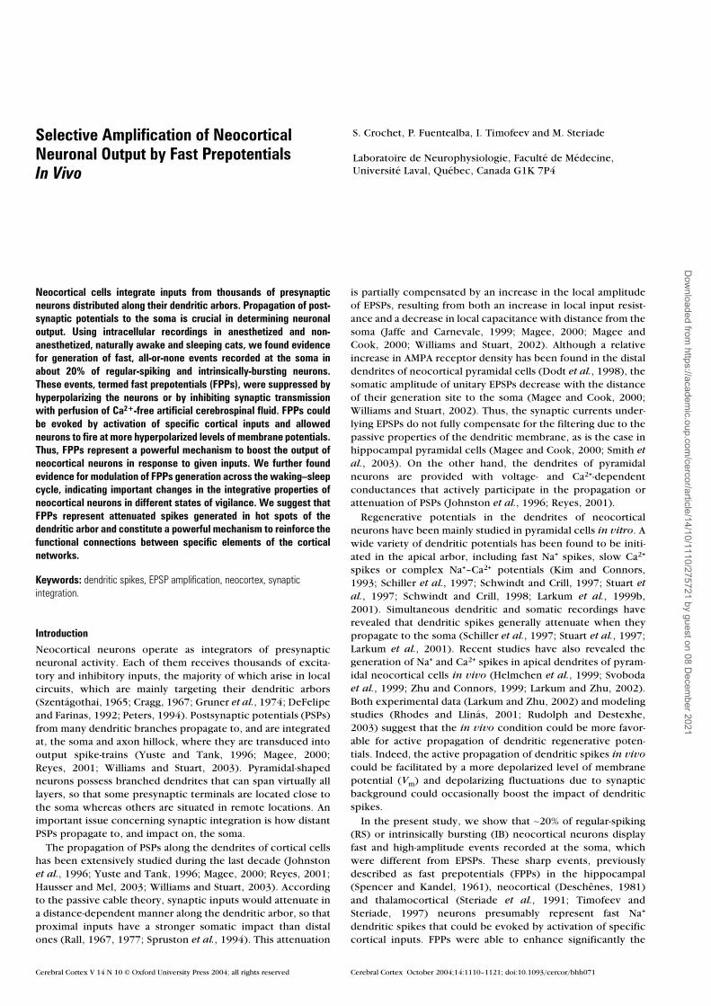
(ii) However, in 22% of the cells (18/83), some high-ampli-tude and fast-rising events were extracted. These eventsappeared as a population distinct from spontaneous EPSPs, asrevealed by plotting the amplitude versus the maximal slopefor all events (Fig. 1, middle panel right). Their mean amplituderanged from cell to cell between 3.6 and 10.2 mV, with anaverage value of 6.16 ± 0.14 mV, and their mean maximal sloperanged from 8 to 20 V/s, with an average value of 12.97 ± 0.30V/s. The rise-time of these events (0.49 ± 0.009 ms) was signif-icantly shorter than the rise time of the faster EPSPs (1.09 ±0.041 ms, P < 0.001) and significantly longer than the rise timeof full-blown APs (0.28 ± 0.008 m, P < 0.001). Their fallingphase was characterized by a biphasic decay best fitted by adouble exponential: the τ of the initial fast decay was 0.79 ±0.03 ms, while the τ of slower decay was 6.93 ± 0.29 ms.Because of these features, fast-rising events were very similar tothe FPPs initially described in hippocampal neurons bySpencer and Kandel (1961), and hereafter we will call themFPPs.
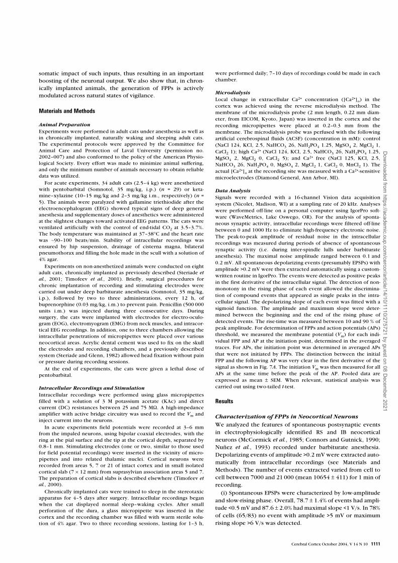
All neurons in which FPPs were detected had APs >0.6 ms athalf amplitude (mean = 0.81 ± 0.01 ms). The cortical depthdistribution of neurons was calculated from the depth of therecording indicated by the micro-driver and correctedaccording to the angle of the pipette within the cortex (Fig. 2).The neurons displaying FPPs were distributed mainly between200 and 800 µm, and between 1200 and 1600 µm, indicating
that most of them were recorded in layers II/III and V/VI. Theinput resistance of neurons displaying FPPs (17.43 ± 0.95 MΩ)
was not significantly different from that of neurons withoutFPPs (18.44 ± 1.00 MΩ, P > 0.8).
FPPs appeared as all-or-none events that crowned spontane-
ously occurring EPSPs. They appeared either in isolation or led
Figure 1. Fast prepotentials (FPPs) in regular spiking (RS) neocortical neurons in vivo. Barbiturate anesthesia. Top left, electroencephalogram (EEG) and intracellular recording inarea 5. The neuron was electrophysiologically identified as RS (top right). A part of the intracellular recording is expanded below (arrow). Different depolarizing events extracted areindicated in green (EPSPs) and blue (FPPs). Plotting the amplitude vs. the maximum slope for the selected events (middle panel, right) revealed an FPP population characterized byhigh-amplitude and fast-rising phase (dashed blue line). The bottom panel shows, on the left, an overlay of four EPSPs leading to FPPs in isolation or leading to full-blown actionpotential (AP). At right, the superimposition of averaged APs (red trace, truncated), FPPs (blue trace), and ‘fast’ (max. slope >3 V/s) EPSPs (green trace).
Figure 2. Depth distribution of regular-spiking (RS) and intrinsically bursting (IB)neurons recorded in the intact cortex of anesthetized cats (association areas 5 and 7).Histograms show the depth distribution for the neurons in which no FPP were detected(left) and for the neurons with FPPs (right). The depth of the recorded neurons wascorrected according to the angle of the pipette.
Dow
nloaded from https://academ
ic.oup.com/cercor/article/14/10/1110/275721 by guest on 08 D
ecember 2021
Cerebral Cortex October 2004, V 14 N 10 1113
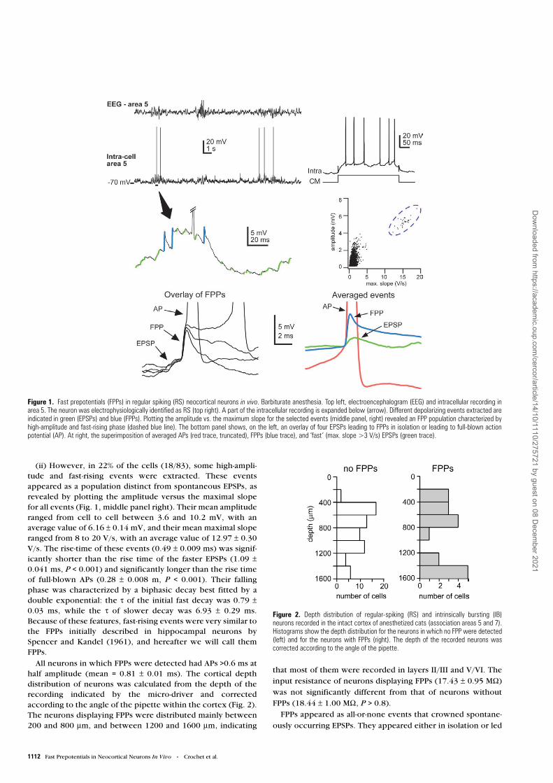
to full-blown APs (see overlay in Fig. 1). The essential conditionfor FPP generation was a relatively depolarized Vm and thepresence of synaptic activity. Actually, hyperpolarizing the cellby intracellular injection of current suppressed FPPs (Fig. 3).However, depolarizing current pulses could not elicit FPPsduring periods without spontaneous synaptic activity (i.e.interspindle lull in barbiturate anesthesia) but, instead,induced trains of APs. In Fig. 3, the cell was held at two Vm
levels by injection of hyperpolarizing current pulses. The histo-gram of Vm (gray) shows a bimodal distribution resulting fromcurrent pulses, with FPPs initiated only at Vms more depolar-ized than –70 mV. In this neuron, the mean threshold for thegeneration of FPPs was –66 mV. In the 18 neurons with FPPs,we compared the Vm at the initiation of FPPs and the Vm at initi-ation of APs. The threshold for FPPs’ generation was differentfrom cell to cell and varied from –72 to –62 mV (mean 67.2 ±0.2 mV), but was always more hyperpolarized than thethreshold for axonal APs initiation (55.9 ± 0.1 mV, P < 0.001)(see below). In all 18 neurons, holding the cell below thethreshold for FPPs’ generation completely suppressed theirappearance.
In order to determine the dependence of FPPs’ generationon synaptic transmission, we used the reverse microdialysistechnique to change the extracellular concentration of calcium([Ca2+]o) in the vicinity of the recorded neurons. The actual[Ca2+]o at the recording site was measured with a Ca2+-sensitiveelectrode. The baseline control [Ca2+]o was 1.2 mM, as previ-ously described in vivo (Massimini and Amzica, 2001);10–15 min after perfusion of the microdialysis probe with highCa2+ ACSF, the [Ca2+]o reached a stable level of 3 mM; after20 min of perfusion with the Ca2+-free ACSF, the [Ca2+]o stabil-
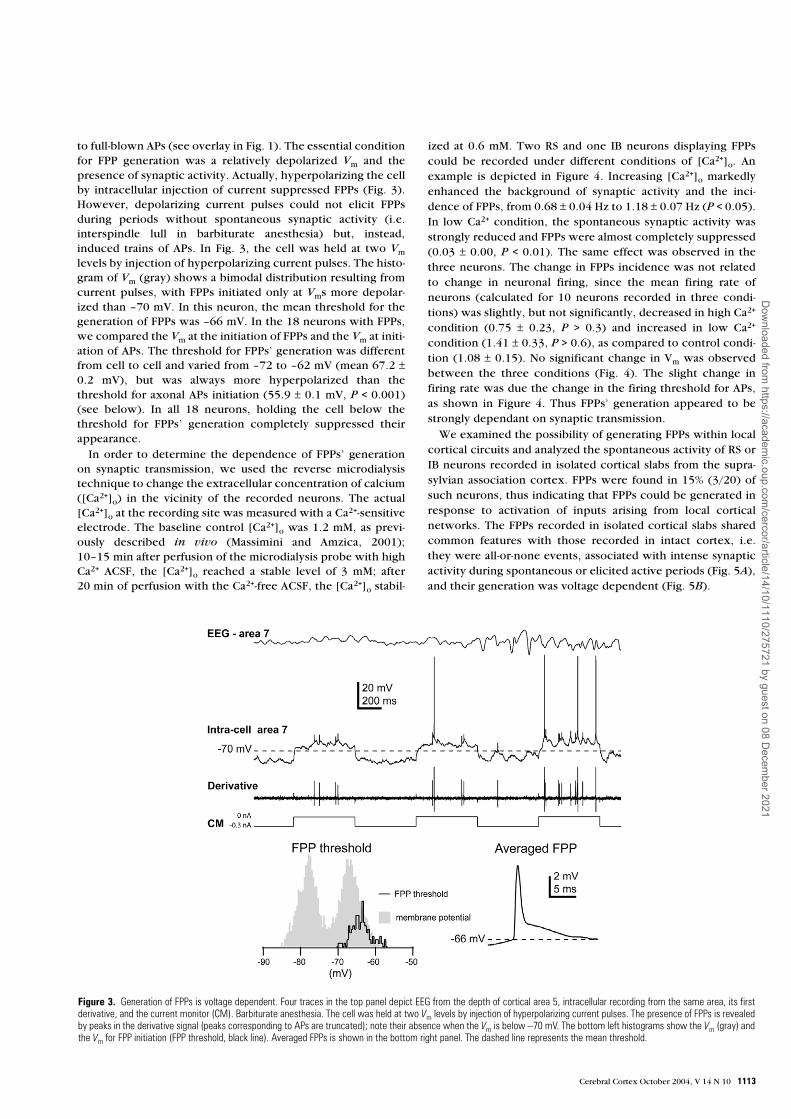
ized at 0.6 mM. Two RS and one IB neurons displaying FPPscould be recorded under different conditions of [Ca2+]o. Anexample is depicted in Figure 4. Increasing [Ca2+]o markedlyenhanced the background of synaptic activity and the inci-dence of FPPs, from 0.68 ± 0.04 Hz to 1.18 ± 0.07 Hz (P < 0.05).In low Ca2+ condition, the spontaneous synaptic activity wasstrongly reduced and FPPs were almost completely suppressed(0.03 ± 0.00, P < 0.01). The same effect was observed in thethree neurons. The change in FPPs incidence was not relatedto change in neuronal firing, since the mean firing rate ofneurons (calculated for 10 neurons recorded in three condi-tions) was slightly, but not significantly, decreased in high Ca2+
condition (0.75 ± 0.23, P > 0.3) and increased in low Ca2+
condition (1.41 ± 0.33, P > 0.6), as compared to control condi-tion (1.08 ± 0.15). No significant change in Vm was observedbetween the three conditions (Fig. 4). The slight change infiring rate was due the change in the firing threshold for APs,as shown in Figure 4. Thus FPPs’ generation appeared to bestrongly dependant on synaptic transmission.
We examined the possibility of generating FPPs within localcortical circuits and analyzed the spontaneous activity of RS orIB neurons recorded in isolated cortical slabs from the supra-sylvian association cortex. FPPs were found in 15% (3/20) ofsuch neurons, thus indicating that FPPs could be generated inresponse to activation of inputs arising from local corticalnetworks. The FPPs recorded in isolated cortical slabs sharedcommon features with those recorded in intact cortex, i.e.they were all-or-none events, associated with intense synapticactivity during spontaneous or elicited active periods (Fig. 5A),and their generation was voltage dependent (Fig. 5B).
Figure 3. Generation of FPPs is voltage dependent. Four traces in the top panel depict EEG from the depth of cortical area 5, intracellular recording from the same area, its firstderivative, and the current monitor (CM). Barbiturate anesthesia. The cell was held at two Vm levels by injection of hyperpolarizing current pulses. The presence of FPPs is revealedby peaks in the derivative signal (peaks corresponding to APs are truncated); note their absence when the Vm is below –70 mV. The bottom left histograms show the Vm (gray) andthe Vm for FPP initiation (FPP threshold, black line). Averaged FPPs is shown in the bottom right panel. The dashed line represents the mean threshold.
Dow
nloaded from https://academ
ic.oup.com/cercor/article/14/10/1110/275721 by guest on 08 D
ecember 2021

1114 Fast Prepotentials in Neocortical Neurons In Vivo • Crochet et al.
Selective Amplification of Cortical InputsIn 13 out of 18 neurons, we tested the efficacy of cortical and/or thalamic inputs to evoked FPPs. FPPs could be elicited bycortical simulation in 8 of the 13 neurons. In general,responses to cortical stimulation consisted in a depolarization-hyperpolarization sequence, the first depolarization beingcomposed of a summation of EPSPs. The early depolarizationwas crowned by an FPP when the response reached thethreshold for their generation (Fig. 6). Interestingly, the gener-
ation of FPPs selectively depended on the stimulated pathway.At variance with an earlier study that pointed to thalamicinputs generating FPPs’ in cortical neurons (Deschênes, 1981),cortical stimulation evoked FPPs while thalamic stimuli,applied to intralaminar centrolateral or lateral posterior nuclei,did not elicit FPPs in the same neuron, even when the responsereached the threshold for FPP generation (Fig. 6A). This wasobserved in the 4 neurons in which we tested both thalamicand cortical stimulation.
In four other neurons, we compared the potency of corticalstimuli applied at different sites to evoke FPPs. Stimuli wereapplied alternatively through two electrodes (see Materials andMethods) inserted in the ipsilateral cortex, at 1–2 mm from therecorded neuron and separated by 2–3 mm. The intensity ofstimulation was adjusted for each electrode to induce EPSPs of
similar amplitude, when subthreshold for FPP. Figure 6B
shows the responses of another cortical neuron to stimuliapplied through two different cortical electrodes. Stimuli deliv-ered to the first electrode evoked an EPSP that was crowned byan FPP when it reached the threshold (left), whereas stimuliapplied to the second electrode evoked a similar responses butno FPP (right). To be sure that the second stimulation did notfail to evoke FPPs because of more depolarized threshold forFPP generation, we held the cell at a more depolarized level ofVm while stimulating through the second electrode. At moredepolarized Vm, the EPSP eventually evoked an AP (not shown)but no FPP was observed. The consequence of FPPs’ genera-tion in response to cortical stimulation was a non-linearity inthe voltage–response relationship, as revealed by plotting themaximum Vm reached by the evoked response versus the Vm
before stimulation. As shown in Figure 6B (bottom), theprogression of the Vm reached by the response increased line-arly with the initial Vm (blue open triangles) until it reached thethreshold for FPP generation. Then, the FPPs induced a jump inthe Vm reached by the response (blue filled triangles). On theother hand, the Vm reached by the response to stimuli appliedthrough the second electrode increased linearly even abovethe threshold for FPP generation (green open circles).
Besides its dependence upon synaptic inputs, the generationof FPPs was also dependent on the temporal sequence of the
Figure 4. Generation of FPPs depends on synaptic transmission. Barbiturate anesthesia. The upper panel shows the EEG and intracellular recording of a cortical neuron in threeconditions of extracellular Ca2+ ([Ca2+]o). The same neurons were recorded while a microdialysis probe inserted in the vicinity of the pipette was perfused with normal artificialcerebrospinal fluid (ACSF) (control, Cont), with ACSF enriched in Ca2+ (High Ca2+, H Ca) and with Ca2+ free ACSF (low Ca2+, L Ca). Note the increase and decrease of thebackground synaptic activity in H Ca and L Ca conditions respectively. The middle panel shows the superimposition of APs and FPPs for the three conditions. Note the presence ofFPPs in isolation, FPPs of leading to APs, and APs arising from summation of EPSPs in Cont and H Ca conditions while in L Ca condition all APs arose from EPSPs. Note also the changein firing threshold for axo-somatic APs. The left bottom panel shows the mean incidence (±SEM) of FPPs in the three conditions of extracellular Ca2+ calculated for the neurondepicted in the upper panels. The middle and rigth bottom histograms show the mean firing rates and mean Vms (±SEM) calculated for 10 cortical neurons in the three conditions.Asterisks represent significant difference: *P < 0.05 and **P < 0.01.
Dow
nloaded from https://academ
ic.oup.com/cercor/article/14/10/1110/275721 by guest on 08 D
ecember 2021
Cerebral Cortex October 2004, V 14 N 10 1115
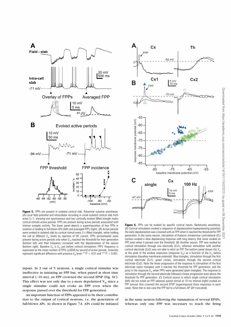
inputs. In 2 out of 5 neurons, a single cortical stimulus wasineffective in initiating an FPP but, when paired at short timeinterval (∼10 ms), an FPP crowned the second EPSP (Fig. 6C).This effect was not only due to a more depolarized Vm since asingle stimulus could not evoke an FPP even when theresponse passed over the threshold for FPP generation.
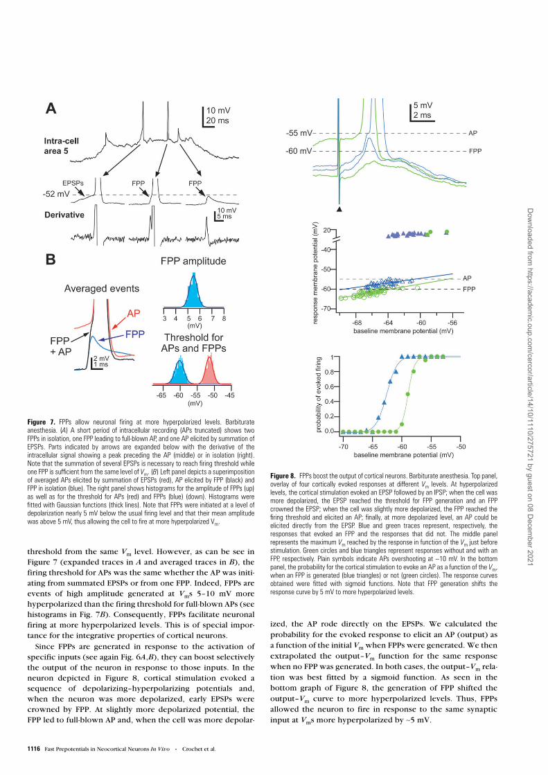
An important function of FPPs appeared to be their contribu-tion to the output of cortical neurons, i.e. the generation offull-blown APs. As shown in Figure 7A, APs could be initiated
in the same neuron following the summation of several EPSPs,whereas only one FPP was necessary to reach the firing
Figure 5. FPPs are present in isolated cortical slab. Ketamine–xylazine anesthesia.(A) Local field potential and intracellular recording in small isolated cortical slab fromareas 5–7, showing one spontaneous and two cortically evoked (filled triangle markscortical stimuli) active periods. FPPs are present during active periods associated withintense synaptic activity. The lower panel depicts a superimposition of four FPPs inisolation or leading to full-blown APs (left) and averaged FPPs (right). (B) Active periodswere evoked in isolated slab by cortical stimuli every 3 s (filled triangle), while holdingthe cell at different Vm levels by injection of DC current. FPPs (arrowheads) werepresent during active periods only when Vm reached the threshold for their generation(bottom left) and their frequency increased with the depolarization of the neuron(bottom right). Baseline Vm is Vm just before cortical stimulation; FPPs’ frequency isexpressed as the mean number of FPPs (±SEM) by second of active periods. Asterisksrepresent significant difference with previous Vm level: **P < 0.01 and ***P < 0.001.
Figure 6. FPPs can be evoked by specific cortical inputs. Barbiturate anesthesia.(A) Cortical stimulation evoked a sequence of depolarization-hyperpolarizing potential;the early depolarization was crowned with an FPP when it reached the threshold for FPPgeneration. In the same neuron, stimulation of thalamic intralaminar centrolateral (CL)nucleus evoked a slow depolarizing response with long latency that never evoked anFPP, even when it passed over the threshold. (B) Another neuron. FPP was evoked bycortical stimulation through one electrode (Cx1), whereas stimulation with anothercortical electrode (Cx2) was not able to elicit an FPP. The bottom panel shows the Vmat the peak of the evoked responses (response Vm) as a function of the Vm beforestimulation (baseline membrane potential). Blue triangles, stimulation through the firstcortical electrode (Cx1); green circles, stimulation through the second corticalelectrode (Cx2). Note the linear progression of the response to stimulation of the firstelectrode (open triangles) until it reaches the threshold for FPP generation, and thejump in the response Vm when FPPs were generated (plain triangles). The response tostimulation through the second electrode followed a linear progression even above thethreshold for FPP generation. (C) Cortical neuron in which single cortical stimulation(left) did not evoke an FPP, whereas paired stimuli at 10 ms interval (right) evoked anFPP (arrow) that crowned the second EPSP (superimposed three responses in eachcase). Note that in one case the FPP led to a full blown AP (AP truncated).
Dow
nloaded from https://academ
ic.oup.com/cercor/article/14/10/1110/275721 by guest on 08 D
ecember 2021
1116 Fast Prepotentials in Neocortical Neurons In Vivo • Crochet et al.
threshold from the same Vm level. However, as can be see inFigure 7 (expanded traces in A and averaged traces in B), thefiring threshold for APs was the same whether the AP was initi-ating from summated EPSPs or from one FPP. Indeed, FPPs areevents of high amplitude generated at Vms 5–10 mV morehyperpolarized than the firing threshold for full-blown APs (seehistograms in Fig. 7B). Consequently, FPPs facilitate neuronalfiring at more hyperpolarized levels. This is of special impor-tance for the integrative properties of cortical neurons.
Since FPPs are generated in response to the activation ofspecific inputs (see again Fig. 6A,B), they can boost selectivelythe output of the neuron in response to those inputs. In theneuron depicted in Figure 8, cortical stimulation evoked asequence of depolarizing–hyperpolarizing potentials and,when the neuron was more depolarized, early EPSPs werecrowned by FPP. At slightly more depolarized potential, theFPP led to full-blown AP and, when the cell was more depolar-
ized, the AP rode directly on the EPSPs. We calculated theprobability for the evoked response to elicit an AP (output) asa function of the initial Vm when FPPs were generated. We thenextrapolated the output–Vm function for the same responsewhen no FPP was generated. In both cases, the output–Vm rela-tion was best fitted by a sigmoid function. As seen in thebottom graph of Figure 8, the generation of FPP shifted theoutput–Vm curve to more hyperpolarized levels. Thus, FPPsallowed the neuron to fire in response to the same synapticinput at Vms more hyperpolarized by ∼5 mV.
Figure 7. FPPs allow neuronal firing at more hyperpolarized levels. Barbiturateanesthesia. (A) A short period of intracellular recording (APs truncated) shows twoFPPs in isolation, one FPP leading to full-blown AP, and one AP elicited by summation ofEPSPs. Parts indicated by arrows are expanded below with the derivative of theintracellular signal showing a peak preceding the AP (middle) or in isolation (right).Note that the summation of several EPSPs is necessary to reach firing threshold whileone FPP is sufficient from the same level of Vm. (B) Left panel depicts a superimpositionof averaged APs elicited by summation of EPSPs (red), AP elicited by FPP (black) andFPP in isolation (blue). The right panel shows histograms for the amplitude of FPPs (up)as well as for the threshold for APs (red) and FPPs (blue) (down). Histograms werefitted with Gaussian functions (thick lines). Note that FPPs were initiated at a level ofdepolarization nearly 5 mV below the usual firing level and that their mean amplitudewas above 5 mV, thus allowing the cell to fire at more hyperpolarized Vm.
Figure 8. FPPs boost the output of cortical neurons. Barbiturate anesthesia. Top panel,overlay of four cortically evoked responses at different Vm levels. At hyperpolarizedlevels, the cortical stimulation evoked an EPSP followed by an IPSP; when the cell wasmore depolarized, the EPSP reached the threshold for FPP generation and an FPPcrowned the EPSP; when the cell was slightly more depolarized, the FPP reached thefiring threshold and elicited an AP; finally, at more depolarized level, an AP could beelicited directly from the EPSP. Blue and green traces represent, respectively, theresponses that evoked an FPP and the responses that did not. The middle panelrepresents the maximum Vm reached by the response in function of the Vm just beforestimulation. Green circles and blue triangles represent responses without and with anFPP, respectively. Plain symbols indicate APs overshooting at ∼10 mV. In the bottompanel, the probability for the cortical stimulation to evoke an AP as a function of the Vm,when an FPP is generated (blue triangles) or not (green circles). The response curvesobtained were fitted with sigmoid functions. Note that FPP generation shifts theresponse curve by 5 mV to more hyperpolarized levels.
Dow
nloaded from https://academ
ic.oup.com/cercor/article/14/10/1110/275721 by guest on 08 D
ecember 2021

Cerebral Cortex October 2004, V 14 N 10 1117
Modulation of FPP Generation across Behavioral StatesFinally, we investigated the possibility that the incidence ofFPPs is modulated by natural states of vigilance and analyzedintracellular recordings of RS and IB neurons in awake andsleeping cats. FPPs were found in 10 out of 56 neurons (18%)in different states of vigilance. FPPs were present in all behav-ioral states but their frequency was different and depended onthe Vm variations (Fig. 9). During slow-wave sleep (SWS), themean Vm was more hyperpolarized and the incidence of FPPswas the lowest. During waking and paradoxical sleep (PS; orREM sleep), the Vm was equally more depolarized and thefrequency of FPPs was higher in both these states than in SWS,reaching the highest values in PS. We measured the incidenceof FPPs in eight neurons, during different states of vigilance.The incidence of FPPs was the lowest during SWS (4.4 ±0.95 Hz) and was significantly higher during waking (15.02 ±1.18 Hz, P < 0.01) and PS (28.97 ± 4.40 Hz, P < 0.01). Despite asimilar Vm level, the incidence of FPPs during PS was signifi-cantly higher than during waking (P < 0.05), suggesting thatneuromodulators implicated in the regulation of behavioralstates may modulate the generation of FPPs.
DiscussionIn the present paper we report the presence of fast and high-amplitude events, FPPs, recorded from the soma of ∼20 % of RS
and IB neocortical neurons. The FPPs were all-or-none events,suppressed by hyperpolarization, and always associated withsynaptic background activity. They were evoked by activationof specific local cortical inputs and exerted a powerful andselective boosting of cortical neuronal output. Finally, wefound that the incidence of FPPs was modulated by naturalstates of vigilance.
Nature and Origin of FPPsFPPs were first described in the hippocampus (Spencer andKandel, 1961) and later also found in neocortical (Deschênes,1981), thalamocortical (Steriade et al., 1991; Timofeev andSteriade, 1997) and other central neurons. They were inter-preted to either represent attenuated dendritic spikes (Turneret al., 1993; Ariav et al., 2003) or reflect APs in electrotonicallycoupled neurons (Llinás et al., 1974; MacVicar and Dudek,1981; Valiante et al., 1995; Gibson et al., 1999; Hughes et al.,2002; Landisman et al., 2002). Although in vitro studies haverevealed abundant electrical coupling via gap-junctionsbetween both pyramidal neurons and interneurons in imma-ture neocortex (Peinado et al., 1993; Bittman et al., 2002), elec-trophysiological evidence for electrical coupling in adultanimal has been only found between local interneurons but notbetween pyramidal neurons (Connors et al., 1983; Galarretaand Hestrin, 1999; Gibson et al., 1999). In the present study,
Figure 9. FPP generation is regulated across the states of vigilance. Four traces in the top panel depict electromyogram (EMG), electro-oculogram (EOG), EEG from the depth ofright cortical area 3 (somatosensory), and intracellular recording from left area 3. The same neuron was recorded during waking (W), slow-wave sleep (SWS) and paradoxical sleep(PS). Parts of the intracellular recording marked by horizontal bars are expanded bellow. The derivative of the intracellular signal revealed the presence of FPPs (APs are truncatedin intracellular recording and its derivative). Note the absence of FPP when the cell is more hyperpolarized than –65 mV. The left bottom panel shows averaged FPPs. The middlebottom panel shows the distribution of Vm of the neuron depicted in the three behavioral states (W, red; SWS, green and PS, blue). The right bottom panel shows the meanincidence of FPPs in the three behavioral states calculated for eight neurons. Asterisks represent significant difference: *P < 0.05 and **P < 0.01.
Dow
nloaded from https://academ
ic.oup.com/cercor/article/14/10/1110/275721 by guest on 08 D
ecember 2021
1118 Fast Prepotentials in Neocortical Neurons In Vivo • Crochet et al.
FPPs were recorded in RS and IB neurons with relatively wideAPs (mean duration at half amplitude: 0.82 ± 0.01 ms), indi-cating that these neurons were most likely not interneurons. Ina few experiments, we stained the recorded neurons withintracellular injection of Neurobiotine. One neuron thatdisplayed FPPs was stained and was identified as a pyramidal-shaped layer III neuron (data not shown). Recent experimentsin the hippocampus demonstrated the generation of spikeletsof 1–10 mV in amplitude mediated by axo-axonal gap junctionsbetween pyramidal cells (Schmitz et al., 2001). However, tothe best of our knowledge, there is no evidence for electricalcoupling between axons of pyramidal neurons in theneocortex. On the other hand, the possibility that electricalcoupling between neocortical pyramidal neurons may persistin adult animals has been raised by molecular studies. Thus,connexin36 might be expressed by a subset of pyramidalneurons (Deans et al., 2001) and the expression of mRNA foranother type of connexin (43) has been found in pyramidalcells of mature neocortex (Simburger et al., 1997).
In the present study, the generation of FPPs was voltage-dependent and always associated with synaptic activity. Simi-larly, the generation of dendritic spikes recorded from pyram-idal neocortical or hippocampal neurons in vitro has beenshown to be voltage-dependent (Kim and Connors, 1993;Schwindt and Crill, 1997; Schiller et al., 2000; Ariav et al.,2003). Finally, we found that FPPs were suppressed whensynaptic transmission is inhibited by perfusion of Ca2+-freeACSF and their incidence increased with perfusion of high-Ca2+
ACSF. These observations do not support the idea that FPPsrepresent APs in electronically coupled neurons, since elec-trical coupling is not affected or even enhanced in low-Ca2+
conditions (Perez-Velazquez et al., 1994; Valiante et al., 1995;Draguhn et al., 1998; Mann-Metzer and Yarom, 1999). Thus,although we cannot completely discard the possibility thatFPPs represent APs in electrotonically coupled neurons, ourresults strongly suggest that they are more likely dendriticregenerative potentials. It is also unlikely that the neurons thatdisplayed FPPs in our study were recorded at dendritic levels:(i) none of them showed characteristic features of establisheddendritic recordings in vitro or in vivo, such as plateau poten-tials or complex spikes (Kim and Connors, 1993; Steriade et al.,1996; Helmchen et al., 1999; Zhu and Connors, 1999; Larkumand Zhu, 2002); (ii) all cells generated trains of full-blown,overshooting APs of constant amplitudes in response todepolarizing current pulses, whereas backpropagating axonalAPs recorded in the dendrites of cortical neurons in vivo showstrong attenuation and broadening with distance from thesoma (Svoboda et al., 1999; Waters et al., 2003). We thereforethink that our recordings were performed at somatic levels ofcortical neurons and thus suggest that FPPs represent attenu-ated dendritic spikes.
As to the mechanisms underlying the generation of FPPs, it iswell established that the dendrites of cortical pyramidal cellscan sustain active regenerative potentials under certain condi-tions. Na+, Ca2+ and NMDA-generated spikes have been demon-strated in the apical and basal dendrites of cortical pyramidalneurons (Magee et al., 1998; Hausser et al., 2000; Schiller andSchiller, 2001; Hausser and Mel, 2003; Williams and Stuart,2003). Because of the slow time course of Ca2+ and NMDAspikes (Kim and Connors, 1993; Schiller et al., 1997; Schwindtand Crill, 1997; Larkum et al., 1999a; Schiller et al., 2000;Larkum et al., 2001; Larkum and Zhu, 2002), we assume that at
least the earliest sharp component of the FPPs represent a Na+
spike whose generation has been shown in the apical dendritesof neocortical and hippocampal pyramidal cells (Kim andConnors, 1993; Stuart et al., 1997; Golding and Spruston, 1998;Schwindt and Crill, 1998) and in the basal dendrites of hippo-campal pyramidal neurons as well (Ariav et al., 2003).However, Na+ spikes generated in apical dendrites are most ofthe time associated with Ca2+ spikes and strongly attenuatewhile propagating to the soma (Schiller et al., 1997; Stuart et
al., 1997; Larkum et al., 2001). As a result, when simultane-ously recorded at the dendrite and soma in vitro, dendritic Na+
spikes do not appear as fast potentials at the soma. Moreover,we were able to block the generation of FPPs by injection ofhyperpolarizing current at the soma. In vitro data demonstratethat alteration of somatic membrane potential by direct currentinjection can affect the membrane potential of the dendrites atleast out to 300–400 µm from the soma (Schwindt and Crill,1995). The high conductance state of cortical neurons in vivo
probably further limits the dendritic impact of somatic currentinjection, suggesting that FPPs are not generated far from thesoma, i.e. in proximal apical or basal dendrites of pyramidalcells. Another possibility is that FPPs were generated in distalapical dendrites and then propagated actively to the soma. Thispossibility was suggested in modeling studies showing thatunder in vivo-like conditions, a depolarized Vm increases theprobability of generation and propagation of dendritic spikes(Rhodes and Llinás, 2001; Rudolph and Destexhe, 2003). Inthis condition, hyperpolarizing the soma could block the prop-agation FPPs (Larkum et al., 2001). However, their relativelysmall amplitude at the soma, compared to the amplitude ofregenerative potentials generated in the dendrites, does notsupport the idea of an active propagation in this case.
In hippocampal pyramidal cells in vitro, coactivation of clus-tered neighboring inputs on basal dendrites can elicit a localspike that consists in fast Na+ and slow NMDA components(Ariav et al., 2003). These dendritic spikes, recorded at thesoma, have a shape very similar to the FPPs described here andare similarly suppressed by injection of hyperpolarizingcurrent at the soma. But in layer V neocortical pyramidal cellsin vitro, only the slow NMDA spike was evoked by the activ-ation of basal dendrites (Schiller et al., 2000). However, in ourexperiments, many neurons with FPPs were localized in layersII/III or VI. It is thus possible that Na+ spikes, similar to thosegenerated in the basal dendrites of hippocampal neurons, canbe generated in the basal dendrites of some subpopulation ofneocortical pyramidal cells in other layers. Therefore, wesuggest that FPPs represent dendritic Na+ spikes generated inthe basal dendrites of neocortical pyramidal cells. The biphasicdecay of the FPPs may reflect an underlying slow componentdue either to Ca2+ or NMDA conductances.
The generation of dendritic spikes is supposed to occurwhen synaptic inputs are activated simultaneously and/orconverge at the same dendritic region. In neocortical pyram-idal neurons, supralinear summation of EPSPs due to activedendritic processes has been found to occur in a time window<30 ms (Nettleton and Spain, 2000). Local spikes in the basaldendrites of both neocortical or hippocampal pyramidal cellscan be initiated by co-activation of clustered neighboring basalinputs (Schiller et al., 2000; Ariav et al., 2003). Based on thelocal amplitude of EPSPs recorded in vitro, Williams and Stuart(2002) have found that the synchronized activation of 4–30presynaptic neurons is required for the initiation of dendritic
Dow
nloaded from https://academ
ic.oup.com/cercor/article/14/10/1110/275721 by guest on 08 D
ecember 2021
Cerebral Cortex October 2004, V 14 N 10 1119
spikes. Thus, initiation of local spikes is expected to occureither if inputs carrying related information selectively inner-vate the same dendritic segments (Archie and Mel, 2000;Poirazi and Mel, 2001) or if the network activity is synchro-nized (Kamondi et al., 1998; Helmchen et al., 1999). Inkeeping with this idea, we found that FPPs could be evoked bygross cortical stimulations, which activate simultaneously apool of neurons and fibers or by paired stimuli at highfrequency. Thus, only converging coincident inputs or closelytime-spaced inputs could generate FPPs, suggesting that FPPsare likely generated in response to an important depolarizationof a particular dendritic segment. Interestingly, the activationof different cortical inputs had various effects on the genera-tion of FPPs, even when the subthreshold evoked responseswere very similar (see Fig. 6). This observation point out to theinput-specific generation of FPPs that might be due either tothe location of these inputs (apical or basal dendrites) or to theconvergence of the simultaneously activated inputs (clusteredor spread on the dendritic arbors). The input-dependent gener-ation of FPPs may also be due to non-uniformly distributedglutamate receptors that form hot spots of glutamate respon-sivity on dendritic arbors. Synaptic inputs impinging uponthese regions would have a higher probability to elicitdendritic spikes (Frick et al., 2001).
Functional Implication of FPPs for the Integrative Properties of Cortical NeuronsActive regenerative dendritic potentials have mainly beenstudied in the apical dendrites of neocortical and hippocampalpyramidal cells. Physiological and modeling studies haveshown that forward-propagating dendritic spikes can boost theinfluence of synapses in distal dendrites on their way to thesoma, thereby circumventing the attenuation produced by thepassive cable properties of dendrites (Cauller and Connors,1994; Schwindt and Crill, 1998; Larkum et al., 2001; Williamsand Stuart, 2002; Rudolph and Destexhe, 2003). It is assumedthat dendritic spikes strongly attenuate as they propagateforward and appear at the soma as slow events undistinguish-able from other postsynaptic potentials (Schiller et al., 1997;Stuart et al., 1997; Golding and Spruston, 1998; Larkum et al.,2001). However, in the present study we found that FPPs weresignificantly higher in amplitude and had faster rising phasethan other spontaneous depolarizing events recorded at thesoma. FPPs induced a non-linearity in the integrative propertiesof cortical neurons (see Fig. 6B), thus providing additionalcomputational properties to the neurons by markedlyenhancing the somatic impact of cortical inputs. We furtherdemonstrate that the boosting of EPSPs by FPPs results in afunctional hierarchy of the inputs by a selective enhancementof the output of cortical neurons for given inputs. Thus, FPPsmay reinforce the functional links between specific elementsof the cortical network. This hypothesis is of interest since wedemonstrated, using dual intracellular recordings of synapti-cally connected neurons in vivo, that the somatic impact ofindividual cortical inputs during active states of the network isvery weak due to a high conductance state and high failure rate(Timofeev and Crochet, 2002). FPPs might also enhanceneuronal output in response to simultaneous arriving inputs.Indeed, the generation of dendritic spikes might require coin-cident activation of different inputs and may thus play animportant role for coincidence detection of cortical inputs.
As demonstrated recently in hippocampal neurons, Na+
dendritic spikes could serve to improve the precision and thestability of the timing of axonal APs, mainly by reducing thetemporal jitter of evoked APs (Ariav et al., 2003). This wasapparently not the case for FPPs in the present study because(i) the generation of FPPs themselves showed relatively hightemporal variability (not shown); and (ii) once an FPP is gener-ated, the timing of the AP was also variable (see Figs 1 and 5A).Physiological studies have also suggested that forward-propagating Ca2+ spikes could be involved in the generation ofburst firing in cortical neurons (Larkum et al., 1999a,b;Schwindt and Crill, 1999; Williams and Stuart, 1999). Incontrast to Ca2+ spikes initiated in apical dendrites, we foundthat FPPs did not evoke a burst of axonal APs, but rather singleAP. This is expected from the fast decay of the FPPs, which aremore likely generated by a Na+ conductance and whichcontrast with the broad Ca2+ spikes evoked in apical dendrites.
Dendritic excitability is regulated by neuromodulators suchas serotonin, acetylcholine or noradrenaline (Hoffman andJohnston, 1999; Carr et al., 2002). These neuromodulators arereleased by ascending activating systems that originate in thebrainstem and basal forebrain, and play a major role in thecontrol of behavioral states (Steriade and McCarley, 1990). Inline with these observations, we found evidence for a modula-tion of FPPs generation across behavioral states of vigilance. Inall recorded neurons, we found changes in the incidence ofspontaneous FPPs associated with shifts from a state of vigi-lance to another. The incidence of FPPs appeared to be thelowest during SWS and the highest during PS. The decrease inFPPs incidence during SWS was expected because of Vm hyper-polarization during this state (Steriade et al., 2001) due todecreased firing rates in thalamocortical (Glenn and Steriade,1982) and other corticipetal systems during SWS, compared toboth waking and PS. The increased incidence of FPPs in PScompared to waking (Fig. 9) fits in well with the higher firingrates, during PS, of mesopontine cholinergic neurons withidentified projections to intralaminar and lateroposteriorthalamic nuclei (Steriade et al., 1990), which are the main affer-ents of cortical association areas at which level the presentrecordings were made. Thus, neuromodulators are able tocontrol the integrative properties of cortical neurons acrossbehavioral states.
Concluding RemarksThis study revealed that ∼20% of RS and IB neocortical neuronsare able to generate FPPs. The electrophysiological featuresof these events strongly suggest that they represent forwardpropagated Na+ dendritic spikes attenuated at the soma. Wepropose that FPPs represent dendritic spikes generated bycoincident inputs converging on the same dendritic segment.These dendritic spikes strongly affect the integrative proper-ties of cortical neurons and may serve to reinforce the func-tional links between specific elements of the cortical network
or for coincidence detection as well. Finally, the generationFPPs appeared to be regulated across behavioral states, indi-cating important changes in the integrative properties ofcortical neurons during states of vigilance.
NotesWe thank P. Giguère and D. Drolet for technical assistance. This workwas supported by grants from the Canadian Institutes for HealthResearch (MT-3689, MOP-36545, MOP-37862) and US National Insti-
Dow
nloaded from https://academ
ic.oup.com/cercor/article/14/10/1110/275721 by guest on 08 D
ecember 2021
1120 Fast Prepotentials in Neocortical Neurons In Vivo • Crochet et al.
tute of Health (RO1, NS-40522). S. Crochet is supported by the Pick-wick Postdoctoral Fellowship program from the National SleepFoundation. I. Timofeev is a Scholar of the Canadian Institutes ofHealth Research.
Address correspondence to M. Steriade, Laboratoire de Neuro-physiologie, Faculté de Médecine, Pavillon Vandry, Université Laval,Québec G1K 7P4, Canada. Email: [email protected].
ReferencesArchie KA , Mel BW (2000) A model for intradendritic computation of
binocular disparity. Nat Neurosci 3:54–63.Ariav G, Polsky A, Schiller J (2003) Submillisecond precision of the
input-output transformation function mediated by fast sodiumdendritic spikes in basal dendrites of CA1 pyramidal neurons. JNeurosci 23:7750–7758.
Bittman K, Becker DL, Cicirata F, Parnavelas JG (2002) Connexinexpression in homotypic and heterotypic cell coupling in thedeveloping cerebral cortex. J Comp Neurol 443:201–212.
Carr DB, Cooper DC, Ulrich SL, Spruston N, Surmeier DJ (2002) Sero-tonin receptor activation inhibits sodium current and dendriticexcitability in prefrontal cortex via a protein kinase C-dependentmechanism. J Neurosci 22:6846–6855.
Cauller LJ, Connors BW (1994) Synaptic physiology of horizontal affer-ents to layer I in slices of rat SI neocortex. J Neurosci 14:751–762.
Connors BW, Gutnick MJ (1990) Intrinsic firing patterns of diverseneocortical neurons. Trends Neurosci 13:99–104.
Connors BW, Benardo LS, Prince DA (1983) Coupling betweenneurons of the developing rat neocortex. J Neurosci 3:773–782.
Cragg B (1967) The density of synapses and neurones in the motor andvisual areas of the cerebral cortex. J Anat 101:639–654.
Deans MR, Gibson JR, Sellitto C, Connors BW, Paul DL (2001) Synchro-nous activity of inhibitory networks in neocortex requires elec-trical synapses containing connexin 36. Neuron 31:477–485.
DeFelipe J, Farinas I (1992) The pyramidal neuron of the cerebralcortex: morphological and chemical characteristics of the synapticinputs. Prog Neurobiol 39:563–607.
Deschênes M (1981) Dendritic spikes induced in fast pyramidal tractneurons by thalamic stimulation. Exp Brain Res 43:304–308.
Dodt HU, Frick A, Kampe K, Zieglgansberger W (1998) NMDA andAMPA receptors on neocortical neurons are differentially distrib-uted. Eur J Neurosci 10:3351–3357.
Draguhn A, Traub RD, Schmitz D, Jefferys JG (1998) Electricalcoupling underlies high-frequency oscillations in the hippo-campus in vitro. Nature 394:189–192.
Frick A, Zieglgansberger W, Dodt HU (2001) Glutamate receptorsform hot spots on apical dendrites of neocortical pyramidalneurons. J Neurophysiol 86:1412–1421.
Galarreta M, Hestrin S (1999) A network of fast-spiking cells in theneocortex connected by electrical synapses. Nature 402:72–75.
Gibson JR, Beierlein M, Connors BW (1999) Two networks of electri-cally coupled inhibitory neurons in neocortex. Nature 402:75–79.
Glenn LL, Steriade M (1982) Discharge rates and excitability of corti-cally projecting intralaminar thalamic neurons during waking andsleep states. J Neurosci 2:1287–1404.
Golding NL , Spruston N (1998) Dendritic sodium spikes are variabletriggers of axonal action potentials in hippocampal CA1 pyramidalneurons. Neuron 21:1189–1200.
Gruner JE, Hirsch JC, Sotelo C (1974) Ultrastructural features of theisolated suprasylvian gyrus in the cat. J Comp Neurol 154:1–27.
Hausser M , Mel B (2003) Dendrites: bug or feature? Curr Opin Neuro-biol 13:372–383.
Hausser M, Spruston N, Stuart GJ (2000) Diversity and dynamics ofdendritic signaling. Science 290:739–744.
Helmchen F, Svoboda K, Denk W, Tank DW (1999) In vivo dendriticcalcium dynamics in deep-layer cortical pyramidal neurons. NatNeurosci 2:989–996.
Hoffman DA, Johnston D (1999) Neuromodulation of dendritic actionpotentials. J Neurophysiol 81:408–411.
Hughes SW, Blethyn KL, Cope DW, Crunelli V (2002) Properties andorigin of spikelets in thalamocortical neurones in vitro. Neuro-science 110:395–401.
Jaffe DB, Carnevale NT (1999) Passive normalization of synaptic inte-gration influenced by dendritic architecture. J Neurophysiol82:3268–3285.
Johnston D, Magee JC, Colbert CM, Cristie BR (1996) Active propertiesof neuronal dendrites. Annu Rev Neurosci 19:165–186.
Kamondi A, Acsady L, Buzsaki G (1998) Dendritic spikes are enhancedby cooperative network activity in the intact hippocampus. JNeurosci 18:3919–3928.
Kim HG, Connors BW (1993) Apical dendrites of the neocortex: corre-lation between sodium- and calcium-dependent spiking andpyramidal cell morphology. J Neurosci 13:5301–5311.
Landisman CE, Long MA, Beierlein M, Deans MR, Paul DL, Connors BW(2002) Electrical synapses in the thalamic reticular nucleus. JNeurosci 22:1002–1009.
Larkum ME, Zhu JJ (2002) Signaling of layer 1 and whisker-evokedCa2+ and Na+ action potentials in distal and terminal dendrites ofrat neocortical pyramidal neurons in vitro and in vivo. J Neurosci22:6991–7005.
Larkum ME, Kaiser KM, Sakmann B (1999a) Calcium electrogenesis indistal apical dendrites of layer 5 pyramidal cells at a criticalfrequency of back-propagating action potentials. Proc Natl Acad SciU S A 96:14600–14604.
Larkum ME, Zhu JJ, Sakmann B (1999b) A new cellular mechanism forcoupling inputs arriving at different cortical layers. Nature398:338–341.
Larkum ME, Zhu JJ, Sakmann B (2001) Dendritic mechanisms under-lying the coupling of the dendritic with the axonal action potentialinitiation zone of adult rat layer 5 pyramidal neurons. J Physiol(Lond) 533:447–466.
Llinás R, Baker R, Sotelo C (1974) Electrotonic coupling betweenneurons in cat inferior olive. J Neurophysiol 37:560–571.
MacVicar BA, Dudek FE (1981) Electrotonic coupling between pyram-idal cells: a direct demonstration in rat hippocampal slices. Science213:782–785.
Magee JC (2000) Dendritic integration of excitatory synaptic input.Nat Rev Neurosci 1:181–190.
Magee JC, Cook EP (2000) Somatic EPSP amplitude is independent ofsynapse location in hippocampal pyramidal neurons. Nat Neurosci3:895–903.
Magee J, Hoffman D, Colbert C, Johnston D (1998) Electrical andcalcium signaling in dendrites of hippocampal pyramidal neurons.Annu Rev Physiol 60:327–346.
Mann-Metzer P, Yarom Y (1999) Electrotonic coupling interacts withintrinsic properties to generate synchronized activity in cerebellarnetworks of inhibitory interneurons. J Neurosci 19:3298–3306.
Massimini M, Amzica F (2001) Extracellular calcium fluctuations andintracellular potentials in the cortex during the slow sleep oscilla-tion. J Neurophysiol 85:1346–1350.
McCormick DA, Connors BW, Lighthall JW, Prince DA (1985) Compar-ative electrophysiology of pyramidal and sparsely spiny stellateneurons of the neocortex. J Neurophysiol 54:782–806.
Nettleton JS, Spain WJ (2000) Linear to supralinear summation ofAMPA-mediated EPSPs in neocortical pyramidal neurons. J Neuro-physiol 83:3310–3322.
Nuñez A, Amzica F, Steriade M (1993) Electrophysiology of cat associ-ation cortical cells in vivo: intrinsic properties and synapticresponses. J Neurophysiol 70:418–430.
Peinado A, Yuste R, Katz LC (1993) Extensive dye coupling betweenrat neocortical neurons during the period of circuit formation.Neuron 10:103–114.
Perez-Velazquez JL, Valiante TA, Carlen PL (1994) Modulation of gapjunctional mechanisms during calcium-free induced field burstactivity: a possible role for electrotonic coupling in epilepto-genesis. J Neurosci 14:4308–4317.
Peters A (1994) Number of neurons and synapses in primary visualcortex. In: Cerebral cortex, further aspects of cortical function;including hippocampus (Jones E, Peters A, eds), pp. 267–294.New York: Plenum.
Poirazi P, Mel BW (2001) Impact of active dendrites and structuralplasticity on the memory capacity of neural tissue. Neuron29:779–796.
Dow
nloaded from https://academ
ic.oup.com/cercor/article/14/10/1110/275721 by guest on 08 D
ecember 2021
Cerebral Cortex October 2004, V 14 N 10 1121
Rall W (1967) Distinguishing theoretical synaptic potentials computedfor different soma-dendritic distributions of synaptic input. JNeurophysiol 30:1138–1168.
Rall W (1977) Core conductor theory and cable properties of neurons.In: Handbook of physiology. The nervous system. Cellular biologyof neurons (Kandel ER, Brookhart JM, Mountcastle VB, eds), pp.39–97. Bethesda, MD: American Physiological Society.
Reyes A (2001) Influence of dendritic conductances on the input-output properties of neurons. Annu Rev Neurosci 24:653–675.
Rhodes PA, Llinás RR (2001) Apical tuft input efficacy in layer 5 pyram-idal cells from rat visual cortex. J Physiol (Lond) 536:167–187.
Rudolph M, Destexhe A (2003) A fast-conducting, stochastic integ-rative mode for neocortical neurons in vivo. J Neurosci23:2466–2476.
Schiller J, Schiller Y (2001) NMDA receptor-mediated dendriticspikes and coincident signal amplification. Curr Opin Neurobiol11:343–348.
Schiller J, Schiller Y, Stuart G, Sakmann B (1997) Calcium actionpotentials restricted to distal apical dendrites of rat neocorticalpyramidal neurons. J Physiol (Lond) 505:605–616.
Schiller J, Major G, Koester HJ, Schiller Y (2000) NMDA spikes in basaldendrites of cortical pyramidal neurons. Nature 404:285–289.
Schmitz D, Schuchmann S, Fisahn A, Draguhn A, Buhl EH, Petrasch-Parwez E, Dermietzel R, Heinemann U, Traub RD (2001) Axo-axonal coupling. a novel mechanism for ultrafast neuronal commu-nication. Neuron 31:831–840.
Schwindt PC, Crill WE (1995) Amplification of synaptic current bypersistent sodium conductance in apical dendrite of neocorticalneurons. J Neurophysiol 74:2220–2224.
Schwindt, PC and Crillm WE (1997) Local and propagated dendriticaction potentials evoked by glutamate iontophoresis on rat neocor-tical pyramidal neurons. J Neurophysiol 77:2466–2483.
Schwindt PC, Crill WE (1998) Synaptically evoked dendritic actionpotentials in rat neocortical pyramidal neurons. J Neurophysiol79:2432–2446.
Schwindt P, Crill W (1999) Mechanisms underlying burst and regularspiking evoked by dendritic depolarization in layer 5 corticalpyramidal neurons. J Neurophysiol 81:1341–1354.
Simburger E, Stang A, Kremer M, Dermiezel R (1997) Expression ofconnexin43 mRNA in adult rodent brain. Histochem Cell Biol107:127–137.
Smith MA, Ellis-Davies GC, Magee JC (2003) Mechanism of thedistance-dependent scaling of Schaffer collateral synapses in ratCA1 pyramidal neurons. J Physiol (Lond) 548:245–258.
Spencer W, Kandel E (1961) Electrophysiology of hippocampalneurons. IV. Fast prepotentials. J Neurophysiol 24:272–285.
Spruston N, Jaffe DB, Johnston D (1994) Dendritic attenuation ofsynaptic potentials and currents: the role of passive membraneproperties. Trends Neurosci 17:161–166.
Steriade M, Glenn LL (1982) Neocortical and caudate projections ofintralaminar thalamic neurons and their synaptic excitation frommidbrain reticular core. J Neurophysiol 48:352–371.
Steriade M, McCarley RW (1990) Brainstem control of wakefulness andsleep. New York: Plenum Press.
Steriade M, Datta S, Paré D, Oakson G, Curró Dossi R (1990) Neuronalactivities in brainstem cholinergic nuclei related to tonic activa-
tion processes in thalamocortical systems. J Neurosci10:2541–2559.
Steriade M, Curró Dossi R, Paré D, Oakson G (1991) Fast oscillations(20–40 Hz) in thalamocortical systems and their potentiation bymesopontine cholinergic nuclei in the cat. Proc Natl Acad Sci U S A88:4396–4400.
Steriade M, Amzica F, Contreras D (1996) Synchronization of fast(30–40 Hz) spontaneous cortical rhythms during brain activation.J Neurosci 16:392–417.
Steriade M, Timofeev I, Grenier F (2001) Natural waking and sleepstates: a view from inside neocortical neurons. J Neurophysiol85:1969–1985.
Stuart G, Schiller J, Sakmann B (1997) Action potential initiation andpropagation in rat neocortical pyramidal neurons. J Physiol (Lond)505:617–632.
Svoboda K, Helmchen F, Denk W, Tank DW (1999) Spread of dendriticexcitation in layer 2/3 pyramidal neurons in rat barrel cortex invivo. Nat Neurosci 2:65–73.
Szentágothai J (1965) The use of degeneration methods in the investi-gation of short neuronal connections. In: Degeneration patterns inthe nervous system, progress in brain research (Singer M, SchadéP, eds), pp. 1–32. Amsterdam: Elsevier.
Timofeev I, Steriade M (1997) Fast (mainly 30–100 Hz) oscillations inthe cat cerebellothalamic pathway and their synchronization withcortical potentials. J Physiol (Lond) 504:153–168.
Timofeev I, Crochet, S (2002) Activity-dependent modulation ofsynaptic responses in vivo. Soc Neurosci Abstr 32: 345.16.
Timofeev I, Grenier F, Bazhenov M, Sejnowski TJ, Steriade M (2000)Origin of slow cortical oscillations in deafferented cortical slabs.Cereb Cortex 10:1185–1199.
Timofeev I, Grenier F, Steriade M (2001) Disfacilitation and active inhi-bition in the neocortex during the natural sleep-wake cycle: anintracellular study. Proc Natl Acad Sci U S A 98:1924–1929.
Turner RW, Meyers DE, Barker JL (1993) Fast pre-potential generationin rat hippocampal CA1 pyramidal neurons. Neuroscience53:949–959.
Valiante TA, Perez Velazquez JL, Jahromi SS, Carlen PL (1995)Coupling potentials in CA1 neurons during calcium-free-inducedfield burst activity. J Neurosci 15:6946–6956.
Waters J, Larkum M, Sakmann B, Helmchen F (2003) Supralinear Ca2+
influx into dendritic tufts of layer 2/3 neocortical pyramidalneurons in vitro and in vivo. J Neurosci 23:8558–8567.
Williams SR, Stuart GJ (1999) Mechanisms and consequences of actionpotential burst firing in rat neocortical pyramidal neurons. JPhysiol (Lond) 521:467–482.
Williams SR, Stuart GJ (2002) Dependence of EPSP efficacy on synapselocation in neocortical pyramidal neurons. Science 295:1907–1910.
Williams SR, Stuart GJ (2003) Role of dendritic synapse location in thecontrol of action potential output. Trends Neurosci 26:147–154.
Yuste R, Tank DW (1996) Dendritic integration in mammalianneurons, a century after Cajal. Neuron 16:701–716.
Zhu JJ, Connors BW (1999) Intrinsic firing patterns and whisker-evoked synaptic responses of neurons in the rat barrel cortex. JNeurophysiol 81:1171–1183.
Dow
nloaded from https://academ
ic.oup.com/cercor/article/14/10/1110/275721 by guest on 08 D
ecember 2021