Embed Size (px)
Citation preview
Dow
nloadedfrom
https://journals.lww.com
/jorthotraumaby
BhDMf5ePH
Kav1zEoum1tQ
fN4a+kJLhEZgbsIH
o4XMi0hC
ywCX1AW
nYQp/IlQ
rHD3qvIfLXp9JlM
QsYSx6G
kK8kbdY426MZO
l2NAabig9+nO
QQgzVEyqffg==
on06/03/2018
Downloadedfromhttps://journals.lww.com/jorthotraumabyBhDMf5ePHKav1zEoum1tQfN4a+kJLhEZgbsIHo4XMi0hCywCX1AWnYQp/IlQrHD3qvIfLXp9JlMQsYSx6GkK8kbdY426MZOl2NAabig9+nOQQgzVEyqffg==on06/03/2018
SUPPLEMENT ARTICLE
Semiextended Intramedullary Nailing of the Tibia Using aSuprapatellar Approach: Radiographic Results and Clinical
Outcomes at a Minimum of 12 Months Follow-up
Roy W. Sanders, MD,* Thomas G. DiPasquale, DO,† Charles J. Jordan, MD,‡John A. Arrington, MD,§ and H. Claude Sagi, MD*
Objective: To evaluate the clinical and radiographic resultsassociated with the use of a percutaneous suprapatellar (SP) portaland accompanying instrumentation for tibial intramedullary nail(IMN) insertion using a semiextended approach.
Design: Prospective, nonrandomized, nonconsecutive study.
Setting: Level 1 trauma center.
Patients and Methods: From June 2007 to January 2011, 56fractures (55 patients) underwent intramedullary nailing of a tibiafracture with a semiextended approach through a SP portal.Radiographic and clinical follow-up examinations were performedat a minimum of 1 year after the index procedure. Measurementsincluded bone healing, tibial alignment, knee range of motion, paindrawings, pain scoring (visual analogue scale), functional outcome(Lysholm and SF-36 scoring), evaluation of prenail and postnailinsertion arthroscopic images of the patella-femoral (PF) joint(subgroup of study patients), and 1-year follow-up magneticresonance imaging (MRI) scans (STIR and T2 gradient echo) ofthe knee to evaluate the PF joint cartilage. MRI scans were reviewedby an independent bone radiologist, whereas arthroscopic imageswere evaluated by an independent sports medicine fellowship-trained orthopaedic surgeon.
Results: Thirty-six patients (37 fractures) were available for follow-up at a minimum of 1 year (range: 12–49 months) after the indexprocedure. All but 2 fractures healed after the index procedure
(94.6%). There was 1 radiographic malunion (2.7%). The meanLysholm knee score was 82.14. Mean SF-36 physical and mentalscores were 40.8 and 46.0, respectively. Mean arc of knee motionwas 124.4 degrees for the affected extremity compared with 127.2degrees for the contralateral knee. One patient (2.7%) complained ofmild pain at the scar, but no patient complained of anterior knee paineither at the PF joint or at the anterior proximal tibia. In 13 of 15patients undergoing an arthroscopic assessment of the PF joint, pre-nail and postnail insertion, no cartilage changes, or pressure pointswere seen either at the patella or at the trochlea groove. Two patientshad grade II chondromalacia of the trochlea immediately after theprocedure, but these did not correspond with either MRI scans orclinical findings at 1 year. When the remainder of the 1-year MRIscans were reviewed, 1 knee (2.7%) in a patient that did not have anarthroscopic examination was found to have grade II chondromalaciain the PF joint, but this did not correlate with the clinical examina-tion, which was normal.
Conclusions: This is the first paper to critically document clinicaland radiographic results using the percutaneous SP portal with thesemiextended approach for IMN of the tibia. Our 1 year resultsindicate that the procedure resulted in excellent tibial alignment,union, and knee range of motion, with rare sequelae in the PF jointbased on immediate arthroscopy and 1-year MRI scans and clinicalexaminations. Even more interesting was the absence of anteriortibial pain often found when a tibial nail is inserted in a standardfashion.
Key Words: suprapatellar, semiextended approach, tibial intrame-dullary nail
Level of Evidence: Therapeutic Level IV. See Instructions forAuthors for a complete description of levels of evidence.
(J Orthop Trauma 2014;28:S29–S39). Reprinted with permission.
INTRODUCTIONThe tibia is the most commonly fractured long bone in
the human body. In the 1940s, Küntscher developed a medul-lary nail for diaphyseal fractures that required reaming forinsertion and canal fit.1 Presently, reamed insertion of anintramedullary nail (IMN), with the additional placement ofinterlocking screws for axial and rotational stability, is thepreferred method for managing unstable tibial diaphysealfractures.2 Entry portal placement for standard tibial nailinghas traditionally been centrally behind the patella tendon in an
Accepted for publication February 10, 2014.From the *Orthopaedic Trauma Service, Florida Orthopaedic Institute, Tampa,
FL; †WellSpan Health, York, PA; ‡UHZ Sports Medicine Institute, Miami,FL; and §University Diagnostic Institute, Tampa, FL.
Supported by a research grant from Stryker Orthopaedics and the TampaGeneral Foundation.
Presented in part at the Annual Meeting of the Orthopaedic TraumaAssociation, October 6, 2012, Minneapolis, MN.
Study performed by the Orthopaedic Trauma Service, Florida OrthopaedicInstitute, Tampa, FL.
R. W. Sanders is a consultant and received royalties from Smith-Nephew.H. C. Sagi is a consultant and receives royalties from Stryker. T. G.DiPasquale is a speaker for AO North America and Smith-Nephew. Theother authors report no conflict of interest.
Supplemental digital content is available for this article. Direct URL citationsappear in the printed text and are provided in the HTML and PDFversions this article on the journal’s Web site (www.jorthotrauma.com).
Reprints: Roy W. Sanders, MD, Orthopaedic Trauma Service, FloridaOrthopaedic Institute, 5 Tampa General Circle, Suite 710, Tampa, FL33606 (e-mail: [email protected]).
Copyright © 2014 by Lippincott Williams & Wilkins
J Orthop Trauma � Volume 28, Number 8 Supplement, August 2014 www.jorthotrauma.com | S29
area between the articular surface (proximally) and the tibialtubercle (distally). This area is reached either through a patel-lar tendon-splitting or tendon-sparing approach, with the kneein flexion or even hyperflexion.3,4
With the development of locking screws, metaphysealfractures became amenable to treatment with IMN. Althoughtheoretically advantageous, IMN insertion for these fracturesremains technically challenging. This is most notable withproximal third tibial fractures where the quadriceps andextensor mechanism complex attempts to extend the proximalfracture fragment, whereas the distal fragment remains flexed,resulting in a procurvatum deformity of the tibia.5 Equallyproblematic is the fact that the metaphysis is conical in shape,making even a slightly angulated entry, result in a coronalplane (valgus/varus) deformity. As a result many additionaltechniques have been developed to solve these problemsincluding blocking (poller) screws and plates.6–8
One technique used to correct proximal tibial malalign-ment at the time of IMN is the semiextended techniquedescribed by Tornetta et al.9 This approach employs a partialmedial parapatellar arthrotomy to subluxate the patella later-ally. This allows IMN insertion with the knee in approxi-mately 15 degrees of flexion. Satisfactory results have beenreported with this technique, but an extensile approach isrequired compared with traditional IMN. As a result, a mod-ification of the semiextended technique, known as the supra-patellar (SP) approach, was developed by Dean Cole, M.D.(personal communication) of Orlando, FL.10 This is a percu-taneous approach that uses an incision 2.5 cm proximal to thesuperior pole of the patella. After the quadriceps tendon issplit in line with its fibers, a cannula system is employed bothfor tibial preparation and for IMN insertion.
This study is designed to review our experience withthe SP approach for semiextended nail insertion. Our goalwas to compare this technique to published reports oftraditional IMN of the tibia with regard to postoperativealignment, healing, function, range of motion (ROM), andpain.
PATIENTS AND METHODS
PatientsFrom June 2007 to January 2011, 501 tibial shaft
fractures were treated with an IMN at our level I traumacenter. Of these, 55 patients (56 fractures) underwent IMNusing a semiextended approach through a SP portal. Choiceof this technique was at the discretion of the treating surgeon,based on fracture pattern, familiarity with the technique, andavailability of instrumentation. This was a nonrandomizednonconsecutive series. All patients were skeletally mature,and both open and closed fractures were included. Allfractures that were treated with this technique involved thetibial shaft including those that had a nondisplaced distaltibial extension. Shaft fractures with associated tibial plateauor pilon fractures requiring tibial plating in addition to the nailwere excluded from the study. The Orthopaedic TraumaAssociation Classification was used to document eachfracture type (OTA 42 A, B, C).11 Each fracture was also
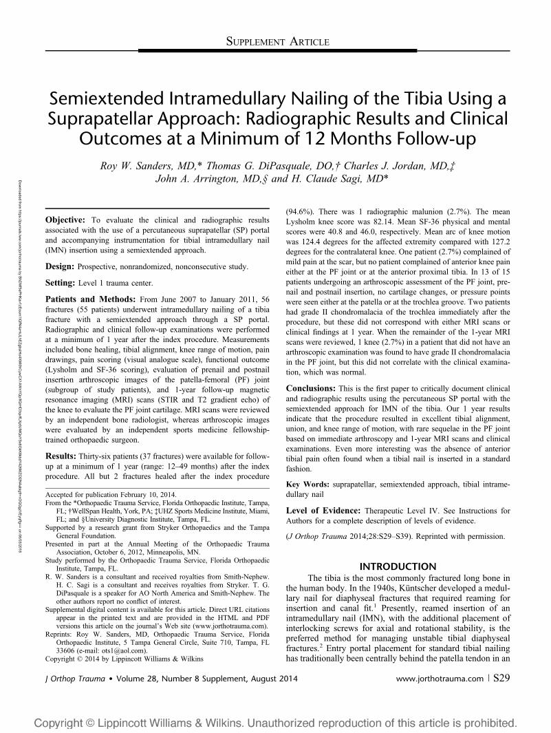
classified based on its location according to the S.P.R.I.N.Tcriteria as proximal, proximal-middle, middle, middle-distal,and distal.12 All fractures were operated on the same hospitaladmission as the initial presentation. All fractures underwentreamed, statically locked intramedullary nailing, with or with-out compression thru a titanium alloy nail, using 1 of 2 spe-cially designed instrumentation systems for SP IMN insertion(T2 Tibial Nail; Stryker Orthopaedics, Mahwah, NJ; TrigenMeta Tibial Nail; Smith and Nephew, Memphis, TN) (Fig. 1,Table 1).
Evaluation of the Patella-Femoral JointDuring the initial period of data collection, we attemp-
ted to determine how best to assess injury to the patella-femoral (PF) joint. Consequently, after the first 26 cases,diagnostic arthroscopy was added to our procedure to betterevaluate the PF joint before and immediately after nailinsertion. Any damage to the PF joint that occurred intra-operatively would therefore be noted. In these cases, after theincision was made, a small amount of fluid was placed intothe PF joint and both PF articular surfaces were inspected,and images were obtained. After completion of the procedure,but before closure, the knee was washed out, the arthroscopewas reinserted, and the PF articular surfaces were againinspected, and images were saved. All images were thenevaluated by an independent sports medicine fellowshiptrained attending to evaluate cartilage injury. Any changeswere recorded using the Outerbridge scale; however, grade Iwas omitted because it is an “active” diagnosis, that is,requires probing of the lesion. Therefore, images were eval-uated for Outerbridge grade II: Fragmentation and fissuring,
FIGURE 1. Cross-sectional representation of specialized can-nula system placed in the SP portal with the knee in thesemiextended position.
Sanders et al J Orthop Trauma � Volume 28, Number 8 Supplement, August 2014
S30 | www.jorthotrauma.com � 2014 Lippincott Williams & Wilkins
less than 0.5-in diameter, grade III: fragmentation and fissur-ing, greater than 0.5-in diameter, or grade IV: Erosion ofcartilage down to exposed subchondral bone.
Postoperative Protocol andOutcome Measures
Patients were made weight bearing as tolerated,immediately postoperatively.12 All patients were taughtactive knee and ankle ROM exercises and a quadricepsstrengthening regimen. Patients were evaluated initially at2 weeks, and at monthly intervals until the fractures were
radiographically healed, based on bridging bone seen on allradiographic views.12 Additionally, all fractures were scoredby the Radiographic Union Scale for Tibial fractures (RUST)method at 1 year.13 During this follow-up time period, patientswere asked to enroll in an institutional review board approvedstudy to prospectively evaluate their outcomes related to thistechnique. For the purposes of the study, patients were broughtback into the office at a minimum of 12 months. Thosepatients that were unavailable at 12 months were brought inwhenever they could be located for an evaluation as long as itwas 12 months after the index procedure.
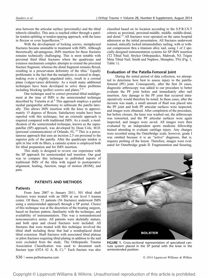
TABLE 1. Patient Data
Sex Age, y Side Nail Type OTA Class Open? Fracture Level Other ORIF? Other Findings
1 F 70 Right Stryker 42B1.3 N MD Ankle PT 70, Infirmed
2 F 37 Right Trigen 42A1.1 N MD Ankle —
3 F 32 Left Stryker 42C2.2 Open PM + MI — R calc, R talus, R navicular, L spine
4 M 25 Left Stryker 42C2.2 Open PM + MI — R patella fx, CHI, skull fx
5 M 50 Left Stryker 42B1.2 Open MD — —
6 M 22 Left Trigen 42B3.3 Open MD Ankle —
7 F 48 Left Trigen 43A2.3 N MD Ankle —
8 M 58 Left Trigen 43A2.2 Open MD Ankle —
9 M 50 Left Stryker 42A2.3 Open MD — Degenerative tears M+L, OA knee
10 M 59 Left Trigen 42A2.1 N MI — —
11 F 50 Left Stryker 42C2 N PM + MI — Pulm cont, spleen lac, SDH, L2-5 fx,L IT, R ankle, R olecranon, pelvis
12 M 24 Right Stryker 42C3.3 Open MI — R fem shaft, C and L spine, spleen lac
13 F 34 Left Stryker 42B3 N MD — —
14 F 59 Left Stryker 42A2.2 Open MI — Preexisting OA knees
15 M 48 Left Trigen 42C2.2 N PM + MI — —
16 M 43 Left Trigen 41A2.3 N MI — —
17 M 76 Left Trigen 42B1.2 N PM + MI — R femur A + V, R dist fem, R tib, Rcalc, R MT, preexisting OA knees
18 M 42 Left Trigen 42A3.3 N MI — Pelvis and sacral fractures
19 M 50 Right Stryker 42B2.3 N MD — —
20 M 50 Right Stryker 42B2.2 Open MD — —
21 M 22 Left Trigen 42A3.3 N MI — —
22 M 18 Left Trigen 42B2.3 N MI — —
23 F 38 Right Stryker 42C2.2 N PM + MI Ankle —
24 M 20 Left Stryker 42C3.2 Open MI — CHI, C2, R acet, L fa, R hum
25 M 21 Left Stryker 42A3.3 N MI — —
26 F 41 Right Stryker 42B3.3 N MD Ankle —
27 F 59 Right Stryker 42B2.3 N MD Ankle Preexisting OA knees
28 M 63 Right Trigen 42B2.3 N MD Ankle Stroke on opposite side
29 F 30 Left Trigen 42C1 N MI + D — —
30 M 67 Right Trigen 42A2.3 N MI — L1, L4 compression fx, concussion
31 M 23 Right Trigen 42B1.1 N MI — —
32 M 25 Left Trigen 42A3.3 N MI — —
33 M 31 Right Trigen 42A2 Open MI — —
34 M 31 Left Stryker 42A2 Open PM — Malunion
35 F (L) 26 Left Trigen 42B2.1 Open MI Ankle —
36 F (R) 26 Right Trigen 42C3.3 Open MD Ankle —
37 F 40 Left Stryker 42B1 Open MI — Morbid obesity, preexisting OAknees
CHI, closed head injury; D, distal; F, female; fx, fracture; IT, intertrochanteric fracture; L, left; M, male; MI, middle; MD, middle distal; N, no; OA, osteoarthritis; P, proximal; PM,proximal middle; R, right.
J Orthop Trauma � Volume 28, Number 8 Supplement, August 2014 Suprapatellar Approach for IMN of the Tibia
� 2014 Lippincott Williams & Wilkins www.jorthotrauma.com | S31
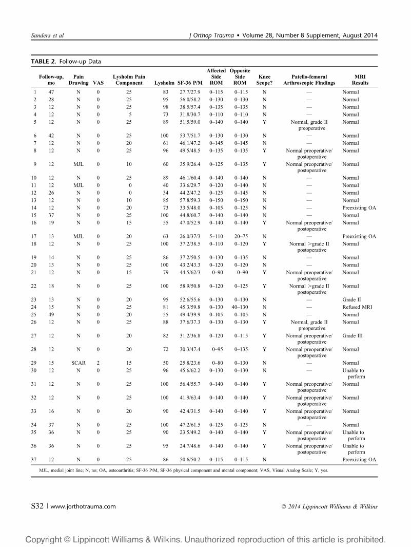
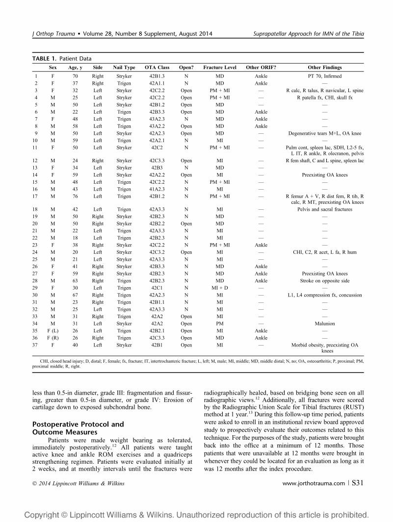
TABLE 2. Follow-up Data
Follow-up,mo
PainDrawing VAS
Lysholm PainComponent Lysholm SF-36 P/M
AffectedSideROM
OppositeSideROM
KneeScope?
Patello-femoralArthroscopic Findings
MRIResults
1 47 N 0 25 83 27.7/27.9 0–115 0–115 N — Normal
2 28 N 0 25 95 56.0/58.2 0–130 0–130 N — Normal
3 12 N 0 25 98 38.5/57.4 0–135 0–135 N — Normal
4 12 N 0 5 73 31.8/30.7 0–110 0–110 N — Normal
5 12 N 0 25 89 51.5/59.0 0–140 0–140 Y Normal, grade IIpreoperative
Normal
6 42 N 0 25 100 53.7/51.7 0–130 0–130 N — Normal
7 12 N 0 20 61 46.1/47.2 0–145 0–145 N — Normal
8 12 N 0 25 96 49.5/48.5 0–135 0–135 Y Normal preoperative/postoperative
Normal
9 12 MJL 0 10 60 35.9/26.4 0–125 0–135 Y Normal preoperative/postoperative
Normal
10 12 N 0 25 89 46.1/60.4 0–140 0–140 N — Normal
11 12 MJL 0 0 40 33.6/29.7 0–120 0–140 N — Normal
12 26 N 0 0 34 44.2/47.2 0–125 0–145 N — Normal
13 12 N 0 10 85 57.8/59.3 0–150 0–150 N — Normal
14 12 N 0 20 73 33.5/48.0 0–105 0–125 N — Preexisting OA
15 37 N 0 25 100 44.8/60.7 0–140 0–140 N — Normal
16 19 N 0 15 55 47.0/52.9 0–140 0–140 Y Normal preoperative/postoperative
Normal
17 13 MJL 0 20 63 26.0/37/3 5–110 20–75 N — Preexisting OA
18 12 N 0 25 100 37.2/38.5 0–110 0–120 Y Normal .grade IIpostoperative
Normal
19 14 N 0 25 86 37.2/50.5 0–130 0–135 N — Normal
20 13 N 0 25 100 43.2/43.3 0–120 0–120 N — Normal
21 12 N 0 15 79 44.5/62/3 0–90 0–90 Y Normal preoperative/postoperative
Normal
22 18 N 0 25 100 58.9/50.8 0–120 0–125 Y Normal .grade IIpostoperative
Normal
23 13 N 0 20 95 52.6/55.6 0–130 0–130 N — Grade II
24 15 N 0 25 81 45.3/59.8 0–130 40–130 N — Refused MRI
25 49 N 0 20 55 49.4/39.9 0–105 0–105 N — Normal
26 12 N 0 25 88 37.6/37.3 0–130 0–130 Y Normal, grade IIpreoperative
Normal
27 12 N 0 20 82 31.2/36.8 0–120 0–115 Y Normal preoperative/postoperative
Grade III
28 12 N 0 20 72 30.3/47.4 0–95 0–135 Y Normal preoperative/postoperative
Normal
29 15 SCAR 2 15 50 25.8/23.6 0–80 0–130 N — Normal
30 12 N 0 25 96 45.6/62.2 0–130 0–130 N — Unable toperform
31 12 N 0 25 100 56.4/55.7 0–140 0–140 Y Normal preoperative/postoperative
Normal
32 12 N 0 25 100 41.9/63.4 0–140 0–140 Y Normal preoperative/postoperative
Normal
33 16 N 0 20 90 42.4/31.5 0–140 0–140 Y Normal preoperative/postoperative
Normal
34 37 N 0 25 100 47.2/61.5 0–125 0–125 N — Normal
35 36 N 0 25 90 23.5/49.2 0–140 0–140 Y Normal preoperative/postoperative
Unable toperform
36 36 N 0 25 95 24.7/48.6 0–140 0–140 Y Normal preoperative/postoperative
Unable toperform
37 12 N 0 25 86 50.6/50.2 0–115 0–115 N — Preexisting OA
MJL, medial joint line; N, no; OA, osteoarthritis; SF-36 P/M, SF-36 physical component and mental component; VAS, Visual Analog Scale; Y, yes.
Sanders et al J Orthop Trauma � Volume 28, Number 8 Supplement, August 2014
S32 | www.jorthotrauma.com � 2014 Lippincott Williams & Wilkins
At each visit, an independent research nurse docu-mented ROM of both knees using a goniometer andquadriceps strength manually.14 Postoperative function wasdetermined using 2 validated outcome tools, the generalSF-36 and the knee specific Lysholm score.15 The SF-36 isa generic validated functional outcome measure designed tooffer both mental and physical well-being scores comparedwith a general population. Originally designed for assessmentof ligament injuries of the knee, the Lysholm knee score hasalso been validated for the evaluation of patients with chon-dral disorders of the knee and those with an acute patellardislocation. It is a condition-specific 100-point outcome scorethat contains 8 domains: limp, locking, pain, stair climbing,use of supports, instability, swelling, and squatting (95–100excellent, 84–94 good, 65–83 fair, and ,65 points, a pooroutcome).
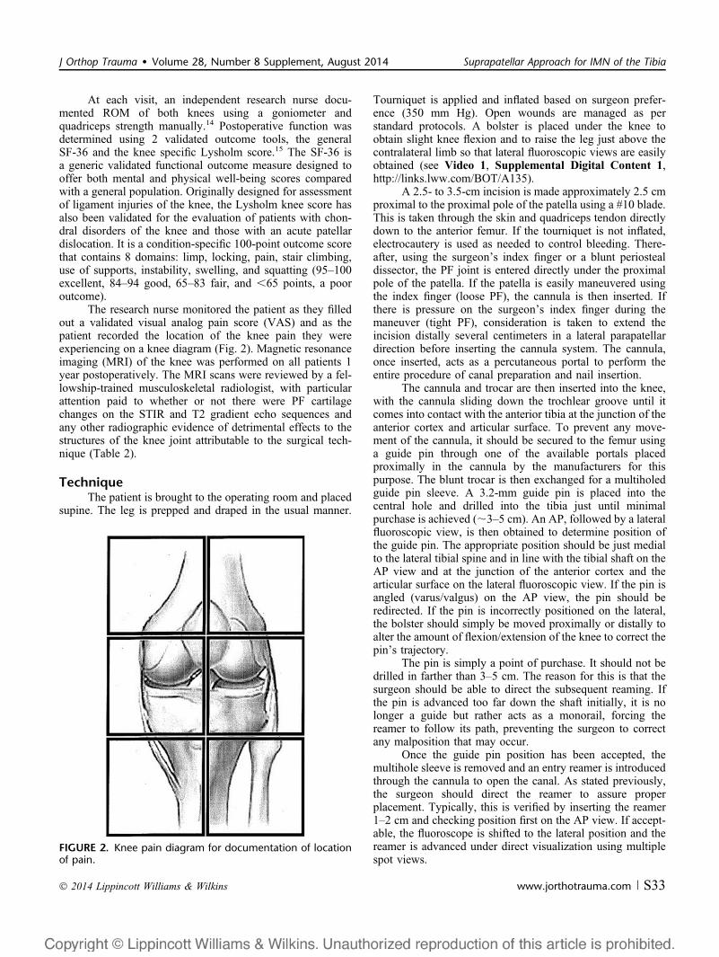
The research nurse monitored the patient as they filledout a validated visual analog pain score (VAS) and as thepatient recorded the location of the knee pain they wereexperiencing on a knee diagram (Fig. 2). Magnetic resonanceimaging (MRI) of the knee was performed on all patients 1year postoperatively. The MRI scans were reviewed by a fel-lowship-trained musculoskeletal radiologist, with particularattention paid to whether or not there were PF cartilagechanges on the STIR and T2 gradient echo sequences andany other radiographic evidence of detrimental effects to thestructures of the knee joint attributable to the surgical tech-nique (Table 2).
TechniqueThe patient is brought to the operating room and placed
supine. The leg is prepped and draped in the usual manner.
Tourniquet is applied and inflated based on surgeon prefer-ence (350 mm Hg). Open wounds are managed as perstandard protocols. A bolster is placed under the knee toobtain slight knee flexion and to raise the leg just above thecontralateral limb so that lateral fluoroscopic views are easilyobtained (see Video 1, Supplemental Digital Content 1,http://links.lww.com/BOT/A135).
A 2.5- to 3.5-cm incision is made approximately 2.5 cmproximal to the proximal pole of the patella using a #10 blade.This is taken through the skin and quadriceps tendon directlydown to the anterior femur. If the tourniquet is not inflated,electrocautery is used as needed to control bleeding. There-after, using the surgeon’s index finger or a blunt periostealdissector, the PF joint is entered directly under the proximalpole of the patella. If the patella is easily maneuvered usingthe index finger (loose PF), the cannula is then inserted. Ifthere is pressure on the surgeon’s index finger during themaneuver (tight PF), consideration is taken to extend theincision distally several centimeters in a lateral parapatellardirection before inserting the cannula system. The cannula,once inserted, acts as a percutaneous portal to perform theentire procedure of canal preparation and nail insertion.
The cannula and trocar are then inserted into the knee,with the cannula sliding down the trochlear groove until itcomes into contact with the anterior tibia at the junction of theanterior cortex and articular surface. To prevent any move-ment of the cannula, it should be secured to the femur usinga guide pin through one of the available portals placedproximally in the cannula by the manufacturers for thispurpose. The blunt trocar is then exchanged for a multiholedguide pin sleeve. A 3.2-mm guide pin is placed into thecentral hole and drilled into the tibia just until minimalpurchase is achieved (;3–5 cm). An AP, followed by a lateralfluoroscopic view, is then obtained to determine position ofthe guide pin. The appropriate position should be just medialto the lateral tibial spine and in line with the tibial shaft on theAP view and at the junction of the anterior cortex and thearticular surface on the lateral fluoroscopic view. If the pin isangled (varus/valgus) on the AP view, the pin should beredirected. If the pin is incorrectly positioned on the lateral,the bolster should simply be moved proximally or distally toalter the amount of flexion/extension of the knee to correct thepin’s trajectory.
The pin is simply a point of purchase. It should not bedrilled in farther than 3–5 cm. The reason for this is that thesurgeon should be able to direct the subsequent reaming. Ifthe pin is advanced too far down the shaft initially, it is nolonger a guide but rather acts as a monorail, forcing thereamer to follow its path, preventing the surgeon to correctany malposition that may occur.
Once the guide pin position has been accepted, themultihole sleeve is removed and an entry reamer is introducedthrough the cannula to open the canal. As stated previously,the surgeon should direct the reamer to assure properplacement. Typically, this is verified by inserting the reamer1–2 cm and checking position first on the AP view. If accept-able, the fluoroscope is shifted to the lateral position and thereamer is advanced under direct visualization using multiplespot views.
FIGURE 2. Knee pain diagram for documentation of locationof pain.
J Orthop Trauma � Volume 28, Number 8 Supplement, August 2014 Suprapatellar Approach for IMN of the Tibia
� 2014 Lippincott Williams & Wilkins www.jorthotrauma.com | S33
The reamer should not be introduced across the fractureunless the fracture is reduced. If reduction is required, then thereamer is removed and a reduction tool is inserted through thecannula to reduce the fracture. Because the leg is essentially flaton the operating table, gravity is not an issue, and the fracturecan be easily reduced and/or held by an assistant while thereduction tool is used and reduction achieved. If the fracture ishighly comminuted, reduction forceps, clamps, blockingscrews, etc, can be used easily to assist the reduction. Oncethe fracture is reduced (by whatever means), a straight guidewire is placed. Nail length is determined using a speciallydesigned ruler through the cannula.
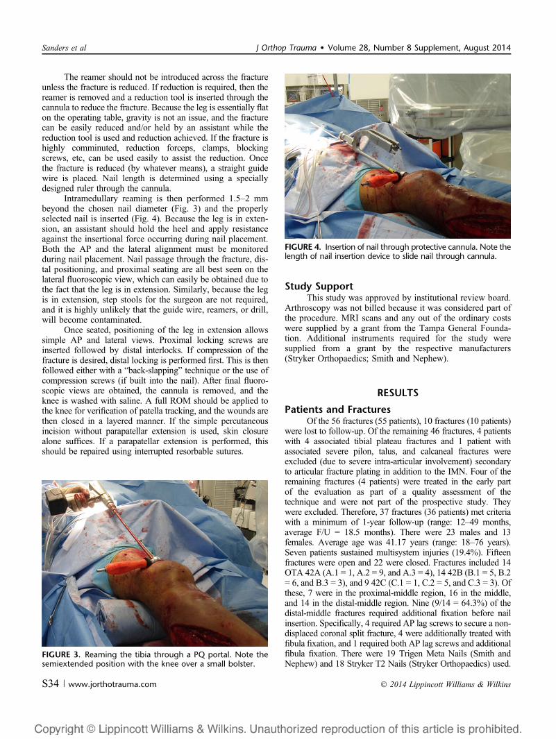
Intramedullary reaming is then performed 1.5–2 mmbeyond the chosen nail diameter (Fig. 3) and the properlyselected nail is inserted (Fig. 4). Because the leg is in exten-sion, an assistant should hold the heel and apply resistanceagainst the insertional force occurring during nail placement.Both the AP and the lateral alignment must be monitoredduring nail placement. Nail passage through the fracture, dis-tal positioning, and proximal seating are all best seen on thelateral fluoroscopic view, which can easily be obtained due tothe fact that the leg is in extension. Similarly, because the legis in extension, step stools for the surgeon are not required,and it is highly unlikely that the guide wire, reamers, or drill,will become contaminated.
Once seated, positioning of the leg in extension allowssimple AP and lateral views. Proximal locking screws areinserted followed by distal interlocks. If compression of thefracture is desired, distal locking is performed first. This is thenfollowed either with a “back-slapping” technique or the use ofcompression screws (if built into the nail). After final fluoro-scopic views are obtained, the cannula is removed, and theknee is washed with saline. A full ROM should be applied tothe knee for verification of patella tracking, and the wounds arethen closed in a layered manner. If the simple percutaneousincision without parapatellar extension is used, skin closurealone suffices. If a parapatellar extension is performed, thisshould be repaired using interrupted resorbable sutures.
Study SupportThis study was approved by institutional review board.
Arthroscopy was not billed because it was considered part ofthe procedure. MRI scans and any out of the ordinary costswere supplied by a grant from the Tampa General Founda-tion. Additional instruments required for the study weresupplied from a grant by the respective manufacturers(Stryker Orthopaedics; Smith and Nephew).
RESULTS
Patients and FracturesOf the 56 fractures (55 patients), 10 fractures (10 patients)
were lost to follow-up. Of the remaining 46 fractures, 4 patientswith 4 associated tibial plateau fractures and 1 patient withassociated severe pilon, talus, and calcaneal fractures wereexcluded (due to severe intra-articular involvement) secondaryto articular fracture plating in addition to the IMN. Four of theremaining fractures (4 patients) were treated in the early partof the evaluation as part of a quality assessment of thetechnique and were not part of the prospective study. Theywere excluded. Therefore, 37 fractures (36 patients) met criteriawith a minimum of 1-year follow-up (range: 12–49 months,average F/U = 18.5 months). There were 23 males and 13females. Average age was 41.17 years (range: 18–76 years).Seven patients sustained multisystem injuries (19.4%). Fifteenfractures were open and 22 were closed. Fractures included 14OTA 42A (A.1 = 1, A.2 = 9, and A.3 = 4), 14 42B (B.1 = 5, B.2= 6, and B.3 = 3), and 9 42C (C.1 = 1, C.2 = 5, and C.3 = 3). Ofthese, 7 were in the proximal-middle region, 16 in the middle,and 14 in the distal-middle region. Nine (9/14 = 64.3%) of thedistal-middle fractures required additional fixation before nailinsertion. Specifically, 4 required AP lag screws to secure a non-displaced coronal split fracture, 4 were additionally treated withfibula fixation, and 1 required both AP lag screws and additionalfibula fixation. There were 19 Trigen Meta Nails (Smith andNephew) and 18 Stryker T2 Nails (Stryker Orthopaedics) used.
FIGURE 3. Reaming the tibia through a PQ portal. Note thesemiextended position with the knee over a small bolster.
FIGURE 4. Insertion of nail through protective cannula. Note thelength of nail insertion device to slide nail through cannula.
Sanders et al J Orthop Trauma � Volume 28, Number 8 Supplement, August 2014
S34 | www.jorthotrauma.com � 2014 Lippincott Williams & Wilkins
Limited arthroscopy was employed to evaluate the PF jointalone in 40.5% (15/37) of the cases.
Fracture HealingAt the 1-year evaluation, all but one fracture had healed
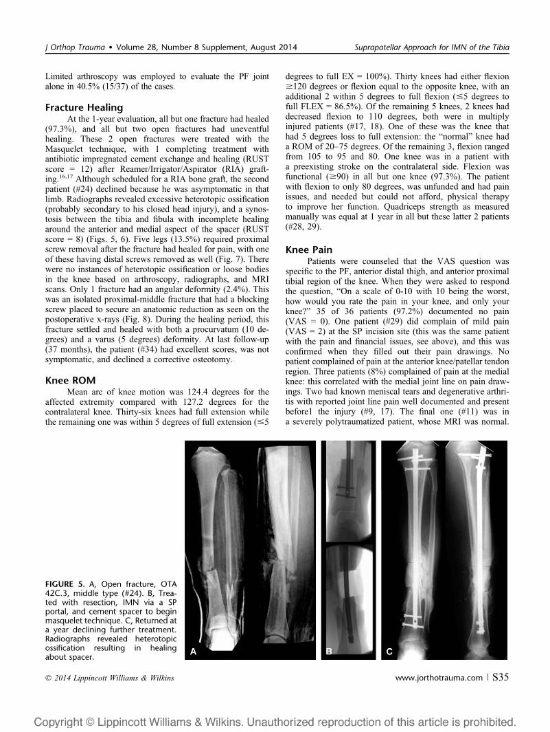
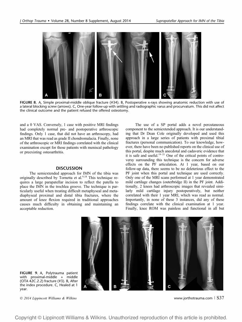
(97.3%), and all but two open fractures had uneventfulhealing. These 2 open fractures were treated with theMasquelet technique, with 1 completing treatment withantibiotic impregnated cement exchange and healing (RUSTscore = 12) after Reamer/Irrigator/Aspirator (RIA) graft-ing.16,17 Although scheduled for a RIA bone graft, the secondpatient (#24) declined because he was asymptomatic in thatlimb. Radiographs revealed excessive heterotopic ossification(probably secondary to his closed head injury), and a synos-tosis between the tibia and fibula with incomplete healingaround the anterior and medial aspect of the spacer (RUSTscore = 8) (Figs. 5, 6). Five legs (13.5%) required proximalscrew removal after the fracture had healed for pain, with oneof these having distal screws removed as well (Fig. 7). Therewere no instances of heterotopic ossification or loose bodiesin the knee based on arthroscopy, radiographs, and MRIscans. Only 1 fracture had an angular deformity (2.4%). Thiswas an isolated proximal-middle fracture that had a blockingscrew placed to secure an anatomic reduction as seen on thepostoperative x-rays (Fig. 8). During the healing period, thisfracture settled and healed with both a procurvatum (10 de-grees) and a varus (5 degrees) deformity. At last follow-up(37 months), the patient (#34) had excellent scores, was notsymptomatic, and declined a corrective osteotomy.
Knee ROMMean arc of knee motion was 124.4 degrees for the
affected extremity compared with 127.2 degrees for thecontralateral knee. Thirty-six knees had full extension whilethe remaining one was within 5 degrees of full extension (#5
degrees to full EX = 100%). Thirty knees had either flexion$120 degrees or flexion equal to the opposite knee, with anadditional 2 within 5 degrees to full flexion (#5 degrees tofull FLEX = 86.5%). Of the remaining 5 knees, 2 knees haddecreased flexion to 110 degrees, both were in multiplyinjured patients (#17, 18). One of these was the knee thathad 5 degrees loss to full extension: the “normal” knee hada ROM of 20–75 degrees. Of the remaining 3, flexion rangedfrom 105 to 95 and 80. One knee was in a patient witha preexisting stroke on the contralateral side. Flexion wasfunctional ($90) in all but one knee (97.3%). The patientwith flexion to only 80 degrees, was unfunded and had painissues, and needed but could not afford, physical therapyto improve her function. Quadriceps strength as measuredmanually was equal at 1 year in all but these latter 2 patients(#28, 29).
Knee PainPatients were counseled that the VAS question was
specific to the PF, anterior distal thigh, and anterior proximaltibial region of the knee. When they were asked to respondthe question, “On a scale of 0-10 with 10 being the worst,how would you rate the pain in your knee, and only yourknee?” 35 of 36 patients (97.2%) documented no pain(VAS = 0). One patient (#29) did complain of mild pain(VAS = 2) at the SP incision site (this was the same patientwith the pain and financial issues, see above), and this wasconfirmed when they filled out their pain drawings. Nopatient complained of pain at the anterior knee/patellar tendonregion. Three patients (8%) complained of pain at the medialknee: this correlated with the medial joint line on pain draw-ings. Two had known meniscal tears and degenerative arthri-tis with reported joint line pain well documented and presentbefore1 the injury (#9, 17). The final one (#11) was ina severely polytraumatized patient, whose MRI was normal.
FIGURE 5. A, Open fracture, OTA42C.3, middle type (#24). B, Trea-ted with resection, IMN via a SPportal, and cement spacer to beginmasquelet technique. C, Returned ata year declining further treatment.Radiographs revealed heterotopicossification resulting in healingabout spacer.
J Orthop Trauma � Volume 28, Number 8 Supplement, August 2014 Suprapatellar Approach for IMN of the Tibia
� 2014 Lippincott Williams & Wilkins www.jorthotrauma.com | S35
Patients did, however, complain of residual pain secondary toprominent locking/blocking screws, scars from open wounds,fasciotomies, soft tissue flaps, or the fracture site itself(Figs. 9, 10). When asked about pain as part of the Lysholm
score, 21 patients exhibited no pain (25/25), whereas8 patients exhibited slight pain during athletics (20/25). Ofthe 7 patients with a Lysholm pain score of #15 of 25, allwere related to either their injury or the preexisting conditions(vide infra).
Functional Outcome ScoresThe Lysholm score was used to assess overall knee
function (37 knees) as it related to gait, stair climbing, andwalking/running. The mean Lysholm knee score was 82.14.The breakdown was as follows: 14 excellent, 8 good, 7 fair,and 8 poor. Of the 8 poor results (8 knees), all had complaintsrelated to prolonged walking/running and stair climbing. Fourof these patients suffered from polytrauma and 1 frompreexisting arthritis and could not separate out their limita-tions when filling out the questionnaire. Mean SF-36 physicaland mental scores were 41.8 and 47.9, respectively.
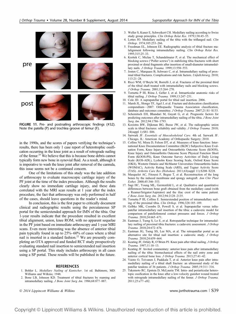
Evaluation of the PF JointIn those patients undergoing an arthroscopic assessment
of the PF joint prenail and postnail insertion, no cartilagechanges, gouges, or pressure changes from the cannula wereseen in 13 of 15 patients (86.7%) (Fig. 11). Two kneesshowed evidence of grade II chondromalacia limited to thetrochlea groove immediately after completion of the procedureas seen on arthroscopic images. Thirty-three of 37 knees had anMRI scan performed at the 1-year follow-up. One patient(1 fracture) refused the scan and 3 patients (3 fractures) couldnot undergo the test secondary to metallic implants elsewhere.Of the remaining 33 MRI scans, 1 patient had grade II PFchanges and 1 had grade III PF changes. For the patients thathad an arthroscopic evaluation of their PF joint, there was norelationship between the arthroscopic findings and MRI find-ings. The 2 cases exhibiting arthroscopic grade II changes inthe PF joint immediately postoperatively were read as normalon their 1-year MRI scan. Both had a Lysholm score of 100
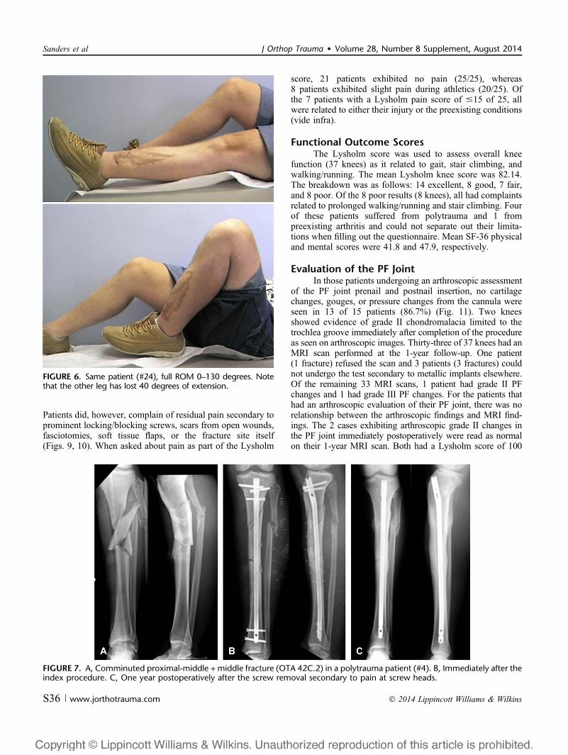
FIGURE 6. Same patient (#24), full ROM 0–130 degrees. Notethat the other leg has lost 40 degrees of extension.
FIGURE 7. A, Comminuted proximal-middle + middle fracture (OTA 42C.2) in a polytrauma patient (#4). B, Immediately after theindex procedure. C, One year postoperatively after the screw removal secondary to pain at screw heads.
Sanders et al J Orthop Trauma � Volume 28, Number 8 Supplement, August 2014
S36 | www.jorthotrauma.com � 2014 Lippincott Williams & Wilkins
and a 0 VAS. Conversely, 1 case with positive MRI findingshad completely normal pre- and postoperative arthroscopicfindings. Only 1 case, that did not have an arthroscopy, hadan MRI that was read as grade II chondromalacia. Finally, noneof the arthroscopic or MRI findings correlated with the clinicalexamination except for those patients with meniscal pathologyor preexisting osteoarthritis.
DISCUSSIONThe semiextended approach for IMN of the tibia was
originally described by Tornetta et al.9,18 This technique re-quires a large parapatellar incision to reflect the patella toplace the IMN in the trochlea groove. The technique is par-ticularly useful when treating difficult metaphyseal and meta-diaphyseal proximal and distal tibia fractures, where theamount of knee flexion required in traditional approachescauses much difficulty in obtaining and maintaining anacceptable reduction.
The use of a SP portal adds a novel percutaneouscomponent to the semiextended approach. It is our understand-ing that Dr Dean Cole originally developed and used thisapproach in a large series of patients with proximal tibialfractures (personal communication). To our knowledge, how-ever, there have been no published reports on the clinical use ofthis portal, despite much anecdotal and cadaveric evidence thatit is safe and useful.19–21 One of the critical points of contro-versy surrounding this technique is the concern for adverseeffects on the PF articulation. At 1 year, based on ourfollow-up data, there seems to be no deleterious effect to thePF joint when this portal and technique are used correctly.Only one of the MRI scans performed at 1 year demonstratedmild cartilage changes (outerbridge II) in the PF joint. Addi-tionally, 2 knees had arthroscopic images that revealed simi-larly mild cartilage injury postoperatively, but neithercorrelated with their 1 year MRI, which was read as normal.Importantly, in none of these 3 instances, did any of thesefindings correlate with the clinical examination at 1 year.Finally, knee ROM was painless and functional in all but
FIGURE 8. A, Simple proximal-middle oblique fracture (#34). B, Postoperative x-rays showing anatomic reduction with use ofa lateral blocking screw (arrows). C, One-year follow-up with settling and radiographic varus and procurvatum. This did not affectthe clinical outcome and the patient refused the offered osteotomy.
FIGURE 9. A, Polytrauma patientwith proximal-middle + middle(OTA 42C.2.2) fracture (#3). B, Afterthe index procedure. C, Healed at 1year.
J Orthop Trauma � Volume 28, Number 8 Supplement, August 2014 Suprapatellar Approach for IMN of the Tibia
� 2014 Lippincott Williams & Wilkins www.jorthotrauma.com | S37
two knees when compared with the normal contralateral side.Clearly, these patients will need to be followed at a minimumof 2–5 years to document any late effects that may occur, butthe 1-year outcomes seem promising in this regard.
It is even more remarkable that none of our SP portalpatients complained of any PF or anterior knee pain whatso-ever. The reasons are unknown, but in patients with traditionalnail insertion techniques, it may be that the patella tendon andthe soft tissue surrounding that tendon simply do not respondwell to open manipulation.22,23 Vaisto et al noted that differingthe traditional approach (paratendinous vs. transtendinous)made no difference when evaluating the cause of anterior kneepain after IMN.24 Interestingly, retrograde femoral IMN pa-tients do not seem to complain about knee pain using essen-tially the same standard inferior patella tendon approach, buta different bone window. Furthermore, because the bone win-dow for tibial nail insertion remains the same whether a stan-dard or SP portal is used, the difference may simply be theprolonged knee flexion associated with tibial nail insertion in
contrast to both the semiextended tibial and retrograde femoralnail insertion method, where the knee is largely in extension.19
It is possible that using a percutaneous portal proximal to thepatella avoids the patella tendon altogether and therefore, theassociated pain. When comparing our study to other reportsthat describe pain in the leg after injury and IMN insertion, itmust be stated that our evaluation was specifically focused onwhether the portal and/or the technique caused knee pain andnot generalized leg pain, which in the trauma patient, and inour patients, is multifactorial. Whatever the reason, the fact thatnone of the patients experienced any PF or anterior knee pain atall is remarkable. A larger study population will be necessary,however, before definitive comments about lack of knee paincan be stated with certainty.
Although our patients did not experience any heterotopicossification or joint mice as a result of the procedure, concernsarise regarding reaming debris as well. Again the literature forretrograde nailing is illuminating. Despite the thousands ofretrograde nails placed because the technique became popular
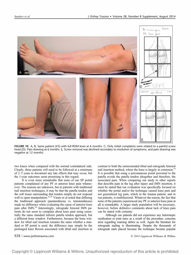
FIGURE 10. A, B, Same patient (#3) with full ROM knee at 6 months. C, Only initial complaints were related to a painful screwhead (D). Pain drawing at 6 months. E, Screw removal was declined secondary to resolution of symptoms, and pain drawing wasnegative at 12 months.
Sanders et al J Orthop Trauma � Volume 28, Number 8 Supplement, August 2014
S38 | www.jorthotrauma.com � 2014 Lippincott Williams & Wilkins
in the 1990s, and the scores of papers verifying the technique’sresults, there has been only 1 case report of heterotrophic ossifi-cation occurring in the knee joint as a result of retrograde nailingof the femur.25 We believe that this is because bone debris cannottypically form new bone in synovial fluid. As a result, although itis imperative to wash the knee joint after removal of the cannula,this issue seems not be a continued concern.
One of the limitations of this study was the late additionof arthroscopy to evaluate macroscopic cartilage injury of thePF joint at the time of the index procedure. Although the resultsclearly show no immediate cartilage injury, and these datacorrelated with the MRI scan results at 1 year after the indexprocedure, the fact that arthroscopy was only performed in halfof the cases, should leave questions in the reader’s mind.
In conclusion, this is the first paper to critically documentclinical and radiographic results using the percutaneous SPportal for the semiextended approach for IMN of the tibia. Our1-year results indicate that the procedure resulted in excellenttibial alignment, union, knee ROM, with no apparent sequelaein the PF joint based on immediate arthroscopy and 1-year MRIscans. Even more interesting was the absence of anterior tibialpain typically found in up to 25%–60% of cases where a tibialnail is inserted in a standard fashion.22 We are presently com-pleting an OTA approved and funded RCT study prospectivelyevaluating standard nail insertion to semiextended nail insertionusing a SP portal. This study includes arthroscopy in all casesusing a SP portal. These results will be published in the future.
REFERENCES1. Bohler L. Medullary Nailing of Kuntscher. 1st ed. Baltimore, MD:
Williams and Wilkins; 1948.2. Bone LB, Johnson KD. Treatment of tibial fractures by reaming and
intramedullary nailing. J Bone Joint Surg Am. 1986;68:877–887.
3. Weller S, Kuner E, Schweikert CH. Medullary nailing according to Swissstudy group principles. Clin Orthop Relat Res. 1979;138:45–55.
4. Lottes JO. Medullary nailing of the tibia with the triflanged nail. ClinOrthop. 1974;105:253–266.
5. Freedman EL, Johnson EE. Radiographic analysis of tibial fracture ma-lalignment following intramedullary nailing. Clin Orthop Relat Res.1995;315:25–33.
6. Krettek C, Miclau T, Schandelmaier P, et al. The mechanical effect ofblocking screws (“Poller screws”) in stabilizing tibia fractures with shortproximal or distal fragments after insertion of small-diameter intramedul-lary nails. J Orthop Trauma. 1999;13:550–553.
7. Josten C, Marquass B, Schwarz C, et al. Intramedullary nailing of prox-imal tibial fractures. Complications and risk factors. Unfallchirurg. 2010;113:21–28.
8. Ricci WM, O’Boyle M, Borrelli J, et al. Fractures of the proximal thirdof the tibial shaft treated with intramedullary nails and blocking screws.J Orthop Trauma. 2001;15:264–270.
9. Tornetta P III, Riina J, Geller J, et al. Intraarticular anatomic risks oftibial nailing. J Orthop Trauma. 1999;13:247–251.
10. Cole JD. A suprapatellar portal for tibial nail insertion. 2006.11. Marsh JL, Slongo TF, Agel J, et al. Fracture and dislocation classification
compendium—2007: Orthopaedic Trauma Association classification,database and outcomes committee. J Orthop Trauma. 2007;21:S1–S133.
12. Schemitsch EH, Bhandari M, Guyatt G, et al. Prognostic factors forpredicting outcomes after intramedullary nailing of the tibia. J Bone JointSurg Am. 2012;94:1786–1793.
13. Kooistra BW, Dijkman BG, Busse JW, et al. The radiographic unionscale in tibial fractures: reliability and validity. J Orthop Trauma. 2010;24(suppl 1):S81–S86.
14. Sarwark JF. Essentials of Musculoskeletal Care. 4th ed. Sarwark JF.Chicago, IL: American Academy of Orthopaedic Surgery; 2010.
15. Collins NJ, Misra D, Felson DT, et al. Measures of knee function: Inter-national Knee Documentation Committee (IKDC) Subjective Knee Eval-uation Form, Knee Injury and Osteoarthritis Outcome Score (KOOS),Knee Injury and Osteoarthritis Outcome Score Physical Function ShortForm (KOOS-PS), Knee Outcome Survey Activities of Daily LivingScale (KOS-ADL), Lysholm Knee Scoring Scale, Oxford Knee Score(OKS), Western Ontario and McMaster Universities Osteoarthritis Index(WOMAC), Activity Rating Scale (ARS), and Tegner Activity Score(TAS). Arthritis Care Res (Hoboken). 2011;63(suppl 11):S208–S228.
16. Masquelet AC, Fitoussi F, Begue T, et al. Reconstruction of the longbones by the induced membrane and spongy autograft. Ann Chir PlastEsthet. 2000;45:346–353.
17. Sagi HC, Young ML, Gerstenfeld L, et al. Qualitative and quantitativedifferences between bone graft obtained from the medullary canal (witha Reamer/Irrigator/Aspirator) and the iliac crest of the same patient.J Bone Joint Surg Am. 2012;94:2128–2135.
18. Tornetta P III, Collins E. Semiextended position of intramedullary nail-ing of the proximal tibia. Clin Orthop. 1996;328:185–189.
19. Gelbke MK, Coombs D, Powell S, et al. Suprapatellar versus infra-patellar intramedullary nail insertion of the tibia: a cadaveric model forcomparison of patellofemoral contact pressures and forces. J OrthopTrauma. 2010;24:665–671.
20. Eastman J, Tseng S, Lo E, et al. Retropatellar technique for intramedul-lary nailing of proximal tibia fractures: a cadaveric assessment. J OrthopTrauma. 2010;24:672–676.
21. Eastman JG, Tseng SS, Lee MA, et al. The retropatellar portal as analternative site for tibial nail insertion: a cadaveric study. J OrthopTrauma. 2010;24:659–664.
22. Keating JF, Orfaly R, O’Brien PJ. Knee pain after tibial nailing. J OrthopTrauma. 1997;11:10–13.
23. Keating JF. Invited commentary: anterior knee pain after intramedullarynailing of the tibia: biomechanical effects of the nail entry zone andanterior cortical bone loss. J Orthop Trauma. 2013;27:41–42.
24. Vaisto O, Toivanen J, Paakkala T, et al. Anterior knee pain after intra-medullary nailing of a tibial shaft fracture: an ultrasound study of thepatellar tendons of 36 patients. J Orthop Trauma. 2005;19:311–316.
25. Takemoto RC, Epstein D, McLaurin TM. Intra- and periarticular hetero-topic ossification in the knee after a low-velocity gunshot wound treatedwith retrograde intramedullary nailing of the femur. J Orthop Trauma.2011;25:e77–e82.
FIGURE 11. Pre- and postnailing arthroscopic findings (#32).Note the patella (P) and trochlea groove of femur (F).
J Orthop Trauma � Volume 28, Number 8 Supplement, August 2014 Suprapatellar Approach for IMN of the Tibia
� 2014 Lippincott Williams & Wilkins www.jorthotrauma.com | S39